Review

Cellular and Molecular Mechanisms Modulated by Genistein in Cancer

by
Valeria Naponelli
1,*,
Annamaria Piscazzi
2 and
Domenica Mangieri
2,*
1
Department of Medicine and Surgery, University of Parma, Plesso Biotecnologico Integrato, Via Volturno 39, 43126 Parma, Italy
2
Department of Clinical and Experimental Medicine, University of Foggia, Via Pinto 1, 71122 Foggia, Italy
*
Authors to whom correspondence should be addressed.
Int. J. Mol. Sci. 2025, 26(3), 1114; https://doi.org/10.3390/ijms26031114
Submission received: 27 November 2024 / Revised: 21 January 2025 / Accepted: 23 January 2025 / Published: 27 January 2025
(This article belongs to the Special Issue Natural-Derived Bioactive Compounds in Disease Treatment)

Abstract

:
Genistein (4′,5,7-trihydroxyisoflavone) is a phytoestrogen belonging to a subclass of natural flavonoids that exhibits a wide range of pharmacological functions, including antioxidant and anti-inflammatory properties. These characteristics make genistein a valuable phytochemical compound for the prevention and/or treatment of cancer. Genistein effectively inhibits tumor growth and dissemination by modulating key cellular mechanisms. This includes the suppression of angiogenesis, the inhibition of epithelial–mesenchymal transition, and the regulation of cancer stem cell proliferation. These effects are mediated through pivotal signaling pathways such as JAK/STAT, PI3K/Akt/mTOR, MAPK/ERK, NF-κB, and Wnt/β-catenin. Moreover, genistein interferes with the function of specific cyclin/CDK complexes and modulates the activation of Bcl-2/Bax and caspases, playing a critical role in halting tumor cell division and promoting apoptosis. The aim of this review is to discuss in detail the key cellular and molecular mechanisms underlying the pleiotropic anticancer effects of this flavonoid.

1. Introduction

Cancer encompasses a diverse group of complex and dynamic diseases, defined by distinct characteristics such as uncontrolled cell proliferation, resistance to apoptosis, epigenetic reprogramming, immune evasion, and phenotypic plasticity [1,2]. Despite their limitations, including severe cytotoxicity, and the development of multidrug resistance, the most employed anticancer and antimetastatic treatments include chemotherapy, radiotherapy, surgery, and immunotherapy, often administered in combination [3]. Furthermore, as one of the leading causes of death worldwide, cancer highlights the need for effective therapeutic approaches, with chemoprevention emerging as one of the most promising strategies [4,5,6]. In this context, several members of the naturally occurring flavonoids, already known for their multiple health benefits, also show efficacy in chemoprevention [7,8,9,10]. Genistein (4′,5,7-trihydroxyisoflavone, a member of the flavonoid family) is a phytoestrogen also acting as a potent inhibitor of tyrosine kinases [11] that has already been successfully tested in the management of breast and prostate cancers [12,13,14,15,16]. Further experimental evidence has demonstrated genistein’s ability to influence a broad spectrum of cell signaling pathways, thus interfering with the progression of several other types of cancer [17,18]. Building on these premises, this review explores the most significant findings on the anticancer properties of genistein, with a focus on its capacity to modulate key molecular pathways involved in angiogenesis, tumor cell migration and invasion, epithelial–mesenchymal transition (EMT), maintenance of cancer stem cells (CSCs), cell cycle arrest, and the induction of tumor cell death.

2. Chemistry

Genistein (5,7-dihydroxy-3-(4-hydroxyphenyl)-chromen-4-one) is a naturally occurring isoflavone in soy products [19]. Isoflavones are members of the flavonoid family, which comprises over 5000 compounds [15]. The aglycone form of isoflavones is the biologically active form. The biologically active isoflavone is the aglycosylated form obtained after food processing of genistin, found in natural sources [19,20,21]. Knowing the chemical structure of genistein helps to understand its biological activity. Genistein is a 4′,5,7-trihydroxyisoflavone (C15H10O5) with a low molecular weight of 270.24. It is a light-sensitive plant secondary metabolite formed by two aromatic benzene rings (A and C) and a non-aromatic heterocyclic pyran ring (B) (3-phenylchromen-4-one backbone), and the substituents at positions 4′, 5, and 7 of rings A and B are hydroxyl groups (Figure 1) [22]. These phenols give this class of compounds significant antioxidant activity. Moreover, genistein is poorly soluble in water and is soluble in acetone, ethanol, and other polar solvents [21]. Due to its structural similarity to human endogenous estrogen, namely 17β-estradiol (E2), genistein can compete with it and bind to estrogen receptors (ERs) α and β through the two hydroxyl groups located on carbons 4 and 7 [21,23]. Moreover, genistein is a recognized inhibitor of protein kinases [15,24]. This activity is thought to be due to the C4′ phenolic group of this phytoestrogen, which is structurally similar to the phosphoacceptor group of tyrosine [15].

3. Sources

Genistein is an isoflavone abundantly found in the roots and seeds of plants belonging to the Fabaceae (Leguminosae) family [25]. The phytoestrogen is abundant in soy foods, soy drinks, and soybeans (Table 1) [21,26]. The genistein content of mature soybeans ranges from 5.6 to 276 mg/100 g, with an average of 81 mg/100 g [26,27]. These differences in genistein content of soybeans are not influenced by the soil cultivation system [28] whereas the technological tillage of soybeans has a significant effect on this parameter. Genistein concentrations are significantly higher in fermented soy products (e.g., miso, tempeh, sufu, natto, soy sauce) compared to unfermented soy and soy-based products such as soy milk, soy drinks, okara, tofu, and soy cheeses [22,29]. The second most abundant source of this isoflavone is represented by legumes with an average content ranging from 0.2 and 0.6 mg/100 g [21,30,31]. Fruits, nuts, and vegetables contain variable amounts of genistein, in the range of 0.03 to 0.2 mg/100 g [31]. A more comprehensive list of genistein content in food classes is available online [26].
Various treatments (such as hulling, flaking, and defatting) are used to isolate soya proteins from the soya bean [33]. Fermentation is a common low-cost process to improve the bioavailability of soy [34]. The isoflavone is extracted from soybean meal in organic solvents and genistin-containing fractions are converted to genistein by acid hydrolysis [35]. The products are solubilized in water and genistein is crystallized from the solution [36]. Recently, the methods for the isolation of genistein have been improved and have become more complex and sensitive [37]. Most of the separation and purification methods are based on thin-layer chromatography and other chromatographic techniques that are based on a solid stationary phase, such as semi-preparative and preparative HPLC [38,39]. In recent years, high-speed countercurrent chromatography (HSCCC) has become the method of choice for isoflavone preparation [38,40].

4. Bioavailability and Metabolism

While it is possible to produce synthetic forms of genistein to increase the supply [27,41], the main source for most people remains soy products in their diet [20,42].
In soybeans, soy foods, and natural sources, isoflavones are generally present as a mixture of glycoside conjugates linked to either glucose, malonyl, or acetyl derivatives of glucose in cell vacuoles [32,43,44,45,46,47]. In soybeans, the glycoside (sugar conjugate) form of genistein, known as genistin, is the more abundant while the biologically active and free form (aglycone) is present in small amounts [47,48]. Although studies on human bioavailability have produced conflicting results, at least partly due to the different formulations used, isoflavones generally exhibit low bioavailability [49,50]. However, compared to aglycones, the glycosylated forms of isoflavones tend to be more water-soluble and polar and, for this reason, they are poorly absorbed from the gastrointestinal tract and show weak biological activity [49,50]. Hydrolysis of the carbohydrate moiety is required to ensure the bioavailability of these forms [44]. Various reports, however, suggest that the oral bioavailability of genistin is higher than that of genistein, as the former compound is found more rapidly in plasma than the latter [45,51,52].
Free genistein concentrations in soy users’ blood are in the low nanomolar range [15]. Instead, isoflavone aglycones can be rapidly transported from the jejunum by non-ionic passive diffusion but they have a low bioavailability due to their lipophilicity [44]. Indeed, in humans, isoflavones appear in blood plasma faster and in higher concentrations after oral aglycone rather than glycoside administration [53]. The isoflavones are hydrolyzed by the gut microbiota [54] or gut wall enzymes (such as phloridzin-lactate hydrolase and other intestinal β-glucosidases [35,55]) and their sugar moiety is released [56,57]. Subsequently, genistein and its derivatives are metabolized in the gut or liver to glucorinidated and sulfated forms [56,57]. They are then transported back into the intestinal lumen and the blood [16,17,58,59]. The conjugation of genistein with sugars ensures rapid elimination by biliary and urinary excretion [17,60]. Several approaches are being used to optimize the solubility and bioavailability of genistein including nanostructured lipid carriers or other molecular carriers, complexation with chemically modified cyclodextrins, or co-crystal engineering [58,59,60,61].

5. Genistein and Angiogenesis

Angiogenesis involves the dynamic formation of new blood vessels from a preformed vascular system [62]. Angiogenesis is tightly regulated by pro- and antiangiogenic mediators and primarily occurs during embryonic development. In healthy adults, it is generally infrequent and is associated with specific physiological events, such as the female menstrual cycle and pregnancy [62,63,64,65]. In the tumor context, the aberrant metabolism coupled with the typical hypoxic conditions leads to proangiogenic factor overexpression, including vascular endothelial growth factor (VEGF), platelet-derived growth factor (PDGF), fibroblast growth factor (FGF), and matrix metalloproteases (MMPs) [66,67]. Consequently, angiogenesis is a crucial process during all phases of the tumor disease promoting cancer cell proliferation, migration, and invasion, as well as metastases formation [68,69]. Therefore, targeting tumor angiogenesis appears to be a strategic method to interfere with cancer expansion [69]. As part of its chemopreventive properties, genistein functions as a potent antiangiogenic agent and has demonstrated efficacy in managing breast and prostate cancers [12,13,14,16]. For example, as extensively reviewed by Varinska et al. (2015), by interfering with the activity of various proangiogenic mediators including VEGF, epithelial growth factor receptor (EGFR), MMPs, nuclear factor-kappa B (NF-κB), phosphatidylinositol 3-kinase (PI3K)/protein kinase B (PI3K/Akt), and GEN (ERK1/2) signaling pathways, GEN strongly inhibited angiogenesis in human breast cancer (BC) [70]. Experimental evidence demonstrates the antiangiogenic properties of the flavonoid, showing that genistein can bind to hypoxia-inducible factor-1α (HIF-1α) in an in vitro model of human BC [71]. HIF-1α signaling is a crucial hub implicated in activating the angiogenic switch, sustaining tumor growth and metastases diffusion. Thus, it is reasonable that targeting the HIF-1α/HIF axis may represent a promising antiangiogenic and anticancer therapeutic approach [72]. Furthermore, in line with the action of the flavonoid explained above, by using a dedicated multiplex-array assay, Uifalean and co-workers (2018) showed genistein dose-dependently hindered C-X-C motif chemokine ligand 16 (CXCL16) and vascular endothelial growth factor-A (VEGFA) secretion in BC in vitro (in MCF-7 estrogen-dependent and MDA-MB-231 estrogen-independent cell lines) [73]. Interestingly, both CXCL16 and VEGFA not only trigger angiogenesis but also promote metastasis [73,74]. More investigations reveal that genistein inhibits angiogenesis in other tumor types [75,76]. For example, it was found that the compound repressed the expression/secretion of different angiogenic factors (including VEGF and PDGF) and matrix-degrading enzymes (such as urokinase-type plasminogen activator (uPA), MMP-2, and MMP-9) in human bladder cancer cells, upregulating angiogenesis inhibitor levels (such as plasminogen activator inhibitor-1 (PAI-1), angiostatin, endostatin, and thrombospondin-1 (TSP-1)) [75]. In addition, genistein treatment caused a relevant decrease in VEGF mRNA expression in thyroid carcinoma cells [76]. Of note, this suppressive effect was more emphasized in the case of the genistein–thymoquinone combination [76]. Taken together, this evidence emphasizes genistein’s inhibitory effect on tumor angiogenesis as part of its chemopreventive efficacy (Figure 2).

6. Genistein Inhibits Cancer Invasion and Metastases

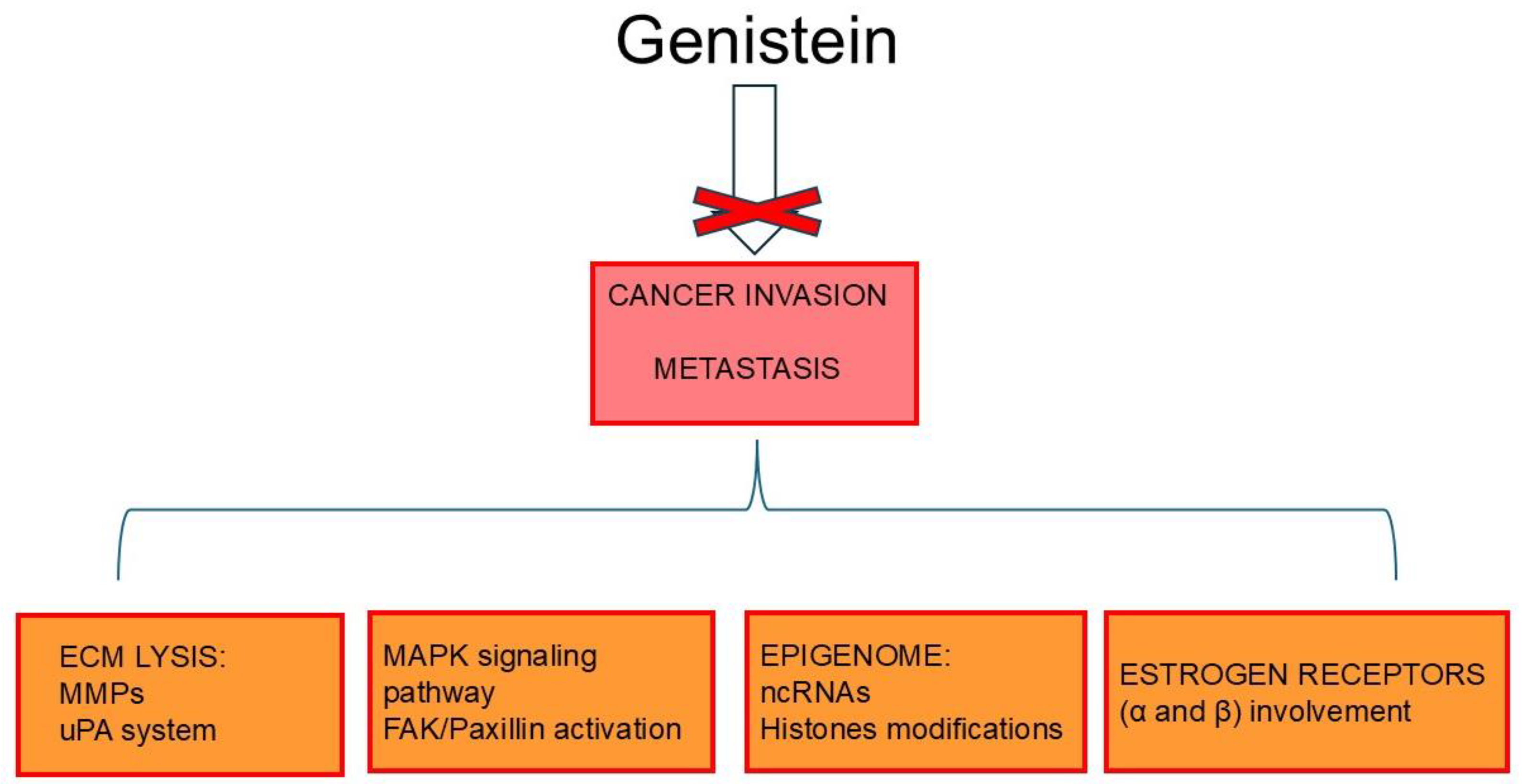
Extracellular matrix (ECM) remodeling and degradation are critical in cancer progression [77,78]. Matrix-degrading enzymes, including Ca2+- and Zn2+-dependent endopeptidases, namely MMPs and the uPA system, are the main mediators responsible for ECM molecule lysis [79,80,81,82]. As part of its chemopreventive action, by modulating the different molecular signaling pathways discussed below, genistein negatively influences the expression/activation of matrix-lysis enzymes, thereby acting on multiple aspects of cancer metastasis [79,83]. In this regard, a study by Kousidou and colleagues demonstrated that genistein downregulates the mRNA expression of several members of the MMP family in human BC cells, including estrogen-receptor-negative MDA-MB-231 and MCF-7 cell lines. Functionally, this reduced invasion of the cancer cells [82]. In addition, genistein dose-dependently efficiently inhibits transforming growth factor beta1 (TGF-β1)-induced invasion and metastatic potential of human pancreatic cancer in vitro by downregulating MMP-2 and uPA expression [84]. The anti-invasive impact of genistein on pancreatic cancer cells was also supported by the targeting of Forkhead box protein M1 (FOXM1) as shown by Wang et al. (2010) [85]. Furthermore, Xiao and co-workers demonstrated that the isoflavone inhibited (in a dose-dependent manner) the invasiveness of human colon–rectal cancer (CRC) cells, as well as metastasis formation in murine orthotopic implantation models, through the selective suppression of the proangiogenic marker Fms-related tyrosine kinase 4 (FLT4) (i.e., VEGFR2) and MMP-2 [86]. Additionally, Hussain et al. (2021) revealed that genistein, in a time-dependent manner, inhibited the invasive ability of HeLa cells by modulating MMP-9 and tissue inhibitor of metalloproteinases 1 (TIMP-1) [87].
The invasiveness of cancer cells is closely related to the abundance of focal adhesions (FAs), large and dynamic protein complexes, that link the cytoskeleton to the ECM; of note, FAs particularly accumulate in specialized membrane protrusions (i.e., invadopodia and podosomes) that contribute to the lysis of ECM through the action of MMPs [88]. Focal adhesion kinase (FAK) and paxillin are the main FA-associated kinases, and their activation is crucial in controlling cell migration and invasion [89]. Given this, genistein, by downregulating p125FAK, inhibited the metastatic activity of hepatocellular carcinoma (HCC) cells (Bel 7402) coupled with angiogenesis suppression in a murine xenograft model [90].
As already emphasized, genistein is recognized as a phytoestrogen due to its ability to interact with ERs [91]. Not surprisingly, its biological functions are largely modulated by the ERα and ERβ subtypes [92]. On this premise, Chan et al. (2018) noticed that genistein (in a dose-dependent trend) significantly reduced both migration and invasive ability of ER-positive ovarian cancer cells (SKOV-3 positive for ERα and A2780CP expressing ERβ) by suppressing FAK signaling [93]. Similarly, the invasion ability of human choriocarcinoma cells (JAR cells) was silenced by genistein via ERβ binding, followed by metastasis-associated proteins 3 (MTA3)/Snail/E-cadherin pathway activation [94].
The mitogen-activated protein kinase (MAPK) pathway transmits extracellular signals from the membrane to intracellular compartments and it is involved in several cellular processes including cell proliferation, migration, invasion, as well as cell death [95]. A study by Chen and colleagues demonstrated that genistein, in a dose-dependent manner, inhibited the metastatic potential of human cervical cancer cells (HeLa) in vitro by interfering with MAPK signaling pathways and FAK-paxillin activation [96]. Furthermore, by inhibiting MMP-9 expression and interrupting specific transductors of the MAPK pathway (namely MEK/ERK and JNK signaling pathways), genistein (in a dose-dependent manner) was able to suppress both migration and invasion of squamous cell carcinoma in vitro (SK-MEL-28 cells) [97].
Interestingly, in 16F10 melanoma cells, different doses of genistein were found to affect cancer cell invasiveness. High doses of the phytoestrogen (100 μM), by inactivating the FAK/paxillin signaling cascade, inhibited the cell adhesion, migration, and invasion triggering apoptosis; on the contrary, a low dosage of genisetin (12.5–50 μM) significantly promoted both invasion and migration of melanoma cells by activating the FAK/paxillin and MAPK signaling pathways [98]. Moreover, using a B16 mouse model of murine melanoma (B164A5 melanoma cell line and C57BL/6J mice), genistein showed antimetastatic activity and reduced the tumor’s size [99].
Tetradecanoylphorbol-13-acetate (TPA) is one of the most widely used agents to study the mechanisms of carcinogenesis and metastasis; in fact, it induces MMP expression/activation by modulating different signaling pathways [100]. Wang et al. (2014) observed that genistein blocks TPA-mediated metastasis via the downregulation of MMP-9 and epidermal growth factor receptor (EGFR) followed by the suppression of nuclear factor-κB (NF-κB) and activating protein-1 (AP-1) transcription factors and inhibition of MAPK, IκB, and PI3K/Akt signaling pathways in an HCC model [101].
Recently, Khongsti and co-workers found that human BC cells (MDA-MB-435 and MDA-MB-231) treated with genistein showed a reduction in osteopontin (OPN) secretion associated with increased phosphorylation of ERK1/2 and mitogen-activated protein kinase kinase 1/2 (MEK1/2) and upregulation of a member of the NAD (+)-dependent histone deacetylase family, namely SIRT1. Thus, the authors speculated that OPN inhibition via genistein may be epigenetically regulated by MAPK-pathway-induced SIRT1 [102]. Notably, OPN plays a pivotal role in cancer [103].
In prostate cancer (PC), the effects of genistein are rather inconsistent [104,105,106,107]. As a matter of fact, in a study by Touny et al. (2009) genistein curiously promoted and exacerbated the metastatic propensity of undiagnosed early-stage human PC through an estrogen- and PI3K-dependent mechanism, also involving OPN upregulation [104]. Conversely, in another investigation, genistein inhibited PC invasion and MMP-2 activation, switching off TGF-β-mediated phosphorylation of MAPK-activated protein kinase 2 (MAPKAPK2) and heat shock protein 27 (HSP27), both downstream regulators of p38MAP kinase signaling [105]. The antimetastatic effect of genistein on PC cells (1532CPTX, 1532NPTX, 1542NPTX, 1542CPTX, PC3, and PC3-M cell lines) was further supported by Xu et al. (2009) who showed suppression of MMP-2 by targeting the mitogen-activated protein kinase kinase 4 (MEK4) [107]. In this circumstance, the authors also noted the clinical relevance of genistein treatment in inhibiting signaling pathways involved in PC invasion. By analyzing normal prostate epithelial cells isolated from the prostate tissue of patients enrolled in a prospective, randomized phase II trial, they observed lower levels of MMP-2 expression compared to cells from untreated patients [107]. To address the controversial impact of genistein treatment on PC, Nakamura’s group developed a clinically relevant xenograft model generated from a patient’s prostatectomy specimen and demonstrated a prometastatic effect of the flavonoid. Thus, the authors speculated that genistein may have heterogeneous antimetastatic effects correlated with differential ERβ expressions among patients [106]. Additionally, as discussed below, non-coding RNAs (ncRNAs), including different microRNAs (miRs, 18–25 nt), that act as oncogenes and/or tumor suppressors in different cancers, can be regulated by genistein [108]. Indeed, as discovered by Chiyomaru and co-workers, genistein, both in vitro and in mouse models, by upregulating miR-574-3p expression and affecting related signaling pathways, decreased the invasion and metastatic activity of androgen-independent PC cell lines (PC3 and DU145, both expressing the ERβ subtype) [109].
Extensive evidence suggests that a dysregulated expression of KCNK9 (a member of the two-pore domain potassium (K2P) channel family) supports cancer progression [110,111]. Pertinently, a study by Cheng’s group demonstrated that genistein, by downregulating KCNK9 expression, acted on the Wnt/β-catenin signaling pathway, suppressing the malignant phenotype of human colon cancer [112].
The catalytic subunit of DNA-PK kinase (DNA-PKcs), which plays a major role in DNA damage signaling repair, also shows a proinvasive action [113]. As radiotherapy increases the invasive tendency of DNA-PKcs-positive glioblastoma multiforme (GBM), it has recently been shown that genistein can specifically bind to DNA-PKcs, suppressing the DNA-PKcs/Akt2/Rac1 signaling pathway, thereby successfully inhibiting the radiation-induced invasiveness of GBM cells in vitro and in vivo [114].
Epigenetic plasticity (i.e., DNA methylation/demethylation, histone modifications, as well as short and long ncRNA involvement) can contribute to cancer onset and progression [115]. For example, the methylation in the promoter of the gene coding Wnt inhibitory factor 1 (WIFI) favors cancer development by switching off the Wnt/β-catenin signaling pathway [116]. Pertinently, Zhu and co-workers found that genistein inhibited the invasion and migration of colon cancer cells (HT29) by inducing WIF1 gene demethylation, thereby restoring its activity. These effects were also supported by the regulation of cancer cell invasion-associated genes, including MMP-9, MMP-2, TIMP-1, E-cadherin, and Wnt signaling pathway mediators including β-catenin, c-Myc proto-oncogene protein, and cyclin D [117]. As mentioned above, the epigenetic modulator miRs can represent a target of genistein [108]. Consistently, it was observed that the phytochemical, by targeting miR-27a, inhibited invasion, suppressed cell growth, and induced apoptosis in pancreatic cancer cells [118]. In addition, Hirata et al. (2021) demonstrated that isoflavones, by regulating miR-1260b expression, inhibited Wnt signaling effector genes such as secreted frizzled-related protein 1 (sFRP1), Dickkopf 2 (Dkk2), and Smad4 in renal cancer cells (786-O, A-498, Caki-2 cell lines); functionally, cancer cell invasiveness together with proliferation and apoptosis phenomena was significantly decreased [119]. Another interesting study showed that genistein interrupted the metastatic activity of human colorectal cancer (CRC) by suppressing the interplay between the long ncRNA TATTY18 and the Akt pathway [120]. In more detail, genistein-treated CRC cells (SW480), among other phenomena, showed downregulated expressions of TATTY18 and reduced cell migration, coupled to a decrease in glucocorticoid-regulated kinase 1 (SGK1) and reduced Akt and p38-MAPK phosphorylation; these results were confirmed in vivo using genistein-treated tumor-bearing nude mice [118]. Circular RNAs (circRNAs), like other ncRNAs (i.e., micro- and long-ncRNAs), can act as oncogenes [121,122,123]. Recently, members of the flavonoid family have been reported to interfere with cancer progression by targeting specific circRNAs [122,123]. FOXM1, a cell-cycle-regulating transcription factor upregulated in cancer, has the role of maintaining malignant hallmarks by modulating the expression of target genes at the transcriptional level [124]. Interestingly, Yu et al. (2021) discovered that genistein exposure regulated non-small cell lung cancer (NSCLC) cell migration and invasion by decreasing the circRNA circ_0031250; in parallel, they showed that miR-873-5p is a target of circ_0031250 to finally conclude that genistein restricts NSCLC invasiveness and progression by involving the circ_0031250/miR-873-5p/FOXM1 axis [125].
It has been observed that genistein can synergistically hinder cancer spreading when combined with other compounds including specific phytochemicals. For example, a mixture of genistein and retinoic acid (ATRA) significantly inhibits the invasion ability of human adenocarcinoma cells (A549 cell line) by downregulating the transmembrane protein mucin 1 (MUC1) as well as intercellular adhesion molecules-1 (ICAM1) expression [126]. Furthermore, adding rottlerin to genistein on neuroblastoma cells (SH-SY5Y and Kelly) led to notable decreased levels of eukaryotic elongation factor 2 kinase (EF2K) which is known to influence invasion/metastasis and the integrin/Src/FAK axis [127].
To sum up, genistein can interfere with cancer invasion and metastasis by acting on multiple molecular mechanisms (Figure 3).

7. Genistein and Epithelial Mesenchymal Transition

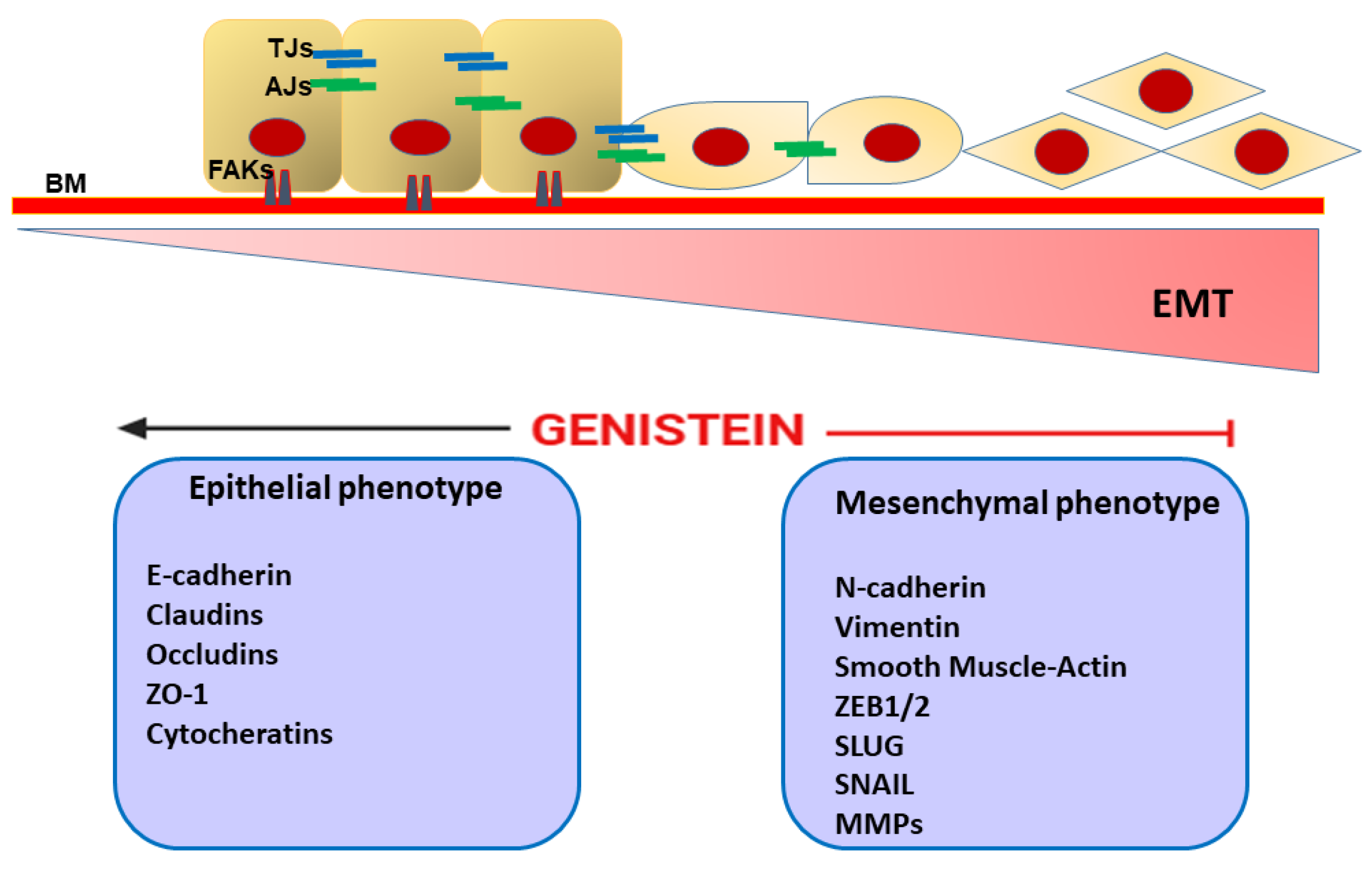
EMT is a dynamic and complex process by which epithelial cells lose their junctions and polarity and acquire a migratory mesenchymal phenotype, involving reorganization of the cytoskeletal scaffold [128,129]. Thus, typical epithelial markers including E-cadherin (CDH1), claudins, zonula occludens-1 (ZO-1), desmoplakin, and plakophilin are replaced by mesenchymal proteins such as N-cadherin (CDH2), vimentin (Vim), and α-smooth muscle actin (α-SMA) [128]. Multiple and interacting signaling pathways orchestrate the EMT phenomenon, such as tumor growth factor-β (TGF-β), epidermal growth factor (EGF), FGF, PDGF, Notch, Wnt/β-catenin, PI3K-Akt, FAK/paxillin, MAPK signaling, and the Hippo-Yes-associated protein (YAP)/PDZ-binding motif (TAZ) pathways, as well as several ncRNAs [128,130,131,132]. In synergy with the above molecular signals, numerous transcription factors such as Snail, Slug, Twist, NF-κB, HIF1/2 and zinc finger E-box binding homeobox 1/2 (ZEB1/2), and basic helix–loop–helix (bHLH), are involved in EMT [133]. Physiologically, EMT plays a crucial role in organogenesis and tissue repair [134]. In the tumor context, this process contributes to cancer malignancy by eliciting cell invasion, cancer stem cell (CSC) grouping, and drug resistance and supporting metastasis formation [135]. Therefore, inhibiting or reversing EMT may be a clinically relevant strategy to prevent cancer spread. As explored by Hsieh et al. (2020), genistein was able to suppress (in a dose-dependent trend) the prometastatic propensity of human head and neck cancer (HNC) coupled with a decrease in multidrug resistance by regulation of typical EMT markers (i.e., E-cadherin, Vim, Slug, and ZEB1) [136]. Additionally, by introducing an in vivo model, the authors concluded that genistein inhibited the aggressiveness of the HNC cells by perturbing the miR-34a/RTCB axis [136]. Another study showed that genistein, in combination with a specific miR-223 inhibitor, reversed the EMT process in gemcitabine-resistant pancreatic cancer cells, as supported by a downregulation in the expressions of mesenchymal markers (such as Slug, Vim, Snail, ZEB1, and ZEB2), thus enhancing the drug sensitivity and inhibiting cell motility and invasion [137]. As previously highlighted, genistein as a phytoestrogen can exert multiple biological functions by binding to ERs, including chemoprevention [16,18,19,21]. E2, as well as the typical endocrine-disrupting chemicals (EDCs) such as bisphenol A (BPA) and nonylphenol (NP), has been shown to promote EMT and invasiveness in estrogen-responsive cancers, including ovarian cancer [138]. On these premises, Kim and co-workers found that genistein was effective in reversing the ER-mediated EMT process activated by E2, as well as BPA and NP, in human ovarian cancer cells (BG-1) and reducing the resulting increase in expression of invasion markers such as MMP-2 and cathepsin D. In a parallel setting, the same investigators showed that the phytoestrogen was also able to reactivate the TGF-β signaling pathway in the BG-1 cells after its suppression by the compounds mentioned above [138]. Furthermore, in human HCC, genistein dose-dependently reversed EMT in vitro by partly suppressing the nuclear factor of activated T cell 1 (NFAT1) expression and thus showed antimetastatic activity in nude mice bearing liver orthotopic tumor implants [139]. In human colon cancer cells (HT-29) genistein (200 µgmol/L) decreased the expression of typical EMT molecules (i.e., N-cadherin, Snail2/Slug, ZEB1, ZEB2, FOXC1, FOXC2, and TWIST1), perturbed the Notch-1/NF-κB axis, and induced apoptosis [140]. Moreover, in human pancreatic cancer cell line Panc-1, genistein, through a Smad4-dependent signaling pathway, in a dose-dependent fashion, suppressed TGF-β1-induced EMT and invasiveness [84]. As discussed in other contexts of this review, the role of the genistein in PC remains disputable [104,105,106,108,109]. However, using dedicated PC cell lines (highly metastatic IA8-ARCaP cells and LNCaP/HIF-1α cells that stably overexpress HIF-1α), Zhang et al. (2008) found that low-dose genistein (0.2–15 μmol/L) inhibited invasion in vitro by reversing the EMT process [141]. Differently, a study performed by Du and co-workers found that genistein treatment inhibited endothelial growth factor (EGF)-dependent EMT in laryngeal cancer cells (Hep-2 cell line), inhibited cancer cell growth and migration, and promoted apoptosis; moreover, all these antitumor effects were more evident when trichostatin A (TSA) was added to genistein [109]. Another investigation demonstrated that genistein treatment of papillary thyroid carcinoma (PTC) cells, by preventing nucleus translocation of β-catenin, was able to block the EMT process as documented by alteration of EMT-related modulators (i.e., E- and N-cadherin; Vim and Snail), suppressed cell cycle/proliferation, and promoted cell death [142].
Taken together, this experimental evidence suggests that genistein, by acting on multiple EMT-related mechanisms, can effectively contribute to blocking cell invasion and the metastatic propensity of different tumor entities (Figure 4).

8. Genistein Eradicates Cancer Stem Cells

CSCs constitute a cluster of highly invasive cancer cells able to trigger tumorigenesis, supporting the metastatic cascade [143]. Like normal stem cells, CSCs are capable of self-renewal and differentiation [144]. However, various aberrant signaling pathways are involved in the maintenance and propagation of this tumor subpopulation, including TGF-β, Wnt/β-catenin, Notch, Hedgehog, PI3K/Akt/mTOR, NF-κB signaling, as well as the Hippo-YAP/TAZ pathways [145,146,147,148]. Furthermore, several biomarkers, including cell surface molecules (i.e., cluster of differentiation (CD) 24, CD90, and CD133), and various transcription factors, such as octamer-binding transcription factors 3 and 4 (Oct-3 and -4), Nanog, and SEX-determining region (SRY) homology box 2 (Sox2), are frequently used to identify and isolate CSCs [149]. A peculiar aptitude of CSCs shown in in vitro conditions is their ability to form “spheroids” (known as mammospheres in the case of breast CSCs); moreover, CSCs have an instrumental role in conferring drug resistance as well as tumor recurrence; thus, given their potent role in tumor aggressiveness, CSCs represent a considerable target for cancer therapy [150].
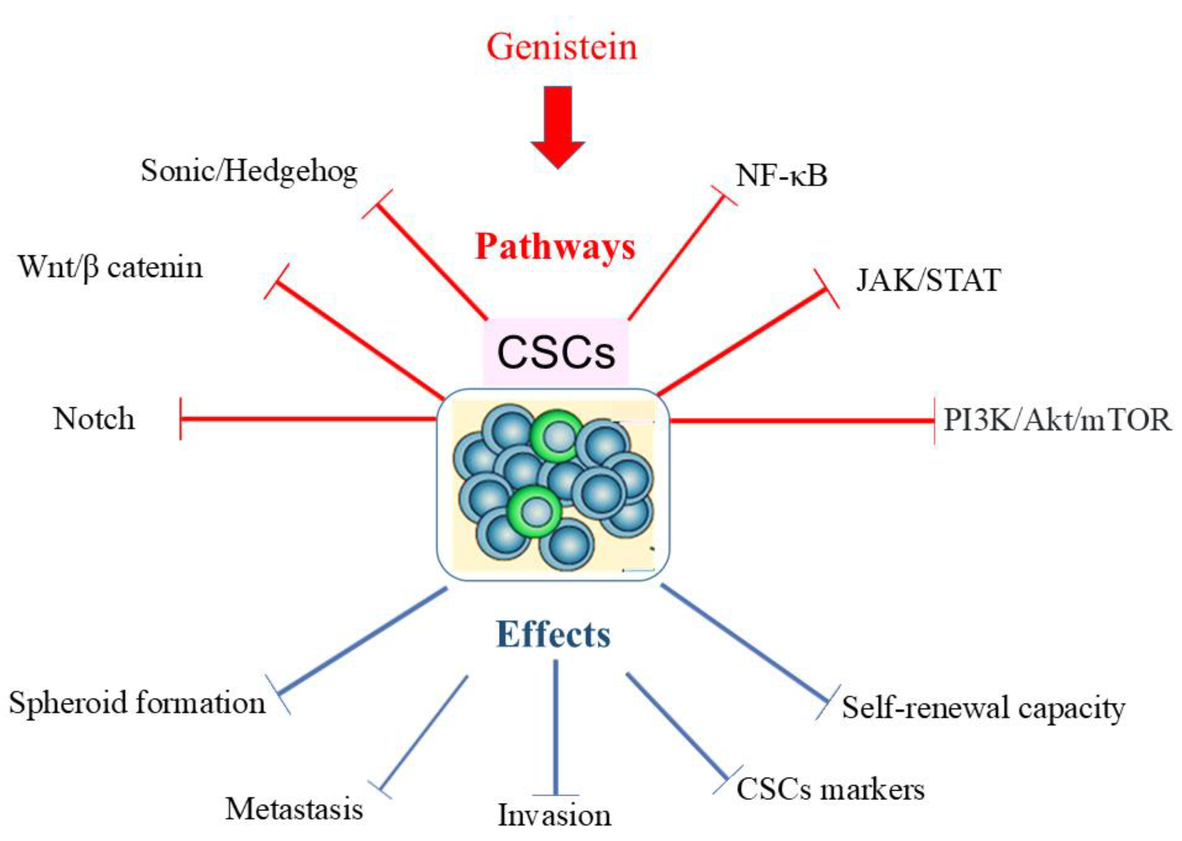
Recent studies have highlighted the potential of various phytochemicals, such as isoflavones, to interfere with numerous signaling pathways involved in CSC propagation, effectively reducing their ability to metastasize [151]. In this scenario, it has been demonstrated that genistein, by acting on specific signaling pathways (i.e., Sonic Hedgehog (SHH), Wnt/β-catenin, Notch, NF-κB JAK-STAT, PI3K/Akt/mTOR signaling), affects CSC proliferation, thereby inhibiting tumor invasiveness and metastasis (Figure 5) [152]. Particularly, it is postulated that genistein exerts its anticancer effects by modulating specific signaling pathways such as the Wnt/β-catenin, Notch, and Hedgehog pathways, which are crucial for stemness maintenance and self-renewal [108]. Moreover, genistein is associated with several pharmacological activities, including inhibition of various kinases, transmembrane channels, and other crucial molecular mechanisms [153]. For instance, topoisomerase II, tyrosine kinases, MAPK, ATP-binding cassette (Abc) transporters, P13K/Akt, polo-like kinase 1 (PLK1), SHH, and Wnt/β catenin signaling pathways are all mechanisms impaired by the above-mentioned isoflavonoid [21,153,154].
Specifically, these activities, together with induction of cell cycle arrest and apoptosis, prevention of reactive oxygen species (ROS), resolution of inflammation, inhibition of angiogenesis, EMT, regulation of steroid hormones, and specific metabolic pathways, contribute to the anticancer properties of genistein [153,155,156,157].
Originally, among other effects, genistein was recognized as a key compound with wide-ranging medicinal activities and has also been used to limit cancer invasiveness [152]. Specifically, genistein was found to reduce PC stem cells both in vitro and in xenograft mice by inhibiting the Hedgehog pathway and interfering with CD44 expression; ectopically, tumor growth was found to be effectively reduced [152]. As clarified above, CSCs can form spheroids in vitro, preserving, among other stem-like properties, immortality and invasiveness. Thus, in the above experimental context, genistein dose-dependently significantly inhibited tumorsphere formation (22RV1- and DU145-derived stem cell formation) [152]. Furthermore, in a study performed by Fan and co-workers, it was demonstrated that genistein exerts multiple effects on MCF-7 BC cells, not only suppressing proliferation and inducing apoptosis but also specifically inhibiting the CSC subpopulations with consequent inhibition of mammosphere formation through downregulation of the Hedgehog–Gli1 pathway [158]. Moreover, the phytoestrogen induced BC stem/progenitor cell differentiation by interacting with ER-expressing cancer cells through a paracrine mechanism correlated with PI3K/Akt and MEK/ERK signaling [159]. Yu et al. (2014) found that genistein markedly reduced the Gli1 level, which is considered a mediator of the SHH signaling pathway; such a mechanism involves specific CSC hallmarks of gastric cancer (GC) including CD44 expression, stem-cell-related genes (i.e., Oct-4, Bmi, Nestin, and adenosine triphosphate (ATP)-binding cassette efflux transporter G2(ABCG2)), spheroid formation, as well as migration and invasion propulsion [160]. Of note, as observed by Huang’s group, genistein treatment also alters the resistance of CSCs to chemotherapeutic drugs such as 5-fluorouracil (5-FU) and cisplatin, and it can also reduce tumor size in animal models of GC cells [161]. Similarly, more recent studies demonstrate that genistein inhibits proliferation and induces apoptosis by inhibiting the SHH signaling pathway in CSCs derived from human renal cancer [162]. The same SHH-targeting mechanism was found in human nasopharyngeal cancer stemness eradication, including the arrangement of tumor spheroids [163]. In addition, many researchers agree that 7-difluoromethoxy-5′,4′-di-n-octyl genistein, a genistein analog characterized by high bioavailability, can effectively eradicate CSCs derived from GC and ovarian cancer by multiple mechanisms, including inactivation of Akt, ERK, and NF-κB signaling, downregulation of FOXM1, as well as the propensity of stem-like cells to originate spheroids and suppress EMT [164,165,166]. Fu et al. (2020) showed that genistein (20 and 40 µM) inhibited the sphere formation aptitude of CSCs derived from human NSLC cells, which was associated with a decreased expression of stem-cell-related markers such as CD133, CD44, Bmi1, and Nanog. In a parallel setting, the authors found that genistein also suppressed the migratory and invasive activities of these specific CSCs by modulating manganese superoxide dismutase (MnSOD) and FoxM1 signaling pathways [167]. Furthermore, inhibition of PI3K/Akt signaling, coupled with overexpression of phosphatase and tensin homolog deleted on chromosome 10 (PTEN), has been shown to be a relevant pathway by which CSC behavior can be controlled by certain dietary factors, including genistein [168]. Similarly, previous studies by Montales et al. (2012) demonstrated that genistein inhibits the formation of CSC spheres originating from human breast tumors, showing that the lowest dose of genistein (40 nM) consistently attenuated the formation of primary and secondary mammospheres from transformed cell lines and primary epithelial cells isolated from BC cells (MCF-7 (expressing ERα) and MDA-MB-231). In contrast, a supraphysiological dose of genistein (2 μM) was less effective in eliciting a similar biological outcome [168]. Additionally, for a specific MDA-MB-231 subpopulation expressing CD44 and epithelial-specific antigen (ESA) (being particularly enriched in CSC content), both 2 μM and 40 nM doses of genistein were effective in suppressing mammosphere formation. Furthermore, the inhibition of PI3K/Akt signaling, coupled with PTEN overexpression, has been shown to be a relevant pathway by which CSC behavior can be controlled by dietary factors, including genistein [168].
Given the potent anticancer effects of genistein, in particular targeting the CSC phenotype, recent clinical studies have been designed to investigate the preventive and therapeutic efficacy of this natural compound in different human tumors, including CRC (ClinicalTrials.gov NCT01985763 nct.gov, 2019), PC (ClinicalTrials.gov NCT01126879 nct.gov, 2019), BC (ClinicalTrials.gov NCT00290756 nct.gov, 2017), and urothelial cancer (ClinicalTrials.gov, NCT00118040 nct.gov, 2017, NCT01489813 nct.gov, 2018).
Thus, based on the above experimental evidence, it is reasonable to ponder genistein as a useful drug to attenuate the aggressiveness of CSCs.

9. Genistein and Cell Cycle Arrest

The cell cycle is the ordered and finely regulated sequence of events that occur in cells that are about to divide [169,170]. Progression through each step of the cell cycle is controlled by checkpoint proteins belonging to the cyclin-dependent kinase (CDK) family [170,171]. Disorders that disrupt the CDK family of proteins deeply affect the rate of cell division and are often implicated in the development of primary tumors [172]. DNA damage causes a pause in the cell cycle to allow for repair mechanisms before the cell is committed to the subsequent steps [169,170]. Notably, DNA damage checkpoints can be divided into those controlled by the tumor suppressor and transcription factor p53 and those ultimately under the control of the checkpoint kinase 1 (Chk1) [170]. Several proteins are involved in cell cycle checkpoint activation pathways induced by DNA damage and DNA double-strand breaks, including mediator of DNA damage checkpoint protein 1 (MDC1), ataxia telangiectasia mutated (ATM), ATM- and Rad3-related (ATR), Chk1, checkpoint kinase 2 (Chk2), and PLK1 [173,174,175]. Genistein has been shown to modulate several cellular pathways and one of the most studied is the signaling cascade that controls cell cycle arrest [154,176,177,178].
Concentrations of genistein between 5 and 200 µM have been shown to induce cell cycle arrest in different cancer cell lines although the mechanisms are not fully understood [142,179,180,181,182,183,184,185] as these concentrations, even if theoretically achievable, are much higher than those found in the bloodstream after food intake [15].
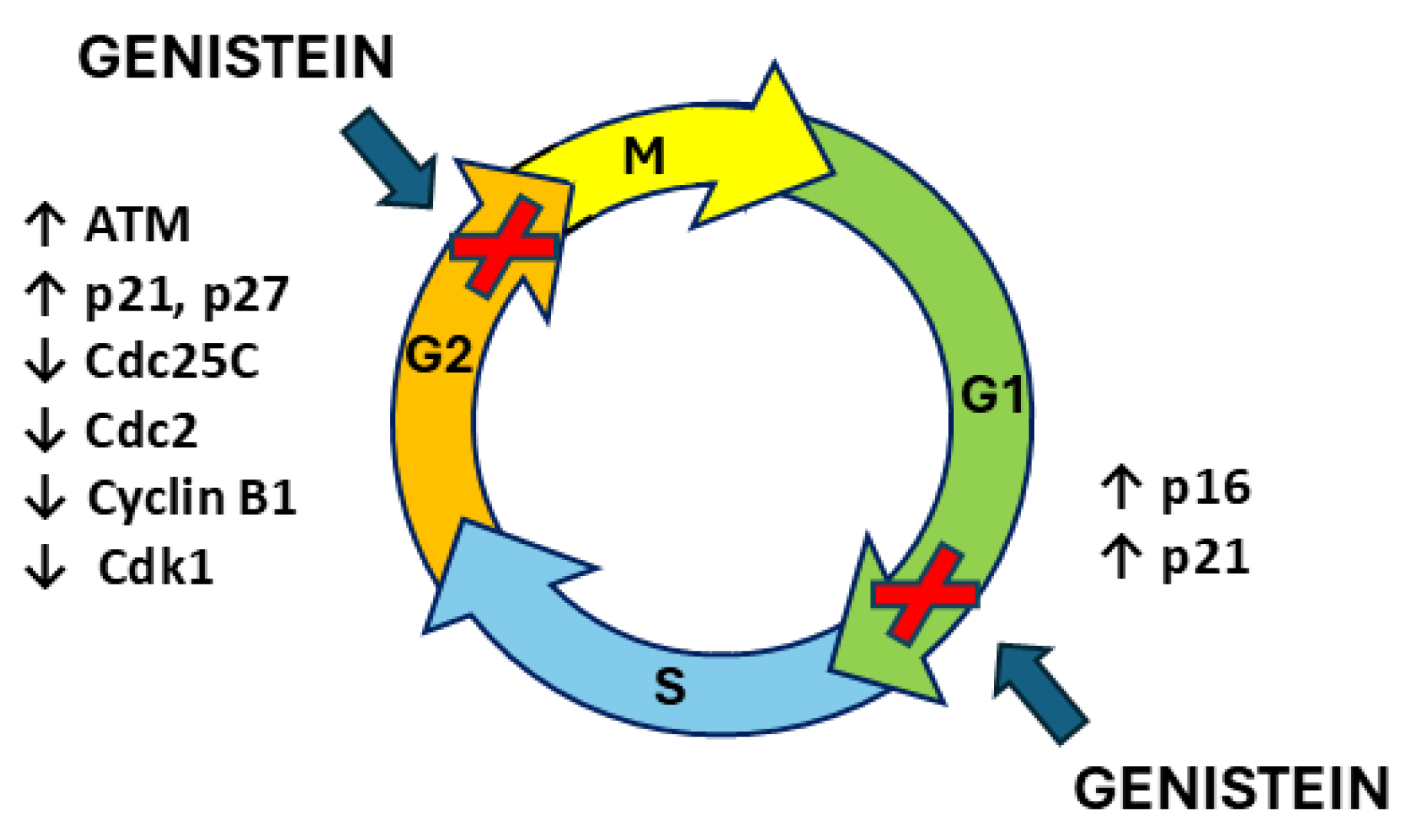
The extensive research on BC and PC evidenced the cell cycle arrest induced by genistein during the G2/M, G0/G1, and G1/S phases [186,187,188,189,190]. The analysis of differentially expressed genes performed on MCF-7 BC cells after treatment with genistein revealed a strong dose-dependent alteration in the expression of genes involved in cell cycle control, such as glioma pathogenesis-related protein 1 (GLIPR1), cell-division cycle protein 20 homolog (Cdc20), budding uninhibited by benzimidazole 1 (BUB1), mini-chromosome maintenance (MCM) complex 2, and cyclin B1 (CCNB1) [191]. Fang et al. (2016) showed that in the human triple-negative BC (TNBC) cell line MDA-MB-231, genistein affects various molecular processes during cell cycle progression, including DNA replication, cohesion complex cleavage, and kinetochore formation, through regulation via phosphorylation at 332 different sites on 226 proteins [192]. Consistently, in MDA-MB-435S, MDA-MB-468, and MCF-7 BC cells genistein induced a concentration-dependent accumulation of cells in the G2/M phase [193,194,195]. The same results were confirmed in MCF-7 and MDA-MB-231 BC cells where the DNA damage checkpoint (pATM) was activated and the levels of inactive pCdc25c and pCdc2 were upregulated, arresting cells in the G2/M phase [196]. In human BC MDA-MB-231 and SKBR3 cells, genistein exerted the same G2/M phase arrest in a dose-dependent manner and the molecular mechanism involved the inhibition of S-phase kinase-associated protein 2 (Skp2) and promotion of its downstream targets p21 and p27 [197]. G2/M phase arrest was confirmed in MDA-MB-231 cells after genistein administration in association with the downregulation of cyclin B1, Bcl-2, and Bcl-xL expression, possibly mediated by NF-κB activation via the Notch-1 signaling pathway [198]. Furthermore, genistein treatment of MDA-MB-231 BC cells resulted in G2/M cell cycle arrest as evidenced by a strong concentration-dependent reduction in the protein levels of cyclin B1, Cdk1, and Cdc25C; in particular, these results were mediated by a genistein-induced stable activation of ERK1/2 in a concentration- and time-dependent manner [199]. Similarly, genistein has been shown to induce G2/M phase block in BRCA1-impaired human BC MDA-MB-231 and HCC1937 cells by downregulating cyclin B1 levels due to genistein-induced suppression of seven-transmembrane receptor G protein-coupled receptor 30 (GPR30) activation and Akt phosphorylation/activation [200]. A similar mechanism involving the PI3K/Akt pathway was found in the PC cell lines PC3 and LNCaP, where 5 or 10 µM genistein induced a significant G2/M cell cycle arrest [201]. Co-treatment with selenite and genistein showed synergistic effects on G2/M cell cycle arrest, although the effect in PC3 cells was less than in LNCaP cells [201]. Higher levels of p21waf1 and Bax were detected in genistein-treated PC3 and LNCaP cells, while AKT phosphorylation was decreased only in PC3 cells with no change in total AKT protein levels [201]. Similarly, in PC cells (PC3 cell line), cell cycle analysis revealed a G2/M phase arrest induced by genistein treatment associated with a 1.1-fold increase in nuclear p21WAF1/Cip1 protein expression and a 14% decrease in nuclear cyclin B1 expression [202]. The combination of genistein and radiation had an even stronger effect on these cells than either treatment alone [202]. Furthermore, the increase in p21 detected in LNCaP and PC3 cell lines after genistein administration is associated with delayed mitosis and has been linked to transcriptional inhibition of PLK-1 [203]. In another human cell line, PCDU145, genistein blocked the cell cycle in the G2/M phase and induced the growth arrest and DNA amage-inducible 45 (GADD45) gene thorough its promoter [189]. The Gadd45 protein is a target gene of p53 and has been reported to play a role in cell cycle regulation [204,205]. Similarly, cell cycle arrest in the G2 phase in response to genistein treatment has been described in BC MCF-7 and PC cell lines (PC3), where genistein downregulated the mouse double minute 2 (MDM2) oncoprotein at the transcriptional level by interacting with the MDM2 promoter and at the post-transcriptional level by inducing MDM2 ubiquitination [188]. The inhibition of MDM2 expression by genistein was confirmed in PC3 xenografts [188].
Indeed, by flow cytometry analysis, genistein was confirmed to induce an accumulation of cells in the G2/M phase of the cell cycle in the PC cell lines LNCaP, DU145, and PC3 [206,207]. Gene expression analysis performed in genistein-treated cells showed that in DU145 cells, the cyclin-dependent kinase 4 (CDK4), cyclin-dependent kinase inhibitor 2A (CDKN2A), and minichromosome maintenance 4 (MCM4) genes were downregulated while only the SERTA domain-containing 1 (SERTAD1) gene was significantly upregulated [206]. In PC3 cells, genistein administration caused a significant decrease in minichromosome maintenance 3 (MCM3) mRNA expression and an increase in cyclin H (CCNH) [206]. In LNCaP cells, the baculoviral IAP repeat-containing 5 (BIRC5), cyclin B2, Chk2, CDC28, protein kinase regulatory subunit 1B (CKS1B), GTSE1, hairy-related 5 (HERC5), minichromosome maintenance 2 (MCM2), MCM4, and proliferating cell nuclear antigen (PCNA) genes were downregulated while, in CDK7, cyclin-dependent kinase inhibitor 1A (CDKN1A) and cyclin-dependent kinase inhibitor 1B (CDKN2B)p were upregulated after genistein treatment [206]. In line with this, a significant G2/M phase arrest was described in the genistein-treated DuPro androgen-insensitive PC cell line; these cells showed suppression of cyclins with concomitant induction of the tumor suppressor genes p21 (WAF1/CIP1/KIP1) and p16 (INK4a) [190]. In particular, genistein has been shown to increase active acetylated histones at p21 and p16 transcription start sites [190]. Two different studies showed that genistein-induced G2/M phase cell cycle arrest in T47D BC cells is mediated by the formation of 5,7,3′,4′-tetrahydroxyisoflavone (THIF), a product of genistein cellular metabolism [121,122]. THIF has been shown to cause cell cycle arrest through activation of ATR, which is a consequence of intracellular oxidative stress, GSH depletion, and increased DNA damage [119]. In addition, THIF induced inhibition of cdc2, phosphorylation of p53 and Chk1, and deactivation of cdc25C phosphatase [121]. Moreover, another study by Nguyen et al. (2018) attributed the effect of THIF to the phosphorylation of p38 MAP kinase, resulting in the inhibition of cyclin B1 and cdc2 activation [208]. Additionally, genistein and a hydantoin-derived antiandrogen–genistein conjugate caused a significant accumulation in the S-phase of LNCaP cells [209].
Although the effect of genistein has often been described in terms of arresting the G2/S phase of the cell cycle, some studies have shown a cell cycle arrest induced by genistein administration in BC cell lines in a different phase. In more detail, genistein induced a G0/G1 phase arrest in MCF-7 and MDA-MB-231 cell lines [210,211]. Lin et al. (2009) confirmed the ability of genistein to inhibit the entry of the BC cell lines HS578T, MDA-MB-231, and MCF-7 into the G0/G1 phase of the cell cycle [212]. Moreover, in the T47D cell line genistein administration resulted in G0/G1 phase cell cycle arrest only when cells reached confluence [213]. In PC LNCaP cells, genistein induced a G0/G1 cell cycle arrest through the increased expression of histone acetyltransferases responsible for the increased transcription of p21 and p16 suppressor genes which induce cell cycle arrest [190].
In MDA-MB-231 and MCF-7 cells, genistein induced the accumulation of cells in the G0/G1 phase, and co-treatment of cells with GEN and a potent steroid hormone precursor called 1,25-dihydroxycholecalciferol (1,25(OH)2D3) confirmed this result with a stronger effect on the MDA-MB-231 cell line [187]. Similarly, in the ER+/HER2-overexpressing BT-474 human BC cell line, genistein and tamoxifen monotherapy significantly increased the G1 phase cell population, and this G1 arrest effect was enhanced when the two molecules were used in combination [214].
In conclusion, genistein appears to have a wide range of effects on the cell cycle, which vary depending on the cell line and the phase of the cell cycle at the time of treatment [185,215]. As a potent modulator of cell-cycle-related proteins and a regulator of several molecular targets involved in cell cycle progression, genistein may have anticancer properties by modulating the effects of deregulated cell cycle checkpoints in cancer cells [185] (Figure 6).

10. Programmed Cell Death

Apoptosis is a programmed cell death (PCD) mediated by multiple signaling pathways triggered by cellular stress, DNA damage, immune surveillance, and other stressing cellular factors [216,217]. Two distinct signaling pathways—the extrinsic and intrinsic apoptotic pathways—trigger apoptosis in most tumor cells [217,218]. The extrinsic pathway involves the activation of death receptors such as Fas and tumor necrosis factor receptors (TNFRs), leading to the activation of caspase-8 and caspase-3 to induce apoptosis [218,219]. The intrinsic pathway is associated with changes in mitochondrial permeability, leading to the mitochondrial release of proapoptotic proteins such as B-cell lymphoma 2 (Bcl-2)-associated X protein (Bax), cytochrome c (cyt c), and apoptosis-inducing factor (AIF) into the cytoplasm and activation of caspase-9 and caspase-3, ultimately triggering apoptosis [218,219,220]. Caspase-3 is responsible for cleaving poly (ADP-ribose) polymerase (PARP) during cell death and is derived from both extrinsic and intrinsic pathways. Caspase-8 can cleave BH3-interacting domain death agonist (BID), a death-inducing member of the B-cell lymphoma 2 (Bcl-2) family, allowing crosstalk between the extrinsic and intrinsic apoptotic pathways [220]. The cleaved BID translocates to mitochondria and induces the release of cyt c, leading to caspase-9-dependent activation [218,220,221]. The ERK and NF-κB pathways can inhibit this apoptotic signal [218,222].
Cancer cells tend to evade cell death by activating antiapoptotic mechanisms, so cancer cell death can be achieved by reversing antiapoptotic processes [223,224,225]. Genistein has anticarcinogenic effects; in fact, several studies have shown that this isoflavone inhibits cell proliferation and induces death in several human cancer cells [18,108,176,183,226]. In this section, we will examine in detail the effects of genistein on the activation of apoptotic death in hormone-dependent cancers, such as prostate and breast cancers, which have been extensively studied [14,16,211,215,227].

10.1. Breast Cancer

The effects of genistein have been studied in BC MCF-7 cells through oligo microarray technology and the expression of multiple Bcl-2 family genes, which resulted in both pro- and antiapoptotic altered genes [228].
A study by Tophkhane et al. (2007) has shown that genistein-induced enhanced cell death and growth inhibition in MCF-7 cells overexpressing high levels of Bcl-2. Genistein administration resulted in increased levels of p85, a major subunit of cleaved PARP, membrane permeability changes, and cyt c release, suggesting that the enhanced activation of the caspase cascade involved in Bcl-2 overexpression mediates the sensitization of MCF-7 cells to genistein. Furthermore, the enhanced activation of the apoptotic process in Bcl-2-overexpressing cells is due to the isoflavone-induced accumulation of Bcl-2 and alteration of Bax anchoring in mitochondria [229]. Similarly, in MCF-7 cells, genistein-induced apoptosis led to a reduction in Bcl-2 expression and induction of Bax. In MCF-7 cells, both the expression of ERα and the proliferation were reduced after the administration of genistein [230]. Furthermore, high concentrations of genistein inhibited both normal MCF-10A and cancerous MDA-MB-231 ERα-negative breast cells, associated with p53- and p21-dependent apoptosis activation. Genistein increased proapoptotic proteins (phospho-p53 (p-p53) and p21) and decreased antiapoptotic proteins (Bcl-xL or cyclin B1) in breast cells [231]. These data were confirmed by the study of Ye et al. (2018) which showed that genistein significantly inhibited cell proliferation and induced pronounced apoptosis in the human BC cell lines MDA-MB-231 and SKBR3. The molecular mechanism involved downregulation and inhibition of S-phase kinase-associated protein 2 (Skp2) and upregulation of its downstream targets p21 and p27 [197]. Genistein promoted a p53-independent decrease in mouse double minute 2 (MDM2), which led to an increase in the half-life of p21 since this protein is a direct target of MDM2 for proteasomal degradation. Given that p21 is involved in genistein-induced apoptosis and G2 arrest in MCF-7 tumor cells, and that p21 protein is stabilized by genistein, it may mediate the antitumor effects of genistein [188]. Moreover, genistein-treated MCF-7, MCF-7-caspase-3 (MCF-7-C3), and T47D BC cells expressed low levels of the cancerous inhibitor of protein phosphatase 2A (CIP2A) which correlated with growth inhibition and apoptosis induction, as evidenced by PARP cleavage and the expression of functional caspase-3 in MCF-7-C3 and T47D [232].
Genistein has been shown to induce apoptosis via an ERα-independent pathway without the involvement of MAP kinase and Akt but associated with reduced Bcl-2/Bax ratio in MCF-7 cells [233]. Typically, genistein has been shown to inhibit the growth of MCF-7 cells and promote the apoptotic pathway through the inactivation of PI3K/Akt signaling, as evidenced by the decrease in phospho-Akt in treated cells, resulting in reduced expression of the downstream target HOX Antisense Intergenic RNA (HOTAIR) [234], a long ncRNA (lncRNA) implicated in cancer invasion and metastasis [235]. Furthermore, treatment with genistein resulted in a significant decrease in PI3K and AKT protein and a significant increase in Fas ligand, FAS-associated protein with death domain (FADD), cyt c truncated Bid, caspase-9, and caspase-3 in MCF-7 cells [236,237]. Similarly, genistein has been shown to induce the extrinsic FAS-receptor-dependent apoptosis pathway in MCF-7 cells engineered to overexpress oncogenic HER2 (MCF-7 HER2) and control vector cells (MCF-7 vec). Specifically, the phytoestrogen administration caused upregulation of p53 and levels of FAS receptor and cleaved caspase-8 and induced PARP cleavage, sustaining an antiproliferative activity explained by the inhibition of NF-κB signaling; in fact, in BC cell lysates, this compound inhibited the phosphorylation of IκBα, preventing IκBα from forming the NF-κB heterodimer (p65 and p50) necessary for the activation of the NF-κB axis, and inhibited the nuclear translocation of p65 (subunit of the NF-κB heterodimer) and its phosphorylation in the nucleus, leading to the inhibition of the transactivation of NF-κB target genes [238]. In a similar way, genistein treatment of MDA-MB-231 cells induced apoptosis in a dose- and time-dependent manner by inhibiting NF-κB activity via the Notch-1 signaling pathway. Moreover, genistein administration suppressed the expression of cyclin B1, Bcl-2, and Bcl-xL, possibly through the activation of NF-κB via the Notch-1 signaling axis [182]. Specifically, the phytoestrogen caused a dose-dependent reduction in the levels of mitogen-activated protein kinase 5 (MEK5), total ERK5, and phospho-ERK5 and a decrease in the levels of NF-κB/p65 nuclear protein, which are associated with inhibition of MDA-MB-231 cell proliferation and induction of apoptosis. Activation of the apoptotic process is confirmed by a dose-dependent increase in Bax protein levels and a decrease in Bcl-2 protein levels, as well as cleavage of caspase-3 and induction of caspase-3 activity [239]. Similarly, genistein has been shown to induce apoptotic cell death, as demonstrated by a decreased Bcl-2/Bax mRNA and protein ratio, in the MCF-7 cell line through a mechanism involving the inactivation of the type 1 insulin-like growth factor receptor (IGF-1R)/p-Akt signaling axis [240]. The p38-MAPK pathway drives cell proliferation and antiapoptosis, so the inhibition of this axis represents a good strategy for the promotion of cancer cell death [241]. In this regard, genistein induced apoptotic MCF-7 cell death through calpain and caspase-7 activation and PARP cleavage. In addition, administration of the phytoestrogen activated the apoptosis signaling kinase 1 (ASK1)-p38 mitogen-activated protein kinase cascades involving Ca2+ release from the endoplasmic reticulum, whereas no effect was observed for ERK1/2 [242]. Similarly, the isoflavone induces apoptosis in MCF-7 BC cells by activating Ca2+-dependent proapoptotic proteases, including mu-calpain and caspase-12, through the increase in intracellular Ca2+ concentration resulting from the depletion of endoplasmic reticulum Ca2+ stores [243].
The MAPK and AKT pathways are often constitutively activated in solid tumors; in contrast, the deletion of PTEN is one of the tumor suppressors often lacking in patients with advanced cancer [244]. In this regard, genistein-treated MCF-7 cells showed increased PTEN protein levels associated with decreased Akt phosphorylation. They increased p27 protein levels, whereas MAPK phosphorylation and cyclin D1 levels regulated by PTEN protein phosphatase activity were not altered, but their mRNA levels were slightly increased in phytoestrogen-stimulated cells [245]. Consistently, at low physiologically relevant concentrations, genistein inhibited cell survival and induced apoptosis in metastatic BC cells, MDA-MB-435 and Hs578t, while not affecting the survival of non-metastatic MCF-7 cells. In association with decreased cell viability and increased apoptosis, genistein decreases miR-155 and increases its proapoptotic targets including Forkhead box (Fox) subclass O3 (FOXO3), PTEN, casein kinase, and p27 [246]. A study by Dave et al. (2005) provided further insight into the PTEN-involving mechanism by which genistein promotes mammary epithelial cell death in vitro and in vivo. The mammary glands of young adult female rats exposed to diets containing casein (CAS) as the sole protein source supplemented with genistein or soy protein isolate (SPI+) showed increased apoptosis compared to rats fed a CAS diet without genistein. Increased mammary apoptosis in genistein and SPI+ rats was accompanied by increased PTEN expression. However, increased expression of the proapoptotic genes p21, Bax, and Bok was only observed in genistein-fed rats. Furthermore, genistein-induced apoptosis in MCF-7 cells was associated with increased PTEN expression and was abolished when PTEN expression was knocked down. MCF-7 cells treated with serum from genistein- or SPI(+)-fed rats showed increased apoptosis and increased levels of PTEN transcript [247].
In addition, genistein-induced apoptosis in MCF-7 cells was estrogen-independent and associated with dysregulation of the Bax/Bcl-2 ratio, downregulation of the antiapoptotic protein survivin, and induction of oxidative stress, as evidenced by decreased expression of the antioxidant enzymes copper–zinc superoxide dismutase (CuZnSOD), MnSOD, and thioredoxin reductases (TrxRs) and increased expression of glutathione peroxidase (GPx) [248]. Similarly, in MDA-MB-231 and MDA-MB-468 cells, genistein administration resulted in the downregulation of antiapoptotic Bcl-2, upregulation of proapoptotic Bax, and activation of caspase-3, leading to the induction of apoptotic death involving the mobilization of endogenous copper, as demonstrated by the fact that the copper-specific chelator neocuproine was able to almost completely reverse these gemistein-induced effects, whereas iron and zinc chelators were ineffective. Furthermore, the involvement of ROS production in genistein-induced apoptosis was demonstrated by its inhibition by ROS scavengers [249].
As specified above, genistein, as a phytoestrogen, can compete and/or interfere with the activity of Erα, and ERβ receptors can act as an estrogen agonist or antagonist in mammals by competing or interfering. In this regard, genistein was reported to have different effects against BC cells at different concentrations and in various cell types (ER-positive and ER-negative cells) [91,213,250].
For example, using proteomic and transcriptomic approaches, it has been shown that genistein has effects on gene and protein expression in T47D ERβ-positive cells, even in the presence of low levels of ERα, and that its final estrogenic effect on cells/tissues depends on both by the phenotype of receptors and the receptor subtype ratios within these cells/tissues. Precisely, this phenotype may be altered by exposure to genistein [251].
The necessity of ERα for estrogen-induced growth inhibition and apoptosis was confirmed by Obiorah et al. (2014) who showed that the phytoestrogen genistein induced endoplasmic reticulum stress, an inflammatory response leading to intrinsic and extrinsic apoptosis in a long-term estrogen-deprived BC cell line (MCF-7:5C) through an ERα-mediated mechanism. Genistein induced the endoplasmic reticulum stress (ERS) marker CHOP and the unfolded protein response (UPR) sensor inositol-requiring protein 1 alpha (IRE1α), whereas PCD activation was evidenced by upregulation of the apoptotic genes BCL2L11/BIM, tumor necrosis factor (TNF), FAS, and FADD [252]. Similarly, genistein at general physiological concentrations significantly reduced proliferation and increased apoptosis in primary BC cells, as evidenced by increases in apoptosis markers such as FADD, tBid, cyt c, caspase-8, and caspase-3. However, in the presence of E2, the ability of genistein to induce apoptotic effects in malignant breast cells ex vivo was completely abolished [253]. Furthermore, cellular responses have been shown to be triggered by different signaling mechanisms depending on the concentration of genistein. Physiological concentrations of this isoflafone (<10 μM) plus E2 induced apoptosis of MDA-MB-231 (ERβ-positive/ERα-negative) cells by dysregulating the Bax/Bcl-2 ratio and reducing the phosphorylation of ERK1/2, sustaining the antitumoral role of genistein against ERβ-positive/ERα-negative BC cells. At higher concentrations (10 to 100 μM), genistein reduced cell proliferation and increased apoptosis, both in the presence and absence of E2, without the involvement of Bax/Bcl-2 or the phosphorylation of ERK1/2 [254]. Pons et al. (2014) demonstrated that the outcome of genistein treatment depends on the ERα/ERβ ratio in BC cells. Co-administration of genistein with E2 induced cell proliferation and inhibited apoptosis in MCF-7 cells characterized by a high ERα/ERβ ratio, whereas in T47D cells with a low ERα/ERβ ratio, E2 and especially GEN exerted antiproliferative and proapoptotic effects as indicated by a significant decrease in the P-STAT3/STAT3 ratio [213].
In addition, genistein exhibited a biphasic effect on MCF-7 BC cell growth and ERα expression; in fact, low concentrations (<10 µM) of the phytoestrogen induced marked increases in proliferation and ERα expression, whereas high concentrations of the isoflavone caused inhibition of cell proliferation and apoptotic morphological features in treated cells. Furthermore, the expression of ERα and erbB2 was dose-dependently reduced by genistein for both SKBR3 and ZR-75-1 cells, and apoptotic changes were detected after exposure of ZR-75-1 cells to either daidzein or genistein at a concentration of 100 µM for 72 h [255]. Similarly, the isoflavone induced apoptosis in estrogen-sensitive MCF-7 cells at 50 and 100 µM, but estrogen interfered with the apoptotic activity of genistein at 50 µM. The estrogen-modulated increase in cyclin B1 levels was contrasted by co-treatment with genistein (100 μM), suggesting that the antiapoptotic regulation of cells by estrogen may involve cyclin B1 and that genistein exerted an estrogen-antagonistic action at a higher concentration [256].
The proapoptotic effect of genistein has been shown to be dependent on estrogen levels only in cultured cells, whereas in animal models estrogen levels did not affect the results. In detail, genistein administration induced apoptosis in MCF-7 cells in the absence of estrogen, as evidenced by DNA fragmentation, reduced levels of Bcl-2, and upregulated Bax protein. In the presence of estrogen, p21 and p53 protein expressions were upregulated by high concentrations of genistein. In female rats, the Bcl-2/Bax ratio was decreased by genistein treatment in the presence or absence of estrogen. These results indicate that the proapoptotic property of genistein may be strongly influenced by the concentration of estrogen in vitro, but this influence by estrogen is not evident in vivo [257]. Furthermore, the proliferation of MCF-7 cells was induced by both E2 alone and E2 plus genistein at concentrations ≥ 10−6 mol/L while apoptotic cell death was induced by GEN monotherapy in the same cell line [257]. The combination of genistein and E2 caused the increase in proliferating cell nuclear antigen, PI3K, and p-Akt and the inhibition of the increase in FADD, cyt c, truncated Bid, caspase-9, caspase-3, and ERβ [258]. Consistently, Lucki et al. (2011) have provided a mechanism to explain genistein’s ability to promote the proliferative response of MCF-7 cells via an estrogen-dependent ER-binding pathway, which predicts that genistein induces acid ceramidase (ASAH1) gene expression by activating a cell surface receptor-coupled G-protein (GRP30)-dependent pathway leading to the phosphorylation of ERK1/2 which in turn triggers the proliferative response of BC cells via an estrogen-dependent pathway. Moreover, ERK1/2 activation provokes the phosphorylation of ERα and the formation of a complex with ERα, Sp1, and SRC1 bound to the ASAH1 promoter. As a result of ASAH1 transcription, there is an increase in protein expression and enzymatic activity, which in turn leads to an increase in sphingosine 1-phosphate (S1P) production. By binding on cell surface S1P receptors, S1P can then be exported and activate proliferative pathways; in parallel, ASAH1 controls the expression of cyclin B2, thereby promoting mitosis and cell growth [259]. Instead, co-treatment of MCF-7 cells with different doses of E2 and genistein (10−6 M) did not result in antagonism of E2 activity and did not affect the proliferation rate. Co-treating MCF-7 cells with E2 and GEN resulted in a significantly increased inhibition of apoptosis, at least in combination with E2 (10−10 M) [260].
Moreover, the SUM1315MO2 cell model carrying the 185delAG BRCA1 mutation was markedly more sensitive to GEN than BRCA1 wild-type cell lines, probably due to the expression of ERβ, which is a major mechanism of physiological action of genistein, as already explained [261]. Genistein induced apoptosis more efficiently in BRCA1-mutant BC cell lines (HCC1937, SUM149, and SUM1315 cells) than in the MDA-MB-231 cell line, which harbors a functional wild-type BRCA1 gene. After genistein administration, all cells showed increased p21 protein levels and decreased Akt signaling, consistent with activation of the apoptotic process, except for MDA-MB-231 cells, in which mRNA and AKT protein levels were strongly increased and p21 protein levels slightly decreased, resulting in resistance to genistein [262].
The treatment with various concentrations of soya aglycone-rich extract (SARE) and flaxseed aglycone-rich extract (FSARE) caused higher caspase levels in MDA-MB-231 than in MCF-7 cells. Based on the docking score and binding energy, the best-fit protein target genistein was aldose reductase which showed a dose-dependent decrease in its expression after SARE administration [263]. In the study by Stocco et al. (2015), an extract of soy biotransformed by the fungus Aspergillus awamori was administered to estrogen-dependent (MCF-7) and non-estrogen-dependent (SKBR3) BC cell lines. The data suggested that the components of the extract, which was implemented in terms of the amount of isoflavones produced by the process of soy biotransformation, induced cell death by apoptosis and necrosis, mainly in MCF-7 cells, through a process responsive to caspase-3 activation involving, among other proapoptotic factors, Bad [264].

10.2. Prostate Cancer

In PC, similar effects were exhibited after the isoflavone administration: specifically, genistein administration to PC3 cells induced apoptotic death by increasing caspase-3 expression and activity. Cell growth was suppressed by the reduction of the p38 mitogen-activated protein kinase (p38MAPK) gene expression and protein level, while cell aggressiveness was strongly suppressed by the reduction in MMP-2 activity [265]. A similar result has been described by Kumi-Diaka et al. (2006); specifically, genistein exposure promoted the inhibition of cell growth through apoptotic cell death in both PC3 and LNCaP cells [266]. A significant dose- and time-dependent inhibition of MMP-2 expression levels in both cells was correlated with increased genistein concentrations [266]. In the study by Li et al. (2004), the effects of GEN on PC3 cells and experimental PC3 bone tumors created by injecting PC3 cells into human bone fragments previously implanted in severe combined immunodeficient (SCID) mice were evaluated. It was found that the isoflavone modulated the expression of genes involved in cell growth, apoptosis, and metastasis both in vitro and in vivo. Genistein administration caused the suppression of MMP-2, -11, -13, -14, and membrane-type matrix MMPs (MT-MMPs) and the upregulation of osteoprotegerin in PC3 bone tumors. Furthermore, MMP-9 expression was inhibited in PC3 cells in vitro and PC3 bone tumors in vivo after genistein administration [267]. Differently, the administration of genistein to PC3 cells resulted in the downregulation of the MDM2 oncogene mRNA, protein upregulation of p21, and induction of apoptosis. The downregulation of MDM2 protein is achieved by genistein-induced MDM2 ubiquitination, with the MDM2 promoter being important for the effects of genistein. Overexpression of MDM2 abolished genistein-induced apoptosis in vitro. These results were confirmed in vivo, as genistein inhibited tumor growth in PC3 xenografts [188]. By contrast, the effect of genistein in LNCaP and PC3 cancer cell lines was associated with the induction of apoptosis through the downregulation of PLK-1 protein and the upregulation of p21 [203]. Through epigenetic changes, genistein also affects gene expression patterns. In this regard, the modulation effects of genistein on DNA methylation led to the inhibition of cell growth and induction of apoptosis. In detail, the methylation profiles of 58 genes were altered by genistein administration in DU145 and LNCaP PC cells. In addition, the methylation frequencies of the mitotic arrest deficient 1-like protein 1 (MAD1L1), TNF receptor-associated factor 7 (TRAF7), lysine demethylase 4B (KDM4B), and human telomerase reverse transcriptase (hTERT) genes were remarkably modified by genistein treatment [268]. Furthermore, genistein has been shown to suppress growth and induce apoptotic death in PC3, DU145, and LNCaP cell lines in vitro by inducing the expression of aplasia Ras homology I (ARHI), a tumor suppressor gene downregulated in various malignancies including PC. ARHI was reported to upregulate the Hect domain and RLD5 (HERC5), CDNK1A, growth arrest, and DNA damage-inducible alpha (GADD45A) at transcriptional and protein levels. The effect of genistein was mediated by downregulation of miR-221 and miR-222 [269]. In addition, miR-574-3p was significantly upregulated in genistein-treated DU145 and PC3 PC cells compared to vehicle control. The expression of miR-574-3p was significantly lower in PC cell lines and clinical PC tissues than in normal prostate cells (RWPE-1) and adjacent normal tissues. miR-574-3p restoration induced apoptosis through reduction of Bcl-xL and activation of caspase-9 and caspase-3 [270]. Moreover, GEN administration modulated epigenetics and gene expression by altering histone acetylation through increased HAT1 protein levels resulting in increased H3K9 acetylation and increased SOX7, a Wnt inhibitory gene, and cell death promotion in PC3, DU145, ARCaP-E, ARCaP-M, and LNCaP cancer cell lines [271]. Similarly, genistein significantly induced apoptosis of DU145 cells, but no significant effect on apoptosis was observed in PC3 cells. lncRNA profiling showed that genistein modulated increased HOTAIR expression in castration-resistant PC cell lines compared to normal prostate cells. Indeed, PC cell growth and invasion were suppressed by HOTAIR knockdown (siRNA) in association with induction of apoptosis. miR-34a was also upregulated by GEN and directly targeted HOTAIR in both PC3 and DU145 PC cells [272]. Therefore, global gene expression patterns showed that maximal physiologically achievable concentrations of genistein (≤10 μM) had proliferative effects in PC3 cells by inducing activation of CDKs, MAPKs and small ribosomal subunit proteins (RPSKs), whereas high concentrations of genistein (>10 μM) appeared to modulate a different signaling axis leading to apoptotic cell death, specifically downregulating TGF-β by specifically decreasing SMAD 2/3, 4 in the downstream TGF-β signaling cascade [273]. This effect has also been shown in LNCaP cells where the isoflavones did not alter death receptor expression but significantly augmented TRAIL-induced disruption of mitochondrial membrane potential causing cytotoxic and apoptotic effects [274]. The oxidative-stress-promoting effect of GEN was confirmed in PC3 cells under 3D culture conditions, where a concentration of 480 μM of the isoflavone reduced cancer cell viability by inducing apoptosis through a non-mitochondrial pathway; furthermore, genistein reduced the production of cellular nitric oxide (NO) and increased catalase and glutathione production [275]. Furthermore, genistein inhibited the growth of DU145 and LNCaP cancer cells, leading to the death of such cells, by inducing ROS production and interfering with the expression of the two copper transporter genes, CTR1 and ATP7A. The copper chelator neocuproine reversed this effect, highlighting the role of copper in isoflavone-induced cytotoxicity [276].
In an animal model study by Nakamura et al. (2011), genistein has been shown to promote metastatic activity in a patient-derived PC (LTL163a) xenograft NOD-SCID mouse model, where increased lymph node and secondary organ metastases were demonstrated in genistein-treated mice compared to controls. More proliferating and fewer apoptotic cancer cells were found in paraffin sections from genistein-treated groups compared to the untreated group. Immunoblotting data showed that the activities of tyrosine kinases, EGFR and its downstream mediator Src, were increased in the genistein-treated groups [106]. Furthermore, 18 months of consumption of 19.2 g/day of whole soy protein isolate containing 24 mg genistein by middle-aged to older males reduced circulating testosterone and sex-hormone-binding globulin (SHBG) but not free testosterone SHBG compared with the casein-based placebo. Indeed, serum concentrations of estradiol, VEGF, insulin growth factor-1 (IGF-1), insulin-like growth-factor-binding protein-3 (IGFBP-3), IGF-1/IGFBP-3 ratio, soluble Fas, Fas-ligand, and sFas/Fas-ligand ratio were not affected, and the apoptotic process was not induced [277]. Similar effects have been described using natural mixtures of isoflavones with high genistein content as analyzed below. For example, an isoflavone mixture (83.3% genistein, 14.6% daidzein, and 0.26% glycitein) inhibited the phosphorylation of Akt and FOXO3a, regulated the phosphorylation of Src, and increased the expression of glycogen synthase kinase-3β (GSK-3β), leading to the downregulation of androgen receptor (AR) and its target gene prostate-specific antigen (PSA) with the result of induction of apoptosis in both androgen-sensitive and -insensitive PC cells [278]. Furthermore, in LNCaP and PC3 cells, soy extract was shown to be more effective than soy isoflavones, inducing cell cycle arrest and apoptosis. Soybean extract induced cell cycle arrest, activated caspases, and increased Bax via NF-κB-independent routes [279]. Genistein combined polysaccharide (GCP), a dietary supplement containing the isoflavones genistein, daidzein, and glycitein, mediated growth inhibition and apoptosis of PC cells by multiple mechanisms, including molecular mimicry of androgen ablation (via AR downregulation) and by an AR-independent proapoptotic signal (mTOR inhibition) [280]. Furthermore, GCP and perifosine were able to induce growth arrest in LNCaP (androgen sensitive), LNCaP-R273H, C4-2, Cds1, and PC3 (androgen insensitive) PC cell lines, associated with increased inhibition of Akt activity and induction of p21 expression. Only LNCaP cells showed increased apoptosis, further enhanced after AR knockdown [281]. Similarly, GCP potentiated the activity of the AR antagonist bicalutamide, the antimicrotubule taxane docetaxel, and the Src kinase inhibitor pp2 in LNCaP, CWR22Rv1, PC3, and LNCaP-R273H cell lines, causing growth inhibition and increased apoptosis. In more detail, the combination of GCP and bicalutamide had enhanced activity in both the LNCaP and LNCaP-R273H lines whereas LNCaP cells exhibited increased apoptosis when docetaxel administration was followed by GCP [282]. Similar results were reported by Bemis et al. (2004), demonstrating that GCP inhibited LNCaP and PC3 cell growth by inducing apoptosis in LNCaP cells but not in PC3 cells. GCP induced p27 and p53 protein expression only in LNCaP cells and suppressed phosphorylated Akt in both cell lines. Furthermore, a 2% GCP-supplemented diet delayed tumor growth and increased apoptosis in LNCaP xenograft tumor-bearing immunodeficient mice [283]. A clinical trial in PC patients showed that lycopene and soy isoflavones delayed the progression of both hormone-refractory and hormone-sensitive PC; however, no additive effect between the two compounds was demonstrated [284]. The main targets of genistein in the activation of apoptotic death in prostate cancer and BC are summarized in Table 2.
While the aforementioned studies focus on genistein’s antitumor activity in hormone-dependent tumors, particularly PC and BC, evidence also suggests that isoflavones may exhibit antitumor activity in other solid tumors [215,285,286]. Furthermore, the antitumor activity of genistein against various hematological tumors has also been confirmed by several studies [287,288,289].

10.3. Combinatorial Strategy

Combinations of genistein agents with different molecular mechanisms of action are promising, as they may be more effective and have fewer systemic toxicities. More specifically, synergism and the interaction of the isoflavone with different types of chemotherapeutics or natural bioactive have been studied. Genistein, administered alone or in combination with doxorubicin, induced apoptotic death in the resistant derivative cell line MCF-7/Adr by downregulating Her2/neu mRNA expression in a dose-dependent manner, but had no effect on multidrug resistance 1 (mdr-1) mRNA expression. Her2, a member of the EGFR family, is a membrane tyrosine kinase (TK) that is overexpressed in several types of cancer, where it leads to the activation of downstream oncogenic cascades such as the Ras/MAPK and PI3K/Akt pathways [290]. Furthermore, pretreatment of PC3 and MDA-MB-231 with genistein in vitro and in vivo inactivates NF-κB, thereby sensitizing cancer cells to chemotherapeutic growth inhibition and apoptosis. Genistein at 15 to 30 μM in combination with 100 to 500 nM of cisplatin, 0.5 to 2 nM of docetaxel, or 50 ng/mL of doxorubicin resulted in a significantly greater inhibition of PC3 (PC), MDA-MB-231 (BC) cell growth, and induction of apoptosis compared to any of these agents alone. In addition, NF-κB activity was significantly increased within 2 h of treatment with cisplatin and docetaxel, and the NF-κB-inducing activity of these agents was completely abolished in cells pretreated with genistein [291]. These results were further confirmed by animal experiments performed in vivo in PC3 tumors established in SCID mice treated with doxacetal, which demonstrated that a specific target (NF-κB) was affected in vivo [291]. Consistently, genistein was shown to enhance the antitumor effects of cisplatin via mitochondria-mediated apoptosis in ERα-deficient MDA-MB-231 cells (ERα−/ERβ+) but not in normal MDA-MB-231 cells. Indeed, Bax/Bcl-2 and p21 expression ratios appeared even more sensitive to cisplatin or genistein + cisplatin treatment, suggesting that p53-independent regulation of Bax/Bcl-2 and p21 may play a role in cisplatin’s antitumor effects and genistein’s procisplatin bioactivity [292]. Conversely, a study by Hu et al. (2014) showed that genistein counteracted the antitumor potential of cisplatin only in the absence of E2 through the modulation of apoptosis and proliferation of MCF-7 cells. Through a mechanism mediated by ERα, the presence of E2 allowed abolition of the anticisplatin effect of genistein as shown by the increase in Bax/Bcl-xL [293]. These results were confirmed in vivo where oral administration of genistein inhibited the antitumor effects of cisplatin treatment in ovariectomized BC EMT6 xenograft tumor mouse models. At the molecular level, the Bax/Bcl-2-associated mitochondria-dependent apoptosis was blocked by genistein [294]. Moreover, in cells with a high ERα/ERβ ratio (MCF-7), genistein affects the efficacy of chemotherapeutic agents by reducing ROS production and apoptosis induction (in cells treated with cisplatin) or autophagic cell death (in cells treated with tamoxifen), whereas in cells with a low ERα/ERβ ratio (T47D and MCF-7 + ERβ), the effect of genistein administration is less pronounced [295].
Whole-genome expression analysis showed that the combination of genistein and the histone deacetylase inhibitor vorinostat induced apoptotic cell death by decreasing baculoviral IAP repeat-containing protein 7 (BIRC7/Livin), transforming growth factor beta-1-induced transcript 1 protein (TGFB1I1/ARA55), hairy and enhancer of split-1 (HES1), and Snail family transcriptional repressor 2 (SLUG), which are involved in the TNF-NF-κB and androgen signaling pathways in PC3, DU145, ARCaP-E, ARCaP-M, and LNCaP cancer cell lines [271].
A mechanism opposing the action of specific drugs has been shown for genistein in several cancer cell lines; in fact, the isoflavone counteracted the induction of apoptosis by the chemotherapeutic tubulin-binding compounds paclitaxel or vincristine by reducing Bcl-2 phosphorylation and suppressing cyclin B1 and CDC2 kinase expression without altering Bax protein expression in MCF-7 and MDA-MB-231 cells [296].
Conversely, genistein enhanced the effect of the chemotherapeutic cabazitaxel by promoting both the upregulation of the proapoptotic protein Bax and the activation of apoptotic signaling in mCRPC cells. In a PC3 luciferase xenograft model, the combined treatment significantly delayed mCRPC growth compared to vehicle control or monotherapy. Tissue staining confirmed the in vivo effect of genistein on Bax induction and apoptosis activation [297]. In addition, both genistein and topotecan induce cell death in LNCaP cells, with the combination of genistein and topotecan being significantly more effective in reducing the viability of LNCaP cells than either genistein or topotecan alone. Cell death was primarily apoptotic via activation of caspase-3 and -9, which are involved in the intrinsic pathway, and associated with increased ROS generation with genistein–topotecan combination treatment [298].
Genistein may affect estrogen-modulated pathways by antagonizing or agonizing the action of estrogen pathway modulators. The combination of tamoxifen, a drug used for the initial treatment and prevention of estrogen-receptor-positive (ER+) breast tumors, and genistein synergized in vitro inhibition of the growth of ER+/HER2-overexpressing BT-474 human BC cells. Co-administration of genistein and tamoxifen induced apoptotic cell death as evidenced by an increase in DNA fragmentation and downregulation of mRNA and protein expression of survivin, one of the apoptotic effectors, and downregulation of EGFR, HER2, and ER expression [214]. Moreover, the combination of genistein and centchroman (CC), a selective ER modulator used as a non-steroidal oral contraceptive, caused a ROS-dependent induction of apoptosis in MCF-7 and MDA-MB-231 cells, as evidenced by increased Bax/Bcl-2 ratio, activation of caspases-3, -7, and -9, and PARP cleavage. The PCD is at least partly achieved by strong suppression of the PI3K/Akt/NF-κB pathway after genistein and CC administration, as shown by decreased phosphorylation of PI3K and Akt and decreased levels of NF-κB in both cell lines and decreased phosphorylated mTOR found only in MCF-7 cells. These data were confirmed by in vivo evidence, where the combination therapy of CC and genistein was well tolerated and induced a significant reduction in tumor growth in comparison to the single therapies in the mouse 4Ti breast tumor model [299]. The co-administration of the steroid hormone precursor 1,25(OH)2D3 and the phytoestrogen genistein exerted a synergistic effect on apoptosis of MCF-7 and MDA-MB-231 BC cells as evidenced by an increase in BAX and CASP3 gene expression and downregulation of the BCL-2 gene compared to the single treatment [187]. Similarly, the sensitivity of PC DU145 cells to the growth inhibitory effects of 1alpha-25-dihydroxyvitamin D3 (1,25(OH)2D3) is increased by co-treatment with genistein. The mechanism underlying the effect of genistein is a direct non-competitive inhibition of mitochondrial CYP24 enzyme activity. Genistein potentiated the action of 1,25(OH)2D3 by directly inhibiting CYP24 enzyme activity and prolonging the half-life of 1,25(OH)2D3 with upregulation of the vitamin D receptor (VDR) both at the mRNA and protein levels [300].
The combination of genistein and the cholesterol-lowering agent 2-hydroxypropyl-beta-cyclodextrin (HPCD) resulted in greater inhibition of LNCaP PC cell growth compared to genistein treatment alone. Apoptosis induction was demonstrated by PARP cleavage and caspase-3 activation. In addition, the EGFR-mediated phosphorylation cascade of Akt, GSK-3beta, and p70S6k was significantly inhibited by the combination treatment, and downregulation of AR was detected in the lipid raft microdomain [301]. A non-toxic dose of terazosin synergized the antitumor activity of genistein on DU145 human PC cells. The combination of genistein and terazosin caused a more significant decrease in Bcl-XL levels in DU145 cells compared to genistein treatment alone [302].
A mechanism of increased cell killing by combined genistein and radiation treatment has been proposed to be triggered by inhibition of NF-κB, leading to altered transcription of cell cycle regulatory proteins such as cyclin B and/or p21WAF1/Cip1, thereby promoting apoptotic cell death. Increased apoptotic cell death was confirmed by observing significantly increased expression levels of cleaved PARP protein in cells treated with genistein and irradiation compared to each modality alone, demonstrating increased apoptotic cell death [202]. The sensitivity of MCF-7 and MDA-MB-231 cells to genistein increased after cell X-ray irradiation leading to cell apoptotic death as evidenced by upregulation of Bax and protein 73 (p73) protein levels and downregulation of Bcl-2 protein expression. Of note, p73 is a tumor suppressor protein related to the tumor protein p53 [196]. Compared to GEN or radiation alone, the combination of genistein and radiation produced an increase in giant cells, apoptosis, inflammatory cells, and fibrosis with decreased tumor cell proliferation consistent with increased tumor cell destruction. Genistein alone increased the size of heavily infiltrated lymph nodes and prostate tumors were larger and had more necrotic cells, apoptotic cells, and giant cells than control tumors observed after radiation and GEN treatment [303]. The combination of genistein and ionizing radiation (IR) induced apoptosis, prolonged cell cycle arrest, and disrupted damage repair in DU145 PC cells [304].
The combination of genistein and photodynamic therapy with hypericin was effective in inducing apoptosis in MCF-7 and MDA-MB-231 breast adenocarcinoma cells in particular. This process was associated with the increment in Bax/Bcl-2 ratio in both cell lines and suppression of Akt and Erk1/2 phosphorylation induced by photoactivated hypericin in MCF-7 cells, whereas Akt and Erk1/2 phosphorylation, which was not stimulated by photodynamic therapy with hypericin in MDA-MB-231 cells, was effectively suppressed in combinatory treatment [305].
A synergistic effect has been described between genistein and specific inhibitors of enzymes involved in important cellular pathways as described below. Treatment with genistein alone or in combination with the TK inhibitor (tyrphostin) AG1024 increased the radiosensitivity of PC cells (DU145) to X-irradiation by cell cycle arrest and induction of apoptosis [306]. Although genistein hurt the efficacy of the aromatase inhibitors letrozole (Let) and anastrozole (Ana), its combination with the aromatase inhibitor Exe potentiated the antiproliferative and apoptotic effect of the single treatment in sensitive (MCF-7-aro) and resistant (LTEDaro) BC cells evidenced by the increase in the ratio of cleaved PARP/PARP [307]. Co-administration of genistein and the kinesin spindle protein (KSP) inhibitor SB715992 resulted in significantly greater inhibition of PC3 cell growth and induction of apoptosis compared to the effects of either agent alone [308].
The ability of genistein to epigenetically modulate aberrant gene expression patterns in BC cells was confirmed by Lubecka et al. (2018) who showed that exposure of MCF-7 and MDA-MB-231 cells to the combination of genistein and clofarabine (2-chloro-2′-fluoro-2′-deoxyarabinosyladenine, ClF), a second-generation 2′-deoxyadenosine analog capable of regulating epigenetic processes, resulted in strong upregulation and activation of DNA-methylation-silenced tumor suppressors, including PTEN, retinoic acid receptor beta (RARB), and CDKN1A, and consequent activation of apoptosis [309]. Another proposed strategy has combined genistein and CD36 siRNA-loaded self-assembled DNA nanoprisms (NP-siCD36) as a treatment for TNBC cells (MDA-MB-231). CD36 is a glycoprotein involved in the transport of fatty acids. Both genistein and NP-siCD36 have a more potent effect on CD36 and phospho-p38 suppression compared to the individual monotherapies [310]. Genistein mediates apoptosis via the caspase-3 cascade in DU145 cells. Combined treatment with survivin RNA interference (RNAi) and varying concentrations of genistein showed a stronger inducible apoptotic effect on DU145 prostate cells [311].
Administration of genistein and the ortho-naphthoquinone beta-lapachone (bLap) promoted apoptosis in PC3 cells by targeting mainly caspase-3 (CPP32) and NAD(P)H quinone oxidoreductase (NQO1), respectively [312].
Several natural bioactive agents have been shown to exert a synergistic antitumoral effect with genistein. For example, daidzein and genistein showed a synergistic effect in inhibiting cell proliferation and inducing apoptosis in early-stage androgen-dependent PC cells (LNCaP) and bone-metastatic LNCaP-derived PC cells (C4-2B cells). In more detail, 25 or 50 μM daidzein/50 μM genistein significantly increased the apoptotic effects on C4-2B cells, although they had no effect when used alone [313]. Furthermore, apoptosis induction was achieved by the combination of genistein and equol, a metabolite of daidzein, in MCF-7 cells but not in SK-BR-3 cells, as evidenced by the increase in the Bax/Bcl-xL expression ratio, without affecting the activities of Akt and mTOR [314]. In addition, genistein-induced apoptosis in MCF-7 BC cells, and combined treatment with genistein and pomegranate extracts enhanced apoptosis compared to a single treatment [315]. Moreover, the combination of genistein and sulforaphane, a natural compound found in cruciferous vegetables, exerted synergistic effects in reducing the cellular viability of MDA-MB-231 and MCF-7 BC cell lines. The mechanism involved the downregulation of Krüppel-like factor 4 (KLF4), an oncogene-acting gene in BC, and HDAC2 and HDAC3 activity, whose inhibitor-mediated inhibition is dependent on KLF4. In addition, hTERT, which is also overactivated by KLF4 in BC, was downregulated by the combination of genistein and sulforaphane [316]. In another study, the combination of sulforaphane, sodium butyrate, a short-chain fatty acid, and genistein exerted a synergistic induction of the apoptotic pathway in MDA-MB-231 and MCF-7 BC cells compared to the compounds administered alone. The tri-combinatorial treatment caused genome-wide epigenetic modification through the downregulation of DNA methyltransferases, histone deacetylases, and histone methylases and the upregulation of histone acetyltransferases [317]. Selenite and genistein have shown synergistic effects on apoptosis, cell cycle arrest, and associated signaling pathways in p53-expressing LNCaP and p53-null PC3 PC cells. Selenite or genistein treatment alone increased p53 protein levels only in LNCaP cells, whereas p21(waf1) and Bax were upregulated in both cell lines. In addition, only genistein inhibited AKT phosphorylation [201].
The target pathways hit by the combinatory treatments with genistein in breast and prostate cancers are summarized in Table 3.

10.4. Autophagy and Ferroptosis

Genistein has been shown to induce other types of PCD (i.e., autophagy and ferroptosis). In MCF-7 cells, genistein has been shown to trigger apoptosis and autophagy through the induction of oxidative stress, as evidenced by the reduction of antioxidant enzymes and upregulation of GPx expression. Autophagy was demonstrated by the presence of LC3-positive puncta characteristic of autophagosome formation in treated cells [248].
The combination of genistein and daidzein induced ferroptotic death only in MDA-MB-231 cells, whereas this mechanism was not responsible for genistein- or daidzein-induced cell death in MCF-7 cells. Furthermore, both phytochemicals induced biochemical markers of ferroptosis, including lipid peroxidation and iron ion levels, and decreased GSH/GSSG levels. The mRNA expression levels of the most important antiferroptosis genes, Gpx4 and Fsp-1, were reduced by treatment with both phytochemicals. Pretreatment of MDA-MB-231 cells with ferrostatin-1 reversed the viability of these cells, confirming the activation of the ferroptotic process [318].
Taken together, this experimental evidence provides pivotal details about the prodeath action of genistein on cancer in both in vitro and in vivo circumstances.

11. Conclusions and Future Perspectives

Cancer is nowadays a major public health problem affecting an increasing number of people and is still the second leading cause of death worldwide [319]. Over the last 10 years, considerable evidence has confirmed that several natural compounds found in many edible plants have chemopreventive and antitumoral properties, with fewer side effects than current pharmacological strategies, which are often expensive, unspecific, and even significantly toxic [320,321,322,323]. In addition, a healthy and vegetable-rich diet is a valuable tool in the prevention, management, and treatment of many chronic and degenerative diseases, including cancer [324,325]. Consistently, several epidemiological studies have shown an association between a soy-rich diet and cancer prevention, attributed to the presence of genistein, a biologically active isoflavone [215,326,327,328]. Genistein has been shown to modulate a wide range of signaling pathways commonly altered in cancer, acting mainly by inhibiting angiogenesis and metastasis, affecting both EMT and the invasive potential of CSCs and altering the cell cycle and programmed cell death. The antioxidant and anti-inflammatory activities of this isoflavone are also known [19]. This review summarizes the antitumor activities of genistein in vitro and in vivo, emphasizing, in detail, the currently known mechanism of action. Isoflavones show structural similarity and biological activity to estrogen hormones and are also known as phytoestrogens. They are therefore able to compete with E2 for binding to the ERs and exert agonistic or antagonistic activity. In hormone-dependent tumors such as breast and prostate cancers, the effects of genistein can be either antiapoptotic and proliferative or proapoptotic and inhibitory to cell growth, depending on the concentration of phytoestrogen and the target cell type. The same synergistic or agonistic effect has been shown in studies in which genistein has been administered with different types of chemotherapeutic agents and natural compounds with known antitumor activity. In this case, again, the outcome of the combination was dependent on the concentration of the bioactive compound and the characteristics of the target cells. Chemically, the highly hydrophobic nature of genistein, and consequently its poor bioavailability, limits its biological applications. For this reason, current research tends to investigate and identify new methods and approaches to increase the bioavailability of genistein and its delivery to specific cells via nanovectors such as nanovesicles and liposomes [329,330,331,332,333]. Therefore, further long-term prospective studies and clinical trials are needed to fully understand the mechanism of action of this isoflavone in different cellular and pathological conditions, with a perspective of a possible wider use of genistein in cancer management.

Author Contributions

Conceptualization, D.M. and V.N.; writing—original draft preparation, V.N., A.P. and D.M.; writing—review and editing, V.N. and D.M.; supervision, D.M. All authors have read and agreed to the published version of the manuscript.

Funding

This research received no external funding.

Acknowledgments

The authors would like to thank Marco Falasca for constructive criticism of the manuscript and English language editing, and and the European Union–NextGenerationUE as part PNRR MUR–M4C2–Investimento 1.3–Public Call “Partenariati Estesi”-D. D. n. 341/2022.

Conflicts of Interest

The authors declare no conflicts of interest.

Abbreviations

AIFapoptosis-inducing factor
Aktprotein kinase B
AMPKAMP-activated protein kinase
ARHI/DIRAS3Aplasia Ras Homology I
ARTataxia telangiectasia and Rad3-related kinase
ASK1apoptosis signaling kinase 1
BaxBCL2-associated X, apoptosis regulator
Bcl-2B-cell lymphoma 2
BimBcl-2 Interacting Mediator of cell death
CCNHcyclin H
CDcluster of differentiation
cdccell division cycle
CDKcyclin-dependent kinase
CDKN2Acyclin-dependent kinase inhibitor 2A
CHEK2checkpoint kinase 2
CHOPC/EBP homologous protein
CRCcolorectal cancer
CSCscancer stem cells
CuZnSODcopper–zinc superoxide dismutase
CXCL16C-X-C Motif Chemokine Ligand 16
DNAdeoxyribonucleic acid
DNA-PKcsDNA kinase catalytic subunit
ECMextracellular matrix
EGFRepidermal growth factor receptor
EMTepithelial–mesenchymal transition
ERsestrogen receptors
ERKextracellular signal-related kinase
ESAepithelial-specific antigen
FADDFAS-associated protein with death domain
FAKsfocal adhesion kinases
FGFfibroblast growth factor
FOXM1Forkhead box protein M1
FSAREflaxseed aglycone-rich extract FTH1 ferritin heavy chain 1
5-FU5-fluorouracil
Gli1GLI Family Zinc Finger 1
GPR30G protein-coupled receptor 30
GPXglutathione peroxidase
GSK-3βglycogen synthase kinase 3-beta
HAT1histone acetyl transferase 1
HCChepatocellular carcinoma
HeLahuman cervical carcinoma cell
HERC5Hect domain and RLD5
HIF-1αhypoxia-inducible factor 1α
HOTAIRHOX Antisense Intergenic RNA
HPCD2-hydroxpropyl-beta-cyclodextrin
ICAM1intercellular adhesion molecules-1
IGF-IRtype I insulin growth factor receptor
JAKJanus kinase
JNKJun N-terminal kinase
KCNK9potassium channel proteins containing two pore-forming P domains
MAPKmitogen-activated protein kinase
MCMminichromosome maintenance
MEKMAP kinase-ERK kinase
miRNAsmicroRNA
MMPsmetalloproteases
MnSODmanganese muperoxide dismutase
mRNAmessenger ribonucleic acid
mTORmammalian target of rapamycin
ncRNAsnon-coding RNAs
NFAT1nuclear factor of activated T cells 1
NF-kBnuclear factor-kappa B
Notchsignal transducer and activator of transcription
NSCLCnon-small cell lung carcinoma
Octoctamer-binding transcription factor
OPNosteopontin
PAI-1plasminogen activator inhibitor-1
PARPpoly-ADP ribose polymerase
PCprostate cancer
PCDprogrammed cell death
PCNAproliferating cell nuclear antigen
PDGFplatelet-derived growth factor
PI3Kphosphatidylinositol-3-kinase
PLK-1polo-like kinase 1
PTENphosphatase and tensin homolog
ROSreactive oxygen species
SAREsoya aglycone-rich extract
SIRT1sirtuin 1
SnailSnail homolog 1/2 of drosophila
SHHSonic Hedgehog
Sox2SEX-determining region (SRY) homology box 2
SPIsoy protein isolate
STATsignal transducer and activator of transcription
TAZPDZ-binding motif
TGF-βtransforming growth factor-beta
TNFtumor necrosis factor
TNFRtumor necrosis factor receptor
TRAILTNF-related apoptosis-inducing ligand
TPAtetradecanoylphorbol-13-acetate
TSAtrichostatin A
TIMP-1tissue inhibitor of metalloproteinases 1
Trxthioredoxin
TSP-1thrombospondin-1
TWISTTwist family bHLH transcription factor
ULK1 kinaseUNC51-like kinase-1
uPAurokinase-type plasminogen activator
VEGFvascular endothelial growth factor
YAPHippo-Yes-associated protein
ZEB1/2zinc finger E-box binding homeobox 1/2
ZO-1zonula occludens-1

References

  1. Zhu, L.; Yang, X.; Zhu, R.; Yu, L. Identifying Discriminative Biological Function Features and Rules for Cancer-Related Long Non-Coding RNAs. Front. Genet. 2020, 11, 598773. [Google Scholar] [CrossRef] [PubMed]
  2. Hanahan, D. Hallmarks of Cancer: New Dimensions. Cancer Discov. 2022, 12, 31–46. [Google Scholar] [CrossRef] [PubMed]
  3. Suhail, Y.; Cain, M.P.; Vanaja, K.; Kurywchak, P.A.; Levchenko, A.; Kalluri, R. Kshitiz Systems Biology of Cancer Metastasis. Cell Syst. 2019, 9, 109–127. [Google Scholar] [CrossRef]
  4. Bray, F.; Laversanne, M.; Sung, H.; Ferlay, J.; Siegel, R.L.; Soerjomataram, I.; Jemal, A. Global Cancer Statistics 2022: GLOBOCAN Estimates of Incidence and Mortality Worldwide for 36 Cancers in 185 Countries. CA Cancer J. Clin. 2024, 74, 229–263. [Google Scholar] [CrossRef]
  5. Kucuk, O. Cancer Chemoprevention. Cancer Metastasis Rev. 2002, 21, 189–197. [Google Scholar] [CrossRef] [PubMed]
  6. Steward, W.P.; Brown, K. Cancer Chemoprevention: A Rapidly Evolving Field. Br. J. Cancer. 2013, 109, 1–7. [Google Scholar] [CrossRef]
  7. Panche, A.N.; Diwan, A.D.; Chandra, S.R. Flavonoids: An Overview. J. Nutr. Sci. 2016, 5, e47. [Google Scholar] [CrossRef] [PubMed]
  8. Ullah, A.; Munir, S.; Badshah, S.L.; Khan, N.; Ghani, L.; Poulson, B.G.; Emwas, A.H.; Jaremko, M. Important Flavonoids and Their Role as a Therapeutic Agent. Molecules 2020, 25, 5243. [Google Scholar] [CrossRef] [PubMed]
  9. Hazafa, A.; Rehman, K.U.; Jahan, N.; Jabeen, Z. The Role of Polyphenol (Flavonoids) Compounds in the Treatment of Cancer Cells. Nutr. Cancer 2020, 72, 386–397. [Google Scholar] [CrossRef]
  10. Kopustinskiene, D.M.; Jakstas, V.; Savickas, A.; Bernatoniene, J. Flavonoids as Anticancer Agents. Nutrients 2020, 12, 457. [Google Scholar] [CrossRef] [PubMed]
  11. Spinozzi, F.; Pagliacci, M.C.; Migliorati, G.; Moraca, R.; Grignani, F.; Riccardi, C.; Nicoletti, I. The Natural Tyrosine Kinase Inhibitor Genistein Produces Cell Cycle Arrest and Apoptosis in Jurkat T-Leukemia Cells. Leuk. Res. 1994, 18, 431–439. [Google Scholar] [CrossRef] [PubMed]
  12. Boutas, I.; Kontogeorgi, A.; Dimitrakakis, C.; Kalantaridou, S.N. Soy Isoflavones and Breast Cancer Risk: A Meta-Analysis. In Vivo 2022, 36, 556–562. [Google Scholar] [CrossRef]
  13. Sivoňová, M.K.; Kaplán, P.; Tatarková, Z.; Lichardusová, L.; Dušenka, R.; Jurečeková, J. Androgen Receptor and Soy Isoflavones in Prostate Cancer. Mol. Clin. Oncol. 2019, 10, 191–204. [Google Scholar] [CrossRef]
  14. Van der Eecken, H.; Joniau, S.; Berghen, C.; Rans, K.; De Meerleer, G. The Use of Soy Isoflavones in the Treatment of Prostate Cancer: A Focus on the Cellular Effects. Nutrients 2023, 15, 4856. [Google Scholar] [CrossRef]
  15. Pavese, J.M.; Farmer, R.L.; Bergan, R.C. Inhibition of Cancer Cell Invasion and Metastasis by Genistein. Cancer Metastasis Rev. 2010, 29, 465–482. [Google Scholar] [CrossRef]
  16. Bhat, S.S.; Prasad, S.K.; Shivamallu, C.; Prasad, K.S.; Syed, A.; Reddy, P.; Cull, C.A.; Amachawadi, R.G. Genistein: A Potent Anti-Breast Cancer Agent. Curr. Issues Mol. Biol. 2021, 43, 1502–1517. [Google Scholar] [CrossRef] [PubMed]
  17. Banerjee, S.; Li, Y.; Wang, Z.; Sarkar, F.H. Multi-Targeted Therapy of Cancer by Genistein. Cancer Lett. 2008, 269, 226–242. [Google Scholar] [CrossRef]
  18. Spagnuolo, C.; Russo, G.L.; Orhan, I.E.; Habtemariam, S.; Daglia, M.; Sureda, A.; Nabavi, S.F.; Devi, K.P.; Loizzo, M.R.; Tundis, R.; et al. Genistein and Cancer: Current Status, Challenges, and Future Directions. Adv. Nutr. 2015, 6, 408–419. [Google Scholar] [CrossRef] [PubMed]
  19. Sharifi-Rad, J.; Quispe, C.; Imran, M.; Rauf, A.; Nadeem, M.; Gondal, T.A.; Ahmad, B.; Atif, M.; Mubarak, M.S.; Sytar, O.; et al. Genistein: An Integrative Overview of Its Mode of Action, Pharmacological Properties, and Health Benefits. Oxidative Med. Cell. Longev. 2021, 2021, 3268136. [Google Scholar] [CrossRef]
  20. Smeriglio, A.; Calderaro, A.; Denaro, M.; Laganà, G.; Bellocco, E. Effects of Isolated Isoflavones Intake on Health. Curr. Med. Chem. 2019, 26, 5094–5107. [Google Scholar] [CrossRef] [PubMed]
  21. Jaiswal, N.; Akhtar, J.; Singh, S.P.; Ahsan, F. An Overview on Genistein and Its Various Formulations. Drug Res. 2019, 69, 305–313. [Google Scholar] [CrossRef] [PubMed]
  22. Garbiec, E.; Cielecka-Piontek, J.; Kowalówka, M.; Hołubiec, M.; Zalewski, P. Genistein-Opportunities Related to an Interesting Molecule of Natural Origin. Molecules 2022, 27, 815. [Google Scholar] [CrossRef]
  23. Tham, D.M.; Gardner, C.D.; Haskell, W.L. Clinical Review 97: Potential Health Benefits of Dietary Phytoestrogens: A Review of the Clinical, Epidemiological, and Mechanistic Evidence. J. Clin. Endocrinol. Metab. 1998, 83, 2223–2235. [Google Scholar] [CrossRef]
  24. Akiyama, T.; Ishida, J.; Nakagawa, S.; Ogawara, H.; Watanabe, S.; Itoh, N.; Shibuya, M.; Fukami, Y. Genistein, a Specific Inhibitor of Tyrosine-Specific Protein Kinases. J. Biol. Chem. 1987, 262, 5592–5595. [Google Scholar] [CrossRef]
  25. Hsiao, Y.H.; Ho, C.T.; Pan, M.H. Bioavailability and Health Benefits of Major Isoflavone Aglycones and Their Metabolites. J. Funct. Foods 2020, 74, 104164. [Google Scholar] [CrossRef]
  26. Bhagwat, S.; Haytowitz, D.B.; Holden, J.M. USDA Database for the Isoflavone Content of Selected Foods, Release 2.0; U.S. Department of Agriculture, Agricultural Research Service, Nutrient Data Laboratory: Beltsville, MD, USA, 2008. Available online: https://www.ars.usda.gov/ARSUserFiles/80400535/Data/isoflav/Isoflav_R2.pdf (accessed on 1 November 2024).
  27. Hou, S. Genistein: Therapeutic and Preventive Effects, Mechanisms, and Clinical Application in Digestive Tract Tumor. Evid.-Based Complement. Altern. Med. 2022, 2022, 5957378. [Google Scholar] [CrossRef]
  28. Mureșan, L.; Clapa, D.; Borsai, O.; Teodor, R.; Wang, T.T.Y.; Park, J.B. Potential Impacts of Soil Tillage System on Isoflavone Concentration of Soybean as Functional Food Ingredients. Land 2020, 9, 386. [Google Scholar] [CrossRef]
  29. Zaheer, K.; Humayoun Akhtar, M. An Updated Review of Dietary Isoflavones: Nutrition, Processing, Bioavailability and Impacts on Human Health. Crit. Rev. Food Sci. Nutr. 2017, 57, 1280–1293. [Google Scholar] [CrossRef] [PubMed]
  30. Leuner, O.; Havlik, J.; Hummelova, J.; Prokudina, E.; Novy, P.; Kokoska, L. Distribution of Isoflavones and Coumestrol in Neglected Tropical and Subtropical Legumes. J. Sci. Food Agric. 2013, 93, 575–579. [Google Scholar] [CrossRef]
  31. Liggins, J.; Bluck, L.J.C.; Runswick, S.; Atkinson, C.; Coward, W.A.; Bingham, S.A. Daidzein and Genistein Contents of Vegetables. Br. J. Nutr. 2000, 84, 717–725. [Google Scholar] [CrossRef]
  32. Sureda, A.; Sanches Silva, A.; Sánchez-Machado, D.I.; López-Cervantes, J.; Daglia, M.; Nabavi, S.F.; Nabavi, S.M. Hypotensive Effects of Genistein: From Chemistry to Medicine. Chem. Biol. Interact. 2017, 268, 37–46. [Google Scholar] [CrossRef] [PubMed]
  33. Qin, P.; Wang, T.; Luo, Y. A Review on Plant-Based Proteins from Soybean: Health Benefits and Soy Product Development. J. Agric. Food Res. 2022, 7, 100265. [Google Scholar] [CrossRef]
  34. Jayachandran, M.; Xu, B. An Insight into the Health Benefits of Fermented Soy Products. Food Chem. 2019, 271, 362–371. [Google Scholar] [CrossRef] [PubMed]
  35. Mortensen, A.; Kulling, S.E.; Schwartz, H.; Rowland, I.; Ruefer, C.E.; Rimbach, G.; Cassidy, A.; Magee, P.; Millar, J.; Hall, W.L.; et al. Analytical and Compositional Aspects of Isoflavones in Food and Their Biological Effects. Mol. Nutr. Food Res. 2009, 53 (Suppl. S2), S266–S309. [Google Scholar] [CrossRef] [PubMed]
  36. Saxena, S.; Sharma, A. Phytoestrogen “Genistein”: Its Extraction and Isolation from Soybean Seeds. Int. J. Pharmacogn. Phytochem. Res. 2015, 7, 1121–1126. [Google Scholar]
  37. Wang, D.; Khan, M.S.; Cui, L.; Song, X.; Zhu, H.; Ma, T.; Li, X.; Sun, R. A Novel Method for the Highly Efficient Biotransformation of Genistein from Genistin Using a High-Speed Counter-Current Chromatography Bioreactor. RSC Adv. 2019, 9, 4892–4899. [Google Scholar] [CrossRef]
  38. He, J.; Fan, P.; Feng, S.; Shao, P.; Sun, P. Isolation and Purification of Two Isoflavones from Hericium Erinaceum Mycelium by High-Speed Counter-Current Chromatography. Molecules 2018, 23, 560. [Google Scholar] [CrossRef] [PubMed]
  39. Zhang, E.J.; Ka, M.N.; Luo, K.Q. Extraction and Purification of Isoflavones from Soybeans and Characterization of Their Estrogenic Activities. J. Agric. Food Chem. 2007, 55, 6940–6950. [Google Scholar] [CrossRef] [PubMed]
  40. Benedetti, B.; Di Carro, M.D.; Magi, E. Phytoestrogens in Soy-Based Meat Substitutes: Comparison of Different Extraction Methods for the Subsequent Analysis by Liquid Chromatography-Tandem Mass Spectrometry. J. Mass Spectrom. 2018, 53, 862–870. [Google Scholar] [CrossRef]
  41. Rusin, A.; Krawczyk, Z.; Grynkiewicz, G.; Gogler, A.; Zawisza-Puchałka, J.; Szeja, W. Synthetic Derivatives of Genistein, Their Properties and Possible Applications. Acta Biochim. Pol. 2010, 57, 23–34. [Google Scholar] [CrossRef]
  42. Cai, J.S.; Feng, J.Y.; Ni, Z.J.; Ma, R.H.; Thakur, K.; Wang, S.; Hu, F.; Zhang, J.G.; Wei, Z.J. An Update on the Nutritional, Functional, Sensory Characteristics of Soy Products, and Applications of New Processing Strategies. Trends Food Sci. Technol. 2021, 112, 676–689. [Google Scholar] [CrossRef]
  43. de Oliveira, I.C.; Santana, D.C.; de Oliveira, J.L.G.; Silva, E.V.M.; da Silva Candido Seron, A.C.; Blanco, M.; Teodoro, L.P.R.; da Silva Júnior, C.A.; Baio, F.H.R.; Alves, C.Z.; et al. Flavonoids and Their Relationship with the Physiological Quality of Seeds from Different Soybean Genotypes. Sci. Rep. 2024, 14, 17008. [Google Scholar] [CrossRef]
  44. Setchell, K.D.R.; Brown, N.M.; Zimmer-Nechemias, L.; Brashear, W.T.; Wolfe, B.E.; Kirschner, A.S.; Heubi, J.E. Evidence for Lack of Absorption of Soy Isoflavone Glycosides in Humans, Supporting the Crucial Role of Intestinal Metabolism for Bioavailability. Am. J. Clin. Nutr. 2002, 76, 447–453. [Google Scholar] [CrossRef]
  45. Vitale, D.C.; Piazza, C.; Melilli, B.; Drago, F.; Salomone, S. Isoflavones: Estrogenic Activity, Biological Effect and Bioavailability. Eur. J. Drug Metab. Pharmacokinet. 2013, 38, 15–25. [Google Scholar] [CrossRef] [PubMed]
  46. Dias, M.C.; Pinto, D.C.G.A.; Silva, A.M.S. Plant Flavonoids: Chemical Characteristics and Biological Activity. Molecules 2021, 26, 5377. [Google Scholar] [CrossRef] [PubMed]
  47. Konstantinou, E.K.; Gioxari, A.; Dimitriou, M.; Panoutsopoulos, G.I.; Panagiotopoulos, A.A. Molecular Pathways of Genistein Activity in Breast Cancer Cells. Int. J. Mol. Sci. 2024, 25, 5556. [Google Scholar] [CrossRef] [PubMed]
  48. Wang, C.; Sherrard, M.; Pagadala, S.; Wixon, R.; Scott, R.A. Isoflavone Content among Maturity Group 0 to II Soybeans. J. Am. Oil Chem. Soc. 2000, 77, 483–487. [Google Scholar] [CrossRef]
  49. Hollman, P.C.H.; Katan, M.B. Dietary Flavonoids: Intake, Health Effects and Bioavailability. Food Chem. Toxicol. 1999, 37, 937–942. [Google Scholar] [CrossRef]
  50. Ross, J.A.; Kasum, C.M. Dietary Flavonoids: Bioavailability, Metabolic Effects, and Safety. Annu. Rev. Nutr. 2002, 22, 19–34. [Google Scholar] [CrossRef]
  51. Kwon, S.H.; Kang, M.J.; Huh, J.S.; Ha, K.W.; Lee, J.R.; Lee, S.K.; Lee, B.S.; Han, I.H.; Lee, M.S.; Lee, M.W.; et al. Comparison of Oral Bioavailability of Genistein and Genistin in Rats. Int. J. Pharm. 2007, 337, 148–154. [Google Scholar] [CrossRef] [PubMed]
  52. Motlekar, N.; Khan, M.A.; Youan, B.B.C. Preparation and Characterization of Genistein Containing Poly(Ethylene Glycol) Microparticles. J. Appl. Polym. Sci. 2006, 101, 2070–2078. [Google Scholar] [CrossRef]
  53. Izumi, T.; Piskula, M.K.; Osawa, S.; Obata, A.; Tobe, K.; Saito, M.; Kataoka, S.; Kubota, Y.; Kikuchi, M. Soy Isoflavone Aglycones Are Absorbed Faster and in Higher Amounts than Their Glucosides in Humans. J. Nutr. 2000, 130, 1695–1699. [Google Scholar] [CrossRef] [PubMed]
  54. Murota, K.; Nakamura, Y.; Uehara, M. Flavonoid Metabolism: The Interaction of Metabolites and Gut Microbiota. Biosci. Biotechnol. Biochem. 2018, 82, 600–610. [Google Scholar] [CrossRef] [PubMed]
  55. Franke, A.A.; Lai, J.F.; Halm, B.M. Absorption, Distribution, Metabolism, and Excretion of Isoflavonoids after Soy Intake. Arch. Biochem. Biophys. 2014, 559, 24. [Google Scholar] [CrossRef] [PubMed]
  56. Yang, Z.; Kulkarni, K.; Zhu, W.; Hu, M. Bioavailability and Pharmacokinetics of Genistein: Mechanistic Studies on Its ADME. Anti-Cancer Agents Med. Chem. 2012, 12, 1264–1280. [Google Scholar] [CrossRef]
  57. Kobayashi, S.; Shinohara, M.; Nagai, T.; Konishi, Y. Transport Mechanisms for Soy Isoflavones and Microbial Metabolites Dihydrogenistein and Dihydrodaidzein across Monolayers and Membranes. Biosci. Biotechnol. Biochem. 2013, 77, 2210–2217. [Google Scholar] [CrossRef]
  58. Wang, Z.; Li, Q.; An, Q.; Gong, L.; Yang, S.; Zhang, B.; Su, B.; Yang, D.; Zhang, L.; Lu, Y.; et al. Optimized Solubility and Bioavailability of Genistein Based on Cocrystal Engineering. Nat. Prod. Bioprospect. 2023, 13, 30. [Google Scholar] [CrossRef]
  59. Aditya, N.P.; Shim, M.; Lee, I.; Lee, Y.; Im, M.H.; Ko, S. Curcumin and Genistein Coloaded Nanostructured Lipid Carriers: In Vitro Digestion and Antiprostate Cancer Activity. J. Agric. Food Chem. 2013, 61, 1878–1883. [Google Scholar] [CrossRef] [PubMed]
  60. Zhang, W.; Li, X.; Ye, T.; Chen, F.; Sun, X.; Kong, J.; Yang, X.; Pan, W.; Li, S. Design, Characterization, and in Vitro Cellular Inhibition and Uptake of Optimized Genistein-Loaded NLC for the Prevention of Posterior Capsular Opacification Using Response Surface Methodology. Int. J. Pharm. 2013, 454, 354–366. [Google Scholar] [CrossRef] [PubMed]
  61. Stancanelli, R.; Mazzaglia, A.; Tommasini, S.; Calabrò, M.L.; Villari, V.; Guardo, M.; Ficarra, P.; Ficarra, R. The Enhancement of Isoflavones Water Solubility by Complexation with Modified Cyclodextrins: A Spectroscopic Investigation with Implications in the Pharmaceutical Analysis. J. Pharm. Biomed. Anal. 2007, 44, 980–984. [Google Scholar] [CrossRef]
  62. Adair, T.H.; Montani, J.-P. Angiogenesis; Morgan & Claypool Life Sciences, Ed.; Morgan & Claypool Life Sciences: San Rafael, CA, USA, 2010. [Google Scholar]
  63. Liekens, S.; De Clercq, E.; Neyts, J. Angiogenesis: Regulators and Clinical Applications. Biochem. Pharmacol. 2001, 61, 253–270. [Google Scholar] [CrossRef]
  64. Kazerounian, S.; Lawler, J. Integration of Pro- and Anti-Angiogenic Signals by Endothelial Cells. J. Cell Commun. Signal. 2018, 12, 171–179. [Google Scholar] [CrossRef] [PubMed]
  65. Groothuis, P.G. Angiogenesis and Vascular Remodelling in Female Reproductive Organs. Angiogenesis 2005, 8, 87–88. [Google Scholar] [CrossRef] [PubMed]
  66. Moeller, B.J.; Cao, Y.; Vujaskovic, Z.; Li, C.Y.; Haroon, Z.A.; Dewhirst, M.W. The Relationship between Hypoxia and Angiogenesis. Semin. Radiat. Oncol. 2004, 14, 215–221. [Google Scholar] [CrossRef]
  67. García-Caballero, M.; Sokol, L.; Cuypers, A.; Carmeliet, P. Metabolic Reprogramming in Tumor Endothelial Cells. Int. J. Mol. Sci. 2022, 23, 11052. [Google Scholar] [CrossRef]
  68. Jiang, X.; Wang, J.; Deng, X.; Xiong, F.; Zhang, S.; Gong, Z.; Li, X.; Cao, K.; Deng, H.; He, Y.; et al. The Role of Microenvironment in Tumor Angiogenesis. J. Exp. Clin. Cancer Res. 2020, 39, 204. [Google Scholar] [CrossRef]
  69. Bridges, E.M.; Harris, A.L. The Angiogenic Process as a Therapeutic Target in Cancer. Biochem. Pharmacol. 2011, 81, 1183–1191. [Google Scholar] [CrossRef] [PubMed]
  70. Varinska, L.; Gal, P.; Mojzisova, G.; Mirossay, L.; Mojzis, J. Soy and Breast Cancer: Focus on Angiogenesis. Int. J. Mol. Sci. 2015, 16, 11728–11749. [Google Scholar] [CrossRef] [PubMed]
  71. Mukund, V.; Saddala, M.S.; Farran, B.; Mannavarapu, M.; Alam, A.; Nagaraju, G.P. Molecular Docking Studies of Angiogenesis Target Protein HIF-1α and Genistein in Breast Cancer. Gene 2019, 701, 169–172. [Google Scholar] [CrossRef] [PubMed]
  72. Masoud, G.N.; Li, W. HIF-1α Pathway: Role, Regulation and Intervention for Cancer Therapy. Acta Pharm. Sin. B 2015, 5, 378–389. [Google Scholar] [CrossRef]
  73. Uifalean, A.; Rath, H.; Hammer, E.; Ionescu, C.; Iuga, C.A.; Lalk, M. Influence of Soy Isoflavones in Breast Cancer Angiogenesis: A Multiplex Glass ELISA Approach. J. BUON 2018, 23, 53–59. [Google Scholar]
  74. Deng, W.; Liu, X.; Huang, S.; Wu, Z.; Alessandro, F.; Lin, Q.; Cai, Z.; Zhang, Z.; Huang, Y.; Wang, H.; et al. CXCL16 Promotes Tumor Metastasis by Regulating Angiogenesis in the Tumor Micro-Environment of BRAF V600E Mutant Colorectal Cancer. Transl. Oncol. 2024, 41, 101854. [Google Scholar] [CrossRef] [PubMed]
  75. Kim, M.H. Flavonoids Inhibit VEGF/BFGF-Induced Angiogenesis in Vitro by Inhibiting the Matrix-Degrading Proteases. J. Cell. Biochem. 2003, 89, 529–538. [Google Scholar] [CrossRef] [PubMed]
  76. Ozturk, S.A.; Alp, E.; Yar Saglam, A.S.; Konac, E.; Menevse, E.S. The Effects of Thymoquinone and Genistein Treatment on Telomerase Activity, Apoptosis, Angiogenesis, and Survival in Thyroid Cancer Cell Lines. J. Cancer Res. Ther. 2018, 14, 328–334. [Google Scholar] [CrossRef] [PubMed]
  77. Rocchetti, M.T.; Bellanti, F.; Zadorozhna, M.; Fiocco, D.; Mangieri, D. Multi-Faceted Role of Luteolin in Cancer Metastasis: EMT, Angiogenesis, ECM Degradation and Apoptosis. Int. J. Mol. Sci. 2023, 24, 8824. [Google Scholar] [CrossRef] [PubMed]
  78. Brassart-Pasco, S.; Brézillon, S.; Brassart, B.; Ramont, L.; Oudart, J.B.; Monboisse, J.C. Tumor Microenvironment: Extracellular Matrix Alterations Influence Tumor Progression. Front. Oncol. 2020, 10, 397. [Google Scholar] [CrossRef] [PubMed]
  79. Basset, P.; Okada, A.; Chenard, M.P.; Kannan, R.; Stoll, I.; Anglard, P.; Bellocq, J.P.; Rio, M.C. Matrix Metalloproteinases as Stromal Effectors of Human Carcinoma Progression: Therapeutic Implications. Matrix Biol. 1997, 15, 535–541. [Google Scholar] [CrossRef]
  80. John, A.; Tuszynski, G. The Role of Matrix Metalloproteinases in Tumor Angiogenesis and Tumor Metastasis. Pathol. Oncol. Res. 2001, 7, 14–23. [Google Scholar] [CrossRef]
  81. Deryugina, E.I.; Quigley, J.P. Matrix Metalloproteinases and Tumor Metastasis. Cancer Metastasis Rev. 2006, 25, 9–34. [Google Scholar] [CrossRef]
  82. Kousidou, O.C.; Mitropoulou, T.N.; Roussidis, A.E.; Kletsas, D.; Theocharis, A.D.; Karamanos, N.K. Genistein Suppresses the Invasive Potential of Human Breast Cancer Cells through Transcriptional Regulation of Metalloproteinases and Their Tissue Inhibitors. Int. J. Oncol. 2005, 26, 1101–1109. [Google Scholar] [CrossRef]
  83. Rajendran, P. Unveiling the Power of Flavonoids: A Dynamic Exploration of Their Impact on Cancer through Matrix Metalloproteinases Regulation. Biomedicine 2024, 14, 12–28. [Google Scholar] [CrossRef]
  84. Han, L.; Zhang, H.W.; Zhou, W.P.; Chen, G.M.; Guo, K.J. The Effects of Genistein on Transforming Growth Factor-Β1-Induced Invasion and Metastasis in Human Pancreatic Cancer Cell Line Panc-1 in Vitro. Chin. Med. J. 2012, 125, 2032–2040. [Google Scholar] [PubMed]
  85. Wang, Z.; Ahmad, A.; Banerjee, S.; Azmi, A.; Kong, D.; Li, Y.; Sarkar, F.H. FoxM1 Is a Novel Target of a Natural Agent in Pancreatic Cancer. Pharm. Res. 2010, 27, 1159–1168. [Google Scholar] [CrossRef] [PubMed]
  86. Xiao, X.; Liu, Z.; Wang, R.; Wang, J.; Zhang, S.; Cai, X.; Wu, K.; Bergan, R.C.; Xu, L.; Fan, D. Genistein Suppresses FLT4 and Inhibits Human Colorectal Cancer Metastasis. Oncotarget 2015, 6, 3225–3239. [Google Scholar] [CrossRef] [PubMed]
  87. Hussain, A.; Harish, G.; Prabhu, S.A.; Mohsin, J.; Khan, M.A.; Rizvi, T.A.; Sharma, C. Inhibitory Effect of Genistein on the Invasive Potential of Human Cervical Cancer Cells via Modulation of Matrix Metalloproteinase-9 and Tissue Inhibitors of Matrix Metalloproteinase-1 Expression. Cancer Epidemiol. 2012, 36, e387–e393. [Google Scholar] [CrossRef] [PubMed]
  88. Saykali, B.A.; El-Sibai, M. Invadopodia, Regulation, and Assembly in Cancer Cell Invasion. Cell Commun. Adhes. 2014, 21, 207–212. [Google Scholar] [CrossRef]
  89. Brullo, C.; Tasso, B. New Insights on Fak and Fak Inhibitors. Curr. Med. Chem. 2021, 28, 3318–3338. [Google Scholar] [CrossRef] [PubMed]
  90. Gu, Y.; Zhu, C.F.; Iwamoto, H.; Chen, J.S. Genistein Inhibits Invasive Potential of Human Hepatocellular Carcinoma by Altering Cell Cycle, Apoptosis, and Angiogenesis. World J. Gastroenterol. 2005, 11, 6512–6517. [Google Scholar] [CrossRef]
  91. Malik, P.; Singh, R.; Kumar, M.; Malik, A.; Mukherjee, T.K. Understanding the Phytoestrogen Genistein Actions on Breast Cancer: Insights on Estrogen Receptor Equivalence, Pleiotropic Essence and Emerging Paradigms in Bioavailability Modulation. Curr. Top. Med. Chem. 2023, 23, 1395–1413. [Google Scholar] [CrossRef]
  92. Paterni, I.; Granchi, C.; Katzenellenbogen, J.A.; Minutolo, F. Estrogen Receptors Alpha (ERα) and Beta (ERβ): Subtype-Selective Ligands and Clinical Potential. Steroids 2014, 90, 13–29. [Google Scholar] [CrossRef]
  93. Chan, K.K.L.; Siu, M.K.Y.; Jiang, Y.X.; Wang, J.J.; Leung, T.H.Y.; Ngan, H.Y.S. Estrogen Receptor Modulators Genistein, Daidzein and ERB-041 Inhibit Cell Migration, Invasion, Proliferation and Sphere Formation via Modulation of FAK and PI3K/AKT Signaling in Ovarian Cancer. Cancer Cell Int. 2018, 18, 65. [Google Scholar] [CrossRef] [PubMed]
  94. Liu, X.; Li, X.; Yin, L.; Ding, J.; Jin, H.; Feng, Y. Genistein Inhibits Placental Choriocarcinoma Cell Line JAR Invasion through ERβ/MTA3/Snail/E-Cadherin Pathway. Oncol. Lett. 2011, 2, 891–897. [Google Scholar] [CrossRef] [PubMed]
  95. Bahar, M.E.; Kim, H.J.; Kim, D.R. Targeting the RAS/RAF/MAPK Pathway for Cancer Therapy: From Mechanism to Clinical Studies. Signal Transduct. Target. Ther. 2023, 8, 455. [Google Scholar] [CrossRef] [PubMed]
  96. Chen, C.; Wang, Y.; Chen, S.; Ruan, X.; Liao, H.; Zhang, Y.; Sun, J.; Gao, J.; Deng, G. Genistein Inhibits Migration and Invasion of Cervical Cancer HeLa Cells by Regulating FAK-Paxillin and MAPK Signaling Pathways. Taiwan J. Obstet. Gynecol. 2020, 59, 403–408. [Google Scholar] [CrossRef] [PubMed]
  97. Li, K.; Hong, S.; Lin, S.; Chen, K. Genistein Inhibits the Proliferation, Migration and Invasion of the Squamous Cell Carcinoma Cells via Inhibition of MEK/ERK and JNK Signalling Pathways. J. BUON 2020, 25, 1172–1177. [Google Scholar] [PubMed]
  98. Cui, S.; Wang, J.; Wu, Q.; Qian, J.; Yang, C.; Bo, P. Genistein Inhibits the Growth and Regulates the Migration and Invasion Abilities of Melanoma Cells via the FAK/Paxillin and MAPK Pathways. Oncotarget 2017, 8, 21674–21691. [Google Scholar] [CrossRef]
  99. Danciu, C.; Borcan, F.; Bojin, F.; Zupko, I.; Dehelean, C. Effect of the Isoflavone Genistein on Tumor Size, Metastasis Potential and Melanization in a B16 Mouse Model of Murine Melanoma. Nat. Prod. Commun. 2013, 8, 343–346. [Google Scholar] [CrossRef]
  100. Hung, C.Y.; Lee, C.H.; Chiou, H.L.; Line, C.L.; Chen, P.N.; Lin, M.T.; Hsieh, Y.H.; Chou, M.C. Praeruptorin-B Inhibits 12-O-Tetradecanoylphorbol-13-Acetate-Induced Cell Invasion by Targeting AKT/NF-ΚB via Matrix Metalloproteinase-2/-9 Expression in Human Cervical Cancer Cells. Cell Physiol. Biochem. 2019, 52, 1255–1266. [Google Scholar] [CrossRef]
  101. Wang, S.D.; Chen, B.C.; Kao, S.T.; Liu, C.J.; Yeh, C.C. Genistein Inhibits Tumor Invasion by Suppressing Multiple Signal Transduction Pathways in Human Hepatocellular Carcinoma Cells. BMC Complement. Altern. Med. 2014, 14, 26. [Google Scholar] [CrossRef] [PubMed]
  102. Khongsti, K.; Das, K.B.; Das, B. MAPK Pathway and SIRT1 Are Involved in the Down-Regulation of Secreted Osteopontin Expression by Genistein in Metastatic Cancer Cells. Life Sci. 2021, 265, 118787. [Google Scholar] [CrossRef]
  103. Panda, V.K.; Mishra, B.; Nath, A.N.; Butti, R.; Yadav, A.S.; Malhotra, D.; Khanra, S.; Mahapatra, S.; Mishra, P.; Swain, B.; et al. Osteopontin: A Key Multifaceted Regulator in Tumor Progression and Immunomodulation. Biomedicines 2024, 12, 1527. [Google Scholar] [CrossRef] [PubMed]
  104. Touny, L.H.E.; Banerjee, P.P. Identification of a Biphasic Role for Genistein in the Regulation of Prostate Cancer Growth and Metastasis. Cancer Res. 2009, 69, 3695–3703. [Google Scholar] [CrossRef]
  105. Xu, L.; Bergan, R.C. Genistein Inhibits Matrix Metalloproteinase Type 2 Activation and Prostate Cancer Cell Invasion by Blocking the Transforming Growth Factor Beta-Mediated Activation of Mitogen-Activated Protein Kinase-Activated Protein Kinase 2-27-KDa Heat Shock Protein Pathway. Mol. Pharmacol. 2006, 70, 869–877. [Google Scholar] [CrossRef] [PubMed]
  106. Nakamura, H.; Wang, Y.; Kurita, T.; Adomat, H.; Cunha, G.R.; Wang, Y. Genistein Increases Epidermal Growth Factor Receptor Signaling and Promotes Tumor Progression in Advanced Human Prostate Cancer. PLoS ONE 2011, 6, e20034. [Google Scholar] [CrossRef]
  107. Xu, L.; Ding, Y.; Catalona, W.J.; Yang, X.J.; Anderson, W.F.; Jovanovic, B.; Wellman, K.; Killmer, J.; Huang, X.; Scheidt, K.A.; et al. MEK4 Function, Genistein Treatment, and Invasion of Human Prostate Cancer Cells. J. Natl. Cancer Inst. 2009, 101, 1141–1155. [Google Scholar] [CrossRef] [PubMed]
  108. Javed, Z.; Khan, K.; Herrera-Bravo, J.; Naeem, S.; Iqbal, M.J.; Sadia, H.; Qadri, Q.R.; Raza, S.; Irshad, A.; Akbar, A.; et al. Genistein as a Regulator of Signaling Pathways and MicroRNAs in Different Types of Cancers. Cancer Cell Int. 2021, 21, 388. [Google Scholar] [CrossRef] [PubMed]
  109. Lau, K.M.; LaSpina, M.; Long, J.; Ho, S.M. Expression of Estrogen Receptor (ER)-Alpha and ER-Beta in Normal and Malignant Prostatic Epithelial Cells: Regulation by Methylation and Involvement in Growth Regulation. Cancer Res. 2000, 60, 3175–3182. [Google Scholar]
  110. Patel, A.J.; Lazdunski, M. The 2P-Domain K+ Channels: Role in Apoptosis and Tumorigenesis. Pflug. Arch. 2004, 448, 261–273. [Google Scholar] [CrossRef]
  111. Kim, C.J.; Cho, Y.G.; Jeong, S.W.; Kim, Y.S.; Kim, S.Y.; Nam, S.W.; Lee, S.H.; Yoo, N.J.; Lee, J.Y.; Park, W.S. Altered Expression of KCNK9 in Colorectal Cancers. APMIS 2004, 112, 588–594. [Google Scholar] [CrossRef]
  112. Cheng, Y.; Tang, Y.; Tan, Y.; Li, J.; Zhang, X. KCNK9 Mediates the Inhibitory Effects of Genistein on Hepatic Metastasis from Colon Cancer. Clinics 2023, 78, 100141. [Google Scholar] [CrossRef]
  113. Kotula, E.; Berthault, N.; Agrario, C.; Lienafa, M.C.; Simon, A.; Dingli, F.; Loew, D.; Sibut, V.; Saule, S.; Dutreix, M. DNA-PKcs Plays Role in Cancer Metastasis through Regulation of Secreted Proteins Involved in Migration and Invasion. Cell Cycle 2015, 14, 1961–1972. [Google Scholar] [CrossRef]
  114. Liu, X.; Wang, Q.; Liu, B.; Zheng, X.; Li, P.; Zhao, T.; Jin, X.; Ye, F.; Zhang, P.; Chen, W.; et al. Genistein Inhibits Radiation-Induced Invasion and Migration of Glioblastoma Cells by Blocking the DNA-PKcs/Akt2/Rac1 Signaling Pathway. Radiother. Oncol. 2021, 155, 93–104. [Google Scholar] [CrossRef] [PubMed]
  115. Flavahan, W.A.; Gaskell, E.; Bernstein, B.E. Epigenetic Plasticity and the Hallmarks of Cancer. Science 2017, 357, eaal2380. [Google Scholar] [CrossRef]
  116. Huang, M.; Chen, C.; Geng, J.; Han, D.; Wang, T.; Xie, T.; Wang, L.; Wang, Y.; Wang, C.; Lei, Z.; et al. Targeting KDM1A Attenuates Wnt/β-Catenin Signaling Pathway to Eliminate Sorafenib-Resistant Stem-like Cells in Hepatocellular Carcinoma. Cancer Lett. 2017, 398, 12–21. [Google Scholar] [CrossRef] [PubMed]
  117. Zhu, J.; Ren, J.; Tang, L. Genistein Inhibits Invasion and Migration of Colon Cancer Cells by Recovering WIF1 Expression. Mol. Med. Rep. 2018, 17, 7265–7273. [Google Scholar] [CrossRef]
  118. Xia, J.; Cheng, L.; Mei, C.; Ma, J.; Shi, Y.; Zeng, F.; Wang, Z.; Wang, Z. Genistein Inhibits Cell Growth and Invasion through Regulation of MiR-27a in Pancreatic Cancer Cells. Curr. Pharm. Des. 2014, 20, 5348–5353. [Google Scholar] [CrossRef]
  119. Hirata, H.; Ueno, K.; Nakajima, K.; Tabatabai, Z.L.; Hinoda, Y.; Ishii, N.; Dahiya, R. Genistein Downregulates Onco-MiR-1260b and Inhibits Wnt-Signalling in Renal Cancer Cells. Br. J. Cancer 2013, 108, 2070–2078. [Google Scholar] [CrossRef] [PubMed]
  120. Chen, X.; Wu, Y.; Gu, J.; Liang, P.; Shen, M.; Xi, J.; Qin, J. Anti-Invasive Effect and Pharmacological Mechanism of Genistein against Colorectal Cancer. Biofactors 2020, 46, 620–628. [Google Scholar] [CrossRef]
  121. Kristensen, L.S.; Jakobsen, T.; Hager, H.; Kjems, J. The Emerging Roles of CircRNAs in Cancer and Oncology. Nat. Rev. Clin. Oncol. 2022, 19, 188–206. [Google Scholar] [CrossRef]
  122. Pu, Y.; Han, Y.; Ouyang, Y.; Li, H.; Li, L.; Wu, X.; Yang, L.; Gao, J.; Zhang, L.; Zhou, J.; et al. Kaempferol Inhibits Colorectal Cancer Metastasis through Circ_0000345 Mediated JMJD2C/β-Catenin Signalling Pathway. Phytomedicine 2024, 128, 155261. [Google Scholar] [CrossRef]
  123. Wang, W.; Xie, Y.; Malhotra, A. Potential of Curcumin and Quercetin in Modulation of Premature Mitochondrial Senescence and Related Changes during Lung Carcinogenesis. J. Environ. Pathol. Toxicol. Oncol. 2021, 40, 53–60. [Google Scholar] [CrossRef]
  124. Liao, G.B.; Li, X.Z.; Zeng, S.; Liu, C.; Yang, S.M.; Yang, L.; Hu, C.J.; Bai, J.Y. Regulation of the Master Regulator FOXM1 in Cancer. Cell Commun. Signal. 2018, 16, 57. [Google Scholar] [CrossRef] [PubMed]
  125. Yu, Y.; Xing, Y.; Zhang, Q.; Zhang, Q.; Huang, S.; Li, X.; Gao, C. Soy Isoflavone Genistein Inhibits Hsa_circ_0031250/MiR-873-5p/FOXM1 Axis to Suppress Non-Small-Cell Lung Cancer Progression. IUBMB Life 2021, 73, 92–107. [Google Scholar] [CrossRef] [PubMed]
  126. Cheng, J.; Qi, J.; Li, X.-T.; Zhou, K.; Xu, J.-H.; Zhou, Y.; Zhang, G.-Q.; Xu, J.-P.; Zhou, R.-J. ATRA and Genistein Synergistically Inhibit the Metastatic Potential of Human Lung Adenocarcinoma Cells. Int. J. Clin. Exp. Med. 2015, 8, 4220–4227. [Google Scholar] [PubMed]
  127. Erdogan, M.A.; Yılmaz, O.A. Rottlerin and Genistein Inhibit Neuroblastoma Cell Proliferation and Invasion through EF2K Suppression and Related Protein Pathways. Naunyn-Schmiedeberg’s Arch. Pharmacol. 2023, 396, 2481–2500. [Google Scholar] [CrossRef] [PubMed]
  128. Lamouille, S.; Xu, J.; Derynck, R. Molecular Mechanisms of Epithelial-Mesenchymal Transition. Nat. Rev. Mol. Cell Biol. 2014, 15, 178–196. [Google Scholar] [CrossRef]
  129. Huang, R.Y.J.; Guilford, P.; Thiery, J.P. Early Events in Cell Adhesion and Polarity during Epithelial-Mesenchymal Transition. J. Cell Sci. 2012, 125, 4417–4422. [Google Scholar] [CrossRef]
  130. Gonzalez, D.M.; Medici, D. Signaling Mechanisms of the Epithelial-Mesenchymal Transition. Sci. Signal. 2014, 7, re8. [Google Scholar] [CrossRef]
  131. Akrida, I.; Bravou, V.; Papadaki, H. The Deadly Cross-Talk between Hippo Pathway and Epithelial-Mesenchymal Transition (EMT) in Cancer. Mol. Biol. Rep. 2022, 49, 10065–10076. [Google Scholar] [CrossRef] [PubMed]
  132. Khanbabaei, H.; Ebrahimi, S.; García-Rodríguez, J.L.; Ghasemi, Z.; Pourghadamyari, H.; Mohammadi, M.; Kristensen, L.S. Non-Coding RNAs and Epithelial Mesenchymal Transition in Cancer: Molecular Mechanisms and Clinical Implications. J. Exp. Clin. Cancer Res. 2022, 41, 278. [Google Scholar] [CrossRef] [PubMed]
  133. Debnath, P.; Huirem, R.S.; Dutta, P.; Palchaudhuri, S. Epithelial-Mesenchymal Transition and Its Transcription Factors. Biosci. Rep. 2022, 42, BSR20211754. [Google Scholar] [CrossRef] [PubMed]
  134. Kim, D.H.; Xing, T.; Yang, Z.; Dudek, R.; Lu, Q.; Chen, Y.H. Epithelial Mesenchymal Transition in Embryonic Development, Tissue Repair and Cancer: A Comprehensive Overview. J. Clin. Med. 2017, 7, 1. [Google Scholar] [CrossRef] [PubMed]
  135. Kozak, J.; Forma, A.; Czeczelewski, M.; Kozyra, P.; Sitarz, E.; Radzikowska-büchner, E.; Sitarz, M.; Baj, J. Inhibition or Reversal of the Epithelial-Mesenchymal Transition in Gastric Cancer: Pharmacological Approaches. Int. J. Mol. Sci. 2020, 22, 277. [Google Scholar] [CrossRef]
  136. Hsieh, P.L.; Liao, Y.W.; Hsieh, C.W.; Chen, P.N.; Yu, C.C. Soy Isoflavone Genistein Impedes Cancer Stemness and Mesenchymal Transition in Head and Neck Cancer through Activating MiR-34a/RTCB Axis. Nutrients 2020, 12, 1924. [Google Scholar] [CrossRef]
  137. Ma, J.; Zeng, F.; Ma, C.; Pang, H.; Fang, B.; Lian, C.; Yin, B.; Zhang, X.; Wang, Z.; Xia, J. Synergistic Reversal Effect of Epithelial-to-Mesenchymal Transition by MiR-223 Inhibitor and Genistein in Gemcitabine-Resistant Pancreatic Cancer Cells. Am. J. Cancer Res. 2016, 6, 1384–1395. [Google Scholar]
  138. Kim, Y.S.; Choi, K.C.; Hwang, K.A. Genistein Suppressed Epithelial-Mesenchymal Transition and Migration Efficacies of BG-1 Ovarian Cancer Cells Activated by Estrogenic Chemicals via Estrogen Receptor Pathway and Downregulation of TGF-β Signaling Pathway. Phytomedicine 2015, 22, 993–999. [Google Scholar] [CrossRef]
  139. Dai, W.; Wang, F.; He, L.; Lin, C.; Wu, S.; Chen, P.; Zhang, Y.; Shen, M.; Wu, D.; Wang, C.; et al. Genistein Inhibits Hepatocellular Carcinoma Cell Migration by Reversing the Epithelial-Mesenchymal Transition: Partial Mediation by the Transcription Factor NFAT1. Mol. Carcinog. 2015, 54, 301–311. [Google Scholar] [CrossRef] [PubMed]
  140. Zhou, P.; Wang, C.; Hu, Z.; Chen, W.; Qi, W.; Li, A. Genistein Induces Apoptosis of Colon Cancer Cells by Reversal of Epithelial-to-Mesenchymal via a Notch1/NF-ΚB/Slug/E-Cadherin Pathway. BMC Cancer 2017, 17, 813. [Google Scholar] [CrossRef]
  141. Zhang, L.L.; Li, L.; Wu, D.P.; Fan, J.H.; Li, X.; Wu, K.J.; Wang, X.Y.; He, D.L. A Novel Anti-Cancer Effect of Genistein: Reversal of Epithelial Mesenchymal Transition in Prostate Cancer Cells. Acta Pharmacol. Sin. 2008, 29, 1060–1068. [Google Scholar] [CrossRef] [PubMed]
  142. Zhang, C.; Lv, B.; Yi, C.; Cui, X.; Sui, S.; Li, X.; Qi, M.; Hao, C.; Han, B.; Liu, Z. Genistein Inhibits Human Papillary Thyroid Cancer Cell Detachment, Invasion and Metastasis. J. Cancer 2019, 10, 737–748. [Google Scholar] [CrossRef]
  143. Ayob, A.Z.; Ramasamy, T.S. Cancer Stem Cells as Key Drivers of Tumour Progression. J. Biomed. Sci. 2018, 25, 20. [Google Scholar] [CrossRef]
  144. Atashzar, M.R.; Baharlou, R.; Karami, J.; Abdollahi, H.; Rezaei, R.; Pourramezan, F.; Zoljalali Moghaddam, S.H. Cancer Stem Cells: A Review from Origin to Therapeutic Implications. J. Cell. Physiol. 2020, 235, 790–803. [Google Scholar] [CrossRef]
  145. Yang, L.; Shi, P.; Zhao, G.; Xu, J.; Peng, W.; Zhang, J.; Zhang, G.; Wang, X.; Dong, Z.; Chen, F.; et al. Targeting Cancer Stem Cell Pathways for Cancer Therapy. Signal Transduct. Target. Ther. 2020, 5, 8. [Google Scholar] [CrossRef]
  146. Zhang, Y.; Wang, X. Targeting the Wnt/β-Catenin Signaling Pathway in Cancer. J. Hematol. Oncol. 2020, 13, 165. [Google Scholar] [CrossRef] [PubMed]
  147. Lian, I.; Kim, J.; Okazawa, H.; Zhao, J.; Zhao, B.; Yu, J.; Chinnaiyan, A.; Israel, M.A.; Goldstein, L.S.B.; Abujarour, R.; et al. The Role of YAP Transcription Coactivator in Regulating Stem Cell Self-Renewal and Differentiation. Genes Dev. 2010, 24, 1106–1118. [Google Scholar] [CrossRef]
  148. Ajani, J.A.; Song, S.; Hochster, H.S.; Steinberg, I.B. Cancer Stem Cells: The Promise and the Potential. Semin. Oncol. 2015, 42 (Suppl. S1), S3–S17. [Google Scholar] [CrossRef]
  149. Sharpe, B.; Beresford, M.; Bowen, R.; Mitchard, J.; Chalmers, A.D. Searching for Prostate Cancer Stem Cells: Markers and Methods. Stem Cell Rev. Rep. 2013, 9, 721–730. [Google Scholar] [CrossRef]
  150. Song, S.Y.; Seo, D. Cancer Stem Cells: Biological Features and Targeted Therapeutics. Hanyang Med. Rev. 2015, 35, 250. [Google Scholar] [CrossRef]
  151. Naujokat, C.; McKee, D.L. The “Big Five” Phytochemicals Targeting Cancer Stem Cells: Curcumin, EGCG, Sulforaphane, Resveratrol and Genistein. Curr. Med. Chem. 2021, 28, 4321–4342. [Google Scholar] [CrossRef]
  152. Zhang, L.; Li, L.; Jiao, M.; Wu, D.; Wu, K.; Li, X.; Zhu, G.; Yang, L.; Wang, X.; Hsieh, J.T.; et al. Genistein Inhibits the Stemness Properties of Prostate Cancer Cells through Targeting Hedgehog-Gli1 Pathway. Cancer Lett. 2012, 323, 48–57. [Google Scholar] [CrossRef] [PubMed]
  153. Naujokata, C.; Laufer, S. Targeting Cancer Stem Cells with Defined Compounds and Drugs. J. Cancer Res. Updates 2013, 2, 36–67. [Google Scholar] [CrossRef]
  154. Chae, H.S.; Xu, R.; Won, J.Y.; Chin, Y.W.; Yim, H. Molecular Targets of Genistein and Its Related Flavonoids to Exert Anticancer Effects. Int. J. Mol. Sci. 2019, 20, 2420. [Google Scholar] [CrossRef] [PubMed]
  155. Lee, G.A.; Hwang, K.A.; Choi, K.C. Roles of Dietary Phytoestrogens on the Regulation of Epithelial-Mesenchymal Transition in Diverse Cancer Metastasis. Toxins 2016, 8, 162. [Google Scholar] [CrossRef]
  156. Mukund, V.; Mukund, D.; Sharma, V.; Mannarapu, M.; Alam, A. Genistein: Its Role in Metabolic Diseases and Cancer. Crit. Rev. Oncol. Hematol. 2017, 119, 13–22. [Google Scholar] [CrossRef] [PubMed]
  157. Křížová, L.; Dadáková, K.; Kašparovská, J.; Kašparovský, T. Isoflavones. Molecules 2019, 24, 1076. [Google Scholar] [CrossRef] [PubMed]
  158. Fan, P.; Fan, S.; Wang, H.; Mao, J.; Shi, Y.; Ibrahim, M.M.; Ma, W.; Yu, X.; Hou, Z.; Wang, B.; et al. Genistein Decreases the Breast Cancer Stem-like Cell Population through Hedgehog Pathway. Stem Cell Res. Ther. 2013, 4, 146. [Google Scholar] [CrossRef]
  159. Liu, Y.; Zou, T.; Wang, S.; Chen, H.; Su, D.; Fu, X.; Zhang, Q.; Kang, X. Genistein-Induced Differentiation of Breast Cancer Stem/Progenitor Cells through a Paracrine Mechanism. Int. J. Oncol. 2016, 48, 1063–1072. [Google Scholar] [CrossRef]
  160. Yu, D.; Shin, H.S.; Lee, Y.S.; Lee, D.; Kim, S.; Lee, Y.C. Genistein Attenuates Cancer Stem Cell Characteristics in Gastric Cancer through the Downregulation of Gli1. Oncol. Rep. 2014, 31, 673–678. [Google Scholar] [CrossRef] [PubMed]
  161. Huang, W.; Wan, C.; Luo, Q.; Huang, Z.; Luo, Q. Genistein-Inhibited Cancer Stem Cell-like Properties and Reduced Chemoresistance of Gastric Cancer. Int. J. Mol. Sci. 2014, 15, 3432–3443. [Google Scholar] [CrossRef] [PubMed]
  162. Li, E.; Zhang, T.A.O.; Sun, X.; Li, Y.; Geng, H.A.O.; Yu, D.; Zhong, C. Sonic Hedgehog Pathway Mediates Genistein Inhibition of Renal Cancer Stem Cells. Oncol. Lett. 2019, 18, 3081–3091. [Google Scholar] [CrossRef] [PubMed]
  163. Zhang, Q.; Cao, W.S.; Wang, X.Q.; Zhang, M.; Lu, X.M.; Chen, J.Q.; Chen, Y.; Ge, M.M.; Zhong, C.Y.; Han, H.Y. Genistein Inhibits Nasopharyngeal Cancer Stem Cells through Sonic Hedgehog Signaling. Phytother. Res. 2019, 33, 2783–2791. [Google Scholar] [CrossRef] [PubMed]
  164. Cao, X.; Ren, K.; Song, Z.; Li, D.; Quan, M.; Zheng, Y.; Cao, J.; Zeng, W.; Zou, H. 7-Difluoromethoxyl-5,4′-Di-n-Octyl Genistein Inhibits the Stem-like Characteristics of Gastric Cancer Stem-like Cells and Reverses the Phenotype of Epithelial-Mesenchymal Transition in Gastric Cancer Cells. Oncol. Rep. 2016, 36, 1157–1165. [Google Scholar] [CrossRef] [PubMed]
  165. Ning, Y.; Xu, M.; Cao, X.; Chen, X.; Luo, X. Inactivation of AKT, ERK and NF-ΚB by Genistein Derivative, 7-Difluoromethoxyl-5,4′-Di-n-Octylygenistein, Reduces Ovarian Carcinoma Oncogenicity. Oncol. Rep. 2017, 38, 949–958. [Google Scholar] [CrossRef] [PubMed]
  166. Ning, Y.X.; Li, Q.X.; Ren, K.Q.; Quan, M.F.; Cao, J.G. 7-Difluoromethoxyl-5,4′-Di-n-Octyl Genistein Inhibits Ovarian Cancer Stem Cell Characteristics through the Downregulation of FOXM1. Oncol. Lett. 2014, 8, 295–300. [Google Scholar] [CrossRef] [PubMed]
  167. Fu, Z.; Cao, X.; Liu, L.; Cao, X.; Cui, Y.; Li, X.; Quan, M.; Ren, K.; Chen, A.; Xu, C.; et al. Genistein Inhibits Lung Cancer Cell Stem-like Characteristics by Modulating MnSOD and FoxM1 Expression. Oncol. Lett. 2020, 20, 2506–2515. [Google Scholar] [CrossRef]
  168. Montales, M.T.E.; Rahal, O.M.; Kang, J.; Rogers, T.J.; Prior, R.L.; Wu, X.; Simmen, R.C.M. Repression of Mammosphere Formation of Human Breast Cancer Cells by Soy Isoflavone Genistein and Blueberry Polyphenolic Acids Suggests Diet-Mediated Targeting of Cancer Stem-like/Progenitor Cells. Carcinogenesis 2012, 33, 652–660. [Google Scholar] [CrossRef] [PubMed]
  169. Wenzel, E.S.; Singh, A.T.K. Cell-Cycle Checkpoints and Aneuploidy on the Path to Cancer. In Vivo 2018, 32, 1–5. [Google Scholar] [CrossRef] [PubMed]
  170. Barnum, K.J.; O’Connell, M.J. Cell Cycle Regulation by Checkpoints. Methods Mol. Biol. 2014, 1170, 29. [Google Scholar] [CrossRef]
  171. Ding, L.; Cao, J.; Lin, W.; Chen, H.; Xiong, X.; Ao, H.; Yu, M.; Lin, J.; Cui, Q. The Roles of Cyclin-Dependent Kinases in Cell-Cycle Progression and Therapeutic Strategies in Human Breast Cancer. Int. J. Mol. Sci. 2020, 21, 1960. [Google Scholar] [CrossRef] [PubMed]
  172. Malumbres, M.; Barbacid, M. Cell Cycle, CDKs and Cancer: A Changing Paradigm. Nat. Rev. Cancer 2009, 9, 153–166. [Google Scholar] [CrossRef] [PubMed]
  173. Stewart, Z.A.; Westfall, M.D.; Pietenpol, J.A. Cell-Cycle Dysregulation and Anticancer Therapy. Trends Pharmacol. Sci. 2003, 24, 139–145. [Google Scholar] [CrossRef]
  174. Pietenpol, J.A.; Stewart, Z.A. Cell Cycle Checkpoint Signaling: Cell Cycle Arrest versus Apoptosis. Toxicology 2002, 181, 475–481. [Google Scholar] [CrossRef] [PubMed]
  175. Smith, H.L.; Southgate, H.; Tweddle, D.A.; Curtin, N.J. DNA Damage Checkpoint Kinases in Cancer. Expert Rev. Mol. Med. 2020, 22, e2. [Google Scholar] [CrossRef] [PubMed]
  176. Ravindranath, M.H.; Muthugounder, S.; Presser, N.; Viswanathan, S. Anticancer Therapeutic Potential of Soy Isoflavone, Genistein. Adv. Exp. Med. Biol. 2004, 546, 121–165. [Google Scholar] [CrossRef]
  177. Farghadani, R.; Naidu, R. The Anticancer Mechanism of Action of Selected Polyphenols in Triple-Negative Breast Cancer (TNBC). Biomed. Pharmacother. 2023, 165, 115170. [Google Scholar] [CrossRef]
  178. Mukund, V. Genistein: Its Role in Breast Cancer Growth and Metastasis. Curr. Drug Metab. 2020, 21, 6–10. [Google Scholar] [CrossRef] [PubMed]
  179. Park, C.; Cha, H.J.; Lee, H.; Hwang-Bo, H.; Ji, S.Y.; Kim, M.Y.; Hong, S.H.; Jeong, J.W.; Han, M.H.; Choi, S.H.; et al. Induction of G2/M Cell Cycle Arrest and Apoptosis by Genistein in Human Bladder Cancer T24 Cells through Inhibition of the ROS-Dependent PI3k/Akt Signal Transduction Pathway. Antioxidants 2019, 8, 327. [Google Scholar] [CrossRef]
  180. Liu, Y.L.; Zhang, G.Q.; Yang, Y.; Zhang, C.Y.; Fu, R.X.; Yang, Y.M. Genistein Induces G2/M Arrest in Gastric Cancer Cells by Increasing the Tumor Suppressor PTEN Expression. Nutr. Cancer 2013, 65, 1034–1041. [Google Scholar] [CrossRef] [PubMed]
  181. Robak, E.; Gerlicz-Kowalczuk, Z.; Dziankowska-Bartkowiak, B.; Wozniacka, A.; Bogaczewicz, J. Genistein-Triggered Anticancer Activity against Liver Cancer Cell Line HepG2 Involves ROS Generation, Mitochondrial Apoptosis, G2/M Cell Cycle Arrest and Inhibition of Cell Migration. Arch. Med. Sci. 2019, 15, 706–712. [Google Scholar] [CrossRef] [PubMed]
  182. Bi, Y.L.; Min, M.; Shen, W.; Liu, Y. Genistein Induced Anticancer Effects on Pancreatic Cancer Cell Lines Involves Mitochondrial Apoptosis, G0/G1cell Cycle Arrest and Regulation of STAT3 Signalling Pathway. Phytomedicine 2018, 39, 10–16. [Google Scholar] [CrossRef] [PubMed]
  183. Tuli, H.S.; Tuorkey, M.J.; Thakral, F.; Sak, K.; Kumar, M.; Sharma, A.K.; Sharma, U.; Jain, A.; Aggarwal, V.; Bishayee, A. Molecular Mechanisms of Action of Genistein in Cancer: Recent Advances. Front. Pharmacol. 2019, 10, 1336. [Google Scholar] [CrossRef] [PubMed]
  184. Mahmoud, A.M.; Yang, W.; Bosland, M.C. Soy Isoflavones and Prostate Cancer: A Review of Molecular Mechanisms. J. Steroid Biochem. Mol. Biol. 2014, 140, 116. [Google Scholar] [CrossRef] [PubMed]
  185. Meeran, S.M.; Katiyar, S.K. Cell Cycle Control as a Basis for Cancer Chemoprevention through Dietary Agents. Front. Biosci. 2008, 13, 2191–2202. [Google Scholar] [CrossRef] [PubMed]
  186. Cappelletti, V.; Fioravanti, L.; Miodini, P.; Di Fronzo, G. Genistein Blocks Breast Cancer Cells in the G(2)M Phase of the Cell Cycle. J. Cell. Biochem. 2000, 79, 594–600. [Google Scholar] [CrossRef]
  187. Alatawi, F.S.; Faridi, U. Anticancer and Anti-Metastasis Activity of 1,25 Dihydroxycholecalciferols and Genistein in MCF-7 and MDA-MB-231 Breast Cancer Cell Lines. Heliyon 2023, 9, e21975. [Google Scholar] [CrossRef] [PubMed]
  188. Li, M.; Zhang, Z.; Hill, D.L.; Chen, X.; Wang, H.; Zhang, R. Genistein, a Dietary Isoflavone, down-Regulates the MDM2 Oncogene at Both Transcriptional and Posttranslational Levels. Cancer Res. 2005, 65, 8200–8208. [Google Scholar] [CrossRef] [PubMed]
  189. Oki, T.; Sowa, Y.; Hirose, T.; Takagaki, N.; Horinaka, M.; Nakanishi, R.; Yasuda, C.; Yoshida, T.; Kanazawa, M.; Satomi, Y.; et al. Genistein Induces Gadd45 Gene and G2/M Cell Cycle Arrest in the DU145 Human Prostate Cancer Cell Line. FEBS Lett. 2004, 577, 55–59. [Google Scholar] [CrossRef]
  190. Majid, S.; Kikuno, N.; Nelles, J.; Noonan, E.; Tanaka, Y.; Kawamoto, K.; Hirata, H.; Li, L.C.; Zhao, H.; Okino, S.T.; et al. Genistein Induces the P21WAF1/CIP1 and P16INK4a Tumor Suppressor Genes in Prostate Cancer Cells by Epigenetic Mechanisms Involving Active Chromatin Modification. Cancer Res. 2008, 68, 2736–2744. [Google Scholar] [CrossRef] [PubMed]
  191. Zhang, L.; Yang, B.; Zhou, K.; Li, H.; Li, D.; Gao, H.; Zhang, T.; Wei, D.; Li, Z.; Diao, Y. Potential Therapeutic Mechanism of Genistein in Breast Cancer Involves Inhibition of Cell Cycle Regulation. Mol. Med. Rep. 2015, 11, 1820–1826. [Google Scholar] [CrossRef]
  192. Fang, Y.; Zhang, Q.; Wang, X.; Yang, X.; Wang, X.; Huang, Z.; Jiao, Y.; Wang, J. Quantitative Phosphoproteomics Reveals Genistein as a Modulator of Cell Cycle and DNA Damage Response Pathways in Triple-Negative Breast Cancer Cells. Int. J. Oncol. 2016, 48, 1016. [Google Scholar] [CrossRef] [PubMed]
  193. Li, Z.; Liu, W.; Mo, B.; Hu, C.; Liu, H.; Qi, H.; Wang, X.; Xu, J. Caffeine Overcomes Genistein-Induced G2/M Cell Cycle Arrest in Breast Cancer Cells. Nutr. Cancer 2008, 60, 382–388. [Google Scholar] [CrossRef]
  194. Balabhadrapathruni, S.; Thomas, T.J.; Yurkow, E.J.; Amenta, P.S.; Thomas, T. Effects of Genistein and Structurally Related Phytoestrogens on Cell Cycle Kinetics and Apoptosis in MDA-MB-468 Human Breast Cancer Cells. Oncol. Rep. 2000, 7, 3–12. [Google Scholar] [CrossRef] [PubMed]
  195. Ono, M.; Takeshima, M.; Nishi, A.; Higuchi, T.; Nakano, S. Genistein Suppresses V-Src-Driven Proliferative Activity by Arresting the Cell-Cycle at G2/M through Increasing P21 Level in Src-Activated Human Gallbladder Carcinoma Cells. Nutr. Cancer 2021, 73, 1471–1479. [Google Scholar] [CrossRef] [PubMed]
  196. Liu, X.; Sun, C.; Jin, X.; Li, P.; Ye, F.; Zhao, T.; Gong, L.; Li, Q. Genistein Enhances the Radiosensitivity of Breast Cancer Cells via G2/M Cell Cycle Arrest and Apoptosis. Molecules 2013, 18, 13200–13217. [Google Scholar] [CrossRef] [PubMed]
  197. Ye, D.; Li, Z.; Wei, C. Genistein Inhibits the S-Phase Kinase-Associated Protein 2 Expression in Breast Cancer Cells. Exp. Ther. Med. 2018, 15, 1069–1075. [Google Scholar] [CrossRef] [PubMed]
  198. Pan, H.; Zhou, W.; He, W.; Liu, X.; Ding, Q.; Ling, L.; Zha, X.; Wang, S. Genistein Inhibits MDA-MB-231 Triple-Negative Breast Cancer Cell Growth by Inhibiting NF-ΚB Activity via the Notch-1 Pathway. Int. J. Mol. Med. 2012, 30, 337–343. [Google Scholar] [CrossRef] [PubMed]
  199. Li, Z.; Li, J.; Mo, B.; Hu, C.; Liu, H.; Qi, H.; Wang, X.; Xu, J. Genistein Induces G2/M Cell Cycle Arrest via Stable Activation of ERK1/2 Pathway in MDA-MB-231 Breast Cancer Cells. Cell Biol. Toxicol. 2008, 24, 401–409. [Google Scholar] [CrossRef]
  200. Kim, G.Y.; Suh, J.; Jang, J.-H.; Kim, D.-H.; Park, O.J.; Park, S.K.; Surh, Y.-J. Genistein Inhibits Proliferation of BRCA1 Mutated Breast Cancer Cells: The GPR30-Akt Axis as a Potential Target. J. Cancer Prev. 2019, 24, 197. [Google Scholar] [CrossRef] [PubMed]
  201. Zhao, R.; Xiang, N.; Domann, F.E.; Zhong, W. Effects of Selenite and Genistein on G2/M Cell Cycle Arrest and Apoptosis in Human Prostate Cancer Cells. Nutr. Cancer 2009, 61, 397–407. [Google Scholar] [CrossRef]
  202. Raffoul, J.J.; Wang, Y.; Kucuk, O.; Forman, J.D.; Sarkar, F.H.; Hillman, G.G. Genistein Inhibits Radiation-Induced Activation of NF-KappaB in Prostate Cancer Cells Promoting Apoptosis and G2/M Cell Cycle Arrest. BMC Cancer 2006, 6, 107. [Google Scholar] [CrossRef] [PubMed]
  203. Seo, Y.J.; Kim, B.S.; Chun, S.Y.; Park, Y.K.; Kang, K.S.; Kwon, T.G. Apoptotic Effects of Genistein, Biochanin-A and Apigenin on LNCaP and PC-3 Cells by P21 through Transcriptional Inhibition of Polo-like Kinase-1. J. Korean Med. Sci. 2011, 26, 1489–1494. [Google Scholar] [CrossRef] [PubMed]
  204. Salvador, J.M.; Brown-Clay, J.D.; Fornace, A.J. Gadd45 in Stress Signaling, Cell Cycle Control, and Apoptosis. Adv. Exp. Med. Biol. 2013, 793, 1–19. [Google Scholar] [CrossRef] [PubMed]
  205. Humayun, A.; Fornace, A.J. GADD45 in Stress Signaling, Cell Cycle Control, and Apoptosis. Adv. Exp. Med. Biol. 2022, 1360, 1–22. [Google Scholar] [CrossRef]
  206. Rabiau, N.; Kossaï, M.; Braud, M.; Chalabi, N.; Satih, S.; Bignon, Y.J.; Bernard-Gallon, D.J. Genistein and Daidzein Act on a Panel of Genes Implicated in Cell Cycle and Angiogenesis by Polymerase Chain Reaction Arrays in Human Prostate Cancer Cell Lines. Cancer Epidemiol. 2010, 34, 200–206. [Google Scholar] [CrossRef]
  207. Shenouda, N.S.; Zhou, C.; Browning, J.D.; Ansell, P.J.; Sakla, M.S.; Lubahn, D.B.; MacDonald, R.S. Phytoestrogens in Common Herbs Regulate Prostate Cancer Cell Growth in Vitro. Nutr. Cancer 2004, 49, 200–208. [Google Scholar] [CrossRef]
  208. Nguyen, D.T.; Hernandez-Montes, E.; Vauzour, D.; Schönthal, A.H.; Rice-Evans, C.; Cadenas, E.; Spencer, J.P.E. The Intracellular Genistein Metabolite 5,7,3′,4′-Tetrahydroxyisoflavone Mediates G2-M Cell Cycle Arrest in Cancer Cells via Modulation of the P38 Signaling Pathway. Free Radic. Biol. Med. 2006, 41, 1225–1239. [Google Scholar] [CrossRef] [PubMed]
  209. George, A.; Raji, I.; Cinar, B.; Kucuk, O.; Oyelere, A.K. Design, Synthesis, and Evaluation of the Antiproliferative Activity of Hydantoin-Derived Antiandrogen-Genistein Conjugates. Bioorg. Med. Chem. 2018, 26, 1481. [Google Scholar] [CrossRef]
  210. Tsuboy, M.S.; Marcarini, J.C.; De Souza, A.O.; De Paula, N.A.; Dorta, D.J.; Mantovani, M.S.; Ribeiro, L.R. Genistein at Maximal Physiologic Serum Levels Induces G0/G1 Arrest in MCF-7 and HB4a Cells, But Not Apoptosis. J. Med. Food 2014, 17, 218–225. [Google Scholar] [CrossRef]
  211. Kabała-Dzik, A.; Rzepecka-Stojko, A.; Kubina, R.; Iriti, M.; Wojtyczka, R.D.; Buszman, E.; Stojko, J. Flavonoids, Bioactive Components of Propolis, Exhibit Cytotoxic Activity and Induce Cell Cycle Arrest and Apoptosis in Human Breast Cancer Cells MDA-MB-231 and MCF-7—A Comparative Study. Cell. Mol. Biol. 2018, 64, 1–10. [Google Scholar] [CrossRef] [PubMed]
  212. Lin, Y.J.; Hou, Y.C.; Lin, C.H.; Hsu, Y.A.; Sheu, J.J.C.; Lai, C.H.; Chen, B.H.; Lee Chao, P.D.; Wan, L.; Tsai, F.J. Puerariae Radix Isoflavones and Their Metabolites Inhibit Growth and Induce Apoptosis in Breast Cancer Cells. Biochem. Biophys. Res. Commun. 2009, 378, 683–688. [Google Scholar] [CrossRef] [PubMed]
  213. Pons, D.G.; Nadal-Serrano, M.; Blanquer-Rossello, M.M.; Sastre-Serra, J.; Oliver, J.; Roca, P. Genistein Modulates Proliferation and Mitochondrial Functionality in Breast Cancer Cells Depending on ERalpha/ERbeta Ratio. J. Cell. Biochem. 2014, 115, 949–958. [Google Scholar] [CrossRef]
  214. Mai, Z.; Blackburn, G.L.; Zhou, J.R. Genistein Sensitizes Inhibitory Effect of Tamoxifen on the Growth of Estrogen Receptor-Positive and HER2-Overexpressing Human Breast Cancer Cells. Mol. Carcinog. 2007, 46, 534–542. [Google Scholar] [CrossRef]
  215. Kaufman-Szymczyk, A.; Jalmuzna, J.; Lubecka-Gajewska, K. Soy-Derived Isoflavones as Chemo-Preventive Agents Targeting Multiple Signalling Pathways for Cancer Prevention and Therapy. Br. J. Pharmacol. 2024. [Google Scholar] [CrossRef]
  216. Kashyap, D.; Garg, V.K.; Goel, N. Intrinsic and Extrinsic Pathways of Apoptosis: Role in Cancer Development and Prognosis. Adv. Protein Chem. Struct. Biol. 2021, 125, 73–120. [Google Scholar] [CrossRef]
  217. Carneiro, B.A.; El-Deiry, W.S. Targeting Apoptosis in Cancer Therapy. Nat. Rev. Clin. Oncol. 2020, 17, 395–417. [Google Scholar] [CrossRef]
  218. Kiraz, Y.; Adan, A.; Kartal Yandim, M.; Baran, Y. Major Apoptotic Mechanisms and Genes Involved in Apoptosis. Tumour Biol. 2016, 37, 8471–8486. [Google Scholar] [CrossRef] [PubMed]
  219. Peng, F.; Liao, M.; Qin, R.; Zhu, S.; Peng, C.; Fu, L.; Chen, Y.; Han, B. Regulated Cell Death (RCD) in Cancer: Key Pathways and Targeted Therapies. Signal Transduct. Target. Ther. 2022, 7, 286. [Google Scholar] [CrossRef]
  220. Edlich, F. BCL-2 Proteins and Apoptosis: Recent Insights and Unknowns. Biochem. Biophys. Res. Commun. 2018, 500, 26–34. [Google Scholar] [CrossRef]
  221. Warren, C.F.A.; Wong-Brown, M.W.; Bowden, N.A. BCL-2 Family Isoforms in Apoptosis and Cancer. Cell Death Dis. 2019, 10, 177. [Google Scholar] [CrossRef] [PubMed]
  222. Oeckinghaus, A.; Hayden, M.S.; Ghosh, S. Crosstalk in NF-ΚB Signaling Pathways. Nat. Immunol. 2011, 12, 695–708. [Google Scholar] [CrossRef]
  223. Mukhtar, E.; Mustafa Adhami, V.; Khan, N.; Mukhtar, H. Apoptosis and Autophagy Induction as Mechanism of Cancer Prevention by Naturally Occurring Dietary Agents. Curr. Drug Targets 2012, 13, 1831–1841. [Google Scholar] [CrossRef] [PubMed]
  224. Shahar, N.; Larisch, S. Inhibiting the Inhibitors: Targeting Anti-Apoptotic Proteins in Cancer and Therapy Resistance. Drug Resist. Updates 2020, 52, 100712. [Google Scholar] [CrossRef]
  225. Mohammad, R.M.; Muqbil, I.; Lowe, L.; Yedjou, C.; Hsu, H.Y.; Lin, L.T.; Siegelin, M.D.; Fimognari, C.; Kumar, N.B.; Dou, Q.P.; et al. Broad Targeting of Resistance to Apoptosis in Cancer. Semin. Cancer Biol. 2015, 35, S78–S103. [Google Scholar] [CrossRef] [PubMed]
  226. Rasheed, S.; Rehman, K.; Shahid, M.; Suhail, S.; Akash, M.S.H. Therapeutic Potentials of Genistein: New Insights and Perspectives. J. Food Biochem. 2022, 46, e14228. [Google Scholar] [CrossRef] [PubMed]
  227. Steiner, C.; Arnould, S.; Scalbert, A.; Manach, C. Isoflavones and the Prevention of Breast and Prostate Cancer: New Perspectives Opened by Nutrigenomics. Br. J. Nutr. 2008, 99 (Suppl. S1), ES78–ES108. [Google Scholar] [CrossRef] [PubMed]
  228. Lavigne, J.A.; Takahashi, Y.; Chandramouli, G.V.R.; Liu, H.; Perkins, S.N.; Hursting, S.D.; Wang, T.T.Y. Concentration-Dependent Effects of Genistein on Global Gene Expression in MCF-7 Breast Cancer Cells: An Oligo Microarray Study. Breast Cancer Res. Treat. 2008, 110, 85–98. [Google Scholar] [CrossRef]
  229. Tophkhane, C.; Yang, S.; Bales, W.; Archer, L.; Osunkoya, A.; Thor, A.D.; Yang, X. Bcl-2 Overexpression Sensitizes MCF-7 Cells to Genistein by Multiple Mechanisms. Int. J. Oncol. 2007, 31, 867–874. [Google Scholar] [CrossRef]
  230. Choi, E.J.; Jung, J.Y.; Kim, G.H. Genistein Inhibits the Proliferation and Differentiation of MCF-7 and 3T3-L1 Cells via the Regulation of ERα Expression and Induction of Apoptosis. Exp. Ther. Med. 2014, 8, 454–458. [Google Scholar] [CrossRef] [PubMed]
  231. Seo, H.S.; Ju, J.H.; Jang, K.; Shin, I. Induction of Apoptotic Cell Death by Phytoestrogens by Up-Regulating the Levels of Phospho-P53 and P21 in Normal and Malignant Estrogen Receptor α-Negative Breast Cells. Nutr. Res. 2011, 31, 139–146. [Google Scholar] [CrossRef] [PubMed]
  232. Zhao, Q.X.; Zhao, M.; Parris, A.B.; Xing, Y.; Yang, X. Genistein Targets the Cancerous Inhibitor of PP2A to Induce Growth Inhibition and Apoptosis in Breast Cancer Cells. Int. J. Oncol. 2016, 49, 1203–1210. [Google Scholar] [CrossRef]
  233. Sakamoto, T.; Horiguchi, H.; Oguma, E.; Kayama, F. Effects of Diverse Dietary Phytoestrogens on Cell Growth, Cell Cycle and Apoptosis in Estrogen-Receptor-Positive Breast Cancer Cells. J. Nutr. Biochem. 2010, 21, 856–864. [Google Scholar] [CrossRef] [PubMed]
  234. Chen, J.; Lin, C.; Yong, W.; Ye, Y.; Huang, Z. Calycosin and Genistein Induce Apoptosis by Inactivation of HOTAIR/p-Akt Signaling Pathway in Human Breast Cancer MCF-7 Cells. Cell. Physiol. Biochem. 2015, 35, 722–728. [Google Scholar] [CrossRef]
  235. Hakami, M.A.; Hazazi, A.; Abdulaziz, O.; Almasoudi, H.H.; Alhazmi, A.Y.M.; Alkhalil, S.S.; Alharthi, N.S.; Alhuthali, H.M.; Almalki, W.H.; Gupta, G.; et al. HOTAIR: A Key Regulator of the Wnt/β-Catenin Signaling Cascade in Cancer Progression and Treatment. Pathol. Res. Pract. 2024, 253, 154957. [Google Scholar] [CrossRef]
  236. Chen, F.P.; Chien, M.H. Phytoestrogens Induce Apoptosis through a Mitochondria/Caspase Pathway in Human Breast Cancer Cells. Climacteric 2014, 17, 385–392. [Google Scholar] [CrossRef]
  237. Chan, M.M.; Chen, R.; Fong, D. Targeting Cancer Stem Cells with Dietary Phytochemical—Repositioned Drug Combinations. Cancer Lett. 2018, 433, 53–64. [Google Scholar] [CrossRef]
  238. Seo, H.S.; Choi, H.S.; Choi, H.S.; Choi, Y.K.; Um, J.Y.; Choi, I.; Shin, Y.C.; Ko, S.G. Phytoestrogens Induce Apoptosis via Extrinsic Pathway, Inhibiting Nuclear Factor-KappaB Signaling in HER2-Overexpressing Breast Cancer Cells. Anti-Cancer Res. 2011, 31, 3301–3313. [Google Scholar]
  239. Li, Z.; Li, J.; Mo, B.; Hu, C.; Liu, H.; Qi, H.; Wang, X.; Xu, J. Genistein Induces Cell Apoptosis in MDA-MB-231 Breast Cancer Cells via the Mitogen-Activated Protein Kinase Pathway. Toxicol Vitr. 2008, 22, 1749–1753. [Google Scholar] [CrossRef] [PubMed]
  240. Chen, J.; Duan, Y.; Zhang, X.; Ye, Y.; Ge, B.; Chen, J. Genistein Induces Apoptosis by the Inactivation of the IGF-1R/p-Akt Signaling Pathway in MCF-7 Human Breast Cancer Cells. Food Funct. 2015, 6, 995–1000. [Google Scholar] [CrossRef] [PubMed]
  241. Sarg, N.H.; Zaher, D.M.; Abu Jayab, N.N.; Mostafa, S.H.; Ismail, H.H.; Omar, H.A. The Interplay of P38 MAPK Signaling and Mitochondrial Metabolism, a Dynamic Target in Cancer and Pathological Contexts. Biochem. Pharmacol. 2024, 225, 116307. [Google Scholar] [CrossRef]
  242. Shim, H.Y.; Park, J.H.; Paik, H.D.; Nah, S.Y.; Kim, D.S.H.L.; Han, Y.S. Genistein-Induced Apoptosis of Human Breast Cancer MCF-7 Cells Involves Calpain-Caspase and Apoptosis Signaling Kinase 1-P38 Mitogen-Activated Protein Kinase Activation Cascades. Anti-Cancer Drugs 2007, 18, 649–657. [Google Scholar] [CrossRef]
  243. Sergeev, I.N. Genistein Induces Ca2+ -Mediated, Calpain/Caspase-12-Dependent Apoptosis in Breast Cancer Cells. Biochem. Biophys. Res. Commun. 2004, 321, 462–467. [Google Scholar] [CrossRef] [PubMed]
  244. Zhang, J.; Xiang, Z.; Malaviarachchi, P.A.; Yan, Y.; Baltz, N.J.; Emanuel, P.D.; Liu, Y.L. PTEN Is Indispensable for Cells to Respond to MAPK Inhibitors in Myeloid Leukemia. Cell Signal. 2018, 50, 72–79. [Google Scholar] [CrossRef] [PubMed]
  245. Waite, K.A.; Sinden, M.R.; Eng, C. Phytoestrogen Exposure Elevates PTEN Levels. Hum. Mol. Genet. 2005, 14, 1457–1463. [Google Scholar] [CrossRef]
  246. De La Parra, C.; Castillo-Pichardo, L.; Cruz-Collazo, A.; Cubano, L.; Redis, R.; Calin, G.A.; Dharmawardhane, S. Soy Isoflavone Genistein-Mediated Downregulation of MiR-155 Contributes to the Anticancer Effects of Genistein. Nutr. Cancer 2016, 68, 154–164. [Google Scholar] [CrossRef] [PubMed]
  247. Dave, B.; Eason, R.R.; Till, S.R.; Geng, Y.; Velarde, M.C.; Badger, T.M.; Simmen, R.C.M. The Soy Isoflavone Genistein Promotes Apoptosis in Mammary Epithelial Cells by Inducing the Tumor Suppressor PTEN. Carcinogenesis 2005, 26, 1793–1803. [Google Scholar] [CrossRef] [PubMed]
  248. Prietsch, R.F.; Monte, L.G.; Da Silva, F.A.; Beira, F.T.; Del Pino, F.A.B.; Campos, V.F.; Collares, T.; Pinto, L.S.; Spanevello, R.M.; Gamaro, G.D.; et al. Genistein Induces Apoptosis and Autophagy in Human Breast MCF-7 Cells by Modulating the Expression of Proapoptotic Factors and Oxidative Stress Enzymes. Mol. Cell. Biochem. 2014, 390, 235–242. [Google Scholar] [CrossRef] [PubMed]
  249. Ullah, M.F.; Ahmad, A.; Zubair, H.; Khan, H.Y.; Wang, Z.; Sarkar, F.H.; Hadi, S.M. Soy Isoflavone Genistein Induces Cell Death in Breast Cancer Cells through Mobilization of Endogenous Copper Ions and Generation of Reactive Oxygen Species. Mol. Nutr. Food Res. 2011, 55, 553–559. [Google Scholar] [CrossRef]
  250. Hwang, C.S.; Kwak, H.S.; Lim, H.J.; Lee, S.H.; Kang, Y.S.; Choe, T.B.; Hur, H.G.; Han, K.O. Isoflavone Metabolites and Their in Vitro Dual Functions: They Can Act as an Estrogenic Agonist or Antagonist Depending on the Estrogen Concentration. J. Steroid Biochem. Mol. Biol. 2006, 101, 246–253. [Google Scholar] [CrossRef]
  251. Sotoca, A.M.; Sollewijn Gelpke, M.D.; Boeren, S.; Ström, A.; Gustafsson, J.-Å.; Murk, A.J.; Rietjens, I.M.C.M.; Vervoort, J. Quantitative Proteomics and Transcriptomics Addressing the Estrogen Receptor Subtype-Mediated Effects in T47D Breast Cancer Cells Exposed to the Phytoestrogen Genistein. Mol. Cell. Proteom. 2011, 10, M110.002170. [Google Scholar] [CrossRef] [PubMed]
  252. Obiorah, I.E.; Fan, P.; Jordan, V.C. Breast Cancer Cell Apoptosis with Phytoestrogens Is Dependent on an Estrogen-Deprived State. Cancer Prev. Res. 2014, 7, 939–949. [Google Scholar] [CrossRef]
  253. Chen, F.P.; Chien, M.H. Effects of Phytoestrogens on the Activity and Growth of Primary Breast Cancer Cells Ex Vivo. J. Obstet. Gynaecol. Res. 2019, 45, 1352–1362. [Google Scholar] [CrossRef]
  254. Rajah, T.T.; Peine, K.J.; Du, N.; Serret, C.A.; Drews, N.R. Physiological Concentrations of Genistein and 17β-Estradiol Inhibit MDA-MB-231 Breast Cancer Cell Growth by Increasing BAX/BCL-2 and Reducing PERK1/2. Anti-Cancer Res. 2012, 32, 1181–1191. [Google Scholar]
  255. Choi, E.J.; Kim, G.H. Antiproliferative Activity of Daidzein and Genistein May Be Related to ERα/c-ErbB-2 Expression in Human Breast Cancer Cells. Mol. Med. Rep. 2013, 7, 781–784. [Google Scholar] [CrossRef]
  256. Ock, P.J. Comparison of Estrogen and Genistein in Their Antigenotoxic Effects, Apoptosis and Signal Transduction Protein Expression Patterns. Biofactors 2004, 21, 379–382. [Google Scholar] [CrossRef]
  257. Park, O.J.; Shin, J.I. Proapoptotic Potentials of Genistein under Growth Stimulation by Estrogen. Ann. N. Y. Acad. Sci. 2004, 1030, 410–418. [Google Scholar] [CrossRef]
  258. Chen, F.P.; Chien, M.H.; Chern, I.Y.Y. Impact of Lower Concentrations of Phytoestrogens on the Effects of Estradiol in Breast Cancer Cells. Climacteric 2015, 18, 574–581. [Google Scholar] [CrossRef] [PubMed]
  259. Lucki, N.C.; Sewer, M.B. Genistein Stimulates MCF-7 Breast Cancer Cell Growth by Inducing Acid Ceramidase (ASAH1) Gene Expression. J. Biol. Chem. 2011, 286, 19399–19409. [Google Scholar] [CrossRef]
  260. Schmidt, S.; Michna, H.; Diel, P. Combinatory Effects of Phytoestrogens and 17beta-Estradiol on Proliferation and Apoptosis in MCF-7 Breast Cancer Cells. J. Steroid Biochem. Mol. Biol. 2005, 94, 445–449. [Google Scholar] [CrossRef] [PubMed]
  261. Privat, M.; Aubel, C.; Arnould, S.; Communal, Y.; Ferrara, M.; Bignon, Y.J. Breast Cancer Cell Response to Genistein Is Conditioned by BRCA1 Mutations. Biochem. Biophys. Res. Commun. 2009, 379, 785–789. [Google Scholar] [CrossRef]
  262. Privat, M.; Aubel, C.; Arnould, S.; Communal, Y.; Ferrara, M.; Bignon, Y.J. AKT and P21 WAF1/CIP1 as Potential Genistein Targets in BRCA1-Mutant Human Breast Cancer Cell Lines. Anti-Cancer Res. 2010, 30, 2049–2054. [Google Scholar]
  263. Dutta, S.; Kharkar, P.S.; Sahu, N.U.; Khanna, A. Molecular Docking Prediction and in Vitro Studies Elucidate Anti-Cancer Activity of Phytoestrogens. Life Sci. 2017, 185, 73–84. [Google Scholar] [CrossRef] [PubMed]
  264. Stocco, B.; Toledo, K.A.; Fumagalli, H.F.; Bianchini, F.J.; Fortes, V.S.; Fonseca, M.J.V.; Toloi, M.R.T. Biotransformed Soybean Extract Induces Cell Death of Estrogen-Dependent Breast Cancer Cells by Modulation of Apoptotic Proteins. Nutr. Cancer 2015, 67, 612–619. [Google Scholar] [CrossRef] [PubMed]
  265. Shafiee, G.; Saidijam, M.; Tayebinia, H.; Khodadadi, I. Beneficial Effects of Genistein in Suppression of Proliferation, Inhibition of Metastasis, and Induction of Apoptosis in PC3 Prostate Cancer Cells. Arch. Physiol. Biochem. 2022, 128, 694–702. [Google Scholar] [CrossRef]
  266. Kumi-Diaka, J.K.; Hassanhi, M.; Merchant, K.; Horman, V. Influence of Genistein Isoflavone on Matrix Metalloproteinase-2 Expression in Prostate Cancer Cells. J. Med. Food 2006, 9, 491–497. [Google Scholar] [CrossRef] [PubMed]
  267. Li, Y.; Che, M.; Bhagat, S.; Ellis, K.L.; Kucuk, O.; Doerge, D.R.; Abrams, J.; Cher, M.L.; Sarkar, F.H. Regulation of Gene Expression and Inhibition of Experimental Prostate Cancer Bone Metastasis by Dietary Genistein. Neoplasia 2004, 6, 354–363. [Google Scholar] [CrossRef]
  268. Karsli-Ceppioglu, S.; Ngollo, M.; Adjakly, M.; Dagdemir, A.; Judes, G.; Lebert, A.; Boiteux, J.P.; Penault-Llorca, F.; Bignon, Y.J.; Guy, L.; et al. Genome-Wide DNA Methylation Modified by Soy Phytoestrogens: Role for Epigenetic Therapeutics in Prostate Cancer? OMICS J. Integr. Biol. 2015, 19, 209–219. [Google Scholar] [CrossRef] [PubMed]
  269. Chen, Y.; Saif Zaman, M.; Deng, G.; Majid, S.; Saini, S.; Liu, J.; Tanaka, Y.; Dahiya, R. MicroRNAs 221/222 and Genistein-Mediated Regulation of ARHI Tumor Suppressor Gene in Prostate Cancer. Cancer Prev. Res. 2011, 4, 76–86. [Google Scholar] [CrossRef]
  270. Chiyomaru, T.; Yamamura, S.; Fukuhara, S.; Hidaka, H.; Majid, S.; Saini, S.; Arora, S.; Deng, G.; Shahryari, V.; Chang, I.; et al. Genistein Up-Regulates Tumor Suppressor MicroRNA-574-3p in Prostate Cancer. PLoS ONE 2013, 8, e58929. [Google Scholar] [CrossRef]
  271. Phillip, C.J.; Giardina, C.K.; Bilir, B.; Cutler, D.J.; Lai, Y.H.; Kucuk, O.; Moreno, C.S. Genistein Cooperates with the Histone Deacetylase Inhibitor Vorinostat to Induce Cell Death in Prostate Cancer Cells. BMC Cancer 2012, 12, 145. [Google Scholar] [CrossRef] [PubMed]
  272. Chiyomaru, T.; Yamamura, S.; Fukuhara, S.; Yoshino, H.; Kinoshita, T.; Majid, S.; Saini, S.; Chang, I.; Tanaka, Y.; Enokida, H.; et al. Genistein Inhibits Prostate Cancer Cell Growth by Targeting MiR-34a and Oncogenic HOTAIR. PLoS ONE 2013, 8, e70372. [Google Scholar] [CrossRef] [PubMed]
  273. Terzioglu-Usak, S.; Yildiz, M.T.; Goncu, B.; Ozten-Kandas, N. Achieving the Balance: Biphasic Effects of Genistein on PC-3 Cells. J. Food Biochem. 2019, 43, e12951. [Google Scholar] [CrossRef]
  274. Szliszka, E.; Krol, W. Soy Isoflavones Augment the Effect of TRAIL-Mediated Apoptotic Death in Prostate Cancer Cells. Oncol. Rep. 2011, 26, 533–541. [Google Scholar] [CrossRef]
  275. Khamesi, S.M.; Barough, M.S.; Zargan, J.; Shayesteh, M.; Banaee, N.; Noormohammadi, A.H.; Alikhani, H.K.; Mousavi, M. Evaluation of Anticancer and Cytotoxic Effects of Genistein on PC3 Prostate Cell Line under Three-Dimensional Culture Medium. Iran. Biomed. J. 2022, 26, 380–388. [Google Scholar] [CrossRef] [PubMed]
  276. Farhan, M.; El Oirdi, M.; Aatif, M.; Nahvi, I.; Muteeb, G.; Alam, M.W. Soy Isoflavones Induce Cell Death by Copper-Mediated Mechanism: Understanding Its Anticancer Properties. Molecules 2023, 28, 2925. [Google Scholar] [CrossRef]
  277. Bosland, M.C.; Huang, J.; Schlicht, M.J.; Enk, E.; Xie, H.; Kato, I. Impact of 18-Month Soy Protein Supplementation on Steroid Hormones and Serum Biomarkers of Angiogenesis, Apoptosis, and the Growth Hormone/IGF-1 Axis: Results of a Randomized, Placebo-Controlled Trial in Males Following Prostatectomy. Nutr. Cancer 2022, 74, 110–121. [Google Scholar] [CrossRef]
  278. Li, Y.; Wang, Z.; Kong, D.; Li, R.; Sarkar, S.H.; Sarkar, F.H. Regulation of Akt/FOXO3a/GSK-3beta/AR Signaling Network by Isoflavone in Prostate Cancer Cells. J. Biol. Chem. 2008, 283, 27707–27716. [Google Scholar] [CrossRef] [PubMed]
  279. Hsu, A.; Bray, T.M.; Helferich, W.G.; Doerge, D.R.; Ho, E. Differential Effects of Whole Soy Extract and Soy Isoflavones on Apoptosis in Prostate Cancer Cells. Exp. Biol. Med. 2010, 235, 90–97. [Google Scholar] [CrossRef]
  280. Tepper, C.G.; Vinall, R.L.; Wee, C.B.; Xue, L.; Shi, X.B.; Burich, R.; Mack, P.C.; White, R.W.D.V. GCP-Mediated Growth Inhibition and Apoptosis of Prostate Cancer Cells via Androgen Receptor-Dependent and -Independent Mechanisms. Prostate 2007, 67, 521–535. [Google Scholar] [CrossRef] [PubMed]
  281. Vinall, R.L.; Hwa, K.; Ghosh, P.; Pan, C.X.; Lara, P.N.; De Vere White, R.W. Combination Treatment of Prostate Cancer Cell Lines with Bioactive Soy Isoflavones and Perifosine Causes Increased Growth Arrest and/or Apoptosis. Clin. Cancer Res. 2007, 13, 6204–6216. [Google Scholar] [CrossRef] [PubMed]
  282. Burich, R.A.; Holland, W.S.; Vinall, R.L.; Tepper, C.; DeVere White, R.W.; Mack, P.C. Genistein Combined Polysaccharide Enhances Activity of Docetaxel, Bicalutamide and Src Kinase Inhibition in Androgen-Dependent and Independent Prostate Cancer Cell Lines. BJU Int. 2008, 102, 1458–1466. [Google Scholar] [CrossRef] [PubMed]
  283. Bemis, D.L.; Capodice, J.L.; Desai, M.; Buityan, R.; Katz, A.E. A Concentrated Aglycone Isoflavone Preparation (GCP) That Demonstrates Potent Anti-Prostate Cancer Activity in Vitro and in Vivo. Clin. Cancer Res. 2004, 10, 5282–5292. [Google Scholar] [CrossRef] [PubMed]
  284. Vaishampayan, U.; Hussain, M.; Banerjee, M.; Seren, S.; Sarkar, F.H.; Fontana, J.; Forman, J.D.; Cher, M.L.; Powell, I.; Pontes, J.E.; et al. Lycopene and Soy Isoflavones in the Treatment of Prostate Cancer. Nutr. Cancer 2007, 59, 1–7. [Google Scholar] [CrossRef] [PubMed]
  285. Ardito, F.; Di Gioia, G.; Pellegrino, M.R.; Muzio, L. Lo Genistein as a Potential Anticancer Agent Against Head and Neck Squamous Cell Carcinoma. Curr. Top. Med. Chem. 2018, 18, 174–181. [Google Scholar] [CrossRef]
  286. Das, R.; Woo, J. Identifying the Multitarget Pharmacological Mechanism of Action of Genistein on Lung Cancer by Integrating Network Pharmacology and Molecular Dynamic Simulation. Molecules 2024, 29, 1913. [Google Scholar] [CrossRef]
  287. Hsiao, Y.C.; Peng, S.F.; Lai, K.C.; Liao, C.L.; Huang, Y.P.; Lin, C.C.; Lin, M.L.; Liu, K.C.; Tsai, C.C.; Ma, Y.S.; et al. Genistein Induces Apoptosis in Vitro and Has Antitumor Activity against Human Leukemia HL-60 Cancer Cell Xenograft Growth in Vivo. Environ. Toxicol. 2019, 34, 443–456. [Google Scholar] [CrossRef] [PubMed]
  288. Sánchez, Y.; Amrán, D.; Fernández, C.; De Blas, E.; Aller, P. Genistein Selectively Potentiates Arsenic Trioxide-Induced Apoptosis in Human Leukemia Cells via Reactive Oxygen Species Generation and Activation of Reactive Oxygen Species-Inducible Protein Kinases (P38-MAPK, AMPK). Int. J. Cancer 2008, 123, 1205–1214. [Google Scholar] [CrossRef]
  289. Raynal, N.J.M.; Momparler, L.; Charbonneau, M.; Momparler, R.L. Antileukemic Activity of Genistein, a Major Isoflavone Present in Soy Products. J. Nat. Prod. 2008, 71, 3–7. [Google Scholar] [CrossRef]
  290. Xue, J.P.; Wang, G.; Zhao, Z.B.; Wang, Q.; Shi, Y. Synergistic Cytotoxic Effect of Genistein and Doxorubicin on Drug-Resistant Human Breast Cancer MCF-7/Adr Cells. Oncol. Rep. 2014, 32, 1647–1653. [Google Scholar] [CrossRef] [PubMed]
  291. Li, Y.; Ahmed, F.; Ali, S.; Philip, P.A.; Kucuk, O.; Sarkar, F.H. Inactivation of Nuclear Factor KappaB by Soy Isoflavone Genistein Contributes to Increased Apoptosis Induced by Chemotherapeutic Agents in Human Cancer Cells. Cancer Res. 2005, 65, 6934–6942. [Google Scholar] [CrossRef] [PubMed]
  292. Liu, R.; Xu, X.; Liang, C.; Chen, X.; Yu, X.; Zhong, H.; Xu, W.; Cheng, Y.; Wang, W.; Wu, Y.; et al. ERβ Modulates Genistein’s Cisplatin-Enhancing Activities in Breast Cancer MDA-MB-231 Cells via P53-Independent Pathway. Mol. Cell. Biochem. 2019, 456, 205–216. [Google Scholar] [CrossRef] [PubMed]
  293. Hu, X.J.; Xie, M.Y.; Kluxen, F.M.; Diel, P. Genistein Modulates the Anti-Tumor Activity of Cisplatin in MCF-7 Breast and HT-29 Colon Cancer Cells. Arch. Toxicol. 2014, 88, 625–635. [Google Scholar] [CrossRef] [PubMed]
  294. Ma, X.; Yu, X.; Min, J.; Chen, X.; Liu, R.; Cui, X.; Cheng, J.; Xie, M.; Diel, P.; Hu, X. Genistein Interferes with Antitumor Effects of Cisplatin in an Ovariectomized Breast Cancer Xenograft Tumor Model. Toxicol. Lett. 2022, 355, 106–115. [Google Scholar] [CrossRef] [PubMed]
  295. Pons, D.G.; Nadal-Serrano, M.; Torrens-Mas, M.; Oliver, J.; Roca, P. The Phytoestrogen Genistein Affects Breast Cancer Cells Treatment Depending on the ERα/ERβ Ratio. J. Cell. Biochem. 2016, 117, 218–229. [Google Scholar] [CrossRef] [PubMed]
  296. Liao, C.H.; Pan, S.L.; Guh, J.H.; Teng, C.M. Genistein Inversely Affects Tubulin-Binding Agent-Induced Apoptosis in Human Breast Cancer Cells. Biochem. Pharmacol. 2004, 67, 2031–2038. [Google Scholar] [CrossRef] [PubMed]
  297. Zhang, S.; Wang, Y.; Chen, Z.; Kim, S.; Iqbal, S.; Chi, A.; Ritenour, C.; Wang, Y.A.; Kucuk, O.; Wu, D. Genistein Enhances the Efficacy of Cabazitaxel Chemotherapy in Metastatic Castration-Resistant Prostate Cancer Cells. Prostate 2013, 73, 1681–1689. [Google Scholar] [CrossRef]
  298. Hörmann, V.; Kumi-Diaka, J.; Durity, M.; Rathinavelu, A. Anticancer Activities of Genistein-Topotecan Combination in Prostate Cancer Cells. J. Cell. Mol. Med. 2012, 16, 2631–2636. [Google Scholar] [CrossRef] [PubMed]
  299. Kaushik, S.; Shyam, H.; Agarwal, S.; Sharma, R.; Nag, T.C.; Dwivedi, A.K.; Balapure, A.K. Genistein Potentiates Centchroman Induced Antineoplasticity in Breast Cancer via PI3K/Akt Deactivation and ROS Dependent Induction of Apoptosis. Life Sci. 2019, 239, 117073. [Google Scholar] [CrossRef] [PubMed]
  300. Swami, S.; Krishnan, A.V.; Peehl, D.M.; Feldman, D. Genistein Potentiates the Growth Inhibitory Effects of 1,25-Dihydroxyvitamin D3 in DU145 Human Prostate Cancer Cells: Role of the Direct Inhibition of CYP24 Enzyme Activity. Mol. Cell. Endocrinol. 2005, 241, 49–61. [Google Scholar] [CrossRef] [PubMed]
  301. Oh, H.Y.; Leem, J.; Yoon, S.J.; Yoon, S.; Hong, S.J. Lipid Raft Cholesterol and Genistein Inhibit the Cell Viability of Prostate Cancer Cells via the Partial Contribution of EGFR-Akt/P70S6k Pathway and down-Regulation of Androgen Receptor. Biochem. Biophys. Res. Commun. 2010, 393, 319–324. [Google Scholar] [CrossRef]
  302. Chang, K.L.; Cheng, H.L.; Huang, L.W.; Hsieh, B.S.; Hu, Y.C.; Chih, T.T.; Shyu, H.W.; Su, S.J. Combined Effects of Terazosin and Genistein on a Metastatic, Hormone-Independent Human Prostate Cancer Cell Line. Cancer Lett. 2009, 276, 14–20. [Google Scholar] [CrossRef]
  303. Hillman, G.G.; Wang, Y.; Kucuk, O.; Che, M.; Doerge, D.R.; Yudelev, M.; Joiner, M.C.; Marples, B.; Forman, J.D.; Sarkar, F.H. Genistein Potentiates Inhibition of Tumor Growth by Radiation in a Prostate Cancer Orthotopic Model. Mol. Cancer Ther. 2004, 3, 1271–1279. [Google Scholar] [CrossRef] [PubMed]
  304. Yan, S.X.; Ejima, Y.; Sasaki, R.; Zheng, S.S.; Demizu, Y.; Soejima, T.; Sugimura, K. Combination of Genistein with Ionizing Radiation on Androgen-Independent Prostate Cancer Cells. Asian J. Androl. 2004, 6, 285–290. [Google Scholar] [PubMed]
  305. Ferenc, P.; Solár, P.; Kleban, J.; Mikeš, J.; Fedoročko, P. Down-Regulation of Bcl-2 and Akt Induced by Combination of Photoactivated Hypericin and Genistein in Human Breast Cancer Cells. J. Photochem. Photobiol. B 2010, 98, 25–34. [Google Scholar] [CrossRef] [PubMed]
  306. Tang, Q.; Ma, J.; Sun, J.; Yang, L.; Yang, F.; Zhang, W.; Li, R.; Wang, L.; Wang, Y.; Wang, H. Genistein and AG1024 Synergistically Increase the Radiosensitivity of Prostate Cancer Cells. Oncol. Rep. 2018, 40, 579–588. [Google Scholar] [PubMed]
  307. Bezerra, P.H.A.; Amaral, C.; Almeida, C.F.; Correia-da-Silva, G.; Torqueti, M.R.; Teixeira, N. In Vitro Effects of Combining Genistein with Aromatase Inhibitors: Concerns Regarding Its Consumption during Breast Cancer Treatment. Molecules 2023, 28, 4893. [Google Scholar] [CrossRef]
  308. Davis, D.A.; Sarkar, S.H.; Hussain, M.; Li, Y.; Sarkar, F.H. Increased Therapeutic Potential of an Experimental Anti-Mitotic Inhibitor SB715992 by Genistein in PC-3 Human Prostate Cancer Cell Line. BMC Cancer 2006, 6, 22. [Google Scholar] [CrossRef]
  309. Lubecka, K.; Kaufman-Szymczyk, A.; Cebula-Obrzut, B.; Smolewski, P.; Szemraj, J.; Fabianowska-Majewska, K. Novel Clofarabine-Based Combinations with Polyphenols Epigenetically Reactivate Retinoic Acid Receptor Beta, Inhibit Cell Growth, and Induce Apoptosis of Breast Cancer Cells. Int. J. Mol. Sci. 2018, 19, 3970. [Google Scholar] [CrossRef] [PubMed]
  310. Wang, B.; Yan, N.; Wu, D.; Dou, Y.; Liu, Z.; Hu, X.; Chen, C. Combination Inhibition of Triple-Negative Breast Cancer Cell Growth with CD36 SiRNA-Loaded DNA Nanoprism and Genistein. Nanotechnology 2021, 32, 395101. [Google Scholar] [CrossRef]
  311. Fan, Y.-J.; Huang, N.-S.; Xia, L. Genistein Synergizes with RNA Interference Inhibiting Survivin for Inducing DU-145 of Prostate Cancer Cells to Apoptosis. Cancer Lett. 2009, 284, 189–197. [Google Scholar] [CrossRef]
  312. Kumi-Diaka, J.; Saddler-Shawnette, S.; Aller, A.; Brown, J. Potential Mechanism of Phytochemical-Induced Apoptosis in Human Prostate Adenocarcinoma Cells: Therapeutic Synergy in Genistein and Beta-Lapachone Combination Treatment. Cancer Cell Int. 2004, 4, 5. [Google Scholar] [CrossRef]
  313. Dong, X.; Xu, W.; Sikes, R.A.; Wu, C. Combination of Low Dose of Genistein and Daidzein Has Synergistic Preventive Effects on Isogenic Human Prostate Cancer Cells When Compared with Individual Soy Isoflavone. Food Chem. 2013, 141, 1923–1933. [Google Scholar] [CrossRef] [PubMed]
  314. Ono, M.; Ejima, K.; Higuchi, T.; Takeshima, M.; Wakimoto, R.; Nakano, S. Equol Enhances Apoptosis-Inducing Activity of Genistein by Increasing Bax/Bcl-XL Expression Ratio in MCF-7 Human Breast Cancer Cells. Nutr. Cancer 2017, 69, 1300–1307. [Google Scholar] [CrossRef] [PubMed]
  315. Jeune, M.A.L.; Kumi-Diaka, J.; Brown, J. Anticancer Activities of Pomegranate Extracts and Genistein in Human Breast Cancer Cells. J. Med. Food 2005, 8, 469–475. [Google Scholar] [CrossRef] [PubMed]
  316. Paul, B.; Li, Y.; Tollefsbol, T.O. The Effects of Combinatorial Genistein and Sulforaphane in Breast Tumor Inhibition: Role in Epigenetic Regulation. Int. J. Mol. Sci. 2018, 19, 1754. [Google Scholar] [CrossRef]
  317. Sharma, M.; Tollefsbol, T.O. Combinatorial Epigenetic Mechanisms of Sulforaphane, Genistein and Sodium Butyrate in Breast Cancer Inhibition. Exp. Cell Res. 2022, 416, 113160. [Google Scholar] [CrossRef] [PubMed]
  318. Arzuk, E.; Armağan, G. Genistein and Daidzein Induce Ferroptosis in MDA-MB-231 Cells. J. Pharm. Pharmacol. 2024, 76, 1599–1608. [Google Scholar] [CrossRef]
  319. Yin, W.; Wang, J.; Jiang, L.; James Kang, Y. Cancer and Stem Cells. Exp. Biol. Med. 2021, 246, 1791. [Google Scholar] [CrossRef] [PubMed]
  320. Sauter, E.R. Cancer Prevention and Treatment Using Combination Therapy with Natural Compounds. Expert Rev. Clin. Pharmacol. 2020, 13, 265–285. [Google Scholar] [CrossRef] [PubMed]
  321. Fontana, F.; Raimondi, M.; Marzagalli, M.; Di Domizio, A.; Limonta, P. Natural Compounds in Prostate Cancer Prevention and Treatment: Mechanisms of Action and Molecular Targets. Cells 2020, 9, 460. [Google Scholar] [CrossRef] [PubMed]
  322. Naeem, A.; Hu, P.; Yang, M.; Zhang, J.; Liu, Y.; Zhu, W.; Zheng, Q. Natural Products as Anticancer Agents: Current Status and Future Perspectives. Molecules 2022, 27, 8367. [Google Scholar] [CrossRef]
  323. Izzo, S.; Naponelli, V.; Bettuzzi, S. Flavonoids as Epigenetic Modulators for Prostate Cancer Prevention. Nutrients 2020, 12, 1010. [Google Scholar] [CrossRef]
  324. Montano, L.; Maugeri, A.; Volpe, M.G.; Micali, S.; Mirone, V.; Mantovani, A.; Navarra, M.; Piscopo, M. Mediterranean Diet as a Shield against Male Infertility and Cancer Risk Induced by Environmental Pollutants: A Focus on Flavonoids. Int. J. Mol. Sci. 2022, 23, 1568. [Google Scholar] [CrossRef]
  325. Alzate-Yepes, T.; Pérez-Palacio, L.; Martínez, E.; Osorio, M. Mechanisms of Action of Fruit and Vegetable Phytochemicals in Colorectal Cancer Prevention. Molecules 2023, 28, 4322. [Google Scholar] [CrossRef] [PubMed]
  326. Perabo, F.G.E.; Von Löw, E.C.; Ellinger, J.; von Rücker, A.; Müller, S.C.; Bastian, P.J. Soy Isoflavone Genistein in Prevention and Treatment of Prostate Cancer. Prostate Cancer Prostatic Dis. 2008, 11, 6–12. [Google Scholar] [CrossRef] [PubMed]
  327. Zhang, Q.; Feng, H.; Qluwakemi, B.; Wang, J.; Yao, S.; Cheng, G.; Xu, H.; Qiu, H.; Zhu, L.; Yuan, M. Phytoestrogens and Risk of Prostate Cancer: An Updated Meta-Analysis of Epidemiologic Studies. Int. J. Food Sci. Nutr. 2017, 68, 28–42. [Google Scholar] [CrossRef]
  328. Taylor, C.K.; Levy, R.M.; Elliott, J.C.; Burnett, B.P. The Effect of Genistein Aglycone on Cancer and Cancer Risk: A Review of in Vitro, Preclinical, and Clinical Studies. Nutr. Rev. 2009, 67, 398–415. [Google Scholar] [CrossRef]
  329. Ferrado, J.B.; Perez, A.A.; Baravalle, M.E.; Renna, M.S.; Ortega, H.H.; Santiago, L.G. Genistein Loaded in Self-Assembled Bovine Serum Albumin Nanovehicles and Their Effects on Mouse Mammary Adenocarcinoma Cells. Colloids Surf. B Biointerfaces 2021, 204, 111777. [Google Scholar] [CrossRef]
  330. Tian, J.Y.; Chi, C.L.; Bian, G.; Xing, D.; Guo, F.J.; Wang, X.Q. PSMA Conjugated Combinatorial Liposomal Formulation Encapsulating Genistein and Plumbagin to Induce Apoptosis in Prostate Cancer Cells. Colloids Surf. B Biointerfaces 2021, 203, 111723. [Google Scholar] [CrossRef] [PubMed]
  331. Phan, V.; Walters, J.; Brownlow, B.; Elbayoumi, T. Enhanced Cytotoxicity of Optimized Liposomal Genistein via Specific Induction of Apoptosis in Breast, Ovarian and Prostate Carcinomas. J. Drug Target. 2013, 21, 1001–1011. [Google Scholar] [CrossRef] [PubMed]
  332. Tian, J.; Guo, F.; Chen, Y.; Li, Y.; Yu, B.; Li, Y. Nanoliposomal Formulation Encapsulating Celecoxib and Genistein Inhibiting COX-2 Pathway and Glut-1 Receptors to Prevent Prostate Cancer Cell Proliferation. Cancer Lett. 2019, 448, 1–10. [Google Scholar] [CrossRef] [PubMed]
  333. Shukla, R.P.; Dewangan, J.; Urandur, S.; Banala, V.T.; Diwedi, M.; Sharma, S.; Agrawal, S.; Rath, S.K.; Trivedi, R.; Mishra, P.R. Multifunctional Hybrid Nanoconstructs Facilitate Intracellular Localization of Doxorubicin and Genistein to Enhance Apoptotic and Anti-Angiogenic Efficacy in Breast Adenocarcinoma. Biomater. Sci. 2020, 8, 1298–1315. [Google Scholar] [CrossRef] [PubMed]
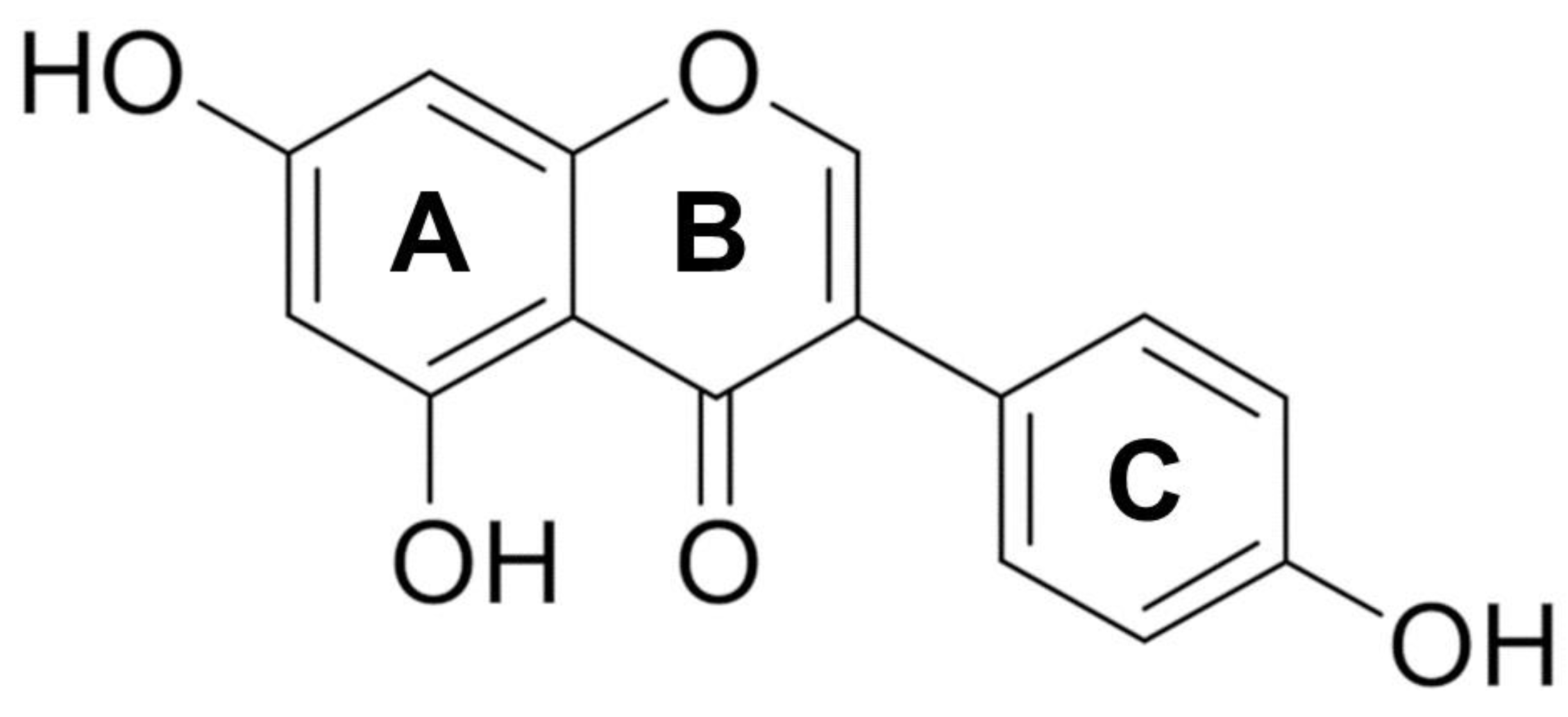
Figure 1. Chemical structure of genistein (5,7-dihydroxy-3-(4-hydroxyphenyl)-chromen-4-one). It is formed by two aromatic benzene rings (A and C) and a non-aromatic heterocyclic pyran ring (B), and the substituents at positions 4′, 5, and 7 of rings A and B are hydroxyl groups.
Ijms 26 01114 g001
Figure 2. Schematic representation of the inhibitory effect of genistein on angiogenesis. The isoflavonoid suppresses tumor neovascularization by interfering with the action of specific growth factors (such as VEGF, FGF, and PDGF) and matrix-degrading enzymes, including MMPs. Full names of the proteins and other abbreviations can be found in the Abbreviations section.
Ijms 26 01114 g002
Figure 3. Influence of genistein on cancer progression. Genistein abrogates cancer invasion and metastasis by interfering with multiple mechanisms including EMC lysis, MAPK/FAK/paxillin activation, epigenetic changes (ncRNA engagement and histone modifications), and estrogenic receptor involvement. Full names of the proteins and other abbreviations can be found in the Abbreviations section.
Ijms 26 01114 g003
Figure 4. Picture showing the main molecular targets genistein interferes with during cancer-related EMT. Specific signaling pathways and transcription factors are mentioned in the epithelial phenotype box and mesenchymal phenotype box, respectively. Of note, genistein drives cytoskeleton rearrangement coupled to cellular junction disintegration. TJs: Tight junctions; AJs: Adherent junctions; FAKs: Focal adhesions (including integrins and paxilin). Full names of the proteins and other abbreviations can be found in the Abbreviations section.
Ijms 26 01114 g004
Figure 5. Diagram showing the main repercussions of genistein in CSC behavior. Genistein treatment affects several signaling pathways (i.e., Sonic/Hedgehog, Wnt/β-catenin, Notch, NF-κB JAK-STAT, PI3K/Akt/mTOR signaling) that are involved in the maintenance of self-renewal capacity, stemness markers, invasion and metastasis propensity, as well as in the aptitude to form spheroids. Full names of the proteins and other abbreviations can be found in the Abbreviations section.
Ijms 26 01114 g005
Figure 6. Genistein arrests the cell cycle of several human cancer cell lines in the G2/M or G1/S phase through the modulation of cell-cycle-related proteins. The main targets of genistein are indicated. Downward arrow ↓, decrease; Upward arrow ↑, increase. Full names of the proteins and other abbreviations can be found in the Abbreviations section.
Ijms 26 01114 g006
Table 1. Foods with the highest genistein content [32].
SourceContent (mg/100 g)
Soybeans seeds5.56–267.2
Miso33.69–67.20
Natto21.52–59.37
Tempeh 1.11–112.21
Pistachio nuts0.10–3.40
Chickpeas0.069–0.214
Peanuts0.02–0.39
Lentils0.00–0.36
Parsley0.057
Almonds 0.00–0.01
Table 2. Cell death induced by genistein in breast and prostate cancers.
Genistein-Treated Cells/AnimalsMolecular Pathway/ProteinEffectRef.
Breast cancerMCF-7 overexpressing Bcl-2↑ Bcl-2; ↑ p85; ↑ cyt c↑ apoptosis[229]
MCF-7↓ Bcl-2; ↑ Bax; ERα↑ apoptosis[230]
MDA-MB-231↑ p-p53; ↑ p21; ↓ Bcl-xL; ↓ cyclin B1↑ apoptosis[231]
MCF-7↓ Bcl-2/Bax↑ apoptosis[233]
MDA-MB-231 and SKBR3↓S kp2; ↑ p21; ↑ p27↑ apoptosis[197]
MCF-7↓ MDM2; ↑ p21↑ apoptosis[188]
MCF-7↓ p-Akt; ↓ HOTAIR↑ apoptosis[234]
MCF-7-caspase-3 and T47D↓ CIP2A; ↑ caspase-3; ↑ c-PARP↑ apoptosis[232]
MCF-7 ↓ PI3K; ↓ Akt; ↑ Fas; ↑ FADD; ↑ cyt c; ↑ t-Bid; ↑ caspase-9; ↑ caspase-3;↑ apoptosis[236]
MCF-7 overexpressing HER2↑ p53; ↑ Fas receptor; ↑ c-PARP; ↑ caspase-9; ↓ p-IκBα; ↓ NF-κB↑ apoptosis[238]
MDA-MB-231↓ NF-κB; ↓ Notch-1; ↓ cyclin B1, ↓ Bcl-2; ↓ Bcl-xL,↑ apoptosis[198]
MDA-MB-231↓ MEK5; ↓ ERK5; ↓ p-ERK5; ↓ NF-κB/p65; ↑ Bax; ↓ Bcl-2; ↑ caspase-3 ↑ apoptosis[199]
MCF-7↓ Bcl-2/Bax; ↓ IGF-1R/p-Akt↑ apoptosis[240]
MCF-7↑ calpain; ↑ Ca++; ↑ caspase-7; ↑ c-PARP; ↑ ASK1-p38 MAPK↑ apoptosis[242]
MCF-7↑ Ca++; ↑ mu-calpain; ↑ caspase-12↑ apoptosis[243]
MCF-7↑ PTEN; ↓ p-Akt; ↑ p27↑ apoptosis[245]
MDA-MB-435 and Hs578t↓ miR-155; ↑ FOXO3; ↑ PTEN; ↑ casein kinase; ↑ p27↑ apoptosis[246]
Rats
MCF-7
↑ PTEN; ↑ p21; ↑ Bax; ↑ Bok;
↑ PTEN
↑ apoptosis
↑ apoptosis
[247]
MCF-7↓ Bcl-2/Bax; ↓ survivin; ↓ CuZnSOD; ↓ MnSOD; ↓ TrxR; ↑ GPx↑ apoptosis
[248]
MDA-MB-231 and MDA-MB-468↓ Bcl-2; ↑ Bax;↑ caspase-3; ↑ ROS; ↑ Cu↑ apoptosis
[249]
T47D↑ IRE1α; ↑ CHOP; ↑ Bim; ↑ TNF; ↑ FAS; ↑ FADD↑ apoptosis
[252]
Primary breast cancer cells

+17β-estradiol
↑ FADD; ↑ tBid; ↑ cyt c; ↑ caspase-8; ↑ caspase-3
↑ apoptosis
↓ apoptosis
[253]
MDA-MB-231
GEN (<10 μM) + 17 β-estradiol
GEN (10–100 μM) ± 17β-estradiol

↓ Bax/Bcl-2; ↓ p-ERK1/2

↑ apoptosis
↑ apoptosis
[254]
MCF-7 + 17β -estradiol
T47D + 17β -estradiol

↓ p-STAT3/STAT3
↑ proliferation
↑ apoptosis
[213]
MCF-7 SK; BR 3 and ZR-75-1
GEN (<10 μM)
GEN (10–100 μM)

↑ ERα
↓ ERα and erbB2

↑ proliferation
↑ apoptosis
[255]
MCF-7
GEN (50 μM) + 17β -estradiol
GEN (100 μM) + 17β -estradiol

↑ Cyclin B1
↓ Cyclin B1

↓ apoptosis
↑ apoptosis
[256]
MCF-7
+17 β-estradiol
Female rats ±17 β-estradiol
↓ Bcl-2; ↑ Bax;
↑ p21; ↑ p53
↓ Bcl-2; ↑ Bax;
↑ apoptosis
No apoptosis
↓ tumor growth
[257]
MCF-7
+17β -estradiol
↑ cell nuclear antigen; ↑ PI3K; ↑ p-Akt; ↓ FADD; ↓ cyt c; ↓ tBid; ↓ caspase-9; ↓ caspase-3; ↓ ERβ↓ apoptosis[258]
MCF-7
20 nm GEN or 5 nm E2
↑ ASAH1; ↑ GRP30; ↑ p-ERK1/2; ↑ p-Erα; ↑ S1P↑ proliferation[259]
MCF-7
+17β -estradiol [10−10 M]
↓ apoptosis[260]
SUM1315MO2 (185delAG BRCA1)↓ ERβ↑ apoptosis[261]
HCC1937, SUM149, SUM1315
MDA-MB-231
↑ p21; ↓ Akt
↑ Akt; ↓ p21
↑ apoptosis
↓ apoptosis
[262]
Prostate cancerPC3↑ caspase-3; ↓ p38MAPK; ↓ MMP-2↑ apoptosis[265]
PC3 and LNCaP↓ MMP-2↑ apoptosis[266]
PC3

PC3-SCID mouse
↓ MMP-2; ↓ MMP-11; ↓ MMP-13; ↓ MMP-14; ↓ MT-MMP; ↑ osteoprotegerin
↓ MMP-9n
↑ apoptosis

↑ apoptosis
[267]
PC3
PC3 xenograft
↓ MMP-2; ↑ p21↑ apoptosis
↓ tumor growth
[188]
LNCaP and PC3↓ PLK-1; ↑ p21↑ apoptosis[203]
PC3, DU145 and LNCap↑ ARHI; ↑ HERC5; ↑ CDNK1A; ↑ GADD45A; ↓ miR-221; ↓ miR-222↑ apoptosis[269]
DU145 and PC3↑ miR-574-3p; ↓ Bcl-xL; ↑ caspase-9; ↑ caspase-3↑ apoptosis[270]
PC3, DU145, ARCaP-E, ARCaP-M, and LNCaP↑ HAT1; ↑ H3K9 acetylation; ↑ SOX7↑ apoptosis[281]
DU145
PC3
↑ HOTAIR; ↑ miR-34a
↑ HOTAIR; ↑ miR-34a
↑ apoptosis
no change
[272]
PC3
GEN (≤10 μM)
GEN (>10 μM)

↑ CDKs, ↑ MAPKs; ↑ RPSKs, ↓ TGF-β; ↓ SMAD 2/3,4

↑ proliferation
↑ apoptosis
[273]
LNCaP↑ TRAIL; ↓ MMP ↑ apoptosis[274]
3D culture PC3 ↓ NO; ↑ catalase; ↑ GSH ↑ apoptosis[275]
DU145 and LNCaP↑ ROS; ↑ CTR1; ↑ ATP7A↑ apoptosis[276]
LTL163a) xenograft NOD-SCID mouse↑ tyrosine kinases; ↑ EGFR; ↑ Src↑ proliferation
↓ apoptosis
[106]
Akt/PKB, phospho-protein kinase B; ARHI/DIRAS3, Aplasia Ras Homology I; ASAH1, N-Acylsphingosine Amidohydrolase 1; ASK1, Apoptosis signal-regulating kinase 1; ATP7A, ATPase Copper Transporting Alpha; Bax, BCL2 Associated X; Bcl-2, B-cell lymphoma-2; Bcl-xL, B-cell lymphoma extra-large; Bid, BH3-interacting-domain death agonist; Bim, Bcl-2 Interacting Mediator of cell death; Bok, Bcl-2-related ovarian killer; CDK, cyclin-dependent kinase; CDNK1A, cyclin-dependent kinase inhibitor 1A; CHOP, C/EBP homologous protein; CIP2A, Cellular Inhibitor Of PP2A; Cyt c, cytochrome c; c-PARP, cleaved-Poly-ADP ribose polymerase; CTR1, High affinity copper uptake protein 1; EGFR, epidermal growth factor receptor; ERα, estrogen receptor α; ERβ, estrogen receptor β; ErbB2, c-Neu or human EGF receptor 2; ERK, extracellular signal-regulated protein kinases; FADD, FAS-associated protein with death domain; Fas, apoptosis stimulating fragment; FOXO3, forkhead box O3; GPx, Glutathione peroxidase; GSH, glutathione; GADD45A, growth arrest and DNA damage-inducible, alpha; GRP30, G protein-coupled receptor 30; HAT1, histone acetyltransferase 1; HERC5, Hect domain and RLD5; HOTAIR, HOX Antisense Intergenic RNA; IRE1α, Inositol-requiring transmembrane kinase/endoribonuclease 1α; MAPK, mitogen-activated protein kinase; MDM2, Mouse Double Minute 2; MMP- Metalloprotease-; NF-κB, nuclear factor kappa-B; NO, nitric oxide; p-Akt, phospho-protein kinase B; PARP, Poly-ADP ribose polymerase; p-ERK, phospho-extracellular signal-regulated protein kinases; PI3K, phosphoinositide 3-kinase; p-IκBα, phosphor-nuclear factor of kappa light polypeptide gen enhancer in B-cells inhibitor alpha; PLK-1, polo-like kinase 1;PP2A, Protein phosphatase 2; p-STAT, phospho-signal transducer and activator of transcription PTEN, Phosphatase and tensin homolog; ROS, reactive oxygen species; RPSK, Small ribosomal subunit protein uS11SMAD, Suppressor of Mothers against Decapentaplegic; Skp2, S-Phase Kinase Associated Protein 2; Src, Proto-Oncogene Tyrosine-Protein Kinase; STAT, signal transducer and activator of transcription SOD, superoxide dismutase; SOX7, SRY-Box Transcription Factor 7; S1P, Sphingosine 1-phosphate; t-Bid, truncated Bid; TGF-β Transforming growth factor-beta; TNF-α, tumor necrosis factor alpha; TRAIL, TNF-related apoptosis-inducing ligand; Trx, thioredoxin; ↑ = increase; ↓ = decrease.
Table 3. Combinatory treatment with genistein in breast and prostate cancers.
Genistein-Treated Cells/AnimalsCombination with GenisteinMolecular Pathway/ProteinEffectRef.
MCF-7/Adrdoxorubicin↓ Her2/neu↑ apoptosis[290]
PC3 and MDA-MB-231

PC3 SCID mice
cisplatin, docetaxel, or doxorubicin
docetaxel
↓ NF-κB↑ apoptosis

↑ growth inhibition
[291]
ERα- MDA-MB-231cisplatin↓ Bcl-2/Bax; ↑ p21↑ apoptosis[292]
MCF-7scisplatin (-17β-estradiol)↑ Bax/Bcl-xl↓ apoptosis[293]
EMT6 xenograft miceCisplatin↑ Bcl-2/Bax↓ apoptosis[294]
MCF-7 (high ERa/Erb)
T47D (low ERa/Erb)
MCF7 + ERβ
cisplatin↓ ROS↓ apoptosis
↑ apoptosis
↑ apoptosis
[295]
PC3, DU145, ARCaP-E, ARCaP-M, and LNCaPvorinostat↓ BIRC7/Livin; ↓ TGFB1I1/ARA55; ↓ HES1; ↓ SLUG↑ apoptosis[271]
MCF-7 and MDA-MB-231paclitaxel or vincristine↓ p-Bcl-2; ↓ cyclin B1; ↓ CDC2↓ apoptosis[296]
mCRPCcabazitaxel↑ Bax↑ apoptosis[297]
LNCaPtopotecan↑ caspase-3; ↑ caspase-9; ↑ ROS↑ apoptosis[298]
BT-474-overexpressing ER+/HER2tamoxifen↓ survivin; ↓ EGFR; ↓ Her2; ↓ ERα↑ apoptosis[214]
MCF-7 and MDA-MB-231
mouse 4Ti breast tumor
centchroman↑ ROS; ↑ Bax/Bcl-2; ↑ caspase-3; ↑ caspase-7; ↑ caspase-9; ↑ c-PARP; ↓ p-PI3K; ↓ p-Akt; ↓ NF-κB↑ apoptosis[299]
MCF-7 and MDA-MB-2311,25(OH)2D3↑ Bax and ↑ caspase-3; ↓ Bcl-2↑ apoptosis[187]
DU1451,25(OH)2D3↓ CYP24; ↑ VDR↑ growth inhibition[300]
LNCaPHPCDc-PARP; caspase-3; ↓ EGFR-Akt-GSK-3beta-p70S6k↑ apoptosis[301]
DU145terazosin↓ Bcl-xl↑ apoptosis[302]
PC3radiation↓ NF-κB; ↓ cyclin B and/or ↑ p21; ↑ c-PARP↑ apoptosis[202]
MCF-7 and MDA-MB-231X-ray irradiation↑ Bax; ↑ p73; ↓ Bcl-2↑ apoptosis[196]
prostate cancer orthotopic modelradiation ↑ apoptosis[303]
DU145ionizing radiation↓ damage repair↑ apoptosis[304]
MCF-7 and MDA-MB-231hypericin↓ Bcl-2; ↑ Bax; ↓ p-Akt; ↓ p-Erk1/2↑ apoptosis[305]
DU145tyrphostin AG1024 and X-irradiation ↑ apoptosis[306]
MCF7-aro and LTEDaroExe Exemestane↑ c-PARP/PARP↑ apoptosis[307]
PC3KSP inhibitor SB715992 ↑ apoptosis[308]
MCF7 and MDA-MB-231clofarabine↑ PTEN; ↑ RARB; ↑ CDKN1A↑ apoptosis[309]
MDA-MB-231NP-siCD36↓ p-p38↑ growth inhibition[310]
DU145survivin RNAi↑ caspase-3↑ apoptosis[311]
PC3beta-lapachone (bLap)↓ caspase-3; ↑ NQO1↑ apoptosis[312]
LNCaP; C4-2Bdaidzein ↑ apoptosis[313]
MCF-7 cellsequol↑ Bax/Bcl-xl↑ apoptosis[314]
MCF-7pomegranate extracts ↑ apoptosis[315]
MDA-MB-231; MCF-7sulforaphane↓ KLF4; ↓ HDAC2; ↓ HDAC3↑ growth inhibition[316]
MDA-MB-231 and MCF-7sulforaphane, sodium butyrate↓ DNMT; ↓ HDAC; ↓ HMT; ↑ HAT methyltransferases and histone methylases ↑ apoptosis[317]
LNCaP and p53-null PC3selenite↑ p21; ↑ Bax ↑ apoptosis[201]
Bax, BCL2 Associated X; Bcl-2, B-cell lymphoma-2; BIRC7/livin, Baculoviral IAP repeat-containing 7; CDC2, Cyclin Dependent Kinase 2; CDNK1A, cyclin-dependent kinase inhibitor 1A; c-PARP, cleaved-Poly-ADP ribose polymerase; CYP24, Cytochrome P450 Family 24; DNMT, DNA methyltransferase; EGFR, epidermal growth factor receptor; ERα, estrogen receptor α; ERK, extracellular signal-regulated protein kinases; GSK-3β, glycogen synthase kinase-3 beta; HAT, histone acetyltransferase; HDAC, histone deacetylases; Her2/neu, human epidermal growth factor receptor 2; HES1, hairy and enhancer of split-1; HMT, histone methylase; HPCD, 2-hydroxypropyl-beta--hydroxypropyl-beta-cyclodextrin; KLF4, Krüppel-like factor 4; KSP, kinesin spindle protein; NF-κB, nuclear factor kappa-B; NQO1, NAD(P)H quinone oxidoreductase 1; p-Akt, phospho-protein kinase B; PARP, Poly-ADP ribose polymerase; p-Bcl-2, phospho B-cell lymphoma-2; PI3K, phosphoinositide 3-kinase; PTEN, Phosphatase and tensin homolog; p70S6K, p70 ribosomal S6 kinase; RARB, retinoic acid receptor beta; ROS, reactive oxygen species; SLUG/SNAI2,Snail Family Transcriptional Repressor 2; TGFB1I1/ARA55, transforming growth factor beta-1-induced transcript 1 protein; VDR, vitamin D receptor; ↑ = increase; ↓ = decrease.
Disclaimer/Publisher’s Note: The statements, opinions and data contained in all publications are solely those of the individual author(s) and contributor(s) and not of MDPI and/or the editor(s). MDPI and/or the editor(s) disclaim responsibility for any injury to people or property resulting from any ideas, methods, instructions or products referred to in the content.