Article

Nephroprotective and Antioxidant Effects of Jatropha dioica Extract Against Ischemia–Reperfusion Injury in Wistar Rats

by
Diana Raquel Rodríguez-Rodríguez
1,†,
Oscar Humberto Mendoza-Hernández
1,†,
Paula Cordero-Pérez
1,*,
Verónica Mayela Rivas-Galindo
2,
Diana Patricia Moreno-Peña
1,
Ramiro Tijerina-Márquez
1,
Alondra Michelle Garza-Villarreal
1,
Gabriela Alarcón-Galván
3,
Linda Elsa Muñoz-Espinosa
1,
Homero Arturo Zapata-Chavira
4,
Marco Antonio Hernández-Guedea
4,
Guadalupe Yazmín Solis-Cruz
2 and
Liliana Torres-González
1,*
1
Liver Unit, Department of Internal Medicine, University Hospital “Dr. José E. González”, Universidad Autónoma de Nuevo León, Monterrey 64460, Nuevo León, Mexico
2
Department of Analytical Chemistry, School of Medicine, Universidad Autónoma de Nuevo León, Monterrey 64460, Nuevo León, Mexico
3
Basic Science Department, School of Medicine, Universidad de Monterrey, Monterrey 66238, Nuevo León, Mexico
4
Transplant Service, University Hospital “Dr. José E. González”, Universidad Autónoma de Nuevo León, Monterrey 64460, Nuevo León, Mexico
*
Authors to whom correspondence should be addressed.
These authors contributed equally to this work, all of them are the first author of the paper.
Int. J. Mol. Sci. 2025, 26(5), 1838; https://doi.org/10.3390/ijms26051838
Submission received: 17 January 2025 / Revised: 19 February 2025 / Accepted: 19 February 2025 / Published: 21 February 2025

Abstract

:
Plant extracts with antioxidant activities have shown nephroprotection against IR injury. Jatropha dioica (Jd) possesses antioxidant activity. Our aim was to evaluate the effects of a hydroalcoholic Jd extract against IR injury in Wistar rats. Rats were divided into groups (n = 6): sham (SH); no toxicity (JdTox; 300 mg/kg/day of extract for 7 days); IR (on day 7 [I: 45 min/R: 24 h]); and Jd+IR (same treatment as JdTox; same surgical procedure as IR). AST and LDH were significantly lower in the JdTox. IR exhibited significantly higher CrS, BUN, and MDA compared with SH; Jd+IR showed reductions in these markers. GSH and SOD levels were significantly lower in IR compared with SH; an increase in these markers was observed in the Jd+IR. Histologically, IR showed significant increases in medullary tubular necrosis, medullary protein casts, and medullary vascular congestion compared with SH and JdTox. In Jd+IR, a significant decrease was observed only in medullary tubular necrosis. Therefore, the evaluated hydroalcoholic Jd extract dose showed no nephrotoxicity and hepatotoxicity. Jd extract pretreatment attenuated IR-induced renal injury, as evidenced by the improved serum markers of renal damage and oxidative stress.

1. Introduction

Acute kidney injury (AKI) in critically ill patients is an independent risk factor that increases short- and long-term morbidity and mortality, with a tremendous economic impact in terms of health costs. Several investigations have shown that minor increases in serum creatinine (CrS) correlate with a worse prognosis [1]. Numerous risk factors, including drug and toxin exposure, sepsis, and ischemia–reperfusion (IR), frequently contribute to the onset of AKI, leading to a decline in the glomerular filtration rate and acute tubular cell death [2]. In terms of this nephropathy, key biological processes have been identified, including cell death, cellular proliferation, inflammation, fibrosis, and the release of pro-oxidant free radicals, that can exacerbate renal injury and play a crucial role in the development of severe complications [3,4].
IR is a pathological process defined by an initial, transient deprivation of blood supply to a specific organ, culminating in the subsequent re-establishment of perfusion and the simultaneous reoxygenation of tissues. In this final process, paradoxically, a marked exacerbation of tissue damage and the onset of a significant inflammatory cascade occur [5]. IR-elicited tissue injury contributes significantly to morbidity and mortality across a wide range of pathologies, including myocardial infarction, ischemic stroke, acute kidney injury, trauma, circulatory arrest, sickle cell disease, and sleep apnea [6]. IR injury is clinically linked to acute kidney failure secondary to shock or severe hypotension, severe trauma, renal artery thrombosis, renal venous thrombosis, and intra-abdominal hypertension [7]. The consequences of IR injury are particularly critical in medical contexts like surgical procedures and kidney transplantation, where it plays a key role in determining patient outcomes [8].
Given the complex nature of IR injury, various animal models have been developed to understand its impact on kidney function better. Among these, murine experimental models are widely used to investigate the mechanisms that underlie the induction and progression of IR-induced renal damage, as well as to identify potential therapeutic targets to mitigate kidney injury caused by IR events. One such model is IR injury, which induces the necrosis and apoptosis of tubular cells, inflammation, and oxidative stress [2,9,10], ultimately leading to impaired renal function, as assessed by blood urea nitrogen (BUN) and the serum creatinine level (CrS). This IR injury is associated with transiently elevated rates of reactive oxygen species (ROS) production after ischemia, resulting in a complex spectrum of cellular changes, including alterations in mitochondrial function, immune activation, transcriptional reprogramming, microvascular inflammation, endothelial–mesenchymal transition, and cell death [5,11,12,13,14,15,16]. The initial acute response is characterized by the ischemic activation of oxygen-sensitive transcription factors, such as hypoxia-inducible factors, and the activation of the nuclear factor-kappa B signaling pathway [17,18]; this is accompanied by detrimental alteration of antioxidant defense and the significant reduction in the activity of antioxidant enzymes [superoxide dismutase (SOD), reduced glutathione (GSH), catalase, etc.] [19,20].
The use of plants has been shown to attenuate experimentally induced renal IR injury in murine models; this nephroprotective action is mainly attributed to the antioxidant capacity of substances derived from different plant parts (i.e., leaves, flowers, stems, roots, or fruits), which are obtained through various extraction methods [21,22,23,24,25,26,27,28,29].
The genus Jatropha of the family Euphorbiaceae comprises a wide variety of dioecious and monoecious plants, which are primarily native to the Americas, Asia, and Africa, and to which multiple pharmacological properties are attributed. These properties include anti-leishmanial, antimicrobial, antihelminthic, cytotoxic, antiprotozoal, anticoagulant, antispasmodic, antitumoral, antioxidant, analgesic, immunomodulatory, and gastroprotective properties [30,31,32,33].
Jatropha dioica (Jd) is a species that is native to Mexico and the southern United States. This heat-tolerant shrub has orange rhizomes, narrow brown-reddish branches, elongated leaves, and sap that turns red when exposed to air, which is why it is traditionally referred to as ‘sangre de drago’ (‘dragon’s blood’) [34,35]. Studies of extracts derived from this plant have reported antioxidant, antibacterial, antiviral, antifungal, hypoglycemic, antidiabetic, cytotoxic, and chemoprotective activities [36,37,38,39,40,41,42,43,44,45,46,47].
Based on the antioxidant and other properties described for this plant, the aim herein was to evaluate a hydroalcoholic extract of Jd as a potential nephroprotective agent against IR injury in Wistar rats.

2. Results

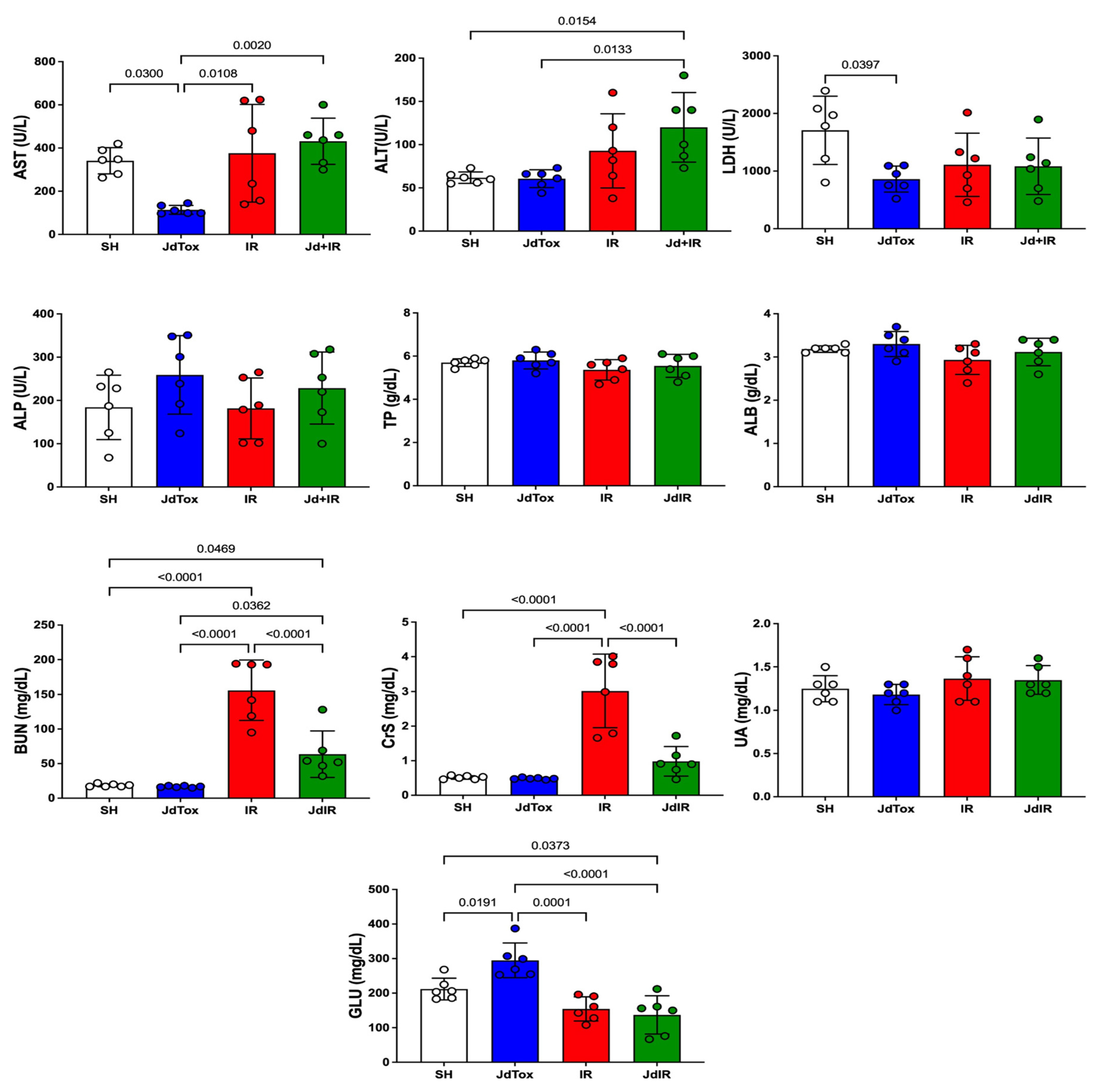
2.1. Jd Nontoxicity Evaluation

Aspartate aminotransferase (AST) and lactate dehydrogenase (LDH) levels were significantly lower in the Jd extract no toxicity group (JdTox) compared with the sham group (SH), with results of 114 ± 21 IU/L vs. 341 ± 61 IU/L (p = 0.03) and 861.3 ± 226.4 U/L vs. 1708 ± 591.8 U/L (p = 0.03), respectively. Serum glucose (GLU) level in JdTox was significantly higher compared with the level in other groups. There were no significant differences between SH and JdTox in alanine aminotransferase (ALT) (62 ± 7 IU/L vs. 61 ± 10 IU/L), CrS (0.52 ± 0.05 mg/dL vs. 0.48 ± 0.02 mg/dL), BUN (19 ± 2 mg/dL vs. 17 ± 1 mg/dL), albumin (ALB) (3.18 ± 0.08 vs. 3.30 ± 0.29), total protein (TP) (5.7 ± 0.2 vs. 5.8 ± 0.4), alkaline phosphatase (ALP) (184 ± 74 vs. 259 ± 91), uric acid (UA) (1.25 ± 0.15 vs. 1.18 ± 0.12) (Figure 1), malondialdehyde (MDA) (110 ± 14 μM/mg vs. 133 ± 22 μM/mg), GSH (1.1 ± 0.1 nM/mg vs. 1.0 ± 0.2 nM/mg), or SOD (11 ± 3 U/mg vs. 12 ± 1 U/mg) (Figure 2).

2.2. Effects of Jd Extract on IR-Induced Renal Damage

CrS and BUN levels were significantly higher in the IR group (IR) compared with the SH group (3.02 ± 1.06 mg/dL vs. 0.52 ± 0.05 mg/dL and 156 ± 44 mg/dL vs. 19 ± 2 mg/dL, respectively; p < 0.0001) (Figure 1). Jd extract pretreatment before IR injury (IR vs. Jd with IR group [Jd+IR]) significantly reduced CrS and BUN levels (3.02 ± 1.06 mg/dL vs. 0.98 ± 0.43 mg/dL and 156 ± 44 mg/dL vs. 64 ± 34 mg/dL, respectively; p < 0.0001) (Figure 1). There were no significant differences in the other biochemical parameters between IR and Jd+IR.

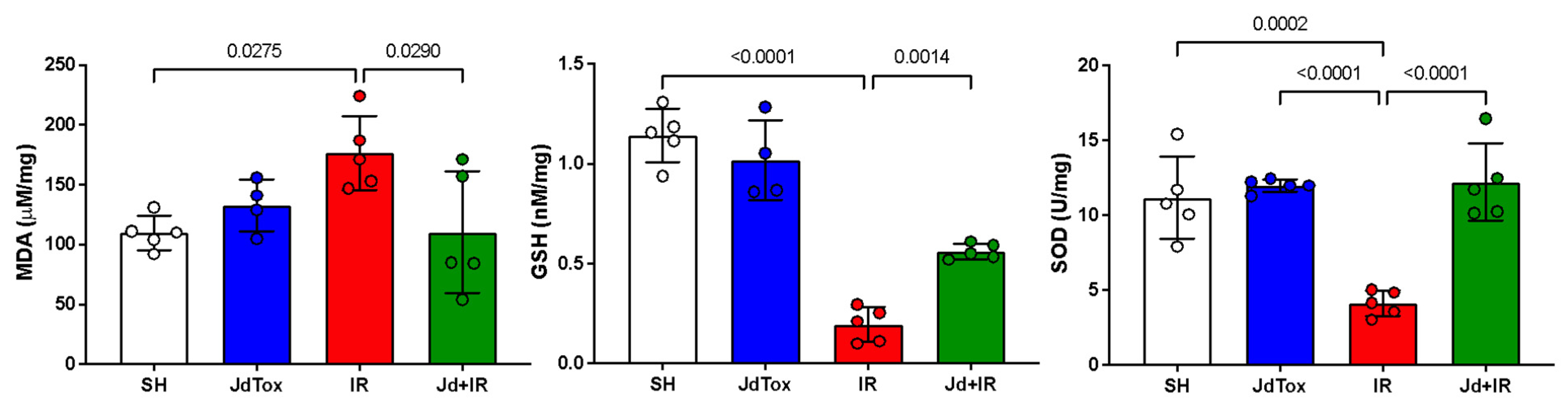
2.3. Effects of Jd Extract on IR-Induced Oxidation

MDA concentration was significantly higher in IR compared with SH (176 ± 31 μM/mg vs. 110 ± 14 μM/mg; p = 0.0275). GSH and SOD levels were significantly lower in IR compared with SH (0.19 ± 0.08 nM/mg vs. 1.14 ± 0.13 nM/mg [p < 0.0001] and 4 ± 1 U/mg vs. 11 ± 3 U/mg [p = 0.0003], respectively) (Figure 2). The MDA concentration in IR was significantly higher than in Jd+IR (176 ± 31 μM/mg vs. 110 ± 51 μM/mg; p = 0.0290). GSH and SOD levels were significantly lower in IR compared with Jd+IR (0.56 ± 0.03 nM/mg vs. 19.00 ± 0.08 nM/mg [p = 0.0014] and 4 ± 1 U/mg vs. 12 ± 3 U/mg [p < 0.0001], respectively) (Figure 2).

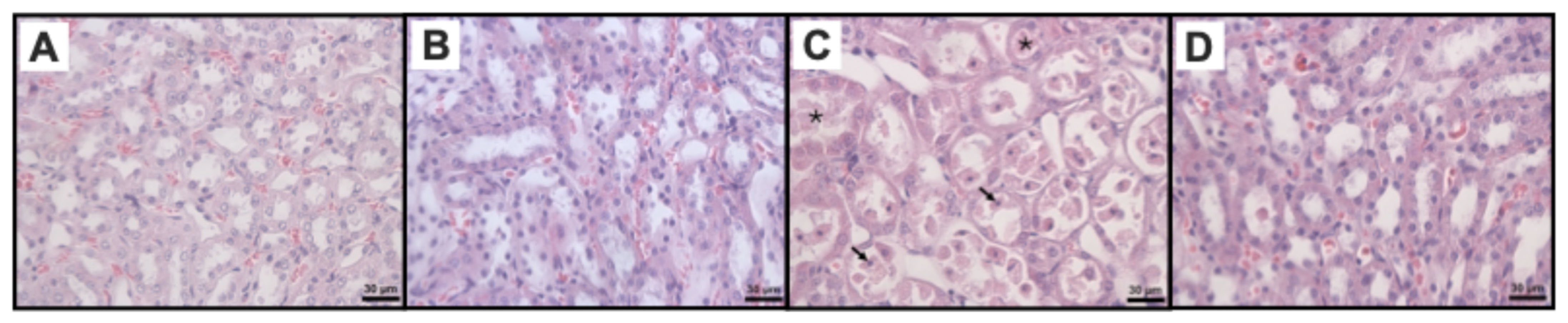
2.4. Effects of Jd Extract on IR-Histophatological Renal

The SH group showed no renal cellular structure damage (Figure 3A). The JdTox group exhibited a renal cellular structure like SH (Figure 3B). The IR group showed pathological changes with the medullary watery degeneration or medullary vacuolar degeneration of renal tubular epithelial cells, medullary tubular necrosis, the medullary exfoliation of some renal tubular epithelial cells, medullary protein casts, and medullary vascular congestion (Figure 3C). Renal pathological changes in Jd+IR group were attenuated compared with the IR group (Figure 3D). Histopathological scores are shown in Table 1. A significant increase was observed in the three renal damage histological markers in the IR group compared with the SH group and JdTox group. Pretreatment with the extract before injury induction significantly reduced the medullary tubular necrosis damage only.

3. Discussion

In this study, a total hydroalcoholic extract of Jd was evaluated as a potential nephroprotective agent against IR injury in Wistar rats. The results demonstrated that the extract was nontoxic based on hepatic and renal evaluations of various markers. Importantly, it was shown that the extract mitigated renal damage induced by IR, as evidenced by biochemical markers, oxidative stress indicators, and histological findings.
The use of plant extracts as a strategy to attenuate experimentally induced renal IR injury was well documented in previous studies [21,22,23,24,25,26,27,28,29]. Given the wide molecular diversity of plant extracts, it was essential to initially evaluate the nontoxicity of the Jd extract; therefore, we assessed the hepatic and renal effects of the Jd extract using biochemical markers and oxidative stress indicators. It has been reported that increased AST, ALT, and LDH levels indicate the loss of hepatocyte membrane integrity during toxin-induced damage [48]. We observed that a 300 mg/kg dose of Jd extract reduced AST and LDH levels, which could be attributed to compounds previously reported in our studies, such as citlalitrione, 6-epi-riolozatrione, riolozatrione, and jatrophatrione [49], and others like β-sitosterol [50,51], catechin [52], ellagic acid [38,53,54,55], gallic acid [56,57,58], jatrophone A, and jatrophone B [59]; these compounds may have attenuated the damage associated with the surgical procedure, as other authors have reported that this type of compound inhibits the activity of these enzymes [60,61]. Interestingly, ALT levels did not change compared to the SH group, suggesting there was no toxicity at this dose. Other markers, such as TP, ALB, ALP, and UA, showed no changes, which is consistent with previous reports assessing plant extract nontoxicity [62,63].
Renal damage following the metabolism of toxic substances involves multiple mechanisms, evidenced by the increase in CrS, BUN, and MDA levels [64], while SOD and GSH function as potent detoxifiers [65]. In our study, the JdTox group did not show significant changes in CrS, BUN, MDA, SOD, or GSH compared to the SH group, suggesting that the Jd extract does not exhibit renal toxicity. These results are consistent with the reported nontoxicity of Jd in Artemia salina lethality bioassays [46,66] and toxicity assays in rats after transplacental exposure [67].
An interesting observation in our study was that GLU levels were significantly higher in the JdTox group compared to other groups. This may be related to the role of peroxisome proliferator-activated receptors (PPARs) in glucose homeostasis [68]. Genetic studies in humans and heterozygous knockout mice have described that reduced PPAR-gamma activity paradoxically improves insulin sensitivity [69]. Jatropha curcas has been shown to activate PPAR-gamma in a dose-dependent manner [70]. Furthermore, the use of Jd extract at 34 mg/kg did not reduce glucose tolerance test levels in rats, and at doses of 25, 50, and 100 mg/kg, glucose levels were not reduced in alloxan-induced diabetic rats [45,46]. This may explain the significant increase in glucose observed in the JdTox group. Therefore, it is necessary to evaluate whether Jd extract also acts on PPAR-gamma, as reported with other species of this genus.
Once it was confirmed that the extract showed no toxicity at the evaluated dose, its effect on the ischemia–reperfusion process was analyzed. Regarding renal function, markers such as CrS and BUN are commonly used to assess the glomerular filtration rate [71]. Previous studies conducted by our group on drugs like misoprostol and plant extracts (e.g., Juglans mollis, Sonchus oleraceus) demonstrated significant increases in CrS and BUN in renal IR groups compared to sham controls [27,72]. In this study, we observed elevated CrS and BUN levels in the IR group compared to the SH group, and the Jd+IR group exhibited a significant reduction in these markers, suggesting the Jd extract has nephroprotective effects. However, other biochemical parameters evaluated in our study for liver and kidney function, such as ALP, ALB, UA, and TP, did not show significant differences between treatment groups, indicating that liver function was not affected the at the time evaluated. This is consistent with previous reports [73].
Oxidative stress plays a key role in IR injury, as MDA acts as an indirect marker of lipid peroxidation, whereas SOD and GSH serve as markers of antioxidant capacity, playing a role in reversing pathological variations due to oxidative stress damage; thus, they have key functions in the endogenous defense system against ROS [74,75]. As we previously reported in our group’s studies with other treatments in this damage model, we observed an increase in MDA levels and a decrease in SOD levels in renal IR groups compared with sham groups. We also showed how these mediators were modified by drug or extract pretreatment before IR [26,27,72], whereas Karimi G. et al. reported a decrease in GSH in IR compared with sham groups [75]. Here, we also found increased MDA and decreased SOD and GSH levels in the IR group compared to the SH group, while the Jd+IR group showed significant reductions in MDA and increases in SOD and GSH, highlighting the antioxidant potential of the Jd extract.
Histologically, the IR group showed greater renal tissue damage compared with the SH group and was characterized by the presence of protein casts, medullary vascular congestion, and tubular necrosis. These findings are consistent with other studies in which IR-induced renal damage, mainly associated with tubular necrosis, was reported 24 h post-reperfusion [76,77]. Jd administration before IR-induced damage only reduced tubular necrosis, aligning with the reported nephroprotective effects shown by reduced distal and proximal tubules in alloxan-induced diabetic rats [46].
Phytochemical research on Jd has identified secondary metabolites with nephroprotective actions, including β-sitosterol [50,51], catechin [52], ellagic acid [38,53,54,55], and gallic acid [56,57,58]. It has also established the gastroprotective effect of jatrophone A and jatrophone B [59]. Previous studies conducted by our group using HPLC reported that the main composition of the root extract of Jd used in the present study consists primarily of citlalitrione, 6-epi-riolozatrione, riolozatrione, and jatrophatrione [49]. These compounds were previously identified through NMR spectroscopy and X-ray diffraction [41,44]. It is important to mention that the hydroalcoholic extract also contained a variety of glycosidic compounds, which, when analyzed by 13C NMR, showed signals between 60 and 100 ppm, corresponding mainly to carbohydrates. These carbohydrates could not be detected using our validated HPLC method [49]. The antioxidant activity of Jd is largely attributed to polyphenols, which can act as reducing agents, hydrogen donors, and radical scavengers [36,40,78,79]. Specifically, ellagic acid has been shown to protect the kidneys from oxidative stress and mitochondrial dysfunction [38,54,55], while gallic acid significantly improves oxidative stress markers and serum biochemical parameters in renal damage models [56,57,58]. These findings suggest that the nephroprotective effects of Jd observed in this study may be due to the presence of antioxidant compounds. This research emphasizes the biological activity of a total hydroalcoholic extract of Jd, expanding the perspective beyond isolated compounds. The extract’s diverse bioactive constituents, including polyphenols, flavonoids, and terpenoids, may act synergistically, enhancing its therapeutic potential. Furthermore, the use of the total extract, rather than isolated molecules, provides a more practical and resource-efficient approach. This approach favors application in preclinical models and lays the foundation for developing a standardized extract with stringent quality control through chemical fingerprinting.
One limitation of this study was the absence of measurements for additional renal injury markers, such as cystatin C, KIM-1, and NGAL, which could have provided further insight into the extent of renal damage induced by IR. Another important aspect not addressed was the evaluation of the inflammatory signaling pathway associated with the nephroprotective activity of the extract, which limited the understanding of the underlying mechanisms.

4. Materials and Methods

4.1. Plant and Extraction

Jd var. sessiliflora (Hook) was collected in the municipality of Villaldama, Nuevo León, Mexico, and authenticated at the Institutional Herbarium of the Faculty of Biological Sciences of the Autonomous University of Nuevo León (specimen registration number UAN-24077). Its crude hydroalcoholic extract was obtained according to the method reported by Castro-Ríos et al. [49].

4.2. Animals and Ethical Considerations

The experimental procedures were conducted in accordance with the guidelines established by the Official Mexican Standard NOM-062-ZOO-1999 for the production, care, and use of laboratory animals [80]. This project was also approved by the Research Ethics Committee and the Institutional Committee for the Care and Use of Laboratory Animals of the Dr. José Eleuterio González Faculty of Medicine and University Hospital, Autonomous University of Nuevo León (registration number HI22-00002).
Twenty-four Wistar albino rats (both sexes) with an average weight of 265 ± 28 g were used. Rats were kept under standard conditions, being housed in polycarbonate cages in a room at a constant temperature of 24 ± 3 °C on a 12 h light–dark cycle. They had ad libitum access to water and commercial rat pellets (Nutrimix de México, S.A. de C.V., Mexico City, Mexico).

4.3. Experimental Study Groups

Rats were randomly distributed (n = 6 per group, 3 females and 3 males) to the following groups:
Sham (SH): vehicle of extract administration (physiological solution with 3% Tween-20) 1 mL/day orally (p.o.) for 7 days, then laparotomy without renal hilum obliteration.
Jd extract no toxicity (JdTox): 300 mg/kg/day extract treatment according to Morales-Velázquez et al. [67] dissolved in 1 mL of the vehicle of extract administration p.o. per day for 7 days, then the same surgical procedure as the SH group.
IR (IR) group: The same treatment as the SH group, then post-administration laparotomy for injury induction via bilateral renal ischemia (45 min) and subsequent reperfusion (24 h).
Jd with IR (Jd+IR) group: The same treatment as the JdTox group, then the same surgical procedure as the IR group.

4.4. Laparotomy and IR Injury Induction

In the SH, JdTox, IR, and Jd+IR groups rats were anesthetized using xylazine (Sedaject; Vedilab S.A. de C.V. Reg. SAGARPA Q0088-122; Mexico City, Mexico) via intraperitoneal (i.p.) injection at a dose of 10 mg/kg body weight. Ketamine was used as an analgesic (Anesket; PiSA Agropecuaria, S.A. de C.V. Reg. SAGARPA Q7833-028; Mexico City, Mexico) and administered i.p. at a dose of 100 mg/kg body weight, according to the manufacturer’s specifications.
Following anesthesia, abdominal trichotomy and asepsis were performed using antiseptic solution Microdacyn (Oculus Technologies of Mexico, S.A. de C.V., Guadalajara, Jal., Mexico). This followed by the addition of 20% chlorhexidine gluconate (Farmacéuticos Altamirano de México, S.A. de C.V., Mexico City, Mexico). A midline incision was made to expose the abdominal viscera. In the IR and Jd+IR groups, after dissection, the occlusion of the renal pedicle was performed by placing a vascular clamp (ischemia) bilaterally for 45 min, after which the clamps were removed, initiating the reperfusion time. The acute kidney injury induction was completed with the primary closure of the surgical wound in two layers, using continuous Nylon 3/0 sutures. In the SH and JdTox groups, the same procedure was not performed, as no clamping was applied.
There was a 24 h reperfusion period, during which the rats had free access to food and tramadol solution in water as an analgesic (50 mg/L; Grünenthal GmbH, Stolberg, Germany). At the end of the reperfusion period, rats were administered xylazine and ketamine i.p. at doses of 5 mg/kg and 50 mg/kg, respectively. The surgical wound was reopened, and samples were obtained by exsanguination euthanasia; 5 mL of arterial blood was taken by abdominal aortic puncture, immediately after which both kidneys were removed. The blood sample was centrifuged at 3500 rpm for 15 min to obtain serum for biochemical marker assessments. One half of each kidney was preserved for histological evaluation, while the other half was frozen at −80 °C for subsequent the homogenization and determination of oxidative stress markers malondialdehyde (MDA), superoxide dismutase (SOD), and reduced glutathione (GSH).

4.5. Biochemical and Oxidative Stress Marker Analysis

Serum concentrations of CrS, BUN, GLU, UA, ALB, TP, ALT, AST, LDH, and ALP were determined using spectrophotometry (ILab Aries; Instrumentation Laboratory, Milan, Italy) with commercial kits (Instrumentation Laboratory) according to the manufacturer’s specifications.
The degree of lipid peroxidation was quantified by determining the concentration of MDA using the TBARS Assay Kit (Cayman Chemical Company, Ann Arbor, MI, USA). To measure the concentrations, we added 100 μL of tissue homogenate supernatant or standard, 100 μL of sodium dodecyl sulfate, and 4 mL of color reagent to each vial. The vial was heated at 100 °C for 1 h and immediately cooled in an ice bath. It was then centrifuged at 11,000 rpm for 15 min at 4 °C. Subsequently, 150 μL from each vial was transferred to microplate wells. Product absorbance was measured at a wavelength of 540 nm using a microplate reader (Thermo Fisher Scientific Multiskan FC, Carlsbad, CA, USA). Results are expressed as micromoles of MDA formed per liter, normalized to protein concentration using the Bradford method [81].
SOD activity was determined using the SOD Assay Kit (Cayman Chemical Company, Ann Arbor, MI, USA), with a colorimetric assay that uses tetrazolium salt to detect superoxide radicals generated by xanthine oxidase and hypoxanthine. To measure SOD activity, 200 μL of diluted radical detector and 10 μL of tissue homogenate supernatant or standard were added to each well, along with 20 μL of xanthine oxidase. Product absorbance was measured at a wavelength of 460 nm after 20 min using a microplate reader (Thermo Fisher Scientific Multiskan FC). Results are expressed as U/mL, normalized to protein concentration using the Bradford method [81]. One unit (UI) of SOD is defined as the amount of enzyme needed to exhibit 50% dismutation of the superoxide radical.
The GSH concentration was determined using the GSH Assay Kit (Sigma-Aldrich, St. Louis, MO, USA). It was quantified through a kinetic assay in which the catalytic amount (in nanomoles) of GSH continuously reduced 5,52-dithiobis(2-nitrobenzoic acid) to 5-thio-2-nitrobenzoic acid (TNB), and the resulting GSH disulfide was recycled by GSH reductase and NADPH. Subsequently, TNB was measured by spectrophotometry at 412 nm using a microplate reader (Thermo Fisher Scientific Multiskan FC). Results are expressed as nM/L, normalized to the protein concentration using the Bradford method [81].

4.6. Histopathological Renal Evaluation

Representative tissue samples were obtained from both kidneys, preserved in 10% phosphate-buffered formalin (pH 7.4), and subsequently embedded in paraffin. Paraffin blocks were sectioned using a microtome at a thickness of 4 nm. After deparaffinization and hydration, sections were stained with hematoxylin and eosin (H&E). Each section was examined under an optical microscope for the histopathological evaluation of renal lesions. Using the indicators proposed in the modified Kobuchi scale [47], a score was assigned for the following: tubular necrosis and protein cylinders (no damage = 0, mild damage = 1, moderate damage = 2, severe damage = 3, very severe damage = 4) and medullary vascular congestion (no congestion = 0, the identification of erythrocytes at 400 magnification = 1, the identification of erythrocytes at 200 = 2, the identification of erythrocytes at 100 = 3, the identification of erythrocytes at 40 = 4).

4.7. Statistical Analysis

Data were analyzed using one-way analysis of variance (ANOVA) or the nonparametric Kruskal–Wallis test, alongside Tukey’s or Dunn’s post hoc test, respectively, based on the data distribution, as assessed by the Shapiro–Wilk normality test. Prism software (v. 10.0.0; GraphPad, San Diego, CA, USA) was used for analyses. Data were expressed as mean ± standard deviation or median (interquartile range), as appropriate. p < 0.05 was considered statistically significant.

5. Conclusions

The Jd hydroalcoholic extract at the dose evaluated herein does not show nephrotoxicity and hepatotoxicity. The fact that Jd extract pretreatment before IR attenuates renal injury was evidenced by the improvement in serum markers of renal damage and oxidative stress. Further studies are needed to evaluate specific, previously reported compounds with antioxidant activities, which may be responsible for the effects observed herein.

Author Contributions

D.R.R.-R., O.H.M.-H., P.C.-P. and L.T.-G. conceptualization, supervision, methodology, investigation, validation, formal analysis, writing—original draft preparation, writing—review and editing, visualization, resources, funding acquisition, and project administration; D.P.M.-P., methodology, investigation, validation, formal analysis, writing—original draft preparation, writing—review and editing; R.T.-M. and A.M.G.-V., methodology, investigation, validation, and formal analysis; G.A.-G., V.M.R.-G. and G.Y.S.-C. methodology and investigation; validation, formal analysis, writing—review and editing; L.E.M.-E., H.A.Z.-C. and M.A.H.-G., writing—review, editing and visualization. All authors have read and agreed to the published version of the manuscript.

Funding

This research was funded by Scientific and Technological Research Support Program of the Universidad Autónoma de Nuevo León, Project number 167-CS-2022.

Institutional Review Board Statement

The experimental procedures were conducted in accordance with the guidelines established by the Official Mexican Standard NOM-062-ZOO-1999 for the production, care, and use of laboratory animals. This project was also approved by the Research Ethics Committee and the Institutional Committee for the Care and Use of Laboratory Animals of the “Dr. José Eleuterio González” Faculty of Medicine and University Hospital, Autonomous University of Nuevo León (registration number HI22-00002) in 27 June 2022.

Informed Consent Statement

Not applicable.

Data Availability Statement

The original contributions presented in the study are included in the article; further inquiries can be directed to the corresponding author.

Acknowledgments

We thank Lucila Catalina Treviño Lozano for her support in the laboratory, to Norma Elena Estrella and Mario Sánchez Terán for their administrative support.

Conflicts of Interest

The authors declare no conflicts of interest.

References

  1. Chertow, G.M.; Burdick, E.; Honour, M.; Bonventre, J.V.; Bates, D.W. Acute kidney injury, mortality, length of stay and costs in hospitalised patients. J. Am. Soc. Nephrol. 2005, 16, 3365–3370. [Google Scholar] [CrossRef] [PubMed]
  2. Sanz, A.B.; Sanchez-Niño, M.D.; Martín-Cleary, C.; Ortiz, A.; Ramos, A.M. Progress in the development of animal models of acute kidney injury and its impact on drug discovery. Expert Opin. Drug Discov. 2013, 8, 879–895. [Google Scholar] [CrossRef] [PubMed]
  3. Andreucci, M.; Faga, T.; Pisani, A.; Perticone, M.; Michael, A. The ischemic/nephrotoxic acute kidney injury and the use of renal biomarkers in clinical practice. Eur. J. Intern. Med. 2017, 39, 1–8. [Google Scholar] [CrossRef] [PubMed]
  4. Endre, Z.H.; Pickering, J.W.; Walker, R.J.; Devarajan, P.; Edelstein, C.L.; Bonventre, J.V.; Frampton, C.M.; Bennett, M.R.; Ma, Q.; Sabbisetti, V.S.; et al. Improved performance of urinary biomarkers of acute kidney injury in the critically ill by stratification for injury duration and baseline renal function. Kidney Int. 2011, 79, 1119–1130. [Google Scholar] [CrossRef]
  5. Zhang, M.; Liu, Q.; Meng, H.; Duan, H.; Liu, X.; Wu, J.; Gao, F.; Wang, S.; Tan, R.; Yuan, J. Ischemia-reperfusion injury: Molecular mechanisms and therapeutic targets. Signal Transduct. Target. Ther. 2024, 9, 12. [Google Scholar] [CrossRef]
  6. Eltzschig, H.K.; Eckle, T. Ischemia and reperfusion from mechanism to translation. Nat. Med. 2011, 17, 1391–1401. [Google Scholar] [CrossRef]
  7. Zhao, H.; Alam, A.; Soo, A.P.; George, A.J.T.; Ma, D. Ischemia-Reperfusion Injury Reduces Long Term Renal Graft Survival: Mechanism and Beyond. EBioMedicine 2018, 28, 31–42. [Google Scholar] [CrossRef] [PubMed]
  8. Menke, J.; Sollinger, D.; Schamberger, B.; Heemann, U.; Lutz, J. The effect of ischemia/reperfusion on the kidney graft. Curr. Opin. Organ. Transplant. 2014, 19, 395–400. [Google Scholar] [CrossRef] [PubMed]
  9. Rovcanin, B.; Medic, B.; Kocic, G.; Cebovic, T.; Ristic, M.; Prostran, M. Molecular Dissection of Renal Ischemia-Reperfusion: Oxidative Stress and Cellular Events. Curr. Med. Chem. 2016, 23, 1965–1980. [Google Scholar] [CrossRef] [PubMed]
  10. Zhou, S.; Sun, Y.; Zhuang, Y.; Zhao, W.; Chen, Y.; Jiang, B.; Guo, C.; Zhang, Z.; Peng, H.; Chen, Y. Effects of kallistatin on oxidative stress and inflammation on renal ischemia-reperfusion injury in mice. Curr. Vasc. Pharmacol. 2015, 13, 265–273. [Google Scholar] [CrossRef]
  11. Trujillo-Rangel, W.Á.; García-Valdés, L.; Méndez-Del Villar, M.; Castañeda-Arellano, R.; Totsuka-Sutto, S.E.; García-Benavides, L. Therapeutic Targets for Regulating Oxidative Damage Induced by Ischemia-Reperfusion Injury: A Study from a Pharmacological Perspective. Oxidative Med. Cell. Longev. 2022, 2022, 8624318. [Google Scholar] [CrossRef] [PubMed]
  12. Welbourn, C.R.; Goldman, G.; Paterson, I.S.; Valeri, C.R.; Shepro, D.; Hechtman, H.B. Pathophysiology of ischaemia reperfusion injury: Central role of the neutrophil. Br. J. Surg. 1991, 78, 651–655. [Google Scholar] [CrossRef]
  13. Cowled, P.; Fitridge, R. Pathophysiology of Reperfusion Injury. In Mechanisms of Vascular Disease: A Reference Book for Vascular Specialists; Fitridge, R., Thompson, M., Eds.; University of Adelaide Press: Adelaide, Australia, 2011; p. 18. [Google Scholar]
  14. Dorweiler, B.; Pruefer, D.; Andrasi, T.B.; Maksan, S.M.; Schmiedt, W.; Neufang, A.; Vahl, C.F. Ischemia-Reperfusion Injury Pathophysiology and Clinical Implications. Eur. J. Trauma Emerg. Surg. 2007, 33, 600–612. [Google Scholar] [CrossRef] [PubMed]
  15. Lasorsa, F.; Rutigliano, M.; Milella, M.; d’Amati, A.; Crocetto, F.; Pandolfo, S.D.; Barone, B.; Ferro, M.; Spilotros, M.; Battaglia, M.; et al. Ischemia–Reperfusion Injury in Kidney Transplantation: Mechanisms and Potential Therapeutic Targets. Int. J. Mol. Sci. 2024, 25, 4332. [Google Scholar] [CrossRef]
  16. Sabet Sarvestani, F.; Azarpira, N.; Al-Abdullah, I.H.; Tamaddon, A.M. MicroRNAs in liver and kidney ischemia reperfusion injury: Insight to improve transplantation outcome. Biomed. Pharmacother. 2021, 133, 110944. [Google Scholar] [CrossRef]
  17. LaGory, E.L.; Giaccia, A.J. The ever-expanding role of HIF in tumour and stromal biology. Nat. Cell Biol. 2016, 18, 356–365. [Google Scholar] [CrossRef] [PubMed]
  18. Eltzschig, H.K.; Carmeliet, P. Hypoxia and inflammation. N. Engl. J. Med. 2011, 364, 656–665. [Google Scholar] [CrossRef] [PubMed]
  19. He, L.; Wei, Q.; Liu, J.; Yi, M.; Liu, Y.; Liu, H.; Sun, L.; Peng, Y.; Liu, F.; Venkatachalam, M.A.; et al. AKI on CKD: Heightened injury, suppressed repair, and the underlying mechanisms. Kidney Int. 2017, 92, 1071–1083. [Google Scholar] [CrossRef] [PubMed]
  20. Sureshbabu, A.; Ryter, S.W.; Choi, M.E. Oxidative stress and autophagy: Crucial modulators of kidney injury. Redox Biol. 2015, 4, 208–214. [Google Scholar] [CrossRef] [PubMed]
  21. Aboutaleb, N.; Jamali, H.; Abolhasani, M.; Pazoki Toroudi, H. Lavender oil (Lavandula angustifolia) attenuates renal ischemia/reperfusion injury in rats through suppression of inflammation, oxidative stress and apoptosis. Biomed. Pharmacother. 2019, 110, 9–19. [Google Scholar] [CrossRef] [PubMed]
  22. Turgut, F.; Bayrak, O.; Catal, F.; Bayrak, R.; Atmaca, A.F.; Koc, A.; Akbas, A.; Akcay, A.; Unal, D. Antioxidant and protective effects of silymarin on ischemia and reperfusion injury in the kidney tissues of rats. Int. Urol. Nephrol. 2008, 40, 453–460. [Google Scholar] [CrossRef] [PubMed]
  23. Bayrak, O.; Uz, E.; Bayrak, R.; Turgut, F.; Atmaca, A.F.; Sahin, S.; Yildirim, M.E.; Kaya, A.; Cimentepe, E.; Akcay, A. Curcumin protects against ischemia/reperfusion injury in rat kidneys. World J. Urol. 2008, 26, 285–291. [Google Scholar] [CrossRef] [PubMed]
  24. Akinrinde, A.S.; Oduwole, O.; Akinrinmade, F.J.; Bolaji-Alabi, F.B. Nephroprotective effect of methanol extract of “Moringa oleifera” leaves on acute kidney injury induced by ischemia-reperfusion in rats. Afr. Health Sci. 2020, 20, 1382–1396. [Google Scholar] [CrossRef]
  25. Tienda-Vázquez, M.A.; Morreeuw, Z.P.; Sosa-Hernández, J.E.; Cardador-Martínez, A.; Sabath, E.; Melchor-Martínez, E.M.; Iqbal, H.M.N.; Parra-Saldívar, R. Nephroprotective Plants: A Review on the Use in Pre-Renal and Post-Renal Diseases. Plants 2022, 11, 818. [Google Scholar] [CrossRef] [PubMed]
  26. Torres-González, L.; Cienfuegos-Pecina, E.; Perales-Quintana, M.M.; Alarcon-Galvan, G.; Muñoz-Espinosa, L.E.; Pérez-Rodríguez, E.; Cordero-Pérez, P. Nephroprotective Effect of Sonchus oleraceus Extract against Kidney Injury Induced by Ischemia-Reperfusion in Wistar Rats. Oxidative Med. Cell. Longev. 2018, 2018, 9572803. [Google Scholar] [CrossRef] [PubMed]
  27. Perez-Meseguer, J.; Torres-González, L.; Gutiérrez-González, J.A.; Alarcón-Galván, G.; Zapata-Chavira, H.; Waksman-de Torres, N.; Moreno-Peña, D.P.; Muñoz-Espinosa, L.E.; Cordero-Pérez, P. Anti-inflammatory and nephroprotective activity of Juglans mollis against renal ischemia-reperfusion damage in a Wistar rat model. BMC Complement. Altern. Med. 2019, 19, 186. [Google Scholar] [CrossRef] [PubMed]
  28. Mahmoudzadeh, L.; Najafi, H.; Ashtiyani, S.C.; Yarijani, Z.M. Anti-inflammatory and protective effects of saffron extract in ischaemia/reperfusion-induced acute kidney injury. Nephrology 2017, 22, 748–754. [Google Scholar] [CrossRef]
  29. Najafi, H.; Mohamadi Yarijani, Z.; Changizi-Ashtiyani, S.; Mansouri, K.; Modarresi, M.; Madani, S.H.; Bastani, B. Protective effect of Malva sylvestris L. extract in ischemia-reperfusion induced acute kidney and remote liver injury. PLoS ONE 2017, 12, e0188270. [Google Scholar] [CrossRef]
  30. Cavalcante, N.B.; Diego da Conceição Santos, A.; Guedes da Silva Almeida, J.R. The genus Jatropha (Euphorbiaceae): A review on secondary chemical metabolites and biological aspects. Chem. Biol. Interact. 2020, 318, 108976. [Google Scholar] [CrossRef] [PubMed]
  31. Sabandar, C.W.; Ahmat, N.; Jaafar, F.M.; Sahidin, I. Medicinal property, phytochemistry and pharmacology of several Jatropha species (Euphorbiaceae): A review. Phytochemistry 2013, 85, 7–29. [Google Scholar] [CrossRef]
  32. Zhang, X.P.; Zhang, M.L.; Su, X.H.; Huo, C.H.; Gu, Y.C.; Shi, Q.W. Chemical constituents of the plants from genus Jatropha. Chem. Biodivers. 2009, 6, 2166–2183. [Google Scholar] [CrossRef] [PubMed]
  33. Devappa, R.K.; Makkar, H.P.; Becker, K. Nutritional, biochemical, and pharmaceutical potential of proteins and peptides from jatropha: Review. J. Agric. Food Chem. 2010, 58, 6543–6555. [Google Scholar] [CrossRef]
  34. Ramírez-Moreno, A.; Delgadillo-Guzmán, D.; Bautista-Robles, V.; Marszalek, J.E.; Keita, H.; Kourouma, A.; García, S.A.R.; Amado, J.R.R.; Tavares-Carvalho, J.C. Jatropha dioica, an Aztec plant with promising pharmacological properties: A systematic review. Afr. J. Pharm. Pharmacol. 2020, 14, 169–178. [Google Scholar] [CrossRef]
  35. Bautistaa, E.; Lozano-Gamboaa, S.; Fragoso-Serranob, M.; Rivera-Chávezc, J.; Salazar-Olivoa, L.A. Jatrophenediol, a pseudoguaiane sesquiterpenoid from Jatropha dioica rhizomes. Tetrahedron Lett. 2022, 104, 154040. [Google Scholar] [CrossRef]
  36. Valenzuela-Soto, R.; Jiménez-Villarreal, J.; García-Garza, R.; Betancourt-Martínez, N.D.; Lozoya-Martínez, R.; Almaraz-Celis, D.; Morán-Martínez, J. Evaluation of the Antioxidant Activity of Cnidoscolus chayamansa (Chaya), Euphorbia prostrata (Herb of the Swallow) and Jatropha dioica (Drago blood) in Wistar rats Induced to Hyperglycemia. Int. J. Morphol. 2019, 37, 36–42. [Google Scholar] [CrossRef]
  37. Martínez, N.; Almaguer, G.; Vázquez-Alvarado, P.; Figueroa, A.; Zúñiga, C.; Hernández-Ceruleos, A. Phytochemical analysis of Jatropha dioica and determination of its antioxidant and chemoprotective effects on the genotoxic potential of cyclophosphamide, daunorubicin, and methylmethanesulfonate, evaluated through the comet assay. BLACPMA 2014, 13, 437–457. [Google Scholar]
  38. Aguilera-Carbo, A.F.; Augur, C.; Prado-Barragan, L.A.; Aguilar, C.N.; Favela-Torres, E. Extraction and analysis of ellagic acid from novel complex sources. Chem. Pap. 2008, 62, 440–444. [Google Scholar] [CrossRef]
  39. Devappa, R.K.; Makkar, H.P.S.; Becker, K. Jatropha Diterpenes: A Review. J. Am. Oil Chem. Soc. 2011, 88, 301–322. [Google Scholar] [CrossRef]
  40. Ramírez-Moreno, A.; Serrano-Gallardo, L.B.; Barragán-Ledezma, L.E.; Quintanar- Escorza, M.A.; Arellano-Pérez-Vertti, R.D. Polyphenolic compounds determination in Jatropha dioica extracts and total antioxidant capacity. Rev. Mex. Cienc. Farm. 2016, 47, 42–48. [Google Scholar]
  41. Melchor-Martínez, E.M.; Silva-Mares, D.A.; Torres-López, E.; Waksman-Minsky, N.; Pauli, G.F.; Chen, S.N.; Niemitz, M.; Sánchez-Castellanos, M.; Toscano, A.; Cuevas, G.; et al. Stereochemistry of a Second Riolozane and Other Diterpenoids from Jatropha dioica. J. Nat. Prod. 2017, 80, 2252–2262. [Google Scholar] [CrossRef] [PubMed]
  42. Villarreal, A.M.; Dominguez, X.A.; Williams, H.J.; Scott, A.I.; Reibenspies, J. Citlalitrione, a new diterpene from Jatropha dioica var. sessiliflora. J. Nat. Prod. 1988, 51, 749–753. [Google Scholar] [CrossRef] [PubMed]
  43. Silva-Belmares, Y.; Rivas-Morales, C.; Viveros-Valdez, E.; de la Cruz-Galicia, M.G.; Carranza-Rosales, P. Antimicrobial and cytotoxic activities from Jatropha dioica roots. Pak. J. Biol. Sci. 2014, 17, 748–750. [Google Scholar] [CrossRef] [PubMed]
  44. Silva-Mares, D.; Torres-López, E.; Rivas-Estilla, A.M.; Cordero-Pérez, P.; Waksman-Minsky, N.; Rivas-Galindo, V.M. Plants from northeast Mexico with anti-HSV activity. Nat. Prod. Commun. 2013, 8, 297–298. [Google Scholar] [CrossRef] [PubMed]
  45. Alarcon-Aguilara, F.J.; Roman-Ramos, R.; Perez-Gutierrez, S.; Aguilar-Contreras, A.; Contreras-Weber, C.C.; Flores-Saenz, J.L. Study of the anti-hyperglycemic effect of plants used as antidiabetics. J. Ethnopharmacol. 1998, 61, 101–110. [Google Scholar] [CrossRef] [PubMed]
  46. Ramírez-Moreno, A.; García, R.; Pedroza, D.; Soto, A.; Flores-Loyola, E.; Castillo, I.; Keita, H.; Sharara, I.; Delgadillo, D. Antioxidant Effect of Jatropha dioica Extract on Immunoreactivity of Claudin 2 in the Kidney of Rats with Induced Diabetes. Nat. Prod. Commun. 2023, 18, 1934578X231157183. [Google Scholar] [CrossRef]
  47. Kobuchi, S.; Shintani, T.; Sugiura, T.; Tanaka, R.; Suzuki, R.; Tsutsui, H.; Fujii, T.; Ohkita, M.; Ayajiki, K.; Matsumura, Y. Renoprotective effects of gamma-aminobutyric acid on ischemia/reperfusion-induced renal injury in rats. Eur. J. Pharmacol. 2009, 623, 113–118. [Google Scholar] [CrossRef] [PubMed]
  48. Fu, S.; Wu, D.; Jiang, W.; Li, J.; Long, J.; Jia, C.; Zhou, T. Molecular Biomarkers in Drug-Induced Liver Injury: Challenges and Future Perspectives. Front. Pharmacol. 2020, 10, 1667. [Google Scholar] [CrossRef] [PubMed]
  49. Castro-Ríos, R.; Melchor-Martínez, E.M.; Solís-Cruz, G.Y.; Rivas-Galindo, V.M.; Silva-Mares, D.A.; Cavazos-Rocha, N.C. HPLC Method Validation for Jatropha dioica Extracts Analysis. J. Chromatogr. Sci. 2020, 58, 445–453. [Google Scholar] [CrossRef] [PubMed]
  50. Al-Fatlawi, A.A.Y.; Al-Salih, A.R.H.; Reda-Yassen, M.A. β-sitosterol protects against cisplatin-induced nephrotoxicity through amelioration of oxidative stress in rats. Muthan Med. J. 2017, 4, 60–74. [Google Scholar] [CrossRef]
  51. Sharmila, R.; Sindhu, G.; Arockianathan, P.M. Nephroprotective effect of β-sitosterol on N-diethylnitrosamine initiated and ferric nitrilotriacetate promoted acute nephrotoxicity in Wistar rats. J. Basic Clin. Physiol. Pharmacol. 2016, 27, 473–482. [Google Scholar] [CrossRef] [PubMed]
  52. Chander, V.; Singh, D.; Chopra, K. Catechin, a natural antioxidant protects against rhabdomyolysis-induced myoglobinuric acute renal failure. Pharmacol. Res. 2003, 48, 503–539. [Google Scholar] [CrossRef] [PubMed]
  53. Shabani, M.; Bayrami, D.; Moghadam, A.A.; Jamali, Z.; Salimi, A. Pretreatment of ellagic acid protects ifosfamide-induced acute nephrotoxicity in rat kidneys: A mitochondrial, histopathological and oxidative stress approaches. Toxicol. Rep. 2023, 10, 441–447. [Google Scholar] [CrossRef] [PubMed]
  54. Bhattacharjee, A.; Kulkarni, V.H.; Chakraborty, M.; Habbu, P.V.; Ray, A. Ellagic acid restored lead-induced nephrotoxicity by anti-inflammatory, anti-apoptotic and free radical scavenging activities. Heliyon 2021, 7, e05921. [Google Scholar] [CrossRef] [PubMed]
  55. Chen, J.H.; Wu, P.T.; Chyau, C.C.; Wu, P.H.; Lin, H.H. The Nephroprotective Effects of Hibiscus sabdariffa Leaf and Ellagic Acid In Vitro and In Vivo Models of Hyperuricemic Nephropathy. J. Agric. Food Chem. 2023, 71, 382–397. [Google Scholar] [CrossRef] [PubMed]
  56. Nouri, A.; Heibati, F.; Heidarian, E. Gallic acid exerts anti-inflammatory, anti-oxidative stress, and nephroprotective effects against paraquat-induced renal injury in male rats. Naunyn-Schmiedeberg’s Arch. Pharmacol. 2021, 394, 1–9. [Google Scholar] [CrossRef]
  57. Moradi, A.; Abolfathi, M.; Javadian, M.; Heidarian, E.; Roshanmehr, H.; Khaledi, M.; Nouri, A. Gallic Acid Exerts Nephroprotective, Anti-Oxidative Stress, and Anti-Inflammatory Effects Against Diclofenac-Induced Renal Injury in Malerats. Arch. Med. Res. 2021, 52, 380–388. [Google Scholar] [CrossRef] [PubMed]
  58. Ahmadvand, H.; Nouryazdan, N.; Nasri, M.; Adibhesami, G.; Babaeenezhad, E. Renoprotective effects of Gallic acid against gentamicin nephrotoxicity through amelioration of oxidative stress in rats. Braz. Arch. Biol. Technol. 2020, 63, e20200131. [Google Scholar] [CrossRef]
  59. Pertino, M.; Schmeda-Hirschmann, G.; Rodríguez, J.A.; Theoduloz, C. Gastroprotective effect and cytotoxicity of semisynthetic jatropholone derivatives. Planta Medica 2007, 73, 1095–1100. [Google Scholar] [CrossRef]
  60. Han, J.H.; Lee, E.J.; Park, W.; Ha, K.T.; Chung, H.S. Natural compounds as lactate dehydrogenase inhibitors: Potential therapeutics for lactate dehydrogenase inhibitors-related diseases. Front. Pharmacol. 2023, 14, 1275000. [Google Scholar] [CrossRef]
  61. Ouassou, H.; Bouhrim, M.; Daoudi, N.E.; Mekhfi, H.; Ziyyat, A.; Legssyer, A.; Aziz, M.; Bnouham, M. Evaluation of Hepatoprotective Activity of Caralluma europaea Stem Extract against CCl4-Induced Hepatic Damage in Wistar Rats. Adv. Pharmacol. Pharm. Sci. 2021, 2021, 8883040. [Google Scholar] [CrossRef]
  62. Kuatsienu, L.E.; Ansah, C.; Adinortey, M.B. Toxicological evaluation and protective effect of ethanolic leaf extract of Launaea taraxacifolia on gentamicin induced rat kidney injury. Asian Pac. J. Trop. Biomed. 2017, 7, 640–646. [Google Scholar] [CrossRef]
  63. Iqbal, S.M.; Hussain, L.; Hussain, M.; Akram, H.; Asif, M.; Jamshed, A.; Saleem, A.; Siddique, R. Nephroprotective Potential of a Standardized Extract of Bambusa arundinacea: In Vitro and In Vivo Studies. ACS Omega 2022, 7, 18159–18167. [Google Scholar] [CrossRef] [PubMed]
  64. Mattew, L.P.; Gordon, M.B. Drugs and toxins that damage the kidney. Medicine 2007, 35, 399–403. [Google Scholar] [CrossRef]
  65. Hong, Y.A.; Park, C.W. Catalytic Antioxidants in the Kidney. Antioxidants 2021, 10, 130. [Google Scholar] [CrossRef] [PubMed]
  66. Serrano-Gallardo, L.B.; Castillo-Maldonado, I.; Borjón-Ríos, C.G.; Rivera-Guillén, M.A.; Morán-Martínez, J.; Tellez-López, M.A.; García-Salcedo, J.J.; Pedroza-Escobar, D.; Vega-Menchaca, M.C. Antimicrobial activity and toxicity of plants from northern México. Indian J. Tradit. Knowl. 2017, 16, 203–207. [Google Scholar]
  67. Morales-Velazquez, G.; Lazalde-Ramos, B.P.; Gómez-Meda, B.C.; Zúñiga-González, G.M.; Ortiz-García, Y.M.; Gutiérrez-Hernández, R.; Guerrero-Velazquez, C.; Sánchez, S.V.; Zamora-Perez, A.L. Genome Damage in Rats after Transplacental Exposure to Jatropha dioica Root Extract. Evid. Based Complement. Altern. Med. 2019, 2019, 2962950. [Google Scholar] [CrossRef]
  68. Ahmadian, M.; Suh, J.M.; Hah, N.; Liddle, C.; Atkins, A.R.; Downes, M.; Evans, R.M. PPARγ signaling and metabolism: The good, the bad and the future. Nat. Med. 2013, 19, 557–566. [Google Scholar] [CrossRef]
  69. Picard, F.; Auwerx, J. PPAR(gamma) and glucose homeostasis. Annu. Rev. Nutr. 2002, 22, 167–197. [Google Scholar] [CrossRef]
  70. Rau, O.; Wurglics, M.; Dingermann, T.; Abdel-Tawab, M.; Schubert-Zsilavecz, M. Screening of herbal extracts for activation of the human peroxisome proliferator-activated receptor. Die Pharm.-Int. J. Pharm. Sci. 2006, 61, 952–956. [Google Scholar]
  71. Karimi, G.; Ramezani, M.; Tahoonian, Z. Cisplatin nephrotoxicity and protection by milk thistle extract in rats. Evid. Based Complement. Altern. Med. 2005, 2, 383–386. [Google Scholar] [CrossRef]
  72. Cura-Esquivel, I.; Delgado-Chávez, E.N.; García-Narro, J.H.; Torres-González, L.; Alarcón-Galván, G.; Moreno-Peña, D.P.; Esquivel-Figueroa, D.; Cantú-Machuca, D.V.; Muñoz-Espinosa, L.E.; Garza-Ocañas, L.; et al. Attenuation of pro-inflammatory cytokines and oxidative stress by misoprostol in renal ischemia/reperfusion in rats. Die Pharm.-Int. J. Pharm. Sci. 2018, 73, 537–540. [Google Scholar] [CrossRef]
  73. Sánchez-Martínez, C.; Torres-González, L.; Alarcón-Galván, G.; Muñoz-Espinosa, L.E.; Zapata-Chavira, H.A.; Moreno-Peña, D.P.; Náñez-Terreros, H.; Pérez-Rodríguez, E.; Garza-Ocañas, L.; Guzmán-de la Garza, F.J.; et al. Anti-inflammatory and antioxidant activity of essential amino acid α-ketoacid analogues against renal ischemia-reperfusion damage in Wistar rats. Biomédica 2020, 40, 336–348. [Google Scholar] [CrossRef]
  74. Flemming, N.; Mikkelsen, B.B.; Nielsen, J.B.; Andersen, H.R.; Grandjean, P. Plasma malondialdehyde as biomarker for oxidative stress: Reference interval and effects of life-style factors. Clin. Chem. 1997, 43, 1209–1214. [Google Scholar] [CrossRef]
  75. Zhong, D.; Wang, H.; Liu, M.; Li, X.; Huang, M.; Zhou, H.; Lin, S.; Lin, Z.; Yang, B. Ganoderma lucidum polysaccharide peptide prevents renal ischemia reperfusion injury via counteracting oxidative stress. Sci. Rep. 2015, 5, 16910. [Google Scholar] [CrossRef]
  76. William, P.; López, H.; Britt, D.; Chan, C.; Erzin, A.; Hottendorf, R. Characterization of renal ischemia-reperfusion injury in rats. J. Pharmacol. Toxicol. Methods 1997, 37, 1–7. [Google Scholar] [CrossRef] [PubMed]
  77. Mozaffari Godarzi, S.; Valizade Gorji, A.; Gholizadeh, B.; Mard, S.A.; Mansouri, E. Antioxidant effect of p-coumaric acid on interleukin 1-â and tumor necrosis factor-á in rats with renal ischemic reperfusion. Nefrología 2020, 40, 311–319. [Google Scholar] [CrossRef] [PubMed]
  78. Diwani, G.; Rafie, S.E.; Hawash, S. Antioxidant activity of extracts obtained from residues of nodes leaves stem and root of Egyptian Jatropha curcas. Afr. J. Pharm. Pharmacol. 2009, 3, 521–530. [Google Scholar]
  79. Gulcin, I. Antioxidants and antioxidant methods: An updated overview. Arch. Toxicol. 2020, 94, 651–715. [Google Scholar] [CrossRef] [PubMed]
  80. Norma Oficial Mexicana. (6 de Diciembre de 1999). Especificaciones Técnicas para la Producción, Cuidado y uso de los Animales de Laboratorio. [NOM-062-ZOO-1999]. Diario Oficial de la Federación, México. [Official Mexican Standard. Technical Specifications for the Production, Care and Use of Laboratory Animals. NOM-062-ZOO-1999]. Available online: https://www.gob.mx/cms/uploads/attachment/file/203498/NOM-062-ZOO-1999_220801 (accessed on 18 February 2025).
  81. Kruger, N.J. The Bradford method for protein quantitation. In The Protein Protocols Handbook; Walker, J.M., Ed.; Springer Protocols Handbooks; Human Press: Totowa, NJ, USA, 2009. [Google Scholar] [CrossRef]
Figure 1. Serum biochemical markers among study groups. AST: aspartate aminotransferase; ALT: alanine aminotransferase; LDH: lactate dehydrogenase; ALP: alkaline phosphatase; TP: total protein; ALB: albumin; BUN: blood urea nitrogen; CrS: creatinine serum level; UA: uric acid; GLU: glucose. Between-group differences were analyzed using one-way ANOVA with Tukey’s post hoc test. Results are expressed as mean ± standard deviation.
Ijms 26 01838 g001
Figure 2. Oxidative stress tissue markers among study groups. MDA: malondialdehyde; GSH: reduced glutathione; SOD: superoxide dismutase. Between-group differences were analyzed using one-way ANOVA followed by Tukey’s post hoc test. Results are expressed as mean ± standard deviation.
Ijms 26 01838 g002
Figure 3. Representative renal micrographs among study groups. H&E staining (400× magnification). (A) SH group, (B) JdTox group, (C) IR group, (D) Jd+IR group. The asterisk indicates medullary vascular congestion, while the black arrow indicates medullary tubular necrosis.
Ijms 26 01838 g003
Table 1. Histological markers of renal damage [47].
Histological Markers of Renal DamageStudy Groupsp
SHJdToxIRJd+IR
Medullary tubular necrosis0 (0–0) a0 (0–0.25) a4 (3.75–4)0 (0–1) a0.0003
Medullary protein casts0 (0–0.25) a0 (0–1) a4 (3–4)1.5 (1–2.25)0.0003
Medullary vascular congestion0.5 (0–1) a0.5 (0–1) a4 (3.75–4)3 (2–3)0.0002
a group vs. IR, p < 0.05. Between-group differences tested using Kruskal–Wallis with Dunn’s post hoc test. Results are expressed as median (interquartile range).
Disclaimer/Publisher’s Note: The statements, opinions and data contained in all publications are solely those of the individual author(s) and contributor(s) and not of MDPI and/or the editor(s). MDPI and/or the editor(s) disclaim responsibility for any injury to people or property resulting from any ideas, methods, instructions or products referred to in the content.

