Article

Identification and Molecular Mechanism of Novel α-Glucosidase Inhibitory Peptides from the Hydrolysate of Hemp Seed Proteins: Peptidomic Analysis, Molecular Docking, and Dynamics Simulation

1
College of Food and Bioengineering, Henan University of Science and Technology, Luoyang 471023, China
2
Academy of Military Medical Sciences, Beijing 100850, China
3
Henan Engineering Research Center of Food Microbiology, Luoyang 471023, China
4
National Demonstration Center for Experimental Food Processing and Safety Education, Luoyang 471023, China
*
Author to whom correspondence should be addressed.
Int. J. Mol. Sci. 2025, 26(5), 2222; https://doi.org/10.3390/ijms26052222
Submission received: 27 January 2025 / Revised: 25 February 2025 / Accepted: 27 February 2025 / Published: 28 February 2025

Abstract

:
There is a growing demand for natural and potent α-glucosidase inhibitors due to the rising prevalence of diabetes. In this study, newly identified α-glucosidase inhibitory peptides were identified from the tryptic hydrolysate of hemp seed proteins based on peptidomics and in silico analysis. A total of 424 peptides, primarily derived from four cupin-type-1 domain-containing proteins, were identified, and 13 ultimately were selected for validation based on their higher PeptideRanker scores, solubility, non-toxicity, and favorable ADMET properties. Molecular docking revealed that these 13 peptides primarily interacted with α-glucosidase via hydrogen bonding and hydrophobic interactions. Among them, three novel peptides—NPVSLPGR (−8.7 kcal/mol), LSAERGFLY (−8.5 kcal/mol), and PDDVLANAF (−8.4 kcal/mol)—demonstrated potent α-glucosidase inhibitory activity due to their lower binding energies than acarbose (−8.1 kcal/mol), the first approved α-glucosidase inhibitor for type 2 diabetes treatment. The molecular mechanism analysis revealed that the peptides NPVSLPGR and LSAERGFLY inhibited α-glucosidase by simultaneously blocking substrate entry through occupying the entrance of the active site gorge and preventing catalysis by binding to active sites. In contrast, the peptide PDDVLANAF primarily exerted inhibitory effects by occupying the entrance of the active site gorge. Molecular dynamics simulation validated the stability of the complexes and provided additional insights into the molecular mechanism determined through docking. These findings contribute essential knowledge for the advancement of natural α-glucosidase inhibitors and offer a promising approach to effectively manage diabetes.

1. Introduction

The global prevalence of diabetes has gradually increased in recent decades due to improved living conditions and lifestyle changes, such as excessive calorie intake, lack of exercise, and late-night habits. According to International Diabetes Federation (IDF), in 2021, an estimated 537 million people worldwide were affected by diabetes, with a prevalence of 10.5%. This number is expected to rise to 783.2 million by 2045, corresponding to a prevalence of 12.2% [1]. Diabetes can cause significant damage and dysfunction to organs and tissues, leading to progressive metabolic complications. These complications further contribute to increased morbidity and mortality, shortened life expectancy of patients and substantial economic burden on both society and individuals [2]. Diabetes is classified into type 1, type 2, and gestational diabetes, with type 2 diabetes (T2DM) accounting for approximately 80% of all cases. T2DM is characterized by hyperglycemia caused by improper insulin secretion and regulation, resulting in elevated blood glucose levels that exceed safe thresholds [3]. In terms of the treatment of T2DM, strategies that prevent carbohydrate digestion, such as the use of α-glucosidase inhibitors, are widely adopted. These inhibitors are recommended by IDF as first-, second-, and third-line treatment options [4].
Acarbose, a naturally derived pseudo-oligosaccharide, was the first α-glucosidase inhibitor approved for the treatment of T2DM [5]. Despite its therapeutic efficacy and low cost, the use of acarbose is associated with several side effects, including flatulence, diarrhea, abdominal distention, and nausea, as demonstrated in clinical trials [6]. Alternatively, recent studies have suggested that bioactive peptides possess a wide range of biological activities, with α-glucosidase inhibitory activity being particularly notable for its potential role in the prevention and management of T2DM. In this regard, finding peptides with potent α-glucosidase inhibitory activity from natural resources has become a new way to address the side effects of existing α-glucosidase inhibitors.
A large number of α-glucosidase inhibitory peptides derived from various food sources have been reported in recent studies. For example, the hydrolysate prepared from the hot-pressed peanut meal using the combination of Alcalase and Neutrase demonstrated favorable α-glucosidase inhibitory activity with an IC50 of 5.63 ± 0.19 mg/mL. From the hydrolysate, four novel α-glucosidase inhibitory peptides, i.e., FYNPAAGR, PGVLPVAS, FFVPPSQQ, and FSYNPQAG, were identified by nano-LC-MS/MS, and their inhibitory mechanisms were analyzed through molecular docking [7]. Similarly, many other plants are also reported as the resources for α-glucosidase inhibitory peptides, such as sweet potato [8], soy [9], Ginkgo biloba [10], and foxtail millet [11]. On the other hand, animal proteins are also attractive for discovering novel α-glucosidase inhibitory peptides. For instance, four peptides, including LPFQR, PSFD, PSFDF, and VPFPR, were identified from the defatted Antarctic krill powder serially by optimized hydrolysis with Neutrase, gel chromatography, and LC-MS/MS techniques. These peptides exert their α-glucosidase inhibitory activities by binding strongly to the active sites of α-glucosidase via hydrogen bonding and hydrophobic interactions [12]. Additionally, other animal proteins, such as soft-shelled turtle egg [13], Andrias davidianus collagen [14], and Binglangjiang buffalo casein [15], have also been shown to be promising resources for producing α-glucosidase inhibitory peptides. Therefore, the screening of α-glucosidase inhibitory peptides from natural food resources offers a promising approach for developing functional, natural alternatives to synthetic α-glucosidase inhibitors for diabetes management.
Industrial hemp is a nondrug type of Cannabis sativa L. that is commonly used in the field of fiber production or as an abundant resource for bioactive phytochemicals [16]. The hemp seed has been used as food for a long history due to its favorable nutritional content, including 30–40% dietary fiber, 25–30% lipid, and less than 30% protein [17]. Due to its high protein content, hemp seed has become a renewed resource for bioactive peptide screening and identification. In recent studies, xanthine oxidase inhibitory peptides have been identified from hemp seed protein (HSP) by virtual screening and validated using directional proteolysis by pancreatic elastase [18]. In the elastase hydrolysate of HSP, the peptide DDNPRRFY demonstrated the highest xanthine oxidase inhibitory activity, with an IC50 of 2.10 ± 0.06 mg/mL, acting as a mixed inhibitor. Additionally, angiotensin-converting enzyme (ACE) inhibitory peptides, including GVLY, IEE, LGV, and RVR, were also discovered from extensive chemical hydrolysate of HSP using 6 M HCl, confirming that hemp seeds may be a valuable source of hypotensive peptides. In addition, pancreatic lipase inhibitory activity [19], antioxidant activity [20], and immunomodulatory activity [21] have also been observed in the hydrolysate of HSP.
Hypoglycemic peptides from HSP are also reported. For instance, dipeptidyl peptidase IV (DPP-IV) inhibitory peptides were screened from the simulated hydrolysate of HSP based on their binding affinity to DPP-IV, and sixteen potent DPP-IV inhibitory peptides were obtained with IC50 values lower than 0.5 mM [22]. As another important target, some α-glucosidase inhibitory peptides have been reported from HSP. For example, the HSP hydrolysate prepared by alkaline protease was found to possess α-glucosidase inhibitory activity, which was further enhanced after graded ethanol precipitation. The hemp protein peptides were suggested to regulate blood glucose and blood lipids in hyperglycemic rats, demonstrating their efficacy for the treatment of diabetes [23]. In another study, the hydrolysate prepared from hemp seed meal with α-glucosidase inhibitory activity prevented intracellular disaccharidase and the transport of glucose [24]. All of this evidence suggests that the HSP-derived peptides are a promising reservoir for the discovery of α-glucosidase inhibitors.
On such a basis, and aiming to discover novel α-glucosidase inhibitory peptides from HSP, the peptide profile of its tryptic hydrolysate was determined after comparing various hydrolysates prepared using different proteases in terms of hydrolysis degree and α-glucosidase inhibitory activity. Through in silico analysis, thirteen peptides were screened out, among which the top three peptides were further investigated using molecular docking and dynamics simulation to elucidate their molecular mechanism of α-glucosidase inhibition.

2. Results and Discussion

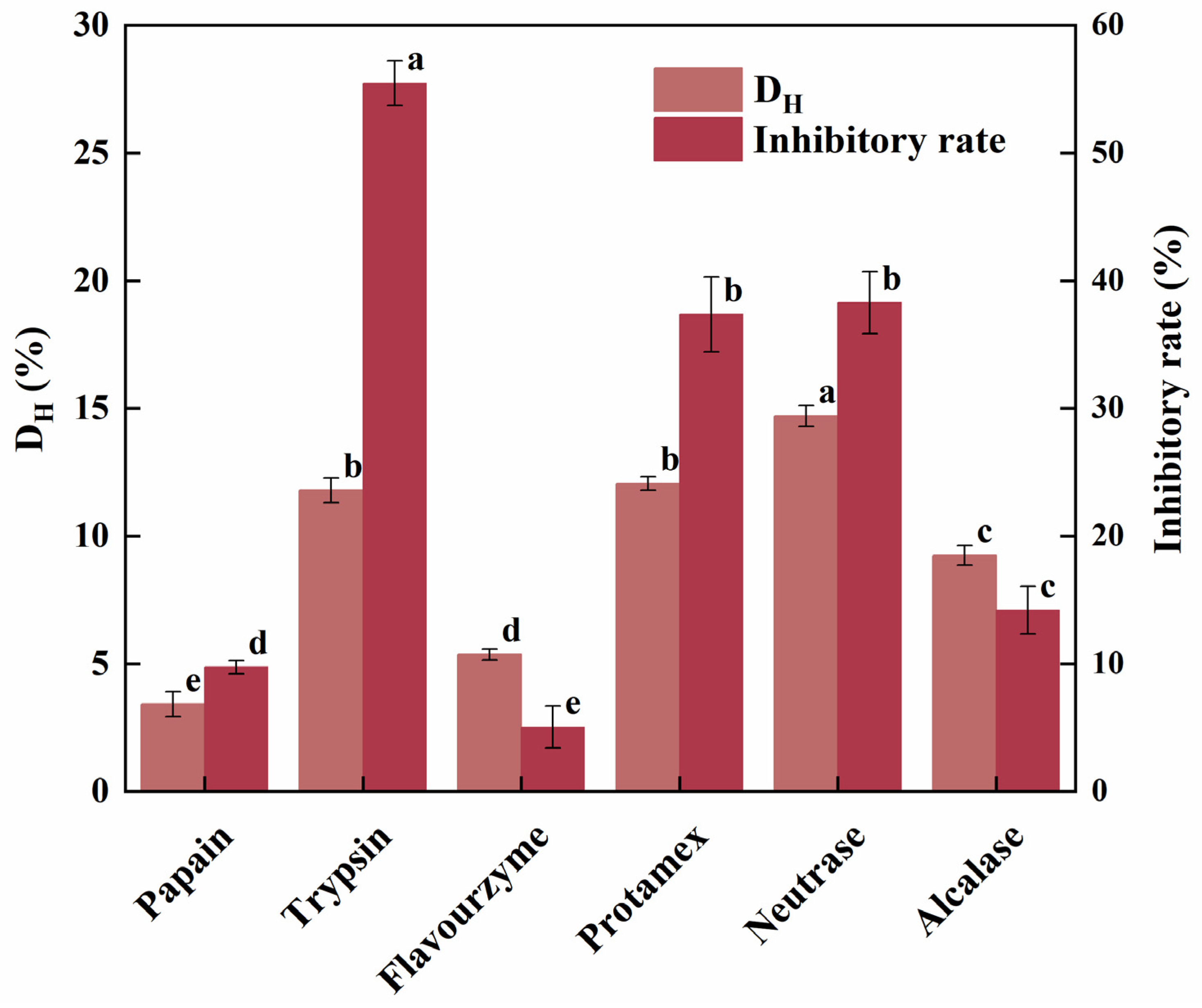
2.1. Protease Screening for HSP Hydrolysis

In this study, the effects of various proteases on the hydrolysis of HSP were compared in terms of DH and α-glucosidase inhibitory activity of the corresponding hydrolysates (Figure 1). The DH serves as an indicator of the extent of protein cleavage during hydrolysis and is also a key factor that may indirectly reflect the sequence length and molecular weight range of the peptides in the resulting hydrolysate. Overall, the DH of HSP ranged from about 3% to 15%, with Neutrase achieving the highest DH (14.70%), followed by Protamex (12.05%) and trypsin (11.79%). On the other hand, papain exhibited the lowest DH, with a value of 3.41%.
Additionally, α-glucosidase inhibitors prevent carbohydrate digestion by competitively inhibiting α-glucosidase residing in the small intestine. Therefore, the α-glucosidase inhibitory rate can also be used for protease screening, aiming at discovery of antidiabetic peptides. In this study, tryptic hydrolysate exhibited the highest inhibitory activity against α-glucosidase, reaching 55.47%, followed by Neutrase (38.28%) and Protamex hydrolysates (37.36%), whereas the flavourzyme hydrolysate exhibited the lowest inhibitory activity (Figure 1). A previous study on the hydrolysis of camellia seed meal has revealed that the α-glucosidase inhibitory rates of the hydrolysates produced by various proteases ranged from 10% to 55%, with the Protamex hydrolysate showing the highest inhibition, followed by trypsin and alkaline protease, and the flavourzyme hydrolysates showing the lowest inhibition [25]. This finding is similar to our results, which may be attributed to the specific terminal groups produced after cleavage, due to the specific cleavage site of each protease for the proteins. For instance, Ibrahim et al. found that an N-terminal hydroxyl-containing residue is critical for α-glucosidase inhibitory activity [26]. Additionally, Stefano et al. reported that peptides containing serine, threonine, tyrosine, lysine, or arginine at the N-terminus, and proline closer to the C-terminus occupied by methionine or alanine, exhibited higher α-glucosidase inhibitory activity [27].
Furthermore, multiple comparisons revealed significant intergroup variations (p < 0.01), and Pearson correlation analysis showed a strong positive correlation between DH and α-glucosidase inhibitory rate (r = 0.8125, p = 0.0495). This suggests that a higher degree of hydrolysis tends to produce hydrolysates with stronger α-glucosidase inhibition. Based on both of DH and α-glucosidase inhibition, the tryptic hydrolysate of HSP was selected for peptidomics analysis.

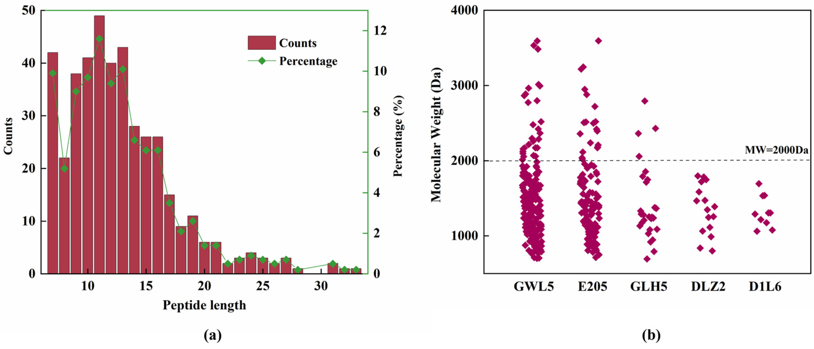
2.2. Peptidomics Analysis

Nano LC-MS/MS was used to analyze the peptide profile of the tryptic hydrolysate of HSP, resulting in the identification of 424 peptide sequences.
The molecular weights of the peptides ranged from 693 Da to 3595 Da, with sequence lengths varying from 7 to 33 amino acids (Figure 2a). Among the 424 peptides detected, 49 were decapeptides, making up the largest proportion (11.56%), followed by tridecapeptides and heptapeptides. A total of 143 peptides were decapeptides or smaller peptides, accounting for approximately 33.72% of the total peptides. Additionally, 329 peptides were pentadecapeptides or smaller, representing about 80% of the total peptides. No hexapeptides or smaller peptides were detected, possibly due to the relatively low DH of HSP by trypsin.
Regarding the origin of the peptides, most were derived from cupin-type-1 domain-containing proteins, specifically those with UniProt ID A0A7J6GWL5 (GWL5), A0A7J6E205 (E205), A0A7J6GLH5 (GLH5), and A0A7J6DLZ2 (DLZ2), which contributed 209, 122, 27, and 16 peptides, respectively (Figure 2b). Together, these proteins accounted for nearly 90% of the total peptides. GWL5 and E205 are commonly classified as 11S seed storage proteins, often referred to as edestin in hemp seeds, which account for 60–80% of the total hemp seed protein content [11]. This explains why they were the main contributors to the peptides in the hydrolysates. After these four proteins, albumin A0A219D1L6 (D1L6) produced 10 peptides. In hemp seed, albumins are more prevalent than globulins, further demonstrating that the distribution of peptides in this study is consistent with the known protein composition of hemp seed.
Among the total 424 peptide sequences, 68 peptides were carbamidomethylated at cysteine residues, 25 peptides were oxidized at methionine residues, 1 peptide showed N-terminal acetylation, 5 peptides had both carbamidomethylation and oxidation, and 5 peptides had both acetylation and oxidation modifications. After excluding these modified peptides, a total of 323 peptides were used for subsequent activity prediction.

2.3. Prediction of Potential Activity of the Peptides in Tryptic Hydrolysates of HSP

In this study, the PeptideRanker program was used to evaluate the potential biological activity of the identified peptides. PeptideRanker utilizes a novel N-to-1 neural network to predict the likelihood that a peptide has biological activity. Any peptide with a score greater than 0.500 is considered to have potential biological activity. Of the 323 peptides evaluated in this study, 47 peptides scored above the threshold of 0.500 (Table 1), with lengths ranging from 7 to 17 amino acids. Among these, there were nine nonapeptides, eight heptapeptides, seven dodecapeptides, five octapeptides, and five undecapeptides. The heptapeptides, octapeptides, and nonapeptides accounted for about a quarter of the total peptides, showing a relatively high proportion of shorter peptides, which tend to have higher bioactivity potential as mentioned before. Previous studies have indicated that short peptides containing phenylalanine and glycine are more likely to exhibit biological activity [13]. However, the results of this study showed that the biological activity, i.e., PeptideRanker score, had little correlation with the phenylalanine and glycine content in the peptides, but it did show a relationship with the length of the peptide sequence (Supplementary Figure S1).

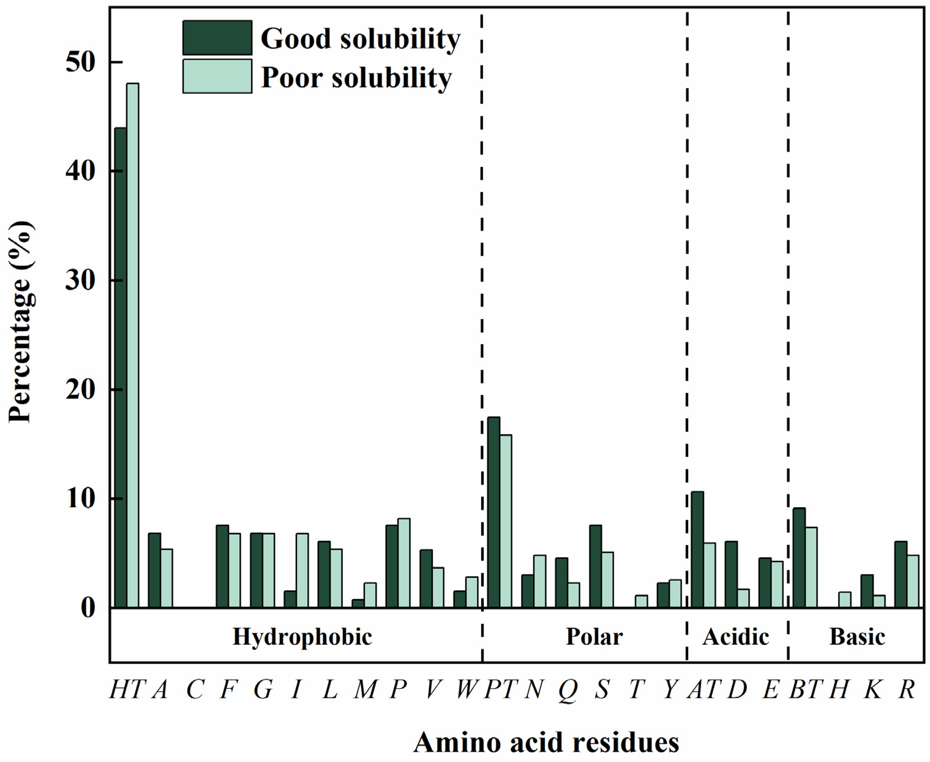
2.4. Analysis of Physicochemical Properties of the Peptides

In this study, the physicochemical properties of the 47 peptides with potential biological activity were analyzed (Table 2). The isoelectric points (pI) of these peptides ranged from 0.60 to 11.29. Among them, 29 peptides were acidic (pI < 7.0), whereas 18 were basic (pI > 7.0). The acidic peptides included 8 peptides with pI < 3.0, 8 peptides with pI between 3.0 and 6.0, and 13 peptides with pI between 6.0 and 7.0. Among the basic peptides, 4 peptides were weakly basic (pI between 7.0 and 8.0), 13 peptides were basic (pI between 8.0 and 11.0), and 1 peptide was extremely basci (pI > 11.0). The net charge of these peptides at pH 7.0 ranged from −2.0 to 4.0, showing a good positive correlation with the isoelectric points (r = 0.88) (Supplementary Figure S1). However, although isoelectric points are not a significant variable for α-glucosidase inhibition, a net charge of 0.0 or +1.0 was predicted for the highly active peptides [26]. In this study, 30 peptides were selected with a net charge at pH 7.0 of 0.0 or +1.0, accounting for 63.8% of the total, indicating a favorable selection outcome.
Furthermore, the water solubility of the peptides was also predicted as it is a crucial factor for their in vivo biological functioning [28]. Among these 47 peptides, 13 peptides exhibited good water solubility, accounting for more than a quarter. Two key factors were generally considered to influence water solubility, namely, the type of amino acid residues and the peptide length [29]. For these reasons, the effect of the amino acid composition on the water solubility was initially analyzed. Generally, more polar amino acids, acidic amino acids and basic amino acids, and less hydrophobic amino acids were observed in the water-soluble peptides (Figure 3). Specifically, as compared with the poorly water-soluble peptides, the contents of the polar amino acid glutarnine, the acidic amino acid aspartic acid and the basic amino acid lysine are significantly higher, and the contents of the hydrophobic amino acid isoleucine and methionine are significantly lower in the water-soluble peptides. Moreover, the presence of arginine or phenylalanine at C-terminus was observed to enhance peptide water solubility, aligning with findings from previous research [30]. On the other hand, no clear relationship was found between sequence length and water solubility in this study, which might be attributed to the fact that most peptide lengths were concentrated within a narrow range and did not vary significantly.

2.5. Toxicity and ADMET Property Analysis of the Peptides

Safety is crucial for the potential application of peptides in food, healthcare products, and drugs. Therefore, the ToxinPred program was used to evaluate the toxicity of 13 water-soluble peptides with potential biological activity. ToxinPred uses a support vector machine (SVM) algorithm to predict the potential toxicity of peptides on biological systems and has been widely validated for its reliability [15]. In this study, the SVM scores for all 13 peptides were less than 0 (Table 3), indicating that these peptides are non-toxic.
When used for virtual screening of bioactive agents, the ADMET properties of peptides are crucial [31]. In this study, the ADMET properties of these 13 peptides were assessed using the AdmetSAR program. For distribution, none of the peptides crossed the blood–brain barrier, likely due to their relatively large molecular weights (Table 3). However, for adsorption, 8 out of the 13 peptides were predicted to be absorbable in the human intestinal tract, indicating that most of the peptides could be administered orally (Table 3). Regarding metabolism, the interaction of the peptides with CYP450 2C9, a key enzyme of the CYP450 family, was analyzed. CYP450 family enzymes are important for toxicity and therapeutic efficacy, as they degrade the molecules from circulation in the liver. Thus, inhibition of these enzymes can lead to abnormal biodegradation of the molecules [32]. In this analysis, none of the peptides were found to act as either substrates or inhibitors of CYP450 2C9 (Table 3). Similar results were also observed for other enzymes in the CYP450 family, suggesting a reduced risk of abnormal metabolic interactions.

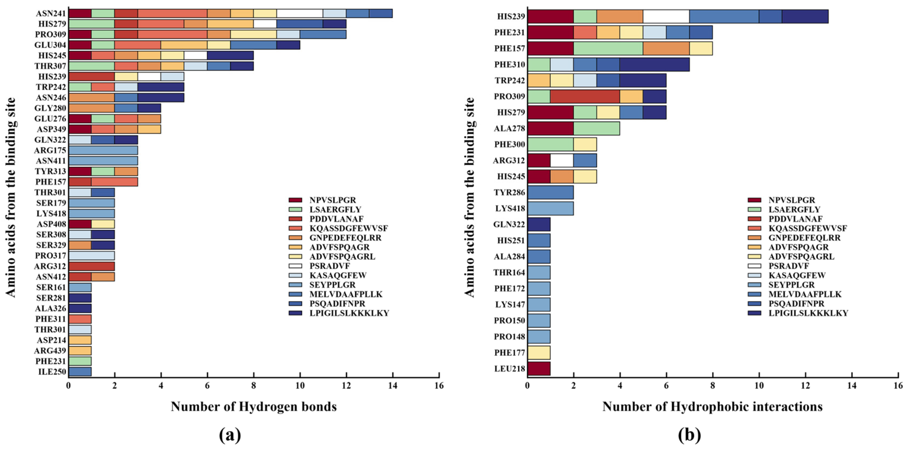
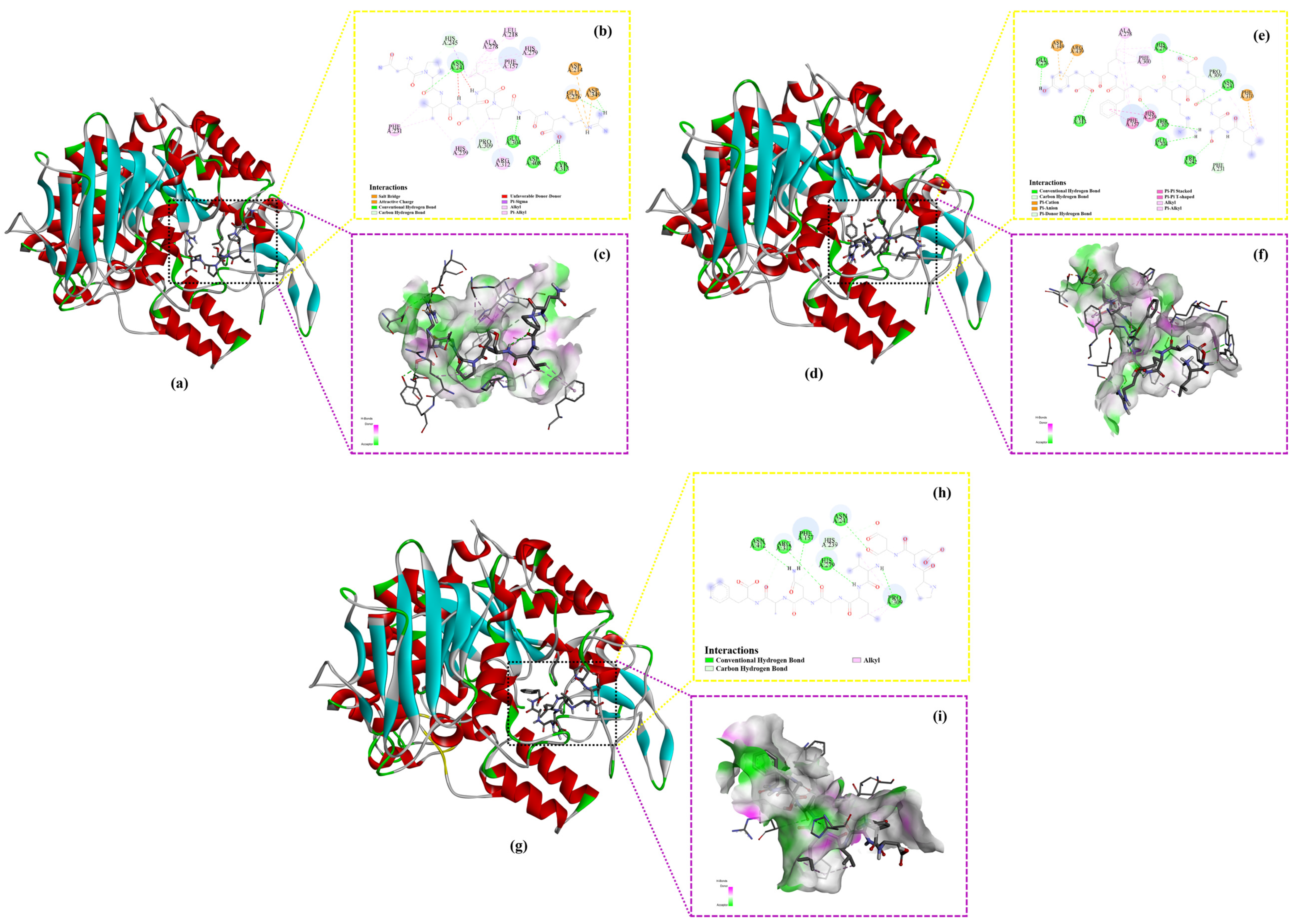
2.6. Molecular Docking

Generally, binding energy plays a crucial role in protein–ligand interactions, as lower binding energy reflects stronger stability of the protein–ligand complex [33]. Therefore, it is commonly used for the rapid screening of inhibitors for specific proteins, including ACE [34], DPP-IV [35], xanthine oxidase [36] and tyrosinase [30], etc. On such a basis, molecular docking was performed to evaluate the binding affinities between the peptides and α-glucosidase, further validating the inhibitory effects of the selected peptides on α-glucosidase (Table 4). In this study, all of the selected peptides exhibited negative binding energies lower than −5.0 kcal/mol, indicating strong and stable interactions with the protein receptor, as demonstrated in previous research [37].
Furthermore, interaction between the peptides and α-glucosidase primarily consisted of hydrogen bonds and hydrophobic interactions, with the number of hydrogen bonds higher than that of hydrophobic interactions. This result aligns with previous studies demonstrating that hydrogen bonds and hydrophobic interactions play a dominant role in α-glucosidase inhibition [38,39].
As shown in the binding site analysis (Figure 4), the key residues on α-glucosidase involved in hydrogen bond formation with the peptides include Asn241, His279, Pro309, Glu304, His245, and Thr307, while the residues responsible for hydrophobic interactions primarily comprise His239, Phe231, Phe157, and Phe310. In S. cerevisiae α-glucosidase, the gatekeeping residues, including Phe231, His239, Asn241, His279, Glu304, and Arg312, control the entry and exit of the substrate into the active site [40]. These results suggest that the peptides in this study exert their α-glucosidase inhibitory effect mainly by occupying the entrance of the active site gorge, as further illustrated by the docking conformation in Figure 5.
In addition, a few electrostatic interactions were observed between the peptides and α-glucosidase at specific amino acid residues, mainly including Asp349, Glu276, and Asp214. In the active sites of S. cerevisiae α-glucosidase, Asp214, Glu276, and Asp349 form a catalytic triad, where Asp214 acts as a nucleophile, Glu276 serves as a proton donor, and Asp349 stabilizes the substrate’s transition state [40]. Although the number of electrostatic interactions between the peptides and α-glucosidase was fewer than that of hydrogen bonds and hydrophobic interactions, their role cannot be overlooked due to their critical involvement in the interactions with the active sites.
In order to further evaluate the inhibitory effect of the peptides on α-glucosidase, their binding affinities were compared with that of acarbose, a commonly used positive control in the screening of potent α-glucosidase inhibitors. The binding energy of acarbose obtained in this study was −8.1 kcal/mol (Table 4), which was similar to that reported in a previous study (−7.9 kcal/mol) [41]. Compared with acarbose, three peptides, i.e., NPVSLPGR (−8.7 kcal/mol), LSAERGFLY (−8.5 kcal/mol), and PDDVLANAF (−8.4 kcal/mol), were found to have lower binding energies, indicating stronger and more stable binding, and thus, a potentially more potent inhibition on α-glucosidase. As shown in a previous study, compounds derived from the marine macro brown alga Dictyopteris hoytii, which exhibited lower binding energies to α-glucosidase, also demonstrated lower IC50 values, indicating a positive correlation between binding energy and IC50 values [26]. A similar observation was also achieved in this study, further confirming the efficacy of these three α-glucosidase inhibitory peptides.
As illustrated by the binding conformations, all three peptides bind to the active pocket of α-glucosidase through hydrogen bonding, hydrophobic interaction, and electrostatic interaction (except for PDDVLANAF) (Figure 5 and Table 4). Since hydrogen bonding plays a critical role in non-bonding interactions and dominates the interactions between the peptides and α-glucosidase, the hydrogen bond surfaces were analyzed in greater detail (Figure 5c,f,i). Hydrogen bonds comprise hydrogen bond donors (purple) and hydrogen bond acceptors (green). In the hydrogen bond surface, the hydrogen bond donor defines the area with lower electron density in the electronegative atom compared with the hydrogen bond acceptor [42]. In this study, all three peptides exhibited more hydrogen bond acceptor areas than hydrogen bond donor areas.
Based on the findings discussed above, it can be inferred that the peptides NPVSLPGR and LSAERGFLY inhibit α-glucosidase by occupying the entrance of the active site gorge to block the entry of substrates, and binding to active sites to prevent catalysis, while the peptide PDDVLANAF primarily inhibits α-glucosidase by occupying the entrance of the active site gorge.

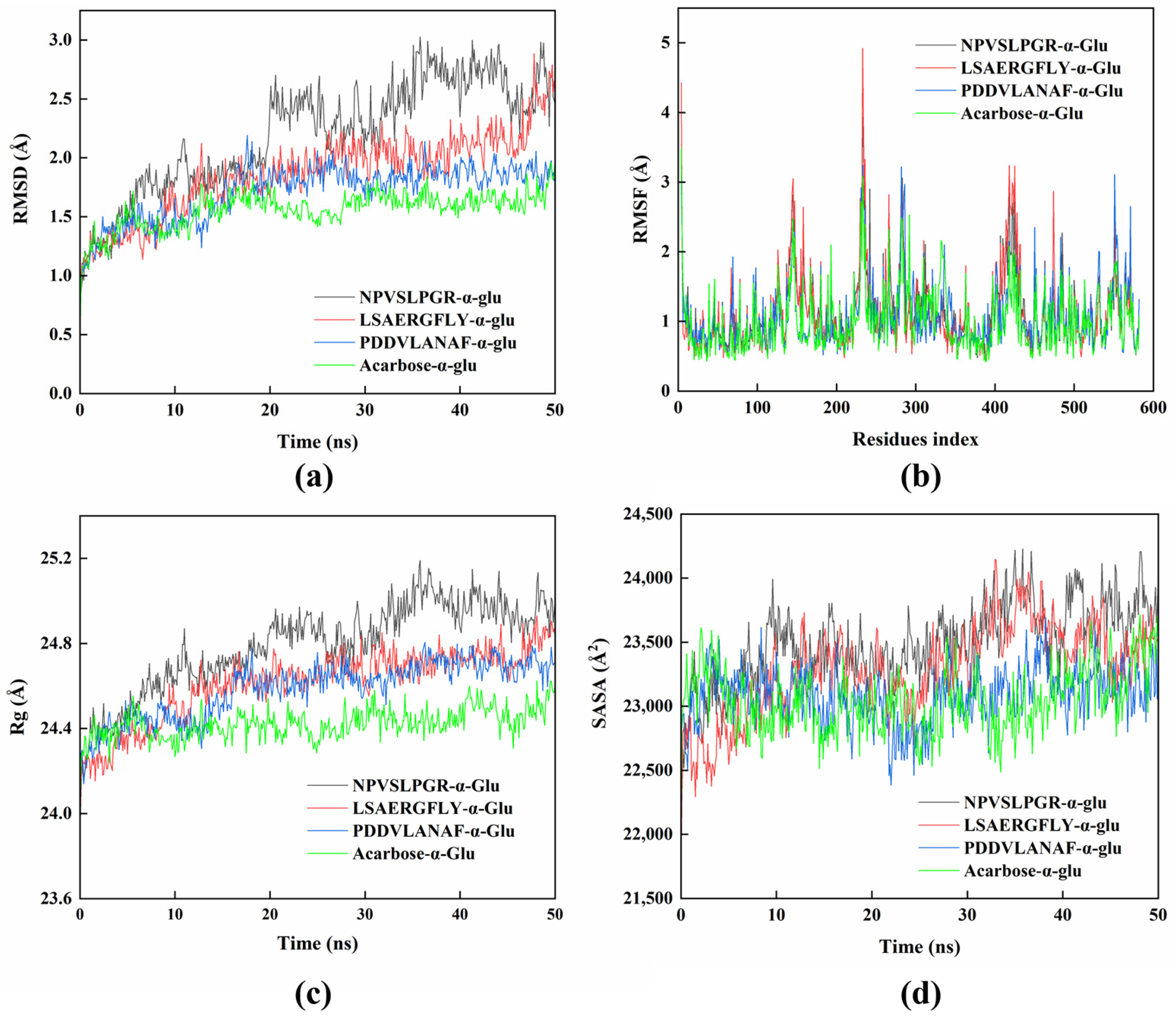
2.7. Molecular Dynamics

In this study, MD simulations were conducted to further elucidate the structural dynamic changes in the complexes formed by the peptides and α-glucosidase. Before the MD simulation, the complex of NPVSLPGR-α-glucosidase was pre-simulated for 200 ns to determine the time required for stabilization (Supplementary Figure S2). The system stabilized after approximately 20 ns, with minimal variation in the average RMSD value (2.77 ± 0.25 Å, from 20 ns to 200 ns). This was not significantly different from the RMSD values observed between 20 ns and 50 ns. Therefore, the MD analyses were conducted for 50 ns for all complexes.
Generally, lower RMSD value indicates a more stable global structure [43]. In this study, the RMSD values showed a slight increase from approximately 1 Å to about 2.5 Å (the complex of NPVSLPGR-α-glucosidase), about 2 Å (the complex of LSAERGFLY-α-glucosidase and PDDVLANAF-α-glucosidase) or about 1.5 Å (the complex of acarbose-α-glucosidase) during the first 20 ns, after which they stabilized with variations less than 0.3 Å, indicating a stable complex formation (Figure 6a). The average RMSD values for the complexes NPVSLPGR-α-glucosidase, LSAERGFLY-α-glucosidase, PDDVLANAF-α-glucosidase, and acarbose-α-glucosidase (from 20 ns to 50 ns) were 2.53 ± 0.23 Å, 2.09 ± 0.20 Å, 1.85 ± 0.08 Å, and 1.62 ± 0.09 Å, respectively, indicating a relatively stable backbone structure of the protein. These results align with previous findings on luteolin-α-glucosidase complex, where the RMSD values of the backbone atoms ranged from 2 to 3 Å [44].
RMSF values reflect the fluctuation degree of the amino acid residues in the receptor protein within the complex. In this study, the average RMSF values for the complexes NPVSLPGR-α-glucosidase, LSAERGFLY-α-glucosidase, PDDVLANAF-α-glucosidase, and acarbose-α-glucosidase were 1.15 ± 0.51 Å, 1.13 ± 0.59 Å, 1.09 ± 0.46 Å, and 1.01 ± 0.44 Å, respectively (Figure 6b), indicating a stable structure during MD simulation. Furthermore, the fluctuation patterns of RMSF were consistent with a previous study on the α-glucosidase inhibitory compounds from Chlorella minutissima, which exhibited significant fluctuation at residues 130–170, 220–250, 280–300, and 400–450 [45]. As reported previously, residues 280–300 and 400–450 of the α-glucosidase correspond to the α-helices and the loop regions near the active site, respectively [45]. The increased fluctuation in both regions enhances the flexibility of the entrance pocket residues, thereby facilitating peptide binding at the active site. This observation corroborates the findings from molecular docking.
The Rg value generally determines the compactness of the protein structure, with lower Rg values reflecting a more closely packed protein. The average Rg values for the complexes NPVSLPGR-α-glucosidase (24.79 ± 0.21 Å), LSAERGFLY-α-glucosidase (24.61 ± 0.17 Å), PDDVLANAF-α-glucosidase (24.58 ± 0.14 Å), and acarbose-α-glucosidase (24.43 ± 0.07 Å) were at similar level and varied minimally (Figure 6c). In a previous study on α-glucosidase inhibitory peptides from Ginkgo biloba seed cake, the Rg values for the peptide-α-glucosidase complexes ranged from 23.5 to 23.7 Å, which are comparable to the values observed in this study [46].
SASA is commonly used to describe the folding stability of a receptor protein and its surface volume change [47]. In this study, the SASA values for the complexes increased slightly with time (Figure 6d), suggesting that the protein pocket bound by the peptides expanded slightly, resulting in a larger surface area.

3. Materials and Methods

3.1. Materials and Reagents

HSP with protein content > 70% was provided by Shaanxi Jinrun Biotechnology Co., Ltd. (Xi’an, China). The proteases used in this study include Flavourzyme, Alcalase 2.4L, Protamex, and Neutrase from Novozymes Biotechnology Co., Ltd. (Tianjin, China), trypsin from Sigma-Aldrich (Shanghai, China), and papain from Shanghai Aladin Biotech Co., Ltd. (Shanghai, China). α-Glucosidase from Saccharomyces cerevisiae (EC 3.2.1.20) and p-nitrophenyl-α-D-glucopyranoside (pNPG) were purchased from Sigma-Aldrich (Shanghai, China).

3.2. Hydrolysis of HSP

HSP powder was exactly weighed out and thoroughly mixed with a buffer as specified (Supplementary Table S1) in a ratio of 5% (HSP: buffer, w/v), after which the protease was added at an E/S ratio of 1%. The hydrolysis was carried out for 6 h at an optimal temperature for respective protease (Supplementary Table S1). After hydrolysis, the mixture was boiled for 10 min to stop the reaction, and then centrifuged at 10,000× g for 10 min. The supernatant was collected, lyophilized, and stored at −20 °C.

3.3. Determination of Degree of Hydrolysis (DH)

The DH was determined using the o-phthalaldehyde (OPA) assay as described by Zhang et al. [48]. Briefly, OPA working solution was initially prepared by thoroughly mixing 25.0 mL of 100 mM aqueous sodium tetraborate solution, 10.0 mL of 5.0% (w/v) aqueous sodium dodecyl sulfate solution, 1.0 mL of 40 mg/mL OPA solution in methanol, 0.1 mL of β-mercaptoethanol, and 13.9 mL of water. Subsequently, 50.0 µL of HSP hydrolysate was mixed with 2.0 mL of the OPA working solution and reacted at room temperature for 8 min. After the reaction, the absorbance at 340 nm was measured using a spectrophotometer (L5S, INESA Analytical Instrument Co., LTD, Shanghai, China), and the free amino content was calculated by the detected absorbance against the calibration curve established using glycine–glycine dipeptide as the standard. The DH of the HSP hydrolysate was calculated according to the following formula:
DH (%) = (C2 − C1)/(C0 − C1) × 100
C₁ is the free amino content of HSP before hydrolysis (mol/L); C₂ is the free amino content of HSP after hydrolysis (mol/L); and C₀ is the total amino content of HSP determined by the Kjeldahl method (mol/L).

3.4. Determination of α-Glucosidase Inhibitory Activity

The α-glucosidase inhibitory activity of HSP hydrolysates was determined according to the method described by Liu et al. [49]. Briefly, 100.0 µL of sample was mixed with 400.0 µL of α-glucosidase solution (0.5 U/mL) and incubated at 37 °C for 10 min. Subsequently, 50.0 µL of pNPG (5 mM) was added to the mixture, thoroughly mixed, and incubated at 37 °C for another 30 min. After the reaction, 100.0 µL of 1 M Na₂CO₃ solution was added to terminate the reaction, and the absorbance at 405 nm was measured. The α-glucosidase inhibitory activity was calculated using the following formula:
α-glucosidase inhibitory rate (%) = (1 − (A1 − A2)/(A3 − A4)) × 100
A₁ refers to the absorbance of the sample, i.e., the HSP hydrolysate; A₂ refers to the absorbance of the sample blank; A₃ refers to the absorbance of the negative control (where the sample is replaced by buffer); and A₄ refers to the absorbance of the negative control blank.

3.5. Analysis of Peptide Profile

The peptide profile of the tryptic hydrolysate of HSP was determined based on Nano LC-MS/MS using an Ultimate 3000 system coupled with Q Active™ Hybrid Quadrupole-Orbitrap™ mass spectrometry equipped with ESI Nanospray Source (Thermo Fisher Scientific, Waltham, MA, USA) [30].

3.6. Evaluation of Potential Biological Activity of the Peptides

The potential biological activity of the identified peptides was evaluated using the PeptideRanker program (http://distilldeep.ucd.ie/PeptideRanker/ (accessed on 24 April 2024)) with the threshold set at 0.500, since the program was trained at this threshold [50]. Thus, any peptide with a PeptideRanker score higher than 0.500 was deemed bioactive.

3.7. Analysis of Physicochemical Properties of the Peptides

The physicochemical properties, including molecular weight, net charge at pH 7.0, isoelectric point, and water solubility, were predicted using an online program Peptide Property Calculator (http://www.innovagen.com/proteomics-tools/ (accessed on 24 April 2024)) for the identified peptides of potential biological activity.

3.8. Peptide Toxicity Analysis

The ToxinPred program (https://webs.iiitd.edu.in/raghava/toxinpred/ (accessed on 24 April 2024)) was used to predict the toxicity of the soluble peptides with potential biological activity as identified above [51].

3.9. ADMET Evaluation

ADMET properties of the soluble peptides with potential biological activity were analyzed using AdmetSAR (http://lmmd.ecust.edu.cn/admetsar2/ (accessed on 24 April 2024)). The human intestinal absorption (HIA), blood–brain barrier penetration (BBB), and cytochrome P450 (CYP 450) 2D9 interaction were analyzed to reflect the absorption, distribution, and metabolic properties of the peptides [52].

3.10. Molecular Docking

Molecular docking was conducted using AutoDock Vina (version 1.1.2), a widely used software for virtual screening of bioactive peptides, despite not being specifically designed for peptide docking [53]. The 3D structure of the peptides for molecular docking was created using Chembio3D Ultra (version 14.0) and saved in MOL2 format. Prior to docking, the 3D structure of the peptides was processed using AutoDock (V 4.2.6). On the other hand, the 3D structure of Saccharomyces cerevisiae α-glucosidase, which was the same enzyme used in the in vitro experiments and belongs to the Glycoside Hydrolase (GH) Family 13, was built based on homology modeling using SWISS-MODEL (https://swissmodel.expasy.org/ (accessed on 2 May 2024)), due to the unavailability of the crystal structure of α-glucosidase [45]. The primary sequence of S. cerevisiae α-glucosidase (UniProt ID: P53341) was obtained from UniProt (https://www.uniprot.org/ (accessed on 2 May 2024)) and used for homology modeling with the crystal structure of S. cerevisiae isomaltase (PDB ID: 3aj7.1A) as a template (GMQE = 0.95). The 3D structure of α-glucosidase obtained from homology modeling was processed by AutoDock before docking to remove water and add hydrogen. Subsequently, site-specific docking between the peptides and α-glucosidase was conducted with the grid centered at center_x = 15.77, center_y = −2.99, and center_z = 17.65 and a grid box size of 60.00 Å × 60.00 Å × 60.00 Å. Conformation with the lowest binding energy was visualized using BIOVIA Discovery Studio Visualizer (V 17.2.0).

3.11. Molecular Dynamics (MD) Simulation

MD simulation was conducted based on the molecular docking conformation with the lowest binding energy using YASARA Dynamics (Version 14.10.5) for 50 ns. Simulation was performed using the AMBER14 force field. The simulation cell was defined as “around all atoms” with a size automatically set at 5.00 Å. The environment was set at 298 K and pH 7.4, using 0.9% NaCl and water solvent system. The cell boundaries were set as “periodic”, and the system was energy minimized before MD. All other parameters were used with default settings. In this study, to evaluate the stability of ligand–receptor interactions, the MD simulation results, including changes in root mean square deviation (RMSD), root mean square fluctuation (RMSF), radius of gyration (Rg), and solvent-accessible surface area (SASA), were analyzed.

3.12. Statistical Analysis

The results in this study were expressed as mean ± standard deviation (S.D.) and analyzed using Duncan’s multiple range test performed with the IBM SPSS Statistics 20.0 (Chicago, IL, USA) for statistical significance at a level of p < 0.05. Additionally, Pearson correlation analysis was conducted using the same software to examine potential relationships between variables.

4. Conclusions

In this study, the peptide profile of the tryptic hydrolysate of HSP was determined, as it exhibited favorable DH and the best α-glucosidase inhibitory activity compared to other HSP hydrolysates prepared using different proteases. A total of 424 peptides, with molecular weights ranging from 693 Da to 3595 Da, were identified, originating mainly from four cupin-type-1 domain-containing proteins. Among these peptides, 47 scored above the threshold of 0.500 by PeptideRanker, indicating their potential as bioactive candidates. Based on physiochemical properties, toxicity, and ADMET analysis, 13 peptides were ultimately selected for validation as potential α-glucosidase inhibitors. These 13 peptides interacted with α-glucosidase primarily through hydrogen bonding and hydrophobic interactions at negative binding energies below –6.0 kcal/mol, indicating stable binding, and consequently, strong α-glucosidase inhibition. Among them, three novel peptides, i.e., NPVSLPGR, LSAERGFLY, and PDDVLANAF, displayed binding energies lower than that of the positive control acarbose, suggesting potent α-glucosidase inhibitory activities. The peptides NPVSLPGR and LSAERGFLY inhibited α-glucosidase by both occupying the entrance of the active site gorge to block substrate entry and binding to active sites to prevent catalysis, whereas the peptide PDDVLANAF primarily inhibits α-glucosidase by occupying the entrance of the active site gorge. Furthermore, MD simulation confirmed the stability of the complexes formed between the peptides and the enzyme and corroborated the findings from molecular docking. Based on the findings from computational studies, these peptides demonstrate promising α-glucosidase inhibitory potential and may serve as viable natural alternatives to synthetic inhibitors. However, further validation in vivo and through clinical trials is essential to ensure their effectiveness in real-world applications.

Supplementary Materials

The following supporting information can be downloaded at: https://www.mdpi.com/article/10.3390/ijms26052222/s1.

Author Contributions

Conceptualization, Z.B.; methodology, Z.N. and Q.Z.; software, Z.B. and Z.N.; validation, Y.W., Z.T. and R.G.; formal analysis, C.C. and W.F.; investigation, Z.M. and C.C.; data curation, Z.M.; writing—original draft preparation, Z.M.; writing—review and editing, Q.Z. and Z.B.; visualization, W.F., Y.W., Z.T. and R.G.; supervision, Z.N. and Q.Z.; project administration, Z.B.; funding acquisition, Z.B. All authors have read and agreed to the published version of the manuscript.

Funding

This research was funded by the National Natural Science Foundation of China (No. 31701536 and 82200738) and the Student Research Training Program (SRTP) of Henan Province (No. 202410464070).

Institutional Review Board Statement

Not applicable.

Informed Consent Statement

Not applicable.

Data Availability Statement

The datasets used and/or analyzed during the current study are available from the corresponding author upon reasonable request.

Conflicts of Interest

The authors declare no conflicts of interest.

References

  1. Sun, H.; Saeedi, P.; Karuranga, S.; Pinkepank, M.; Ogurtsova, K.; Duncan, B.B.; Stein, C.; Basit, A.; Chan, J.C.N.; Mbanya, J.C.; et al. IDF Diabetes Atlas: Global, Regional and Country-Level Diabetes Prevalence Estimates for 2021 and Projections for 2045. Diabetes Res. Clin. Pract. 2022, 183, 109119. [Google Scholar] [CrossRef] [PubMed]
  2. Lu, H.; Xie, T.; Wu, Q.; Hu, Z.; Luo, Y.; Luo, F. α-Glucosidase Inhibitory Peptides: Sources, Preparations, Identifications, and Action Mechanisms. Nutrients 2023, 15, 4267. [Google Scholar] [CrossRef] [PubMed]
  3. Uuh Narvaez, J.J.; Segura Campos, M.R. Combination Therapy of Bioactive Compounds with Acarbose: A Proposal to Control Hyperglycemia in Type 2 Diabetes. J. Food Biochem. 2022, 46, e14268. [Google Scholar] [CrossRef] [PubMed]
  4. Joshi, S.R.; Standl, E.; Tong, N.; Shah, P.; Kalra, S.; Rathod, R. Therapeutic Potential of α-Glucosidase Inhibitors in Type 2 Diabetes Mellitus: An Evidence-Based Review. Expert Opin. Pharmacother. 2015, 16, 1959–1981. [Google Scholar] [CrossRef]
  5. Tsunoda, T.; Samadi, A.; Burade, S.; Mahmud, T. Complete Biosynthetic Pathway to the Antidiabetic Drug Acarbose. Nat. Commun. 2022, 13, 3455. [Google Scholar] [CrossRef]
  6. Dong, Y.; Sui, L.; Yang, F.; Ren, X.; Xing, Y.; Xiu, Z. Reducing the Intestinal Side Effects of Acarbose by Baicalein through the Regulation of Gut Microbiota: An in Vitro Study. Food Chem. 2022, 394, 133561. [Google Scholar] [CrossRef]
  7. Yang, X.; Wang, D.; Dai, Y.; Zhao, L.; Wang, W.; Ding, X. Identification and Molecular Binding Mechanism of Novel α-Glucosidase Inhibitory Peptides from Hot-Pressed Peanut Meal Protein Hydrolysates. Foods 2023, 12, 663. [Google Scholar] [CrossRef]
  8. Hu, Y.; Sun, H.-N.; Zhang, M.; Mu, T.-H. Production and Characterization of α-Glucosidase Inhibitory Peptides from Sweet Potato Protein by Ultrasound-Assisted Enzymatic Hydrolysis and in Vitro Gastrointestinal Digestion. Eur. Food Res. Technol. 2024, 251, 257–267. [Google Scholar] [CrossRef]
  9. Xu, Y.; Yang, Y.; Ma, C.; Bian, X.; Liu, X.; Wang, Y.; Chen, F.; Wang, B.; Zhang, G.; Zhang, N. Characterization of the Structure, Antioxidant Activity and Hypoglycemic Activity of Soy (Glycine max L.) Protein Hydrolysates. Food Res. Int. 2023, 173, 113473. [Google Scholar] [CrossRef]
  10. Wang, X.; Deng, Y.; Xie, P.; Liu, L.; Zhang, C.; Cheng, J.; Zhang, Y.; Liu, Y.; Huang, L.; Jiang, J. Novel Bioactive Peptides from Ginkgo Biloba Seed Protein and Evaluation of Their α-Glucosidase Inhibition Activity. Food Chem. 2023, 404, 134481. [Google Scholar] [CrossRef]
  11. Fu, Y.; Liu, Z.; Wang, H.; Zhang, F.; Guo, S.; Shen, Q. Comparison of the Generation of α-Glucosidase Inhibitory Peptides Derived from Prolamins of Raw and Cooked Foxtail Millet: In Vitro Activity, de Novo Sequencing, and in Silico Docking. Food Chem. 2023, 411, 135378. [Google Scholar] [CrossRef] [PubMed]
  12. Zheng, K.; Wu, Y.; Dai, Q.; Yan, X.; Liu, Y.; Sun, D.; Yu, Z.; Jiang, S.; Ma, Q.; Jiang, W. Extraction, Identification, and Molecular Mechanisms of α-Glucosidase Inhibitory Peptides from Defatted Antarctic Krill (Euphausia superba) Powder Hydrolysates. Int. J. Biol. Macromol. 2024, 266, 131126. [Google Scholar] [CrossRef] [PubMed]
  13. Li, N.; Qiu, L.; Song, H.; Xiao, T.; Luo, T.; Deng, Z.; Zheng, L. Inhibition Mechanism of Oligopeptides from Soft-Shelled Turtle Egg against α-Glucosidase and Their Gastrointestinal Digestive Properties. J. Food Biochem. 2022, 46, e14328. [Google Scholar] [CrossRef] [PubMed]
  14. Dong, Z.-H.; Pan, R.-Y.; Ren, G.-Y.; Zhou, M.; Zhang, B.; Fan, J.-L.; Qiu, Z.-J. A Novel Antidiabetic Peptide GPAGAP from Andrias davidianus Collagen Hydrolysates: Screening, Action Mechanism Prediction and Improving Insulin Resistance in HepG2 Cells. Food Med. Homol. 2024, 1, 9420010. [Google Scholar] [CrossRef]
  15. Zhao, Q.; Wei, G.; Li, K.; Duan, S.; Ye, R.; Huang, A. Identification and Molecular Docking of Novel α-Glucosidase Inhibitory Peptides from Hydrolysates of Binglangjiang Buffalo Casein. LWT 2022, 156, 113062. [Google Scholar] [CrossRef]
  16. Callaway, J.C. Hempseed as a Nutritional Resource: An Overview. Euphytica 2004, 140, 65–72. [Google Scholar] [CrossRef]
  17. Rizzo, G.; Storz, M.A.; Calapai, G. The Role of Hemp (Cannabis sativa L.) as a Functional Food in Vegetarian Nutrition. Foods 2023, 12, 3505. [Google Scholar] [CrossRef]
  18. Huang, X.; Liao, J.; Shi, P.; Pei, X.; Wang, C. Virtual Screening and Directional Preparation of Xanthine Oxidase Inhibitory Peptides Derived from Hemp Seed Protein. Food Sci. Hum. Wellness 2024, 13, 3652–3660. [Google Scholar] [CrossRef]
  19. Zhang, Z.; Ge, J.; Wei, L.; Shi, J.; Ji, Y.; Xing, X.; Shi, Y.; Dong, Y. Isolation and Identification of Novel Hemp Seed Protein-Derived Pancreatic Lipase Inhibitory Peptides. Food Biosci. 2025, 63, 105834. [Google Scholar] [CrossRef]
  20. Montserrat-de la Paz, S.; Rivero-Pino, F.; Villanueva, A.; Toscano-Sanchez, R.; Martin, M.E.; Millan, F.; Millan-Linares, M.C. Nutritional Composition, Ultrastructural Characterization, and Peptidome Profile of Antioxidant Hemp Protein Hydrolysates. Food Biosci. 2023, 53, 102561. [Google Scholar] [CrossRef]
  21. Montserrat-de la Paz, S.; Villanueva-Lazo, A.; Millan, F.; Martin-Santiago, V.; Rivero-Pino, F.; Millan-Linares, M.C. Production and Identification of Immunomodulatory Peptides in Intestine Cells Obtained from Hemp Industrial By-Products. Food Res. Int. 2023, 174, 113616. [Google Scholar] [CrossRef] [PubMed]
  22. Chen, H.-H.; Li, W.; Wang, Y.; Xu, B.; Hu, X.; Li, X.-B.; Liu, J.-Y.; Zhang, C.; Zhang, C.-Y.; Xing, X.-H. Mining and Validation of Novel Hemp Seed-Derived DPP-IV-Inhibiting Peptides Using a Combination of Multi-Omics and Molecular Docking. J. Agric. Food Chem. 2023, 71, 9164–9174. [Google Scholar] [CrossRef] [PubMed]
  23. Cai, L.; Wu, S.; Jia, C.; Cui, C.; Sun-Waterhouse, D. Active Peptides with Hypoglycemic Effect Obtained from Hemp (Cannabis sativa L) Protein through Identification, Molecular Docking, and Virtual Screening. Food Chem. 2023, 429, 136912. [Google Scholar] [CrossRef] [PubMed]
  24. Cai, L.; Wu, S.; Jia, C.; Cui, C. Hydrolysates of Hemp (Cannabis sativa L.) Seed Meal: Characterization and Their Inhibitory Effect on α-Glucosidase Activity and Glucose Transport in Caco-2 Cells. Ind. Crops Prod. 2023, 205, 117559. [Google Scholar] [CrossRef]
  25. Zhang, Y.; Wu, F.; He, Z.; Fang, X.; Liu, X. Optimization and Molecular Mechanism of Novel α-Glucosidase Inhibitory Peptides Derived from Camellia Seed Cake through Enzymatic Hydrolysis. Foods 2023, 12, 393. [Google Scholar] [CrossRef]
  26. Ibrahim, M.A.; Bester, M.J.; Neitz, A.W.H.; Gaspar, A.R.M. Structural Properties of Bioactive Peptides with α-glucosidase Inhibitory Activity. Chem. Biol. Drug Des. 2018, 91, 370–379. [Google Scholar] [CrossRef]
  27. Di Stefano, E.; Oliviero, T.; Udenigwe, C.C. Functional Significance and Structure–Activity Relationship of Food-Derived α-Glucosidase Inhibitors. Curr. Opin. Food Sci. 2018, 20, 7–12. [Google Scholar] [CrossRef]
  28. Fan, Y.; Yu, Z.; Zhao, W.; Ding, L.; Zheng, F.; Li, J.; Liu, J. Identification and Molecular Mechanism of Angiotensin-Converting Enzyme Inhibitory Peptides from Larimichthys Crocea Titin. Food Sci. Hum. Wellness 2020, 9, 257–263. [Google Scholar] [CrossRef]
  29. Wang, Y.; Luan, J.; Tang, X.; Zhu, W.; Xu, Y.; Bu, Y.; Li, J.; Cui, F.; Li, X. Identification of Umami Peptides Based on Virtual Screening and Molecular Docking from Atlantic Cod (Gadus morhua). Food Funct. 2023, 14, 1510–1519. [Google Scholar] [CrossRef]
  30. Zhao, Y.; Zhang, T.; Ning, Y.; Wang, D.; Li, F.; Fan, Y.; Yao, J.; Ren, G.; Zhang, B. Identification and Molecular Mechanism of Novel Tyrosinase Inhibitory Peptides from the Hydrolysate of “Fengdan” Peony (Paeonia ostii) Seed Meal Proteins: Peptidomics and in Silico Analysis. LWT 2023, 180, 114695. [Google Scholar] [CrossRef]
  31. Okella, H.; Okello, E.; Mtewa, A.G.; Ikiriza, H.; Kaggwa, B.; Aber, J.; Ndekezi, C.; Nkamwesiga, J.; Ajayi, C.O.; Mugeni, I.M.; et al. ADMET Profiling and Molecular Docking of Potential Antimicrobial Peptides Previously Isolated from African Catfish, Clarias gariepinus. Front. Mol. Biosci. 2022, 9, 1039286. [Google Scholar] [CrossRef] [PubMed]
  32. Nadavapalli, P.; Nadavapalli, P.; Bojja, K.S.; Gawli, K. Befunolol as a Potential Inhibitor of Glycogen Phosphorylase: An in Silico Approach. J. Appl. Biol. Biotechnol. 2024, 12, 127–132. [Google Scholar] [CrossRef]
  33. Li, Y.; Fan, Y.; Liu, J.; Meng, Z.; Huang, A.; Xu, F.; Wang, X. Identification, Characterization and in Vitro Activity of Hypoglycemic Peptides in Whey Hydrolysates from Rubing Cheese by-Product. Food Res. Int. 2023, 164, 112382. [Google Scholar] [CrossRef]
  34. Yu, Z.; Fan, Y.; Zhao, W.; Ding, L.; Li, J.; Liu, J. Novel Angiotensin-Converting Enzyme Inhibitory Peptides Derived from Oncorhynchus Mykiss Nebulin: Virtual Screening and in Silico Molecular Docking Study. J. Food Sci. 2018, 83, 2375–2383. [Google Scholar] [CrossRef]
  35. Dang, K.; Lan, J.; Wang, Y.; Pan, D.; Du, L.; Suo, S.; Dang, Y.; Gao, X. Screening and Evaluation of Novel DPP-IV Inhibitory Peptides in Goat Milk Based on Molecular Docking and Molecular Dynamics Simulation. Food Chem. X 2025, 25, 102217. [Google Scholar] [CrossRef]
  36. Yu, Z.; Cao, Y.; Kan, R.; Ji, H.; Zhao, W.; Wu, S.; Liu, J.; Shiuan, D. Identification of Egg Protein-Derived Peptides as Xanthine Oxidase Inhibitors: Virtual Hydrolysis, Molecular Docking, and in Vitro Activity Evaluation. Food Sci. Hum. Wellness 2022, 11, 1591–1597. [Google Scholar] [CrossRef]
  37. Li, X.; You, Q. Sanguinarine Identified as a Natural Dual Inhibitor of AURKA and CDK2 through Network Pharmacology and Bioinformatics Approaches. Sci. Rep. 2024, 14, 29608. [Google Scholar] [CrossRef]
  38. Tang, H.; Huang, L.; Sun, C.; Zhao, D. Exploring the Structure-Activity Relationship and Interaction Mechanism of Flavonoids and α-Glucosidase Based on Experimental Analysis and Molecular Docking Studies. Food Funct. 2020, 11, 3332–3350. [Google Scholar] [CrossRef]
  39. Dai, T.; Li, T.; He, X.; Li, X.; Liu, C.; Chen, J.; McClements, D.J. Analysis of Inhibitory Interaction between Epigallocatechin Gallate and α-Glucosidase: A Spectroscopy and Molecular Simulation Study. Spectrochim. Acta. A. Mol. Biomol. Spectrosc. 2020, 230, 118023. [Google Scholar] [CrossRef]
  40. Ur Rehman, N.; Rafiq, K.; Khan, A.; Ahsan Halim, S.; Ali, L.; Al-Saady, N.; Hilal Al-Balushi, A.; Al-Busaidi, H.K.; Al-Harrasi, A. α-Glucosidase Inhibition and Molecular Docking Studies of Natural Brominated Metabolites from Marine Macro Brown Alga Dictyopteris Hoytii. Mar. Drugs 2019, 17, 666. [Google Scholar] [CrossRef]
  41. Patil, S.M.; Martiz, R.M.; Satish, A.M.; Shbeer, A.M.; Ageel, M.; Al-Ghorbani, M.; Ranganatha, L.; Parameswaran, S.; Ramu, R. Discovery of Novel Coumarin Derivatives as Potential Dual Inhibitors against α-Glucosidase and α-Amylase for the Management of Post-Prandial Hyperglycemia via Molecular Modelling Approaches. Molecules 2022, 27, 3888. [Google Scholar] [CrossRef] [PubMed]
  42. Hasan, M.K.; Akhter, S.; Fatema, K.; Hossain, M.R.; Sultana, T.; Uzzaman, M. Selective Modification of Diclofenac to Reduce the Adverse Effects; A Computer-Aided Drug Design Approach. Inform. Med. Unlocked 2023, 36, 101159. [Google Scholar] [CrossRef]
  43. Wang, T.; Bo, N.; Sha, G.; Guan, Y.; Yang, D.; Shan, X.; Lv, Z.; Chen, Q.; Yang, G.; Gong, S.; et al. Identification and Molecular Mechanism of Novel Hypoglycemic Peptide in Ripened Pu-Erh Tea: Molecular Docking, Dynamic Simulation, and Cell Experiments. Food Res. Int. Ott. Ont 2024, 194, 114930. [Google Scholar] [CrossRef] [PubMed]
  44. Al-Masri, A.A.; Ameen, F.; Davella, R.; Mamidala, E. Antidiabetic Effect of Flavonoid from Rumex Vesicarius on Alloxan Induced Diabetes in Male Albino Wistar Rats and Its Validation through in Silico Molecular Docking and Dynamic Simulation Studies. Biotechnol. Genet. Eng. Rev. 2024, 40, 4479–4494. [Google Scholar] [CrossRef]
  45. Selvaraju, K.; Raguraman, V.; Yadav, H.N.; Hariprasad, P.; Malik, A. Spectral Characterization and Binding Dynamics of Bioactive Compounds from Chlorella Minutissima against α-Glucosidase: An in Vitro and in Silico Approach. Algal Res. 2023, 75, 103281. [Google Scholar] [CrossRef]
  46. Wang, X.; Deng, Y.; Zhang, Y.; Zhang, C.; Liu, L.; Liu, Y.; Jiang, J.; Xie, P.; Huang, L. Screening and Evaluation of Novel α-Glucosidase Inhibitory Peptides from Ginkgo Biloba Seed Cake Based on Molecular Docking Combined with Molecular Dynamics Simulation. J. Agric. Food Chem. 2023, 71, 10326–10337. [Google Scholar] [CrossRef]
  47. Pan, J.; Nawaz, M.; Liu, J.; Liu, H.; Lv, Z.; Yang, W.; Jiao, Z.; Zhang, Q. Exploring Synergistic Inhibitory Mechanisms of Flavonoid Mixtures on α-Glucosidase by Experimental Analysis and Molecular Dynamics Simulation. Food Chem. 2025, 464, 141560. [Google Scholar] [CrossRef]
  48. Zhang, B.; Sun, Q.; Liu, H.-J.; Li, S.-Z.; Jiang, Z.-Q. Characterization of Actinidin from Chinese Kiwifruit Cultivars and Its Applications in Meat Tenderization and Production of Angiotensin I-Converting Enzyme (ACE) Inhibitory Peptides. LWT 2017, 78, 1–7. [Google Scholar] [CrossRef]
  49. Liu, L.; Chen, J.; Li, X. Novel Peptides with α-Glucosidase Inhibitory Activity from Changii radix Hydrolysates. Process Biochem. 2021, 111, 200–206. [Google Scholar] [CrossRef]
  50. Mooney, C.; Haslam, N.J.; Pollastri, G.; Shields, D.C. Towards the Improved Discovery and Design of Functional Peptides: Common Features of Diverse Classes Permit Generalized Prediction of Bioactivity. PLoS ONE 2012, 7, e45012. [Google Scholar] [CrossRef]
  51. Gupta, S.; Kapoor, P.; Chaudhary, K.; Gautam, A.; Kumar, R.; Open Source Drug Discovery Consortium; Raghava, G.P.S. In Silico Approach for Predicting Toxicity of Peptides and Proteins. PLoS ONE 2013, 8, e73957. [Google Scholar] [CrossRef]
  52. Xiong, G.; Wu, Z.; Yi, J.; Fu, L.; Yang, Z.; Hsieh, C.; Yin, M.; Zeng, X.; Wu, C.; Lu, A.; et al. ADMETlab 2.0: An Integrated Online Platform for Accurate and Comprehensive Predictions of ADMET Properties. Nucleic Acids Res. 2021, 49, W5–W14. [Google Scholar] [CrossRef] [PubMed]
  53. Eberhardt, J.; Santos-Martins, D.; Tillack, A.F.; Forli, S. AutoDock Vina 1.2.0: New Docking Methods, Expanded Force Field, and Python Bindings. J. Chem. Inf. Model. 2021, 61, 3891–3898. [Google Scholar] [CrossRef] [PubMed]
Figure 1. DH and α-glucosidase inhibitory activity of the hydrolysates from HSP. The results were expressed as mean ± S.D. (n = 3), and significant differences (p < 0.05) based on Duncan’s test were labeled using superscript letters a, b, c, d, and e on the bars.
Ijms 26 02222 g001
Figure 2. The peptide profile analysis of the tryptic hydrolysate of HSP. (a) Distribution of peptide length. (b) Molecular weight distribution of the peptides from the top 5 proteins with the most peptides derived. GWL5: A0A7J6GWL5, cupin type-1 domain-containing protein; E205: A0A7J6E205, cupin type-1 domain-containing protein; GLH5: A0A7J6GLH5, cupin type-1 domain-containing protein; DLZ2: A0A7J6DLZ2, cupin type-1 domain-containing protein; D1L6: A0A219D1L6, albumin.
Ijms 26 02222 g002
Figure 3. Effect of amino acid residues on water solubility of the peptides. The amino acid residues on abscissa were expressed using one letter code, and the abbreviations were as follows: HT, total percentage of hydrophobic amino acid; PT, total percentage of polar amino acid; AT, total percentage of acidic amino acid; BT, total percentage of basic amino acid.
Ijms 26 02222 g003
Figure 4. Analysis of the amino acid residues on α-glucosidase involved in (a) hydrogen bonding and (b) hydrophobic interaction with the peptides in molecular docking.
Ijms 26 02222 g004
Figure 5. Interaction between the peptides with α-glucosidase: (a) 3D structure of the complex NPVSLPGR-α-glucosidase; (b) 2D diagram of the interaction between NPVSLPGR and α-glucosidase; (c) hydrogen bond surface of the complex NPVSLPGR-α-glucosidase; (d) 3D structure of the complex LSAERGFLY-α-glucosidase; (e) 2D diagram of the interaction between LSAERGFLY and α-glucosidase; (f) hydrogen bond surface of the complex LSAERGFLY-α-glucosidase; (g) 3D structure of the complex PDDVLANAF-α-glucosidase; (h) 2D diagram of the interaction between PDDVLANAF and α-glucosidase; (i) hydrogen bond surface of the complex PDDVLANAF-α-glucosidase.
Ijms 26 02222 g005
Figure 6. MD simulation (50 ns) of the peptide-α-glucosidase complex. (a) Root mean square deviation (RMSD) values and (b) root mean square fluctuation (RMSF) values of the complex; (c) radius of gyration (Rg), and (d) solvent-accessible surface area (SASA) values.
Ijms 26 02222 g006
Table 1. Activity prediction of the peptides from the tryptic hydrolysate of HSP.
No.SequencePeptideRanker ScoreNo.SequencePeptideRanker Score
1NAPMMFY0.94425NPVSLPGR0.613
2NLPILRF0.85326DIIAIPAGM0.608
3RGLLLPSFLNAPM0.80627AYEPVWAIGTGK0.605
4LHFPPHR0.80628KQASSDGFEWVSF0.603
5GFEWVSF0.80329EGDIIAIPAGM0.587
6PSQADIFNPR0.77930KASAQGFEW0.583
7RGLLLPSFL0.77231FHLAGNPHR0.579
8PPSGGRFTQIL0.76732AMPDDVLANAF0.572
9AGLQFPVGR0.76333PSRADVF0.570
10EGDIIAIPAGMAYW0.75534PDDVLANAF0.558
11NLPILSFLR0.74835LINPVSLPGRFEPF0.556
12SEYPPLGR0.73836EGDIIAIPAGMAY0.547
13NLPILSFL0.73237SAQGFEWIAVK0.546
14SYNLPILR0.69438GNPEDEFEQLRR0.532
15ADVFSPQAGRL0.69339GFEWIAVK0.525
16HAQGSGGTIWPF0.68640ASAQGFEWIAVK0.522
17IAGNPHQEFPQSMM0.67941ADVFSPQAGR0.522
18INPVSLPGRFEPF0.66942HAQGSGGTIWPFGPETR0.518
19QASSDGFEWVSF0.66843LINPVSLPGRFEPFY0.513
20PAGVAYW0.66044LSAERGFLY0.509
21YNLPILR0.65845LINPVSLPGR0.506
22LPIGILSLKKKLKY0.64446MELVDAAFPLLK0.506
23NNYNLPILR0.62247RIGFLEANPNAF0.502
24NLPILSF0.613
Table 2. Physicochemical properties of the peptides with potential biological activity.
No.SequencepINet Charge at pH 7.0Water
Solubility
No.SequencepINet Charge at pH 7.0Water
Solubility
1NAPMMFY3.240.0Poor25NPVSLPGR10.421.0Good
2NLPILRF10.421.0Poor26DIIAIPAGM0.78−1.0Poor
3RGLLLPSFLNAPM10.551.0Poor27AYEPVWAIGTGK6.840.0Poor
4LHFPPHR10.841.2Poor28KQASSDGFEWVSF3.93−1.0Good
5GFEWVSF0.99−1.0Poor29EGDIIAIPAGM0.71−2.0Poor
6PSQADIFNPR7.080.0Good30KASAQGFEW6.650.0Good
7RGLLLPSFL10.551.0Poor31FHLAGNPHR10.591.2Poor
8PPSGGRFTQIL11.291.0Poor32AMPDDVLANAF0.61−2.0Poor
9AGLQFPVGR10.901.0Poor33PSRADVF7.080.0Good
10EGDIIAIPAGMAYW0.60−2.0Poor34PDDVLANAF0.61−2.0Good
11NLPILSFLR10.421.0Poor35LINPVSLPGRFEPF6.860.0Poor
12SEYPPLGR6.580.0Good36EGDIIAIPAGMAY0.67−2.0Poor
13NLPILSFL3.210.0Poor37SAQGFEWIAVK6.590.0Poor
14SYNLPILR9.571.0Poor38GNPEDEFEQLRR4.04−2.0Good
15ADVFSPQAGRL6.710.0Good39GFEWIAVK6.850.0Poor
16HAQGSGGTIWPF7.560.1Poor40ASAQGFEWIAVK6.910.0Poor
17IAGNPHQEFPQSMM5.10−0.9Poor41ADVFSPQAGR6.710.0Good
18INPVSLPGRFEPF6.870.0Poor42HAQGSGGTIWPFGPETR7.570.1Poor
19QASSDGFEWVSF0.70−2.0Poor43LINPVSLPGRFEPFY6.810.0Poor
20PAGVAYW3.780.0Poor44LSAERGFLY6.810.0Good
21YNLPILR9.571.0Poor45LINPVSLPGR10.841.0Poor
22LPIGILSLKKKLKY10.774.0Good46MELVDAAFPLLK3.93−1.0Good
23NNYNLPILR9.411.0Poor47RIGFLEANPNAF6.580.0Poor
24NLPILSF3.280.0Poor
Table 3. Toxicity and ADMET analysis of the potential bioactive and water-soluble peptides.
No.SequenceToxicity
(SVM Score)
BBB 1HIA 2CYP450 2C9
Substrate 3
CYP450 2C9
Inhibitor 4
1PSQADIFNPRnon-toxicity (1.190)-(0.982)+(0.743)-(0.825)-(0.878)
2SEYPPLGRnon-toxicity (0.210)-(0.983)+(0.637)-(0.813)-(0.921)
3ADVFSPQAGRLnon-toxicity (0.800)-(0.986)-(0.539)-(0.801)-(0.867)
4LPIGILSLKKKLKYnon-toxicity (1.320)-(0.993)+(0.829)-(0.866)-(0.938)
5NPVSLPGRnon-toxicity (0.890)-(0.979)-(0.622)-(0.832)-(0.892)
6KQASSDGFEWVSFnon-toxicity (0.810)-(0.936)+(0.802)-(0.838)-(0.884)
7KASAQGFEWnon-toxicity (1.030)-(0.860)+(0.765)-(0.850)-(0.908)
8PSRADVFnon-toxicity (0.520)-(0.920)-(0.577)-(0.787)-(0.911)
9PDDVLANAFnon-toxicity (0.940)-(0.901)+(0.771)-(0.822)-(0.928)
10GNPEDEFEQLRRnon-toxicity (1.160)-(0.992)+(0.673)-(0.770)-(0.876)
11ADVFSPQAGRnon-toxicity (0.710)-(0.986)-(0.663)-(0.796)-(0.892)
12LSAERGFLYnon-toxicity (0.870)-(0.953)+(0.529)-(0.738)-(0.839)
13MELVDAAFPLLKnon-toxicity (1.620)-(0.990)+(0.759)-(0.827)-(0.910)
1 BBB, blood–brain barrier; “-” in the column stands for “not cross BBB”, and + stands for “cross BBB”; the value in the bracket stands for possibility. 2 HIA, human intestinal absorption; “-” in the column stands for “no HIA”, and + stands for “HIA”; the value in the bracket stands for possibility. 3 “-” in the column stands for “not CYP450 2C9 substrate”, and + stands for “CYP450 2C9 substrate”; the value in the bracket stands for possibility. 4 “-” in the column stands for “not CYP450 2C9 inhibitor”, and + stands for “CYP450 2C9 inhibitor”; the value in the bracket stands for possibility.
Table 4. Molecular docking results of the selected peptides with α-glucosidase.
SequenceBinding Energy (kcal/mol)Amino Acid Residues Involved in Hydrogen Bond (Number of Hydrogen Bonds)Amino Acid Residues Involved in Electrostatic Interactions (Number of Electrostatic Interactions)Amino Acid Residues Involved in Hydrophobic Interactions (Number of Hydrophobic Interactions)
NPVSLPGR−8.7Asn241, His245, Glu276, Glu304, Pro309, Tyr313, Asp349, Asp408 (8)Asp214, Glu276, Asp349 (3)His245, Ala278, Leu218, His279, Phe157, Arg312, His239, Phe231 (13)
LSAERGFLY−8.5Phe231, Asn241, Trp242, Glu276, His279, Glu304, Thr307, Pro309, Tyr313 (11)Asp349, Arg439, Phe310 (3)Ala278, His279, Phe300, Pro309, Phe310, His239, Phe157 (11)
PDDVLANAF−8.4Phe157, His239, Asn241, His279, Pro309, Arg312, Asn412 (9)Not involved (0)Pro309 (3)
Acarbose−8.1Ile217, Lys262, Asn263, His258, Ala289, Tyr292, Glu293, Ser295 (9)Not involved (0)Lys262 (1)
KQASSDGFEWVSF−8.0Phe157, Asn241, Trp242, His245, His279, Glu276, Glu304, Thr307, Pro309, Phe311, Asp349 (18)Asp349, Asp408, Glu276, Asp214 (5)Phe231 (1)
GNPEDEFEQLRR−8.0Asp214, Asn241, His245, Asn246, Glu276, His279, Gly280, Thr307, Pro309, Tyr313, Ser329, Asp349, Asn412, Arg439 (16)Asp214, Asp349, Glu276, Asp 408 (4)His245, His239, Phe157 (5)
ADVFSPQAGR−7.7Asn241, His245, His279, Glu304, Thr307, Asp349 (8)Glu276, Asp349 (2)Phe231, Trp242 Pro309 (3)
ADVFSPQAGRL−7.6His239, Asn241, His245, Glu304, Pro309, Asp408 (7)Asp408 (1)His279, Trp242, His245, Phe231, Phe177, Phe157, Phe300 (7)
PSRADVF−7.6His239, Asn241, His245, His279 (5)Not involved (0)His239, Arg312 (3)
KASAQGFEW−7.1His239, Asn241, Trp242, Thr301, Thr307, Ser308, Pro309, Pro317, Gln322 (10)Not involved (0)Phe231, Phe310, Trp242 (3)
SEYPPLGR−7.0Ser161, Arg175, Ser179, Asn411, Lys418 (11)Glu414 (1)Pro148, Pro150, Lys147, Phe172, Lys418, Thr164 (7)
MELVDAAFPLLK−6.9Asn241, Asn246, Ile250, Gly280, Glu304, Thr307, Pro309 (9)Not involved (0)Phe310, His239, His279, Arg312, Phe231, Ala284, Tyr286, His251 (11)
PSQADIFNPR−6.0Asn241, His279, Gln322, Thr301 (5)Glu325 (1)Trp242, His 239, Phe231, Phe310 (4)
LPIGILSLKKKLKY−6.0Trp242, His245, Asn246, His279, Gly280, Ser281, Glu304, Thr307, Ser308, Gln322, Ala326, Ser329 (14)Glu304, Glu325 (4)Gln322, Phe310, Pro309, His279, Trp242, His239 (10)
Disclaimer/Publisher’s Note: The statements, opinions and data contained in all publications are solely those of the individual author(s) and contributor(s) and not of MDPI and/or the editor(s). MDPI and/or the editor(s) disclaim responsibility for any injury to people or property resulting from any ideas, methods, instructions or products referred to in the content.

