Glycosaminoglycans, Instructive Biomolecules That Regulate Cellular Activity and Synaptic Neuronal Control of Specific Tissue Functional Properties
Abstract
1. Introduction
1.1. GAGs Convey Structural and Functional Properties to Tissues
1.2. GAG Biodiversity
1.3. KS Biodiversity
| Antibody Clone | Antibody Specificity | Reference |
|---|---|---|
| 5-D-4 | Hexa sulfated KS octa-saccharide and a linear dodecasaccharide containing N-sulfated glucosamine in KS multisulfated regions | [69,70] |
| MZ-15 | Hepta and octa-saccharide KS oligosaccharides in multisulfated KS regions | [70] |
| IB-4 | Tetrasulfated hexasaccharide in linear KS mono-sulfated region | [70] |
| R10G | Low sulfation KS in mono-sulfated regions | [71,72,73] |
| 294-1B1 | Low sulfation KS decorating podocalyxcin | [74,75] |
| 3D12/H7 | Sulfated fucosylated poly-N-acetyllactosamine linkage region epitope distributed throughout the CS1 and CS2 region of cartilage aggrecan | [58] |
| 6D2/B5 | Fucosyl-KS epitope | [76] |
| SV2 | High sulfation KS chains on SV2 PG | [77,78] |
| EFG-11 | Tri KS disaccharides | [79] |
| 1/14/16H9 | Specific equine KS antibody | [80,81] |
| BKS-1 (+) | KS neo-epitope, 6-sulfated GlcNAc adjacent to a nonsulfated lactosamine disaccharide in reducing terminal PG linkage region exposed by keratanase-1 pre-digestion. | [82] |
| TRA-1-60 | Epitope is sensitive to neuraminidase, keratanase-I/II, and endo-β-D-galactosidase. Epitope identified Galβ1-3GlcNAcβ1-3Galβ1-4GlcNAc and Galβ1-3GlcNAcβ1-3Galβ1-4GlcNAcβ1-6(Galβ1-3GlcNAcβ1-3)Galβ1-4Glc this oligosaccharide, is expressed on podocalyxcin on pluripotent embryonic stem cells | [83,84,85,86,87] |
| 60-mG2a-f | A cancer-specific anti-podocalyxin monoclonal antibody and effective anti-cancer therapeutic. | [88] |
| TRA-1-81 | Epitope is resistant to neuraminidase but sensitive to endo-β-D-galactosidase and keratanase-I/II. Epitope is terminal Galβ1-3GlcNAcβ1-3Galβ1-4GlcNAc and Galβ1-3GlcNAcβ1-3Galβ1-4GlcNAcβ1-6(Galβ1-3GlcNAcβ1-3)Galβ1-4Glc oligosaccharides on cell surface podocalyxcin of pluripotent embryonic stem cells | [83,84,85,86,87] |
| SSEA-1 | Cell surface glycan of murine embryonic pluripotent stem cells, expressed on PG and glycoprotein core proteins and bioactive lipids | [89] |
| “i” antigen | Human autoantibody to a non-branched epitope in non-sulfated poly-N-acetyllactosamine | [90,91,92,93] |
| “I” antigen | Human autoantibody to a branched epitope in non-sulfated poly-N-acetyllactosamine regions of KS | [90,91,92,93] |
| 4C4 | Highly sulfated KS on embryonic tumor cell podocalyxcin | [94] |
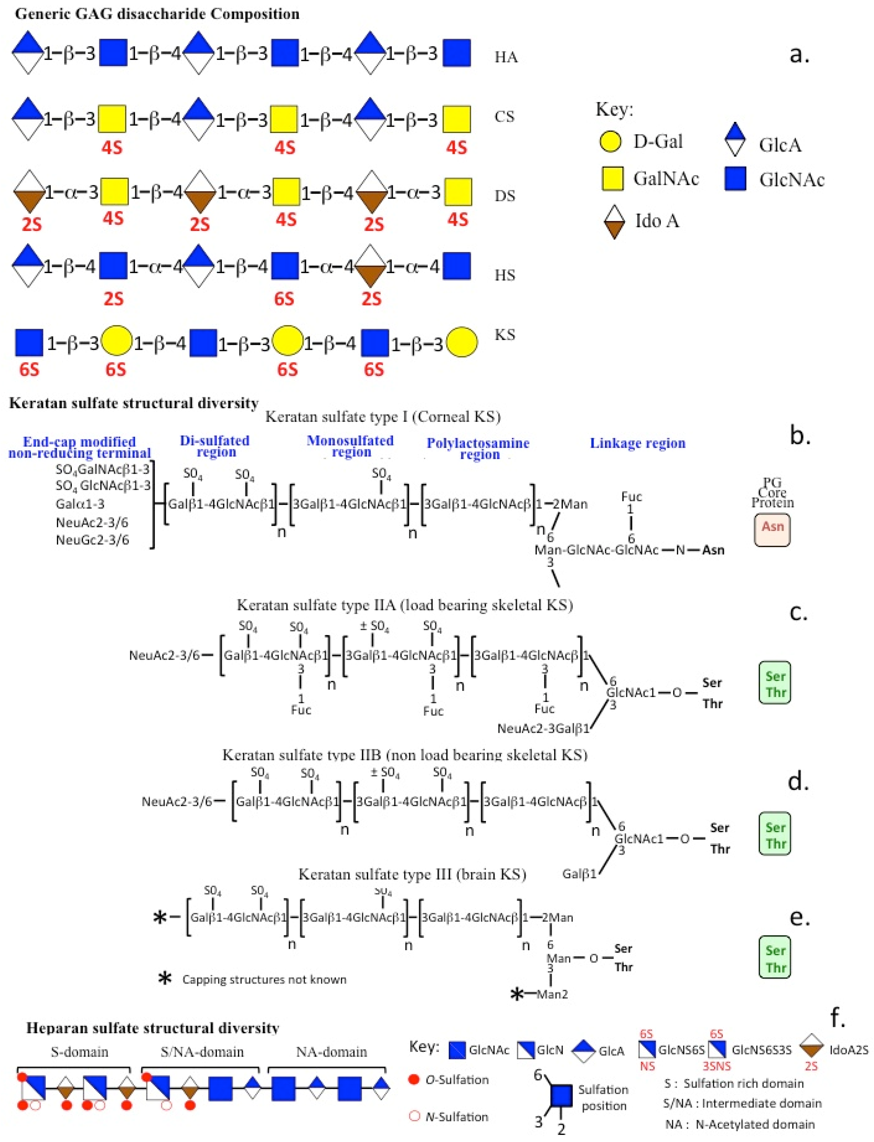
1.4. KS Chain Heterogeneity in a Range of PGs and Mucins
1.5. Low-Sulfation KS-PGs and Glycoconjugates
2. Diversity of KS-PG Functional Roles in Health and Disease
2.1. Aggrecan
2.2. SV2
2.3. Phosphacan
2.4. Podocalyxcin
2.5. Lumican and Fibromodulin
2.6. Keratocan
2.7. Prolargin/PRELP
2.8. Chondroadherin
2.9. Osteomodulin
2.10. Osteoglycin
3. HS Biodiversity
| Antibody Clone | Tissue Antibody and Epitope Reactivities | Refs. |
|---|---|---|
| HepSS1 | Predominantly localised to tissues rich in basement membrane. | [231] |
| JM13 | Predominantly localised to tissues rich in basement membrane. JM13 binding epitope requires the presence of 2-O-sulfated glucuronic acid residues | [232] |
| JM403 | Immunolocalised to basement membrane and some cell surface epitopes, in bovine lung, aorta and Human aorta. JM403 binding epitope is critically dependent on N-unsubstituted GlcN residues, | [233] |
| 10E4 | Immunolocalised to basement membrane and some cell surface epitope in human aorta, bovine intestine and kidney. The 10E4 epitope requires N-unsubstituted glucosamine residues. 10E4 binds to native “mixed” HS domains containing both N-acetylated and N-sulfated disaccharide units Ab reactivity is destroyed by heparinase III digestion | [234] |
| 3G10 | HS neoepitope generated by heparinase III ldigestion of HS chains. The 3G10 desaturated uronate stub epitope is attached to a HS disaccharide unit attached to the reducing terminal HS linkage region to core protein. | [234] |
| MAb865 | N-acetylated regions in HS | [235,236] |
| JM72 | HS-PG core protein epitope | [232,237] |
| Phage display antibodies | Antibody tissue immunoreactivity | |
| HS4C3 | HSMC3 shows strong localization in bovine intestine and kidney, O-and N- linked HS epitopes are important for Ab binding | [221] |
| HS4D10 | HS4D10 epitope immunoreactivity is strong in bovine kidney | |
| HS3G8 | HS3G8 immunoreactivity is strong in bovine kidney and intestine | |
| AO4B08 and HS4E4 | AO4B08 recognizes HS and heparin, interacting with ubiquitous, N-, 2-O-, and 6-O-sulfated saccharide units. HS4E4 preferentially recognized low-sulfated HS motifs containing idoA, N-sulfated and N-acetylated GlcN. | [238] |
4. The Diverse Functional Properties of HS-PGs
4.1. Agrin
4.2. Perlecan
4.3. Collagen XVIII
4.4. The Syndecan Family
4.5. The Glypican Family
4.6. Serglycin
4.7. The Neurexins
4.8. Pikachurin
4.9. Eyes-Shut
4.10. SPOCK
5. HS Functional Motifs That Provide Tissue Functions
6. CS Chain Biodiversity
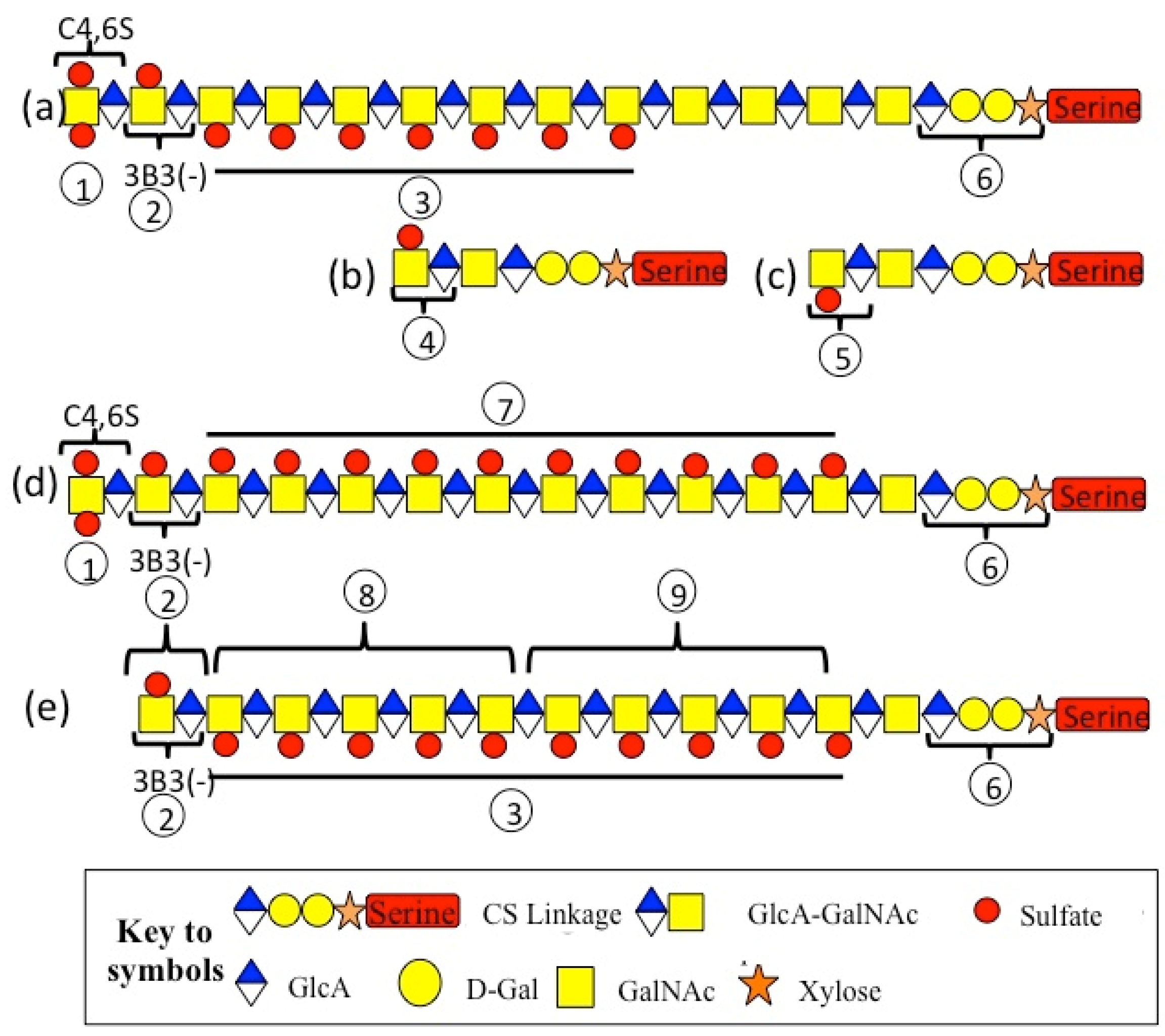
| Structural/Functional Diversity of the CS Side Chains of Aggrecan | ||
| Label | Structural/Functional Features of Annotated Regions | Refs. |
| 1 | Non-reducing terminal disulfated CS epitopes interactive with morphogens such as IHH. | [315,318,327] |
| 2 | New non-reducing terminus generated by HYAL4 digestion generates 3-B-3(−) epitope | [328] |
| 3 | C-4-S epitopes predominate in foetal cartilage | [319,329] |
| 4 | Reducing terminal 3-B-3 (+) stub epitopes attached to CS linkage region generated by chondroitinase ABC. | [330] |
| 5 | Reducing terminal 2-B-6 (+) stub epitopes attached to CS linkage region generated by chondroitinase ABC. | [330] |
| 6 | Linkage region to Serine residues on aggrecan core protein and GAG accepter region involved in the biosynthesis of CS chains. With ageing 6-sulfation levels in GalNAc increase in linkage region accompanied by increased Gal-6-S levels and lower 4-sulfation on GalNAc. | [331] |
| 7 | Increased C-6-S epitope levels in overloaded cartilage regions and with ageing and reduced levels in OA are due to alterations in the expression of sulfotransferases | [332,333,334,335] |
| 8 | Region of CS chain detected by MAb 4-C-3 | [317] |
| 9 | Region of CS chain detected by MAb 7-D-4 | [317] |
| Note. Regions 2, 6, 8, 9 of CS chains detected by MAb’s 3-B-3(−), 4-C-3, and 7-D-4 were identified by graded partial digestion of CS chains using chondroitinase ABC [317]. | ||
7. Variation in the CS Chain Fine Structure in Development and Pathology
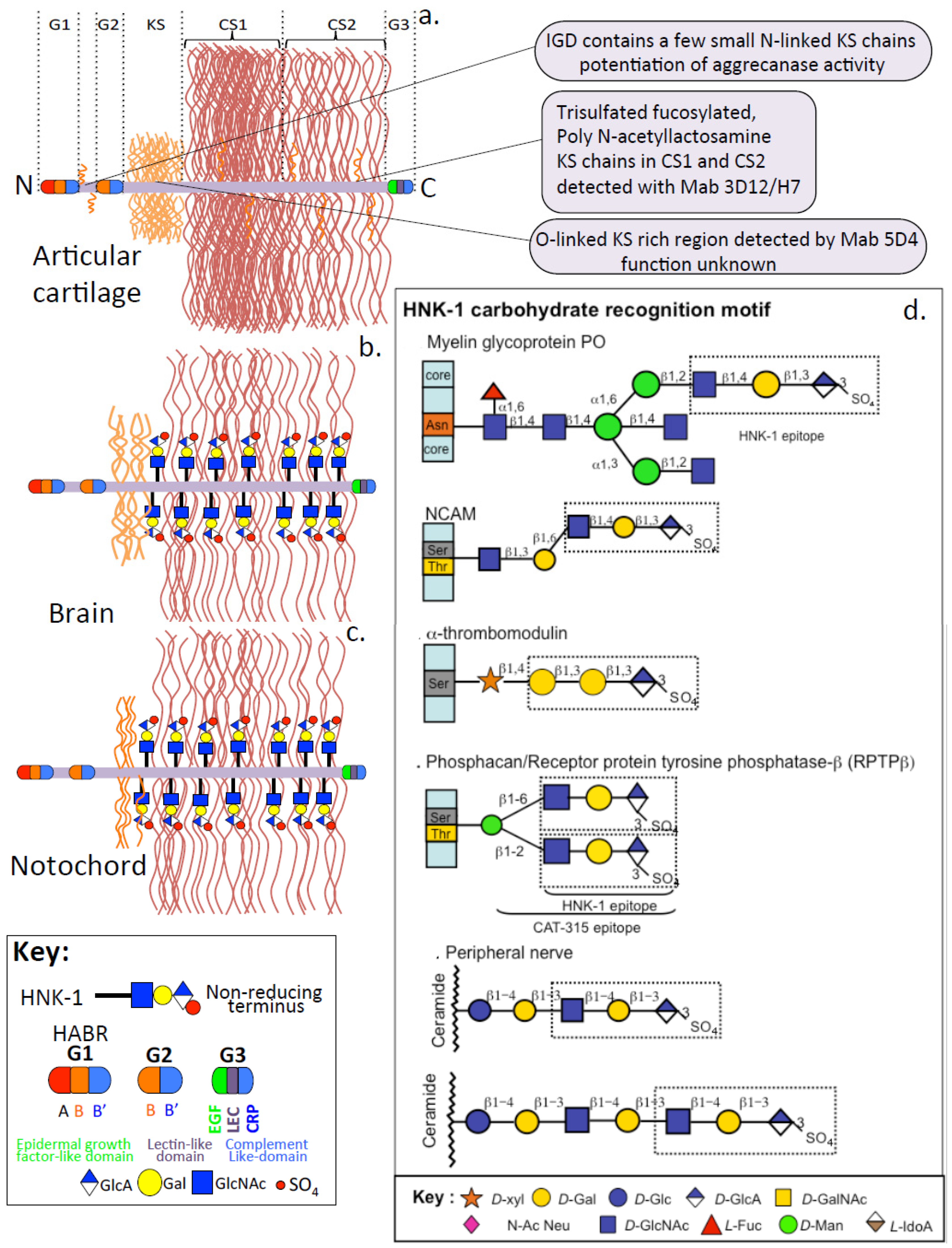
7.1. Aggrecan CS Side Chain Modifications in Specific Tissue Contexts
7.2. Role of IHH in Chondrogenesis
7.3. HNK-1 Trisaccharide

8. The Structural and Functional Diversity of KS, HS and CS-PGs
9. Interactive Properties of GAGs and How They Influence Tissue Development
9.1. Neural GAG Structures Have Significant Functional Roles in Synaptic Activity
HSPG-Specific Roles in Synaptic Stabilization, Specificity of Interaction and Plasticity
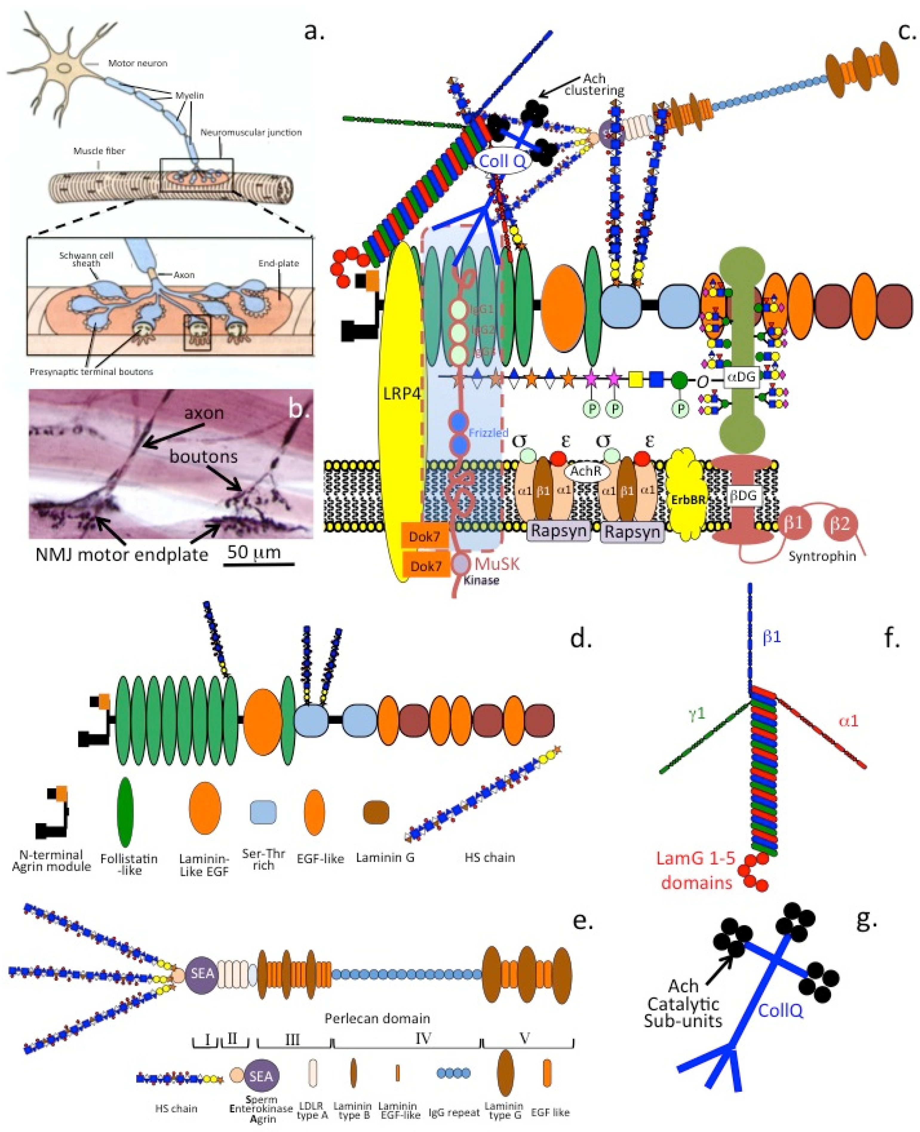
9.2. DG-HSPG Interactive Roles in NMJ Assembly and Neuromuscular Regulation in Health and Disease
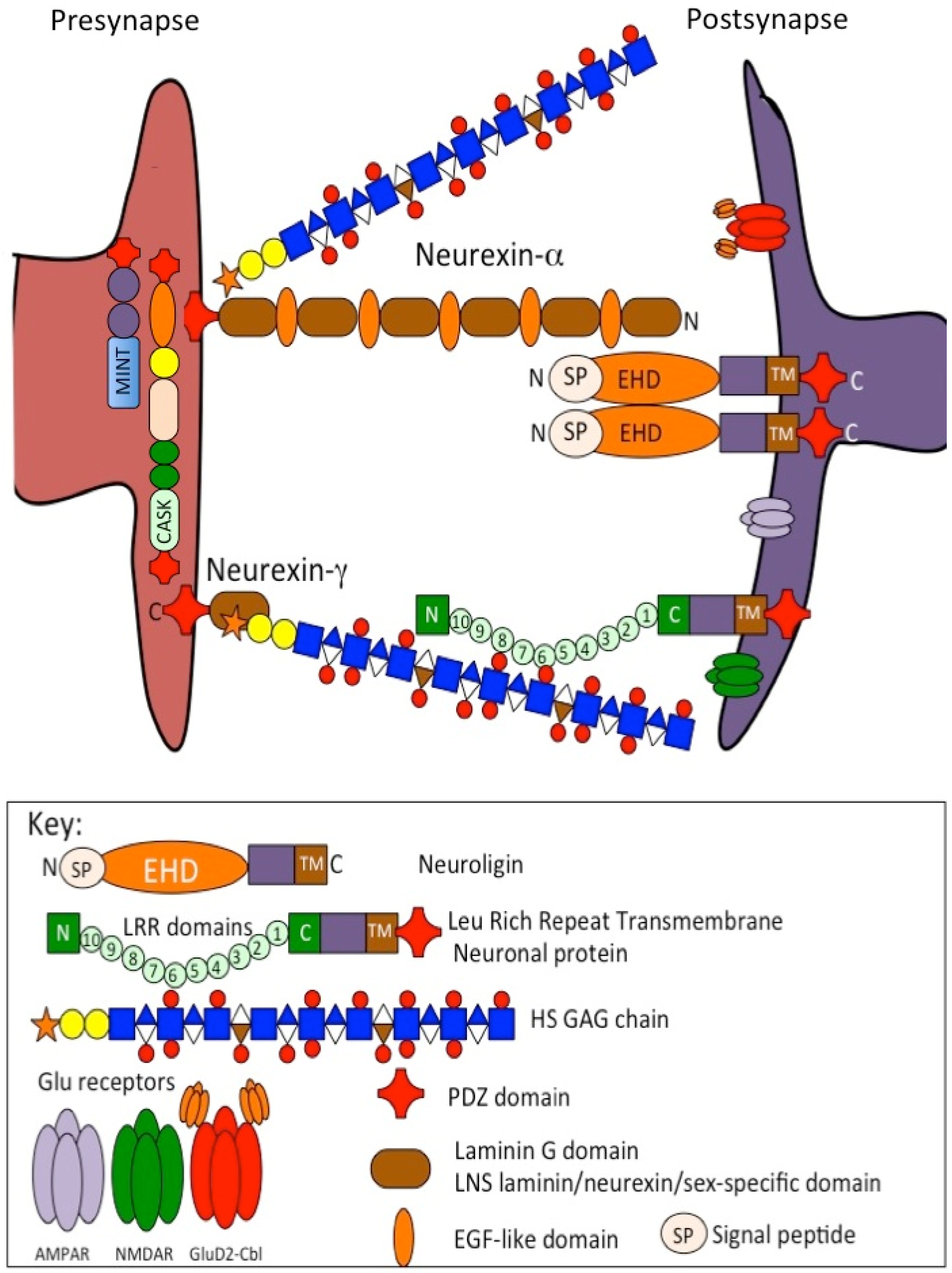

10. Conclusions
Funding
Conflicts of Interest
Abbreviations
| AS | Alport syndrome |
| AMPA | Alpha-amino-3-hydroxy-5-methylisoxazolepropionate |
| AMPAR | α-amino-3-hydroxy-5-methyl-4-isoxazolepropionic acid receptor |
| BMP | Bone morphogenetic protein |
| CASK | Calcium/calmodulin-dependent serine protein kinase |
| Cbl | Cerebellin. |
| CNS | Central nervous system |
| DSD1 | Mouse homolog of phosphacan |
| ECM | ECM |
| FGF(R) | Fibroblast growth factor (receptor) |
| FMOD | Fibromodulin |
| F3 | Contactin |
| GABA | Gamma amino butyric acid |
| GAG | Glycosaminoglycan |
| GlcAT-P | Glucuronyltransferase, B3gat1 |
| GluD2 | Glutamate dehydrogenase-2 (GluA2) |
| GTPase | Guanosine triphosphate hydrolase |
| PG | Proteoglycan |
| HA | Hyaluronan |
| HNK | Human natural killer |
| IHH | Indian hedgehog |
| IVD | Intervertebral disc |
| KERA | Keratocan |
| KSPGs | Keratan sulfate proteoglycans |
| LamG | Laminin G domain |
| L1 | A 200–220 kDa neuronal cell adhesion molecule |
| LUM | Lumican |
| MINT | Molecular interaction |
| MUC | Mucin |
| MuSK | Muscle-specific receptor tyrosine kinase |
| NgCAM | Neural glial cell adhesion molecule |
| NCAM | Neural cell adhesion molecule |
| NMDAR | N-methyl-D-aspartate receptor |
| NMJ | Neuromuscular junction |
| PEN5 | natural killer cell restricted KS-glycoprotein |
| PGSL-1 | Platelet selectin glycoprotein ligand-1 |
| PNS | Peripheral nervous system |
| PTHrP | parathyroid hormone-related peptide |
| SHH | Sonic hedgehog |
| SLRPs | Small leucine rich proteoglycans |
| SMO | Snoothened |
| PRELP | Prolargin |
| PSD-95 | Postsynaptic density protein 95, SAP-90 (synapse protein 90) |
| PSI | Phosphacan short isoform |
| Robo | Roundabout (receptor) |
| RPTP-β | Transmembrane receptor-type protein tyrosine phosphatase-beta, also known as protein tyrosine phosphatase-zeta (PTP-ζ) |
| Sema5A | Semaphorin-5A |
| TSP-1 | Thrombospondin-1 |
| VEGF | Vascular endothelial cell growth factor |
| Wnt | Derived from int and Wg, Wingless-related integration site |
References
- Farach-Carson, M.; Wu, D.; França, C.M. Proteoglycans in Mechanobiology of Tissues and Organs: Normal Functions and Mechanopathology. Proteoglycan Res. 2024, 2, e21. [Google Scholar] [CrossRef] [PubMed]
- Krull, C.; Rife, J.; Klamer, B.; Purmessur, D.; Walter, B.A. Pericellular heparan sulfate proteoglycans: Role in regulating the biosynthetic response of nucleus pulposus cells to osmotic loading. JOR Spine 2022, 5, e1209. [Google Scholar] [CrossRef]
- Guilak, F.; Hayes, A.J.; Melrose, J. Perlecan in Pericellular Mechanosensory Cell-Matrix Communication, Extracellular Matrix Stabilisation and Mechanoregulation of Load-Bearing Connective Tissues. Int. J. Mol. Sci. 2021, 22, 2716. [Google Scholar] [CrossRef]
- Ingber, D.; Wang, N.; Stamenovic, D. Tensegrity, cellular biophysics, and the mechanics of living systems. Rep. Prog. Phys. 2014, 77, 046603. [Google Scholar] [CrossRef] [PubMed]
- Berdiaki, A.; Neagu, M.; Tzanakakis, P.; Spyridaki, I.; Pérez, S.; Nikitovic, D. Extracellular Matrix Components and Mechanosensing Pathways in Health and Disease. Biomolecules 2024, 14, 1186. [Google Scholar] [CrossRef]
- Savransky, S.; White, A.D.; Vilardaga, J.P. Deciphering the role of glycosaminoglycans in GPCR signaling. Cell Signal. 2024, 118, 111149. [Google Scholar] [CrossRef]
- Karamanos, N.; Theocharis, A.D.; Piperigkou, Z.; Manou, D.; Passi, A.; Skandalis, S.S.; Vynios, D.H.; Orian-Rousseau, V.; Ricard-Blum, S.; Schmelzer, C.E.H.; et al. A guide to the composition and functions of the extracellular matrix. FEBS J. 2021, 288, 6850–6912. [Google Scholar] [CrossRef] [PubMed]
- Theocharis, A.; Skandalis, S.S.; Gialeli, C.; Karamanos, N.K. Extracellular matrix structure. Adv. Drug Deliv. Rev. 2016, 97, 4–27. [Google Scholar] [CrossRef]
- Lepucki, A.; Orlińska, K.; Mielczarek-Palacz, A.; Kabut, J.; Olczyk, P.; Komosińska-Vassev, K. The Role of Extracellular Matrix Proteins in Breast Cancer. J. Clin. Med. 2022, 11, 1250. [Google Scholar] [CrossRef]
- Dzobo, K.; Dandara, C. The Extracellular Matrix: Its Composition, Function, Remodeling, and Role in Tumorigenesis. Biomimetics 2023, 8, 146. [Google Scholar] [CrossRef]
- Melrose, J. Hippo cell signaling and HS-proteoglycans regulate tissue form and function, age-dependent maturation, extracellular matrix remodeling, and repair. Am. J. Physiol. Cell Physiol. 2024, 326, C810–C828. [Google Scholar] [CrossRef]
- Scott, R.; Panitch, A. Glycosaminoglycans in biomedicine. Wiley Interdiscip. Rev. Nanomed. Nanobiotechnol. 2013, 5, 388–398. [Google Scholar] [CrossRef] [PubMed]
- Melrose, J. Dystroglycan-HSPG interactions provide synaptic plasticity and specificity. Glycobiology 2024, 34, cwae051. [Google Scholar] [CrossRef] [PubMed]
- Hopkinson, M.; Pitsillides, A.A. Extracellular matrix: Dystroglycan interactions-Roles for the dystrophin-associated glycoprotein complex in skeletal tissue dynamics. Int. J. Exp. Pathol. 2025, 106, e12525. [Google Scholar] [CrossRef]
- Südhof, T. Synaptic Neurexin Complexes: A Molecular Code for the Logic of Neural Circuits. Cell 2017, 171, 745–769. [Google Scholar] [CrossRef] [PubMed]
- Noborn, F.; Sterky, F.H. Role of neurexin heparan sulfate in the molecular assembly of synapses—Expanding the neurexin code? FEBS J. 2021, 290, 252–265. [Google Scholar] [CrossRef]
- Wight, T.N. A role for proteoglycans in vascular disease. Matrix Biol. 2018, 71–72, 396–420. [Google Scholar] [CrossRef]
- Schwartz, N.; Domowicz, M.S. Chemistry and Function of Glycosaminoglycans in the Nervous System. Adv. Neurobiol. 2023, 29, 117–162. [Google Scholar] [CrossRef]
- Zhang, P.; Lu, H.; Peixoto, R.T.; Pines, M.K.; Ge, Y.; Oku, S.; Siddiqui, T.J.; Xie, Y.; Wu, W.; Archer-Hartmann, S.; et al. Heparan Sulfate Organizes Neuronal Synapses through Neurexin Partnerships. Cell 2018, 174, 1450–1464.e23. [Google Scholar] [CrossRef]
- Melrose, J.; Hayes, A.J.; Bix, G. The CNS/PNS Extracellular Matrix Provides Instructive Guidance Cues to Neural Cells and Neuroregulatory Proteins in Neural Development and Repair. Int. J. Mol. Sci. 2021, 22, 5583. [Google Scholar] [CrossRef]
- Melrose, J. CNS/PNS proteoglycans functionalize neuronal and astrocyte niche microenvironments optimizing cellular activity by preserving membrane polarization dynamics, ionic microenvironments, ion fluxes, neuronal activation, and network neurotransductive capacity. J. Neurosci. Res. 2024, 102, e25361. [Google Scholar] [CrossRef] [PubMed]
- Ricard-Blum, S.; Vivès, R.R.; Schaefer, L.; Götte, M.; Merline, R.; Passi, A.; Heldin, P.; Magalhães, A.; Reis, C.A.; Skandalis, S.S.; et al. A biological guide to glycosaminoglycans: Current perspectives and pending questions. FEBS J. 2024, 291, 3331–3366. [Google Scholar] [CrossRef] [PubMed]
- Salmivirta, M.; Jalkanen, M. Syndecan family of cell surface proteoglycans: Developmentally regulated receptors for extracellular effector molecules. Experientia 1995, 51, 863–872. [Google Scholar] [CrossRef]
- Bernfield, M.; Sanderson, R.D. Syndecan, a developmentally regulated cell surface proteoglycan that binds extracellular matrix and growth factors. Philos. Trans. R. Soc. Lond. B Biol. Sci. 1990, 327, 171–186. [Google Scholar] [CrossRef]
- Matsuo, I.; Kimura-Yoshida, C. Extracellular distribution of diffusible growth factors controlled by heparan sulfate proteoglycans during mammalian embryogenesis. Philos. Trans. R. Soc. Lond. B Biol. Sci. 2014, 369, 20130545. [Google Scholar] [CrossRef]
- Thota, L.; Chignalia, A.Z. The role of the glypican and syndecan families of heparan sulfate proteoglycans in cardiovascular function and disease. Am. J. Physiol. Cell Physiol. 2022, 323, C1052–C1060. [Google Scholar] [CrossRef]
- Couchman, J. Transmembrane signaling proteoglycans. Annu. Rev. Cell Dev. Biol. 2010, 26, 89–114. [Google Scholar] [CrossRef] [PubMed]
- Tracy, L.; Minasian, R.A.; Caterson, E.J. Extracellular Matrix and Dermal Fibroblast Function in the Healing Wound. Adv. Wound Care 2016, 5, 119–136. [Google Scholar] [CrossRef]
- Afratis, N.; Nikitovic, D.; Multhaupt, H.A.; Theocharis, A.D.; Couchman, J.R.; Karamanos, N.K. Syndecans—Key regulators of cell signaling and biological functions. FEBS J. 2017, 284, 27–41. [Google Scholar] [CrossRef]
- Zeng, Y. Endothelial glycocalyx as a critical signalling platform integrating the extracellular haemodynamic forces and chemical signalling. J. Cell. Mol. Med. 2017, 21, 1457–1462. [Google Scholar] [CrossRef]
- Couchman, J.; Gopal, S.; Lim, H.C.; Nørgaard, S.; Multhaupt, H.A. Fell-Muir Lecture: Syndecans: From peripheral coreceptors to mainstream regulators of cell behaviour. Int. J. Exp. Pathol. 2015, 96, 1–10. [Google Scholar] [CrossRef] [PubMed]
- Chung, H.; Multhaupt, H.A.; Oh, E.S.; Couchman, J.R. Minireview: Syndecans and their crucial roles during tissue regeneration. FEBS Lett. 2016, 590, 2408–2417. [Google Scholar] [CrossRef]
- De Rossi, G.; Whiteford, J.R. Syndecans in angiogenesis and endothelial cell biology. Biochem. Soc. Trans. 2014, 42, 1643–1646. [Google Scholar] [CrossRef] [PubMed]
- Melrose, J. Mucin-like glycopolymer gels in electrosensory tissues generate cues which direct electrolocation in amphibians and neuronal activation in mammals. Neural Regen. Res. 2019, 14, 1191–1195. [Google Scholar] [CrossRef]
- Josberger, E.; Hassanzadeh, P.; Deng, Y.; Sohn, J.; Rego, M.J.; Amemiya, C.T.; Rolandi, M. Proton conductivity in ampullae of Lorenzini jelly. Sci. Adv. 2016, 2, e1600112. [Google Scholar] [CrossRef]
- Zhang, X.; Xia, K.; Lin, L.; Zhang, F.; Yu, Y.; St Ange, K.; Han, X.; Edsinger, E.; Sohn, J.; Linhardt, R.J. Structural and Functional Components of the Skate Sensory Organ Ampullae of Lorenzini. ACS Chem. Biol. 2018, 13, 1677–1685. [Google Scholar] [CrossRef] [PubMed]
- Melrose, J. Functional Consequences of Keratan Sulfate Sulfation in Electrosensory Tissues and in Neuronal Regulation. Adv. Biosyst. 2019, 3, e1800327. [Google Scholar] [CrossRef]
- Haueisen, M.; Reis, R.E. High resolution in turbid waters: Ampullae of lorenzini in the daggernose shark Carcharhinus oxyrhynchus (Valenciennes, 1839) (Elasmobranchii: Carcharhinidae). J. Fish Biol. 2023, 105, 1540–1554. [Google Scholar] [CrossRef]
- Pedraja, F.; Sawtell, N.B. Collective sensing in electric fish. Nature 2024, 628, 139–144. [Google Scholar] [CrossRef]
- Jones, T.; Allen, K.M.; Moss, C.F. Communication with self, friends and foes in active-sensing animals. J. Exp. Biol. 2021, 224, jeb242637. [Google Scholar] [CrossRef]
- Xu, J.; Cui, X.; Zhang, H. The third form electric organ discharge of electric eels. Sci. Rep. 2021, 11, 6193. [Google Scholar] [CrossRef]
- Proske, U.; Gregory, J.E.; Iggo, A. Sensory receptors in monotremes. Philos. Trans. R. Soc. Lond. B Biol. Sci. 1998, 353, 1187–1198. [Google Scholar] [CrossRef] [PubMed]
- Scheich, H.; Langner, G.; Tidemann, C.; Coles, R.B.; Guppy, A. Electroreception and electrolocation in platypus. Nature 1986, 319, 401–402. [Google Scholar] [CrossRef] [PubMed]
- Gramegna Tota, C.; Leone, A.; Paganini, C.; Khan, A.; Rossi, A.; Superti-Furga, A. Chapter 6. Skeletal dysplasias caused by defects in glycosaminoglycan sulfation. In The Extracellular Matrix in Genetic Skeletal Disorders; Biology of Extracellular Matrix; Rossi, A., Zaucke, F., Eds.; Springer: Berlin/Heidelberg, Germany, 2025; Volume 16, pp. 181–212. [Google Scholar]
- Watt, A.; Chung, K.C. Generalized skeletal abnormalities. Hand Clin. 2009, 25, 265–276. [Google Scholar] [CrossRef]
- Rimoin, D.; Cohn, D.; Krakow, D.; Wilcox, W.; Lachman, R.S.; Alanay, Y. The skeletal dysplasias: Clinical-molecular correlations. Ann. N. Y. Acad. Sci. 2007, 1117, 302–309. [Google Scholar] [CrossRef] [PubMed]
- Caterson, B.; Melrose, J. Keratan sulfate, a complex glycosaminoglycan with unique functional capability. Glycobiology 2018, 28, 182–206. [Google Scholar] [CrossRef]
- Tai, G.; Huckerby, T.N.; Nieduszynski, I.A. Multiple non-reducing chain termini isolated from bovine corneal keratan sulfates. J. Biol. Chem. 1996, 271, 23535–23546. [Google Scholar] [CrossRef]
- Tai, G.; Nieduszynski, I.A.; Fullwood, N.J.; Huckerby, T.N. Human corneal keratan sulfates. J. Biol. Chem. 1997, 272, 28227–28231. [Google Scholar] [CrossRef]
- Oeben, M.; Keller, R.; Stuhlsatz, H.W.; Greiling, H. Constant and variable domains of different disaccharide structure in corneal keratan sulphate chains. Biochem. J. 1987, 248, 85–93. [Google Scholar] [CrossRef]
- Funderburgh, J. Keratan sulfate biosynthesis. IUBMB Life 2002, 54, 187–194. [Google Scholar] [CrossRef]
- Meyer, K.; Linker, A.; Davidson, E.A.; Weissmann, B. The mucopolysaccharides of bovine cornea. J. Biol. Chem. 1953, 205, 611–616. [Google Scholar] [CrossRef] [PubMed]
- Antonsson, P.; Heinegård, D.; Oldberg, A. Posttranslational modifications of fibromodulin. J. Biol. Chem. 1991, 266, 6859–16861. [Google Scholar] [CrossRef]
- Sommarin, Y.; Wendel, M.; Shen, Z.; Hellman, U.; Heinegard, D. Osteoadherin, a cell-binding keratan sulfate proteoglycan in bone, belongs to the family of leucine-rich repeat proteins of the extracellular matrix. J. Biol. Chem. 1998, 273, 16723–16729. [Google Scholar] [CrossRef] [PubMed]
- Nieduszynski, I.; Huckerby, T.N.; Dickenson, J.M.; Brown, G.M.; Tai, G.H.; Morris, H.G.; Eady, S. There are two major types of skeletal keratan sulphates. Biochem. J. 1990, 271, 243–245. [Google Scholar] [CrossRef]
- Skandalis, S.; Theocharis, A.D.; Vynios, D.H.; Theocharis, D.A.; Papageorgakopoulou, N. Proteoglycans in human laryngeal cartilage. Identification of proteoglycan types in successive cartilage extracts with particular reference to aggregating proteoglycans. Biochimie 2004, 86, 221–229. [Google Scholar] [CrossRef] [PubMed]
- Kiani, C.; Chen, L.; Wu, Y.J.; Yee, A.J.; Yang, B.B. Structure and function of aggrecan. Cell Res. 2002, 12, 19–32. [Google Scholar] [CrossRef]
- Fischer, D.; Haubeck, H.D.; Eich, K.; Kolbe-Busch, S.; Stocker, G.; Stuhlsatz, H.W.; Greiling, H. A novel keratan sulphate domain preferentiallyexpressed on the large aggregating proteoglycan from human articularcartilage is recognized by the monoclonal antibody 3D12/H7. Biochem. J. 1996, 318, 1051–1056. [Google Scholar] [CrossRef]
- Melrose, J. Keratan sulfate (KS)-proteoglycans and neuronal regulation in health and disease: The importance of KS-glycodynamics and interactive capability with neuroregulatory ligands. J. Neurochem. 2019, 149, 170–194. [Google Scholar] [CrossRef]
- Funderburgh, J. Keratan sulfate: Structure, biosynthesis, and function. Glycobiology 2000, 19, 951–958. [Google Scholar] [CrossRef]
- Hanisch, F.; Uhlenbruck, G.; Peter-Katalinic, J.; Egge, H.; Dabrowski, U.; Dabrowski, J. Unbranched polylactosamino-O-glycans on human skim milk mucins exhibit Gal beta(1-4)GlcNAc beta(1-6) repeating units. Symp. Soc. Exp. Biol. 1989, 43, 155–162. [Google Scholar]
- Craig, F.; Ralphs, J.R.; Bentley, G.; Archer, C.W. MZ15, a monoclonal antibody recognizing keratan sulphate, stains chick tendon. Histochem. J. 1987, 19, 651–657. [Google Scholar] [CrossRef] [PubMed]
- Hayes, A.; Melrose, J. Keratan Sulphate in the Tumour Environment. Adv. Exp. Med. Biol. 2020, 1245, 39–66. [Google Scholar] [CrossRef]
- Leiphrakpam, P.; Patil, P.P.; Remmers, N.; Swanson, B.; Grandgenett, P.M.; Qiu, F.; Yu, F.; Radhakrishnan, P. Role of keratan sulfate expression in human pancreatic cancer malignancy. Sci. Rep. 2019, 9, 9665. [Google Scholar] [CrossRef] [PubMed]
- Kato, Y.; Hayatsu, N.; Kaneko, M.K.; Ogasawara, S.; Hamano, T.; Takahashi, S.; Nishikawa, R.; Matsutani, M.; Mishima, K.; Narimatsu, H. Increased expression of highly sulfated keratan sulfate synthesized in malignant astrocytic tumors. Biochem. Biophys. Res. Commun. 2008, 369, 1041–1046. [Google Scholar] [CrossRef]
- Hayatsu, N.; Ogasawara, S.; Kaneko, M.K.; Kato, Y.; Narimatsu, H. Expression of highly sulfated keratan sulfate synthesized in human glioblastoma cells. Biochem. Biophys. Res. Commun. 2008, 368, 217–222. [Google Scholar] [CrossRef]
- Aplin, J.; Hey, N.A.; Graham, R.A. Human endometrial MUC1 carries keratan sulfate: Characteristic glycoforms in the luminal epithelium at receptivity. Glycobiology 1998, 8, 269–276. [Google Scholar] [CrossRef] [PubMed]
- Takahashi, K.; Stamenkovic, I.; Cutler, M.; Dasgupta, A.; Tanabe, K.K. Keratan Sulfate Modification of CD44 Modulates Adhesion to Hyaluronate. J. Biol. Chem. 1996, 271, 9490–9496. [Google Scholar] [CrossRef]
- Caterson, B.; Christner, J.E.; Baker, J.R. Identification of a monoclonal antibody that specifically recognizes corneal and skeletal keratan sulfate. Monoclonal antibodies to cartilage proteoglycan. J. Biol. Chem. 1983, 258, 8848–8854. [Google Scholar] [CrossRef]
- Mehmet, H.; Scudder, P.; Tang, P.W.; Hounsell, E.F.; Caterson, B.; Feizi, T. The antigenic determinants recognized by three monoclonal antibodies to keratan sulphate involve sulphated hepta- or larger oligosaccharides ofthe poly(N-acetyllactosamine) series. Eur. J. Biochem. 1986, 157, 385–391. [Google Scholar] [CrossRef]
- Nakao, H.; Nagai, Y.; Kojima, A.; Toyoda, H.; Kawasaki, N.; Kawasaki, T. Binding specificity of R-10G and TRA-1-60/81, and substrate specificityof keratanase II studied with chemically synthesized oligosaccharides. Glycoconj. J. 2017, 34, 789–795. [Google Scholar] [CrossRef]
- Makanga, J.; Kobayashi, M.; Ikeda, H.; Christianto, A.; Toyoda, H.; Yamada, M.; Kawasaki, T.; Inazu, T. Generation of rat induced pluripotent stemcells using a plasmid vector and possible application of a keratan sulfateglycan recognizing antibody in discriminating teratoma formation pheno-types. Biol. Pharm. Bull. 2015, 38, 127–133. [Google Scholar] [CrossRef] [PubMed]
- Kawabe, K.; Tateyama, D.; Toyoda, H.; Kawasaki, N.; Hashii, N.; Nakao, H.; Matsumoto, S.; Nonaka, M.; Matsumura, H.; Hirose, Y.; et al. A novel antibody for human induced pluripotent stem cells and embryonic stem cells recognizes a type of keratan sulfate lacking oversulfated structures. Glycobiology 2013, 23, 322–336. [Google Scholar] [CrossRef]
- Hoshino, H.; Chen, Y.Y.; Inoue, D.; Yoshida, Y.; Khoo, K.H.; Akama, T.O.; Kobayashi, M. Expression of low-sulfated keratan sulfate in non-mucinous ovarian carcinoma. Glycobiology 2024, 34, cwad056. [Google Scholar] [CrossRef] [PubMed]
- Muramoto, A.; Hoshino, H.; Inamura, S.; Murahashi, M.; Akama, T.O.; Terada, N.; Kobayashi, M. Expression of Podocalyxin Potentially Decorated with Low-sulfated Keratan Sulfate in Human Testicular Embryonal Carcinoma. J. Histochem. Cytochem. 2024, 72, 453–465. [Google Scholar] [CrossRef]
- Baker, J.; Walker, T.; Morrison, K.; Neame, P.; Christner, J. The specificity of a mouse monoclonal antibody to human aorta proteoglycans. Matrix 1989, 9, 92–98. [Google Scholar] [CrossRef]
- Sinouris, E.; Skandalis, S.S.; Kilia, V.; Theocharis, A.D.; Theocharis, D.A.; Ravazoula, P.; Vynios, D.H.; Papageorgakopoulou, N. Keratan sulfate-containing proteoglycans in sheep brain with particular reference to phosphacan and synaptic vesicle proteoglycan isoforms. Biomed. Chromatogr. 2009, 23, 455–463. [Google Scholar] [CrossRef]
- Scranton, T.; Iwata, M.; Carlson, S.S. The SV2 protein of synaptic vesicles is a keratan sulfate proteoglycan. J. Neurochem. 1993, 61, 29–44. [Google Scholar] [CrossRef]
- Papageorgakopoulou, N.; Theocharis, A.D.; Skandalis, S.S.; Vynios, D.H.; Theocharis, D.A.; Tsiganos, C.P. Immunological studies of sheep brain keratan sulphate proteoglycans. Biochimie 2002, 84, 1225–1228. [Google Scholar] [CrossRef] [PubMed]
- Okumura, M.; Tagami, M.; Fujinaga, T. Consideration of the role of antigenic keratan sulphate reacting to a 1/14/16H9 antibody as a molecular marker to monitor cartilage metabolism in horses. J. Vet. Med. Sci. 2000, 62, 281–285. [Google Scholar] [CrossRef]
- Okumura, M.; Fujinaga, T. Establishment of a monoclonal antibody (1/14/16H9) for detection of equine keratan sulfate. Am. J. Vet. Res. 1998, 59, 1203–1208. [Google Scholar] [CrossRef]
- Hayes, A.; Melrose, J. Immunolocalization of Keratan Sulfate in Rat Spinal Tissues Using the Keratanase Generated BKS-1(+) Neoepitope: Correlation of Expression Patterns with the Class II SLRPs, Lumican and Keratocan. Cells 2020, 9, 826. [Google Scholar] [CrossRef] [PubMed]
- Adewumi, O.; Aflatoonian, B.; Ahrlund-Richter, L.; Amit, M.; Andrews, P.W.; Beighton, G.; Bello, P.A.; Benvenisty, N.; Berry, L.S.; Bevan, S.; et al. Characterization of human embryonic stem cell lines by the International Stem Cell Initiative. Nat. Biotechnol. 2007, 25, 803–816. [Google Scholar] [PubMed]
- Andrews, P.; Banting, G.; Damjanov, I.; Arnaud, D.; Avner, P. Three monoclonal antibodies defining distinct differentiation antigens associated with different high molecular weight polypeptides on the surface of human embryonal carcinoma cells. Hybridoma 1984, 3, 347–361. [Google Scholar] [CrossRef]
- Badcock, G.; Pigott, C.; Goepel, J.; Andrews, P.W. The human embryonal carcinoma marker antigen TRA-1-60 is a sialylated keratan sulfate proteoglycan. Cancer Res. 1999, 59, 4715–4719. [Google Scholar] [PubMed]
- Natunen, S.; Satomaa, T.; Pitkanen, V.; Salo, H.; Mikkola, M.; Natunen, J.; Otonkoski, T.; Valmu, L. The binding specificity of the marker antibodies Tra-1-60 and Tra-1-81 reveals a novel pluripotency-associated type 1 lactosamine epitope. Glycobiology 2011, 21, 1125–1130. [Google Scholar] [CrossRef]
- Schopperle, W.; DeWolf, W.C. The TRA-1-60 and TRA-1-81 human pluripotent stem cell markers are expressed on podocalyxin in embryonal carcinoma. Stem Cells 2007, 25, 723–730. [Google Scholar] [CrossRef]
- Kaneko, M.; Ohishi, T.; Kawada, M.; Kato, Y. A cancer-specific anti-podocalyxin monoclonal antibody (60-mG2a-f) exerts antitumor effects in mouse xenograft models of pancreatic carcinoma. Biochem. Biophys. Rep. 2020, 24, 100826. [Google Scholar] [CrossRef]
- Ozawa, M.; Muramatsu, T.; Solter, D. SSEA-1, a stage-specific embryonic antigen of the mouse, is carried by the glycoprotein-bound large carbohydrate in embryonal carcinoma cells. Cell Differ. 1985, 16, 169–173. [Google Scholar] [CrossRef]
- Feizi, T.; Kabat, E.A.; Vicari, G.; Anderson, B.; Marsh, W.L. Immunochemical studies on blood groups.XLIX. The I antigen complex: Specificity differences among anti-I sera revealed by quantitative precipitin studies; partial structure of the I determinant specific for one anti-I serum. J. Immunol. 1971, 106, 1578–1592. [Google Scholar] [CrossRef]
- Feizi, T.; Childs, R.A.; Watanabe, K.; Hakomori, S.I. Three types of blood group I specificity among monoclonal anti-I autoantibodies revealed by analogues of a branched erythrocyte glycolipid. J. Exp. Med. 1979, 149, 975–980. [Google Scholar] [CrossRef]
- Young, R.; Akama, T.O.; Liskova, P.; Ebenezer, N.D.; Allan, B.; Kerr, B.; Caterson, B.; Fukuda, M.N.; Quantock, A.J. Differential immunogold localisation of sulphated and unsulphated keratan sulphate proteoglycans in normal and macular dystrophy cornea using sulphation motif-specific antibodies. Histochem. Cell Biol. 2007, 127, 115–120. [Google Scholar] [CrossRef] [PubMed]
- Young, R.; Gealy, E.C.; Liles, M.; Caterson, B.; Ralphs, J.R.; Quantock, A.J. Keratan sulfate glycosaminoglycan and the association with collagen fibrils in rudimentary lamellae in the developing avian cornea. Investig. Ophthalmol. Vis. Sci. 2007, 48, 3083–3088. [Google Scholar] [CrossRef]
- Fukuma, M.; Abe, H.; Okita, H.; Yamada, T.; Hata, J. Monoclonal antibody 4C4-mAb specifically recognizes keratan sulphate proteoglycan on human embryonal carcinoma cells. J. Pathol. 2003, 201, 90–98. [Google Scholar] [CrossRef]
- Symbol Nomenclature for Glycans (SNFG). Symbol Nomenclature for Graphical Representation of Glycans. Glycobiology 2015, 25, 1323–1324. [Google Scholar] [CrossRef]
- Symbol Nomenclature for Glycans (SNFG). Updates to the Symbol Nomenclature for Glycans guidelines. Glycobiology 2019, 29, 620–624. [Google Scholar] [CrossRef]
- Johnson, J.; Young, T.L.; Rada, J.A. Small leucine rich repeat proteoglycans (SLRPs) in the human sclera: Identification of abundant levels of PRELP. Mol. Vis. 2006, 12, 1057–1066. [Google Scholar] [PubMed]
- Barry, F.; Rosenberg, L.C.; Gaw, J.U.; Gaw, J.U.; Koob, T.J.; Neame, P.J. N- and O-linked keratan sulfate on the hyaluronan binding region of aggrecan from mature and immature bovine cartilage. J. Biol. Chem. 1995, 270, 20516–20524. [Google Scholar] [CrossRef] [PubMed]
- Fosang, A.; Last, K.; Poon, C.J.; Plaas, A.H. Keratan sulphate in the interglobular domain has a microstructure that is distinct from keratan sulphate elsewhere on pig aggrecan. Matrix Biol. 2009, 28, 53–61. [Google Scholar] [CrossRef]
- Hayashida, Y.; Akama, T.O.; Beecher, N.; Lewis, P.; Young, R.D.; Meek, K.M.; Kerr, B.; Hughes, C.; Caterson, B.; Tanigami, A.; et al. Matrix morphogenesis in cornea is mediated by the modification of keratan sulfate by GlcNAc 6-O-sulfotransferase. Proc. Natl. Acad. Sci. USA 2006, 103, 13333–13338. [Google Scholar] [CrossRef]
- Linden, S.; Sutton, P.; Karlsson, N.G.; Korolik, V.; McGuckin, M.A. Mucins in the mucosal barrier to infection. Mucosal Immunol. 2008, 1, 183–197. [Google Scholar] [CrossRef]
- Sun, L.; Zhang, Y.; Li, W.; Zhang, J.; Zhang, Y. Mucin Glycans: A Target for Cancer Therapy. Molecules 2023, 28, 7033. [Google Scholar] [CrossRef] [PubMed]
- Hollingsworth, M.; Swanson, B.J. Mucins in cancer: Protection and control of the cell surface. Nat. Rev. Cancer 2004, 4, 45–60. [Google Scholar] [CrossRef]
- Chugh, S.; Gnanapragassam, V.S.; Jain, M.; Rachagani, S.; Ponnusamy, M.P.; Batra, S.K. Pathobiological implications of mucin glycans in cancer: Sweet poison and novel targets. Biochim. Biophys. Acta 2015, 1856, 211–225. [Google Scholar] [CrossRef] [PubMed]
- Thomsson, K.; Vitiazeva, V.; Mateoiu, C.; Jin, C.; Liu, J.; Holgersson, J.; Weijdegård, B.; Sundfeldt, K.; Karlsson, N.G. Sulfation of O-glycans on Mucin-type Proteins From Serous Ovarian Epithelial Tumors. Mol. Cell. Proteom. 2021, 20, 100150. [Google Scholar] [CrossRef] [PubMed]
- Carpenter, J.; Kesimer, M. Membrane-bound mucins of the airway mucosal surfaces are densely decorated with keratan sulfate: Revisiting their role in the Lung’s innate defense. Glycobiology 2021, 31, 436–443. [Google Scholar] [CrossRef]
- Croce, M.; Rabassa, M.E.; Price, M.R.; Segal-Eiras, A. MUC1 mucin and carbohydrate associated antigens as tumor markers in head and neck squamous cell carcinoma. Pathol. Oncol. Res. 2001, 7, 284–291. [Google Scholar] [CrossRef]
- Wilczak, M.; Surman, M.; Przybyło, M. Altered Glycosylation in Progression and Management of Bladder Cancer. Molecules 2023, 28, 3436. [Google Scholar] [CrossRef]
- Melrose, J. Keratan Sulfate, An Electrosensory Neurosentient Bioresponsive Cell Instructive Glycosaminoglycan. Glycobiology 2024, 34, cwae014. [Google Scholar] [CrossRef]
- Cavalcante, L.; Garcia-Abreu, J.; Mendes, F.A.; Moura Neto, V.; Silva, L.C.; Onofre, G.; Weissmüller, G.; Carvalho, S.L. Sulfated proteoglycans as modulators of neuronal migration and axonal decussation in the developing midbrain. Braz. J. Med. Biol. Res. 2003, 36, 993–1002. [Google Scholar] [CrossRef]
- Takeda-Uchimura, Y.; Uchimura, K.; Sugimura, T.; Yanagawa, Y.; Kawasaki, T.; Komatsu, Y.; Kadomatsu, K. Requirement of keratan sulfate proteoglycan phosphacan with a specific sulfation pattern for critical period plasticity in the visual cortex. Exp. Neurol. 2015, 274, 145–155. [Google Scholar] [CrossRef]
- Killick, R.; Richardson, G.P. Antibodies to the sulphated, high molecular mass mouse tectorin stain hair bundles and the olfactory mucus layer. Hear. Res. 1997, 103, 131–141. [Google Scholar] [CrossRef] [PubMed]
- Fischer, D.; Kuth, A.; Winkler, M.; Handt, S.; Hauptmann, S.; Rath, W.; Haubeck, H.D. A large keratan sulfate proteoglycan present in human cervical mucous appears to be involved in the reorganization of the cervical extracellular matrix at term. J. Soc. Gynecol. Investig. 2001, 8, 277–284. [Google Scholar] [CrossRef]
- Hoadley, M.; Seif, M.W.; Aplin, J.D. Menstrual-cycle-dependent expression of keratan sulphate in human endometrium. Biochem. J. 1990, 266, 757–763. [Google Scholar] [CrossRef]
- Hayes, A.; Melrose, J. Glycans and glycosaminoglycans in neurobiology: Key regulators of neuronal cell function and fate. Biochem. J. 2018, 475, 2511–2545. [Google Scholar] [CrossRef]
- Nia, H.; Ortiz, C.; Grodzinsky, A. Aggrecan: Approaches to Study Biophysical and Biomechanical Properties. Methods Mol. Biol. 2022, 2303, 209–226. [Google Scholar] [CrossRef] [PubMed]
- Chandran, P.; Horkay, F. Aggrecan, an unusual polyelectrolyte: Review of solution behavior and physiological implications. Acta Biomater. 2012, 8, 3–12. [Google Scholar] [CrossRef] [PubMed]
- Plaas, A.; Moran, M.M.; Sandy, J.D.; Hascall, V.C. Aggrecan and Hyaluronan: The Infamous Cartilage Polyelectrolytes—Then and Now. Adv. Exp. Med. Biol. 2023, 1402, 3–29. [Google Scholar] [CrossRef]
- Wang, C.; Kahle, E.R.; Li, Q.; Han, L. Nanomechanics of Aggrecan: A New Perspective on Cartilage Biomechanics, Disease and Regeneration. Adv. Exp. Med. Biol. 2023, 1402, 69–82. [Google Scholar] [CrossRef]
- Nia, H.; Han, L.; Bozchalooi, I.S.; Roughley, P.; Youcef-Toumi, K.; Grodzinsky, A.J.; Ortiz, C. Aggrecan nanoscale solid-fluid interactions are a primary determinant of cartilage dynamic mechanical properties. ACS Nano 2015, 9, 2614–2625. [Google Scholar] [CrossRef]
- Eschweiler, J.; Horn, N.; Rath, B.; Betsch, M.; Baroncini, A.; Tingart, M.; Migliorini, F.T. The Biomechanics of Cartilage—An Overview. Life 2021, 11, 302. [Google Scholar] [CrossRef]
- Donnan, F. “Theorie der Membrangleichgewichte und Membranpotentiale bei Vorhandensein von nicht dialysierenden Elektrolyten. Ein Beitrag zur physikalisch-chemischen Physiologie” [The theory of membrane equilibrium and membrane potential in the presence of a non-dialyzable electrolyte. A contribution to physical-chemical physiology]. Z. Elektrochem. Angew. Phys. Chem. 1911, 17, 572–581. [Google Scholar] [CrossRef]
- Huang, K.; Wu, L.D. Aggrecanase and aggrecan degradation in osteoarthritis: A review. J. Int. Med. Res. 2008, 36, 1149–1160. [Google Scholar] [CrossRef] [PubMed]
- Arner, E. Aggrecanase-mediated cartilage degradation. Curr. Opin. Pharmacol. 2002, 2, 322–329. [Google Scholar] [CrossRef]
- Morawski, M.; Brückner, G.; Arendt, T.; Matthews, R.T. Aggrecan: Beyond cartilage and into the brain. Int. J. Biochem. Cell Biol. 2012, 44, 690–693. [Google Scholar] [CrossRef]
- Rowlands, D.; Lensjø, K.K.; Dinh, T.; Yang, S.; Andrews, M.R.; Hafting, T.; Fyhn, M.; Fawcett, J.W.; Dick, G. Aggrecan Directs Extracellular Matrix-Mediated Neuronal Plasticity. J. Neurosci. 2018, 38, 10102–10113. [Google Scholar] [CrossRef] [PubMed]
- Hayes, A.; Melrose, J. Aggrecan, the Primary Weight-Bearing Cartilage Proteoglycan, Has Context-Dependent, Cell-Directive Properties in Embryonic Development and Neurogenesis: Aggrecan Glycan Side Chain Modifications Convey Interactive Biodiversity. Biomolecules 2020, 10, 1244. [Google Scholar] [CrossRef]
- Koch, C.; Lee, C.M.; Apte, S.S. Aggrecan in Cardiovascular Development and Disease. J. Histochem. Cytochem. 2020, 68, 777–795. [Google Scholar] [CrossRef] [PubMed]
- Bartholome, O.; Van den Ackerveken, P.; Sanchez Gil, J.; de la Brassinne Bonardeaux, O.; Leprince, P.; Franzen, R.; Rogister, B. Puzzling Out Synaptic Vesicle 2 Family Members Functions. Front. Mol. Neurosci. 2017, 10, 148. [Google Scholar] [CrossRef]
- Dunn, A.R.; Stout, K.A.; Ozawa, M.; Lohr, K.M.; Hoffman, C.A.; Bernstein, A.I.; Li, Y.; Wang, M.; Sgobio, C.; Sastry, N.; et al. Synaptic vesicle glycoprotein 2C (SV2C) modulates dopamine release and is disrupted in Parkinson disease. Proc. Natl. Acad. Sci. USA 2017, 114, E2253–E2262. [Google Scholar] [CrossRef]
- Reigada, D.; Diez-Perez, I.; Gorostiza, P.; Verdaguer, A.; Gomez de Aranda, I.; Pineda, O.; Vilarrasa, J.; Marsal, J.; Blasi, J.; Aleu, J.; et al. Control of neurotransmitter release by an internal gel matrix in synaptic vesicles. Proc. Natl. Acad. Sci. USA 2003, 100, 3485–3490. [Google Scholar] [CrossRef]
- Nowack, A.; Yao, J.; Custer, K.L.; Bajjalieh, S.M. SV2 regulates neurotransmitter release via multiple mechanisms. Am. J. Physiol. Cell Physiol. 2010, 299, C960–C967. [Google Scholar] [CrossRef] [PubMed]
- Wan, Q.F.; Zhou, Z.Y.; Thakur, P.; Vila, A.; Sherry, D.M.; Janz, R.; Heidelberger, R. SV2 acts via presynaptic calcium to regulate neurotransmitter release. Neuron 2010, 66, 884–895. [Google Scholar] [CrossRef] [PubMed]
- Pyle, R.A.; Schivell, A.E.; Hidaka, H.; Bajjalieh, S.M. Phosphorylation of synaptic vesicle protein 2 modulates binding to synaptotagmin. J. Biol. Chem. 2000, 275, 17195–17200. [Google Scholar] [CrossRef] [PubMed]
- Carlson, S. SV2proteoglycan: A potential synaptic vesicle transporter and nerve terminal extracellular matrix receptor. Perspect. Dev. Neurobiol. 1996, 3, 373–386. [Google Scholar]
- Sudhof, T. A molecular machine for neurotransmitter release: Synaptotagmin and beyond. Nat. Med. 2013, 19, 1227–1231. [Google Scholar] [CrossRef]
- Bajjalieh, S.; Frantz, G.D.; Weimann, J.M.; McConnell, S.K.; Scheller, R.H. Differential expression of synaptic vesicle protein 2 (SV2) isoforms. J. Neurosci. 1994, 14, 5223–5235. [Google Scholar] [CrossRef]
- Vogl, C.; Tanifuji, S.; Danis, B.; Daniels, V.; Foerch, P.; Wolff, C.; Whalley, B.J.; Mochida, S.; Stephens, G.J. Synaptic vesicle glycoprotein 2A modulates vesicular release and calcium channel function at peripheral sympathetic synapses. Eur. J. Neurosci. 2015, 41, 398–409. [Google Scholar] [CrossRef]
- Morgans, C.; Kensel-Hammes, P.; Hurley, J.B.; Burton, K.; Idzerda, R.; McKnight, G.S.; Bajjalieh, S.M. Loss of the Synaptic Vesicle Protein SV2B results in reduced neurotransmission and altered synaptic vesicle protein expression in the retina. PLoS ONE 2009, 4, e5230. [Google Scholar] [CrossRef]
- von Kriegstein, K.; Schmitz, F. The expression pattern and assembly profile of synaptic membrane proteins in ribbon synapses of the developing mouse retina. Cell Tissue Res. 2003, 311, 159–173. [Google Scholar] [CrossRef]
- Von Kriegstein, K.; Schmitz, F.; Link, E.; Sudhof, T.C. Distribution of synaptic vesicle proteins in the mammalian retina identifies obligatory and facultative components of ribbon synapses. Eur. J. Neurosci. 1999, 11, 1335–1348. [Google Scholar] [CrossRef]
- Stout, K.; Dunn, A.R.; Hoffman, C.; Miller, G.W. The Synaptic Vesicle Glycoprotein 2: Structure, Function, and Disease Relevance. ACS Chem. Neurosci. 2019, 10, 3927–3938. [Google Scholar] [CrossRef] [PubMed]
- Maurel, P.; Rauch, U.; Flad, M.; Margolis, R.K.; Margolis, R.U. Phosphacan, a chondroitin sulfate proteoglycan of brain that interacts with neurons and neural cell-adhesion molecules, is an extracellular variant of a receptor-type protein tyrosine phosphatase. Proc. Natl. Acad. Sci. USA 1994, 91, 2512–2516. [Google Scholar] [CrossRef]
- Garwood, J.; Schnädelbach, O.; Clement, A.; Schütte, K.; Bach, A.; Faissner, A. DSD-1-proteoglycan is the mouse homolog of phosphacan and displays opposing effects on neurite outgrowth dependent on neuronal lineage. J. Neurosci. 1999, 19, 3888–3899. [Google Scholar] [CrossRef] [PubMed]
- Faissner, A.; Heck, N.; Dobbertin, A.; Garwood, J. DSD-1-Proteoglycan/Phosphacan and receptor protein tyrosine phosphatase-beta isoforms during development and regeneration of neural tissues. Adv. Exp. Med. Biol. 2006, 557, 25–53. [Google Scholar] [CrossRef]
- Garwood, J.; Heck, N.; Reichardt, F.; Faissner, A. Phosphacan short isoform, a novel non-proteoglycan variant of phosphacan/receptor protein tyrosine phosphatase-beta, interacts with neuronal receptors and promotes neurite outgrowth. J. Biol. Chem. 2003, 278, 24164–24173. [Google Scholar] [CrossRef] [PubMed]
- Hayashi, N.; Mizusaki, M.J.; Kamei, K.; Harada, S.; Miyata, S. Chondroitin sulfate proteoglycan phosphacan associates with parallel fibers and modulates axonal extension and fasciculation of cerebellar granule cells. Mol. Cell. Neurosci. 2005, 30, 364–377. [Google Scholar] [CrossRef]
- Fujikawa, A.; Chow, J.P.H.; Matsumoto, M.; Suzuki, R.; Kuboyama, K.; Yamamoto, N.; Noda, M. Identification of novel splicing variants of protein tyrosine phosphatase receptor type Z. J. Biochem. 2017, 162, 381–390. [Google Scholar] [CrossRef]
- Dino, M.; Harroch, S.; Hockfield, S.; Matthews, R.T. Monoclonal antibody Cat-315 detects a glycoform of receptor protein tyrosine phosphatase beta/phosphacan early in CNS development that localizes to extrasynaptic sites prior to synapse formation. Neuroscience 2006, 142, 1055–1069. [Google Scholar] [CrossRef]
- Ida, M.; Shuo, T.; Hirano, K.; Tokita, Y.; Nakanishi, K.; Matsui, F.; Aono, S.; Fujita, H.; Fujiwara, Y.; Kaji, T.; et al. Identification and functions of chondroitin sulfate in the milieu of neural stem cells. J. Biol. Chem. 2006, 281, 5982–5991. [Google Scholar] [CrossRef]
- Maeda, N.; Noda, M. 6B4 proteoglycan/phosphacan is a repulsive substratum but promotes morphological differentiation of cortical neurons. Development 1996, 122, 647–658. [Google Scholar] [CrossRef]
- Maeda, N. Proteoglycans and neuronal migration in the cerebral cortex during development and disease. Front. Neurosci. 2015, 9, 98. [Google Scholar] [CrossRef] [PubMed]
- Grumet, M.; Friedlander, D.R.; Sakurai, T. Functions of brain chondroitin sulfate proteoglycans during developments: Interactions with adhesion molecules. Perspect. Dev. Neurobiol. 1996, 3, 319–330. [Google Scholar]
- Harroch, S.; Furtado, G.C.; Brueck, W.; Rosenbluth, J.; Lafaille, J.; Chao, M.; Buxbaum, J.D.; Schlessinger, J. A critical role for the protein tyrosine phosphatase receptor type Z in functional recovery from demyelinating lesions. Nat. Genet. 2002, 32, 411–414. [Google Scholar] [CrossRef]
- Harroch, S.; Palmeri, M.; Rosenbluth, J.; Custer, A.; Okigaki, M.; Shrager, P.; Blum, M.; Buxbaum, J.D.; Schlessinger, J. No obvious abnormality in mice deficient in receptor protein tyrosine phosphatase beta. Mol. Cell. Biol. 2000, 20, 7706–7715. [Google Scholar] [CrossRef] [PubMed]
- Kuboyama, K.; Fujikawa, A.; Suzuki, R.; Tanga, N.; Noda, M. Role of Chondroitin Sulfate (CS) Modification in the Regulation of Protein-tyrosine Phosphatase Receptor Type Z (PTPRZ) Activity: Pleiotrophin-PTPRZ signalling is involved in oligodendrocyte differentiation. J. Biol. Chem. 2016, 291, 18117–18128. [Google Scholar] [CrossRef]
- Kadomatsu, K.; Kishida, S.; Tsubota, S. The heparin-binding growth factor midkine: The biological activities and candidate receptors. J. Biochem. 2013, 153, 511–521. [Google Scholar] [CrossRef] [PubMed]
- Sinha, A.; Kawakami, J.; Cole, K.S.; Ladutska, A.; Nguyen, M.Y.; Zalmai, M.S.; Holder, B.L.; Broerman, V.M.; Matthews, R.T.; Bouyain, S. Protein-protein interactions between tenascin-R and RPTPζ/phosphacan are critical to maintain the architecture of perineuronal nets. J. Biol. Chem. 2023, 299, 104952. [Google Scholar] [CrossRef]
- Bouyain, S.; Watkins, D.J. The protein tyrosine phosphatases PTPRZ and PTPRG bind to distinct members of the contactin family of neural recognition molecules. Proc. Natl. Acad. Sci. USA 2010, 107, 2443–2448. [Google Scholar] [CrossRef]
- Nikolaienko, R.; Hammel, M.; Dubreuil, V.; Zalmai, R.; Hall, D.R.; Mehzabeen, N.; Karuppan, S.J.; Harroch, S.; Stella, S.L.; Bouyain, S. Structural Basis for Interactions Between Contactin Family Members and Protein-tyrosine Phosphatase Receptor Type G in Neural Tissues. J. Biol. Chem. 2016, 291, 21335–21349. [Google Scholar] [CrossRef]
- Holland, S.; Peles, E.; Pawson, T.; Schlessinger, J. Cell-contact-dependent signalling in axon growth and guidance: Eph receptor tyrosine kinases and receptor protein tyrosine phosphatase beta. Curr. Opin. Neurobiol. 1998, 8, 117–127. [Google Scholar] [CrossRef]
- Vitureira, N.; Andres, R.; Perez-Martinez, E.; Martinez, A.; Bribian, A.; Blasi, J.; Chelliah, S.; Lopez-Domenech, G.; De Castro, F.; Burgaya, F.; et al. Podocalyxin is a novel polysialylated neural adhesion protein with multiple roles in neural development and synapse formation. PLoS ONE 2010, 5, e12003. [Google Scholar] [CrossRef]
- Vitureira, N.; McNagny, K.; Soriano, E.; Burgaya, F. Pattern of expression of the podocalyxin gene in the mouse brain during development. Gene Expr. Patterns 2005, 5, 349–354. [Google Scholar] [CrossRef] [PubMed]
- Toyoda, H.; Nagai, Y.; Kojima, A.; Kinoshita-Toyoda, A. Podocalyxin as a major pluripotent marker and novel keratan sulfate proteoglycan in human embryonic and induced pluripotent stem cells. Glycoconj. J. 2017, 34, 817–823. [Google Scholar] [CrossRef]
- Binder, Z.A.; Siu, I.M.; Eberhart, C.G.; Ap Rhys, C.; Bai, R.Y.; Staedtke, V.; Zhang, H.; Smoll, N.R.; Piantadosi, S.; Piccirillo, S.G.; et al. Podocalyxin-like protein is expressed in glioblastoma multiforme stem-like cells and is associated with poor outcome. PLoS ONE 2013, 8, e75945. [Google Scholar] [CrossRef]
- Hayatsu, N.; Kaneko, M.K.; Mishima, K.; Nishikawa, R.; Matsutani, M.; Price, J.E.; Kato, Y. Podocalyxin expression in malignant astrocytic tumors. Biochem. Biophys. Res. Commun. 2008, 374, 394–398. [Google Scholar] [CrossRef] [PubMed]
- He, J.; Liu, Y.; Xie, X.; Zhu, T.; Soules, M.; DiMeco, F.; Vescovi, A.L.; Fan, X.; Lubman, D.M. Identification of cell surface glycoprotein markers for glioblastoma-derived stem-like cells using a lectin microarray and LC-MS/MS approach. J. Proteome Res. 2010, 9, 2565–2572. [Google Scholar] [CrossRef] [PubMed]
- Liu, B.; Liu, Y.; Jiang, Y. Podocalyxin promotes glioblastoma multiforme cell invasion and proliferation by inhibiting angiotensin-(1-7)/Mas signaling. Oncol. Rep. 2015, 33, 2583–2591. [Google Scholar] [CrossRef]
- Liu, Y.; Yang, L.; Liu, B.; Jiang, Y.G. Podocalyxin promotes glioblastoma multiforme cell invasion and proliferation via beta-catenin signaling. PLoS ONE 2014, 9, e111343. [Google Scholar] [CrossRef]
- Nielsen, J.S.; McNagny, K.M. The role of podocalyxin in health and disease. J. Am. Soc. Nephrol. 2009, 20, 1669–1676. [Google Scholar] [CrossRef]
- Wang, J.; Zhao, Y.; Qi, R.; Zhu, X.; Huang, C.; Cheng, S.; Wang, S.; Qi, X. Prognostic role of podocalyxin-like protein expression in various cancers: A systematic review and meta-analysis. Oncotarget 2017, 8, 52457–52464. [Google Scholar] [CrossRef]
- Kwon, S.E.; Chapman, E.R. Synaptophysin regulates the kinetics of synaptic vesicle endocytosis in central neurons. Neuron 2011, 70, 847–854. [Google Scholar] [CrossRef] [PubMed]
- Bykhovskaia, M. Synapsin regulation of vesicle organization and functional pools. Semin. Cell Dev. Biol. 2011, 22, 387–392. [Google Scholar] [CrossRef] [PubMed]
- Cesca, F.; Baldelli, P.; Valtorta, F.; Benfenati, F. The synapsins: Key actors of synapse function and plasticity. Prog. Neurobiol. 2010, 91, 313–348. [Google Scholar] [CrossRef]
- Fornasiero, E.F.; Bonanomi, D.; Benfenati, F.; Valtorta, F. The role of synapsins in neuronal development. Cell. Mol. Life Sci. 2010, 67, 1383–1396. [Google Scholar] [CrossRef]
- Song, S.H.; Augustine, G.J. Synapsin Isoforms and Synaptic Vesicle Trafficking. Mol. Cells 2015, 38, 936–940. [Google Scholar] [CrossRef] [PubMed]
- Svensson, L.; Narlid, I.; Oldberg, A. Fibromodulin and lumican bind to the same region on collagen type I fibrils. FEBS Lett. 2000, 470, 178–182. [Google Scholar] [CrossRef]
- Kalamajski, S.; Oldberg, A. Homologous sequence in lumican and fibromodulin leucine-rich repeat 5–7 competes for collagen binding. J. Biol. Chem. 2009, 284, 523–539. [Google Scholar] [CrossRef]
- Tillgren, V.; Morgelin, M.; Onnerfjord, P.; Kalamajski, S.; Aspberg, A. The Tyrosine Sulfate Domain of Fibromodulin Binds Collagen and Enhances Fibril Formation. J. Biol. Chem. 2016, 291, 23744–23755. [Google Scholar] [CrossRef]
- Tillgren, V.; Onnerfjord, P.; Haglund, L.; Heinegard, D. The tyrosine sulfate-rich domains of the LRR proteins fibromodulin and osteoadherin bind motifs of basic clusters in a variety of heparin-binding proteins, including bioactive factors. J. Biol. Chem. 2009, 284, 28543–28553. [Google Scholar] [CrossRef]
- Hildebrand, A.; Romaris, M.; Rasmussen, L.M.; Heinegard, D.; Twardzik, D.R.; Border, W.A.; Ruoslahti, E. Interaction of the small interstitial proteoglycans biglycan, decorin and fibromodulin with transforming growth factor beta. Biochem. J. 1994, 302, 527–534. [Google Scholar] [CrossRef]
- Sjoberg, A.; Onnerfjord, P.; Morgelin, M.; Heinegard, D.; Blom, A.M. The extracellular matrix and inflammation: Fibromodulin activates the classical pathway of complement by directly binding C1q. J. Biol. Chem. 2005, 280, 32301–32308. [Google Scholar] [CrossRef]
- Niewiarowska, J.; Brezillon, S.; Sacewicz-Hofman, I.; Bednarek, R.; Maquart, F.X.; Malinowski, M.; Wiktorska, M.; Wegrowski, Y.; Cierniewski, C.S. Lumican inhibits angiogenesis by interfering with alpha2beta1 receptor activity and downregulating MMP-14 expression. Thromb. Res. 2011, 128, 452–457. [Google Scholar] [CrossRef] [PubMed]
- Stasiak, M.; Boncela, J.; Perreau, C.; Karamanou, K.; Chatron-Colliet, A.; Proult, I.; Przygodzka, P.; Chakravarti, S.; Maquart, F.X.; Kowalska, M.A.; et al. Lumican Inhibits SNAIL-Induced Melanoma Cell Migration Specifically by Blocking MMP-14 Activity. PLoS ONE 2016, 11, e0150226. [Google Scholar] [CrossRef] [PubMed]
- Pietraszek, K.; Chatron-Colliet, A.; Brezillon, S.; Perreau, C.; Jakubiak-Augustyn, A.; Krotkiewski, H.; Maquart, F.X.; Wegrowski, Y. Lumican: A new inhibitor of matrix metalloproteinase-14 activity. FEBS Lett. 2014, 588, 4319–4324. [Google Scholar] [CrossRef]
- Bengtsson, E.; Neame, P.J.; Heinegard, D.; Sommarin, Y. The primary structure of a basic leucine-rich repeat protein, PRELP, found in connective tissues. J. Biol. Chem. 1995, 270, 25639–25644. [Google Scholar] [CrossRef]
- Grover, J.; Chen, X.-N.; Korenberg, J.R.; Recklies, A.D.; Roughley, P.J. The gene organization, chromosome location, and expression of a 55-kDa matrix protein (PRELP) of human articular cartilage. Genomics 1996, 38, 109–117. [Google Scholar] [CrossRef] [PubMed]
- Bengtsson, E.; Mörgelin, M.; Sasaki, T.; Timpl, R.; Heinegård, D.; Aspberg, A. The leucine-rich repeat protein PRELP binds perlecan and collagens and may function as a basement membrane anchor. J. Biol. Chem. 2002, 277, 15061–15068. [Google Scholar] [CrossRef]
- Castells, X.; García-Gómez, J.M.; Navarro, A.; Acebes, J.J.; Godino, O.; Boluda, S.; Barceló, A.; Robles, M.; Ariño, J.; Arús, C. Automated brain tumor biopsy prediction using single-labeling cDNA microarrays-based gene expression profiling. Diagn. Mol. Pathol. 2009, 18, 206–218. [Google Scholar] [CrossRef]
- Castells, X.; Acebes, J.J.; Boluda, S.; Moreno-Torres, A.; Pujol, J.; Julià-Sapé, M.; Candiota, A.P.; Ariño, J.; Barceló, A.; Arús, C. Development of a predictor for human brain tumors based on gene expression values obtained from two types of microarray technologies. OMICS 2010, 14, 157–164. [Google Scholar] [CrossRef]
- Doppler, K.; Lindner, A.; Schütz, W.; Schütz, M.; Bornemann, A. Gain and loss of extracellular molecules in sporadic inclusion body myositis and polymyositis—A proteomics-based study. Brain Pathol. 2012, 22, 32–40. [Google Scholar] [CrossRef]
- Horiguchi, K.; Syaidah, R.; Fujiwara, K.; Tsukada, T.; Ramadhani, D.; Jindatip, D.; Kikuchi, M.; Yashiro, T. Expression of small leucine-rich proteoglycans in rat anterior pituitary gland. Cell Tissue Res. 2013, 351, 207–212. [Google Scholar] [CrossRef] [PubMed]
- Capulli, M.; Olstad, O.K.; Onnerfjord, P.; Tillgren, V.; Muraca, M.; Gautvik, K.M.; Heinegård, D.; Rucci, N.; Teti, A. The C-terminal domain of chondroadherin: A new regulator of osteoclast motility counteracting bone loss. J. Bone Miner. Res. 2014, 29, 1833–1846. [Google Scholar] [CrossRef]
- Gesteira, T.; Verma, S.; Coulson-Thomas, V.J. Small leucine rich proteoglycans: Biology, function and their therapeutic potential in the ocular surface. Ocul. Surf. 2023, 29, 521–536. [Google Scholar] [CrossRef] [PubMed]
- Lin, W.; Zhu, X.; Gao, L.; Mao, M.; Gao, D.; Huang, Z. Osteomodulin positively regulates osteogenesis through interaction with BMP2. Cell Death Dis. 2021, 12, 147. [Google Scholar] [CrossRef] [PubMed]
- Zhao, W.; von Kroge, S.; Jadzic, J.; Milovanovic, P.; Sihota, P.; Luther, J.; Brylka, L.; von Brackel, F.N.; Bockamp, E.; Busse, B.; et al. Osteomodulin deficiency in mice causes a specific reduction of transversal cortical bone size. J. Bone Miner. Res. 2024, 39, 1025–1041. [Google Scholar] [CrossRef]
- Tashima, T.; Nagatoishi, S.; Sagara, H.; Ohnuma, S.; Tsumoto, K. Osteomodulin regulates diameter and alters shape of collagen fibrils. Biochem. Biophys. Res. Commun. 2015, 463, 292–296. [Google Scholar] [CrossRef]
- Matsushima, N.; Takatsuka, S.; Miyashita, H.; Kretsinger, R.H. Leucine Rich Repeat Proteins: Sequences, Mutations, Structures and Diseases. Protein Pept. Lett. 2019, 26, 108–131. [Google Scholar] [CrossRef]
- Deckx, S.; Heymans, S.; Papageorgiou, A.P. The diverse functions of osteoglycin: A deceitful dwarf, or a master regulator of disease? FASEB J. 2016, 30, 2651–2661. [Google Scholar] [CrossRef]
- Lee, N.; Ali, N.; Zhang, L.; Qi, Y.; Clarke, I.; Enriquez, R.F.; Brzozowska, M.; Lee, I.C.; Rogers, M.J.; Laybutt, D.R.; et al. Osteoglycin, a novel coordinator of bone and glucose homeostasis. Mol. Metab. 2018, 13, 30–44. [Google Scholar] [CrossRef]
- Hayes, A.; Melrose, J. HS, an Ancient Molecular Recognition and Information Storage Glycosaminoglycan, Equips HS-Proteoglycans with Diverse Matrix and Cell-Interactive Properties Operative in Tissue Development and Tissue Function in Health and Disease. Int. J. Mol. Sci. 2023, 24, 1148. [Google Scholar] [CrossRef]
- Esko, J.; Lindahl, U. Molecular diversity of heparan sulfate. J. Clin. Investig. 2001, 108, 169–173. [Google Scholar] [CrossRef] [PubMed]
- Lortat-Jacob, H.; Grosdidier, A.; Imberty, A. Structural diversity of heparan sulfate binding domains in chemokines. Proc. Natl. Acad. Sci. USA 2002, 99, 1229–1234. [Google Scholar] [CrossRef] [PubMed]
- Gabius, H. Cell surface glycans: The why and how of their functionality as biochemical signals in lectin-mediated information transfer. Crit. Rev. Immunol. 2006, 26, 43–79. [Google Scholar] [CrossRef]
- Gallagher, J. Structure-activity relationship of heparan sulphate. Biochem. Soc. Trans. 1997, 25, 1206–1209. [Google Scholar] [CrossRef] [PubMed]
- Bernfield, M.; Gotte, M.; Park, P.W.; Reizes, O.; Fitzgerald, M.L.; Lincecum, J.; Zako, M. Functions of cell surface heparan sulfate proteoglycans. Annu. Rev. Biochem. 1999, 68, 729–777. [Google Scholar] [CrossRef]
- Townley, R.; Bulow, H.E. Deciphering functional glycosaminoglycan motifs in development. Curr. Opin. Struct. Biol. 2018, 50, 144–154. [Google Scholar] [CrossRef]
- Tumova, S.; Woods, A.; Couchman, J.R. Heparan sulfate proteoglycans on the cell surface: Versatile coordinators of cellular functions. Int. J. Biochem. Cell Biol. 2000, 32, 269–288. [Google Scholar] [CrossRef]
- Kreuger, J.; Kjellén, L. Heparan sulfate biosynthesis: Regulation and variability. J. Histochem. Cytochem. 2012, 60, 898–907. [Google Scholar] [CrossRef]
- Turnbull, J.; Powell, A.; Guimond, S. Heparan sulfate: Decoding a dynamic multifunctional cell regulator. Trends Cell Biol. 2001, 11, 75–82. [Google Scholar] [CrossRef]
- Turnbull, J.; Miller, R.L.; Ahmed, Y.; Puvirajesinghe, T.M.; Guimond, S.E. Glycomics profiling of heparan sulfate structure and activity. Methods Enzymol. 2010, 480, 65–85. [Google Scholar]
- Turnbull, J.E. Heparan sulfate glycomics: Towards systems biology strategies. Biochem. Soc. Trans. 2010, 38, 1356–1360. [Google Scholar] [CrossRef]
- Whitelock, J.; Iozzo, R.V.; Whitelock, J.; Iozzo, R.V. Heparan sulfate: A complex polymer charged with biological activity. Chem. Rev. 2005, 105, 2745–2764. [Google Scholar] [CrossRef] [PubMed]
- Misra, J.; Irvine, K.D. The Hippo signaling network and its biological functions. Annu. Rev. Genet. 2018, 52, 1–23. [Google Scholar] [CrossRef] [PubMed]
- Milton, C.; Grusche, F.A.; Degoutin, J.L.; Yu, E.; Dai, Q.; Lai, E.C.; Harvey, K.F. The Hippo pathway regulates hematopoiesis in Drosophila melanogaster. Curr. Biol. 2014, 24, 2673–2680. [Google Scholar] [CrossRef] [PubMed]
- Santucci, M.; Vignudelli, T.; Ferrari, S.; Mor, M.; Scalvini, L.; Bolognesi, M.L.; Uliassi, E.; Costi, M.P. The Hippo Pathway and YAP/TAZ-TEAD Protein-Protein Interaction as Targets for Regenerative Medicine and Cancer Treatment. J. Med. Chem. 2015, 58, 4857–4873. [Google Scholar] [CrossRef]
- Pandya, M.; Gopinathan, G.; Tillberg, C.; Wang, J.; Luan, X.; Diekwisch, T.G.H. The Hippo Pathway Effectors YAP/TAZ Are Essential for Mineralized Tissue Homeostasis in the Alveolar Bone/Periodontal Complex. J. Dev. Biol. 2022, 10, 14. [Google Scholar] [CrossRef]
- Stepan, J.; Anderzhanova, E.; Gassen, N.C. Hippo Signaling: Emerging Pathway in Stress-Related Psychiatric Disorders? Front. Psychiatry 2018, 9, 715. [Google Scholar] [CrossRef]
- Wada, K.; Itoga, K.; Okano, T.; Yonemura, S.; Sasaki, H. Hippo pathway regulation by cell morphology andstress fibers. Development 2011, 138, 3907–3914. [Google Scholar] [CrossRef]
- Thompson, S.; Fernig, D.G.; Jesudason, E.C.; Losty, P.D.; van de Westerlo, E.M.; van Kuppevelt, T.H.; Turnbull, J.E. Heparan sulfate phage display antibodies identify distinct epitopes with complex binding characteristics: Insights into protein binding specificities. J. Biol. Chem. 2009, 284, 5621–5631. [Google Scholar] [CrossRef]
- van Kuppevelt, T.; Dennissen, M.A.; van Venrooij, W.J.; Hoet, R.M.; Veerkamp, J.H. Generation and application of type-specific anti-heparan sulfate antibodies using phage display technology. Further evidence for heparan sulfate heterogeneity in the kidney. J. Biol. Chem. 1998, 273, 12960–12966. [Google Scholar] [CrossRef]
- Solari, V.; Rudd, T.R.; Guimond, S.E.; Powell, A.K.; Turnbull, J.E.; Yates, E.A. Heparan sulfate phage display antibodies recognise epitopes defined by a combination of sugar sequence and cation binding. Org. Biomol. Chem. 2015, 13, 6066–6072. [Google Scholar] [CrossRef] [PubMed][Green Version]
- Damen, L.; van de Westerlo, E.M.A.; Versteeg, E.M.M.; van Wessel, T.; Daamen, W.F.; van Kuppevelt, T.H. Construction and evaluation of an antibody phage display library targeting heparan sulfate. Glycoconj. J. 2020, 37, 445–455. [Google Scholar] [CrossRef] [PubMed]
- Dennissen, M.; Jenniskens, G.J.; Pieffers, M.; Versteeg, E.M.; Petitou, M.; Veerkamp, J.H.; van Kuppevelt, T.H. Large, tissue-regulated domain diversity of heparan sulfates demonstrated by phage display antibodies. J. Biol. Chem. 2002, 277, 10982–10986. [Google Scholar] [CrossRef]
- van Kuppevelt, T. Phage display-derived antibodies: New tools to study heparan sulfate diversity. Int. J. Exp. Pathol. 2004, 85, A54–A55. [Google Scholar] [CrossRef]
- Jenniskens, G.; Oosterhof, A.; Brandwijk, R.; Veerkamp, J.H.; van Kuppevelt, T.H. Heparan sulfate heterogeneity in skeletal muscle basal lamina: Demonstration by phage display-derived antibodies. J. Neurosci. 2000, 20, 4099–4111. [Google Scholar] [CrossRef]
- Bernsen, M.; Smetsers, T.F.; van de Westerlo, E.; Ruiter, D.J.; Håkansson, L.; Gustafsson, B.; Van Kuppevelt, T.H.; Krysander, L.; Rettrup, B.; Håkansson, A. Heparan sulphate epitope-expression is associated with the inflammatory response in metastatic malignant melanoma. Cancer Immunol. Immunother. 2003, 52, 780–783. [Google Scholar] [CrossRef] [PubMed]
- Rudd, T.; Guimond, S.E.; Skidmore, M.A.; Duchesne, L.; Guerrini, M.; Torri, G.; Cosentino, C.; Brown, A.; Clarke, D.T.; Turnbull, J.E.; et al. Influence of substitution pattern and cation binding on conformation and activity in heparin derivatives. Glycobiology 2007, 17, 983–993. [Google Scholar] [CrossRef]
- Meneghetti, M.; Hughes, A.J.; Rudd, T.R.; Nader, H.B.; Powell, A.K.; Yates, E.A.; Lima, M.A. Heparan sulfate and heparin interactions with proteins. J. R. Soc. Interface 2015, 12, 0589. [Google Scholar] [CrossRef]
- Xu, D.; Esko, J.D. Demystifying heparan sulfate-protein interactions. Annu. Rev. Biochem. 2014, 83, 129–157. [Google Scholar] [CrossRef]
- Kure, S.; Yoshie, O. A syngeneic monoclonal antibody to murine Meth-A sarcoma (HepSS-1) recognizes heparan sulfate glycosaminoglycan (HS-GAG): Cell density and transformation dependent alteration in cell surface HS-GAG defined by HepSS-1. J. Immunol. 1986, 137, 3900–3908. [Google Scholar] [CrossRef]
- van den Born, J.; van den Heuvel, L.P.; Bakker, M.A.; Veerkamp, J.H.; Assmann, K.J.; Berden, J.H. Production and characterization of a monoclonal antibody against human glomerular heparan sulfate. Lab. Investig. 1991, 65, 287–289. [Google Scholar] [PubMed]
- van den Born, J.; van den Heuvel, L.P.; Bakker, M.A.; Veerkamp, J.H.; Assmann, K.J.; Berden, J.H. A monoclonal antibody against GBM heparan sulfate induces an acute selective proteinuria in rats. Kidney Int. 1992, 41, 115–123. [Google Scholar] [CrossRef] [PubMed]
- David, G.; Bai, X.M.; Van der Schueren, B.; Cassiman, J.J.; Van den Berghe, H. Developmental changes in heparan sulfate expression: In situ detection with mAbs. J. Cell Biol. 1992, 119, 961–975. [Google Scholar] [CrossRef] [PubMed]
- Born, J.; Jann, K.; Assmann, K.J.; Lindahl, U.; Berden, J.H. N-Acetylated domains in heparan sulfates revealed by a monoclonal antibody against the Escherichia coli K5 capsular polysaccharide. Distribution of the cognate epitope in normal human kidney and transplant kidney with chronic vascular rejection. J. Biol. Chem. 1996, 271, 22802–22809. [Google Scholar] [CrossRef][Green Version]
- Peters, H.; Jürs, M.; Jann, B.; Jann, K.; Timmis, K.N.; Bitter-Suermann, D. Monoclonal antibodies to enterobacterial common antigen and to Escherichia coli lipopolysaccharide outer core: Demonstration of an antigenic determinant shared by enterobacterial common antigen and E. coli K5 capsular polysaccharide. Infect. Immun. 1985, 50, 459–466. [Google Scholar] [CrossRef]
- van den Born, J.; van den Heuvel, L.P.; Bakker, M.A.; Veerkamp, J.H.; Assmann, K.J.; Berden, J.H. Monoclonal antibodies against the protein core and glycosaminoglycan side chain of glomerular basement membrane heparan sulfate proteoglycan: Characterization and immunohistological application in human tissues. J. Histochem. Cytochem. 1994, 42, 89–102. [Google Scholar] [CrossRef]
- Kurup, S.; Wijnhoven, T.J.; Jenniskens, G.J.; Kimata, K.; Habuchi, H.; Li, J.P.; Lindahl, U.; van Kuppevelt, T.H.; Spillmann, D. Characterization of anti-heparan sulfate phage display antibodies AO4B08 and HS4E4. J. Biol. Chem. 2007, 282, 21032–21042. [Google Scholar] [CrossRef]
- Eldridge, S.; Nalesso, G.; Ismail, H.; Vicente-Greco, K.; Kabouridis, P.; Ramachandran, M.; Niemeier, A.; Herz, J.; Pitzalis, C.; Perretti, M.; et al. Agrin mediates chondrocyte homeostasis and requires both LRP4 and α-dystroglycan to enhance cartilage formation in vitro and in vivo. Ann. Rheum. Dis. 2016, 75, 1228–1235. [Google Scholar] [CrossRef]
- Asai, N.; Ohkawara, B.; Ito, M.; Masuda, A.; Ishiguro, N.; Ohno, K. LRP4 induces extracellular matrix productions and facilitates chondrocyte differentiation. Biochem. Biophys. Res. Commun. 2014, 451, 302–307. [Google Scholar] [CrossRef]
- Ohazama, A.; Porntaveetus, T.; Ota, M.S.; Herz, J.; Sharpe, P.T. Lrp4: A novel modulator of extracellular signaling in craniofacial organogenesis. Am. J. Med. Genet. A 2010, 152A, 2974–2983. [Google Scholar] [CrossRef]
- Van der Kraan, P.; Blaney Davidson, E.N.; van den Berg, W.B. Bone morphogenetic proteins and articular cartilage: To serve and protect or a wolf in sheep clothing’s? Osteoarthr. Cartil. 2010, 18, 735–741, 746. [Google Scholar] [CrossRef] [PubMed]
- Burgess, R.; Nguyen, Q.T.; Son, Y.J.; Lichtman, J.W.; Sanes, J.R. Alternatively spliced isoforms of nerve- and muscle-derived agrin: Their roles at the neuromuscular junction. Neuron 1999, 23, 33–44. [Google Scholar] [CrossRef]
- Glass, D.; Yancopoulos, G.D. Sequential roles of agrin, MuSK and rapsyn during neuromuscular junction formation. Curr. Opin. Neurobiol. 1997, 7, 379–384. [Google Scholar] [CrossRef] [PubMed]
- Prömer, J.; Barresi, C.; Herbst, R. From phosphorylation to phenotype—Recent key findings on kinase regulation, downstream signaling and disease surrounding the receptor tyrosine kinase MuSK. Cell Signal. 2023, 104, 110584. [Google Scholar] [CrossRef] [PubMed]
- Kosco, E.; Jing, H.; Chen, P.; Xiong, W.C.; Samuels, I.S.; Mei, L. DOK7 Promotes NMJ Regeneration After Nerve Injury. Mol. Neurobiol. 2023, 60, 1453–1464. [Google Scholar] [CrossRef]
- Herbst, R. MuSk function during health and disease. Neurosci. Lett. 2020, 716, 134676. [Google Scholar] [CrossRef]
- Guarino, S.; Canciani, A.; Forneris, F. Dissecting the Extracellular Complexity of Neuromuscular Junction Organizers. Front. Mol. Biosci. 2020, 6, 156. [Google Scholar] [CrossRef]
- Ohkawara, B.; Ito, M.; Ohno, K. Secreted Signaling Molecules at the Neuromuscular Junction in Physiology and Pathology. Int. J. Mol. Sci. 2021, 22, 245. [Google Scholar] [CrossRef]
- Melrose, J. Perlecan, a modular instructive proteoglycan with diverse functional properties. Int. J. Biochem. Cell Biol. 2020, 128, 105849. [Google Scholar] [CrossRef]
- Kirn-Safran, C.; Farach-Carson, M.C.; Carson, D.D. Multifunctionality of extracellular and cell surface heparan sulfate proteoglycans. Cell. Mol. Life Sci. 2009, 66, 3421–3434. [Google Scholar] [CrossRef]
- Melrose, J.; Roughley, P.; Knox, S.; Smith, S.; Lord, M.; Whitelock, J. The structure, location, and function of perlecan, a prominent pericellular proteoglycan of fetal, postnatal, and mature hyaline cartilages. J. Biol. Chem. 2006, 281, 36905–36914. [Google Scholar] [CrossRef] [PubMed]
- Gomes, R.J.; Farach-Carson, M.C.; Carson, D.D. Perlecan functions in chondrogenesis: Insights from in vitro and in vivo models. Cells Tissues Organs 2004, 176, 79–86. [Google Scholar] [CrossRef]
- Hayes, A.; Lord, M.S.; Smith, S.M.; Smith, M.M.; Whitelock, J.M.; Weiss, A.S.; Melrose, J. Colocalization in vivo and association in vitro of perlecan and elastin. Histochem. Cell Biol. 2011, 136, 437–454. [Google Scholar] [CrossRef]
- Siegel, G.; Malmsten, M.; Ermilov, E. Anionic biopolyelectrolytes of the syndecan/perlecan superfamily: Physicochemical properties and medical significance. Adv. Colloid Interface Sci. 2014, 205, 275–318. [Google Scholar] [CrossRef] [PubMed]
- Wang, B.; Lai, X.; Price, C.; Thompson, W.R.; Li, W.; Quabili, T.R.; Tseng, W.J.; Liu, X.S.; Zhang, H.; Pan, J.; et al. Perlecan-containing pericellular matrix regulates solute transport and mechanosensing within the osteocyte lacunar-canalicular system. J. Bone Miner. Res. 2014, 29, 878–891. [Google Scholar] [CrossRef]
- Hayes, A.; Farrugia, B.L.; Biose, I.J.; Bix, G.J.; Melrose, J. Perlecan, A Multi-Functional, Cell-Instructive, Matrix-Stabilizing Proteoglycan with Roles in Tissue Development Has Relevance to Connective Tissue Repair and Regeneration. Front. Cell Dev. Biol. 2022, 10, 856261. [Google Scholar] [CrossRef] [PubMed]
- Seppinen, L.; Pihlajaniemi, T. The multiple functions of collagen XVIII in development and disease. Matrix Biol. 2011, 30, 83–92. [Google Scholar] [CrossRef]
- Myllyharju, J.; Kivirikko, K.I. Collagens and collagen-related diseases. Ann. Med. 2001, 33, 7–21. [Google Scholar] [CrossRef]
- Gopal, S.; Arokiasamy, S.; Pataki, C.; Whiteford, J.R.; Couchman, J.R. Syndecan receptors: Pericellular regulators in development and inflammatory disease. Open Biol. 2021, 11, 200377. [Google Scholar] [CrossRef]
- Alexopoulou, A.; Multhaupt, H.A.; Couchman, J.R. Syndecans in wound healing, inflammation and vascular biology. Int. J. Biochem. Cell Biol. 2007, 39, 505–528. [Google Scholar] [CrossRef]
- Fears, C.; Woods, A. The role of syndecans in disease and wound healing. Matrix Biol. 2006, 25, 443–456. [Google Scholar] [CrossRef] [PubMed]
- Whitelock, J.; Melrose, J. Heparan sulfate proteoglycans in healthy and diseased systems. Wiley Interdiscip. Rev. Syst. Biol. Med. 2011, 3, 739–751. [Google Scholar] [CrossRef] [PubMed]
- Echtermeyer, F.; Bertrand, J.; Dreier, R.; Meinecke, I.; Neugebauer, K.; Fuerst, M.; Lee, Y.J.; Song, Y.W.; Herzog, C.; Theilmeier, G.; et al. Syndecan-4 regulates ADAMTS-5 activation and cartilage breakdown in osteoarthritis. Nat. Med. 2009, 15, 1072–1076. [Google Scholar] [CrossRef]
- Echtermeyer, F.; Betrand, J.; Meinecke, I.; Neugebauer, K.; Herzog, C.; Lee, Y.J.; Song, Y.W.; Dreier, R.; Pap, T. Syndecan-4 regulates cartilage degradation in osteoarthritis. Ann. Rheum. Dis. 2010, 69, A23–A24. [Google Scholar] [CrossRef]
- Saoncella, S.; Echtermeyer, F.; Denhez, F.; Nowlen, J.K.; Mosher, D.F.; Robinson, S.D.; Hynes, R.O.; Goetinck, P.F. Syndecan-4 signals cooperatively with integrins in a Rho-dependent manner in the assembly of focal adhesions and actin stress fibers. Proc. Natl. Acad. Sci. USA 1999, 96, 2805–2810. [Google Scholar] [CrossRef]
- Fisher, M.; Li, Y.; Seghatoleslami, M.R.; Dealy, C.N.; Kosher, R.A. Heparan sulfate proteoglycans including syndecan-3 modulate BMP activity during limb cartilage differentiation. Matrix Biol. 2006, 25, 27–39. [Google Scholar] [CrossRef]
- Nakanishi, T.; Kadomatsu, K.; Okamoto, T.; Ichihara-Tanaka, K.; Kojima, T.; Saito, H.; Tomoda, Y.; Muramatsu, T. Expression of syndecan-1 and -3 during embryogenesis of the central nervous system in relation to binding with midkine. J. Biochem. 1997, 121, 197–205. [Google Scholar]
- Liu, Y.; Wierbowski, B.M.; Salic, A. Hedgehog pathway modulation by glypican 3-conjugated heparan sulfate. J. Cell Sci. 2022, 135, jcs259297. [Google Scholar] [CrossRef]
- Pan, J.; Ho, M. Role of glypican-1 in regulating multiple cellular signaling pathways. Am. J. Physiol. Cell Physiol. 2021, 321, C846–C858. [Google Scholar] [CrossRef]
- Li, G.; Feng, H.; Shi, X.; Chen, M.; Liang, J.; Zhou, Z. Highly sensitive electrochemical aptasensor for Glypican-3 based on reduced graphene oxide-hemin nanocomposites modified on screen-printed electrode surface. Bioelectrochemistry 2021, 138, 107696. [Google Scholar] [CrossRef]
- Ogi, H.; Omori, T.; Hatanaka, K.; Hirao, M.; Nishiyama, M. Detection of Glypican-3 Proteins for Hepatocellular Carcinoma Marker Using Wireless-Electrodeless Quartz-Crystal Microbalance. Jpn. J Appl. Phys. 2008, 47, 4021. [Google Scholar] [CrossRef]
- Shemesh, J.; Jalilian, I.; Shi, A.; Heng Yeoh, G.; Knothe Tate, M.L.; Ebrahimi Warkiani, M. Flow-induced stress on adherent cells in microfluidic devices. Lab Chip 2015, 15, 4114–4127. [Google Scholar] [CrossRef] [PubMed]
- Scuruchi, M.; D’Ascola, A.; Avenoso, A.; Mandraffino, G.G.; Campo, S.S.; Campo, G.M. Serglycin as part of IL-1β induced inflammation in human chondrocytes. Arch. Biochem. Biophys. 2019, 669, 80–86. [Google Scholar] [CrossRef]
- D’Ascola, A.; Scuruchi, M.; Avenoso, A.; Bruschetta, G.; Campo, S.; Mandraffino, G.; Campo, G.M. Serglycin is involved in inflammatory response in articular mouse chondrocytes. Biochem. Biophys. Res. Commun. 2018, 499, 506–512. [Google Scholar] [CrossRef]
- Matsumoto, R.; Sali, A.; Ghildyal, N.; Karplus, M.; Stevens, R.L. Packaging of proteases and proteoglycans in the granules of mast cells and other hematopoietic cells. A cluster of histidines on mouse mast cell protease 7 regulates its binding to heparin serglycin proteoglycans. J. Biol. Chem. 1995, 270, 19524–19531. [Google Scholar] [CrossRef]
- Scully, O.J.; Chua, P.J.; Harve, K.S.; Bay, B.H.; Yip, G.W. Serglycin in health and diseases. Anat. Rec. 2012, 295, 1415–1420. [Google Scholar] [CrossRef]
- Dai, J.; Liakath-Ali, K.; Golf, S.R.; Südhof, T.C. Distinct neurexin-cerebellin complexes control AMPA- and NMDA-receptor responses in a circuit-dependent manner. eLife 2022, 11, e78649. [Google Scholar] [CrossRef] [PubMed]
- Traunmüller, L.; Schulz, J.; Ortiz, R.; Feng, H.; Furlanis, E.; Gomez, A.M.; Schreiner, D.; Bischofberger, J.; Zhang, C.; Scheiffele, P. A cell-type-specific alternative splicing regulator shapes synapse properties in a trans-synaptic manner. Cell Rep. 2023, 42, 112173. [Google Scholar] [CrossRef]
- Zhang, W.; Jiang, H.H.; Luo, F. Diverse organization of voltage-gated calcium channels at presynaptic active zones. Front. Synaptic Neurosci. 2022, 14, 1023256. [Google Scholar] [CrossRef]
- Kim, J.; Wulschner, L.E.G.; Oh, W.C.; Ko, J. Trans-synaptic mechanisms orchestrated by mammalian synaptic cell adhesion molecules. Bioessays 2022, 44, e2200134. [Google Scholar] [CrossRef]
- Cuttler, K.; Hassan, M.; Carr, J.; Cloete, R.; Bardien, S. Emerging evidence implicating a role for neurexins in neurodegenerative and neuropsychiatric disorders. Open Biol. 2021, 11, 210091. [Google Scholar] [CrossRef] [PubMed]
- Luo, F.; Sclip, A.; Merrill, S.; Südhof, T.C. Neurexins regulate presynaptic GABAB-receptors at central synapses. Nat. Commun. 2021, 12, 2380. [Google Scholar] [CrossRef]
- Furukawa, T.; Ueno, A.; Omori, Y. Molecular mechanisms underlying selective synapse formation of vertebrate retinal photoreceptor cells. Cell. Mol. Life Sci. 2020, 77, 1251–1266. [Google Scholar] [CrossRef] [PubMed]
- Orlandi, C.; Omori, Y.; Wang, Y.; Cao, Y.; Ueno, A.; Roux, M.J.; Condomitti, G.; de Wit, J.; Kanagawa, M.; Furukawa, T.; et al. Transsynaptic Binding of Orphan Receptor GPR179 to Dystroglycan-Pikachurin Complex Is Essential for the Synaptic Organization of Photoreceptors. Cell Rep. 2018, 25, 130–145.e5. [Google Scholar] [CrossRef] [PubMed]
- Sugita, Y.; Araki, F.; Chaya, T.; Kawano, K.; Furukawa, T.; Miura, K. Role of the mouse retinal photoreceptor ribbon synapse in visual motion processing for optokinetic responses. PLoS ONE 2015, 10, e0124132. [Google Scholar] [CrossRef]
- Kanagawa, M.; Omori, Y.; Sato, S.; Kobayashi, K.; Miyagoe-Suzuki, Y.; Takeda, S.; Endo, T.; Furukawa, T.; Toda, T. Post-translational maturation of dystroglycan is necessary for pikachurin binding and ribbon synaptic localization. J. Biol. Chem. 2010, 285, 31208–31216. [Google Scholar] [CrossRef]
- Omori, Y.; Araki, F.; Chaya, T.; Kajimura, N.; Irie, S.; Terada, K.; Muranishi, Y.; Tsujii, T.; Ueno, S.; Koyasu, T.; et al. Presynaptic dystroglycan-pikachurin complex regulates the proper synaptic connection between retinal photoreceptor and bipolar cells. J. Neurosci. 2012, 32, 6126–6137. [Google Scholar] [CrossRef]
- Sato, S.; Omori, Y.; Katoh, K.; Kondo, M.; Kanagawa, M.; Miyata, K.; Funabiki, K.; Koyasu, T.; Kajimura, N.; Miyoshi, T.; et al. Pikachurin, a dystroglycan ligand, is essential for photoreceptor ribbon synapse formation. Nat. Neurosci. 2008, 11, 923–931. [Google Scholar] [CrossRef]
- Husain, N.; Pellikka, M.; Hong, H.; Klimentova, T.; Choe, K.M.; Clandinin, T.R.; Tepass, U. The agrin/perlecan-related protein eyes shut is essential for epithelial lumen formation in the Drosophila retina. Dev. Cell 2006, 11, 483–493. [Google Scholar] [CrossRef]
- Liu, Y.; Yu, M.; Shang, X.; Nguyen, M.H.H.; Balakrishnan, S.; Sager, R.; Hu, H. Eyes shut homolog (EYS) interacts with matriglycan of O-mannosyl glycans whose deficiency results in EYS mislocalization and degeneration of photoreceptors. Sci. Rep. 2020, 10, 7795. [Google Scholar] [CrossRef]
- Messchaert, M.; Dona, M.; Broekman, S.; Peters, T.A.; Corral-Serrano, J.C.; Slijkerman, R.W.N.; van Wijk, E.; Collin, R.W.J. Eyes shut homolog is important for the maintenance of photoreceptor morphology and visual function in zebrafish. PLoS ONE 2018, 13, e0200789. [Google Scholar] [CrossRef]
- Lo, J.; Cheng, C.Y.; Yang, C.H.; Yang, C.M.; Chen, Y.C.; Huang, Y.S.; Chen, P.L.; Chen, T.C. Genotypes Influence Clinical Progression in EYS-Associated Retinitis Pigmentosa. Transl. Vis. Sci. Technol. 2022, 11, 6. [Google Scholar] [CrossRef] [PubMed]
- Suvannaboon, R.; Pawestri, A.R.; Jinda, W.; Tuekprakhon, A.; Trinavarat, A.; Atchaneeyasakul, L.O. Genotypic and phenotypic profiles of EYS gene-related retinitis pigmentosa: A retrospective study. Sci. Rep. 2022, 12, 21494. [Google Scholar] [CrossRef]
- Vannahme, C.; Schübel, S.; Herud, M.; Gösling, S.; Hülsmann, H.; Paulsson, M.; Hartmann, U.; Maurer, P. Molecular cloning of testican-2: Defining a novel calcium-binding proteoglycan family expressed in brain. J. Neurochem. 1999, 73, 12–20. [Google Scholar] [CrossRef] [PubMed]
- Hartmann, U.; Maurer, P. Proteoglycans in the nervous system—The quest for functional roles in vivo. Matrix Biol. 2001, 20, 23–35. [Google Scholar] [CrossRef]
- Kohfeldt, E.; Maurer, P.; Vannahme, C.; Timpl, R. Properties of the extracellular calcium binding module of the proteoglycan testican. FEBS Lett. 1997, 414, 517–561. [Google Scholar] [CrossRef]
- Hartmann, U.; Hülsmann, H.; Seul, J.; Röll, S.; Midani, H.; Breloy, I.; Hechler, D.; Müller, R.; Paulsson, M. Testican-3: A brain-specific proteoglycan member of the BM-40/SPARC/osteonectin family. J. Neurochem. 2013, 125, 399–409. [Google Scholar] [CrossRef]
- Thacker, B.; Xu, D.; Lawrence, R.; Esko, J.D. Heparan sulfate 3-O-sulfation: A rare modification in search of a function. Matrix Biol. 2014, 35, 60–72. [Google Scholar] [CrossRef]
- Rezaie, A.; Giri, H. Anticoagulant and signaling functions of antithrombin. J. Thromb. Haemost. 2020, 18, 3142–3153. [Google Scholar] [CrossRef]
- Azhar, A.; Singh, P.; Rashid, Q.; Naseem, A.; Khan, M.S.; Jairajpuri, M.A. Antiangiogenic function of antithrombin is dependent on its conformational variation: Implication for other serpins. Protein Pept. Lett. 2013, 20, 403–411. [Google Scholar]
- Quinsey, N.; Greedy, A.L.; Bottomley, S.P.; Whisstock, J.C.; Pike, R.N. Antithrombin: In control of coagulation. Int. J. Biochem. Cell Biol. 2004, 36, 386–389. [Google Scholar] [CrossRef] [PubMed]
- Mersmann, H. Lipoprotein and hormone-sensitive lipases in porcine adipose tissue. J. Anim. Sci. 1998, 76, 1396–1404. [Google Scholar] [CrossRef]
- Fuerer, C.; Habib, S.J.; Nusse, R. A study on the interactions between heparan sulfate proteoglycans and Wnt proteins. Dev. Dyn. 2010, 239, 184–190. [Google Scholar] [CrossRef]
- Mii, Y.; Takada, S. Heparan Sulfate Proteoglycan Clustering in Wnt Signaling and Dispersal. Front. Cell Dev. Biol. 2020, 8, 631. [Google Scholar] [CrossRef] [PubMed]
- Gao, W.; Xu, Y.; Liu, J.; Ho, M. Epitope mapping by a Wnt-blocking antibody: Evidence of the Wnt binding domain in heparan sulfate. Sci. Rep. 2016, 6, 26245. [Google Scholar] [CrossRef] [PubMed]
- Bisiak, F.; McCarthy, A.A. Structure and Function of Roundabout Receptors. In Macromolecular Protein Complexes II: Structure and Function; Subcellular Biochemistry; Harris, J., Marles-Wright, J., Eds.; Springer: Berlin/Heidelberg, Germany, 2019; Volume 93. [Google Scholar]
- Gao, Q.; Chen, C.Y.; Zong, C.; Wang, S.; Ramiah, A.; Prabhakar, P.; Morris, L.C.; Boons, G.J.; Moremen, K.W.; Prestegard, J.H. Structural Aspects of Heparan Sulfate Binding to Robo1-Ig1-2. ACS Chem. Biol. 2016, 11, 3106–3113. [Google Scholar] [CrossRef]
- Zhang, F.; Moniz, H.A.; Walcott, B.; Moremen, K.W.; Linhardt, R.J.; Wang, L. Characterization of the interaction between Robo1 and heparin and other glycosaminoglycans. Biochimie 2013, 95, 2345–2353. [Google Scholar] [CrossRef]
- Lancaster, M.; Schroth, J.; Gleeson, J.G. Subcellular spatial regulation of canonical Wnt signalling at the primary cilium. Nat. Cell Biol. 2011, 13, 700–707. [Google Scholar] [CrossRef]
- Ramsbottom, S.; Pownall, M.E. Regulation of Hedgehog Signalling Inside and Outside the Cell. J. Dev. Biol. 2016, 4, 23. [Google Scholar] [CrossRef]
- Teran, M.; Nugent, M.A. Synergistic Binding of Vascular Endothelial Growth Factor-A and Its Receptors to Heparin Selectively Modulates Complex Affinity. J. Biol. Chem. 2015, 290, 16451–16462. [Google Scholar] [CrossRef]
- Robinson, C.; Mulloy, B.; Gallagher, J.T.; Stringer, S.E. VEGF165-binding sites within heparan sulfate encompass two highly sulfated domains and can be liberated by K5 lyase. J. Biol. Chem. 2006, 281, 1731–1740. [Google Scholar] [CrossRef] [PubMed]
- Midura, R.; Calabro, A.; Yanagishita, M.; Hascall, V.C. Nonreducing end structures of chondroitin sulfate chains on aggrecan isolated from Swarm rat chondrosarcoma cultures. J. Biol. Chem. 1995, 270, 8009–8015. [Google Scholar] [CrossRef]
- Plaas, A.; West, L.A.; Wong-Palms, S.; Nelson, F.R. Glycosaminoglycan sulfation in human osteoarthritis. Disease-related alterations at the non-reducing termini of chondroitin and dermatan sulfate. J. Biol. Chem. 1998, 273, 12642–12649. [Google Scholar] [CrossRef]
- Heinegard, D. Fell-Muir Lecture: Proteoglycans and more—From molecules to biology. Int. J. Exp. Pathol. 2009, 90, 575–586. [Google Scholar] [CrossRef] [PubMed]
- Caterson, B. Fell-Muir Lecture: Chondroitin sulphate glycosaminoglycans: Fun for some and confusion for others. Int. J. Exp. Pathol. 2012, 93, 1–10. [Google Scholar] [CrossRef]
- Kluppel, M.; Wight, T.N.; Chan, C.; Hinek, A.; Wrana, J.L. Maintenance of chondroitin sulfation balance by chondroitin-4-sulfotransferase 1 is required for chondrocyte development and growth factor signaling during cartilage morphogenesis. Development 2005, 132, 3989–4003. [Google Scholar] [CrossRef] [PubMed]
- Melrose, J.; Smith, M.M.; Hughes, C.E.; Little, C.B.; Caterson, B.; Hayes, A.J. The 7D4, 4C3 and 3B3 (-) Chondroitin Sulphation Motifs are expressed at Sites of Cartilage and Bone Morphogenesis during Foetal Human Knee Joint Development. J. Glycobiol. 2016, 5, 2–9. [Google Scholar] [CrossRef]
- Caterson, B.; Mahmoodian, F.; Sorrell, J.M.; Hardingham, T.E.; Bayliss, M.T.; Carney, S.L.; Ratcliffe, A.; Muir, H. Modulation of native chondroitin sulphate structure in tissue development and in disease. J. Cell Sci. 1990, 97 Pt 3, 411–417. [Google Scholar] [CrossRef]
- Hayes, A.; Hughes, C.E.; Ralphs, J.R.; Caterson, B. Chondroitin sulphate sulphation motif expression in the ontogeny of the intervertebral disc. Eur. Cell Mater. 2011, 21, 1–14. [Google Scholar] [CrossRef]
- Hayes, A.; Sugahara, K.; Farrugia, B.; Whitelock, J.M.; Caterson, B.; Melrose, J. Biodiversity of CS-proteoglycan sulphation motifs: Chemical messenger recognition modules with roles in information transfer, control of cellular behaviour and tissue morphogenesis. Biochem. J. 2018, 475, 587–620. [Google Scholar] [CrossRef]
- Hayes, A.; Smith, S.M.; Caterson, B.; Melrose, J. Concise Review: Stem/Progenitor Cell Proteoglycans Decorated with 7-D-4, 4-C-3, and 3-B-3(-) Chondroitin Sulfate Motifs Are Morphogenetic Markers of Tissue Development. Stem Cells 2018, 36, 1475–1486. [Google Scholar] [CrossRef] [PubMed]
- Mark, M.; Baker, J.R.; Kimata, K.; Ruch, J.V. Regulated changes in chondroitin sulfation during embryogenesis: An immunohistochemical approach. Int. J. Dev. Biol. 1990, 34, 191–204. [Google Scholar] [PubMed]
- Slater, R.R., Jr.; Bayliss, M.T.; Lachiewicz, P.F.; Visco, D.M.; Caterson, B. Monoclonal antibodies that detect biochemical markers of arthritis in humans. Arthritis Rheum. 1995, 38, 655–659. [Google Scholar] [CrossRef]
- Sorrell, J.; Lintala, A.M.; Mahmoodian, F.; Caterson, B. Epitope-specific changes in chondroitin sulfate/dermatan sulfate proteoglycans as markers in the lymphopoietic and granulopoietic compartments of developing bursae of Fabricius. J. Immunol. 1988, 140, 4263–4270. [Google Scholar] [CrossRef] [PubMed]
- Cortes, M.; Baria, A.T.; Schwartz, N.B. Sulfation of chondroitin sulfate proteoglycans is necessary for proper Indian hedgehog signaling in the developing growth plate. Development 2009, 136, 1697–1706. [Google Scholar] [CrossRef]
- Farrugia, B.; Mizumoto, S.; Lord, M.S.; O’Grady, R.L.; Kuchel, R.P.; Yamada, S.; Whitelock, J.M. Hyaluronidase-4 is produced by mast cells and can cleave serglycin chondroitin sulfate chains into lower molecular weight forms. J. Biol. Chem. 2019, 294, 11458–11472. [Google Scholar] [CrossRef]
- Roughley, P.; White, R.J.; Glant, T.T. The structure and abundance of cartilage proteoglycan during early development of the human fetus. Pediatr. Res. 1987, 22, 409–413. [Google Scholar] [CrossRef]
- Bayliss, M.; Osborne, D.; Woodhouse, S.; Davidson, C. Sulfation of chondroitin sulfate in human articular cartilage. The effect of age, topographical position, and zone of cartilage on tissue composition. J. Biol. Chem. 1999, 274, 15892–15900. [Google Scholar] [CrossRef]
- Lauder, R.; Huckerby, T.N.; Brown, G.M.; Bayliss, M.T.; Nieduszynski, I.A. Age-related changes in the sulphation of the chondroitin sulphate linkage region from human articular cartilage aggrecan. Biochem. J. 2001, 358, 523–528. [Google Scholar] [CrossRef]
- Han, J.; Li, D.; Qu, C.; Wang, D.; Wang, L.; Guo, X.; Lammi, M.J. Altered expression of chondroitin sulfate structure modifying sulfotransferases in the articular cartilage from adult osteoarthritis and Kashin-Beck disease. Osteoarthr. Cartil. 2017, 25, 1372–1375. [Google Scholar] [CrossRef]
- Lei, J.; Yan, S.; Zhou, Y.; Wang, L.; Zhang, J.; Guo, X.; Lammi, M.J.; Han, J.; Qu, C. Abnormal expression of chondroitin sulfate sulfotransferases in the articular cartilage of pediatric patients with Kashin-Beck disease. Histochem. Cell Biol. 2020, 153, 153–164. [Google Scholar] [CrossRef]
- Roughley, P.; Mort, J.S. The role of aggrecan in normal and osteoarthritic cartilage. J. Exp. Orthop. 2014, 1, 8. [Google Scholar] [CrossRef] [PubMed]
- Roughley, P.; White, R.J. Age-related changes in the structure of the proteoglycan subunits from human articular cartilage. J. Biol. Chem. 1980, 255, 217–224. [Google Scholar] [CrossRef] [PubMed]
- Horkay, F.; Basser, P.J.; Geissler, E. Cartilage extracellular matrix polymers: Hierarchical structure, osmotic properties, and function. Soft Matter 2024, 20, 6033–6043. [Google Scholar] [CrossRef]
- Johnson, Z.; Shapiro, I.M.; Risbud, M.V. Extracellular osmolarity regulates matrix homeostasis in the intervertebral disc and articular cartilage: Evolving role of TonEBP. Matrix Biol. 2014, 40, 10–16. [Google Scholar] [CrossRef] [PubMed]
- Watanabe, H.; Yamada, Y.; Kimata, K. Roles of aggrecan, a large chondroitin sulfate proteoglycan, in cartilage structure and function. J. Biochem. 1998, 124, 687–693. [Google Scholar] [CrossRef]
- Wei, Q.; Zhang, X.; Zhou, C.; Ren, Q.; Zhang, Y. Roles of large aggregating proteoglycans in human intervertebral disc degeneration. Connect. Tissue Res. 2019, 60, 209–218. [Google Scholar] [CrossRef]
- Miyata, S.; Kitagawa, H. Formation and remodeling of the brain extracellular matrix in neural plasticity: Roles of chondroitin sulfate and hyaluronan. Biochim. Biophys. Acta Gen. Subj. 2017, 1861, 2420–2434. [Google Scholar] [CrossRef]
- Melrose, J. Hyaluronan hydrates and compartmentalises the CNS/PNS extracellular matrix and provides niche environments conducive to the optimisation of neuronal activity. J. Neurochem. 2023, 166, 637–653. [Google Scholar] [CrossRef]
- Visco, D.M.; Johnstone, B.; Hill, M.A.; Jolly, G.A.; Caterson, B. Immunohistochemical analysis of 3-B-(-) and 7-D-4 epitope expression in canine osteoarthritis. Arthritis Rheum. 1993, 36, 1718–1725. [Google Scholar] [CrossRef]
- Hiraoka, K.; Grogan, S.; Olee, T.; Lotz, M. Mesenchymal progenitor cells in adult human articular cartilage. Biorheology 2006, 43, 447–454. [Google Scholar] [CrossRef] [PubMed]
- Akita, K.; von Holst, A.; Furukawa, Y.; Mikami, T.; Sugahara, K.; Faissner, A. Expression of multiple chondroitin/dermatan sulfotransferases in the neurogenic regions of the embryonic and adult central nervous system implies that complex chondroitin sulfates have a role in neural stem cell maintenance. Stem Cells 2008, 26, 798–809. [Google Scholar] [CrossRef] [PubMed]
- Mitsunaga, C.; Mikami, T.; Mizumoto, S.; Fukuda, J.; Sugahara, K. Chondroitin sulfate/dermatan sulfate hybrid chains in the development of cerebellum. Spatiotemporal regulation of the expression of critical disulfated disaccharides by specific sulfotransferases. J. Biol. Chem. 2006, 281, 18942–18952. [Google Scholar] [CrossRef]
- Hayes, A.J.; Tudor, D.; Nowell, M.A.; Caterson, B.; Hughes, C.E. Chondroitin sulfate sulfation motifs as putative biomarkers for isolation of articular cartilage progenitor cells. J. Histochem. Cytochem. 2008, 56, 125–138. [Google Scholar] [CrossRef] [PubMed]
- Novoselov, V.P.; Savchenko, S.V.; Pyatkova, E.V.; Nadeev, A.P.; Ageeva, T.A.; Chikinev, Y.V.; Polyakevich, A.S. Morphological Characteristics of the Cartilaginous Tissue of Human Auricle in Different Age Periods. Bull. Exp. Biol. Med. 2016, 160, 840–843. [Google Scholar] [CrossRef]
- Theocharis, A.; Karamanos, N.K.; Papageorgakopoulou, N.; Tsiganos, C.P.; Theocharis, D.A. Isolation and characterization of matrix proteoglycans from human nasal cartilage. Compositional and structural comparison between normal and scoliotic tissues. Biochim. Biophys. Acta 2002, 1569, 117–126. [Google Scholar] [CrossRef]
- Thalmann, I.; Machiki, K.; Calabro, A.; Hascall, V.C.; Thalmann, R. Uronic acid-containing glycosaminoglycans and keratan sulfate are present in the tectorial membrane of the inner ear: Functional implications. Arch. Biochem. Biophys. 1993, 307, 391–396. [Google Scholar] [CrossRef]
- Rienks, M.; Papageorgiou, A.P.; Frangogiannis, N.G.; Heymans, S. Myocardial extracellular matrix: An ever-changing and diverse entity. Circ. Res. 2014, 114, 872–888. [Google Scholar] [CrossRef]
- Lockhart, M.; Wirrig, E.; Phelps, A.; Wessels, A. Extracellular matrix and heart development. Birth Defects Res. A Clin. Mol. Teratol. 2011, 91, 535–550. [Google Scholar] [CrossRef]
- Fomovsky, G.M.; Thomopoulos, S.; Holmes, J.W. Contribution of extracellular matrix to the mechanical properties of the heart. J. Mol. Cell. Cardiol. 2010, 48, 490–496. [Google Scholar] [CrossRef]
- Miyata, S.; Nadanaka, S.; Igarashi, M.; Kitagawa, H. Structural Variation of Chondroitin Sulfate Chains Contributes to the Molecular Heterogeneity of Perineuronal Nets. Front. Integr. Neurosci. 2018, 12, 3. [Google Scholar] [CrossRef] [PubMed]
- Reichelt, A.C.; Hare, D.J.; Bussey, T.J.; Saksida, L.M. Perineuronal Nets: Plasticity, Protection, and Therapeutic Potential. Trends Neurosci. 2019, 42, 458–470. [Google Scholar] [CrossRef] [PubMed]
- Mak, K.; Kronenberg, H.M.; Chuang, P.T.; Mackem, S.; Yang, Y. Indian hedgehog signals independently of PTHrP to promote chondrocyte hypertrophy. Development 2008, 135, 1947–1956. [Google Scholar] [CrossRef]
- Pathi, S.; Pagan-Westphal, S.; Baker, D.P.; Garber, E.A.; Rayhorn, P.; Bumcrot, D.; Tabin, C.J.; Blake Pepinsky, R.; Williams, K.P. Comparative biological responses to human Sonic, Indian, and Desert hedgehog. Mech. Dev. 2001, 106, 107–117. [Google Scholar] [CrossRef] [PubMed]
- Vortkamp, A.; Lee, K.; Lanske, B.; Segre, G.V.; Kronenberg, H.M.; Tabin, C.J. Regulation of rate of cartilage differentiation by Indian hedgehog and PTH-related protein. Science 1996, 273, 603–622. [Google Scholar] [CrossRef]
- Kronenberg, H. PTHrP and skeletal development. Ann. N. Y. Acad. Sci. 2006, 1068, 1–13. [Google Scholar] [CrossRef]
- Grimsrud, C.; Romano, P.R.; D’Souza, M.; Puzas, J.E.; Schwarz, E.M.; Reynolds, P.R.; Roiser, R.N.; O’Keefe, R.J. BMP signaling stimulates chondrocyte maturation and the expression of Indian hedgehog. J. Orthop. Res. 2001, 19, 18–25. [Google Scholar] [CrossRef]
- Lai, L.; Mitchell, J. Indian hedgehog: Its roles and regulation in endochondral bone development. J. Cell. Biochem. 2005, 96, 1163–1173. [Google Scholar] [CrossRef]
- Chen, X.; Macica, C.M.; Nasiri, A.; Broadus, A.E. Regulation of articular chondrocyte proliferation and differentiation by indian hedgehog and parathyroid hormone-related protein in mice. Arthritis Rheum. 2008, 58, 3788–3797. [Google Scholar] [CrossRef]
- Wei, F.; Zhou, J.; Wei, X.; Zhang, J.; Fleming, B.C.; Terek, R.; Pei, M.; Chen, Q.; Liu, T.; Wei, L. Activation of Indian Hedgehog Promotes Chondrocyte Hypertrophy and Upregulation of MMP-13 in Human Osteoarthritic Cartilage. Osteoarthr. Cartil. 2012, 20, 755–763. [Google Scholar] [CrossRef]
- Ohba, S. Hedgehog Signaling in Endochondral Ossification. J. Dev. Biol. 2016, 4, 20. [Google Scholar] [CrossRef] [PubMed]
- Ruoslahti, E.; Yamaguchi, Y. Proteoglycans as modulators of growth factor activities. Cell 1991, 64, 867–869. [Google Scholar] [CrossRef] [PubMed]
- Domowicz, M.; Cortes, M.; Henry, J.G.; Schwartz, N.B. Aggrecan modulation of growth plate morphogenesis. Dev. Biol. 2009, 329, 242–257. [Google Scholar] [CrossRef]
- Lauing, K.; Cortes, M.; Domowicz, M.S.; Henry, J.G.; Baria, A.T.; Schwartz, N.B. Aggrecan is required for growth plate architecture and differentiation. Dev. Biol. 2014, 396, 224–236. [Google Scholar] [CrossRef] [PubMed]
- Morise, J.; Takematsu, H.; Oka, S. The role of human natural killer-1 (HNK-1) carbohydrate in neuronal plasticity and disease. Biochim. Biophys. Acta Gen. Subj. 2017, 1861, 2455–2461. [Google Scholar] [CrossRef]
- Morise, J.; Kizuka, Y.; Yabuno, K.; Tonoyama, Y.; Hashii, N.; Kawasaki, N.; Manya, H.; Miyagoe-Suzuki, Y.; Takeda, S.; Endo, T.; et al. Structural and biochemical characterization of O-mannose-linked human natural killer-1 glycan expressed on phosphacan in developing mouse brains. Glycobiology 2014, 24, 314–324. [Google Scholar] [CrossRef]
- Voshol, H.; van Zuylen, C.W.; Orberger, G.; Vliegenthart, J.F.; Schachner, M. Structure of the HNK-1 carbohydrate epitope on bovine peripheral myelin glycoprotein P0. J. Biol. Chem. 1996, 271, 22957–22960. [Google Scholar] [CrossRef]
- Morita, I.; Kakuda, S.; Takeuchi, Y.; Itoh, S.; Kawasaki, N.; Kizuka, Y.; Kawasaki, T.; Oka, S. HNK-1 glyco-epitope regulates the stability of the glutamate receptor subunit GluR2 on the neuronal cell surface. J. Biol. Chem. 2009, 284, 30209–30217. [Google Scholar] [CrossRef]
- Morita, I.; Kakuda, S.; Takeuchi, Y.; Kawasaki, T.; Oka, S. HNK-1 (human natural killer-1) glyco-epitope is essential for normal spine morphogenesis in developing hippocampal neurons. Neuroscience 2009, 164, 1685–1694. [Google Scholar] [CrossRef]
- Yabuno, K.; Morise, J.; Kizuka, Y.; Hashii, N.; Kawasaki, N.; Takahashi, S.; Miyata, S.; Izumikawa, T.; Kitagawa, H.; Takematsu, H.; et al. A Sulfated Glycosaminoglycan Linkage Region is a Novel Type of Human Natural Killer-1 (HNK-1) Epitope Expressed on Aggrecan in Perineuronal Nets. PLoS ONE 2015, 10, e0144560. [Google Scholar] [CrossRef]
- Milev, P.; Friedlander, D.R.; Sakurai, T.; Karthikeyan, L.; Flad, M.; Margolis, R.K.; Grumet, M.; Margolis, R.U. Interactions of the chondroitin sulfate proteoglycan phosphacan, the extracellular domain of a receptor-type protein tyrosine phosphatase, with neurons, glia, and neural cell adhesion molecules. J. Cell Biol. 1994, 127, 1703–1715. [Google Scholar] [CrossRef] [PubMed]
- Nakagawa, N.; Izumikawa, T.; Kitagawa, H.; Oka, S. Sulfation of glucuronic acid in the linkage tetrasaccharide by HNK-1 sulfotransferase is an inhibitory signal for the expression of a chondroitin sulfate chain on thrombomodulin. Biochem. Biophys. Res. Commun. 2011, 415, 109–113. [Google Scholar] [CrossRef]
- Hashiguchi, T.; Mizumoto, S.; Nishimura, Y.; Tamura, J.; Yamada, S.; Sugahara, K.I. Involvement of human natural killer-1 (HNK-1) sulfotransferase in the biosynthesis of the GlcUA(3-O-sulfate)-Gal-Gal-Xyl tetrasaccharide found in α-thrombomodulin from human urine. J. Biol. Chem. 2011, 286, 33003–33011. [Google Scholar] [CrossRef]
- Domowicz, M.; Mueller, M.M.; Novak, T.E.; Schwartz, L.E.; Schwartz, N.B. Developmental expression of the HNK-1 carbohydrate epitope on aggrecan during chondrogenesis. Dev. Dyn. 2003, 226, 42–50. [Google Scholar] [CrossRef] [PubMed]
- Melrose, J. Fractone Stem Cell Niche Components Provide Intuitive Clues in the Design of New Therapeutic Procedures/Biomatrices for Neural Repair. Int. J. Mol. Sci. 2022, 23, 5148. [Google Scholar] [CrossRef] [PubMed]
- Nikitovic, D.; Katonis, P.; Tsatsakis, A.; Karamanos, N.K.; Tzanakakis, G.N. Lumican, a small leucine-rich proteoglycan. I. UBMB Life 2008, 60, 818–823. [Google Scholar] [CrossRef]
- Puri, S.; Coulson-Thomas, Y.M.; Gesteira, T.F.; Coulson-Thomas, V.J. Distribution and Function of Glycosaminoglycans and Proteoglycans in the Development, Homeostasis and Pathology of the Ocular Surface. Front. Cell Dev. Biol. 2020, 8, 731. [Google Scholar] [CrossRef]
- Stepp, M.; Menko, A.S. Clearing the light path: Proteoglycans and their important roles in the lens and cornea. Proteoglycan Res. 2024, 2, e20. [Google Scholar] [CrossRef]
- Zheng, Z.; Granado, H.S.; Li, C. Fibromodulin, a Multifunctional Matricellular Modulator. J. Dent. Res. 2023, 102, 125–134. [Google Scholar] [CrossRef]
- Jan, A.; Lee, E.J.; Choi, I. Fibromodulin: A regulatory molecule maintaining cellular architecture for normal cellular function. Int. J. Biochem. Cell Biol. 2016, 80, 66–70. [Google Scholar] [CrossRef]
- Al-Qattan, M.; Al-Qattan, A.M. Fibromodulin: Structure, Physiological Functions, and an Emphasis on its Potential Clinical Applications in Various Diseases. J. Coll. Physicians Surg. Pak. 2018, 28, 783–790. [Google Scholar] [PubMed]
- Zappia, J.; Tong, Q.; Van der Cruyssen, R.; Cornelis, F.M.F.; Lambert, C.; Pinto Coelho, T.; Grisart, J.; Kague, E.; Lories, R.J.; Muller, M.; et al. Osteomodulin downregulation is associated with osteoarthritis development. Bone Res. 2023, 11, 49. [Google Scholar] [CrossRef] [PubMed]
- Nulali, J.; Zhan, M.; Zhang, K.; Tu, P.; Liu, Y.; Song, H. Osteoglycin: An ECM Factor Regulating Fibrosis and Tumorigenesis. Biomolecules 2022, 12, 1674. [Google Scholar] [CrossRef] [PubMed]
- Chen, S.; Birk, D.E. The regulatory roles of small leucine-rich proteoglycans in extracellular matrix assembly. FEBS J. 2013, 280, 2120–2137. [Google Scholar] [CrossRef]
- Starup-Linde, J.; Viggers, R.; Handberg, A. Osteoglycin and Bone—A Systematic Review. Curr. Osteoporos. Rep. 2019, 17, 250–255. [Google Scholar] [CrossRef]
- Woessner, M.; Hiam, D.; Smith, C.; Lin, X.; Zarekookandeh, N.; Tacey, A.; Parker, L.; Landen, S.; Jacques, M.; Lewis, J.R.; et al. Osteoglycin Across the Adult Lifespan. J. Clin. Endocrinol. Metab. 2022, 107, e1426–e1433. [Google Scholar] [CrossRef]
- Leong, I. Osteoglycin—Linking bone and energy homeostasis. Nat. Rev. Endocrinol. 2018, 14, 379. [Google Scholar] [CrossRef]
- Larsson, T.; Sommarin, Y.; Paulsson, M.; Antonsson, P.; Hedbom, E.; Wendel, M.; Heinegård, D. Cartilage matrix proteins. A basic 36-kDa protein with a restricted distribution to cartilage and bone. J. Biol. Chem. 1991, 266, 20428–20433. [Google Scholar] [CrossRef]
- Haglund, L.; Ouellet, J.; Roughley, P. Variation in chondroadherin abundance and fragmentation in the human scoliotic disc. Spine 2009, 34, 1513–1518. [Google Scholar] [CrossRef]
- Mansson, B.; Wenglen, C.; Morgelin, M.; Saxne, T.; Heinegard, D. Association of chondroadherin with collagen type, I.I. J. Biol. Chem. 2001, 276, 32883–32888. [Google Scholar] [CrossRef]
- Hessle, L.; Stordalen, G.A.; Wenglén, C.; Petzold, C.; Tanner, E.; Brorson, S.H.; Baekkevold, E.S.; Önnerfjord, P.; Reinholt, F.P.; Heinegård, D. The skeletal phenotype of chondroadherin deficient mice. PLoS ONE 2013, 8, e63080. [Google Scholar] [CrossRef]
- Camper, L.; Heinegârd, D.; Lundgren-Akerlund, E. Integrin α2β1is a receptor for the cartilage matrix protein chondroadherin. J. Cell Biol. 1997, 138, 1159–1167. [Google Scholar] [CrossRef]
- Burg, M.; Cole, G.J. Claustrin, an antiadhesive neural keratan sulfate proteoglycan, is structurally related to MAP1B. J. Neurobiol. 1994, 25, 1–22. [Google Scholar] [CrossRef] [PubMed]
- Le Tran, N.; Wang, Y.; Nie, G. Podocalyxin in Normal Tissue and Epithelial Cancer. Cancers 2021, 13, 2863. [Google Scholar] [CrossRef] [PubMed]
- Wallace, B. The mechanism of agrin-induced acetylcholine receptor aggregation. Philos. Trans. R. Soc. Lond. B Biol. Sci. 1991, 331, 273–280. [Google Scholar] [CrossRef]
- Gustafsson, E.; Almonte-Becerril, M.; Bloch, W.; Costell, M. Perlecan maintains microvessel integrity in vivo and modulates their formation in vitro. PLoS ONE 2013, 8, e53715. [Google Scholar] [CrossRef]
- Lavorgna, T.; Gressett, T.E.; Chastain, W.H.; Bix, G.J. Perlecan: A review of its role in neurologic and musculoskeletal disease. Front. Physiol. 2023, 14, 1189731. [Google Scholar] [CrossRef]
- Marneros, A.; Olsen, B.R. Physiological role of collagen XVIII and endostatin. FASEB J. 2005, 19, 716–728. [Google Scholar] [CrossRef]
- Kolset, S.; Pejler, G. Serglycin: A structural and functional chameleon with wide impact on immune cells. J. Immunol. 2011, 187, 4927–4933. [Google Scholar] [CrossRef]
- Reissner, C.; Runkel, F.; Missler, M. Neurexins. Genome Biol. 2013, 14, 213. [Google Scholar] [CrossRef]
- Craig, A.; Kang, Y. Neurexin-neuroligin signaling in synapse development. Curr. Opin. Neurobiol. 2007, 17, 43–52. [Google Scholar] [CrossRef]
- Patil, D.; Pantalone, S.; Cao, Y.; Laboute, T.; Novick, S.J.; Singh, S.; Savino, S.; Faravelli, S.; Magnani, F.; Griffin, P.R.; et al. Structure of the photoreceptor synaptic assembly of the extracellular matrix protein pikachurin with the orphan receptor GPR179. Sci. Signal. 2023, 16, eadd9539. [Google Scholar] [CrossRef] [PubMed]
- Sato, K.; Liu, Y.; Yamashita, T.; Ohuchi, H. The medaka mutant deficient in eyes shut homolog exhibits opsin transport defects and enhanced autophagy in retinal photoreceptors. Cell Tissue Res. 2023, 391, 249–267. [Google Scholar] [CrossRef]
- Xiao, M.; Xue, J.; Jin, E. SPOCK: Master regulator of malignant tumors (Review). Mol. Med. Rep. 2024, 30, 231. [Google Scholar] [CrossRef]
- Charbonnier, F.; Chanoine, C.; Cifuentes-Diaz, C.; Gallien, C.L.; Rieger, F.; Alliel, P.M.; Périn, J.P. Expression of the proteoglycan SPOCK during mouse embryo development. Mech. Dev. 2000, 90, 317–321. [Google Scholar] [CrossRef]
- Cifuentes-Diaz, C.; Alliel, P.M.; Charbonnier, F.; de la Porte, S.; Molgó, J.; Goudou, D.; Rieger, F.; Périn, J.P. Regulated expression of the proteoglycan SPOCK in the neuromuscular system. Mech. Dev. 2000, 94, 277–282. [Google Scholar] [CrossRef]
- Zimmermann, D.; Ruoslahti, E. Multiple domains of the large fibroblast proteoglycan, versican. EMBO J. 1989, 8, 2975–2981. [Google Scholar] [CrossRef]
- Andersson-Sjöland, A.; Hallgren, O.; Rolandsson, S.; Weitoft, M.; Tykesson, E.; Larsson-Callerfelt, A.K.; Rydell-Törmänen, K.; Bjermer, L.; Malmström, A.; Karlsson, J.C.; et al. Versican in inflammation and tissue remodeling: The impact on lung disorders. Glycobiology 2015, 25, 243–251. [Google Scholar] [CrossRef] [PubMed] [PubMed Central]
- Matsumoto, K.; Shionyu, M.; Go, M.; Shimizu, K.; Shinomura, T.; Kimata, K.; Watanabe, H. Distinct interaction of versican/PG-M with hyaluronan and link protein. J. Biol. Chem. 2003, 278, 41205–41212. [Google Scholar] [CrossRef]
- Rauch, U.; Feng, K.; Zhou, X.H. Neurocan: A brain chondroitin sulfate proteoglycan. Cell. Mol. Life Sci. 2001, 58, 1842–1856. [Google Scholar] [CrossRef]
- Schmidt, S.; Arendt, T.; Morawski, M.; Sonntag, M. Neurocan Contributes to Perineuronal Net Development. Neuroscience 2020, 442, 69–86. [Google Scholar] [CrossRef]
- Mueller-Buehl, C.; Reinhard, J.; Roll, L.; Bader, V.; Winklhofer, K.F.; Faissner, A. Brevican, Neurocan, Tenascin-C, and Tenascin-R Act as Important Regulators of the Interplay Between Perineuronal Nets, Synaptic Integrity, Inhibitory Interneurons, and Otx2. Front. Cell Dev. Biol. 2022, 10, 886527. [Google Scholar] [CrossRef]
- Friedlander, D.; Milev, P.; Karthikeyan, L.; Margolis, R.K.; Margolis, R.U.; Grumet, M. The neuronal chondroitin sulfate proteoglycan neurocan binds to the neural cell adhesion molecules Ng-CAM/L1/NILE and N-CAM, and inhibits neuronal adhesion and neurite outgrowth. J. Cell Biol. 1994, 125, 669–680. [Google Scholar] [CrossRef]
- Schwartz, N.; Domowicz, M. Proteoglycans in brain development. Glycoconj. J. 2004, 21, 329–341. [Google Scholar] [CrossRef]
- Gubbiotti, M.; Vallet, S.D.; Ricard-Blum, S.; Iozzo, R.V. Decorin interacting network: A comprehensive analysis of decorin-binding partners and their versatile functions. Matrix Biol. 2016, 55, 7–21. [Google Scholar] [CrossRef]
- Neill, T.; Schaefer, L.; Iozzo, R.V. Decorin: A guardian from the matrix. Am. J. Pathol. 2012, 181, 380–387. [Google Scholar] [CrossRef]
- Baghy, K.; Reszegi, A.; Tátrai, P.; Kovalszky, I. Decorin in the Tumor Microenvironment. Adv. Exp. Med. Biol. 2020, 1272, 17–38. [Google Scholar]
- Dong, Y.; Zhong, J.; Dong, L. The Role of Decorin in Autoimmune and Inflammatory Diseases. J. Immunol. Res. 2022, 2022, 1283383. [Google Scholar] [CrossRef]
- Sainio, A.; Järveläinen, H.T. Decorin-mediated oncosuppression—A potential future adjuvant therapy for human epithelial cancers. Br. J. Pharmacol. 2019, 176, 5–15. [Google Scholar] [CrossRef]
- Hausser, H.; Gröning, A.; Hasilik, A.; Schönherr, E.; Kresse, H. Selective inactivity of TGF-beta/decorin complexes. FEBS Lett. 1994, 353, 243–245. [Google Scholar] [CrossRef]
- Nastase, M.; Young, M.F.; Schaefer, L. Biglycan: A multivalent proteoglycan providing structure and signals. J. Histochem. Cytochem. 2012, 60, 963–975. [Google Scholar] [CrossRef] [PubMed]
- Lall, S.; Alsafwani, Z.W.; Batra, S.K.; Seshacharyulu, P. ASPORIN: A root of the matter in tumors and their host environment. Biochim. Biophys. Acta Rev. Cancer 2024, 1879, 189029. [Google Scholar] [CrossRef] [PubMed]
- Nagy, G.; Zhao, X.F.; Karlsson, R.; Wang, K.; Duman, R.; Harlos, K.; El Omari, K.; Wagner, A.; Clausen, H.; Miller, R.L.; et al. Structure and function of Semaphorin-5A glycosaminoglycan interactions. Nat. Commun. 2024, 15, 2723. [Google Scholar] [CrossRef]
- Carulli, D.; de Winter, F.; Verhaagen, J. Semaphorins in Adult Nervous System Plasticity and Disease. Front. Synaptic Neurosci. 2021, 13, 672891. [Google Scholar] [CrossRef]
- Schwend, T.; Lwigale, P.Y.; Conrad, G.W. Nerve repulsion by the lens and cornea during cornea innervation is dependent on Robo–Slit signaling and diminishes with neuron age. Dev. Biol. 2012, 363, 115–127. [Google Scholar] [CrossRef] [PubMed]
- Delloye-Bourgeois, C.; Jacquier, A.; Charoy, C.; Reynaud, F.; Nawabi, H.; Thoinet, K.; Kindbeiter, K.; Yoshida, Y.; Zagar, Y.; Kong, Y.; et al. PlexinA1 is a new Slit receptor and mediates axon guidance function of Slit C-terminal fragments. Nat. Neurosci. 2015, 18, 36–45. [Google Scholar] [CrossRef] [PubMed]
- Schiweck, J.; Beauchamp, M.; Humo, M.; Lelievre, V. Old friends, new story: The role of Slit2C signaling through PlexinA1. Cell Adhes. Migr. 2015, 9, 417–421. [Google Scholar] [CrossRef]
- Kastenhuber, E.; Kern, U.; Bonkowsky, J.L.; Chien, C.B.; Driever, W.; Schweitzer, J. Netrin-DCC, Robo-Slit, and heparan sulfate proteoglycans coordinate lateral positioning of longitudinal dopaminergic diencephalospinal axons. J. Neurosci. 2009, 29, 8914–8926. [Google Scholar] [CrossRef]
- Xu, C.; Fan, C.M. Expression of Robo/Slit and Semaphorin/Plexin/Neuropilin family members in the developing hypothalamic paraventricular and supraoptic nuclei. Gene Expr. Patterns 2008, 8, 502–507. [Google Scholar] [CrossRef]
- Devine, C.; Key, B. Robo-Slit interactions regulate longitudinal axon pathfinding in the embryonic vertebrate brain. Dev. Biol. 2008, 313, 371–383. [Google Scholar] [CrossRef]
- Mambetisaeva, E.; Andrews, W.; Camurri, L.; Annan, A.; Sundaresan, V. Robo family of proteins exhibit differential expression in mouse spinal cord and Robo-Slit interaction is required for midline crossing in vertebrate spinal cord. Dev. Dyn. 2005, 233, 41–51. [Google Scholar] [CrossRef] [PubMed]
- Hayes, A.; Melrose, J. Neural Tissue Homeostasis and Repair Is Regulated via CS and DS Proteoglycan Motifs. Front. Cell Dev. Biol. 2021, 9, 696640. [Google Scholar] [CrossRef] [PubMed]
- Jahncke, J.; Wright, K.M. The many roles of dystroglycan in nervous system development and function: Dystroglycan and neural circuit development: Dystroglycan and neural circuit development. Dev. Dyn. 2023, 252, 61–80. [Google Scholar] [CrossRef] [PubMed]
- Nickolls, A.; Bönnemann, C.G. The roles of dystroglycan in the nervous system: Insights from animal models of muscular dystrophy. Dis. Models Mech. 2018, 11, dmm035931. [Google Scholar] [CrossRef]
- Nam, C.; Chen, L. Postsynaptic assembly induced by neurexin-neuroligin interaction and neurotransmitter. Proc. Natl. Acad. Sci. USA 2005, 102, 6137–6142. [Google Scholar] [CrossRef]
- Ko, J.; Fuccillo, M.V.; Malenka, R.C.; Südhof, T.C. LRRTM2 functions as a neurexin ligand in promoting excitatory synapse formation. Neuron 2009, 64, 791–798. [Google Scholar] [CrossRef]
- Yamagata, A.; Goto-Ito, S.; Sato, Y.; Shiroshima, T.; Maeda, A.; Watanabe, M.; Saitoh, T.; Maenaka, K.; Terada, T.; Yoshida, T.; et al. Structural insights into modulation and selectivity of transsynaptic neurexin-LRRTM interaction. Nat. Commun. 2018, 9, 3964. [Google Scholar] [CrossRef]
- Zanzoni, A.; Montecchi-Palazzi, L.; Quondam, M.; Ausiello, G.; Helmer-Citterich, M.; Cesareni, G. MINT: A Molecular INTeraction database. FEBS Lett. 2002, 513, 135–140. [Google Scholar] [CrossRef] [PubMed]
- Hsueh, Y. The role of the MAGUK protein CASK in neural development and synaptic function. Curr. Med. Chem. 2006, 13, 1915–1927. [Google Scholar] [CrossRef]
- Okamoto, M.; Südhof, T.C. Mints, Munc18-interacting proteins in synaptic vesicle exocytosis. J. Biol. Chem. 1997, 272, 31459–31464. [Google Scholar] [CrossRef]
- Hay, J.; Fisette, P.L.; Jenkins, G.H.; Fukami, K.; Takenawa, T.; Anderson, R.A.; Martin, T.F. ATP-dependent inositide phosphorylation required for Ca(2+)-activated secretion. Nature 1995, 374, 173–177. [Google Scholar] [CrossRef] [PubMed]
- Zhang, R.; Jiang, H.; Liu, Y.; He, G. Structure, function, and pathology of Neurexin-3. Genes Dis. 2022, 10, 1908–1919. [Google Scholar] [CrossRef] [PubMed]
- Jacobson, C.; Côté, P.D.; Rossi, S.G.; Rotundo, R.L.; Carbonetto, S. The dystroglycan complex is necessary for stabilization of acetylcholine receptor clusters at neuromuscular junctions and formation of the synaptic basement membrane. J. Cell Biol. 2001, 152, 435–450. [Google Scholar] [CrossRef]
- Peng, H.; Xie, H.; Rossi, S.G.; Rotundo, R.L. Acetylcholinesterase clustering at the neuromuscular junction involves perlecan and dystroglycan. J. Cell Biol. 1999, 145, 911–921. [Google Scholar] [CrossRef]
- Sciandra, F.; Bozzi, M.; Bigotti, M.G. From adhesion complex to signaling hub: The dual role of dystroglycan. Front. Mol. Biosci. 2023, 10, 1325284. [Google Scholar] [CrossRef] [PubMed]
- Clements, R.; Turk, R.; Campbell, K.P.; Wright, K.M. Dystroglycan Maintains Inner Limiting Membrane Integrity to Coordinate Retinal Development. J. Neurosci. 2017, 37, 8559–8574. [Google Scholar] [CrossRef]
- Cohn, R. Dystroglycan: Important player in skeletal muscle and beyond. Neuromuscul. Disord. 2005, 15, 207–217. [Google Scholar] [CrossRef]
- Dempsey, C.; Bigotti, M.G.; Adams, J.C.; Brancaccio, A. Analysis of α-Dystroglycan/LG Domain Binding Modes: Investigating Protein Motifs That Regulate the Affinity of Isolated LG Domains. Front. Mol. Biosci. 2019, 6, 18. [Google Scholar] [CrossRef]
- Duan, D.; Goemans, N.; Takeda, S.; Mercuri, E.; Aartsma-Rus, A. Duchenne muscular dystrophy. Nat. Rev. Dis. Primers 2021, 7, 13. [Google Scholar] [CrossRef]
- Durbeej, M.; Henry, M.D.; Campbell, K.P. Dystroglycan in development and disease. Curr. Opin. Cell Biol. 1998, 10, 594–601. [Google Scholar] [CrossRef]
- Barresi, R.; Campbell, K.P. Dystroglycan: From biosynthesis to pathogenesis of human disease. J. Cell Sci. 2006, 119, 199–207. [Google Scholar] [CrossRef] [PubMed]
- Endo, T. Glycobiology of α-dystroglycan and muscular dystrophy. J. Biochem. 2015, 157, 1–12. [Google Scholar] [CrossRef] [PubMed]
- Mirouse, V. Evolution and developmental functions of the dystrophin-associated protein complex: Beyond the idea of a muscle-specific cell adhesion complex. Front. Cell Dev. Biol. 2023, 11, 1182524. [Google Scholar] [CrossRef] [PubMed]
- Liao, X.; Wang, Y.; Lai, X.; Wang, S. The role of Rapsyn in neuromuscular junction and congenital myasthenic syndrome. Biomol. Biomed. 2023, 23, 772–784. [Google Scholar] [CrossRef]
- Pearson, R.J.; Carroll, S.L. ErbB transmembrane tyrosine kinase receptors are expressed by sensory and motor neurons projecting into sciatic nerve. J. Histochem. Cytochem. 2004, 52, 1299–1311. [Google Scholar] [CrossRef]
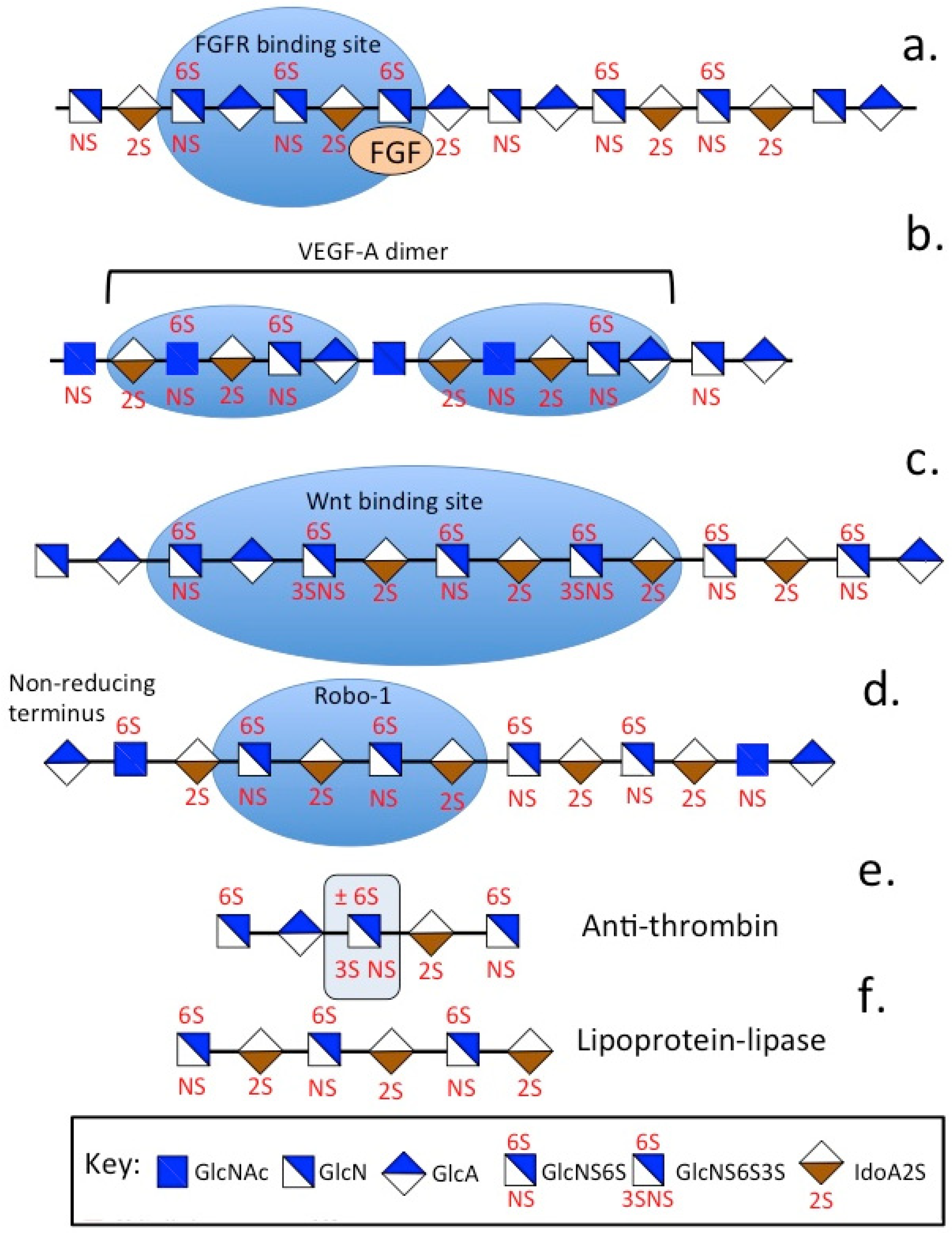
| PG | Structural and Functional Features | Core Protein Size (kDa) | GAG Chains Present |
|---|---|---|---|
| KS-PGs | |||
| Aggrecan, CSPG1 (ACAN) | Hydrates and provides hydrodynamic viscoelastic weight bearing properties to cartilages [57]. | 208–220 | CS, KSI/KSII |
| Lumican (LUM) | MMP inhibitor, anti-angiogenic anti-cancer agent, regulates regularly organized slender collagen fibrils in cornea [378]. | 38 | KSI |
| Keratocan (KERA) | Essential ECM component of the lens capsule, organizes collagen fiber diameters and spacing in the corneal stroma to maintain stromal clarity [194,198,379,380]. | 37–50 | KSI |
| Fibromodulin (FMOD) | Cell regulatory multifunctional matricellular modulator, maintains cellular architecture for normal tissue function, regulates collagen fibrillogenesis [381,382,383]. | 42 | KSI |
| PRELP (Prolargin) | PRELP is an anchoring component in many basement membranes binds type I and II collagens and perlecan to stabilize the basement membrane [188,198]. | 44 | KSI |
| Osteomodulin (osteoadherin) | OMD, is a KS-SLRP that binds to osteoblasts via αςβ3 integrin and regulates osteogenesis through its interaction with BMP2. WNT1 transcriptionally activates expression of OMD [195,196,384] | 42 | KSI |
| Osteoglycin(OGN) (Mimecan) | OGN, is a class II SLRP with diverse roles in ECM assembly, regulates bone formation along with TGF-β1/TGF-β2 that controls collagen fibrillogenesis and has glucose regulatory roles in metabolic health, cancer and diabetes [199,200,385,386,387,388,389] | 35 | KSI |
| Chondroadherin (CHAD) | CHAD, is a 38 kDa member of the KS-SLRP family containing 11 LRRs that bind to α2β1 integrin, type I, II and VI collagen and has an anchoring role in ECM stabilization, binds cells to the ECM and mediates cell-ECM communication through interactions with cell surface PGs such as the syndecans [390,391,392,393,394] | 36–38 | KSI |
| Claustrin | Claustrin is an anti-adhesive neural KS-PG [395] | 105 | KSII |
| Synaptic vesicle PG (SV2) | SV2 is a synaptic vesicle neurotransmitter transporter and smart storage PG, SV2A, SV2B, SV2C paralogs share 60% sequence and 80% structural homology. SV2A controls transmitter release, SV2B is the primary paralog expressed in the retina, SV2C has roles in synaptic plasticity [29,30,31,32,33,34,35]. | Occurs as H 250 kDa and L 100 kDa forms | KS |
| Podocalyxcin (PODXL,TRA-1-60) | Transmembrane, anti-adhesive sialo-KS-PG, up-regulated in many cancers and is a tumor stem cell biomarker [164,396] | 65 | KS |
| Phosphacan | Soluble ectodomain of RPTP-ζ exists as three splice variants, roles in perineuronal net assembly and function in cognitive processes, modulates neurite extension in formation of neural networks [143,144,145,146,148]. | 300 | KS, CS, HNK-1 |
| HS-PGs | |||
| Agrin | 400 kDa HSPG, interacts with LRP4 and α-DG. Promotes chondrocyte differentiation, upregulates SOX9, COL2A1, ACAN [239]. Activates MuSK in NMJ, interacts with rapsyn, LRP,DOK, clusters Ach receptors in NMJ neuromuscular control [397] | 212 | HS |
| Perlecan (HSPG2) | Multifunctional, modular HS/CS PG, interacts with growth factors, controls cell proliferation and differentiation, cell signaling and tissue morphogenesis, facilitates cell-ECM communication, shear flow biosensor important in tissue homeostasis and function [3,250,257,398,399]. | 400–467 | HS/CS |
| Collagen XVIII | Stabilising, basement membrane component in laminin, nidogen HSPG networks [251,263,400]. | 187 | HS |
| The syndecans | SDC 1–4 are G-protein coupled co-receptors in cell proliferation and differentiation, regulating growth factor interactions, tissue development, wound repair, tissue regeneration, inflammation in health and disease [23,24,31,32,260,261]. | 22–48 | HS/CS |
| The glypicans | GPC1-6 have multiple regulatory roles in cell signaling in tissue development and repair processes in health and disease [26,251,269,270]. | 62 | HS |
| Serglycin | Intracellular heparin PG storing bioactive compounds in vesicles [276] in immune [401] and neuroendocrine cells. With varied roles in health and disease [277]. | 17.3 | Heparin |
| Neurexin (NRXN) | NRXN1-3 [402] act as receptors and cell adhesion molecules [19] aiding in synaptic development [403] and stabilization and signaling along with a vast collection of ligands [15,16]. LamG motifs interact with α-DG stabilizing synaptic activity. NRXN3 provides synaptic plasticity. | n/a | HS |
| Pikachurin | Pikachurin has roles in synaptic assembly [404] interacting with α-DG in photoreceptor ribbon synapse assembly [285,289] facilitating interaction with retinal bipolar neural networks in visual processing [288]. | n/a | HS |
| Eyes-shut | Eyes-shut stabilises the photoreceptor primary cilium axenome which connects the inner and outer regions of the photoreceptor and has essential roles to play in phototransduction [291], Eyes shut deficiency leads to autophagy of photoreceptors and impaired vision [405]. | n/a | HS |
| SPOCK (testican, sparc/osteonectin, cwcv and kazal-like domains PG, SPARC (osteonectin) | SPOCK-2 is induced by viral infection or IFN, and is secreted to the ECM, where it blocks virus-cell attachment and entry. SPOCK regulates malignant tumor development [406] and has roles in embryonic development [407] and neuromuscular tissue development [408]. | 48.4 | CS/HS |
| CS-PGs | |||
| Aggrecan | Hydrates and provides hydrodynamic viscoelastic weight bearing properties to cartilages [57,118] but is also a component of heart and brain tissue [125]. HNK-1 in heart and brain aggrecan provides additional interactive properties [372]. | 208–220 | CS, KS |
| Versican (PG-M, CSPG2) | Versican plays diverse roles in cell adhesion, proliferation, migration and angiogenesis and is so named in recognition of its versatile modular structure [409]. Versican has key roles in inflammation through interactions with adhesion molecules on the surfaces of inflammatory leukocytes and chemokines that recruit inflammatory cells [410]. Versican forms macromolecular complexes with HA which are looser than aggrecan-HA aggregates conducive to cell attachment and migration [411]. | 265 | CS |
| Neurocan | Neurocan modulates cell adhesion and migration in brain development and has roles in the formation of perineuronal nets and their functional interactive properties [412,413,414,415,416]. | 145 | CS |
| Brevican (BEHAB,CSPG7) | Brevican is localised to the surface of neurons in the brain and maintains molecular networks around neurons which may slow brain ageing and AD development [416]. | 96 | CS |
| Decorin (DCN) | Widely distributed and highly interactive forming multifunctional networks [417]. DCN has roles in tissue protection [418] and wound repair, angiogenesis, tumor metastasis [419], autophagy, immune regulation and inflammatory diseases [420]. DCN has antifibrotic, anti-inflammatory, antioxidant, antiangiogenic and onco-suppressive properties [421] and inhibits TGFβ activity [422]. | 36 | CS/DS |
| Biglycan (BGN) | BGN is both a structural ECM component and a signaling molecule [423]. BGN LRRs have interactive properties with a range of protein ligands contributing to ECM stabilization and function. When proteolytically released from the ECM, biglycan acts as a danger signal of tissue stress or injury. Biglycan links innate immunity receptors and activators of the inflammasome, stimulating multifunctional proinflammatory signaling in tissue damage [423]. | 38 | CS/DS |
| Asporin (ASPN) | ASPN contains a distinctive group of N-terminal D-Asp-residues which are linked to cancer progression and OA. Regulates TGFβ, Wnt/β-catenin, notch, hedgehog, EGFR, HER2 cell signaling pathways [424]. | 42 | CS |
Disclaimer/Publisher’s Note: The statements, opinions and data contained in all publications are solely those of the individual author(s) and contributor(s) and not of MDPI and/or the editor(s). MDPI and/or the editor(s) disclaim responsibility for any injury to people or property resulting from any ideas, methods, instructions or products referred to in the content. |
© 2025 by the author. Licensee MDPI, Basel, Switzerland. This article is an open access article distributed under the terms and conditions of the Creative Commons Attribution (CC BY) license (https://creativecommons.org/licenses/by/4.0/).
Share and Cite
Melrose, J. Glycosaminoglycans, Instructive Biomolecules That Regulate Cellular Activity and Synaptic Neuronal Control of Specific Tissue Functional Properties. Int. J. Mol. Sci. 2025, 26, 2554. https://doi.org/10.3390/ijms26062554
Melrose J. Glycosaminoglycans, Instructive Biomolecules That Regulate Cellular Activity and Synaptic Neuronal Control of Specific Tissue Functional Properties. International Journal of Molecular Sciences. 2025; 26(6):2554. https://doi.org/10.3390/ijms26062554
Chicago/Turabian StyleMelrose, James. 2025. "Glycosaminoglycans, Instructive Biomolecules That Regulate Cellular Activity and Synaptic Neuronal Control of Specific Tissue Functional Properties" International Journal of Molecular Sciences 26, no. 6: 2554. https://doi.org/10.3390/ijms26062554
APA StyleMelrose, J. (2025). Glycosaminoglycans, Instructive Biomolecules That Regulate Cellular Activity and Synaptic Neuronal Control of Specific Tissue Functional Properties. International Journal of Molecular Sciences, 26(6), 2554. https://doi.org/10.3390/ijms26062554


