Review

Seborrheic Dermatitis: Exploring the Complex Interplay with Malassezia

by
Francesca Piacentini
,
Emanuela Camera
,
Anna Di Nardo
* and
Maria Lucia Dell’Anna
*
Laboratory of Cutaneous Physiopathology and Integrated Center of Metabolomics Research, San Gallicano Dermatological Institute IRCCS, 00144 Rome, Italy
*
Authors to whom correspondence should be addressed.
Int. J. Mol. Sci. 2025, 26(6), 2650; https://doi.org/10.3390/ijms26062650
Submission received: 7 February 2025 / Revised: 13 March 2025 / Accepted: 14 March 2025 / Published: 14 March 2025

Abstract

:
Seborrheic dermatitis (SD) is a chronic inflammatory skin condition often involving the sebaceous-rich areas, characterized by erythematous scaly lesions. It is frequently observed in individuals with immune dysregulation, suggesting the interplay between the immune system and disease development. An altered immune environment leads to an exaggerated inflammatory response with the activation of innate immunity, involving the participation of mast cells, γδ T cells, and the NOD–LRR–pyrin-domain-containing protein 3 (NLRP3) inflammasome. This review aims to assess the complex relationship between Malassezia and the immune system in the pathogenesis of SD. We will explore how an impaired immune response predisposes the skin to Malassezia overgrowth and infection. We will examine the role of adaptive immunity, particularly T helper cells, in driving chronic inflammation in SD. All actors involved, whether part of innate or adaptive immunity, are responsible for the release of pro-inflammatory cytokines, which contribute to the progression of the disease. Therapeutic strategies aimed at the modulation of the immune response in SD have been tested in clinical trials evaluating the efficacy of immunomodulatory treatments in the management of SD. This review synthesizes insights from immunological studies and clinical trials to present an in-depth analysis of the immune mechanisms underpinning SD, thereby proposing targeted therapeutic strategies for its management.

1. Introduction

Seborrheic dermatitis (SD) is a chronic inflammatory skin disease that predominantly affects the seborrheic areas of the body, including the scalp, face (nasolabial folds, ears, and eyebrows), and the upper part of the trunk [1]. The condition manifests more frequently in men. It shows a higher prevalence during specific life stages: infancy within the first three months of life, adolescence and young adulthood, and after the age of 50 years. SD affects approximately 1% to 3% of immunocompetent adults [2,3,4]. SD is more common in patients with neurologic and psychiatric conditions, including Parkinson’s disease [5,6]. The etiology of SD remains a topic of considerable debate in the dermatological field. The current understanding of the pathogenesis of SD points to several factors. Genetic predisposition plays a significant role, making some individuals more susceptible to disease development. Environmental and behavioral factors, such as stress, climatic variations (particularly humidity and cold), sebum excess, and poor hygiene habits, may influence the onset and severity of SD. Furthermore, alterations in the skin barrier are frequently observed in individuals with SD. Recent studies have focused on the alteration of the skin microbiome and have found that changes in microbial communities are associated with the pathogenesis of SD.
  • Seborrheic Dermatitis: Malassezia, Immunity, or Both?
The leading theory on SD etiology postulates a potential association between the fungal genus Malassezia and SD [7].
Malassezia is a lipophilic organism that colonizes lipid-rich areas, such as the skin, and metabolizes sebum components [4]. Recent studies have shown that Malassezia are common commensals on the skin of 75% to 98% of healthy adults. Over millions of years, Malassezia has co-evolved with mammals, acquiring specialized adaptations such as a reduction in fatty acid synthase (FAS) genes and an increase in lipid hydrolases, allowing it to thrive in various human body niches. Dysbiosis, caused by factors like antibiotics, immunosuppression, and environmental changes, can lead to its colonization [8].
Hamido et al. conducted a significant study on the epidemiological prevalence of Malassezia in various skin diseases. They found that M. furfur was most prevalent in pityriasis versicolor and SD lesions, particularly on the scalp, while M. sympodialis was dominant in atopic dermatitis lesions. In healthy individuals, M. furfur, M. globosa, and M. sympodialis were present at lower frequencies [9].
Genetic diversity of Malassezia spp. has been found in SD patients. M. restricta and M. arunalokei exhibited less genetic variation, while M. globosa showed higher genetic diversity [10]. A recent study of Hiruma and coworkers identified a novel species (M. polysorbatinonusus) occurring in a Japanese patient with SD. They molecularly characterized the species, suggesting its similarity to another Malassezia spp. (e.g., M. yamatoensis) already associated with the disease, even if there were some different growth features [11,12].
Recent studies have highlighted significant skin dysbiosis in SD patients, with an increased relative abundance of M. restricta and Staphylococcus spp., which may contribute to disease by disrupting the skin barrier and exacerbating inflammation. Furthermore, SD-affected skin showed reduced fungal diversity, increased M. restricta abundance, and a higher M. restricta/C. acnes ratio compared to healthy skin, alongside increased Staphylococcus spp. and reduced C. acnes abundances [13,14].
Chang et al. provide a critical re-evaluation of the role of Malassezia in SD. While previous studies have suggested a central role for Malassezia in SD, emerging evidence now suggests that immune dysregulation and skin barrier dysfunction are central to the pathogenesis of the disease, with Malassezia acting more as a secondary factor [15]. This shift in perspective is significant for our review, as it emphasizes the need to consider the complex interplay between Malassezia and the host immune system. These findings align with our review’s goal of emphasizing the immune system’s role in SD pathogenesis, alongside microbial factors.
Understanding the relationship between Malassezia and the patient’s inadequate immune response is essential in the pathogenesis of SD. Although Malassezia is a ubiquitous commensal organism on the skin, the distinguishing element in patients with SD is the exaggerated or aberrant immune reactivity towards this yeast. The immune dysregulation serves as both the initiating and perpetuating factor for the chronic inflammation and clinical manifestations [16]. SD is more prevalent in immunocompromised patients, highlighting the critical role of immune mechanisms in the disease’s development. This is particularly evident in individuals with Human Immunodeficiency Virus/Acquired Immunodeficiency Syndrome (HIV/AIDS), where SD tends to be more severe and more resistant to treatment, worsening as the disease progresses [17]. The immune system of individuals with HIV is ineffective in eradicating the yeast due to immune dysregulation, causing excessive yeast proliferation and marked inflammation [18]. The hypo-responsiveness leads to the worsening of SD symptoms, suggesting that Malassezia itself is not the direct cause of SD, but rather it is linked to the type and extent of the immune response to it. This is further supported by the observation that antifungal therapies improve SD clinically, with no definitive resolution [1]. Rather than through a biunivocal mechanism, Malassezia plays a pathogenic role when the immune response is deranged. Both the onset and persistence of the disease are the result of dynamic processes involving the immune and the inflammatory responses. This review aims to examine the role of the immune system in SD, focusing on how alterations in the immune response can facilitate the colonization and proliferation of Malassezia.

2. Skin Barrier

The skin is the body’s primary defense against external threats and is organized as a multilayered tissue formed by the epidermis, the dermis, and the hypodermis. The epidermis consists of keratinocytes in the basal, spinosum, granulosum, and corneum layers, each one characterized by a specific cell renewal and differentiation program aimed at the formation of a competent barrier function. Specialized cells such as melanocytes and Langerhans cells are also present in the epidermis. The impermeable barrier in the epidermis plays a crucial role in the pathophysiology of several skin disorders. Conditions characterized by significant barrier disruption and inflammation include irritant and allergic contact dermatitis, atopic dermatitis, and psoriasis [19].
Presumably, the impairment of the skin barrier is involved in the pathogenesis of SD [20], given the potential for external triggers like Malassezia spp. and its metabolites to penetrate the skin [21].
Park et al. examined the growth of M. furfur under varying pH conditions and found optimal growth at pH 6. Lipid content was highest at pH 6 and lowest at pH 7, suggesting that pH influences lipid metabolism and extracellular lipase activity. Additionally, lipids produced under acidic conditions may play a role in modulating the inflammatory response in skin cells [22].
The lipid composition and the organization of the epidermal matrix are crucial in a skin barrier opposed to the access of microbes [23]. The in-depth characterization of cutaneous barrier function can be achieved through transepidermal water loss (TEWL) measurements combined with ceramide profiling via lipidomic analysis. Such detailed assessments may provide new insights into the involvement of endogenous determinants of SD. Accordingly, the barrier–inflammation axis may play a significant role in mild-to-moderate SD, indicating that it may be more critical than changes in the microbiome [24]. The results corroborate and extend current knowledge regarding the roles of inflammation and microbial factors in SD while enhancing our understanding of barrier dysfunction. Although improving skin barrier function has been suggested as a potential adjunctive therapy for SD [24], current treatment strategies primarily focus on anti-inflammatory and antifungal approaches [25].
Considering these findings, it seems reasonable to posit that disruptions in the skin barrier and immunological mechanisms reinforce each other, initiating and perpetuating skin lesions.

3. Sebaceous Glands

The sebaceous glands (SGs) are holocrine glands that play a crucial role in the pilosebaceous unit and are important for skin homeostasis. They are key in areas such as cell biology, lipid signaling, and hormone regulation. Disruptions in the SGs’ function, resulting into atrophy and modified lipid profiles, can impair the skin’s barrier [26].
SGs have been demonstrated to play an important role in the etiology of numerous dermatological conditions [27,28] and likely contribute to the development of SD [29]. The lipid film on the skin’s surface consists of lipids derived from the disintegration of sebocytes that blend with the lipid matrix in the stratum corneum [30].
Sebum lipids are composed by weight of approximately 50–60% triglycerides and fatty acids, 20–25% wax esters, 12–20% squalene, and 2% cholesterol [31]. Upon secretion of sebum onto the skin’s surface, bacterial and yeast lipases hydrolyze triglycerides into free fatty acids (FFAs) [32].
Lipase activity has been identified in multiple Malassezia species. The isolated lipases from M. globosa cells present specificity for diolein, while intact cells show activity against both diolein and triolein, suggesting the presence of multiple lipases. A specific lipase has been purified, and its gene (LIP1) identified. Expression of this gene has been confirmed in samples from the human scalp, providing a starting point in understanding the role of lipid metabolism in SD [33].
Based on the Malassezia’s capacity to metabolize human triglycerides, it is reasonable that an individual’s susceptibility towards the development of SD is ultimately dependent on their sensitivity to irritant unsaturated FFAs, e.g., oleic acid, and the subsequent inflammatory response [30,34]. This hypothesis is supported by the observation that oleic acid applied topically worsens skin shedding in patients with SD, while it has negligible consequences in unaffected subjects [7].

4. The Innate Immune System Involvement

Cutaneous innate immunity acts as the primary defense against the spread of pathogens from infection sites and is essential for activating the adaptive immune response, the body’s secondary defense mechanism.
During infections, pattern recognition receptors (PRRs) detect pathogen-associated molecular patterns (PAMPs) from invading viruses, thereby triggering antiviral innate immune responses. Notably, immunocompetent skin cells—including keratinocytes, Langerhans cells, dermal dendritic cells, macrophages, mast cells, and fibroblasts—possess PRRs to detect and respond to pathogens [35,36]. Different components of the innate immune system have been involved in SD pathogenesis and in Malassezia infection [37,38].

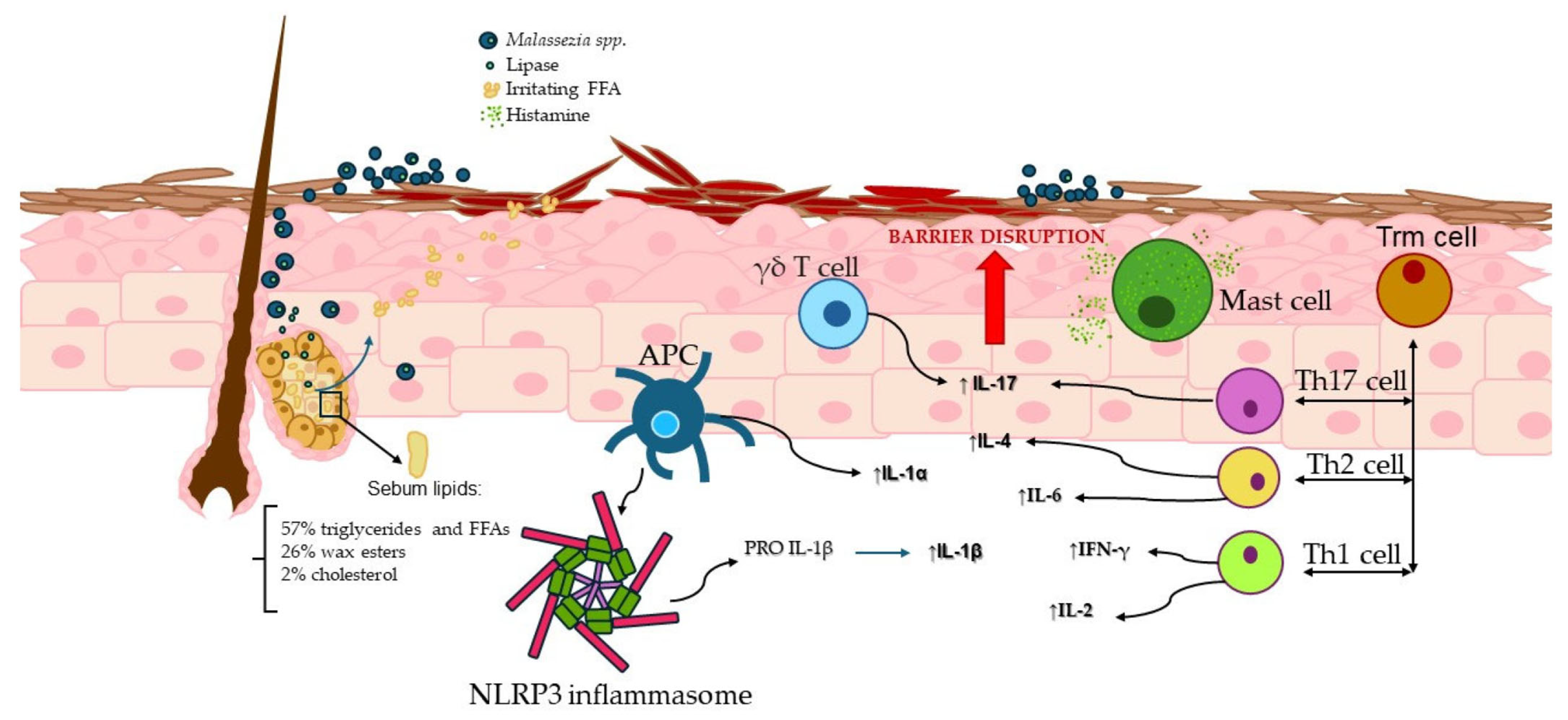
4.1. γδ T Cells and the MPZL3 Gene

γδ T cells are involved in immune-mediated skin diseases such as psoriasis and SD [39,40,41]. γδ T cells have unique properties and can be activated through cytokine signals [42]. In the context of SD, a study observed a significant increase in γδ T cells during the early-onset inflammation in a mouse model lacking Mpzl3 (myelin protein zero-like 3, a type I transmembrane protein). The absence of Mpzl3 in mice led to skin abnormalities and increased interleukin-17 (IL-17) levels primarily produced by γδ T cells [41]. Disruption of the epidermal barrier, coupled with the activation of the innate immune response and subsequent γδ T cell migration to the dermis, likely contributed to the SD-like symptoms.
The disruption of the epidermal barrier caused by the inactive Mpzl3 gene may facilitate the excessive proliferation of Malassezia. The overgrowth of Malassezia likely activates γδ T cells, which in turn promote the release of IL-17 [41]. Ruchti et al. demonstrated that Malassezia colonization of the skin is tightly regulated by the host immune system, particularly through Vγ4+ dermal γδ T cells, which rapidly expand and produce IL-17A to control fungal growth. Vγ4+ T cells can directly respond to Malassezia-derived ligands, which are shared across different Malassezia spp. [43].
Furthermore, a frameshift mutation in zinc finger 750 (ZNF750) has been associated with an SD-like phenotype in humans, with flaky lesions, and follicular hyperkeratotic plugs, all of which contribute to an overgrowth of M. furfur [44,45]. Hyperkeratosis and parakeratosis were apparent in skin biopsies. Mutations in the ZNF750 gene are associated with the impaired expression of late differentiation markers and skin barrier genes [46]. Understanding the ZNF750/MPZL3 pathway could provide insights into the pathogenesis of SD and identify new treatment targets.

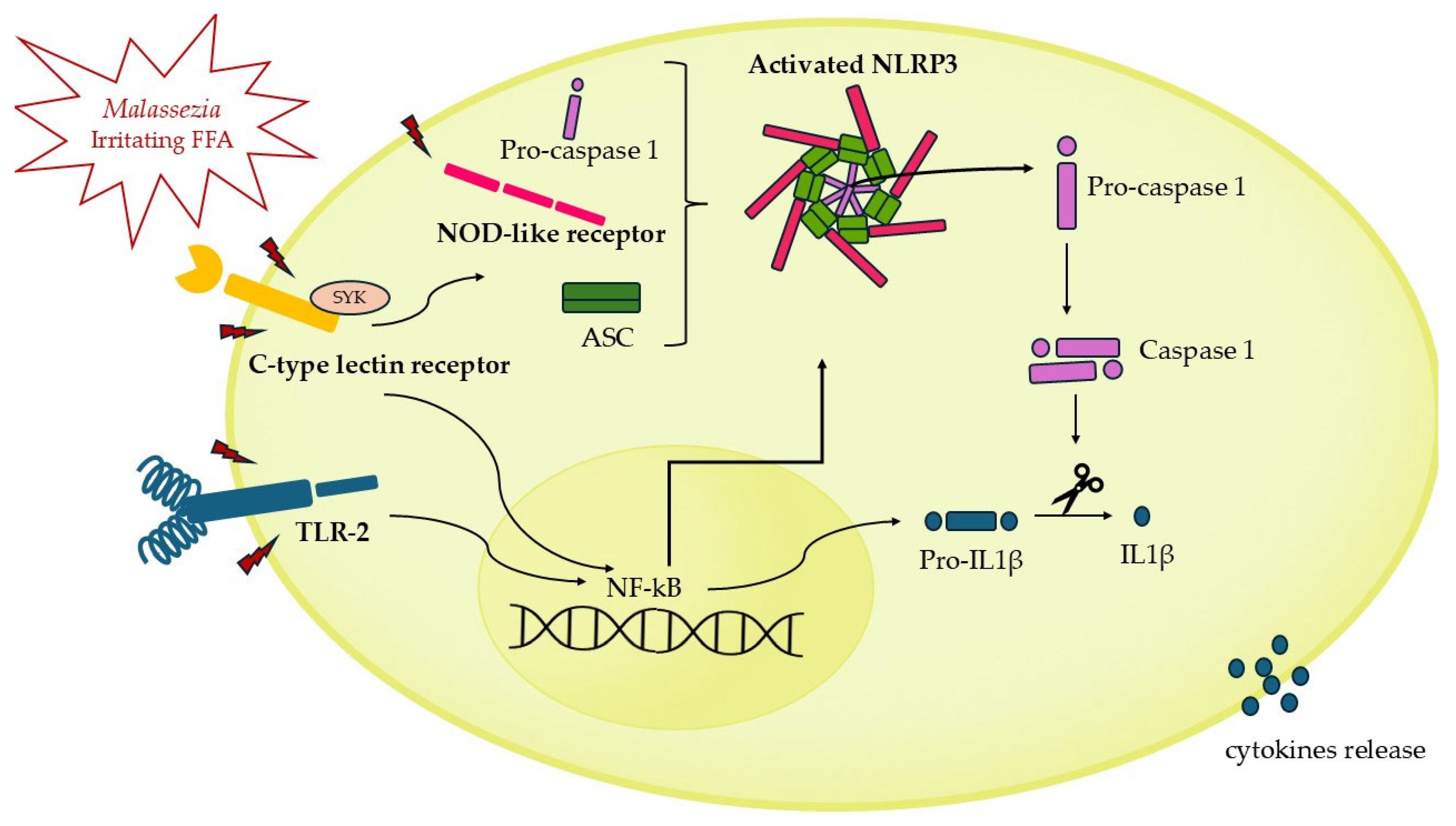
4.2. NLRP3 Inflammasome Activation

Host germline-encoded PRRs initiate the initial immune response, recognizing conserved microbial motifs, endogenous damage signals, and pathogen-induced cellular alterations [47]. Among PRRs are accounted inflammasome-forming sensors that, upon activation, assemble into multimeric complexes [48].
In susceptible individuals, Malassezia activates Antigen-Presenting Cells (APCs) via various PRRs, including Toll-like receptor-2 (TLR-2), NOD-like receptors, and C-type lectin receptors [49,50]. Notably, several clinical isolates of Malassezia spp., in particular M. globosa and M. restricta, [49,51] robustly activate the NLRP3 inflammasome, leading to the secretion of IL-1β (Figure 1). This is particularly significant in the pathogenesis of SD, as the activation of the NLRP3 inflammasome driven by Malassezia may play a central role in this condition. The resulting IL-1β secretion contributes to the chronic inflammation and hallmark symptoms of SD, such as erythema and scaling.

4.3. Mast Cells Potential Implication in Seborrheic Dermatitis

Mast cells (MCs) originate from pluripotent precursors in the bone marrow and circulate in the blood before maturing in various tissues, influenced by factors such as stem cell factor (SCF) and interleukin 3 (IL-3) [52,53,54]. MCs are prevalent in most body tissues, especially those serving as an interface between the host and environment, such as the skin and mucosal surfaces. Due to their distribution, MCs act as a frontline defense against external pathogens and environmental challenges [55,56].
MCs are central to many skin diseases due to their ability to release a wide range of mediators that influence inflammation and tissue remodeling. M. sympodialis, commonly found in atopic dermatitis, can activate MCs, leading to increased IgE-mediated degranulation through the TLR-2/MyD88 pathway [57]. Furthermore, MGL_1304, a protein from M. globosa detected in sweat, has been identified as an allergen capable of inducing IgE-dependent degranulation, highlighting the role of MCs in allergic responses in the skin [58].
The potential interaction between Malassezia spp., commonly associated with SD, allergic responses, and MCs suggests that these fungi may influence the disease process. Specifically, the crosslinking of the high-affinity IgE receptor (FcεRI) with antigen-bound IgE can trigger MC degranulation [57], which potentially exacerbates barrier dysfunction and amplifies the inflammatory responses.
Although MCs have been identified as a critical cellular component in the pathophysiology of certain dermatological conditions, there is currently no conclusive scientific evidence to substantiate their role in the immunological response observed in SD. However, given that MCs are the primary source of histamine in the human body [59] and that elevated histamine levels have been observed in SD patients [29,60], it is reasonable to assume that MCs play a role in the immunology of this disease. This hypothesis is further supported by the observation that histamine levels return to normal following the effective treatment of SD [29,49,61].

5. Adaptive Immunity

The skin-resident memory T cells, known as Trm cells, play a pivotal role in immune surveillance in boundary sites like the skin. They are non-circulating cells characterized by CD69, CD103, and CCR8 expression. Trm cells are involved in various skin pathologies and can influence Th1, Th2, and Th17 responses. Th1 cells are defined by their interleukin-2 (IL-2) and interferon-gamma (IFN-γ) production, while Th2 cells produce cytokines such as IL-4, IL-5, and IL-13. In SD, several studies have reported different findings regarding adaptive immune responses; some do not show any changes [62,63], while others indicate an increased immune response.
Molinero et al. demonstrated an upregulation of inflammatory and Th2 cytokines in skin biopsies from patients with SD, particularly in those with severe manifestations. Furthermore, the expression of the cytotoxicity-inducing MICA, a ligand for γδ-T lymphocytes, CD8+ T cells, and NK cells, was detected in the skin of SD patients using RT-PCR and hybridization with specific probes [64].
Another study conducted a comprehensive analysis of inflammatory cytokines, revealing elevated serum levels of IL-2 and IFN-γ in SD [65]. In addition, Corzo-León et al. highlighted the gene expression and proteomic responses of secreted cytokines in human skin exposed to M. sympodialis, characterized by high levels of TGF-β1 and IL-18 [66].
The role of IL-2 in regulating the immune response in SD may become particularly relevant. A case reported by Kawakami et al. illustrates an unusual adverse effect of systemic recombinant IL-2 (rIL-2) therapy: the development of SD-like eruptions and peri-orbital edema in a patient with renal cell carcinoma. A challenge test confirmed that these symptoms were induced by rIL-2, although the study did not explore the potential involvement of Malassezia. This suggests that IL-2 may contribute to SD-like symptoms through inflammatory pathways, though further investigation is needed to clarify this relationship [67].

6. Th17 and IL-17: Potential Link to Seborrheic Dermatitis?

Th17 cells and IL-17 play critical roles in autoimmune inflammation and maintaining tissue homeostasis. IL-17, mainly produced by various immune cells, regulates immune responses and is linked to inflammatory and autoimmune diseases. The mechanisms behind Th17 responses to Malassezia are not fully understood. It has been shown that C-type lectin receptors, such as Mincle, Dectin-1, and Dectin-2, recognize patterns in Malassezia’s cell wall and activate dendritic cells in vitro. However, in vivo studies have shown that Dectin-2 is the only receptor essential for initiating Th17 cell responses during skin colonization. Moreover, the Th17 response against Malassezia could depend on T-cell-intrinsic MyD88 signaling [68].
Recent studies have explored their involvement in atopic dermatitis, where they are supposed to play a vital role in immune activation and skin barrier function. Fang et al. found that higher levels of Malassezia in psoriasis correlate with increased LL37, IL-17A, TNF-α, and IL-23. The upregulation of LL37 by Malassezia promotes the IL-23/IL-17 pathway, which accelerates psoriasis severity [69]. Additionally, psoriasis lesions exhibit a more significant presence of Th17 cells than normal skin, with some Th17 cells producing TNF-α and IFN-γ. Malassezia activates the IL-23/IL-17 axis, which mediates the inflammatory response to the fungal infection [70]. Thus, targeting this pathway in therapies may inadvertently offer secondary protection against fungal infections. Andersen et al. found that certain HLA alleles may influence the risk of Malassezia-related skin diseases (MRSD). Additionally, they suggested that the genetic interaction between MHC class I and the IL-23/IL-17 pathway could play a role in MRSD, psoriasis, and atopic dermatitis. These findings suggest that Malassezia infections may require specific HLA presentation. However, further studies addressing microbial and protein changes are needed to clarify the immune mechanisms involved [71].
Considering the striking resemblance between psoriasis and SD [72,73], it is plausible that Th17 cells and IL-17 play a pivotal role in the etiology of SD [74]. The similarity of pathogenetic mechanisms indicates that Th17 signals may be involved in the inflammatory and immune responses underlying SD.
Nevertheless, further research is required to substantiate this hypothesis and to elucidate the precise mechanisms through which the IL-17/IL-23 axis may influence the pathogenesis and progression of SD. These studies may potentially lead to the development of novel targeted treatments, thereby improving the clinical management of patients with this condition.

7. Treatment and Future Perspectives

The management of SD includes the regulation of sebum secretion, limiting colonization of the fungus Malassezia spp., and mitigating inflammation. Sebum production can be controlled with different strategies, such as medicated shampoos or topical treatments. Limiting the colonization of Malassezia spp. may be achieved with antifungal agents. Corticosteroids or other anti-inflammatory agents are frequently prescribed for people with SD. Less severe cases of SD are treated topically, in most cases. Severe cases are often managed with off-label topical and systemic medications (Table 1) [1]. Recent studies have highlighted the alteration of the microbiome in people with SD. One study demonstrated that the administration of recombinant human thymosin beta-4 (rhTβ4) led to a significant improvement in scalp condition and microbiome homeostasis [75]. Additionally, the modulation of the skin mycobiome with the administration of a probiotic-enriched oily suspension has been proposed as a treatment for SD [76].

7.1. Topical Antifungal Agents

Antifungal treatments have been proven to be effective in managing SD by reducing yeast populations and controlling inflammation [77]. Topical antifungals, such as ketoconazole, clotrimazole, and miconazole, are often first-line treatments for SD because they inhibit fungal cell wall synthesis [78]. Ketoconazole, introduced in 1979, acts by blocking ergosterol synthesis and has additional anti-inflammatory effects [79,80]. Miconazole is notable for its efficacy and safety comparable to ketoconazole [81], while clotrimazole 1% cream is a broad-spectrum option that is generally well tolerated but occasionally causes localized irritation [82]. Ciclopirox olamine 1% is another broad-spectrum antifungal that inhibits metal-dependent enzymes in fungi and offers anti-inflammatory benefits [78,83]. Despite their efficacy, these treatments have been shown to induce resistance in Malassezia strains. This suggests that topical therapies alone may not always be sufficient [29,84].

7.2. Systemic Antifungal Agents

Systemic antifungal treatments, such as terbinafine and itraconazole, are typically reserved for severe, acute, or treatment-resistant cases of SD, and for conditions that are particularly challenging to manage [1]. Systemic therapy primarily aims to alleviate symptoms rapidly before the subsequent use of topical treatments in the long-term maintenance of the condition. Terbinafine is an antifungal medication with additional antioxidant and anti-inflammatory properties. Oral terbinafine has been demonstrated to be effective in managing SD [82,85]. Itraconazole is a triazole antifungal that is highly effective in the keratin- and lipid-rich environment of the stratum corneum, where Malassezia typically resides [86]. In addition to its antifungal action, itraconazole has anti-inflammatory effects due to inhibition of the formation of 5-lipoxygenase metabolites, which are inflammatory mediators [1,87]

7.3. Anti-Inflammatory Agents

Topical corticosteroids, ranging from potent (e.g., betamethasone valerate) to super potent (e.g., clobetasol propionate), are crucial in the effective management of SD symptoms such as erythema, scaling, and itching through their anti-inflammatory, immunosuppressive, and antiproliferative activities [1,88]. Non-steroidal anti-inflammatory agents offer significant benefits in managing both scalp and non-scalp SD by inhibiting Malassezia through their anti-inflammatory, antimycotic, keratolytic, and antioxidant properties [78,89]. These treatments are generally well-tolerated, with only mild side effects [78,90,91]. Immunomodulators, specifically tacrolimus [83,92,93] and pimecrolimus [78,94,95,96,97], are effective in the treatment of facial SD due to their limited side effects. These drugs inhibit calcineurin, essential for T cell activation and the production of pro-inflammatory cytokines, and may also exhibit antifungal activity against Malassezia. These agents are beneficial when topical corticosteroids and antifungals do not control SD adequately. When conventional treatments are insufficient, as in cases of severe SD, managing CD4+ levels with antiretroviral medications can be advantageous. The prevalence of SD in patients with neurological conditions, such as Parkinson’s disease, often presents as unilateral facial dermatitis and indicates a need for targeted treatments, including antifungals and anti-inflammatory agents [1].

8. Conclusions

In this review, we delved into the intricate role of the immune system in the development of SD, particularly focusing on the interaction between Malassezia and various components of the immune response. Through an in-depth analysis of the existing literature, we have highlighted how skin barrier impairment, innate immunity, and adaptive immunity contribute to the clinical manifestation of SD.
We emphasized how alterations in the skin barrier can facilitate fungal colonization and trigger immune responses, contributing to the disease’s pathogenesis. The importance of the innate immune response, particularly through γδ T cells, MCs, and the NLRP3 inflammasome, emerged as a central factor in amplifying skin inflammation.
Additionally, we explored the role of adaptive immunity, focusing on Th1, Th2, and Th17 cells, suggesting that excessive immune reactivity and an altered immune balance may play a pivotal role in the pathogenesis of SD. Clinical and experimental studies have demonstrated that the immune response is markedly altered in patients with SD. The presence of a hyper-reactive innate immune response, excessive production of pro-inflammatory cytokines, and alterations in Th1 and Th2 responses provide evidence to support an immune-mediated pathogenesis. The current literature also suggests that Th17 cells, known to be implicated in other skin conditions, may also play a role in the pathogenesis of SD.
A hypothetical pathogenic pathway may begin with the colonization of Malassezia spp. under immunocompromised conditions. The lipase produced by the fungus acts on sebaceous lipids, resulting in the release of irritating FFAs. Dendritic cells, γδT cells, MCs, and Trm T cells are activated by Malassezia, initiating an innate immune response. Activated dendritic cells trigger NLRP3, which contributes to the production of IL-1β. MCs release histamine, inducing pruritus. Trm cells, involved in the initial immune response and memory recall, activate Th1, Th2, and Th17 subsets, which produce IL-4, IL-6, IFN-γ, IL-2, and IL-17. These processes ultimately result in the deterioration of the skin barrier, which in turn exacerbates and perpetuates the underlying symptoms. This entire mechanism is described in Figure 2.
In conclusion the review underscores how a deeper understanding of the relationship between Malassezia and the immune system, as well as the central role of inflammation, could guide the development of targeted therapeutic strategies, particularly when various treatment approaches, including both antifungal agents and immunomodulators, are available, thereby enhancing the management of SD and the quality of life of patients.

Funding

This research received no external funding.

Acknowledgments

We acknowledge the National Research Program RF2021-12374216 and the Italian Ministry of Health (Ricerca Corrente 2024).

Conflicts of Interest

The authors declare no conflicts of interest.

Abbreviations

The following abbreviations are used in this manuscript:
APCAntigen-presenting cell
FFAFree fatty acid
FASFatty acid synthase
HIV/AIDSHuman Immunodeficiency Virus/Acquired Immunodeficiency Syndrome
IFNgInterferon-gamma
IL-17Interleukin 17
MCMast cell
MRSDMalassezia-related skin disease
PAMPsPathogen-associated molecular patterns
PRRsPattern recognition receptors
SCFStem cell factor
SDSeborrheic dermatitis
SGsSebaceous glands
TLR2Toll-like receptor 2
TEWLTransepidermal water loss
ZNF750Zinc finger 750

References

  1. Dall’Oglio, F.; Nasca, M.R.; Gerbino, C.; Micali, G. An Overview of the Diagnosis and Management of Seborrheic Dermatitis. Clin. Cosmet. Investig. Dermatol. 2022, 15, 1537–1548. [Google Scholar] [CrossRef] [PubMed]
  2. Gupta, A.K.; Bluhm, R. Seborrheic dermatitis. J. Eur. Acad. Dermatol. Venereol. 2004, 18, 13–20. [Google Scholar] [CrossRef] [PubMed]
  3. De Avelar Breunig, J.; de Almeida, H.L., Jr.; Duquia, R.P.; Souza, P.R.M.; Staub, H.L. Scalp seborrheic dermatitis: Prevalence and associated factors in male adolescents. Int. J. Dermatol. 2012, 51, 46–49. [Google Scholar] [CrossRef] [PubMed]
  4. Dessinioti, C.; Katsambas, A. Seborrheic dermatitis: Etiology, risk factors, and treatments: Facts and controversies. Clin. Dermatol. 2013, 31, 343. [Google Scholar] [CrossRef]
  5. Laurence, M.; Benito-León, J.; Calon, F. Malassezia and Parkinson’s Disease. Front. Neurol. 2019, 10, 758. [Google Scholar] [CrossRef]
  6. Rietcheck, H.R.; Maghfour, J.; Rundle, C.W.; Husayn, S.S.; Presley, C.L.; Sillau, S.H.; Liu, Y.; Leehey, M.A.; Dunnick, C.A.; Dellavalle, R.P. A Review of the Current Evidence Connecting Seborrheic Dermatitis and Parkinson’s Disease and the Potential Role of Oral Cannabinoids. Dermatology 2021, 237, 872–877. [Google Scholar] [CrossRef]
  7. DeAngelis, Y.M.; Gemmer, C.M.; Kaczvinsky, J.R.; Kenneally, D.C.; Schwartz, J.R.; Dawson, T.L., Jr. Three etiologic facets of dandruff and seborrheic dermatitis: Malassezia fungi, sebaceous lipids, and individual sensitivity. J. Investig. Dermatol. Symp. Proc. 2005, 10, 295–297. [Google Scholar] [CrossRef]
  8. Sasikumar, J.; Ebrahim, R.A.; Das, S.P. Diverse Colonisation and Disease Associations of the Human Commensal Malassezia: Our Body’s Secret Tenant. Mycoses 2025, 68, e70014. [Google Scholar] [CrossRef]
  9. Hamdino, M.; Saudy, A.A.; El-Shahed, L.H.; Taha, M. Identification of Malassezia species isolated from some Malassezia associated skin diseases. J. Mycol. Med. 2022, 32, 101301. [Google Scholar] [CrossRef]
  10. Honnavar, P.; Chakrabarti, A.; Joseph, J.; Thakur, S.; Dogra, S.; Lakshmi, P.V.M.; Rudramurthy, S.M. Molecular epidemiology of seborrheic dermatitis/dandruff associated Malassezia species from northern India. Med. Mycol. 2024, 62, myae104. [Google Scholar] [CrossRef]
  11. Hiruma, J.; Nojo, H.; Hiruma, M.; Sugita, T.; Makimura, K.; Harada, K.; Kano, R. Malassezia polysorbatinonusus sp. nov., a Novel Isolate from a Japanese Patient with Seborrheic Dermatitis. Mycopathologia 2025, 190, 15. [Google Scholar] [CrossRef] [PubMed]
  12. Li, J.; Feng, Y.; Liu, C.; Yang, Z.; de Hoog, S.; Qu, Y.; Chen, B.; Li, D.; Xiong, H.; Shi, D. Presence of Malassezia Hyphae Is Correlated with Pathogenesis of Seborrheic Dermatitis. Microbiol. Spectr. 2022, 10, e0116921-21. [Google Scholar] [CrossRef] [PubMed]
  13. Jung, W.H. Alteration in skin mycobiome due to atopic dermatitis and seborrheic dermatitis. Biophys. Rev. 2023, 4, 011309. [Google Scholar] [CrossRef]
  14. Tao, R.; Li, R.; Wang, R. Skin microbiome alterations in seborrheic dermatitis and dandruff: A systematic review. Exp. Dermatol. 2021, 30, 1546–1553. [Google Scholar] [CrossRef]
  15. Chang, C.H.; Chovatiya, R. More yeast, more problems?: Reevaluating the role of Malassezia in seborrheic dermatitis. Arch. Dermatol. Res. 2024, 316, 100–107. [Google Scholar] [CrossRef]
  16. Gaitanis, G.; Magiatis, P.; Hantschke, M.; Bassukas, I.D.; Velegraki, A. The Malassezia genus in skin and systemic diseases. Clin. Microbiol. Rev. 2012, 25, 106–141. [Google Scholar] [CrossRef]
  17. Muñoz-Pérez, M.A.; Rodriguez-Pichardo, A.; Camacho, F.; Colmenero, M.A. Dermatological findings correlated with CD4 lymphocyte counts in a prospective 3 year study of 1161 patients with human immunodeficiency virus disease predominantly acquired through intravenous drug abuse. Br. J. Dermatol. 1998, 139, 33–39. [Google Scholar] [CrossRef]
  18. Chimbetete, T.; Buck, C.; Choshi, P.; Selim, R.; Pedretti, S.; Divito, S.J.; Phillips, E.J.; Lehloenya, R.; Peter, J. HIV-Associated Immune Dysregulation in the Skin: A Crucible for Exaggerated Inflammation and Hypersensitivity. J. Investig. Dermatol. 2023, 143, 362–373. [Google Scholar] [CrossRef]
  19. Proksch, E.; Brandner, J.M.; Jensen, J. The skin: An indispensable barrier. Exp. Dermatol. 2008, 17, 1063–1072. [Google Scholar] [CrossRef]
  20. Wikramanayake, T.C.; Borda, L.J.; Miteva, M.; Paus, R. Seborrheic dermatitis—Looking beyond Malassezia. Exp. Dermatol. 2019, 28, 991. [Google Scholar] [CrossRef]
  21. Goh, J.P.; Ruchti, F.; Poh, S.E.; Koh, W.L.; Tan, K.Y.; Lim, Y.T.; Thng, S.T.; Sobota, R.M.; Hoon, S.S.; Liu, C.; et al. The human pathobiont Malassezia furfur secreted protease Mfsap1 regulates cell dispersal and exacerbates skin inflammation. Proc. Natl. Acad. Sci. USA 2022, 119, e2212533119. [Google Scholar] [CrossRef] [PubMed]
  22. Park, Y.; Yu, B.S.; Heo, Y.M.; Kyung, S.; Lee, K.-E.; Kim, S.; Kang, S.; Han, K.; Kim, D.H. Characteristics of Malassezia furfur at various pH and effects of Malassezia lipids on skin cells. Appl. Microbiol. Biotechnol. 2024, 108, 455. [Google Scholar] [CrossRef] [PubMed]
  23. Uche, L.E.; Gooris, G.S.; Bouwstra, J.A.; Beddoes, C.M. High concentration of the ester-linked ω-hydroxy ceramide increases the permeability in skin lipid model membranes. Biochim. Biophys. Acta Biomembr. 2021, 1863, 183487. [Google Scholar] [CrossRef]
  24. Rousel, J.; Nădăban, A.; Saghari, M.; Pagan, L.; Zhuparris, A.; Theelen, B.; Gambrah, T.; van der Wall, H.E.C.; Vreeken, R.J.; Feiss, G.L.; et al. Lesional skin of seborrheic dermatitis patients is characterized by skin barrier dysfunction and correlating alterations in the stratum corneum ceramide composition. Exp. Dermatol. 2023, 33, e14952. [Google Scholar] [CrossRef]
  25. Desai, S.; McCormick, E.; Friedman, A. An Up-to-Date Approach to the Management of Seborrheic Dermatitis. J. Drugs Dermatol. 2022, 21, 1373–1374. [Google Scholar]
  26. Picardo, M.; Mastrofrancesco, A.; Bíró, T. Sebaceous gland-a major player in skin homoeostasis. Exp. Dermatol. 2015, 24, 485–486. [Google Scholar] [CrossRef]
  27. Cavallo, A.; Camera, E.; Bottillo, G.; Maiellaro, M.; Truglio, M.; Marini, F.; Chavagnac-Bonneville, M.; Fauger, A.; Perrier, E.; Pigliacelli, F.; et al. Biosignatures of defective sebaceous gland activity in sebum-rich and sebum-poor skin areas in adult atopic dermatitis. Exp. Dermatol. 2024, 33, e15066. [Google Scholar] [CrossRef]
  28. Okoro, O.E.; Adenle, A.; Ludovici, M.; Truglio, M.; Marini, F.; Camera, E. Lipidomics of facial sebum in the comparison between acne and non-acne adolescents with dark skin. Sci. Rep. 2021, 11, 16591. [Google Scholar] [CrossRef]
  29. Adalsteinsson, J.A.; Kaushik, S.; Muzumdar, S.; Guttman-Yassky, E.; Ungar, J. An update on the microbiology, immunology and genetics of seborrheic dermatitis. Exp. Dermatol. 2020, 29, 481. [Google Scholar] [CrossRef]
  30. Ro, B.I.; Dawson, T.L. The role of sebaceous gland activity and scalp microfloral metabolism in the etiology of seborrheic dermatitis and dandruff. J. Investig. Dermatol. Symp. Proc. 2005, 10, 194–197. [Google Scholar] [CrossRef]
  31. Picardo, M.; Ottaviani, M.; Camera, E.; Mastrofrancesco, A. Sebaceous gland lipids. Dermatoendocrinology 2009, 1, 68–71. [Google Scholar] [CrossRef] [PubMed]
  32. De Luca, C.; Valacchi, G. Surface lipids as multifunctional mediators of skin responses to environmental stimuli. Mediat. Inflamm. 2010, 2010, 321494. [Google Scholar] [CrossRef] [PubMed]
  33. DeAngelis, Y.M.; Saunders, C.W.; Johnstone, K.R.; Reeder, N.L.; Coleman, C.G.; Kaczvinsky, J.R.; Gale, C.; Walter, R.; Mekel, M.; Lacey, M.P.; et al. Isolation and Expression of a Malassezia globosa Lipase Gene, LIP1. J. Investig. Dermatol. 2007, 127, 2138–2146. [Google Scholar] [CrossRef] [PubMed]
  34. Shi, V.Y.; Leo, M.; Hassoun, L.; Chahal, D.S.; Maibach, H.I.; Sivamani, R.K. Role of sebaceous glands in inflammatory dermatoses. J. Am. Acad. Dermatol. 2015, 73, 856. [Google Scholar] [CrossRef]
  35. Jin, Z.; Song, Y.; He, L. A review of skin immune processes in acne. Front. Immunol. 2023, 14, 1324930. [Google Scholar] [CrossRef]
  36. Kawamura, T.; Ogawa, Y.; Aoki, R.; Shimada, S. Innate and intrinsic antiviral immunity in skin. J. Dermatol. Sci. 2014, 75, 159–166. [Google Scholar] [CrossRef]
  37. Miyachi, H.; Wakabayashi, S.; Sugihira, T.; Aoyama, R.; Saijo, S.; Koguchi-Yoshioka, H.; Fujimoto, M.; Núñez, G.; Matsue, H.; Nakamura, Y. Keratinocyte IL-36 Receptor/MyD88 Signaling Mediates Malassezia-Induced IL-17-Dependent Skin Inflammation. J. Infect. Dis. 2021, 223, 1753–1765. [Google Scholar] [CrossRef]
  38. Sparber, F.; LeibundGut-Landmann, S. Host Responses to Malassezia spp. in the Mammalian Skin. Front. Immunol. 2017, 8, 1614. [Google Scholar] [CrossRef]
  39. Sato, Y.; Ogawa, E.; Okuyama, R. Role of Innate Immune Cells in Psoriasis. Int. J. Mol. Sci. 2020, 21, 6604. [Google Scholar] [CrossRef]
  40. Cai, Y.; Shen, X.; Ding, C.; Qi, C.; Li, K.; Li, X.; Jala, V.R.; Zhang, H.G.; Wang, T.; Zheng, J.; et al. Pivotal role of dermal IL-17-producing γδ T cells in skin inflammation. Immunity 2011, 35, 596–610. [Google Scholar] [CrossRef]
  41. Wikramanayake, T.C.; Hirt, P.; Almastadi, M.; Mitchell, H.; Tomic-Canic, M.; Romero, L.; Garcia, D.; Strbo, N. Increased IL-17-expressing γδ T cells in seborrhoeic dermatitis-like lesions of the Mpzl3 knockout mice. Exp. Dermatol. 2018, 27, 1408. [Google Scholar] [CrossRef] [PubMed]
  42. Bonneville, M.; O’Brien, R.L.; Born, W.K. Gammadelta T cell effector functions: A blend of innate programming and acquired plasticity. Nat. Rev. Immunol. 2010, 10, 467–478. [Google Scholar] [CrossRef] [PubMed]
  43. Ruchti, F.; Tuor, M.; Mathew, L.; E McCarthy, N.; LeibundGut-Landmann, S. γδ T cells respond directly and selectively to the skin commensal yeast Malassezia for IL-17-dependent fungal control. PLoS Pathog. 2024, 20, e1011668. [Google Scholar] [CrossRef] [PubMed]
  44. Birnbaum, R.Y.; Zvulunov, A.; Hallel-Halevy, D.; Cagnano, E.; Finer, G.; Ofir, R.; Geiger, D.; Silberstein, E.; Feferman, Y.; Birk, O.S. Seborrhea-like dermatitis with psoriasiform elements caused by a mutation in ZNF750, encoding a putative C2H2 zinc finger protein. Nat. Genet. 2006, 38, 749–751. [Google Scholar] [CrossRef]
  45. Wikramanayake, T.C.; Nicu, C.; Gherardini, J.; Mello, A.C.; Chéret, J.; Paus, R. Mitochondrially Localized MPZL3 Functions as a Negative Regulator of Sebaceous Gland Size and Sebocyte Proliferation. J. Investig. Dermatol. 2022, 142, 2524–2527.e7. [Google Scholar] [CrossRef]
  46. Cohen, I.; Birnbaum, R.Y.; Leibson, K.; Taube, R.; Sivan, S.; Birk, O.S. ZNF750 is expressed in differentiated keratinocytes and regulates epidermal late differentiation genes. PLoS ONE 2012, 7, e42628. [Google Scholar] [CrossRef]
  47. Li, D.; Wu, M. Pattern recognition receptors in health and diseases. Signal Transduct. Target. Ther. 2021, 6, 291. [Google Scholar] [CrossRef]
  48. Xu, J.; Núñez, G. The NLRP3 inflammasome: Activation and regulation. Trends Biochem. Sci. 2023, 48, 331–344. [Google Scholar] [CrossRef]
  49. Kistowska, M.; Fenini, G.; Jankovic, D.; Feldmeyer, L.; Kerl, K.; Bosshard, P.; Contassot, E.; French, L.E. Malassezia yeasts activate the NLRP3 inflammasome in antigen-presenting cells via Syk-kinase signalling. Exp. Dermatol. 2014, 23, 884–889. [Google Scholar] [CrossRef]
  50. Takeuchi, O.; Akira, S. Pattern Recognition Receptors and Inflammation. Cell 2010, 140, 805. [Google Scholar] [CrossRef]
  51. Park, H.R.; Oh, J.H.; Lee, Y.J.; Park, S.H.; Lee, Y.W.; Lee, S.; Kang, H.; Kim, J.E. Inflammasome-mediated Inflammation by Malassezia in human keratinocytes: A comparative analysis with different strains. Mycoses 2021, 64, 292–299. [Google Scholar] [CrossRef] [PubMed]
  52. Metcalfe, D.D.; Baram, D.; Mekori, Y.A. Mast cells. Physiol. Rev. 1997, 77, 1033–1079. [Google Scholar] [CrossRef] [PubMed]
  53. Gurish, M.F.; Austen, K.F. Developmental origin and functional specialization of mast cell subsets. Immunity 2012, 37, 25–33. [Google Scholar] [CrossRef]
  54. Wernersson, S.; Pejler, G. Mast cell secretory granules: Armed for battle. Nat. Rev. Immunol. 2014, 14, 478. [Google Scholar] [CrossRef]
  55. John, A.L.S.; Abraham, S.N. Innate immunity and its regulation by mast cells. J. Immunol. 2013, 190, 4458–4463. [Google Scholar] [CrossRef]
  56. Voss, M.; Kotrba, J.; Gaffal, E.; Katsoulis-Dimitriou, K.; Dudeck, A. Mast Cells in the Skin: Defenders of Integrity or Offenders in Inflammation? Int. J. Mol. Sci. 2021, 22, 4589. [Google Scholar] [CrossRef]
  57. Selander, C.; Engblom, C.; Nilsson, G.; Scheynius, A.; Andersson, C.L. TLR2/MyD88-dependent and -independent activation of mast cell IgE responses by the skin commensal yeast Malassezia sympodialis. J. Immunol. 2009, 182, 4208–4216. [Google Scholar] [CrossRef]
  58. Hiragun, T.; Ishii, K.; Hiragun, M.; Suzuki, H.; Kan, T.; Mihara, S.; Yanase, Y.; Bartels, J.; Schröder, J.-M.; Hide, M. Fungal protein MGL_1304 in sweat is an allergen for atopic dermatitis patients. J. Allergy Clin. Immunol. 2013, 132, 608–615.e4. [Google Scholar] [CrossRef]
  59. Thangam, E.B.; Jemima, E.A.; Singh, H.; Baig, M.S.; Khan, M.; Mathias, C.B.; Church, M.K.; Saluja, R. The Role of Histamine and Histamine Receptors in Mast Cell-Mediated Allergy and Inflammation: The Hunt for New Therapeutic Targets. Front. Immunol. 2018, 9, 1873. [Google Scholar] [CrossRef]
  60. Schwartz, J.R.; Messenger, A.G.; Tosti, A.; Todd, G.; Hordinsky, M.; Hay, R.J.; Wang, X.; Zachariae, C.; Kerr, K.M.; Henry, J.P.; et al. A Comprehensive Pathophysiology of Dandruff and Seborrheic Dermatitis—Towards a More Precise Definition of Scalp Health. Acta Derm. Venerol. 2013, 93, 131. [Google Scholar] [CrossRef]
  61. Pedrosa, A.F.; Lisboa, C.; Rodrigues, A.G. Malassezia infections: A medical conundrum. J. Am. Acad. Dermatol. 2014, 71, 170–176. [Google Scholar] [CrossRef] [PubMed]
  62. Bergbrant, I.M.; Andersson, B.; Faergemann, J. Cell-mediated immunity to Malassezia furfur in patients with seborrhoeic dermatitis and pityriasis versicolor. Clin. Exp. Dermatol. 1999, 24, 402–406. [Google Scholar] [CrossRef] [PubMed]
  63. Faergemann, J.; Bergbrant, I.-M.; Dohsé, M.; Scott, A.; Westgate, G. Seborrhoeic dermatitis and Pityrosporum (Malassezia) folliculitis: Characterization of inflammatory cells and mediators in the skin by immunohistochemistry. Br. J. Dermatol. 2001, 144, 549–556. [Google Scholar] [CrossRef] [PubMed]
  64. Molinero, L.L.; Gruber, M.; Leoni, J.; Woscoff, A.; Zwirner, N.W. Up-regulated expression of MICA and proinflammatory cytokines in skin biopsies from patients with seborrhoeic dermatitis. Clin. Immunol. 2003, 106, 50–54. [Google Scholar] [CrossRef]
  65. Trznadel-Grodzka, E.; Błaszkowski, M.; Rotsztejn, H. Investigations of seborrheic dermatitis. Part, I. The role of selected cytokines in the pathogenesis of seborrheic dermatitis. Postepy Hig. Med. Dosw. 2012, 66, 843–847. [Google Scholar] [CrossRef]
  66. Corzo-León, D.E.; MacCallum, D.M.; Munro, C.A. Host Responses in an Ex Vivo Human Skin Model Challenged With Malassezia sympodialis. Front. Cell. Infect. Microbiol. 2021, 10, 561382. [Google Scholar] [CrossRef]
  67. Kawakami, Y.; Nakamura-Wakatsuki, T.; Yamamoto, T. Seborrheic dermatitis-like eruption following interleukin-2 administration. Dermatol. Online J. 2010, 16, 12. [Google Scholar] [CrossRef]
  68. Tuor, M.; Stappers, M.H.; Desgardin, A.; Ruchti, F.; Sparber, F.; Orr, S.J.; Gow, N.A.; LeibundGut-Landmann, S. Card9 and MyD88 differentially regulate Th17 immunity to the commensal yeast Malassezia in the murine skin. Mucosal Immunol. 2025, 18, 205–219. [Google Scholar] [CrossRef]
  69. Fang, H.; Hou, Y.; Zhuang, H.; Wang, C. The effects of Malassezia in the activation of Interleukin (IL)-23/IL-17 axis in Psoriasis. New Microbiol. 2022, 45, 130–137. [Google Scholar]
  70. Jia, Q.; Hu, J.; Wang, X.; Deng, Y.; Zhang, J.; Li, H. Malassezia globosa Induces Differentiation of Pathogenic Th17 Cells by Inducing IL-23 Secretion by Keratinocytes. Mycopathologia 2024, 189, 85. [Google Scholar] [CrossRef]
  71. Andersen, P.L.; Jemec, G.B.; Erikstrup, C.; Didriksen, M.; Dinh, K.M.; Mikkelsen, S.; Sørensen, E.; DBDS Genetic Consortium; Nielsen, K.R.; Bruun, M.T.; et al. Human leukocyte antigen system associations in Malassezia-related skin diseases. Arch. Dermatol. Res. 2023, 315, 895–902. [Google Scholar] [CrossRef] [PubMed]
  72. Baş, Y.; Seçkiïn, H.Y.; Kalkan, G.; Takci, Z.; Çitil, R.; Önder, Y.; Şahïn, Ş.; Demir, A.K. Prevalence and related factors of psoriasis and seborrheic dermatitis: A community-based study. Turk. J. Med. Sci. 2016, 46, 303–309. [Google Scholar] [CrossRef] [PubMed]
  73. Kim, S.; Kim, J.; Hwang, M.; Kim, M.; Jo, S.J.; Je, M.; Jang, J.E.; Lee, D.H.; Hwang, J.Y. Smartphone-based multispectral imaging and machine-learning based analysis for discrimination between seborrheic dermatitis and psoriasis on the scalp. Biomed. Opt. Express 2019, 10, 879–891. [Google Scholar] [CrossRef] [PubMed]
  74. Sparber, F.; De Gregorio, C.; Steckholzer, S.; Ferreira, F.M.; Dolowschiak, T.; Ruchti, F.; Kirchner, F.R.; Mertens, S.; Prinz, I.; Joller, N.; et al. The Skin Commensal Yeast Malassezia Triggers a Type 17 Response that Coordinates Anti-fungal Immunity and Exacerbates Skin Inflammation. Cell Host Microbe 2019, 25, 389–403.e6. [Google Scholar] [CrossRef]
  75. Yu, R.; Lin, Q.; Zhai, Y.; Mao, Y.; Li, K.; Gao, Y.; Liu, Y.; Fu, L.; Fang, T.; Zhao, M.; et al. Recombinant human thymosin beta-4 (rhTβ4) improved scalp condition and microbiome homeostasis in seborrheic dermatitis. Microb. Biotechnol. 2021, 14, 2152–2163. [Google Scholar] [CrossRef]
  76. Truglio, M.; Sivori, F.; Cavallo, I.; Abril, E.; Licursi, V.; Fabrizio, G.; Cardinali, G.; Pignatti, M.; Toma, L.; Valensise, F.; et al. Modulating the skin mycobiome-bacteriome and treating seborrheic dermatitis with a probiotic-enriched oily suspension. Sci. Rep. 2024, 14, 2722. [Google Scholar] [CrossRef]
  77. Barak-Shinar, D.; Del Río, R.; Green, L.J. Treatment of Seborrheic Dermatitis Using a Novel Herbal-based Cream. J. Clin. Aesthet. Dermatol. 2017, 10, 17–23. [Google Scholar]
  78. Borda, L.J.; Perper, M.; Keri, J.E. Treatment of seborrheic dermatitis: A comprehensive review. J. Dermatolog Treat. 2019, 30, 158–169. [Google Scholar] [CrossRef]
  79. Choi, F.D.; Juhasz, M.L.; Mesinkovska, N.A. Topical ketoconazole: A systematic review of current dermatological applications and future developments. J. Dermatol. Treat. 2019, 30, 760–771. [Google Scholar] [CrossRef]
  80. Chowdhry, S.; Gupta, S.; D’Souza, P. Topical Antifungals used for Treatment of Seborrheic Dermatitis. J. Bacteriol. Mycol. Open Access 2017, 4, 76. [Google Scholar]
  81. Buechner, S.A. Multicenter, double-blind, parallel group study investigating the non-inferiority of efficacy and safety of a 2% miconazole nitrate shampoo in comparison with a 2% ketoconazole shampoo in the treatment of seborrhoeic dermatitis of the scalp. J. Dermatol. Treat. 2014, 25, 226–231. [Google Scholar] [CrossRef] [PubMed]
  82. Zhang, A.Y.; Camp, W.L.; Elewski, B.E. Advances in topical and systemic antifungals. Dermatol. Clin. 2007, 25, 165–183. [Google Scholar] [CrossRef] [PubMed]
  83. Goldust, M.; Rezaee, E.; Raghifar, R.; Hemayat, S. Treatment of seborrheic dermatitis: The efficiency of sertaconazole 2% cream vs. tacrolimus 0.03% cream. Ann. Parasitol. 2013, 59, 73–77. [Google Scholar] [PubMed]
  84. Gary, G. Optimizing treatment approaches in seborrheic dermatitis. J. Clin. Aesthet. Dermatol. 2013, 6, 44–49. [Google Scholar]
  85. Vena, G.A.; Micali, G.; Santoianni, P.; Cassano, N.; Peruzzi, E. Oral terbinafine in the treatment of multi-site seborrhoic dermatitis: A multicenter, double-blind placebo-controlled study. Int. J. Immunopathol. Pharmacol. 2005, 18, 745–753. [Google Scholar] [CrossRef]
  86. Das, J.; Majumdar, M.; Chakraborty, U.; Majumdar, V.; Mazumdar, G.; Nath, J. Oral itraconazole for the treatment of severe seborrhoeic dermatitis. Indian J. Dermatol. 2011, 56, 515–516. [Google Scholar]
  87. Shemer, A.; Kaplan, B.; Nathansohn, N.; Grunwald, M.H.; Amichai, B.; Trau, H. Treatment of moderate to severe facial seborrheic dermatitis with itraconazole: An open non-comparative study. Isr. Med. Assoc. J. 2008, 10, 417–418. [Google Scholar]
  88. Kastarinen, H.; Oksanen, T.; O Okokon, E.; Kiviniemi, V.V.; Airola, K.; Jyrkkä, J.; Oravilahti, T.; Rannanheimo, P.K.; Verbeek, J.H. Topical anti-inflammatory agents for seborrhoeic dermatitis of the face or scalp. Cochrane Database Syst. Rev. 2014, 2017, CD009446. [Google Scholar] [CrossRef]
  89. Oglio, F.D.; Tedeschi, A.; Fusto, C.M.; Lacarrubba, F.; Dinotta, F.; Micali, G. A novel cosmetic antifungal/anti-inflammatory topical gel for the treatment of mild to moderate seborrheic dermatitis of the face: An open-label trial utilizing clinical evaluation and erythema-directed digital photography. G. Ital. Dermatol. Venereol. 2017, 152, 436–440. [Google Scholar]
  90. Turlier, V.; Viode, C.; Durbise, E.; Bacquey, A.; LeJeune, O.; Soares, R.O.; Lauze, C.; Villeneuve, C.; Rouquier, A.; Casas, C.; et al. Clinical and biochemical assessment of maintenance treatment in chronic recurrent seborrheic dermatitis: Randomized controlled study. Dermatol. Ther. 2014, 4, 43–59. [Google Scholar] [CrossRef]
  91. Youn, H.J.; Kim, S.Y.; Park, M.; Jung, W.H.; Lee, Y.W.; Choe, Y.B.; Ahn, K.J. Efficacy and Safety of Cream Containing Climbazole/Piroctone Olamine for Facial Seborrheic Dermatitis: A Single-Center, Open-Label Split-Face Clinical Study. Ann. Dermatol. 2016, 28, 733–739. [Google Scholar] [CrossRef] [PubMed]
  92. Braza, T.; DiCarlo, J.; Soon, S.; McCall, C. Tacrolimus 0.1% ointment for seborrhoeic dermatitis: An open-label pilot study. Br. J. Dermatol. 2003, 148, 1242–1244. [Google Scholar] [CrossRef] [PubMed]
  93. Kim, T.; Mun, J.; Jwa, S.; Song, M.; Kim, H.; Ko, H.; Kim, M.; Song, K.; Lee, S.; Seo, J.; et al. Proactive treatment of adult facial seborrhoeic dermatitis with 0.1% tacrolimus ointment: Randomized, double-blind, vehicle-controlled, multi-centre trial. Acta Derm. Venereol. 2013, 93, 557–561. [Google Scholar] [CrossRef] [PubMed]
  94. Koc, E.; Arca, E.; Kose, O.; Akar, A. An open, randomized, prospective, comparative study of topical pimecrolimus 1% cream and topical ketoconazole 2% cream in the treatment of seborrheic dermatitis. J. Dermatol. Treat. 2009, 20, 4–9. [Google Scholar] [CrossRef]
  95. Kim, B.-S.; Kim, S.-H.; Kim, M.-B.; Oh, C.-K.; Jang, H.-S.; Kwon, K.-S. Treatment of facial seborrheic dermatitis with pimecrolimus cream 1%: An open-label clinical study in Korean patients. J. Korean Med. Sci. 2007, 22, 868–872. [Google Scholar] [CrossRef]
  96. Rigopoulos, D.; Ioannides, D.; Kalogeromitros, D.; Gregoriou, S.; Katsambas, A. Pimecrolimus cream 1% vs. betamethasone 17-valerate 0.1% cream in the treatment of seborrhoeic dermatitis. A randomized open-label clinical trial. Br. J. Dermatol. 2004, 151, 1071–1075. [Google Scholar] [CrossRef]
  97. Ang-Tiu, C.U.; Meghrajani, C.F.; Maano, C.C. Pimecrolimus 1% cream for the treatment of seborrheic dermatitis: A systematic review of randomized controlled trials. Expert. Rev. Clin. Pharmacol. 2012, 5, 91–97. [Google Scholar] [CrossRef]
Figure 1. The assembly and activation of the NLRP3 inflammasome in response to NOD-like receptor (NLR) stimulated by Malassezia spp. and irritating FFAs. The inflammasome is assembled by three essential components: the sensor (NLR), the adaptor (ASC, apoptosis-associated speck-like protein containing a caspase recruitment domain), and the effector (caspase-1). Upon activation, caspase-1 interacts with clustered ASC leading to the formation of nucleated filaments. The activated caspase-1 cleaves pro-IL-1β to generate active IL-1β. TLR2 heterocomplex triggers NF-κB activation, which upregulates the expression of proinflammatory cytokines, including pro-IL-1β. Syk-coupled C-type lectin receptors activate both NF-κB and NLRP3 inflammasome. Upon NF-κB activation NLRP3 is upregulated, emphasizing the interplay between immune signaling pathways in the regulation of inflammation.
Ijms 26 02650 g001
Figure 2. A hypothetical pathogenic pathway involving the colonization of Malassezia spp. under immunocompromised conditions. The lipase produced by the fungus acts on sebaceous lipids, resulting in the release of irritating FFAs. Dendritic cells, γδT cells, MCs, and Trm cells thus initiate an innate immune response. Activated dendritic cells trigger the NLRP3 inflammasome, which contributes to the production of IL-1β. MCs release histamine, inducing pruritus. Trm cells also involved in memory recall, activate Th1, Th2, and Th17 subsets, which produce IL-4, IL-6, IFN-γ, IL-2, and IL-17.
Ijms 26 02650 g002
Table 1. Overview of treatments for SD. The table summarizes various therapeutic approaches such as antifungals, corticosteroids, topical treatments, and immunomodulators, with their specific mechanisms of action and their respective indications and recommended use.
Treatment CategoryTherapyMechanism of ActionMedical Indications
Topical anti-fungal agentsKetoconazole
Miconazole
Clottrimazole 1%
Ciclopiroxolamine 1%
Inhibition of Ianosterol 14-a demethylase
Depletion of ergosterol
Toxic sterols in membrane
Mild to moderate SD
Systemic anti-fungal agentsTerbinafine
Itraconazole
Inhibition of ergosterol synthesis
Inhibition of squalene epoxidase
Mild to moderate SD
Inhibition of 5-lipoxygenase metabolitesModerate to severe SD
Anti-inflammatory agentsNon-steroidal (ciclopirox7piroctonolamine)Inhibition of P450-mediated reactionsMild to moderate SD
Topical corticosteroids (betamethasone valerate, clobetasol propionate)Anti-inflammatory
Immunosuppressive
Antiproliferative
Moderate to severe SD
ImmunomodulatorsTacrolimus
Picrolimus
Inhibition of calcineurinMild to moderate SD (short-term treatment)
Disclaimer/Publisher’s Note: The statements, opinions and data contained in all publications are solely those of the individual author(s) and contributor(s) and not of MDPI and/or the editor(s). MDPI and/or the editor(s) disclaim responsibility for any injury to people or property resulting from any ideas, methods, instructions or products referred to in the content.

