Article

Chicken Primordial Germ Cells Do Not Proliferate in Insulin-Lacking Media

1
Key Laboratory of Animal Genetics, Breeding and Molecular Design of Jiangsu Province, College of Animal Science and Technology, Yangzhou University, Yangzhou 225009, China
2
Joint International Research Laboratory of Agriculture and Agri-Product Safety of Ministry of Education of China, Yangzhou University, Yangzhou 225009, China
3
Animal & Avian Sciences, University of Maryland, College Park, MA 20742, USA
4
Poultry Institute, Chinese Academy of Agricultural Sciences Poultry Institute of Jiangsu, Yangzhou 225003, China
5
College of Biotechnology, Jiangsu University of Science and Technology, Zhenjiang 212100, China
*
Authors to whom correspondence should be addressed.
These authors contributed equally to this study.
Int. J. Mol. Sci. 2025, 26(7), 3122; https://doi.org/10.3390/ijms26073122
Submission received: 2 March 2025 / Revised: 25 March 2025 / Accepted: 26 March 2025 / Published: 28 March 2025
(This article belongs to the Section Molecular Genetics and Genomics)

Abstract

:
Insulin is an important component of stem cell cultures; however, its role in the proliferation of avian primordial germ cells (PGCs) is unknown. The proliferation of PGCs in cultures varies and the growth factors and signaling pathways necessary to induce the proliferation of PGCs in chickens are unknown. Therefore, we conducted the present study to investigate the effect of insulin on the survival and proliferation of PGCs. In this study, we observed that under this culture system, PGCs proliferate in the presence of insulin, but do not proliferate in the absence of insulin. Furthermore, in insulin-lacking media, the expression of pluripotency genes, including LIN28, NANOG, POUV, and SOX2, was markedly decreased. Similarly, the expression of cell adhesion proteins ZO-1, Occludin, and JAM-A was significantly reduced. Elevated levels of ROS, GSSG, and MDA reduced the redox capacity of the cells and induced apoptosis. Subsequent transcriptome analyses revealed that insulin is one of the key factors in the proliferation of chicken PGCs through the regulation of downstream genes by PI3K/AKT, ECM–receptor interaction, Wnt, and P53 signaling, and that these downstream genes may be important for PGCs’ proliferation and survival.

1. Introduction

Avians are the animal model of choice for studies in developmental biology [1,2,3]. However, the process of embryonic development in avian species is different from other animals and zygotes are difficult to obtain; therefore, research on transgenic avian species has not made outstanding progress thus far. However, because of the various advantages of using avian species, the practical application of avian PGCs has attracted widespread attention, and the isolation and cultivation of PGCs has gained ground in the last decade. Therefore, the prospect of genetically modifying PGCs to prepare transgenic avian species and obtaining the desired production traits is very promising and will also bring inestimable benefits to society. In addition, due to national emphasis on the preservation of genomes in recent years, the use of avian species to study breed preservation technology is also a research trend.
After years of exploration, chicken PGCs have become one of few germ cells that can be cultured in vitro for a long period of time, but there are still problems, such as the inefficient establishment of the lineage and paucity of research on the function of various trophic factors in culture systems [4]. In 2015, Whyte et al. developed a culture system in which PGCs can self-renew in the absence of feeder cells using fibroblast growth factor 2 (FGF2), activin A, and insulin [5]. Determination of media conditions allows for the identification of key molecular pathways required for the self-renewal of PGCs.
Insulin is an important metabolic regulatory hormone secreted by pancreatic β-cells; its ability to induce cell proliferation has been demonstrated in a variety of cells, including hepatic cells [6], umbilical cord-derived mesenchymal stem cells (MSCs) [7], and chondrocytes [8]. Human embryonic stem cells (hESCs) die in the absence of insulin; therefore, insulin is considered to be a necessary media culturing E8-hESCs [9]. Stem cell proliferation and the maintenance of pluripotency are regulated by genes, such as ECM, LIN28, SOX2, and NANOG [10,11,12,13,14,15,16,17,18,19,20]. Cell–cell adhesion via the link between the ECM component fibronectin and integrin forms a complex with ZO-1 in cells at the edge of migrating monolayers, regulating cells’ migration [21]. ZO-1 regulates the assembly of tight junctions by integrating the actin cytoskeleton with transmembrane proteins such as Claudins and Occludin [22]. Remodeling of the ECM and changes in integrin signaling are associated with insulin [23,24]. In addition, the ECM can activate the mitochondria to participate in the oxidative stress response by releasing reactive oxygen species (ROS), which, in turn, oxidize proteins, lipids, and nucleic acids [25,26]. ROS play important roles as secondary messengers, involved in self-renewal, cell proliferation, and apoptosis in embryonic stem cells (ESCs) [27,28,29]. In addition, insulin inhibits apoptosis after cleavage and the dissociation of cysteine aspartate, and activates Wnt and the PI3K/AKT signaling pathway to promote cell survival [30,31,32]. However, whether lack of insulin affects the ECM, oxidative stress, signaling pathway, or the proliferation of PGCs has not been studied.
Although insulin is an important component in stem cell cultures, it is not clear how PGCs behave in the absence of insulin, whether insulin affects PGCs’ pluripotency and cell survival, or what molecular mechanisms are associated with insulin’s function. Here, we investigated the effect of non-insulin media on the proliferation, cell adhesion, apoptosis, and related signaling pathways of PGCs by RT–qPCR, CCK-8, and RNA sequencing. We are mainly interested in the mechanisms by which insulin promotes proliferation and signal transduction. This study provides basic data for establishing an ideal in vitro culture system for chicken embryo PGCs to promote the regulation of reproductive cell development and transgenic research.

2. Results

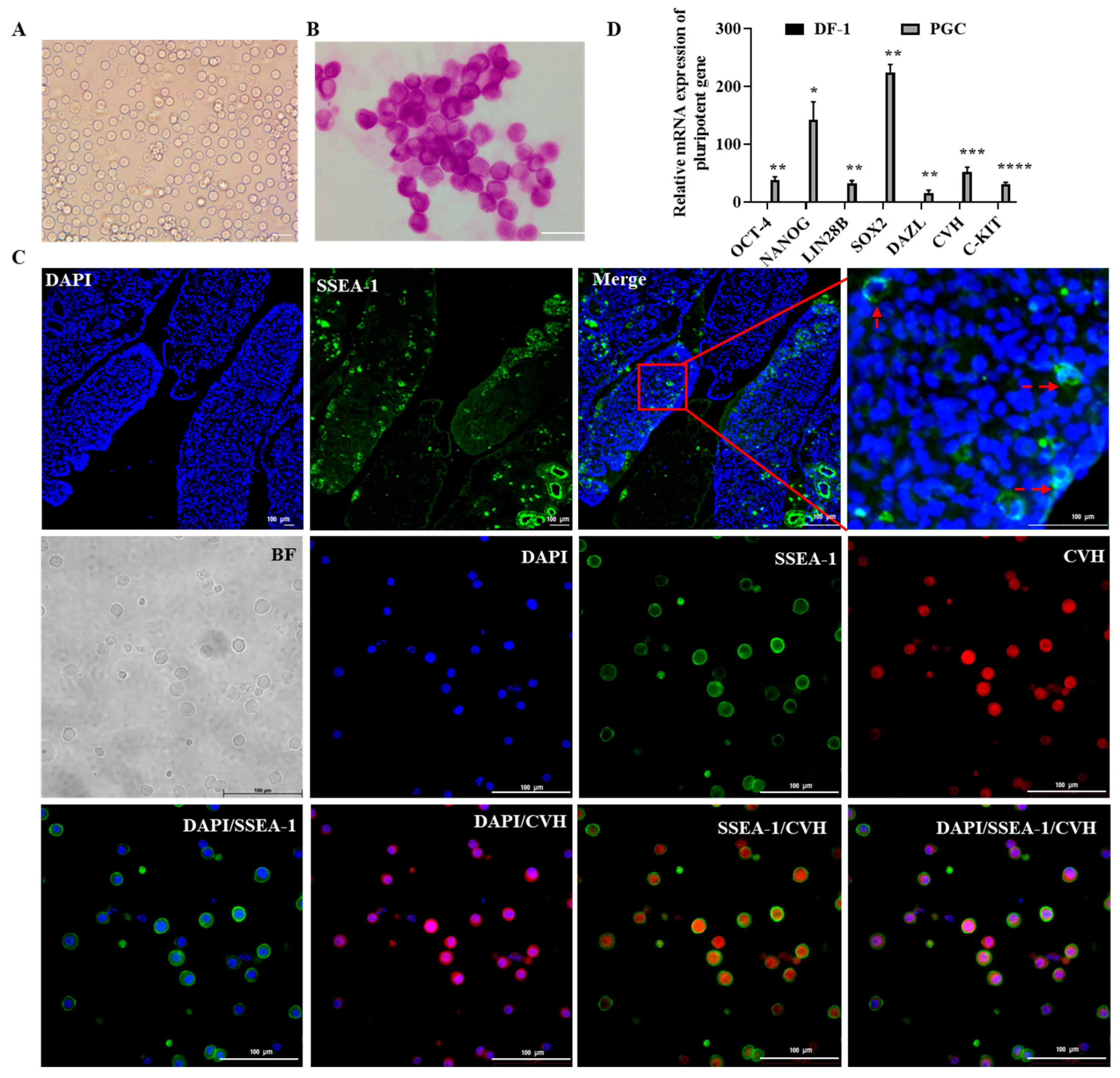
2.1. Culture and Characterization of Chicken Primordial Germ Cells

After 60 days of purification and culture, a morphologically uniform cell line was obtained (Figure S1) [5]. Morphological observation after freezing and thawing showed that the cultured cells grew in suspension and appeared circular and separately dispersed, which was consistent with the basic morphology of PGCs (Figure 1A) [33]. To further demonstrate whether the cultured cells had the biological properties of PGCs, this experiment systematically identified the cultured cells. The results showed that these cells presented PAS positivity and SSEA-1/CVH positivity, indicating that a large number of lipid droplets had attached to the cells’ surface, as well as the cells’ stemness (Figure 1B,C), consistent with the positive expression of SSEA-1 in PGCs in the gonads. PGCs are pluripotent, while somatic cells are not [33]. Therefore, RT–qPCR was then conducted and the results showed that the totipotency genes NANOG, Oct4, SOX2 and germline marker genes DAZL, CVH, and C-kit were significantly more abundant in PGCs than in somatic cells (DF-1) (Figure 1D). All these results indicated that PGCs were successfully cultured in this study.

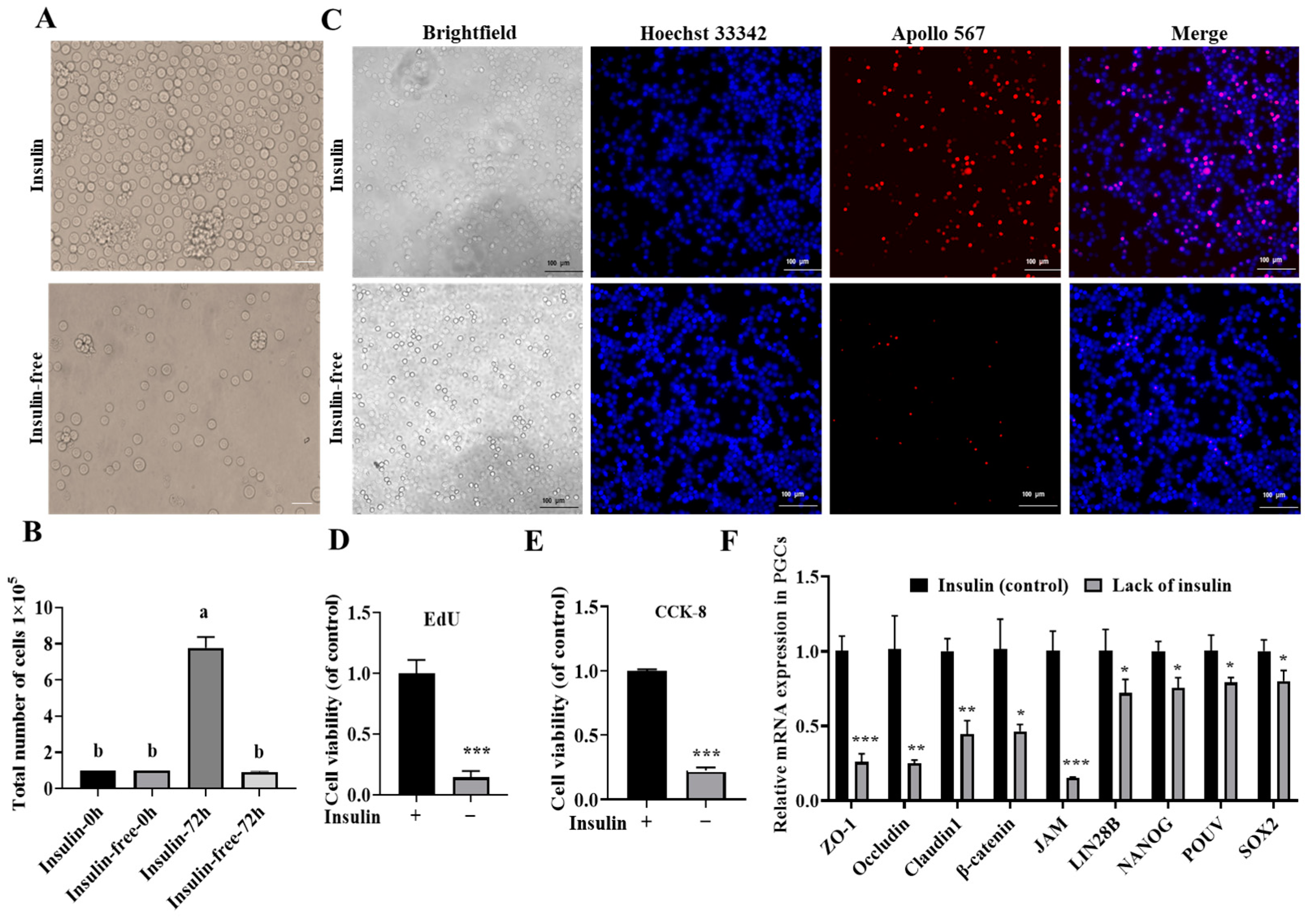
2.2. Insulin Is Indispensable for the Long-Term In Vitro Culture of PGCs

To investigate the effect of insulin on the proliferation of PGCs, B-27™ plus supplement (50×) was replaced with B-27™ minus insulin and the cells were cultured for 72 h and observed under a microscope. The results showed (Figure 2A) that there were significant proliferative changes in the insulin group and cell counts revealed that the cells in the non-insulin group did not proliferate (Figure 2B). Further proliferation assays using the CCK-8/EdU method showed (Figure 2C–E) that the efficiency of proliferation was highly significantly reduced in the non-insulin group compared to the control group (p < 0.001).
The proliferation of chicken embryonic intestinal cells is regulated by cell adhesion [34]. To further investigate whether the lack of insulin affected the cells’ adhesion, this study used qRT–PCR to detect the expression of cell adhesion-related genes. As shown in Figure 2F, the expression levels of ZO-1, Occludin, Claudin1, JAM, and β-catenin mRNA were significantly lower in the non-insulin group than in the insulin group. These results showed that non-insulin media reduce the adhesion capacity of PGCs and thus affect cell proliferation.

2.3. Non-Insulin Media Leads to Decreased Expression of Pluripotency Genes

To investigate the effect of non-insulin media on the pluripotency of PGCs, we first examined the expression of pluripotency genes, including POUV, SOX2, NANOG, and LIN28B. Figure 2F shows that the expression levels of POUV, SOX2, NANOG, and LIN28B were reduced in media without insulin, indicating that non-insulin media reduced the expression of pluripotency genes in PGCs.

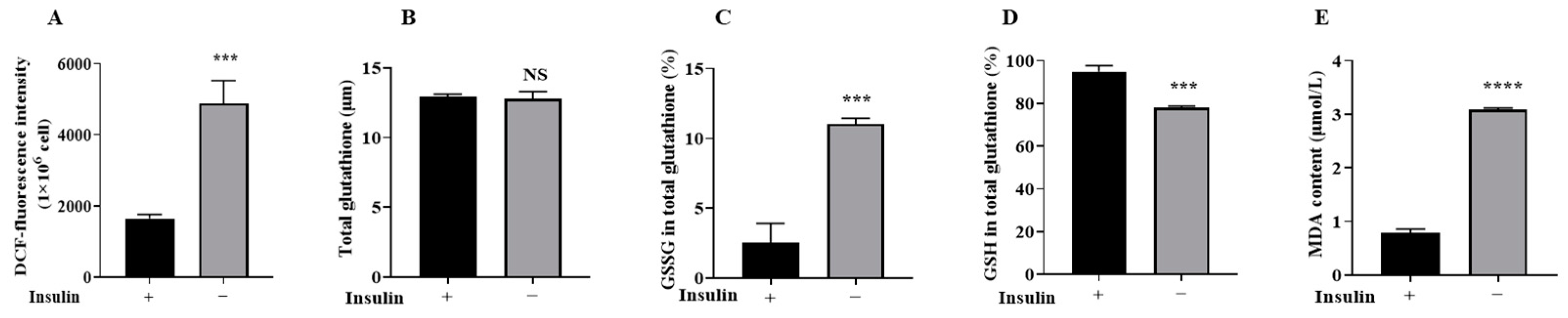
2.4. Non-Insulin Media Reduces Redox Capacity in PGCs

To investigate the effect of non-insulin media on the redox capacity of PGCs, the content of the secondary messenger ROS was examined and it was found (Figure 3A) that the ROS content significantly increased, indicating that the cellular redox capacity of PGCs was reduced. ROS can cause damage to cells through lipid peroxidation [35]; moreover, MDA and GSSG were detected and the results showed (Figure 3B–E) a significant increase in MDA and GSSG content and a decrease in glutathione (GSH) content (p < 0.05). These results suggest that the cellular redox capacity of non-insulin PGCs was reduced.

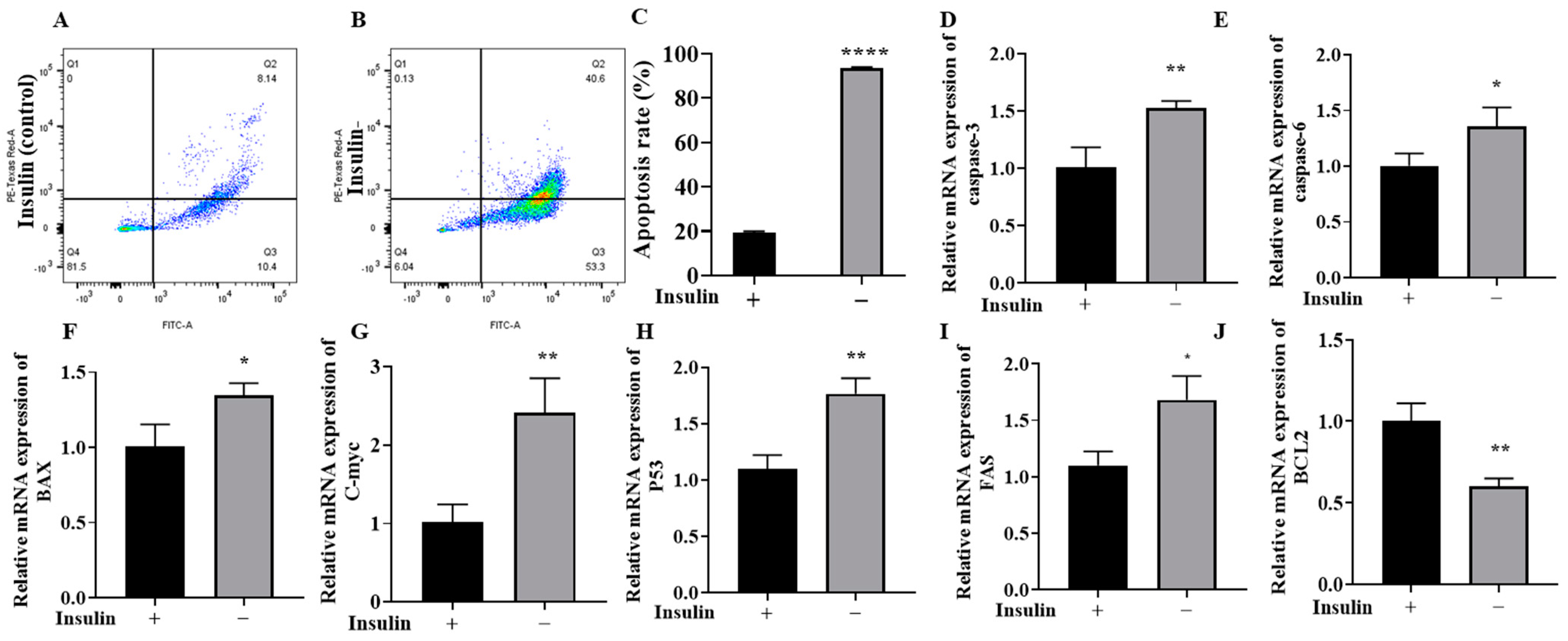
2.5. Non-Insulin Media Can Induce Apoptosis in PGCs

ROS-mediated lipid peroxidation and its products can trigger apoptosis [36]. Therefore, the effect of lack of insulin on apoptosis in PGCs was explored next (Figure 4A–C). We first found a significant increase in apoptosis in the non-insulin group by flow cytometry with FITC/PI labeling (p < 0.05). Next, qRT–PCR was used to detect the expression of apoptosis-related genes. The results showed that the expression levels of the apoptosis-related marker genes caspase-3, caspase-6, C-myc, BAX, P53, and FAS were significantly upregulated in the PGCs after treatment with non-insulin media (Figure 4D–I). In contrast, the expression of the antiapoptotic marker BCL-2 was significantly downregulated (Figure 4J) (p < 0.05). Downregulation of BCL-2 and upregulation of BAX are important markers of apoptosis in cells [37], indicating that the non-insulin media induced apoptosis in the PGCs.

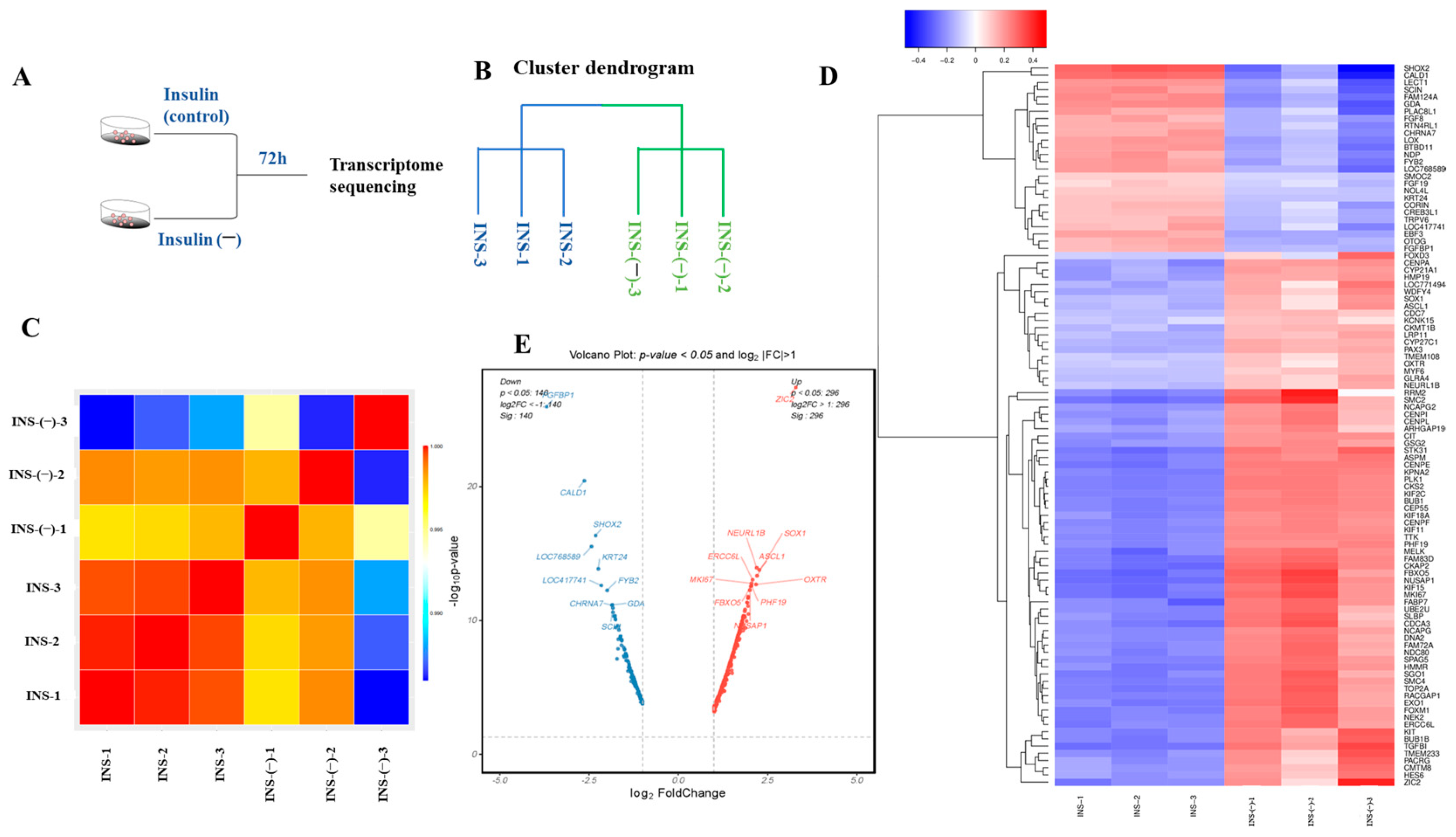
2.6. RNA-Seq Assessment of the Quality of the Data

To investigate the differential genes and signaling pathways between the non-insulin and insulin groups (Figure 5A), this study used the Illumina TruSeqTM RNA sample preKit to construct six libraries and obtain gene expression profiles of the two groups. According to the results of the sample clustering and correlation analysis (Figure 5B,C), duplicate samples were tightly clustered and different samples were relatively dispersed, indicating that the test samples exhibited high repeatability and low dispersion. The results indicated that the sequencing data were reliable and could be used for subsequent analyses of differentially expressed genes and signaling pathway enrichment. DEGs between the different groups were screened based the threshold p < 0.05 and |Log2FC| > 1. The results showed (Figure 5D,E) that there were 436 DEGs between the non-insulin and insulin groups, of which 296 genes were upregulated and 140 genes were downregulated (Table S1).

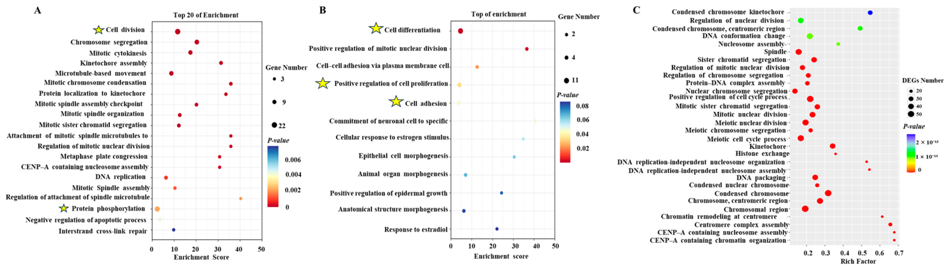
2.7. Enrichment Analysis of DEGs

GO analysis revealed that in all the differential genes, biological processes were mainly enriched in the pathways of cell division and protein phosphorylation (Figure 6A). Among the downregulated genes, GO analysis revealed them to be enriched in the pathways of cell differentiation, positive regulation of cell differentiation by proliferation, and cell adhesion (Figure 6B). Among the upregulated genes, GO analysis found them to be mainly enriched in the nucleosome assembly and sister chromatid segregation pathways (Figure 6C). To further analyze which signaling pathways were involved in regulating insulin to promote the proliferation of chicken PGCs, a KEGG analysis of the differentially expressed genes was performed. All the DEGs were mainly enriched in the calcium signaling pathway, the Wnt signaling pathway, cell adhesion, the mTOR signaling pathway, the FoxO signaling pathway, and ferroptosis (Figure 7A). All upregulated genes were enriched in the oocyte meiosis, progesterone-mediated oocyte maturation, steroid hormone biosynthesis (CYP1B1 and CYP21A1), P53 signaling pathway, and ferroptosis pathways (Figure 7B). All downregulated genes were enriched in the calcium signaling pathway, the Ras signaling pathway, ECM–receptor interactions, cell adhesion, and the PI3K/AKT signaling pathway (EREG and AREG) (Figure 7C). These results suggest that these signaling pathways play important roles in insulin-induced proliferation of chicken PGCs. Further heatmap (Figure 8A–F) analyses of the genes’ enrichment in the signaling pathways and comparison to previous experimental results again verify that lack of insulin reduces cells’ adhesion and pluripotency and promotes apoptosis in PGCs.

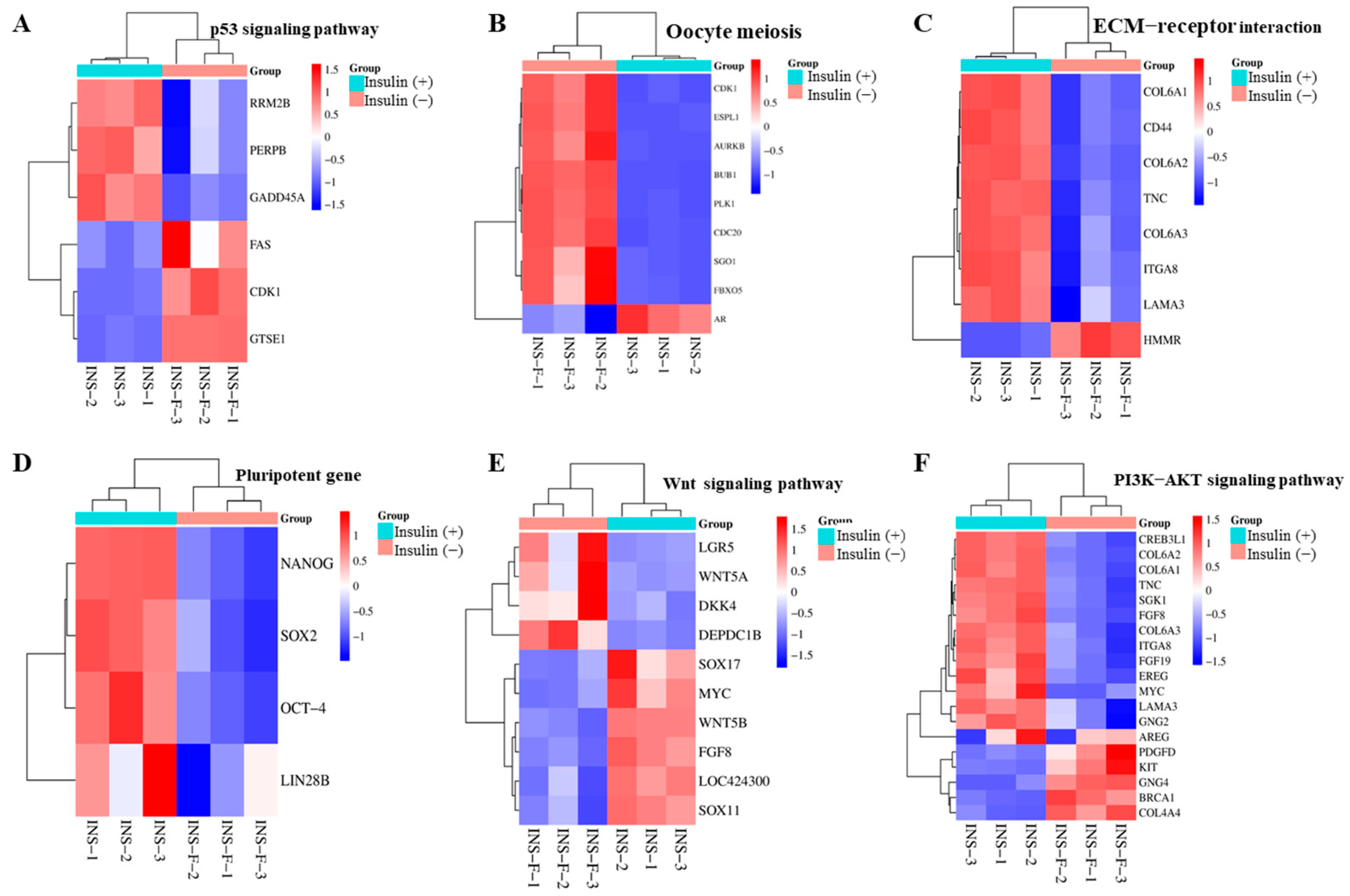
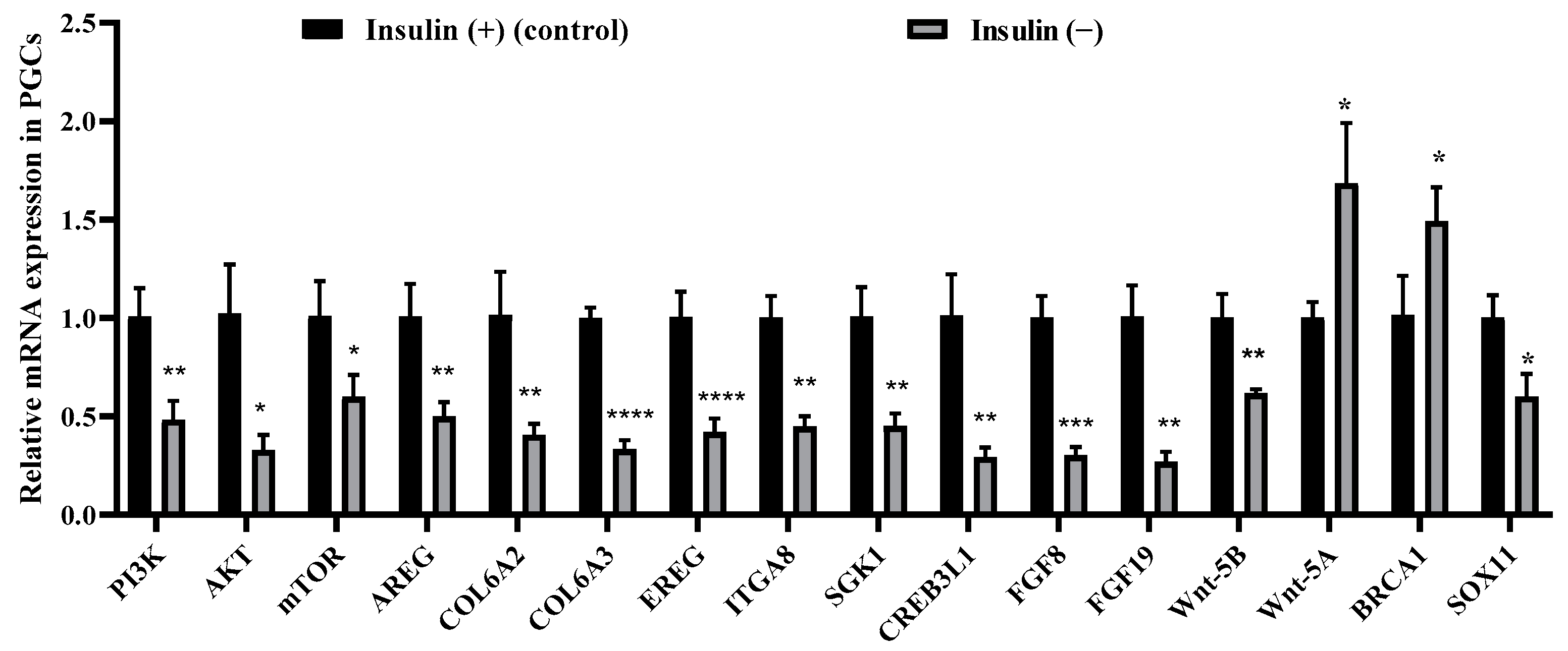
2.8. Non-Insulin Media Affect the PI3K/AKT and Wnt Signaling Pathways in PGCs

Based on the sequencing results described above, we verified the expression of genes related to the PI3K/AKT and Wnt signaling pathways by qRT–PCR (Figure 9A) and compared the fold change of qRT–PCR with the results of the RNA-seq (Figure S2) expression analysis, and the results were consistent. The qRT–PCR results showed that PI3K, AKT, mTOR, AREG, COL6A2, COL6A3, EREG, ITGA8, SGK1, CREB3L1, FGF8, FGF19, Wnt-5B, and SOX11 expression levels were significantly decreased, while the Wnt-5A and BRCA1 expression levels were significantly upregulated. These results suggested that the PI3K/AKT and Wnt signaling pathways in the PGCs were inhibited by non-insulin media, thereby affecting proliferation.

3. Discussion

The proliferation of PGCs is an extremely complex process regulated by multiple factors, such as cytokines, key genes, and signaling pathways [38]. The proliferation of PGCs in cultures is different in that an appropriate growth factor must be added to the medium [39,40]. This experiment found that PGCs in non-insulin media do not proliferate and that decreased pluripotency, cell adhesion, and decreased redox capacity lead to apoptosis. The results from the transcriptomic data analysis suggest that non-insulin media are downregulated the PI3K/AKT and Wnt pathways. The results of this study clarify the role of insulin in the proliferation of chicken PGCs and lays a foundation for further studies of its mechanism.
Insulin has been used to improve stem cell cultures [9], and promotes hESC proliferation and cell adhesion [30]. E-cadherin is a classic cadherin protein [41]. E-cadherin and α/β-catenin are activated during the proliferation of PGCs and the inhibition of E-cadherin leads to cell death and affects the development of PGCs [16,42,43]. A reduction or loss in ZO-1 can lead to slowed cell proliferation and embryonic lethality [44,45]. JAM-A deficiency or overexpression can regulate 293T human embryonic kidney epithelial cell adhesion [46]. The results of this experiment indicate that JAM-A and ZO-1 decreased in the non-insulin group and ECM–receptor interactions were downregulated in the transcriptome, suggesting that the non-insulin media suppresses the expression of adhesion-related factors and, thus, the proliferation of PGCs.
Under physiological conditions, insulin binds to insulin receptor substrate 1 (IRS-1); IRS-1 promotes IGF-1 signaling [47], which, in turn, promotes PI3K, then AKT and downstream targets, such as mTOR, GSK3β, and FOXO [48,49,50]. In human embryonic stem cells, the pluripotency marker gene Lin28 and related protein are decreased after knockdown of IGF-1. Lin28 is essential for the maintenance of chicken PGCs and can regulate the cells’ proliferation in vitro [18]. It has been shown that activation of the IGF1R–/PI3K/AKT pathway increases the expression of cancer stemness markers NANOG and SOX2 [51]. In chicken PGCs, NANOG is responsible for regulating stem cells’ pluripotency during self-renewal [10,13], forming a transcriptional network with OCT4 and SOX2 [52,53]. In this study, these genes were all downregulated, which indicates a gradual loss of pluripotency-related features [52,53]. The results of this study show that in non-insulin media, self-renewal was interrupted and the pluripotency of the PGCs decreased, which are consistent with other research findings.
Insulin induces downregulation in the IGF-1R signaling pathway in adipose-derived stem cells (ADSCs), which, in turn, results in oxidative stress, leading to a significant increase in apoptosis [54]. Redox homeostasis is essential for cellular function [55] and for increased levels of reactive oxygen species (ROS) in ESCs under oxidative stress [27]. When the concentration of ROS increases, it can cause damage to DNA and decrease glutathione (GSH) content [56,57]. The content in the oxidative stress biomarker malondialdehyde (MDA) increases after sustaining damage to cell membranes [25,26,58,59]. Disruption of the ROS/GSH balance leads to inhibited proliferation of cells and even induces cell death [60]. In Type 2 diabetic rats, insulin can reduce MDA levels in the pancreas [61]. Previous research findings indicate that insulin significantly reduces the production of ROS in human plasma [62,63] and our results are consistent with these previous studies.
ROS-mediated lipid peroxidation and its products can trigger apoptosis [36] and antioxidants increase the number of porcine PGCs in vitro by inhibiting apoptosis [64]. In addition, activation of the p53 pathway can lead to reduced proliferation of PGCs [65]. Apoptosis is mainly regulated by the BCL-2 protein family and the ratio of BCL-2/Bax proteins determines cell survival and death [66,67,68,69]. The normal development of germ cells is also highly dependent on the balanced regulation of mitochondrial apoptosis by BCL-2-family proteins and abnormal apoptotic genes impair germ cells’ differentiation [70]. A previous study suggests that the absence of insulin can lead to increased caspase-3 activity in rat hepatocytes, which may induce apoptosis [71]. Insulin significantly prevents apoptosis in rat liver cells by reducing the levels of mitochondrial pro-apoptotic Bax and cytoplasmic cytochrome C [63]. Our results are consistent with previous studies.
Insulin plays a crucial role in the formation of chicken PGCs [5]; however, the mechanism by which it regulates the proliferation of chicken PGCs is unclear. In this study, transcriptome sequencing revealed that the Wnt signaling pathway, oocyte meiosis, progesterone-mediated oocyte maturation, steroid hormone biosynthesis, and the P53 signaling pathway were involved (Figure 7A). Strict regulation of the Wnt signaling pathway is necessary for the proper development of PGCs [72,73,74]. Activation of the P53/BAX signaling pathway can induce apoptosis in hESCs [75], which is also consistent with results from our previous test. It has been shown that insulin is virtually independent of meiosis in goldfish and zebrafish oocytes [76,77] and has also been shown to induce steroidogenesis in vertebrate follicles and to induce maturation in oocytes [78]. Our transcriptomic data suggest that non-insulin media affect oocytes’ meiosis, which may provide some reference for the study of oocyte maturation; however, the exact mechanism deserves further investigation. The cortisol-related biomarker CYP1B1 [79] is the basic enzyme in the production of estrogen metabolites [80]. CYP21A1 is involved in the steroid hormone biosynthesis pathway and the overexpression of CYP21A1 promotes cells’ proliferation and inhibits apoptosis [81]. We infer that CYP1B1 and CYP21A1 do not promote the synthesis of steroid hormones at the gene level, but may promote the synthesis of steroid hormone proteins. The specific reasons need to be studied further. Downregulation in the calcium signaling pathway has also been found, with previous studies showing that insulin reduces calcium ions in mice pancreata, stimulates the activation of integrin in human embryonic stem cells, and promotes cell–matrix and cell–cell adhesion [30,82]. Compared with mammals, avian species have relatively high blood glucose levels. In obese chickens, the regulation of adipogenesis and fat deposition mediated by insulin signaling differs from that in obese mammals. Therefore, caution should be exercised when applying research findings from mammals to poultry studies [83]. The transcriptome results presented in this paper provide a theoretical basis for subsequent research on the mechanism of insulin in poultry.
Insulin activates the PI3K/AKT signaling pathway by binding to its receptor [84]. In human and mouse embryonic stem cells, the PI3K/AKT pathway is important for maintaining pluripotency, proliferation, and survival [85,86,87,88,89,90,91,92]. AREG activates the phosphoinositide 3-kinases (PI3K)/AKT/mammalian target of the rapamycin (mTOR) pathway and promotes the cycling of hematopoietic stem cells (HSCs) [93]. Insulin also can activate the Wnt signaling pathway, notably enhancing the expression of Wnt-5B in embryonic mesenchymal cells [94,95]. The functions of the Wnt and FGF signaling pathways in growing embryos are associated with various developmental processes, including cell proliferation, differentiation, migration, and apoptosis [96]. Notably, overexpression of FGF-8 can promote self-renewal in chicken PGCs [97]. Our results indicate that insulin can activate the PI3K/AKT and Wnt-5B/FGF8 signaling pathways in PGCs, consistent with previous studies.

4. Conclusions

To the best of our knowledge, this study is the first to systematically investigate the effect of insulin on the proliferation of PGCs and to identify the underlying regulatory mechanisms. Our results indicate that PGCs do not proliferate in non-insulin media and that decreased cell adhesion, cell pluripotency, and redox capacity lead to apoptosis and the downregulation of signaling pathways such as PI3K/AKT. This study contributes to a better understanding of the molecular regulation of insulin in PGCs and provides a theoretical reference for the optimization of the culture system, as well as studies signaling pathways in PGCs.

5. Materials and Methods

5.1. PGC Culture Conditions

The left gonads of 4.5-day-old Rugao yellow chicken embryos (30 pieces, repeated three times) were removed and placed in 200 μL of sterile BSA solution (Bioss, Beijing, China), crushed with a pestle, and digested with accutase enzyme (Lianke, China). After digestion for 10 min, an equal volume of the complete PGC medium (Table 1) was added, the supernatant was discarded by centrifugation, and the 48-well cultures were resuspended and incubated at 37 °C in a 5% CO2 incubator (Thermo Fisher Scientific Inc., Shanghai, China). The cultures were observed daily and the medium was changed every 3 days. Following a period of 60 days of purification and cultivation, all PGCs in the culture dish were collected. The efficiency of cell line establishment was 30% (9/30, 9/30, 9/30) and cells were frozen using serum-free cryopreservative (Ncmbio, Changzhou, China). The cells were thawed and subsequently used for downstream experimentation, encompassing all the mentioned procedures, such as PAS staining, drug treatment, cell counting, EdU proliferation assay, RNA extraction and RT–qPCR, ROS, GSH/GSSG, MDA, Western blotting, and transcriptome sequencing.

5.2. Periodic Acid–Schiff Stain

This assay used cells fixed in 4% paraformaldehyde, rinsed in distilled water, placed in Reagent A for 8 min, rinsed, and stained with Reagent B Schiff for 15 min. The membrane was rinsed and sealed, and observed under an Olympus BX41 microscope. The specific steps were conducted as specified by the periodic acid–Schiff (PAS) staining kit (Solarbio, Beijing, China).

5.3. Immunohistochemistry

The gonads were fixed with 4% paraformaldehyde for 24 h, after which they were trimmed and passed through alcohol and xylene for dehydration and transparency and dipped in wax for embedding. The trimmed wax blocks were frozen at −20 °C for 30 min and then sliced. When the tissue temperature was −20 °C, thinner continuous slices could be cut. The slices were submerged in warm water at 40 °C, the specimens were fixed with adhesion microscope slides (CITOTEST, Nantong, China), and the slides were placed in an oven at 37 °C for 12 h, after which they were stored at room temperature. The PGCs were fixed on adhesion microscope slides with 4% paraformaldehyde, infiltrated with Triton X-100, and washed three times with PBST. The cells and gonads were sealed with normal goat serum for 2 h and 6 h (Solarbio, Beijing, China), respectively. After incubation with primary antibodies against SSEA-1 (Abcam, Shanghai, China) or DDX4/MVH (Abcam, Shanghai, China) at 4 °C overnight, the membrane was washed three times with PBST and incubated with the corresponding secondary antibodies (Alexa-Fluor 488 (Abcam, China) and goat anti-rabbit IgG H&L/Cy3 (Bioss, Beijing, China)) for 2 h at room temperature. The sections were rinsed three times with PBST, titrated with the mounting medium and an antifading agent (DAPI) (Beyotime, Shanghai, China), sealed and visualized using a Leica microscope (Leica Microsystems Inc., Wetzlar, Germany).

5.4. Experimental Design

PGCs in the logarithmic growth phase were inoculated into 24-well plates (1 × 10⁵) and divided into two groups. The controls received B-27™ plus supplement (50×) (Thermo Fisher Scientific Inc., Shanghai, China) and the experimental treatment received B-27™ minus insulin (Thermo Fisher Scientific Inc., Shanghai, China) for 72 h for subsequent analyses. The only difference between the two groups was that the experimental group did not have insulin.

5.5. Cell Counting Kit-8 (CCK-8) Assay

For this assay, 10 μL of CCK-8 reagent was added to 100 μL of culture medium. Three parallel wells were set up for the experimental, control, and blank groups and PBS was added to the periphery of the unused wells to prevent evaporation of the liquid. The samples were incubated at 37 °C for 2 h in a 5% CO2 incubator, after which absorbance was measured at 450 nm (SPARK, Grödig, Austria). The cell counting kit (CCK-8) (Dojindo, Beijing, China) was used in these specific steps.

5.6. EdU Proliferation Assay

In total, 1 × 10⁵ cells per well were taken and cultured in EdU medium at a concentration of 50 μm (this concentration was obtained through preliminary experimentation) for 2 h. The culture solution was discarded for washing and the cells were fixed on slides with 4% paraformaldehyde, washed, permeabilized with Triton X-100, and stained with Apollo staining. The sections were incubated with a mounting medium and an antifading solution (DAPI) and sealed and observed with a Leica microscope. The procedure was carried out according to the manufacturer’s instructions from the Cell-Light EdU Apollo567 In Vitro Kit (Ribobio, Guangzhou, China).

5.7. RNA Extraction and Real-Time Quantitative PCR (RT–qPCR)

Total RNA was isolated using 1 mL of TRIzol reagent (Thermo Fisher Scientific Inc., Shanghai, China), according to the manufacturer’s protocol. A nanodropper was used to measure the concentration of RNA (NanoDrop 1000, Shanghai, China). Reverse transcription to cDNA was carried out using FastKing gDNA Dispelling RT SuperMix (TIANGEN, Beijing, China). RT–qPCR amplification was performed on a CFX Connect™ Real-Time System (Bio-Rad, Hercules, CA, USA) using ChamQ Universal SYBR qPCR Master Mix (Vazyme, Nanjing, China). The reaction conditions were 95 °C for 5 min followed by 40 cycles of 95 °C for 10 s and 60 °C for 30 s. After PCR amplification, a melting curve was obtained, namely, 95 °C for 15 s, 60 °C for 1 min, and 95 °C for 15 s, to verify the primers’ specificity. The relative expression of genes was calculated using the 2−ΔΔCt method after normalization to the mRNA expression of the housekeeping gene β-actin. The primers were synthesized by Beijing Tsingke Biotech; their sequences are shown in Table S1.

5.8. Detection of Apoptosis by the FITC/PI Double-Staining Method

After centrifugation at 1000 rpm for 5 min, the cells were collected, washed twice with precooled PBS, and 5 × 10⁵ cells were collected. Next, 100 μL of 1× binding buffer, 5 μL of Annexin V-FITC, and 10 μL of PI staining solution were added. The mixture was incubated for 15 min at room temperature in the dark. After that, 400 μL of 1× binding buffer was added and the mixture was assayed by flow cytometry. For the specific steps, please refer to the Annexin V-FITC/PI Apoptosis Detection Kit (Yeasen Biotechnology, Shanghai, China). We distinguished between living cells, dead cells, early apoptotic cells, and apoptotic cells. The relative proportion of early apoptotic cells (Q3) and apoptotic cells (Q2) was combined as the target of our comparison [98].

5.9. Reactive Oxygen Species Assay

Cells were collected by centrifugation, added to 10 μM DCFH-DA, incubated at 37 °C for 2 h, and mixed every 10 min. The cells were washed with a serum-free cell culture solution to remove DCFH-DA that did not enter the cells, which was detected using a microplate reader. For the specific steps, please refer to the Reactive Oxygen Species (ROS) Assay Kit (Yeasen Biotechnology, Shanghai, China).

5.10. Assay for GSH/GSSG/MDA

The content of reduced glutathione (GSH) and oxidized glutathione disulfide (GSSG) was assayed with a GSH and GSSG assay kit according to the manufacturer’s instructions (Beyotime, Shanghai, China). GSH content of the test samples was calculated as the total glutathione (GSSG) × 2. Malondialdehyde (MDA) content was measured using a lipid peroxidation MDA assay kit according to the manufacturer’s instructions (KeyGEN BioTECH, Nanjing, China). After centrifugation at 12,000× g at 4 °C for 10 min, the supernatant was harvested and assayed with a microplate reader.

5.11. RNA Extraction, Library Preparation, and Illumina HiSeq Sequencing

Total RNA was extracted from the PGCs using TRIzol®® reagent according to the manufacturer’s instructions (Invitrogen, Waltham, MA, USA), and genomic DNA were removed using DNase I (TaKaRa, Kusatsu, Shiga). Then, the quality of RNA was determined using a 2100 bioanalyser (Agilent, Santa Clara, CA, USA) and quantified using the ND-2000 spectrophotometer (NanoDrop Technologies, Wilmington, DE, USA). RNA-seq transcriptome libraries were prepared following instructions from the TruSeqTM RNA Sample Preparation Kit from Illumina (San Diego, CA, USA). The experiments were performed by the Shanghai Biozeron Biotechnology Co., Ltd. (Shanghai, China).

5.12. Differential Expression Analysis and Functional Enrichment

To identify differential expression genes (DEGs) between the two different samples, the expression level of each gene was calculated using the fragments per kilobase of exon per million mapped reads (FRKM) method. The R statistical package edgeR (https://www.bioconductor.org/packages/release/bioc/html/edgeR.html) was used for differential expression analysis (accessed on 27 September 2024). DEGs between two samples were selected using the following criteria: the logarithm of the fold change is greater than 2 and a false discovery rate (FDR) less than 0.05. To understand the functions of the differentially expressed genes, GO functional enrichment and KEGG pathway analyses were performed via https://bioinformatics.com.cn/ (accessed on 27 September 2024).

5.13. Statistical Analysis

Data are presented as the mean ± SEM. Statistical analyses were performed using SPSS (version 25.0, USA) and GraphPad Prism (GraphPad Software, version 10, USA). Histograms were drawn and statistical significance was set to p < 0.05.

Supplementary Materials

The following supporting information can be downloaded at: https://www.mdpi.com/article/10.3390/ijms26073122/s1.

Author Contributions

X.L. (Xin Liu) performed the experiments and drafted the manuscript. J.W. and Y.P. performed the experiments. H.Q., X.L. (Xiaoqian Lv), F.L., K.J., Y.N., and Q.Z. conceived and designed the experiments. J.S. and W.H. were involved in collection and analysis of the data. G.C. and B.L. were involved in the supervision and administration of the project and reviewed and edited the article. All authors have read and agreed to the published version of the manuscript.

Funding

This study was funded by the Biological Breeding-National Science and Technology Major Project (2023ZD0407503), the Excellent Youth Foundation of Jiangsu (BK20220117), the National Key R&D Program of China (2021YFD1200301), the National Natural Science Foundation of China (32272858, 32172718, 32372861, and 32202655), the China Postdoctoral Science Foundation (2022M722697), the International Science and Technology Cooperation Projects of Yangzhou (YZ2022206), the JBGS Project of Seed Industry Revitalization in Jiangsu Province (JBGS (2021) 029) and the Project Funded by the Priority Academic Program Development of Jiangsu Higher Education Institutions.

Institutional Review Board Statement

The animal study protocol was reviewed and approved by the Institutional Animal Care and Use Committee of Yangzhou University (No. YZUDWSY2017-11-07).

Informed Consent Statement

Not applicable.

Data Availability Statement

Data presented in this study are available upon request from the corresponding author.

Conflicts of Interest

The authors declare that this research was conducted in the absence of commercial or financial relationships that could be construed as potential conflicts of interest.

References

  1. Farzaneh, M.; Khoshnam, S.E.; Nokhbatolfoghahai, M. First scientific record of two cases of partial twinning in the chick embryo, Gallus gallus domesticus. Vet. Rec. Case Rep. 2016, 4, e000353. [Google Scholar]
  2. Trefil, P.; Aumann, D.; Koslová, A.; Mucksová, J.; Benešová, B.; Kalina, J.; Wurmser, C.; Fries, R.; Elleder, D.; Schusser, B.; et al. Male fertility restored by transplanting primordial germ cells into testes: A new way towards efficient transgenesis in chicken. Sci. Rep. 2017, 7, 14246. [Google Scholar]
  3. Han, J.Y.; Park, Y.H. Primordial germ cell-mediated transgenesis and genome editing in birds. J. Anim. Ence Biotechnol. 2018, 9, 19. [Google Scholar] [CrossRef] [PubMed]
  4. Woodcock, M.E.; Gheyas, A.A.; Mason, A.S.; Nandi, S.; Taylor, L.; Sherman, A.; Smith, J.; Burt, D.W.; Hawken, R.; McGrew, M.J. Reviving rare chicken breeds using genetically engineered sterility in surrogate host birds. Proc. Natl. Acad. Sci. USA 2019, 116, 20930–20937. [Google Scholar] [PubMed]
  5. Whyte, J.; Glover, J.D.; Woodcock, M.; Brzeszczynska, J.; Taylor, L.; Sherman, A.; Kaiser, P.; McGrew, M.J. FGF, Insulin, and SMAD Signaling Cooperate for Avian Primordial Germ Cell Self-Renewal. Stem Cell Rep. 2015, 5, 1171–1182. [Google Scholar]
  6. Ramos, D.M.; Abdulmalik, S.; Arul, M.R.; Sardashti, N.; Banasavadi-Siddegowda, Y.K.; Nukavarapu, S.P.; Drissi, H.; Kumbar, S.G. Insulin-Functionalized Bioactive Fiber Matrices with Bone Marrow-Derived Stem Cells in Rat Achilles Tendon Regeneration. ACS Appl. Bio Mater. 2022, 5, 2851–2861. [Google Scholar]
  7. Li, P.; Wei, J.; Gao, X.; Wei, B.; Lin, H.; Huang, R.; Niu, Y.; Lim, K.; Jing, K.; Chu, J. Insulin Promotes the Proliferation of Human Umbilical Cord Matrix-Derived Mesenchymal Stem Cells by Activating the Akt-Cyclin D1 Axis. Stem Cells Int. 2017, 2017, 7371615. [Google Scholar]
  8. Yao, Y.; Zhai, Z.; Wang, Y. Evaluation of Insulin Medium or Chondrogenic Medium on Proliferation and Chondrogenesis of ATDC5 Cells. BioMed Res. Int. 2014, 2014, 569241. [Google Scholar]
  9. Chen, G.; Gulbranson, D.R.; Hou, Z.; Bolin, J.M.; Ruotti, V.; Probasco, M.D.; Smuga-Otto, K.; Howden, S.E.; Diol, N.R.; Propson, N.E.; et al. Chemically defined conditions for human iPSC derivation and culture HHS public access. Nat. Methods 2011, 8, 424–429. [Google Scholar] [CrossRef]
  10. Cañón, S.; Herranz, C.; Manzanares, M. Germ cell restricted expression of chick Nanog. Dev. Dyn. Off. Publ. Am. Assoc. Anat. 2006, 235, 2889–2894. [Google Scholar]
  11. Lavial, F.; Acloque, H.; Bertocchini, F.; Macleod, D.J.; Boast, S.; Bachelard, E.; Montillet, G.; Thenot, S.; Sang, H.M.; Stern, C.D.; et al. The Oct4 homologue PouV and Nanog regulate pluripotency in chicken embryonic stem cells. Development 2007, 134, 3549–3563. [Google Scholar] [CrossRef] [PubMed]
  12. Shin, M.; Alev, C.; Wu, Y.; Nagai, H.; Sheng, G. Activin/TGF-beta signaling regulates Nanog expression in the epiblast during gastrulation. Mech. Dev. 2011, 128, 268–278. [Google Scholar] [CrossRef] [PubMed]
  13. Okuzaki, Y.; Kaneoka, H.; Suzuki, T.; Hagihara, Y.; Nakayama, Y.; Murakami, S.; Murase, Y.; Kuroiwa, A.; Iijima, S.; Nishijima, K.I. PRDM14 and BLIMP1 control the development of chicken primordial germ cells. Dev. Biol. 2019, 455, 32–41. [Google Scholar] [CrossRef] [PubMed]
  14. Nakashima, Y.; Omasa, T. What Kind of Signaling Maintains Pluripotency and Viability in Human-Induced Pluripotent Stem Cells Cultured on Laminin-511 with Serum-Free Medium? BioResearch Open Access 2016, 5, 84–93. [Google Scholar] [CrossRef]
  15. Ahmed, M.; Ffrench-Constant, C. Extracellular Matrix Regulation of Stem Cell Behavior. Curr. Stem Cell Rep. 2016, 2, 197–206. [Google Scholar]
  16. Li, L.; Bennett, S.A.; Wang, L. Role of E-cadherin and other cell adhesion molecules in survival and differentiation of human pluripotent stem cells. Cell Adhes. Migr. 2012, 6, 59–70. [Google Scholar] [CrossRef]
  17. Lane, S.W.; Williams, D.A.; Watt, F.M. Modulating the stem cell niche for tissue regeneration. Nat. Biotechnol. 2014, 32, 795–803. [Google Scholar] [CrossRef]
  18. Suzuki, K.; Kwon, S.J.; Saito, D.; Atsuta, Y. LIN28 is essential for the maintenance of chicken primordial germ cells. Cells Dev. 2023, 176, 203874. [Google Scholar] [CrossRef]
  19. Fagerberg, L.; Hallström, B.M.; Oksvold, P.; Kampf, C.; Djureinovic, D.; Odeberg, J.; Habuka, M.; Tahmasebpoor, S.; Danielsson, A.; Edlund, K.; et al. Analysis of the human tissue-specific expression by genome-wide integration of transcriptomics and antibody-based proteomics. Mol. Cell. Proteom. MCP 2014, 13, 397–406. [Google Scholar] [CrossRef]
  20. Mishra, M.; Akatsu, H.; Heese, K. The novel protein MANI modulates neurogenesis and neurite-cone growth. J. Cell. Mol. Med. 2011, 15, 1713–1725. [Google Scholar] [CrossRef]
  21. González-Tarragó, V.; Elosegui-Artola, A.; Bazellières, E.; Oria, R.; Pérez-González, C.; Roca-Cusachs, P. Binding of ZO-1 to α5β1 integrins regulates the mechanical properties of α5β1-fibronectin links. Mol. Biol. Cell 2017, 28, 1847–1852. [Google Scholar] [CrossRef]
  22. Rouaud, F.; Sluysmans, S.; Flinois, A.; Shah, J.; Vasileva, E.; Citi, S. Scaffolding proteins of vertebrate apical junctions: Structure, functions and biophysics. Biochim. Biophys. Acta Biomembr. 2020, 1862, 183399. [Google Scholar]
  23. Williams, A.S.; Kang, L.; Wasserman, D.H. The extracellular matrix and insulin resistance. Trends Endocrinol. Metab. TEM 2015, 26, 357–366. [Google Scholar] [PubMed]
  24. Musale, V.; Murdoch, C.E.; Banah, A.K.; Hasib, A.; Hennayake, C.K.; Dong, B.; Lang, C.C.; Wasserman, D.H.; Kang, L. Limiting extracellular matrix expansion in diet-induced obese mice reduces cardiac insulin resistance and prevents myocardial remodelling. Mol. Metab. 2024, 86, 101970. [Google Scholar] [PubMed]
  25. Tharp, K.M.; Higuchi-Sanabria, R.; Timblin, G.A.; Ford, B.; Garzon-Coral, C.; Schneider, C.; Muncie, J.M.; Stashko, C.; Daniele, J.R.; Moore, A.S.; et al. Adhesion-mediated mechanosignaling forces mitohormesis. Cell Metab. 2021, 33, 1322–1341.e1313. [Google Scholar] [CrossRef]
  26. Jiménez-Uribe, A.P.; Gómez-Sierra, T.; Aparicio-Trejo, O.E.; Orozco-Ibarra, M.; Pedraza-Chaverri, J. Backstage players of fibrosis: NOX4, mTOR, HDAC, and S1P; companions of TGF-β. Cell. Signal. 2021, 87, 110123. [Google Scholar]
  27. Solari, C.; Petrone, M.V.; Vazquez Echegaray, C.; Cosentino, M.S.; Waisman, A.; Francia, M.; Barañao, L.; Miriuka, S.; Guberman, A. Superoxide dismutase 1 expression is modulated by the core pluripotency transcription factors Oct4, Sox2 and Nanog in embryonic stem cells. Mech. Dev. 2018, 154, 116–121. [Google Scholar]
  28. Bigarella, C.L.; Liang, R.; Ghaffari, S. Stem cells and the impact of ROS signaling. Development 2014, 141, 4206–4218. [Google Scholar]
  29. Sart, S.; Song, L.; Li, Y. Controlling Redox Status for Stem Cell Survival, Expansion, and Differentiation. Oxid. Med. Cell Longev. 2015, 105135. [Google Scholar]
  30. Godoy-Parejo, C.; Deng, C.; Liu, W.; Chen, G. Insulin Stimulates PI3K/AKT and Cell Adhesion to Promote the Survival of Individualized Human Embryonic Stem Cells. Stem Cells 2019, 37, 1030–1041. [Google Scholar] [CrossRef]
  31. Li, Y.; Jiang, J.; Yang, J.; Xiao, L.; Hua, Q.; Zou, Y. PI3K/AKT/mTOR signaling participates in insulin-mediated regulation of pathological myopia-related factors in retinal pigment epithelial cells. BMC Ophthalmol. 2021, 21, 218. [Google Scholar] [CrossRef] [PubMed]
  32. Rulifson, I.C.; Karnik, S.K.; Heiser, P.W.; ten Berge, D.; Chen, H.; Gu, X.; Taketo, M.M.; Nusse, R.; Hebrok, M.; Kim, S.K. Wnt signaling regulates pancreatic beta cell proliferation. Proc. Natl. Acad. Sci. USA 2007, 104, 6247–6252. [Google Scholar] [CrossRef] [PubMed]
  33. Tagami, T.; Miyahara, D.; Nakamura, Y. Avian Primordial Germ Cells. Adv. Exp. Med. Biol. 2017, 1001, 1–18. [Google Scholar] [PubMed]
  34. Chen, M.J.; Zhou, J.Y.; Chen, Y.J.; Wang, X.Q.; Yan, H.C.; Gao, C.Q. The in ovo injection of methionine improves intestinal cell proliferation and differentiation in chick embryos by activating the JAK2/STAT3 signaling pathway. Anim. Nutr. (Zhongguo Xu Mu Shou Yi Xue Hui) 2021, 7, 1031–1038. [Google Scholar] [CrossRef]
  35. Kajarabille, N.; Latunde-Dada, G.O. Programmed Cell-Death by Ferroptosis: Antioxidants as Mitigators. Int. J. Mol. Sci. 2019, 20, 4968. [Google Scholar] [CrossRef]
  36. Kaminskyy, V.O.; Zhivotovsky, B. Free radicals in cross talk between autophagy and apoptosis. Antioxid. Redox Signal. 2014, 21, 86–102. [Google Scholar] [CrossRef]
  37. Zhang, Y.; Yang, X.; Ge, X.; Zhang, F. Puerarin attenuates neurological deficits via Bcl-2/Bax/cleaved caspase-3 and Sirt3/SOD2 apoptotic pathways in subarachnoid hemorrhage mice. Biomed. Pharmacother. = Biomed. Pharmacother. 2019, 109, 726–733. [Google Scholar] [CrossRef]
  38. Sasaki, H.; Matsui, Y. Epigenetic events in mammalian germ-cell development: Reprogramming and beyond. Nat. Rev. Genet. 2008, 9, 129–140. [Google Scholar] [CrossRef]
  39. Ge, C.; Yu, M.; Petitte, J.N.; Zhang, C. Epidermal growth factor-induced proliferation of chicken primordial germ cells: Involvement of calcium/protein kinase C and NFKB1. Biol. Reprod. 2009, 80, 528–536. [Google Scholar] [CrossRef]
  40. Choi, J.W.; Kim, S.; Kim, T.M.; Kim, Y.M.; Seo, H.W.; Park, T.S.; Jeong, J.W.; Song, G.; Han, J.Y. Basic fibroblast growth factor activates MEK/ERK cell signaling pathway and stimulates the proliferation of chicken primordial germ cells. PLoS ONE 2010, 5, e12968. [Google Scholar] [CrossRef]
  41. Garcia, M.A.; Nelson, W.J.; Chavez, N. Cell-Cell Junctions Organize Structural and Signaling Networks. Cold Spring Harb. Perspect. Biol. 2018, 10, a029181. [Google Scholar]
  42. Shim, S.R.; Kook, S.; Kim, J.I.; Song, W.K. Degradation of focal adhesion proteins paxillin and p130cas by caspases or calpains in apoptotic rat-1 and L929 cells. Biochem. Biophys. Res. Commun. 2001, 286, 601–608. [Google Scholar] [PubMed]
  43. Okamura, D.; Kimura, T.; Nakano, T.; Matsui, Y. Cadherin-mediated cell interaction regulates germ cell determination in mice. Development 2003, 130, 6423–6430. [Google Scholar] [PubMed]
  44. Xu, J.; Kausalya, P.J.; Phua, D.C.; Ali, S.M.; Hossain, Z.; Hunziker, W. Early embryonic lethality of mice lacking ZO-2, but Not ZO-3, reveals critical and nonredundant roles for individual zonula occludens proteins in mammalian development. Mol. Cell. Biol. 2008, 28, 1669–1678. [Google Scholar] [PubMed]
  45. González-Mariscal, L.; Domínguez-Calderón, A.; Raya-Sandino, A.; Ortega-Olvera, J.M.; Vargas-Sierra, O.; Martínez-Revollar, G. Tight junctions and the regulation of gene expression. Semin. Cell Dev. Biol. 2014, 36, 213–223. [Google Scholar]
  46. Severson, E.A.; Jiang, L.; Ivanov, A.I.; Mandell, K.J.; Nusrat, A.; Parkos, C.A. Cis-dimerization mediates function of junctional adhesion molecule A. Mol. Biol. Cell 2008, 19, 1862–1872. [Google Scholar]
  47. Yoneyama, Y.; Lanzerstorfer, P.; Niwa, H.; Umehara, T.; Shibano, T.; Yokoyama, S.; Chida, K.; Weghuber, J.; Hakuno, F.; Takahashi, S.I. IRS-1 acts as an endocytic regulator of IGF-I receptor to facilitate sustained IGF signaling. eLife 2018, 7, e32893. [Google Scholar] [CrossRef]
  48. Yazıcı, D.; Sezer, H. Insulin Resistance, Obesity and Lipotoxicity. Adv. Exp. Med. Biol. 2017, 960, 277–304. [Google Scholar]
  49. Blázquez, E.; Velázquez, E.; Hurtado-Carneiro, V.; Ruiz-Albusac, J.M. Insulin in the brain: Its pathophysiological implications for States related with central insulin resistance, type 2 diabetes and Alzheimer’s disease. Front. Endocrinol. 2014, 5, 161. [Google Scholar] [CrossRef]
  50. Talbot, K. Brain insulin resistance in Alzheimer’s disease and its potential treatment with GLP-1 analogs. Neurodegener. Dis. Manag. 2014, 4, 31–40. [Google Scholar] [CrossRef]
  51. Zhou, L.; Li, H.; Sun, T.; Wen, X.; Niu, C.; Li, M.; Li, W.; Hoffman, A.R.; Hu, J.-F.; Cui, J. HULC targets the IGF1R–PI3K-AKT axis in trans to promote breast cancer metastasis and cisplatin resistance. Cancer Lett. 2022, 548, 215861. [Google Scholar] [CrossRef] [PubMed]
  52. Naeemipour, M.; Dehghani, H.; Bassami, M.; Bahrami, A. Expression dynamics of pluripotency genes in chicken primordial germ cells before and after colonization of the genital ridges. Mol. Reprod. Dev. 2013, 80, 849–861. [Google Scholar] [CrossRef] [PubMed]
  53. Rengaraj, D.; Won, S.; Jung, K.M.; Woo, S.J.; Lee, H.; Kim, Y.M.; Kim, H.; Han, J.Y. Chicken blastoderms and primordial germ cells possess a higher expression of DNA repair genes and lower expression of apoptosis genes to preserve their genome stability. Sci. Rep. 2022, 12, 49. [Google Scholar] [CrossRef] [PubMed]
  54. Circu, M.L.; Aw, T.Y. Reactive oxygen species, cellular redox systems, and apoptosis. Free Radic. Biol. Med. 2010, 48, 749–762. [Google Scholar] [CrossRef]
  55. Ufer, C.; Wang, C.C. The Roles of Glutathione Peroxidases during Embryo Development. Front. Mol. Neurosci. 2011, 4, 12. [Google Scholar] [CrossRef]
  56. Lima, C.F.; Fernandes-Ferreira, M.; Pereira-Wilson, C. Phenolic compounds protect HepG2 cells from oxidative damage: Relevance of glutathione levels. Life Sci. 2006, 79, 2056–2068. [Google Scholar] [CrossRef]
  57. An, J.; Hu, J.; Shang, Y.; Zhong, Y.; Zhang, X.; Yu, Z. The cytotoxicity of organophosphate flame retardants on HepG2, A549 and Caco-2 cells. J. Environ. Sci. Health. Part A Toxic/Hazard. Subst. Environ. Eng. 2016, 51, 980–988. [Google Scholar] [CrossRef]
  58. Wang, Y.; Lin, S.; Jiang, P.; Song, Y.; Zhao, Y.; Zheng, Y. Focal Adhesion Kinase Inhibitor Inhibits the Oxidative Damage Induced by Central Venous Catheter via Abolishing Focal Adhesion Kinase-Protein Kinase B Pathway Activation. BioMed Res. Int. 2021, 2021, 6685493. [Google Scholar] [CrossRef]
  59. Wang, B.; Wang, Y.; Zhang, J.; Hu, C.; Jiang, J.; Li, Y.; Peng, Z. ROS-induced lipid peroxidation modulates cell death outcome: Mechanisms behind apoptosis, autophagy, and ferroptosis. Arch. Toxicol. 2023, 97, 1439–1451. [Google Scholar] [CrossRef]
  60. Liu, T.; Sun, L.; Zhang, Y.; Wang, Y.; Zheng, J. Imbalanced GSH/ROS and sequential cell death. J. Biochem. Mol. Toxicol. 2022, 36, e22942. [Google Scholar] [CrossRef]
  61. Matsathit, U.; Sermwittayawong, D.; Komolgriengkrai, M.; Khimmaktong, W. Glucan-rich polysaccharides obtained from split gill mushroom [Schizophyllum commune (Fr.)] ameliorate hyperglycemia by enhancing insulin and GLUT2 pancreas in type 2 diabetic rats. Histol. Histopathol. 2024, 39, 191–203. [Google Scholar]
  62. Dandona, P.; Ghanim, H.; Bandyopadhyay, A.; Korzeniewski, K.; Ling Sia, C.; Dhindsa, S.; Chaudhuri, A. Insulin suppresses endotoxin-induced oxidative, nitrosative, and inflammatory stress in humans. Diabetes Care 2010, 33, 2416–2423. [Google Scholar] [PubMed]
  63. Francés, D.E.; Ronco, M.T.; Monti, J.A.; Ingaramo, P.I.; Pisani, G.B.; Parody, J.P.; Pellegrino, J.M.; Sanz, P.M.; Carrillo, M.C.; Carnovale, C.E. Hyperglycemia induces apoptosis in rat liver through the increase of hydroxyl radical: New insights into the insulin effect. J. Endocrinol. 2010, 205, 187–200. [Google Scholar] [PubMed]
  64. Lee, C.K.; Weaks, R.L.; Johnson, G.A.; Bazer, F.W.; Piedrahita, J.A. Effects of protease inhibitors and antioxidants on In vitro survival of porcine primordial germ cells. Biol. Reprod. 2000, 63, 887–897. [Google Scholar] [PubMed]
  65. Teng, H.; Sui, X.; Zhou, C.; Shen, C.; Yang, Y.; Zhang, P.; Guo, X.; Huo, R. Fatty acid degradation plays an essential role in proliferation of mouse female primordial germ cells via the p53-dependent cell cycle regulation. Cell Cycle 2016, 15, 425–431. [Google Scholar]
  66. Singh, R.; Letai, A.; Sarosiek, K. Regulation of apoptosis in health and disease: The balancing act of BCL-2 family proteins. Nat. Rev. Mol. Cell Biol. 2019, 20, 175–193. [Google Scholar]
  67. Deng, J.; Shimamura, T.; Perera, S.; Carlson, N.E.; Cai, D.; Shapiro, G.I.; Wong, K.K.; Letai, A. Proapoptotic BH3-Only BCL-2 Family Protein BIM Connects Death Signaling from Epidermal Growth Factor Receptor Inhibition to the Mitochondrion. Cancer Res. 2007, 67, 11867–11875. [Google Scholar]
  68. Sarosiek, K.A.; Letai, A. Directly targeting the mitochondrial pathway of apoptosis for cancer therapy using BH3 mimetics—Recent successes, current challenges and future promise. FEBS J. 2016, 283, 3523–3533. [Google Scholar]
  69. Ricci, C.; Pastukh, V.; Mozaffari, M.; Schaffer, S.W. Insulin withdrawal induces apoptosis via a free radical-mediated mechanism. Can. J. Physiol. Pharmacol. 2007, 85, 455–464. [Google Scholar]
  70. Okamoto, T.; Coultas, L.; Metcalf, D.; van Delft, M.F.; Glaser, S.P.; Takiguchi, M.; Strasser, A.; Bouillet, P.; Adams, J.M.; Huang, D.C. Enhanced stability of Mcl1, a prosurvival Bcl2 relative, blunts stress-induced apoptosis, causes male sterility, and promotes tumorigenesis. Proc. Natl. Acad. Sci. USA 2014, 111, 261–266. [Google Scholar]
  71. Pagliassotti, M.J.; Wei, Y.; Wang, D. Insulin protects liver cells from saturated fatty acid-induced apoptosis via inhibition of c-Jun NH2 terminal kinase activity. Endocrinology 2007, 148, 3338–3345. [Google Scholar] [PubMed]
  72. Chawengsaksophak, K.; Svingen, T.; Ng, E.T.; Epp, T.; Spiller, C.M.; Clark, C.; Cooper, H.; Koopman, P. Loss of Wnt5a disrupts primordial germ cell migration and male sexual development in mice. Biol. Reprod. 2012, 86, 1–12. [Google Scholar]
  73. Kimura, T.; Nakamura, T.; Murayama, K.; Umehara, H.; Yamano, N.; Watanabe, S.; Taketo, M.M.; Nakano, T. The stabilization of beta-catenin leads to impaired primordial germ cell development via aberrant cell cycle progression. Dev. Biol. 2006, 300, 545–553. [Google Scholar] [PubMed]
  74. Laird, D.J.; Altshuler-Keylin, S.; Kissner, M.D.; Zhou, X.; Anderson, K.V. Ror2 enhances polarity and directional migration of primordial germ cells. PLoS Genet. 2011, 7, e1002428. [Google Scholar]
  75. Zhang, L.; Ma, L.; Yan, T.; Han, X.; Xu, J.; Xu, J.; Xu, X. Activated mitochondrial apoptosis in hESCs after dissociation involving the PKA/p-p53/Bax signaling pathway. Exp. Cell Res. 2018, 369, 226–233. [Google Scholar]
  76. Lessman, C.A. Effect of insulin on meiosis reinitiation induced in vitro by three progestogens in oocytes of the goldfish (Carassius auratus). Dev. Biol. 1985, 107, 259–263. [Google Scholar]
  77. Maitra, S.; Das, D.; Ghosh, P.; Hajra, S.; Roy, S.S.; Bhattacharya, S. High cAMP attenuation of insulin-stimulated meiotic G2-M1 transition in zebrafish oocytes: Interaction between the cAMP-dependent protein kinase (PKA) and the MAPK3/1 pathways. Mol. Cell. Endocrinol. 2014, 393, 109–119. [Google Scholar] [PubMed]
  78. Lessman, C.A.; Schuetz, A.W. Role of follicle wall in meiosis reinitiation induced by insulin in Rana pipiens oocytes. Am. J. Physiol. 1981, 241, E51–E56. [Google Scholar]
  79. Wang, J.J.; Xu, F.B.; Hu, S.; Xu, Y.M.; Wang, X.Z. Identification and validation of cortisol-related hub biomarkers and the related pathogenesis of biomarkers in Ischemic Stroke. Brain Behav. 2024, 14, e3358. [Google Scholar]
  80. Sowers, M.R.; Wilson, A.L.; Kardia, S.R.; Chu, J.; McConnell, D.S. CYP1A1 and CYP1B1 polymorphisms and their association with estradiol and estrogen metabolites in women who are premenopausal and perimenopausal. Am. J. Med. 2006, 119, S44–S51. [Google Scholar]
  81. You, Z.; Yuan, J.; Wang, Y.; Sun, Y.; Ni, A.; Li, Y.; Ma, H.; Ma, T.; Chen, J. Integrated transcriptomic analysis on chicken ovary reveals CYP21A1 affects follicle granulosa cell development and steroid hormone synthesis. Poult. Sci. 2024, 103, 103589. [Google Scholar] [CrossRef] [PubMed]
  82. Bruce, J.I.E.; Sánchez-Alvarez, R.; Sans, M.D.; Sugden, S.A.; Qi, N.; James, A.D.; Williams, J.A. Insulin protects acinar cells during pancreatitis by preserving glycolytic ATP supply to calcium pumps. Nat. Commun. 2021, 12, 4386. [Google Scholar] [CrossRef] [PubMed]
  83. Geng, T.; Xia, L.; Li, F.; Xia, J.; Zhang, Y.; Wang, Q.; Yang, B.; Montgomery, S.; Cui, H.; Gong, D. The role of endoplasmic reticulum stress and insulin resistance in the occurrence of goose fatty liver. Biochem. Biophys. Res. Commun. 2015, 465, 83–87. [Google Scholar] [CrossRef]
  84. Thapa, N.; Chen, M.; Horn, H.T.; Choi, S.; Wen, T.; Anderson, R.A. Phosphatidylinositol-3-OH kinase signalling is spatially organized at endosomal compartments by microtubule-associated protein 4. Nat. Cell Biol. 2020, 22, 1357–1370. [Google Scholar] [CrossRef] [PubMed]
  85. Alva, J.A.; Lee, G.E.; Escobar, E.E.; Pyle, A.D. Phosphatase and tensin homolog regulates the pluripotent state and lineage fate choice in human embryonic stem cells. Stem Cells 2011, 29, 1952–1962. [Google Scholar] [CrossRef]
  86. Armstrong, L.; Hughes, O.; Yung, S.; Hyslop, L.; Stewart, R.; Wappler, I.; Peters, H.; Walter, T.; Stojkovic, P.; Evans, J.; et al. The role of PI3K/AKT, MAPK/ERK and NFkappabeta signalling in the maintenance of human embryonic stem cell pluripotency and viability highlighted by transcriptional profiling and functional analysis. Hum. Mol. Genet. 2006, 15, 1894–1913. [Google Scholar] [CrossRef]
  87. Jirmanova, L.; Afanassieff, M.; Gobert-Gosse, S.; Markossian, S.; Savatier, P. Differential contributions of ERK and PI3-kinase to the regulation of cyclin D1 expression and to the control of the G1/S transition in mouse embryonic stem cells. Oncogene 2002, 21, 5515–5528. [Google Scholar] [CrossRef]
  88. McLean, A.B.; D’Amour, K.A.; Jones, K.L.; Krishnamoorthy, M.; Kulik, M.J.; Reynolds, D.M.; Sheppard, A.M.; Liu, H.; Xu, Y.; Baetge, E.E.; et al. Activin a efficiently specifies definitive endoderm from human embryonic stem cells only when phosphatidylinositol 3-kinase signaling is suppressed. Stem Cells 2007, 25, 29–38. [Google Scholar] [CrossRef]
  89. Paling, N.R.; Wheadon, H.; Bone, H.K.; Welham, M.J. Regulation of embryonic stem cell self-renewal by phosphoinositide 3-kinase-dependent signaling. J. Biol. Chem. 2004, 279, 48063–48070. [Google Scholar] [CrossRef]
  90. Shepherd, P.R.; Navé, B.T.; Siddle, K. Insulin stimulation of glycogen synthesis and glycogen synthase activity is blocked by wortmannin and rapamycin in 3T3-L1 adipocytes: Evidence for the involvement of phosphoinositide 3-kinase and p70 ribosomal protein-S6 kinase. Biochem. J. 1995, 305 Pt 1, 25–28. [Google Scholar] [CrossRef]
  91. Kurtzhals, P.; Schaffer, L.; Sorensen, A.; Kristensen, C.; Jonassen, I.; Schmid, C.; Trub, T. Correlations of receptor binding and metabolic and mitogenic potencies of insulin analogs designed for clinical use. Diabetes 2000, 49, 999–1005. [Google Scholar] [PubMed]
  92. Tennagels, N.; Werner, U. The metabolic and mitogenic properties of basal insulin analogues. Arch. Physiol. Biochem. 2013, 119, 1–14. [Google Scholar] [CrossRef]
  93. Wu, L.; Lin, Q.; Chatla, S.; Amarachintha, S.; Wilson, A.F.; Atale, N.; Gao, Z.J.; Joseph, J.; Wolff, E.V.; Du, W. LepR+ niche cell-derived AREG compromises hematopoietic stem cell maintenance under conditions of DNA repair deficiency and aging. Blood 2023, 142, 1529–1542. [Google Scholar]
  94. Wong, S.K.; Mohamad, N.V.; Jayusman, P.A.; Ibrahim, N. A Review on the Crosstalk between Insulin and Wnt/β-Catenin Signalling for Bone Health. Int. J. Mol. Sci. 2023, 24, 12441. [Google Scholar] [CrossRef] [PubMed]
  95. Heller, R.S.; Dichmann, D.S.; Jensen, J.; Miller, C.; Wong, G.; Madsen, O.D.; Serup, P. Expression patterns of Wnts, Frizzleds, sFRPs, and misexpression in transgenic mice suggesting a role for Wnts in pancreas and foregut pattern formation. Dev. Dyn. 2010, 225, 260–270. [Google Scholar] [CrossRef] [PubMed]
  96. Choe, C.P.; Collazo, A.; Trinh, L.A.; Pan, L.; Moens, C.B.; Crump, J.G. Wnt-dependent epithelial transitions drive pharyngeal pouch formation. Dev. Cell 2013, 24, 296–309. [Google Scholar]
  97. Wang, M.; Zhang, C.; Huang, C.; Cheng, S.; He, N.; Wang, Y.; Ahmed, M.F.; Zhao, R.; Jin, J.; Zuo, Q.; et al. Regulation of fibroblast growth factor 8 (FGF8) in chicken embryonic stem cells differentiation into spermatogonial stem cells. J. Cell. Biochem. 2018, 119, 2396–2407. [Google Scholar]
  98. Chen, Q.; Zhang, F.; Dong, L.; Wu, H.; Xu, J.; Li, H.; Wang, J.; Zhou, Z.; Liu, C.; Wang, Y.; et al. SIDT1-dependent absorption in the stomach mediates host uptake of dietary and orally administered microRNAs. Cell Res. 2021, 31, 247–258. [Google Scholar] [CrossRef]
Figure 1. The culture and characterization of chicken primordial germ cells. (A) Morphological observation of PGCs cells after freezing and thawing; scale bar = 20 μm. (B) PAS staining diagram; bar = 10 μm. (C) Immunofluorescence of SSEA-1/CVH in PGCs in the gonads and PGCs cultured in vitro; bar = 100 μm. Red squares is a partial enlargement of a specific area in the ‘Merge’ image, used to display details more clearly, red arrow indicates PGCs (D) Expression of pluripotency and germline marker genes; n = 3, * p < 0.05, ** p < 0.01, *** p < 0.001, and **** p < 0.0001.
Ijms 26 03122 g001
Figure 2. Insulin is indispensable for the long-term in vitro culture of PGCs. (A) Morphological observation of two groups of cultured PGCs; bar = 20 μm. (B) Cell counts; different superscripts represent significant differences. (C,D) EdU experimental fluorescence plots and statistical results. (E) The statistical results of the CCK-8 assay. (F) Expression of cell adhesion and pluripotency genes; * p < 0.05, ** p < 0.01, *** p < 0.001.
Ijms 26 03122 g002
Figure 3. Non-insulin media reduces redox capacity in PGCs. (A) A statistical graph of the change in ROS content of the two groups. (BD) Statistical analyses of changes in the intracellular GSH/GSSH ratio. (E) The fffect of the non-insulin medium on MDA levels; n = 3, *** p < 0.001, **** p < 0.0001.
Ijms 26 03122 g003
Figure 4. Non-insulin media can induce apoptosis in PGCs. (AC) Apoptosis measured by flow cytometry and graphs of the statistics. (DJ) The effect of non-insulin media on apoptosis genes in the PGCs. n = 3, * p < 0.05, ** p < 0.01, and **** p < 0.0001.
Ijms 26 03122 g004
Figure 5. RNA-seq assessment of the quality of the data. (A) Sample source. (B) An example of a dendrogram comparing the non-insulin and insulin-treated groups. (C) An example of a correlation analysis where red means a strong correlation and blue means a weak correlation. (D) A statistical map of differential genes. The red columns represent upregulated genes and the blue columns represent downregulated genes. (E) A volcano map of differentially expressed genes between the non-insulin and insulin-treated samples. The red dots represent significantly upregulated genes; the blue dots represent significantly downregulated genes.
Ijms 26 03122 g005
Figure 6. The GO enrichment analyses of the DEGs between the non-insulin and insulin-treated samples. (A) The GO analysis of all differentially expressed genes. Biological processes were mainly enriched in cell division and protein phosphorylation. (B) The GO analysis of all downregulated genes. (C) The GO analysis of all upregulated genes. Cell differentiation, cell adhesion, etc., are marked with an asterisk.
Ijms 26 03122 g006
Figure 7. KEGG enrichment analyses of the DEGs between the non-insulin and insulin-treated samples. (A) The KEGG pathway enrichment analysis of all genes. (B) The KEGG pathway enrichment analysis of all upregulated genes. (C) The KEGG pathway enrichment analysis of all downregulated genes. Pathways of interest for the subsequent heatmap analyses are marked with an asterisk.
Ijms 26 03122 g007
Figure 8. Heatmaps showing the clustering of signaling pathway genes. (A): p53 signaling pathway related gene expression heatmap; (B): Heat map of gene expression related to meiosis in oocytes; (C): Heat map of gene expression related to ECM−receptor interaction; (D): Heat map of Pluripotent gene expression; (E): Wnt signaling pathway related gene expression heatmap; (F): PI3K−AKT signaling pathway related gene expression heatmap.
Ijms 26 03122 g008
Figure 9. Non-insulin media inhibit the PI3K/AKT signaling pathway in PGCs. The effect of insulin media on the PI3K/AKT and Wnt signaling pathways genes detected by qRT–PCR; n = 3, * p < 0.05, ** p < 0.01, *** p < 0.001, and **** p < 0.0001.
Ijms 26 03122 g009
Table 1. Cell culture medium.
DMEM Basal Medium
CompositionSourceConcentrationVolume
DMEMMeilunbio-PWL037, Dalian, China75%37.5 mL
Ultra-filtered waterSigma-W3500, Shanghai, China24%12 mL
CaCl2·2H2OSigma-C7902, Shanghai, China0.15 mM0.5 mL
DMEM basal mediumDiscard 3.368ml 46.632 mL
B-27TM supplement (control)Gibco-17504044, Shanghai, China1 mL
B-27TM supplement, minus insulin (experimental group)Gibco-A1895601, Shanghai, China1 mL
GlutaMaxGibco-35050061, Shanghai, China2 mM0.5 mL
MEM NEAAGibco-11140050, Shanghai, China0.5 mL
2-MercaptoethanolGibco-21985023, Shanghai, China0.1 mM91 µL
Chicken serumGibco-16110082, Shanghai, China0.2%100 µL
EmbryoMax nucleosidesSigma-ES-008-D, Shanghai, China0.5 mL
Sodium pyruvateGibco-11360070, Shanghai, China1.2 mM0.6 mL
OvalbumlnSigma-A5503, Shanghai, China0.2%0.1 g
Sodium heparinMCE-HY-17567A, Shanghai, China0.01%50 µL
Basic fibroblast growth factorMCE-HY-P70600, Shanghai, China20 µL
Human Activin AMCE-HY-P70311, Shanghai, China25 µL
Pen strepGibco-15070063, Shanghai, China0.5 mL
Disclaimer/Publisher’s Note: The statements, opinions and data contained in all publications are solely those of the individual author(s) and contributor(s) and not of MDPI and/or the editor(s). MDPI and/or the editor(s) disclaim responsibility for any injury to people or property resulting from any ideas, methods, instructions or products referred to in the content.

