Review

Luteinizing Hormone Regulates Testosterone Production, Leydig Cell Proliferation, Differentiation, and Circadian Rhythm During Spermatogenesis

The Sperm Laboratory, College of Life Sciences, Zhejiang University, Hangzhou 310058, China
*
Author to whom correspondence should be addressed.
Authors contributed equally to this work.
Int. J. Mol. Sci. 2025, 26(8), 3548; https://doi.org/10.3390/ijms26083548
Submission received: 23 February 2025 / Revised: 3 April 2025 / Accepted: 6 April 2025 / Published: 10 April 2025
(This article belongs to the Section Molecular Biology)

Abstract

:
Male reproductive health, particularly the regulation of spermatogenesis, is controlled by a complex combination of factors, including luteinizing hormone (LH) and its effects on Leydig cells (LCs). LH stimulates testosterone synthesis in LCs, which is critical for maintaining spermatogenesis and male fertility. This review examines the pathways through which LH regulates testosterone production, LC proliferation, differentiation, and circadian rhythm in human and non-human species. In particular, the signaling pathways of luteinizing hormone involved in testosterone production are discussed. Additionally, we explore LH’s role in sperm maturation and quality, emphasizing its clinical implications in treating hypogonadotropic hypogonadism and diagnosing gonadal dysfunctions such as androgen insensitivity syndrome and precocious puberty. Furthermore, the potential of LH in assisted reproductive technologies for improving sperm quality is discussed. By highlighting key molecular mechanisms, this work provides insights into the therapeutic potential of LH in addressing male infertility and conditions of LC dysfunction.

1. Introduction

In contemporary society, humans are confronted with a plethora of health issues, with reproductive health concerns garnering increasing attention. A recent study examining sperm quality in Portuguese sperm donors revealed a consistent decline in sperm quality from 2011 to 2018 [1]. Furthermore, 8% to 12% of couples globally are unable to conceive, with male factors accounting for around 50% of cases [2]. Between 1990 and 2017, the rate of age-standardized infertility among men grew by 0.291% year [2]. All of these emphasize the importance of focusing on male reproductive health.
The testis is one of the male sex organs and contains seminiferous tubules and interstitial tissue. Nerves, mast cells, fibrocytes, lymphocytes, LCs, macrophages, and monocytes are all found in the interstitial tissue. The basement membrane separates the seminiferous tubules from interstitial tissues [3]. The synthesis of testosterone by LCs has been demonstrated, and they are crucial for the development of secondary sexual characteristics, gonadal development, and spermatogenesis [4]. Based on their appearance and function, LCs can be classified as either fetal (FLC) or adult (ALC) [5,6].
Spermatogenesis is an extremely important part of the male reproductive process with complex regulatory mechanisms. The process of spermatogenesis is as follows: spermatogonia proliferation, spermatogonia differentiation into spermatocytes, spermatocytes undergo meiosis to produce round spermatids, spermatids undergo metamorphosis to form spermatozoa, and finally spermatozoa release into the lumen of seminiferous tubules [7]. In the human seminiferous tubules, Sertoli cells, peritubular myoid cells, LCs, and the surrounding stroma together form a complex microenvironment that supports spermatogenesis, hormone synthesis, and the maintenance of the blood–testis barrier (reviewed in [8]). During spermatogenesis, some non-coding RNAs, such as miRNAs, piRNAs, and lncRNA, as well as mitochondria, exosomes, and the ubiquitin–proteasome system, have also been found to participate in this precise regulatory process [9,10,11,12,13,14]. In addition, the regulation of spermatogenesis homeostasis is influenced by a number of hormones, including testosterone (T), follicle-stimulating hormone (FSH), and luteinizing hormone (LH) [15]. These hormones regulate spermatogenesis by participating in the regulation of spermatogenic cells or cells around spermatogenic cells [15]. In the regulation of spermatogenesis, each component is very important, and the disorder of any component may cause abnormal spermatogenesis.
LH is one of these hormones that can stimulate LC testosterone release, control LC differentiation, proliferation, and biological rhythm, and ultimately control spermatogenesis [16,17,18]. In addition, LH has been shown to be associated with sperm quality, sperm motility and sperm capacitation [19,20,21]. There is mounting evidence to support the notion that LH is critical to spermatogenesis.
Based on the precise regulation of LH in spermatogenesis, this article reviews the pathways of LH in the process of testosterone production in LCs in the HPG axis, and the specific regulation of LH on the proliferation, differentiation, and biological rhythm of LCs to elucidate the mechanism of LH in the regulation of male spermatogenesis and its role in maintaining spermatogenesis homeostasis. Moreover, considering LH’s pivotal regulatory function and the array of disorders stemming from its aberrant levels, we explore potential clinical applications of LH. By doing so, we aim to offer novel insights and approaches for treating male infertility.

2. LH Affects LC Testosterone Secretion and Thus Spermatogenesis

2.1. Developmental Processes of FLCs and ALCs

In mice, FLCs can first be recognized at embryonic day 12.5 [22]. After that, their numbers continue to increase, reaching a peak at birth, and then they gradually degenerate [23,24]. The precursors of FLCs may be derived from a variety of cells, such as fibroblasts derived from the mesonephros and fibroblasts derived from the gonadal ridge [25], and it has also been suggested that the precursors of FLCs are derived from coelomic epithelial cells, but this may not be the main source [26]. In addition, some studies have suggested that FLCs may originate from the neural crest, pericytes, vascular-associated cells, or NR5A1-positive precursor cells [27,28]. The increase in the number of FLCs is dependent on the continuous differentiation of progenitor cells rather than mitosis [29].
Many factors jointly regulate this process, such as desert hedgehog (Dhh) and platelet-derived growth factor receptor-α (Pdgfrα). Dhh is derived from Sertoli cells, and its receptor is expressed on LCs and induces differentiation of FLC precursors by maintaining high levels of SF1. PGDF signaling is also required for FLC precursor differentiation [26,30,31]. A number of other factors also regulate the process, including nuclear receptor subfamily 5 group A member 1 (NR5A1), insulin-like growth factor 1 (IGF-1), hepatocyte growth factor (HGF), homeobox gene Aristaless (ARX), platelet-derived growth factor-A (PDGF A), and ovalbumin upstream promoter transcription factor II (COUP-TFII) [32,33]. Testosterone, 5α-dihydrotestosterone (DHT), and insulin-like factor 3 (Insl3) are the main hormones that FLCs produce. Testosterone is produced in a manner similar to that of adults, which synthesize testosterone from cholesterol. However, in mice, FLCs lack the enzyme 17 beta-hydroxysteroid dehydrogenase type 3 (Hsd17b3), which is necessary for ALCs to make testosterone. Furthermore, 11β-hydroxylase (Cyp11b1) and 21-hydroxylase (Cyp21), which have functional similarities with adrenocortical cells, can be expressed by FLCs [34]. Notably, although FLCs contain LH receptor (LHR), which responds to LH, unlike ALCs, their testosterone synthesis is gonadotropin-independent [34].
After birth, the FLCs gradually disappear by degeneration, rather than by apoptosis, and a portion of the FLCs persist in the testicles with ALCs, accounting for about 5% ~ 20%. However, since HSD17B3 and HSD3B6 are absent, their contributions to testosterone generation are smaller or even nil [35,36].
Lymphatic endothelial cells, fibroblasts, and/or pericytes are possible sources of ALCs. Studies have shown that the interstitial compartment’s perivascular and peritubular spindle-shaped cells are implicated in the creation of ALCs [6]. It is uncertain whether the source of ALC stem cells is the same as that of FLCs [37]. ALCs develop from stem LCs (SLCs) and have four developmental stages in rats: SLCs, progenitor LCs (PLCs), immature LCs (ILCs), and ALCs [38]. In rats, the SLCs present a spindle shape do not express LH receptors and do not secrete testosterone [38]. PLCs also have a spindle shape but express 3β-HSD and LHR have a strong ability to produce testosterone [38]. PLCs further differentiate into ILCs, which are round in shape and contain many lipid droplets [38]. Finally, ILCs differentiate into ALCs, intracellular lipid droplets disappear, and the levels of testosterone production and the number of LHR receptors reach their maximum [38]. These phases are shared by ALC restitution in adult testes depleted of LCs as well as the postnatal formation of ALCs [38]. Unlike FLCs, LH and testosterone have strong regulatory effects on the development of ALCs [39]. This will be described in detail below. In addition, mature ALCs can also undergo mitosis to proliferate, which is related to the TGF, IGF1, NGF pathway [34].

2.2. Hypothalamic–Pituitary–Gonadal Axis

The hypothalamic–pituitary–gonadal (HPG) axis is a humoral component of the nervous and endocrine systems, which communicate with each other, and participates in the regulation of testosterone production by LH (Figure 1). The gonadotropin-releasing hormone (GnRH) is one of the most important molecules in the HPG axis; it is a kind of decapeptide molecule produced from hypothalamus neuroendocrine cell synthesis, and it is released into the pituitary gland, where it stimulates luteinizing hormone synthesis and release. LCs have LHR on their surface, which LH binds to initiate a signaling cascade that ultimately results in the production of testosterone [40]. Kisspeptin in the HPG axis is also involved in the synthesis of testosterone. Kisspeptin is a polypeptide synthesized and released by kisspeptin neurons located in the hypothalamus. Kisspeptin promotes gonadotropin secretion primarily by stimulating GnRH secretion from the hypothalamus [41]. In addition, there is also substantial experimental evidence that kisspeptin is likely to directly stimulate pituitary secretion of gonadotropins (reviewed in [42]). Testosterone has a negative feedback effect on hypothalamic GnRH synthesis, but since GnRH neurons do not express androgen receptors, it is hypothesized that the effects of testosterone may be transmitted through other neurons, such as kisspeptin neurons [43].

2.3. LH-Stimulated Testosterone Production in LCs

Situated on the surface of LCs, LHRs are members of the G protein-coupled receptor family. Their distinctive feature is their transmembrane domain, which is made up of seven transmembrane helices joined by three extracellular and three intracellular loops [44]. The first identified mutation in LHR was a missense amino acid change, which was found in a family member with familial male precocity, showing a high serum testosterone level and a low serum LH level [45].
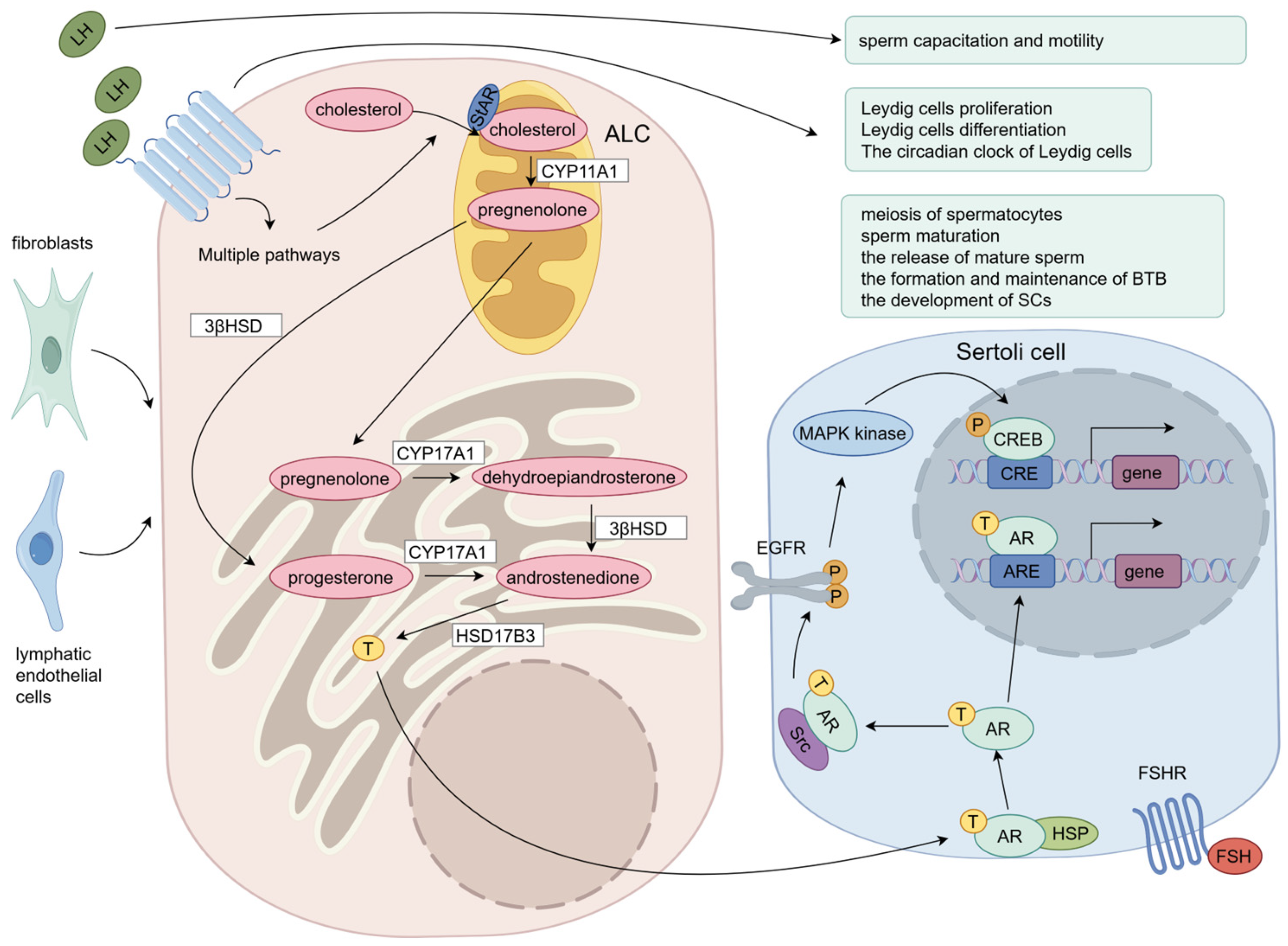
In LCs, after LH binds to LHR, the precursor of testosterone, cholesterol, is carried from the cytoplasmic matrix to the interior mitochondrial membrane by transduction bodies such as PKA, steroidogenic acute regulatory protein (StAR), and others [46,47]. CYP11A1 then converts cholesterol to pregnenolone [46,47]. Pregnenolone then leaves the mitochondria and enters the endoplasmic reticulum, where it is mostly converted into progesterone by 3b-hydroxysteroid dehydrogenase (3βHSD) [46,47]. In the endoplasmic reticulum, pregnenolone is metabolized by CYP17 to form dehydroepiandrosterone, progesterone is metabolized by CYP17 to form androstenedione, and androstenedione is also formed from dehydroepiandrosterone by 3βHSD [46,47]. Dehydroepiandrosterone and progesterone are metabolized by 17βHSD to form testosterone [46,47].
Testosterone production occurs primarily in the endoplasmic reticulum and mitochondria and involves the catalysis of a variety of enzymes (Figure 2). During testosterone production, cholesterol transfer regulated by StAR proteins is a key step in determining the rate of testosterone production [48]. In 1991, Epstein LF and Orme-Johnson NR found the presence of an LH-induced 30kDa phosphorylated protein in LCs, which was later found to be the result of 37kDa protein cleavage and named steroidogenic acute regulatory protein [49,50]. Recent studies have found that in addition to cholesterol transport, StAR proteins in LCs are implicated in DAG buildup in lipid droplets, which may affect DAG signaling mediated cellular function [51]. Apart from StAR (StARD1), several more START domain proteins have been discovered in mammals, among which StARD4 and StARD5 are found to have a weak effect on cholesterol transfer, while StARD6 has strong activity [52].
LH-regulated testosterone production involves the regulation of multiple signaling pathways, which we discuss in detail below.

2.4. Testosterone Regulates Spermatogenesis

LH regulates spermatogenesis by regulating the testosterone secretion of LCs, so how does testosterone regulate spermatogenesis? After being released by LCs, testosterone is partially bound to androgen-binding protein (ABP) for transport [53]. ABP is a glycoprotein secreted by Sertoli cells stimulated by FSH and is present in the testis, epididymis, and liver [53]. In the testis, ABP maintains hormone concentrations, provides a favorable environment for spermatogenesis, and regulates intracellular signals through membrane receptors to affect cell growth, differentiation, and function [53]. When testosterone enters the seminiferous tubules, it binds to androgen receptor (AR) proteins located in peripheral vascular cells such vascular endothelial cells and smooth muscle cells, as well as support cells like Sertoli cells and peritubular myoid cells [54,55]. In the classical pathway, the binding of testosterone to AR leads to its conformational change and detachment of heat shock protein (HSP), and then AR is transferred from the cytoplasm to the nucleus for binding (ARE), thereby expressing activating or inhibiting regulatory proteins to regulate cell activities [56]. In the non-classical signaling pathway, AR is transported to the plasma membrane where it interacts with Src kinase [57]. This ultimately causes the transcription factor CREB to become phosphorylated, which in turn triggers the production of associated proteins [57]. Compared with the classical pathway, the non-classical pathway takes about a few seconds to a few minutes, while the classical pathway takes about 30–45 min [58,59]. Currently, the classical pathway has been found to play a role in neurodevelopment, prostate epithelial homeostasis, bone healing, muscle hypertrophy, reduction in β-amyloid plaques, and reproductive system function (such as spermatogenesis), while the non-classical pathway mainly exists in hippocampal synaptic plasticity, protection of cerebellar granule cells under oxidative stress, migration of prostate cancer cells, anti-apoptosis of neurons, tight junction formation of Sertoli cells in the testis, and alleviation of cardiac ischemia–reperfusion injury [60].
Testosterone, one of the androgens, is essential for spermatogenesis and is found in greater quantity in the testis than in the serum. Testosterone has been found to have a major impact on the meiosis of spermatocytes. According to research by Bartlett JM et al., abnormal spermatogenesis results from acute depression of intratesticular testosterone levels followed by stage-specific degeneration of germ cells, which is mirrored in stage-specific degeneration of pachytene primary spermatocytes at stages VII to VIII of the spermatogenic cycle [61]. In addition, O’Donnell L et al. found that the transition of round spermatids from stages VII to VIII is a crucial T-dependent stage in the spermiogenesis process [62]. Proteomic studies have shown that testosterone may affect sperm meiosis by affecting proteins involved in cell stress and apoptosis, RNA splicing and processing, and DNA repair [63]. In addition to meiosis, testosterone also affects spermatogenesis by influencing the formation and upkeep of the blood–testis barrier, the maturation of sperm, and the growth of Sertoli cells [64,65,66].
In summary, the testosterone secreted by LCs binds to the AR in various cells surrounding spermatogenic cells (primarily Sertoli cells), regulating multiple processes of spermatogenesis through both classical and non-classical pathways (Figure 2).

3. LH Regulates Proliferation, Differentiation, and the Circadian Clock of LCs

3.1. LH Regulates LC Proliferation

It has been demonstrated that LH regulates LC steroidogenesis in a significant way. Nevertheless, the impact of LH on LC proliferation in the early stages remains unclear, and LH is believed to be less involved in LC proliferation because the period of LC proliferation is independent of the rise in LH concentration, and no appreciable DNA synthesis was seen in LCs stimulated by LH in vitro [67]. After LCs in rats were destroyed by treatment with ethane dimethyl sulphonate (EDS), serum LH levels increased due to a feedback regulatory mechanism; however, PLC proliferation seems to be independent of LH in LC regeneration [17,68]. Nonetheless, subsequent studies have found luteinizing hormone to be indispensable in the proliferation of LCs. When 21-day-old rats were deprived of endogenous LH by LH antiserum, the levels of D-cyclins D3 and proliferating cell nuclear antigen (PCNA), which are strongly linked to cell proliferation, were considerably reduced [16]. Moreover, endogenous LH-deprived mice showed mRNA loss of insulin-like growth factor (IGF-1), a growth factor that has been shown to be strongly linked to the proliferation and development of LCs [16]. In LHR knockout mice, LC counts were much lower [69]. Precocious stimulation with LH alone or in conjunction with FSH increased the mean amount of HSD3B-positive rhesus cells by 20–30-fold, according to a study on pubertal LCs in rhesus monkeys [70]. It also significantly increased their nuclear diameter, which is a hallmark of LC proliferation during early puberty [70]. In the LH-mediated regulation of LC proliferation, ERK1/2 cascade signaling pathway was confirmed to be involved in this process, and neuregulin 1 (NRG1) and collagen IV were also involved [71,72,73]. Stimulation of late pubertal rats with bisphenol B revealed LC proliferation and increased phosphorylation of AKT1/AKT2 and ERK1/2, suggesting that these signaling molecules may be involved in LC proliferation [74]. These results suggest that LH plays an important role in the proliferation of LCs, and the mechanism may involve the ERK and PI3k/AKT signaling pathways.

3.2. LH Regulates LC Differentiation

As mentioned above, FLCs, although expressing the LH receptor, are not sensitive to LH stimulation. In the process of ALC formation, there is differentiation from SLCs to PLCs, PLCs to ILCs, and ILCs to ALCs, so what part does LH play in controlling these differentiation processes? LH alone on SLCs isolated from seminiferous tubules did not induce differentiation into PLCs [75]. Moreover, it was discovered that the stem cells developed into cells that could produce testosterone at weeks 2–3 when the spermatogenic tubules were cultured with DHH agonists without LH [37]. These indicate that LH is possibly not necessary for SLCs to differentiate into PLCs. In this process, the testicular microenvironment may participate in the differentiation of SLCs through the leptin-DHH signaling pathway [76]. In adult rats, within 4 weeks following EDS administration, LCs did not develop when LH levels were implanted with testosterone to keep them within the normal range [68]. Numerous 3b-HSD-positive cells were produced by hCG treatment 28 days after EDS delivery, indicating that hCG/LHR is necessary for PLC differentiation [68]. Since rats do not produce CG themselves, LH in rats seems to be important for rat PLC differentiation. In LHR knockout mice, there were no mature LCs and very few progenitor cells [69]. Immature LCs isolated from the testis of 35-day-old rats were treated with LH and found to have a significant increase in steroidogenesis and upregulation of Cyp11a1, Scarb1, Srd5a1, and Cyp17a1, as well as upregulation of biomarkers of mature LCs such as Insl3 [77]. These results indicate that LH can induce ILCs to differentiate into ALCs. However, although LH induces maturation of ILCs, Griffin DK et al. raised the idea that LH may not be an absolute necessity for maturation of LCs in senescent LCs. They found that LHRKO mice treated with testosterone, although highly infertile, exhibited breakthrough spermatogenesis at 12 months of age, suggesting that testosterone can induce LC maturation in aging mice [78]. In conclusion, LH plays an indispensable role in the differentiation of PLCs into ILCs, but it is not necessary for ILC differentiation, although it can promote ILC differentiation into ALCs. In addition, LH may not play a role in the differentiation of SLCs to PLCs.

3.3. LH Regulates the Circadian Clock of LCs

The circadian clock serves as an internal timing system present in various tissues and cells, crucial for organisms to adapt to daily changes. The circadian rhythm of cell life activities is regulated by the activators CLOCK (circadian locomotor output cycles protein kaput) and BMAL1 (brain and muscle ARNT-like 1) heterodimers, which recognize E-box motifs to regulate the expression of thousands of genes, including the repressors Period (Per) and cryptochrome (Cry) [79]. These repressors are translated and assembled into repressor complexes to inhibit CLOCK:BMAL1 function, and such feedback regulation leads to a circadian rhythm of cell life activities with a period of about 24 h [79]. LCs also exhibit a circadian clock governing testosterone secretion. After continuous light treatment of growing rats, testis volume decreased, LH receptor gene expression decreased, testosterone production-related gene expression changed, and mitochondrial function of LCs was impaired, suggesting that the stability of circadian rhythm is crucial to the endocrine function of LCs [80]. It has been shown that BMAL1 protein is rhythmically expressed in mouse LCs, and its loss leads to decreased expression of steroidogenic-related genes and decreased testosterone production [81]. Subsequent studies confirmed that clock and steroidogenic gene expression followed circadian rhythms in mouse LCs both in vivo and in vitro [82]. Rhythmic expression of clock-related genes and steroid-related genes was also found in cultured rat and goat LCs in vitro [83,84].
Aging has a significant effect on the rhythm of LCs. Studies have shown that although the endocrine function of rat LCs maintains a circadian rhythm during aging, the robustness and expression level of the rhythm are decreased, and the uptake of cholesterol is also markedly reduced [83]. In rat LCs, aging enhances cGMP signaling and decreases cAMP signaling in LCs, which may affect the process of testosterone secretion [85]. It has also been found that aging leads to decreased circadian rhythm and steroidogenesis through increased ER stress in mice [86].
In the study of the circadian rhythm of testosterone production in LCs, the important role of LH has gradually emerged. In primary cultured LCs, the activation of LHR-cAMP signaling promotes the expression of the StAR gene and changes the expression levels of many clock-related genes, such as the upregulation of Rorb, Dec1/2, and Per1/PER1 and the downregulation of Bmal1 and Rev-erb/b [18]. In rats with artificial hypogonadotropic hypogonadism, decreased secretion of LH and testosterone with loss of circadian rhythm was observed, and decreased Cyp11a1, Cyp17a1, and cAMP with loss of rhythm was observed in LCs [18]. Some genes associated to circadian rhythms, such as Dec1, Csnk1e, Bmal1, Per2, Cry1, Cry2, Rora, Rorb, Rev-erba/b/REV-ERBB, etc., have altered expression levels in LCs, whereas Npas2 and PER1 have decreased levels [18]. Though it was insufficient to preserve the circadian regularity of testosterone production in LCs, the clock gene was still able to sustain rhythmic expression [18]. This indicates that while LCs possess an intrinsic circadian clock, the circadian rhythm of cAMP and testosterone production relies on the reproductive axis’s rhythmicity [18].
Currently, the circadian rhythm of various testicular cells and spermatogenesis has been observed, but the specific mechanisms remain unclear [87]. Further exploration is needed to elucidate the rhythmic mechanism underlying testosterone production by LCs. Although experimental evidence has established that LH is important for regulating the biological rhythm of LC steroidogenesis, further exploration is needed to elucidate the rhythmic mechanism of testosterone production by LCs.

4. LH Regulates Testosterone Secretion of LCs Through Multiple Signaling Pathways

LH promotes testosterone production through a variety of pathways, including the cGMP-PKG, MAPK, PI3k/AKT, and calcium signaling pathways, etc. Additionally, excess testosterone can induce feedback regulation of LC testosterone production, reducing testosterone generation to maintain homeostatic levels through signaling pathways like the AMPK pathway and the Slit/Robo signaling pathway.
In LCs, after LH binds to LHR, it generates a large amount of cAMP, inositol triphosphate (IP3), and diacylglycerol (DG), which subsequently phosphorylate the protein kinases PKA and PKC [88,89]. It is important to note that the LH-induced intracellular response in granulosa cells may be different from that in LCs, with LH preferentially acting through the ERK/AKT pathway, while hCG preferentially acts through the cAMP/PKA pathway [90]. In human LCs, although hCG can induce more cAMP generation, hCG is not qualitatively different from LH in terms of cAMP and ERK1/2 activation, and they are equal in activating downstream steroidogenic events, which reflects the difference in the intracellular signal activated by LH between different genders [91]. Subsequently, various transcription factors are further activated through pathways such as MAPK and calcium ions, which stimulate downstream gene expression.
In the process of steroid production, multiple transcription factors are involved in the regulation of StAR transcription, including cAMP response element binding (CREB) protein [92,93]. According to Manna et al., StAR promoter activity, StAR mRNA, StAR protein levels, and cAMP-induced progesterone production were all enhanced by transiently expressing CREB in MA-10 murine LC tumor cells [93]. However, these responses were markedly suppressed in CREB mutants that cannot be phosphorylated [93]. This implies that CREB/CREM family member phosphorylation plays a crucial role in testosterone production and StAR expression. In addition to CREB/M, activator protein 1 (Fos and Jun), C/EBPa and C/EBPb of the C/EBP family, SF-1, and GATA-4 are all involved in the regulation of LH in steroidogenesis through the cAMP signaling pathway. They are phosphorylated by PKA or PKC and interact with each other to promote or inhibit the transcription of the StAR gene (view in [94]). Among them, Fos/Jun and CREB have crosstalk on a single cis element, which has a repressive effect on StAR gene transcription [95]. At the same time, there are multiple factors involved in the post-transcriptional processing of StAR mRNA. LCs were found to harbor both HuR, a nucleoplasmic mRNA shuttle protein that stabilizes transcripts, and Tis11b, a zinc finger RNA instability protein, indicating a potential regulatory role for StAR mRNA stabilization [96,97]. Also, CPE-binding proteins (CPEBs), the mitochondrial A-kinase anchoring protein 121 (AKAP121), and DAX-1 may all play roles in StAR mRNA processing [94,98,99].
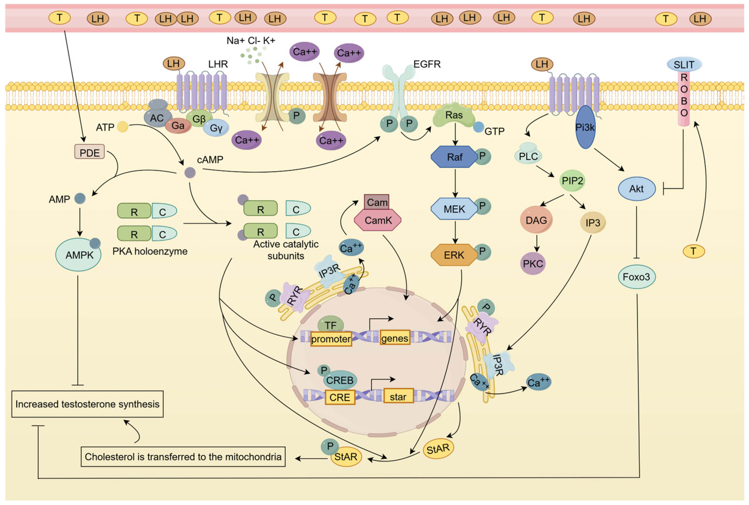
In the following section, we will systematically elucidate the key signaling pathways through which LH promotes testosterone synthesis in LCs, with the regulatory network illustrated in Figure 3.

4.1. EGFR/MAPK Signaling Pathway Is Involved in LH-Induced LC Testosterone Synthesis

The mitogen-activating pathway, or MAPK, is a crucial signal pathway involved in cell proliferation, differentiation, apoptosis, and stress response in both healthy and pathological settings. It is a part of the eukaryotic signal transduction network. Hirakawa T et al. shown that LHR receptors can trigger LC ERK1/2 phosphorylation in testis LCs, which is mediated by cAMP and PKA [100]. Subsequently, further studies revealed that LHR activated ERK1/2 phosphorylation through growth factor receptor (EGFR) phosphorylation [101,102]. In mouse LCs, LH induces EGFR transactivation in two pathways, MMP-mediated and cAMP-mediated [103]. But in LCs, MMP inhibition had no discernible impact on the synthesis of testosterone, suggesting that the MMP-mediated pathway is not thought to be involved in steroidogenesis [104]. In mouse LCs, it was found that in a short period (<60 min), rapid EGFR signaling activates the ERK signaling pathway and induces phosphorylation of intracellular STAR protein to promote steroidogenesis; however, with prolonged hCG treatment (>2 h), the EGFR/ERK signaling pathway was no longer involved in the steroidogenesis of LCs, and cAMP maintained testosterone production in LCs by promoting StAR transcription [105]. Reduced LH-induced steroid synthesis and higher expression of StAR protein were seen in rat primary LCs and tumor LC lines preincubated with the mitogen-activated protein kinase (MEK) inhibitors U0126 and PD98059 [106]. This suggests an important role for EGFR/LHR interaction in steroidogenic homeostasis in LCs of the testis. Furthermore, in the LCs of Mek1f/f, Mek2−/−, and Cre+ adult mice, the expression of several genes encoding androgen metabolism enzymes (Srda1 and Dhrs9) was upregulated, while the expression of many genes involved in testosterone synthesis (StAR, Hsd3b6, Cyp17a1, and Hsd17b3) was decreased, resulting in a net decrease in steroidogenesis [107]. This also demonstrated the regulatory role of ERK signaling in testosterone secretion in LCs. It has recently been shown that ERK5 knockdown decreases steroidogenesis and decreases the expression of StAR and Nr4a1 (nuclear receptor subfamily 4, group A, member 1, also known as NUR77/NGFI-B) in human chorionic gonadotrophin (HCG)-treated MA-10 LCs, through phosphorylation of MEF2 [108]. Since HCG and LH share the same receptor, whether LH can regulate testosterone production through ERK5 signaling deserves further investigation.
In conclusion, LH regulates testosterone synthesis in LCs in a time-dependent (mainly short-term) manner through activating cAMP/PKA and EGFR-ERK1/2/ERK5 signaling pathways, while ERK5 may affect steroid synthetase expression through the MEF2-Nr4a1 pathway.

4.2. Calcium Signaling Pathway Is Involved in LH-Induced LC Testosterone Synthesis

Numerous processes are mediated by calcium signaling and its interaction network, such as embryonic development, gene expression, synaptic transmission, excitation–contraction coupling, stimulus–secretion coupling, and induction of synaptic plasticity [109]. Calcium is also involved in the regulation of LH-induced steroidogenesis in LCs. In LCs, the influx pathway of LH-stimulated changes in calcium concentration is associated with T-type Ca2+ channels at the interstitial membrane, whereas the ER release pathway is dependent on the IP3 receptor (IP3R) and the ryanodine receptor (RyR) [110,111]. LH stimulation of intracellular calcium concentration depends on two pathways: the cAMP-dependent and cAMP-independent pathways. Significant changes in testosterone and [Ca2+]i were found in rat LCs upon stimulation with low concentrations of sheep LH treatment, whereas no increase in cyclic AMP was detected [112]. Therefore, at low concentrations of LH (below 0.1 pg /mL), [Ca2+] may be a regulator of steroidogenesis rather than cyclic AMP. However, when stimulated by a high level of sheep LH, the concentration of calcium ion and cAMP increased, and cAMP could stimulate the increase in calcium ion concentration [112]. However, due to species differences, whether the conclusions obtained by treating rat LCs with sheep LH are consistent with the real response in vivo is worthy of further investigation. In addition, cAMP-activated PKA and PKC can also induce calcium elevation, and the induction is mainly by PKA [113]. The increase in calcium concentration in the cytoplasm depends on two pathways: calcium release from the endoplasmic reticulum and calcium influx from the extracellular matrix. Calcium ions in the testicular stromal cells can be transported to the mitochondria and promote the formation of NADH and NADPH, thus contributing to the mitochondrial testosterone production process [110]. In addition, calcium regulates steroidogenesis by activating the early orphan nuclear receptor NUR77 via calmodulin-dependent protein kinase (CaMK), which in turn promotes the expression of StAR [114,115]. Among various CAMKs, CAMKI has the most significant effect on LC steroidogenesis (reviewed in [116]). One such mechanism is that the co-activator p300 is recruited to the nur77 promoter to activate its expression through AP1, CREB, and MEF2 transcription factors [117].
However, Pandey AK et al. found that after blocking L-type calcium channels in MA-10 mouse LCs, subthreshold cAMP induction (which could not induce StAR expression under normal conditions) significantly increased StAR protein expression and progesterone production, which may be related to the downregulation of DAX-1 expression [118]. The above phenomenon was not produced when L-type calcium channels were blocked alone [118]. This suggests that the blockade of calcium channels “compensates” the sensitivity of LCs to cAMP and that calcium has a complex bidirectional regulation of testosterone production. In hamster LCs, the large-conductance Ca2+-activated K+-channel (BKCa) was discovered [119]. It was discovered that blocking this receptor raised testosterone levels, indicating that it might control steroidogenesis by lowering calcium influx through membrane hyperpolarization [119]. It has recently been discovered that LH can support the regulation of mitochondrial membrane potential, STAR processing, and testosterone production by SH3 and cysteine-rich protein 3 (STAC3) [120]. Since STAC3 plays a key role in calcium signaling, it may act as an important factor in the calcium signaling pathway regulating testosterone synthesis.
In addition to calcium ions, other ions are also involved in the regulation of LC steroid production by LH, including chloride ions. When LCs were cultured in medium without chloride, testosterone was found to be increased, whereas chloride deprivation significantly increased StAR protein levels in cells incubated with (Bu) 2cAMP, suggesting a regulatory effect of chloride on LCs, possibly through StAR protein [121]. And it was also found that chloride channel blocker 4, 4′-diisothiocyanate-stilbene-2, and 2′-disulfonic acid (DIDS) inhibited the LH-stimulated steroidogenesis of LCs but had no effect on dibutyryl cyclic adenosine monophosphate (dbc AMP)-stimulated steroidogenesis [122]. Therefore, it can be inferred that LH-stimulated chloride efflux can affect the level of cAMP, which in turn affects the synthesis of STAR protein to regulate steroidogenesis [122]. Even though chloride ions have been shown to be crucial for controlling steroidogenesis, more research is still needed to fully understand the ion channels and underlying processes.
In summary, LH bidirectionally regulates testosterone synthesis in LCs via calcium signaling (Figure 3), mainly through the cAMP-CamK-Nur77 pathway, and calcium channel blockade may enhance cAMP sensitivity.

4.3. cGMP-PKG Signaling Pathway Has a Bidirectional Effect on LH-Induced Testosterone Production

In addition to cAMP, cGMP may also be involved in the regulation of the LH steroid secretion of LCs, but it is worth noting that it has a biphasic effect. The intracellular messenger cGMP is generated by guanylate cyclase (mGC) and nitric oxide (NO)-dependent soluble guanylate cyclase (sGC) [123]. It regulates effector function either directly or via cGMP-dependent protein kinase (PKG) [123]. In LCs, nitric oxide is produced by the enzyme nitric oxide synthase (NOS), which is present in three isoforms: endothelial (NOS3), neural (NOS1), and inducible (NOS2). The NO-cGTP signaling pathway has biphasic effects on steroidogenesis in mouse LCs: at low concentrations, NO promotes steroidogenesis, while at high concentrations, NO acts as an inhibitory effect [124].
Andric et al. found that in the rat testis, cGMP stimulates testosterone production in LCs through the phosphorylation of StAR protein, which promotes cholesterol transport to mitochondria [125]. Recent studies have shown that senescent LCs are characterized by elevated cGMP, which leads to decreased ATP production as well as altered mitochondrial function [126]. Subsequent downregulation of NO-cGMP signaling improved mitochondrial dynamics and increased the ATP and testosterone levels of LCs [126]. Andric et al. found that prolonged treatment of intact rats with testosterone enanthate resulted in a significant decrease in serum LH and significantly reduced the ability of purified LCs to produce testosterone in vitro, and further experiments showed that testosterone inhibited LC testosterone production by increasing NOS2 synthesis and decreasing cGMP production [127]. This suggests that although the cGMP signaling pathway does not regulate steroidogenesis through the LH receptor pathway, it may be involved in LH-mediated steroidogenesis by the autocrine regulatory mechanism.
These results suggest that cGMP regulates testosterone synthesis in LCs in a concentration-dependent manner (enhanced at low concentration and inhibited at high concentration) through a mechanism involving StAR phosphorylation and mitochondrial function. An abnormal cGMP-NOS2 axis during aging or testosterone-negative feedback leads to dys-synthesis, suggesting that it acts as an indirect LH pathway to affect steroidogenesis through an autocrine mechanism.

4.4. AMPK Signaling Pathway and Slit/Robo Pathway Have Feedback Regulatory Effects on LH-Induced Testosterone Production

In the regulation of LC steroidogenesis by LH, multiple pathways maintain steroid homeostasis through feedback regulation mechanisms. In LCs, phosphodiesterase (PDE) dephosphorylates cAMP and cGMP, resulting in reduced steroid production. PDE5 inhibition was found to increase cGMP and testosterone levels in LCs [125]. In LCs of PDE8A knockout mice, a fourfold increase in their LH-stimulated testosterone production sensitivity was detected [128]. However, in wild-type mice, treatment with 3-isobutyl-1-methylxanthine, a substance that inhibits all cAMP PDEs except PDE8A, resulted in just a slight increase in LH-induced testosterone synthesis; in PDEA8 knockout animals, this increase was more noticeable [128]. This suggests that PDE8A acts in concert with the remaining PDEs to regulate AMP levels in LCs. In addition, it was found that PDE8B is also involved in LH-induced steroid production and that simultaneous inhibition of PDE8s and PDE4s is required for a substantial increase in the PKA-mediated phosphorylation of several proteins related to the regulation of steroid production, including increased expression of the StAR protein [129].
In LCs, PDE converts cAMP to AMP, and AMP activates AMP-activated protein kinase (AMPK), which in turn regulates related transcription factors through the AMPK signaling pathway to regulate steroid production. In knockout mice for AMPK catalytic subunit alpha 1, high levels of testosterone and increases in 3βHSD, CYP17, and StAR proteins are observed, which shows the inhibiting effect of AMPK on steroidogenesis [130]. Further studies showed that the inhibitory effect of AMPK on testosterone production may be through decreasing the expression of the steroidogenic activators c-jun, NUR77, and CREB or increasing the expression of steroidogenic inhibitors such as c-Fos and NR0B1 [131]. Meanwhile, testosterone induced the production of AMPK, which is induced by the degradation of cAMP by PDE, demonstrating the role of AMPK signaling in the regulation of steroid homeostasis. Recently, in the study of the global phosphoproteomic profile of MA-10 LCs in response to Fsk (stimulation of steroidogenesis) and AICAR-mediated AMPK activation (repression of steroidogenesis), it was found that the AMPK signaling pathway downregulates the transcription of StAR and nur77 and affects the phosphorylation of a variety of steroidogenic-related proteins [132]. Notably, AMPK increases CREB levels in cells, and since cleavage of CREB exons results in a switch from activator to repressor to germ cells, it is plausible that active AMPK influences the utilization of CREB exons, and in this case, CREB may prevent the expression of steroidogenic genes [132,133].
Recent studies have shown that the regulation of steroid synthesis in mouse LCs is likewise mediated by Slit/Robo signaling. The homologous single-channel transmembrane receptors of the ring (ROBO) family are bound by secreted glycoproteins known as SLIT ligands, which play a role in controlling cell adhesion, proliferation, and survival in a range of tissues [134]. Slit ligands exist in mouse LCs, and testosterone can increase their expression. Reduced LHR and steroidogenic gene expression, CREB phosphorylation, AKT activity, LH signaling, and testosterone levels were all observed in LCs treated with SLIT in vitro [135]. In the meantime, StAR expression and testosterone synthesis in LCs were observed to be elevated in mice lacking a gene [135]. These findings imply that, in order to preserve the equilibrium of steroids in the body, Slit/Robo signaling may control LH-induced steroid production through feedback regulation. More research is required to elucidate the regulatory mechanism of Slit/Robo, as there are currently only a few studies on the regulatory influence of this protein on the generation of testosterone in LCs.
In summary, LH has been found to maintain steroid homeostasis in LCs through feedback mechanisms of the PDE-cAMP/cGMP-AMPK axis (downregulating StAR/NUR77) and Slit/Robo pathway (inhibiting LHR signaling), where AMPK plays a central inhibitory role through transcription factor reprogramming, while Slit/Robo may serve as a novel testosterone-negative regulator.

5. Clinical Application of LH: Treatment of HH, Diagnosis of Gonadal-Related Diseases, and Monitoring of Sperm Quality

5.1. Treatment of Hypogonadotropic Hypogonadism (HH): By Rescuing the Broken HPG Axis

Given LH’s pivotal role in LC differentiation, proliferation, testosterone production, spermatogenesis, and sperm capacitation, it holds significant clinical relevance in disease treatment. One notable application is its use in treating hypogonadotropic hypogonadism (HH). The clinical manifestations are often a high degree of infertility, lack of prominent secondary sexual characteristics, and gonadal dysgenesis [136]. There are many causes of HH, including congenital factors, such as isolated GnRH deficiency(IGD), LHR receptor mutation, LHB or FSHB mutation, combined pituitary hormone deficiency, genetic syndrome, etc. [137,138]. There are also acquired factors, such as tumor, lymphocytic infiltration disease, and iatrogenic diseases [137]. Some of these patients with isolated GnRH deficiency present with anosmia, which is known as Kaman syndrome [139].
For the treatment of HH, gonadotropin replacement therapy (GnRH, FSH, LH, or a mixture of LH and FSH) can be used to promote gonadal development and improve fertility [137]. However, because LH is expensive, hCG is often used in clinical practice to replace the testosterone-promoting effect of LH. Because LH and hCG have different half-lives and pharmacokinetics, these differences need to be considered when considering the dose to be used [140]. Regarding their efficacy, studies have shown that their results are highly variable, less effective in individuals lacking mini-puberty (the first six months of neonatal life when the HGP axis is most active), and more effective in individuals with partial pubertal development, indicating the importance of the period of gonadotropin treatment for congenital HH [141].
Several experiments have demonstrated the important role of LH or HCG in gonadal development and hormone production in neonates and secondary sexual characteristics development in adolescents, sex hormone production, and spermatogenesis in adult infertility. In one-year-old neonates with congenital hypogonadotropic hypogonadism (CHH), subcutaneous injection of recombinant LH (rLH) and recombinant FSH (rFSH) led to a notable increase in testicular length from 1.6 cm to 2.4 cm, along with a 170% rise in testicular volume [142]. Importantly, no significant adverse effects were observed [142]. In another study, pump-administered rLH and rFSH to two neonates for several months showed therapeutic effects with penile growth, increased testicular volume, and endogenous testosterone production [143]. A recent study of 65 newborns with CHH who received rhLH and rhFSH treatment by either pump or injection showed that both methods significantly improved the development of mini-puberty but that the injection group experienced a significantly greater increase in testosterone levels and penis length than the pump group, and there were no other differences [144]. Numerous studies have demonstrated the good efficacy of treating gonadal development in adolescents with HH. For example, when HCG and rFSH were administered to adolescents with HH for more than four years until testicular development stopped, it was found that testicular volume, sperm concentration, and quality of life were significantly improved in both pre-pubertal and non-pubertal adolescents with or without testosterone treatment, greatly reducing the impact of gonadal dysgenesis [145].
In adult infertility, in addition to T therapy and pulsatile GnRH therapy, gonadotropin therapy is also commonly used [146]. The usual dose of HCG is 75 to 225IU two to three times a week, often adjusted according to the amount of testosterone in the serum [146]. For most patients, injections of 1500 IU of hCG twice weekly can be used to produce normal testosterone levels, but for some patients 10,000 IU may be required [147]. The use of hCG alone can induce semen production in patients with CHH, but if severe oligospermia or azoospermia persists after 3 to 4 months of treatment alone, it is necessary to add 150–225 IU of FSH three times a week for 6 to 24 months, resulting in testicular growth in almost all patients and in 80% to 90% of patients without cryptorchidism [147].
A substantial amount of clinical research evidence has already proven the effectiveness of hCG in improving testosterone production by LCs. However, the study by Park et al. found that hCG can induce ER stress by activating the UPR pathway, which plays a significant role in steroidogenic enzyme expression [148]. Moreover, high-dose hCG exposure can lead to ER stress-mediated apoptosis in mLTC-1 cells and mouse testes [148]. This has prompted us to pay more attention to the dosage range and duration of use of hCG in clinical applications.

5.2. LH Level Can Be Used in the Diagnosis of Androgen Insensitivity Syndrome and Precocious Puberty

As mentioned above, the HPG axis and its involved hormones, such as LH, FSH, and testosterone, play an important role in maintaining male characteristics and spermatogenesis. Abnormalities in this axis often lead to a variety of diseases, among which LH level can be used as one of the bases for disease diagnosis.
Androgen insensitivity syndrome, a disorder associated with the X chromosome due to abnormalities in the androgen receptor (AR) gene, is the most common cause of neutral developmental disorders in 46, XY individuals [149]. Among them, complete type shows normal female external genitalia, partial type shows dysplastic male external genitalia, and mild type shows infertility and gynecomastia [150]. The endocrine features of AIS are characterized by elevated serum androgens and serum LH due to impaired negative feedback regulatory mechanisms of the pituitary gland [150]. A study including 39 Chinese patients with AIS revealed that the basal LH level was about 23 and 17 times higher in CAIS (27.39 ± 3.18 IU/L) and PAIS (18.86 ± 2.25 IU/L) than in healthy children [151]. The peak LH levels were also significantly higher in both conditions than in healthy children [151]. The elevated level of LH can be used as one of the bases for the diagnosis of AIS. AIS is currently tested in children by a human chorionic gonadotropin (hCG) stimulation test and by measuring serum androstenedione, testosterone, and DHT after 72 h. In adults, by measuring elevated/normal total testosterone and increased luteinizing hormone (revealing an overview of androgen resistance), FSH and statin B levels may be within the normal range.
In addition, precocious puberty is also one of the diseases caused by abnormal sex hormones. Precocious puberty refers to the premature development of secondary sexual characteristics, which is manifested as the onset of puberty two standard deviations earlier than the population norm, that is, breast or pubic hair development in girls before the age of 8 years and testicular or penile enlargement in boys before the age of 9 years [152]. The two types of precocious puberty in males are called peripheral precocious puberty, which results from aberrant adrenal and/or testicular dysfunction, and central precocious puberty, which results from premature activation of the hypothalamic–pituitary–gonadal axis. It is especially crucial to identify premature puberty in time since it can result in earlier bone aging and be brought on by organic lesions [153]. LH is the best marker of the onset of puberty, and the detection of LH level can be used as one of the bases for judging precocious puberty [154]. Individuals with central precocious puberty have significantly higher LH levels than normal individuals. In peripheral precocious puberty, the marked excess of LH may be caused by HCG tumors or by defects in activating LHCG receptors (testotoxicosis), which lead to LC proliferation and precocious puberty [152]. LH concentrations can also be used to assess the effect of treatment. If the LH peak value of GnRH stimulation is lower than 2.5–4.5 mL/U, the HPG axis is successfully suppressed [155]. A study also found that first-voided urinary LH level measurement can adequately assess precocious puberty treatment with a cutoff value for LH of 1.01 mIU/mL [156].

5.3. Sperm Quality Monitoring and Regulation

Differences in the morphology and function of sperm affect their ability to fertilize, so it is particularly important to evaluate the quality of sperm, especially in assisted reproductive technology. Studies on sperm quality are mainly concerned with sperm morphology, sperm swimming ability, sperm bioenergetics, and sperm capacitation, which are very important for sperm successful fertilization [157]. The detection methods of sperm quality mainly involve microscope observation, computer-aided system evaluation, extracellular flux analysis, image flow cytometry, etc. [157]. In addition, many studies have shown a strong correlation between LH and sperm quality, and LH may be used as one of the auxiliary means for sperm quality detection and regulation.
The previous discussion emphasized the importance of the LH and HPG axis in reproduction, and it can be said that the level of LH is extremely important for the normal development of spermatogenesis. In terms of sperm concentration and motility, an earlier study comparing normospermic and oligospermic men found that oligospermic individuals exhibited reduced plasma sperm concentration and motility, with blood LH concentration showing significant associations with sperm cell concentration (p < 0.05) and motility (p < 0.02) [19]. This suggests that blood LH concentration may affect sperm concentration as well as sperm motility. Also, after comparing the circulating sex hormones and sperm quality in 338 men, only LH was found to be associated with progressive sperm motility [158]. A study on Holstein bulls also showed that sperm density and fresh sperm motility were associated with LHR genotype [159]. In LHR knockout mice, a significant reduction in sperm morphology, motility, and fertility rate was found, which could only be partially rescued by T treatment, indicating the irreversibly important role of LH in maintaining sperm quality [160].
In sperm capacitation and sperm bioenergetics, the researchers found that a functional LH receptor is present in sperm, located in the head of the sperm, a region that experiences morphological and biochemical changes during capacitation, and its activation leads to an increase in cAMP and PKA [20]. It is speculated that the LHR in sperm may be stimulated by LH from the serum or the female reproductive tract and play a role in sperm capacitation, metamorphosis, and motility. Since LHR receptors are present in the epididymis, and the acquisition of sperm motility occurs during sperm maturation in the epididymis, it is suggested that there is a biochemical process of LH-regulated sperm motility in the epididymis [158,161]. Healthy human sperm treated in vitro with LH did not alter the acrosome reaction, but it did cause alterations in sperm capacitation associated with the phosphorylation of the protein tyrosine, a reduction in sperm rapid progressive motility, and a rise in intracellular calcium concentration [21]. The effects occurred at concentrations of 0.5 and 1 mg/mL of hormone, similar to the peak concentrations achieved during the preovulatory LH elevation, indicating that LH is essential for sperm capacitation in the female reproductive tract, as it occurs there [21,162].
These research findings collectively suggest that LH may play a role in sperm capacitation, motility, and maturation, potentially serving as a supplementary approach in assisted reproduction through its ex vivo manipulation of sperm. Furthermore, the significant correlation observed between LH concentration in blood and sperm quality suggests that LH concentration testing could also serve as a method for assessing sperm quality.

6. Conclusions and Perspectives

In summary, this study systematically revealed the comprehensive mechanism of LH in regulating testosterone synthesis, proliferation, differentiation, and sperm function in LCs through multiple signaling pathways (Table 1).
The homeostasis of testosterone production in LCs plays a crucial role in regulating spermatogenesis. As the central signaling molecule for testosterone synthesis in LCs, LH maintains testosterone production homeostasis through various pathways. Among these pathways, cAMP/PKA, calcium, EGFR/MAPK, cGMP-PKG, and other pathways play an important positive response role, while AMPK and Slit/Robo maintain the homeostasis of testosterone production through a negative feedback regulation mechanism. Additionally, LH is involved in processes such as LC proliferation, differentiation, and circadian rhythm regulation, making it a key factor in maintaining LC function and population. Currently, LH is utilized in clinical practice for the treatment of conditions such as hypogonadotropic hypogonadism, and it is also extensively employed in the diagnosis of disorders associated with gonadal function, including androgen insensitivity syndrome and precocious puberty. Moreover, many studies have shown that LH may affect sperm concentration, sperm motility, and sperm capacitation, although the underlying mechanisms are not fully understood. Therefore, LH holds potential as an adjunctive hormone for in vitro reproduction and shows promise in the treatment of male infertility conditions such as asthenospermia, male gonadal dysgenesis, or abnormal spermatogenesis resulting from LC dysfunction. In addition, although LH plays an important role in the maturation and function of LCs, the precise regulatory mechanisms of LH in LC proliferation, differentiation, and circadian rhythm remain unclear. Identifying the regulatory molecules and targets through transcriptomic and proteomic approaches may offer new avenues for treating male gonadal dysgenesis characterized by low testosterone levels due to LC abnormalities.

Author Contributions

T.L. and W.-X.Y. conceived this study. T.L. wrote the review article. Y.Y. participated in discussions and revising of this paper. W.-X.Y. revised this paper and provided funding. All authors have read and agreed to the published version of the manuscript.

Funding

This work was supported in part by the National Natural Science Foundation of China (No. 32072954 and 32270555).

Institutional Review Board Statement

Not applicable.

Informed Consent Statement

Not applicable.

Data Availability Statement

Not applicable.

Acknowledgments

The authors are grateful to all members of the Sperm Laboratory in Zhejiang University for their support and valuable discussion. Figure support was provided by Figdraw (www.figdraw.com).

Conflicts of Interest

The authors declare no conflicts of interest.

References

  1. Fonseca, A.C.S.; Barreiro, M.; Tomé, A.; Vale-Fernandes, E. Male Reproductive Health—Study of a Sperm Donor Population. JBRA Assist. Reprod. 2022, 26, 247–254. [Google Scholar] [CrossRef] [PubMed]
  2. Agarwal, A.; Baskaran, S.; Parekh, N.; Cho, C.-L.; Henkel, R.; Vij, S.; Arafa, M.; Panner Selvam, M.K.; Shah, R. Male Infertility. Lancet 2021, 397, 319–333. [Google Scholar] [CrossRef] [PubMed]
  3. Svingen, T.; Koopman, P. Building the Mammalian Testis: Origins, Differentiation, and Assembly of the Component Cell Populations. Genes Dev. 2013, 27, 2409–2426. [Google Scholar] [CrossRef]
  4. O’Donnell, L.; Smith, L.B.; Rebourcet, D. Sertoli Cells as Key Drivers of Testis Function. Semin. Cell Dev. Biol. 2022, 121, 2–9. [Google Scholar] [CrossRef]
  5. Haider, S.G. Cell Biology of Leydig Cells in the Testis. Int. Rev. Cytol. 2004, 233, 181–241. [Google Scholar] [CrossRef]
  6. Bhattacharya, I.; Dey, S. Emerging Concepts on Leydig Cell Development in Fetal and Adult Testis. Front. Endocrinol. 2022, 13, 1086276. [Google Scholar] [CrossRef]
  7. Sokwalla, S.M.R. Chapter 2—Reproductive Endocrine Physiology. In Subfertility; Elsevier: Amsterdam, The Netherlands, 2021; pp. 39–64. [Google Scholar]
  8. Neto, F.T.L.; Bach, P.V.; Najari, B.B.; Li, P.S.; Goldstein, M. Spermatogenesis in Humans and Its Affecting Factors. Semin. Cell Dev. Biol. 2016, 59, 10–26. [Google Scholar] [CrossRef]
  9. Walker, W.H. Regulation of Mammalian Spermatogenesis by miRNAs. Semin. Cell Dev. Biol. 2022, 121, 24–31. [Google Scholar] [CrossRef]
  10. Shen, L.; Yu, C.; Gao, F.; Hong, Y.-T. [PIWI/piRNA Complex-Mediated Regulation of Spermatogenesis]. Zhonghua Nan Ke Xue 2021, 27, 262–268. [Google Scholar]
  11. Xiong, B.; Jiang, Y.; Wang, Y.; Han, X.; Zhang, C.; Zhong, R.; Ge, W.; Han, B.; Ge, Z.; Huang, G.; et al. LncRNA8276 Primes Cell-Cell Adhesion for Regulation of Spermatogenesis. Andrology 2022, 10, 1687–1701. [Google Scholar] [CrossRef]
  12. Zhang, Z.; Miao, J.; Wang, Y. Mitochondrial Regulation in Spermatogenesis. Reproduction 2022, 163, R55–R69. [Google Scholar] [CrossRef] [PubMed]
  13. Mohammadi, A.; Shabani, R.; Bashiri, Z.; Rafiei, S.; Asgari, H.; Koruji, M. Therapeutic Potential of Exosomes in Spermatogenesis Regulation and Male Infertility. Biol. Cell 2024, 116, e2300127. [Google Scholar] [CrossRef] [PubMed]
  14. Xiong, Y.; Yu, C.; Zhang, Q. Ubiquitin-Proteasome System-Regulated Protein Degradation in Spermatogenesis. Cells 2022, 11, 1058. [Google Scholar] [CrossRef] [PubMed]
  15. Sofikitis, N.; Giotitsas, N.; Tsounapi, P.; Baltogiannis, D.; Giannakis, D.; Pardalidis, N. Hormonal Regulation of Spermatogenesis and Spermiogenesis. J. Steroid Biochem. Mol. Biol. 2008, 109, 323–330. [Google Scholar] [CrossRef]
  16. Sriraman, V.; Rao, V.S.; Sairam, M.R.; Rao, A.J. Effect of Deprival of LH on Leydig Cell Proliferation: Involvement of PCNA, Cyclin D3 and IGF-1. Mol. Cell. Endocrinol. 2000, 162, 113–120. [Google Scholar] [CrossRef]
  17. Sriraman, V.; Sairam, M.; Rao, A. Evaluation of Relative Roles of LH and FSH in Regulation of Differentiation of Leydig Cells Using an Ethane 1,2-Dimethylsulfonate-Treated Adult Rat Model. J. Endocrinol. 2003, 176, 151–161. [Google Scholar] [CrossRef]
  18. Baburski, A.Z.; Andric, S.A.; Kostic, T.S. Luteinizing Hormone Signaling Is Involved in Synchronization of Leydig Cell’s Clock and Is Crucial for Rhythm Robustness of Testosterone Production. Biol. Reprod. 2019, 100, 1406–1415. [Google Scholar] [CrossRef]
  19. Vasquez, J.M.; Ben-Nun, I.; Greenblatt, R.B.; Mahesh, V.B.; Keel, B.A. Correlation between Follicle-Stimulating Hormone, Luteinizing Hormone, Prolactin, and Testosterone with Sperm Cell Concentration and Motility. Obs. Gynecol. 1986, 67, 86–90. [Google Scholar]
  20. Eblen, A.; Bao, S.; Lei, Z.M.; Nakajima, S.T.; Rao, C.V. The Presence of Functional Luteinizing Hormone/Chorionic Gonadotropin Receptors in Human Sperm. J. Clin. Endocrinol. Metab. 2001, 86, 2643–2648. [Google Scholar] [CrossRef]
  21. López-Torres, A.S.; González-González, M.E.; Mata-Martínez, E.; Larrea, F.; Treviño, C.L.; Chirinos, M. Luteinizing Hormone Modulates Intracellular Calcium, Protein Tyrosine Phosphorylation and Motility during Human Sperm Capacitation. Biochem. Biophys. Res. Commun. 2017, 483, 834–839. [Google Scholar] [CrossRef]
  22. Griswold, S.L.; Behringer, R.R. Fetal Leydig Cell Origin and Development. Sex. Dev. 2009, 3, 1–15. [Google Scholar] [CrossRef] [PubMed]
  23. Kerr, J.B.; Knell, C.M. The Fate of Fetal Leydig Cells during the Development of the Fetal and Postnatal Rat Testis. Development 1988, 103, 535–544. [Google Scholar] [CrossRef] [PubMed]
  24. Brennan, J.; Capel, B. One Tissue, Two Fates: Molecular Genetic Events That Underlie Testis Versus Ovary Development. Nat. Rev. Genet. 2004, 5, 509–521. [Google Scholar] [CrossRef] [PubMed]
  25. Hall, P.F.; Irby, D.C.; Kretser, D.M.D. Conversion of Cholesterol to Androgens by Rat Testes: Comparison of Interstitial Cells and Seminiferous Tubules. Endocrinology 1969, 84, 488–496. [Google Scholar] [CrossRef]
  26. Brennan, J.; Tilmann, C.; Capel, B. Pdgfr-α Mediates Testis Cord Organization and Fetal Leydig Cell Development in the XY Gonad. Genes Dev. 2003, 17, 800–810. [Google Scholar] [CrossRef]
  27. Wen, Q.; Cheng, C.Y.; Liu, Y.-X. Development, Function and Fate of Fetal Leydig Cells. Semin. Cell Dev. Biol. 2016, 59, 89–98. [Google Scholar] [CrossRef]
  28. DeFalco, T.; Takahashi, S.; Capel, B. Two Distinct Origins for Leydig Cell Progenitors in the Fetal Testis. Dev. Biol. 2011, 352, 14–26. [Google Scholar] [CrossRef]
  29. Shima, Y.; Morohashi, K. Leydig Progenitor Cells in Fetal Testis. Mol. Cell. Endocrinol. 2017, 445, 55–64. [Google Scholar] [CrossRef]
  30. Clark, A.M.; Garland, K.K.; Russell, L.D. Desert Hedgehog (Dhh) Gene Is Required in the Mouse Testis for Formation of Adult-Type Leydig Cells and Normal Development of Peritubular Cells and Seminiferous Tubules. Biol. Reprod. 2000, 63, 1825–1838. [Google Scholar] [CrossRef]
  31. Yao, H.H.-C.; Whoriskey, W.; Capel, B. Desert Hedgehog/Patched 1 Signaling Specifies Fetal Leydig Cell Fate in Testis Organogenesis. Genes Dev. 2002, 16, 1433–1440. [Google Scholar] [CrossRef]
  32. Kitamura, K.; Yanazawa, M.; Sugiyama, N.; Miura, H.; Iizuka-Kogo, A.; Kusaka, M.; Omichi, K.; Suzuki, R.; Kato-Fukui, Y.; Kamiirisa, K.; et al. Mutation of ARX Causes Abnormal Development of Forebrain and Testes in Mice and X-Linked Lissencephaly with Abnormal Genitalia in Humans. Nat. Genet. 2002, 32, 359–369. [Google Scholar] [CrossRef] [PubMed]
  33. Inoue, M.; Baba, T.; Morohashi, K. Recent Progress in Understanding the Mechanisms of Leydig Cell Differentiation. Mol. Cell. Endocrinol. 2018, 468, 39–46. [Google Scholar] [CrossRef] [PubMed]
  34. Teerds, K.J.; Huhtaniemi, I.T. Morphological and Functional Maturation of Leydig Cells: From Rodent Models to Primates. Hum. Reprod. Update 2015, 21, 310–328. [Google Scholar] [CrossRef] [PubMed]
  35. Kaftanovskaya, E.M.; Lopez, C.; Ferguson, L.; Myhr, C.; Agoulnik, A.I. Genetic Ablation of Androgen Receptor Signaling in Fetal Leydig Cell Lineage Affects Leydig Cell Functions in Adult Testis. FASEB J. 2015, 29, 2327–2337. [Google Scholar] [CrossRef]
  36. Shima, Y.; Matsuzaki, S.; Miyabayashi, K.; Otake, H.; Baba, T.; Kato, S.; Huhtaniemi, I.; Morohashi, K. Fetal Leydig Cells Persist as an Androgen-Independent Subpopulation in the Postnatal Testis. Mol. Endocrinol. 2015, 29, 1581–1593. [Google Scholar] [CrossRef]
  37. Chen, H.; Wang, Y.; Ge, R.; Zirkin, B.R. Leydig Cell Stem Cells: Identification, Proliferation and Differentiation. Mol. Cell. Endocrinol. 2017, 445, 65–73. [Google Scholar] [CrossRef]
  38. Chen, H.; Stanley, E.; Jin, S.; Zirkin, B.R. Stem Leydig Cells: From Fetal to Aged Animals. Birth Defects Res. Part C Embryo Today Rev. 2010, 90, 272–283. [Google Scholar] [CrossRef]
  39. O’Shaughnessy, P.J.; Fleming, L.M.; Jackson, G.; Hochgeschwender, U.; Reed, P.; Baker, P.J. Adrenocorticotropic Hormone Directly Stimulates Testosterone Production by the Fetal and Neonatal Mouse Testis. Endocrinology 2003, 144, 3279–3284. [Google Scholar] [CrossRef]
  40. Kaprara, A.; Huhtaniemi, I.T. The Hypothalamus-Pituitary-Gonad Axis: Tales of Mice and Men. Metabolism 2018, 86, 3–17. [Google Scholar] [CrossRef]
  41. Xie, Q.; Kang, Y.; Zhang, C.; Xie, Y.; Wang, C.; Liu, J.; Yu, C.; Zhao, H.; Huang, D. The Role of Kisspeptin in the Control of the Hypothalamic-Pituitary-Gonadal Axis and Reproduction. Front. Endocrinol. 2022, 13, 925206. [Google Scholar] [CrossRef]
  42. Pinilla, L.; Aguilar, E.; Dieguez, C.; Millar, R.P.; Tena-Sempere, M. Kisspeptins and Reproduction: Physiological Roles and Regulatory Mechanisms. Physiol. Rev. 2012, 92, 1235–1316. [Google Scholar] [CrossRef] [PubMed]
  43. Bliss, S.P.; Navratil, A.M.; Xie, J.; Roberson, M.S. GnRH Signaling, the Gonadotrope and Endocrine Control of Fertility. Front. Neuroendocr. 2010, 31, 322–340. [Google Scholar] [CrossRef] [PubMed]
  44. Loosfelt, H.; Misrahi, M.; Atger, M.; Salesse, R.; Vu Hai-Luu Thi, M.T.; Jolivet, A.; Guiochon-Mantel, A.; Sar, S.; Jallal, B.; Garnier, J.; et al. Cloning and Sequencing of Porcine LH-hCG Receptor cDNA: Variants Lacking Transmembrane Domain. Science 1989, 245, 525–528. [Google Scholar] [CrossRef] [PubMed]
  45. Kremer, H.; Mariman, E.; Otten, B.J.; Moll, G.W.; Stoellnga, G.B.A.; Wit, J.M.; Jansen, M.; Drop, S.L.; Faas, B.; Ropers, H.-H.; et al. Cosegregation of Missense Mutations of the Luteinizing Hormone Receptor Gene with Familial Male-Limited Precocious Puberty. Hum. Mol. Genet. 1993, 2, 1779–1783. [Google Scholar] [CrossRef]
  46. Miller, W.L.; Auchus, R.J. The Molecular Biology, Biochemistry, and Physiology of Human Steroidogenesis and Its Disorders. Endocr. Rev. 2011, 32, 81–151. [Google Scholar] [CrossRef]
  47. Papadopoulos, V.; Miller, W.L. Role of Mitochondria in Steroidogenesis. Best Pract. Res. Clin. Endocrinol. Metab. 2012, 26, 771–790. [Google Scholar] [CrossRef]
  48. Tugaeva, K.V.; Sluchanko, N.N. Steroidogenic Acute Regulatory Protein: Structure, Functioning, and Regulation. Biochemistry 2019, 84, 233–253. [Google Scholar] [CrossRef]
  49. Epstein, L.F.; Orme-Johnson, N.R. Acute Action of Luteinizing Hormone on Mouse Leydig Cells: Accumulation of Mitochondrial Phosphoproteins and Stimulation of Testosterone Synthesis. Mol. Cell. Endocrinol. 1991, 81, 113–126. [Google Scholar] [CrossRef]
  50. Clark, B.J.; Wells, J.; King, S.R.; Stocco, D.M. The Purification, Cloning, and Expression of a Novel Luteinizing Hormone-Induced Mitochondrial Protein in MA-10 Mouse Leydig Tumor Cells. Characterization of the Steroidogenic Acute Regulatory Protein (StAR). J. Biol. Chem. 1994, 269, 28314–28322. [Google Scholar] [CrossRef]
  51. Galano, M.; Li, Y.; Li, L.; Sottas, C.; Papadopoulos, V. Role of Constitutive STAR in Leydig Cells. Int. J. Mol. Sci. 2021, 22, 2021. [Google Scholar] [CrossRef]
  52. Bose, H.S.; Whittal, R.M.; Ran, Y.; Bose, M.; Baker, B.Y.; Miller, W.L. StAR-like Activity and Molten Globule Behavior of StARD6, A Male Germ-Line Protein. Biochemistry 2008, 47, 2277–2288. [Google Scholar] [CrossRef]
  53. Munell, F.; Suárez-Quian, C.A.; Selva, D.M.; Tirado, O.M.; Reventós, J. Androgen-Binding Protein and Reproduction: Where Do We Stand? J. Androl. 2002, 23, 598–609. [Google Scholar] [CrossRef] [PubMed]
  54. O’Hara, L.; Smith, L.B. Androgen Receptor Roles in Spermatogenesis and Infertility. Best Pract. Res. Clin. Endocrinol. Metab. 2015, 29, 595–605. [Google Scholar] [CrossRef] [PubMed]
  55. Naamneh Elzenaty, R.; du Toit, T.; Flück, C.E. Basics of Androgen Synthesis and Action. Best Pract. Res. Clin. Endocrinol. Metab. 2022, 36, 101665. [Google Scholar] [CrossRef] [PubMed]
  56. Heinlein, C.A.; Chang, C. Androgen Receptor (AR) Coregulators: An Overview. Endocr. Rev. 2002, 23, 175–200. [Google Scholar] [CrossRef]
  57. Cheng, J.; Watkins, S.C.; Walker, W.H. Testosterone Activates Mitogen-Activated Protein Kinase via Src Kinase and the Epidermal Growth Factor Receptor in Sertoli Cells. Endocrinology 2007, 148, 2066–2074. [Google Scholar] [CrossRef]
  58. Walker, W.H. Non-Classical Actions of Testosterone and Spermatogenesis. Philos. Trans. R. Soc. Lond. B Biol. Sci. 2010, 365, 1557–1569. [Google Scholar] [CrossRef]
  59. Shang, Y.; Myers, M.; Brown, M. Formation of the Androgen Receptor Transcription Complex. Mol. Cell 2002, 9, 601–610. [Google Scholar] [CrossRef]
  60. Pillerová, M.; Borbélyová, V.; Hodosy, J.; Riljak, V.; Renczés, E.; Frick, K.M.; Tóthová, Ľ. On the Role of Sex Steroids in Biological Functions by Classical and Non-Classical Pathways. An Update. Front. Neuroendocrinol. 2021, 62, 100926. [Google Scholar] [CrossRef]
  61. Bartlett, J.M.S.; Kerr, J.B.; Sharpe, R.M. The Effect of Selective Destruction and Regeneration of Rat Leydig Cells on the Intratesticular Distribution of Testosterone and Morphology of the Seminiferous Epithelium. J. Androl. 1986, 7, 240–253. [Google Scholar] [CrossRef]
  62. O’Donnell, L.; McLachlan, R.I.; Wreford, N.G.; Robertson, D.M. Testosterone Promotes the Conversion of Round Spermatids between Stages VII and VIII of the Rat Spermatogenic Cycle. Endocrinology 1994, 135, 2608–2614. [Google Scholar] [CrossRef] [PubMed]
  63. Stanton, P.G.; Sluka, P.; Foo, C.F.H.; Stephens, A.N.; Smith, A.I.; McLachlan, R.I.; O’Donnell, L. Proteomic Changes in Rat Spermatogenesis in Response to in Vivo Androgen Manipulation; Impact on Meiotic Cells. PLoS ONE 2012, 7, e41718. [Google Scholar] [CrossRef] [PubMed]
  64. Meng, J.; Holdcraft, R.W.; Shima, J.E.; Griswold, M.D.; Braun, R.E. Androgens Regulate the Permeability of the Blood-Testis Barrier. Proc. Natl. Acad. Sci. USA 2005, 102, 16696–16700. [Google Scholar] [CrossRef] [PubMed]
  65. O’Donnell, L.; Pratis, K.; Wagenfeld, A.; Gottwald, U.; Müller, J.; Leder, G.; McLachlan, R.I.; Stanton, P.G. Transcriptional Profiling of the Hormone-Responsive Stages of Spermatogenesis Reveals Cell-, Stage-, and Hormone-Specific Events. Endocrinology 2009, 150, 5074–5084. [Google Scholar] [CrossRef]
  66. Guo, J.; Nie, X.; Giebler, M.; Mlcochova, H.; Wang, Y.; Grow, E.J.; DonorConnect; Kim, R.; Tharmalingam, M.; Matilionyte, G.; et al. The Dynamic Transcriptional Cell Atlas of Testis Development during Human Puberty. Cell Stem Cell 2020, 26, 262–276.e4. [Google Scholar] [CrossRef]
  67. Sriraman, V.; Anbalagan, M.; Rao, A.J. Hormonal Regulation of Leydig Cell Proliferation and Differentiation in Rodent Testis: A Dynamic Interplay between Gonadotrophins and Testicular Factors. Reprod. BioMed. Online 2005, 11, 507–518. [Google Scholar] [CrossRef]
  68. Teerds, K.J.; de Rooij, D.G.; Rommerts, F.F.G.; van den Hurk, R.; Wensing, C.J.G. Proliferation and Differentiation of Possible Leydig Cell Precursors after Destruction of the Existing Leydig Cells with Ethane Dimethyl Sulphonate: The Role of LH/Human Chorionic Gonadotrophin. J. Endocrinol. 1989, 122, 689-NP. [Google Scholar] [CrossRef]
  69. Lei, Z.M.; Mishra, S.; Zou, W.; Xu, B.; Foltz, M.; Li, X.; Rao, C.V. Targeted Disruption of Luteinizing Hormone/Human Chorionic Gonadotropin Receptor Gene. Mol. Endocrinol. 2001, 15, 184–200. [Google Scholar] [CrossRef]
  70. Verhagen, I.; Ramaswamy, S.; Teerds, K.J.; Keijer, J.; Plant, T.M. Time Course and Role of Luteinizing Hormone and Follicle-Stimulating Hormone in the Expansion of the Leydig Cell Population at the Time of Puberty in the Rhesus Monkey (Macaca mulatta). Andrology 2014, 2, 924–930. [Google Scholar] [CrossRef]
  71. Shiraishi, K.; Ascoli, M. Lutropin/Choriogonadotropin Stimulate the Proliferation of Primary Cultures of Rat Leydig Cells through a Pathway That Involves Activation of the Extracellularly Regulated Kinase 1/2 Cascade. Endocrinology 2007, 148, 3214–3225. [Google Scholar] [CrossRef]
  72. Anbalagan, M.; Rao, A.J. Collagen IV-Mediated Signalling Is Involved in Progenitor Leydig Cell Proliferation. Reprod. BioMed. Online 2004, 9, 391–403. [Google Scholar] [CrossRef] [PubMed]
  73. Umehara, T.; Kawashima, I.; Kawai, T.; Hoshino, Y.; Morohashi, K.; Shima, Y.; Zeng, W.; Richards, J.S.; Shimada, M. Neuregulin 1 Regulates Proliferation of Leydig Cells to Support Spermatogenesis and Sexual Behavior in Adult Mice. Endocrinology 2016, 157, 4899–4913. [Google Scholar] [CrossRef] [PubMed]
  74. Li, Y.; Yan, H.; Yu, Y.; Zou, C.; Tian, L.; Xin, X.; Zhang, S.; Li, Z.; Ma, F.; Ge, R.-S. Bisphenol B Stimulates Leydig Cell Proliferation but Inhibits Maturation in Late Pubertal Rats. Food Chem. Toxicol. 2021, 153, 112248. [Google Scholar] [CrossRef] [PubMed]
  75. Li, X.; Wang, Z.; Jiang, Z.; Guo, J.; Zhang, Y.; Li, C.; Chung, J.; Folmer, J.; Liu, J.; Lian, Q.; et al. Regulation of Seminiferous Tubule-Associated Stem Leydig Cells in Adult Rat Testes. Proc. Natl. Acad. Sci. USA 2016, 113, 2666–2671. [Google Scholar] [CrossRef]
  76. Arora, H.; Qureshi, R.; Khodamoradi, K.; Seetharam, D.; Parmar, M.; Van Booven, D.J.; Issa, I.C.; Sackstein, R.; Lamb, D.; Hare, J.M.; et al. Leptin Secreted from Testicular Microenvironment Modulates Hedgehog Signaling to Augment the Endogenous Function of Leydig Cells. Cell Death Dis. 2022, 13, 208. [Google Scholar] [CrossRef]
  77. Li, X.; Zhu, Q.; Wen, Z.; Yuan, K.; Su, Z.; Wang, Y.; Zhong, Y.; Ge, R.-S. Androgen and Luteinizing Hormone Stimulate the Function of Rat Immature Leydig Cells Through Different Transcription Signals. Front. Endocrinol. 2021, 12, 599149. [Google Scholar] [CrossRef]
  78. Griffin, D.K.; Ellis, P.J.; Dunmore, B.; Bauer, J.; Abel, M.H.; Affara, N.A. Transcriptional Profiling of Luteinizing Hormone Receptor-Deficient Mice Before and after Testosterone Treatment Provides Insight into the Hormonal Control of Postnatal Testicular Development and Leydig Cell Differentiation1. Biol. Reprod. 2010, 82, 1139–1150. [Google Scholar] [CrossRef]
  79. Koronowski, K.B.; Sassone-Corsi, P. Communicating Clocks Shape Circadian Homeostasis. Science 2021, 371, eabd0951. [Google Scholar] [CrossRef]
  80. Marinkovic, D.Z.; Medar, M.L.J.; Becin, A.P.; Andric, S.A.; Kostic, T.S. Growing Up Under Constant Light: A Challenge to the Endocrine Function of the Leydig Cells. Front. Endocrinol. 2021, 12, 653602. [Google Scholar] [CrossRef]
  81. Alvarez, J.D.; Hansen, A.; Ord, T.; Bebas, P.; Chappell, P.E.; Giebultowicz, J.M.; Williams, C.; Moss, S.; Sehgal, A. The Circadian Clock Protein BMAL1 Is Necessary for Fertility and Proper Testosterone Production in Mice. J. Biol. Rhythm. 2008, 23, 26–36. [Google Scholar] [CrossRef]
  82. Li, C.; Zhang, L.; Ma, T.; Gao, L.; Yang, L.; Wu, M.; Pang, Z.; Wang, X.; Yao, Q.; Xiao, Y.; et al. Bisphenol A Attenuates Testosterone Production in Leydig Cells via the Inhibition of NR1D1 Signaling. Chemosphere 2021, 263, 128020. [Google Scholar] [CrossRef] [PubMed]
  83. Baburski, A.Z.; Sokanovic, S.J.; Bjelic, M.M.; Radovic, S.M.; Andric, S.A.; Kostic, T.S. Circadian Rhythm of the Leydig Cells Endocrine Function Is Attenuated during Aging. Exp. Gerontol. 2016, 73, 5–13. [Google Scholar] [CrossRef] [PubMed]
  84. Xiao, Y.; Zhao, L.; Li, W.; Wang, X.; Ma, T.; Yang, L.; Gao, L.; Li, C.; Zhang, M.; Yang, D.; et al. Circadian Clock Gene BMAL1 Controls Testosterone Production by Regulating Steroidogenesis-Related Gene Transcription in Goat Leydig Cells. J. Cell. Physiol. 2021, 236, 6706–6725. [Google Scholar] [CrossRef]
  85. Baburski, A.Z.; Sokanovic, S.J.; Andric, S.A.; Kostic, T.S. Aging Has the Opposite Effect on cAMP and cGMP Circadian Variations in Rat Leydig Cells. J. Comp. Physiol. B 2017, 187, 613–623. [Google Scholar] [CrossRef]
  86. Gao, L.; Gao, D.; Zhang, J.; Li, C.; Wu, M.; Xiao, Y.; Yang, L.; Ma, T.; Wang, X.; Zhang, M.; et al. Age-Related Endoplasmic Reticulum Stress Represses Testosterone Synthesis via Attenuation of the Circadian Clock in Leydig Cells. Theriogenology 2022, 189, 137–149. [Google Scholar] [CrossRef]
  87. He, M.; Liu, K.; Cao, J.; Chen, Q. An Update on the Role and Potential Mechanisms of Clock Genes Regulating Spermatogenesis: A Systematic Review of Human and Animal Experimental Studies. Rev. Endocr. Metab. Disord. 2023, 24, 585–610. [Google Scholar] [CrossRef]
  88. Dufau, M.L.; Winters, C.A.; Hattori, M.; Aquilano, D.; Barañao, J.L.S.; Nozu, K.; Baukal, A.; Catt, K.J. Hormonal Regulation of Androgen Production by the Leydig Cell. J. Steroid Biochem. 1984, 20, 161–173. [Google Scholar] [CrossRef]
  89. Manna, P.R.; Huhtaniemi, I.T.; Stocco, D.M. Mechanisms of Protein Kinase C Signaling in the Modulation of 3′,5′-Cyclic Adenosine Monophosphate-Mediated Steroidogenesis in Mouse Gonadal Cells. Endocrinology 2009, 150, 3308–3317. [Google Scholar] [CrossRef]
  90. Casarini, L.; Lispi, M.; Longobardi, S.; Milosa, F.; La Marca, A.; Tagliasacchi, D.; Pignatti, E.; Simoni, M. LH and hCG Action on the Same Receptor Results in Quantitatively and Qualitatively Different Intracellular Signalling. PLoS ONE 2012, 7, e46682. [Google Scholar] [CrossRef]
  91. Riccetti, L.; De Pascali, F.; Gilioli, L.; Potì, F.; Giva, L.B.; Marino, M.; Tagliavini, S.; Trenti, T.; Fanelli, F.; Mezzullo, M.; et al. Human LH and hCG Stimulate Differently the Early Signalling Pathways but Result in Equal Testosterone Synthesis in Mouse Leydig Cells in Vitro. Reprod. Biol. Endocrinol. 2017, 15, 2. [Google Scholar] [CrossRef]
  92. Clem, B.F.; Hudson, E.A.; Clark, B.J. Cyclic Adenosine 3′,5′-Monophosphate (cAMP) Enhances cAMP-Responsive Element Binding (CREB) Protein Phosphorylation and Phospho-CREB Interaction with the Mouse Steroidogenic Acute Regulatory Protein Gene Promoter. Endocrinology 2005, 146, 1348–1356. [Google Scholar] [CrossRef] [PubMed]
  93. Manna, P.R.; Dyson, M.T.; Eubank, D.W.; Clark, B.J.; Lalli, E.; Sassone-Corsi, P.; Zeleznik, A.J.; Stocco, D.M. Regulation of Steroidogenesis and the Steroidogenic Acute Regulatory Protein by a Member of the cAMP Response-Element Binding Protein Family. Mol. Endocrinol. 2002, 16, 184–199. [Google Scholar] [CrossRef] [PubMed]
  94. Manna, P.R.; Dyson, M.T.; Stocco, D.M. Regulation of the Steroidogenic Acute Regulatory Protein Gene Expression: Present and Future Perspectives. Mol. Hum. Reprod. 2009, 15, 321–333. [Google Scholar] [CrossRef]
  95. Manna, P.R.; Stocco, D.M. Crosstalk of CREB and Fos/Jun on a Single Cis-Element: Transcriptional Repression of the Steroidogenic Acute Regulatory Protein Gene. J. Mol. Endocrinol. 2007, 39, 261–277. [Google Scholar] [CrossRef]
  96. Duan, H.; Jefcoate, C.R. The Predominant cAMP-Stimulated 3.5 Kb StAR mRNA Contains Specific Sequence Elements in the Extended 3′UTR That Confer High Basal Instability. J. Mol. Endocrinol. 2007, 38, 159–179. [Google Scholar] [CrossRef]
  97. De Mattos, K.; Viger, R.S.; Tremblay, J.J. Transcription Factors in the Regulation of Leydig Cell Gene Expression and Function. Front. Endocrinol. 2022, 13, 881309. [Google Scholar] [CrossRef]
  98. Xu, B.; Yang, W.-H.; Gerin, I.; Hu, C.-D.; Hammer, G.D.; Koenig, R.J. Dax-1 and Steroid Receptor RNA Activator (SRA) Function as Transcriptional Coactivators for Steroidogenic Factor 1 in Steroidogenesis. Mol. Cell. Biol. 2009, 29, 1719–1734. [Google Scholar] [CrossRef]
  99. Dyson, M.T.; Jones, J.K.; Kowalewski, M.P.; Manna, P.R.; Alonso, M.; Gottesman, M.E.; Stocco, D.M. Mitochondrial A-Kinase Anchoring Protein 121 Binds Type II Protein Kinase A and Enhances Steroidogenic Acute Regulatory Protein-Mediated Steroidogenesis in MA-10 Mouse Leydig Tumor Cells1. Biol. Reprod. 2008, 78, 267–277. [Google Scholar] [CrossRef]
  100. Hirakawa, T.; Ascoli, M. The Lutropin/Choriogonadotropin Receptor-Induced Phosphorylation of the Extracellular Signal-Regulated Kinases in Leydig Cells Is Mediated by a Protein Kinase A-Dependent Activation of Ras. Mol. Endocrinol. 2003, 17, 2189–2200. [Google Scholar] [CrossRef]
  101. Shiraishi, K.; Ascoli, M. Activation of the Lutropin/Choriogonadotropin Receptor in MA-10 Cells Stimulates Tyrosine Kinase Cascades That Activate Ras and the Extracellular Signal Regulated Kinases (ERK1/2). Endocrinology 2006, 147, 3419–3427. [Google Scholar] [CrossRef]
  102. Shiraishi, K.; Ascoli, M. A Co-Culture System Reveals the Involvement of Intercellular Pathways as Mediators of the Lutropin Receptor (LHR)-Stimulated ERK1/2 Phosphorylation in Leydig Cells. Exp. Cell Res. 2008, 314, 25–37. [Google Scholar] [CrossRef] [PubMed]
  103. Johnson, G.P.; Jonas, K.C. Mechanistic Insight into How Gonadotropin Hormone Receptor Complexes Direct Signaling. Biol. Reprod. 2020, 102, 773–783. [Google Scholar] [CrossRef] [PubMed]
  104. Jamnongjit, M.; Gill, A.; Hammes, S.R. Epidermal Growth Factor Receptor Signaling Is Required for Normal Ovarian Steroidogenesis and Oocyte Maturation. Proc. Natl. Acad. Sci. USA 2005, 102, 16257–16262. [Google Scholar] [CrossRef]
  105. Evaul, K.; Hammes, S.R. Cross-Talk between G Protein-Coupled and Epidermal Growth Factor Receptors Regulates Gonadotropin-Mediated Steroidogenesis in Leydig Cells. J. Biol. Chem. 2008, 283, 27525–27533. [Google Scholar] [CrossRef]
  106. Martinat, N.; Crépieux, P.; Reiter, E.; Guillou, F. Extracellular Signal-Regulated Kinases (ERK) 1, 2 Are Required for Luteinizing Hormone (LH)-Induced Steroidogenesis in Primary Leydig Cells and Control Steroidogenic Acute Regulatory (StAR) Expression. Reprod. Nutr. Dev. 2005, 45, 101–108. [Google Scholar] [CrossRef]
  107. Matzkin, M.E.; Yamashita, S.; Ascoli, M. The ERK1/2 Pathway Regulates Testosterone Synthesis by Coordinately Regulating the Expression of Steroidogenic Genes in Leydig Cells. Mol. Cell. Endocrinol. 2013, 370, 130–137. [Google Scholar] [CrossRef]
  108. de Mattos, K.; Dumas, F.-O.; Campolina-Silva, G.H.; Belleannée, C.; Viger, R.S.; Tremblay, J.J. ERK5 Cooperates With MEF2C to Regulate Nr4a1 Transcription in MA-10 and MLTC-1 Leydig Cells. Endocrinology 2023, 164, bqad120. [Google Scholar] [CrossRef]
  109. Luan, S.; Wang, C. Calcium Signaling Mechanisms Across Kingdoms. Annu. Rev. Cell Dev. Biol. 2021, 37, 311–340. [Google Scholar] [CrossRef]
  110. Costa, R.R.; Varanda, W.A. Intracellular Calcium Changes in Mice Leydig Cells Are Dependent on Calcium Entry through T-Type Calcium Channels: T-Type Calcium Channels in Leydig Cells. J. Physiol. 2007, 585, 339–349. [Google Scholar] [CrossRef]
  111. Costa, R.R.; Varanda, W.A.; Franci, C.R. A Calcium-Induced Calcium Release Mechanism Supports Luteinizing Hormone-Induced Testosterone Secretion in Mouse Leydig Cells. Am. J. Physiol.-Cell Physiol. 2010, 299, C316–C323. [Google Scholar] [CrossRef]
  112. Sullivan, M.H.F.; Cooke, B.A. The Role of Ca2+ in Steroidogenesis in Leydig Cells. Stimulation of Intracellular Free Ca2+ by Lutropin (LH), Luliberin (LHRH) Agonist and Cyclic AMP. Biochem. J. 1986, 236, 45–51. [Google Scholar] [CrossRef] [PubMed]
  113. Costa, R.R.; Dos Reis, R.I.; Aguiar, J.F.; Varanda, W.A. Luteinizing Hormone (LH) Acts through PKA and PKC to Modulate T-Type Calcium Currents and Intracellular Calcium Transients in Mice Leydig Cells. Cell Calcium 2011, 49, 191–199. [Google Scholar] [CrossRef] [PubMed]
  114. Martin, L.J.; Boucher, N.; Brousseau, C.; Tremblay, J.J. The Orphan Nuclear Receptor NUR77 Regulates Hormone-Induced StAR Transcription in Leydig Cells through Cooperation with Ca2+/Calmodulin-Dependent Protein Kinase I. Mol. Endocrinol. 2008, 22, 2021–2037. [Google Scholar] [CrossRef]
  115. Abdou, H.S.; Villeneuve, G.; Tremblay, J.J. The Calcium Signaling Pathway Regulates Leydig Cell Steroidogenesis through a Transcriptional Cascade Involving the Nuclear Receptor NR4A1 and the Steroidogenic Acute Regulatory Protein. Endocrinology 2013, 154, 511–520. [Google Scholar] [CrossRef]
  116. De Mattos, K.; Pierre, K.J.; Tremblay, J.J. Hormones and Signaling Pathways Involved in the Stimulation of Leydig Cell Steroidogenesis. Endocrines 2023, 4, 573–594. [Google Scholar] [CrossRef]
  117. Abdou, H.S.; Robert, N.M.; Tremblay, J.J. Calcium-Dependent Nr4a1 Expression in Mouse Leydig Cells Requires Distinct AP1/CRE and MEF2 Elements. J. Mol. Endocrinol. 2016, 56, 151–161. [Google Scholar] [CrossRef]
  118. Pandey, A.K.; Li, W.; Yin, X.; Stocco, D.M.; Grammas, P.; Wang, X. Blocking L-Type Calcium Channels Reduced the Threshold of cAMP-Induced Steroidogenic Acute Regulatory Gene Expression in MA-10 Mouse Leydig Cells. J. Endocrinol. 2010, 204, 67–74. [Google Scholar] [CrossRef]
  119. Matzkin, M.E.; Lauf, S.; Spinnler, K.; Rossi, S.P.; Köhn, F.M.; Kunz, L.; Calandra, R.S.; Frungieri, M.B.; Mayerhofer, A. The Ca2+-Activated, Large Conductance K+-Channel (BKCa) Is a Player in the LH/hCG Signaling Cascade in Testicular Leydig Cells. Mol. Cell. Endocrinol. 2013, 367, 41–49. [Google Scholar] [CrossRef]
  120. Bi, X.; Liu, J.; Xu, S.; Wang, Y.; Wu, X. Testicular STAC3 Regulates Leydig Cell Steroidogenesis through Potentiating Mitochondrial Membrane Potential and StAR Processing. Cell Tissue Res. 2021, 384, 195–209. [Google Scholar] [CrossRef]
  121. Ramnath, H.I.; Peterson, S.; Michael, A.E.; Stocco, D.M.; Cooke, B.A. Modulation of Steroidogenesis by Chloride Ions in MA-10 Mouse Tumor Leydig Cells: Roles of Calcium, Protein Synthesis, and the Steroidogenic Acute Regulatory Protein. Endocrinology 1997, 138, 2308–2314. [Google Scholar] [CrossRef]
  122. Cooke, B.A.; Ashford, L.; Abayasekara, D.R.E.; Choi, M. The Role of Chloride Ions in the Regulation of Steroidogenesis in Rat Leydig Cells and Adrenal Cells p. J. Steroid Biochem. Mol. Biol. 1999, 69, 359–365. [Google Scholar] [CrossRef] [PubMed]
  123. Gambaryan, S. The Role of NO/sGC/cGMP/PKG Signaling Pathway in Regulation of Platelet Function. Cells 2022, 11, 3704. [Google Scholar] [CrossRef] [PubMed]
  124. Valenti, S.; Cuttica, C.M.; Fazzuoli, L.; Giordano, G.; Giusti, M. Biphasic Effect of Nitric Oxide on Testosterone and Cyclic GMP Production by Purified Rat Leydig Cells Cultured in Vitro. Int. J. Androl. 1999, 22, 336–341. [Google Scholar] [CrossRef] [PubMed]
  125. Andric, S.A.; Janjic, M.M.; Stojkov, N.J.; Kostic, T.S. Protein Kinase G-Mediated Stimulation of Basal Leydig Cell Steroidogenesis. Am. J. Physiol.-Endocrinol. Metab. 2007, 293, E1399–E1408. [Google Scholar] [CrossRef]
  126. Sokanovic, S.J.; Baburski, A.Z.; Kojic, Z.; Medar, M.L.J.; Andric, S.A.; Kostic, T.S. Aging-Related Increase of cGMP Disrupts Mitochondrial Homeostasis in Leydig Cells. J. Gerontol. Ser. A 2021, 76, 177–186. [Google Scholar] [CrossRef]
  127. Andric, S.A.; Janjic, M.M.; Stojkov, N.J.; Kostic, T.S. Testosterone-Induced Modulation of Nitric Oxide-cGMP Signaling Pathway and Androgenesis in the Rat Leydig Cells1. Biol. Reprod. 2010, 83, 434–442. [Google Scholar] [CrossRef]
  128. Vasta, V.; Shimizu-Albergine, M.; Beavo, J.A. Modulation of Leydig Cell Function by Cyclic Nucleotide Phosphodiesterase 8A. Proc. Natl. Acad. Sci. USA 2006, 103, 19925–19930. [Google Scholar] [CrossRef]
  129. Shimizu-Albergine, M.; Tsai, L.-C.L.; Patrucco, E.; Beavo, J.A. cAMP-Specific Phosphodiesterases 8A and 8B, Essential Regulators of Leydig Cell Steroidogenesis. Mol. Pharmacol. 2012, 81, 556–566. [Google Scholar] [CrossRef]
  130. Tartarin, P.; Guibert, E.; Touré, A.; Ouiste, C.; Leclerc, J.; Sanz, N.; Brière, S.; Dacheux, J.-L.; Delaleu, B.; McNeilly, J.R.; et al. Inactivation of AMPKα1 Induces Asthenozoospermia and Alters Spermatozoa Morphology. Endocrinology 2012, 153, 3468–3481. [Google Scholar] [CrossRef]
  131. Abdou, H.S.; Bergeron, F.; Tremblay, J.J. A Cell-Autonomous Molecular Cascade Initiated by AMP-Activated Protein Kinase Represses Steroidogenesis. Mol. Cell. Biol. 2014, 34, 4257–4271. [Google Scholar] [CrossRef]
  132. Demmouche, Z.B.; Tremblay, J.J. Comprehensive and Quantitative Analysis of the Changes in Proteomic and Phosphoproteomic Profiles during Stimulation and Repression of Steroidogenesis in MA-10 Leydig Cells. Int. J. Mol. Sci. 2022, 23, 12846. [Google Scholar] [CrossRef] [PubMed]
  133. Walker, W.H.; Girardet, C.; Habener, J.F. Alternative Exon Splicing Controls a Translational Switch from Activator to Repressor Isoforms of Transcription Factor CREB during Spermatogenesis. J. Biol. Chem. 1996, 271, 20145–20150. [Google Scholar] [CrossRef] [PubMed]
  134. Blockus, H.; Chédotal, A. Slit-Robo Signaling. Development 2016, 143, 3037–3044. [Google Scholar] [CrossRef] [PubMed]
  135. Martinot, E.; Boerboom, D. Slit/Robo Signaling Regulates Leydig Cell Steroidogenesis. Cell Commun. Signal. 2021, 19, 8. [Google Scholar] [CrossRef]
  136. Butler, G.; Srirangalingam, U.; Faithfull, J.; Sangster, P.; Senniappan, S.; Mitchell, R. Klinefelter Syndrome: Going beyond the Diagnosis. Arch. Dis. Child. 2023, 108, 166–171. [Google Scholar] [CrossRef]
  137. Ulloa-Aguirre, A.; Lira-Albarrán, S. Clinical Applications of Gonadotropins in the Male. Prog. Mol. Biol. Transl. Sci. 2016, 143, 121–174. [Google Scholar] [CrossRef]
  138. Szeliga, A.; Kunicki, M.; Maciejewska-Jeske, M.; Rzewuska, N.; Kostrzak, A.; Meczekalski, B.; Bala, G.; Smolarczyk, R.; Adashi, E.Y. The Genetic Backdrop of Hypogonadotropic Hypogonadism. Int. J. Mol. Sci. 2021, 22, 13241. [Google Scholar] [CrossRef]
  139. Stamou, M.I.; Georgopoulos, N.A. Kallmann Syndrome: Phenotype and Genotype of Hypogonadotropic Hypogonadism. Metabolism 2018, 86, 124–134. [Google Scholar] [CrossRef]
  140. Casarini, L.; Santi, D.; Brigante, G.; Simoni, M. Two Hormones for One Receptor: Evolution, Biochemistry, Actions, and Pathophysiology of LH and hCG. Endocr. Rev. 2018, 39, 549–592. [Google Scholar] [CrossRef]
  141. Pitteloud, N.; Dwyer, A. Hormonal Control of Spermatogenesis in Men: Therapeutic Aspects in Hypogonadotropic Hypogonadism. Ann. D’endocrinologie 2014, 75, 98–100. [Google Scholar] [CrossRef]
  142. Main, K.M.; Schmidt, I.M.; Toppari, J.; Skakkebaek, N.E. Early Postnatal Treatment of Hypogonadotropic Hypogonadism with Recombinant Human FSH and LH. Eur. J. Endocrinol. 2002, 146, 75–79. [Google Scholar] [CrossRef] [PubMed]
  143. Bougnères, P.; François, M.; Pantalone, L.; Rodrigue, D.; Bouvattier, C.; Demesteere, E.; Roger, D.; Lahlou, N. Effects of an Early Postnatal Treatment of Hypogonadotropic Hypogonadism with a Continuous Subcutaneous Infusion of Recombinant Follicle-Stimulating Hormone and Luteinizing Hormone. J. Clin. Endocrinol. Metab. 2008, 93, 2202–2205. [Google Scholar] [CrossRef] [PubMed]
  144. Avril, T.; Hennocq, Q.; Lambert, A.-S.; Leger, J.; Simon, D.; Martinerie, L.; Bouvattier, C. Gonadotropin Administration to Mimic Mini-Puberty in Hypogonadotropic Males: Pump or Injections? Endocr. Connect. 2023, 12, e220252. [Google Scholar] [CrossRef] [PubMed]
  145. Rohayem, J.; Hauffa, B.P.; Zacharin, M.; Kliesch, S.; Zitzmann, M.; the “German Adolescent Hypogonadotropic Hypogonadism Study Group”. Testicular Growth and Spermatogenesis: New Goals for Pubertal Hormone Replacement in Boys with Hypogonadotropic Hypogonadism?—A Multicentre Prospective Study of hCG/rFSH Treatment Outcomes during Adolescence. Clin. Endocrinol. 2017, 86, 75–87. [Google Scholar] [CrossRef]
  146. Young, J.; Xu, C.; Papadakis, G.E.; Acierno, J.S.; Maione, L.; Hietamäki, J.; Raivio, T.; Pitteloud, N. Clinical Management of Congenital Hypogonadotropic Hypogonadism. Endocr. Rev. 2019, 40, 669–710. [Google Scholar] [CrossRef]
  147. Han, T.S.; Bouloux, P.M.G. What Is the Optimal Therapy for Young Males with Hypogonadotropic Hypogonadism? Clin. Endocrinol. 2009, 72, 731–737. [Google Scholar] [CrossRef]
  148. Park, S.-J.; Kim, T.-S.; Park, C.-K.; Lee, S.-H.; Kim, J.-M.; Lee, K.-S.; Lee, I.; Park, J.-W.; Lawson, M.A.; Lee, D.-S. hCG-Induced Endoplasmic Reticulum Stress Triggers Apoptosis and Reduces Steroidogenic Enzyme Expression through Activating Transcription Factor 6 in Leydig Cells of the Testis. J. Mol. Endocrinol. 2013, 50, 151–166. [Google Scholar] [CrossRef]
  149. Delli Paoli, E.; Di Chiano, S.; Paoli, D.; Lenzi, A.; Lombardo, F.; Pallotti, F. Androgen Insensitivity Syndrome: A Review. J. Endocrinol. Investig. 2023, 46, 2237–2245. [Google Scholar] [CrossRef]
  150. Batista, R.L.; Costa, E.M.F.; Rodrigues, A.D.S.; Gomes, N.L.; Faria, J.A.; Nishi, M.Y.; Arnhold, I.J.P.; Domenice, S.; Mendonca, B.B.D. Androgen Insensitivity Syndrome: A Review. Arch. Endocrinol. Metab. 2018, 62, 227–235. [Google Scholar] [CrossRef]
  151. Liu, Q.; Yin, X.; Li, P. Clinical, Hormonal and Genetic Characteristics of Androgen Insensitivity Syndrome in 39 Chinese Patients. Reprod. Biol. Endocrinol. 2020, 18, 34. [Google Scholar] [CrossRef]
  152. Banerjee, S.; Bajpai, A. Precocious Puberty. Indian J. Pediatr. 2023, 90, 582–589. [Google Scholar] [CrossRef] [PubMed]
  153. Fudge, E. Precocious Puberty. In Endocrinology and Diabetes; Bandeira, F., Gharib, H., Golbert, A., Griz, L., Faria, M., Eds.; Springer: New York, NY, USA, 2014; pp. 219–233. ISBN 978-1-4614-8683-1. [Google Scholar]
  154. Latronico, A.C.; Brito, V.N.; Carel, J.-C. Causes, Diagnosis, and Treatment of Central Precocious Puberty. Lancet Diabetes Endocrinol. 2016, 4, 265–274. [Google Scholar] [CrossRef] [PubMed]
  155. Cheuiche, A.V.; Da Silveira, L.G.; De Paula, L.C.P.; Lucena, I.R.S.; Silveiro, S.P. Diagnosis and Management of Precocious Sexual Maturation: An Updated Review. Eur. J. Pediatr. 2021, 180, 3073–3087. [Google Scholar] [CrossRef]
  156. Yüce, Ö.; Bideci, A.; Çelik, N.; Çamurdan, O.; Cinaz, P. Diagnostic Value of Urinary Luteinizing Hormone Levels in the Monitoring of Precocious Puberty Treatment. Arch. Endocrinol. Metab. 2020, 64, 121–127. [Google Scholar] [CrossRef]
  157. Roldan, E.R.S. Assessments of Sperm Quality Integrating Morphology, Swimming Patterns, Bioenergetics and Cell Signalling. Theriogenology 2020, 150, 388–395. [Google Scholar] [CrossRef]
  158. Zhao, W.; Jing, J.; Shao, Y.; Zeng, R.; Wang, C.; Yao, B.; Hang, D. Circulating Sex Hormone Levels in Relation to Male Sperm Quality. BMC Urol. 2020, 20, 101. [Google Scholar] [CrossRef]
  159. Sun, L.-P.; Du, Q.-Z.; Song, Y.-P.; Yu, J.-N.; Wang, S.-J.; Sang, L.; Song, L.-W.; Yue, Y.-M.; Lian, Y.-Z.; Zhang, S.-L.; et al. Polymorphisms in Luteinizing Hormone Receptor and Hypothalamic Gonadotropin-Releasing Hormone Genes and Their Effects on Sperm Quality Traits in Chinese Holstein Bulls. Mol. Biol. Rep. 2012, 39, 7117–7123. [Google Scholar] [CrossRef]
  160. Pakarainen, T.; Zhang, F.-P.; Mäkelä, S.; Poutanen, M.; Huhtaniemi, I. Testosterone Replacement Therapy Induces Spermatogenesis and Partially Restores Fertility in Luteinizing Hormone Receptor Knockout Mice. Endocrinology 2005, 146, 596–606. [Google Scholar] [CrossRef]
  161. Sun, L.P.; Song, Y.P.; Liu, J.J.; Liu, X.R.; Guo, A.Z.; Yang, L.G. Differential Expression of Luteinizing Hormone Receptor, Androgen Receptor and Heat-Shock Protein 70 in the Testis of Long-Distance Transported Mice. Genet. Mol. Res. 2015, 14, 9985–9993. [Google Scholar] [CrossRef]
  162. Sherman, B.M.; Korenman, S.G. Hormonal Characteristics of the Human Menstrual Cycle throughout Reproductive Life. J. Clin. Investig. 1975, 55, 699–706. [Google Scholar] [CrossRef]
  163. Choi, Y.S.; Song, J.E.; Kong, B.S.; Hong, J.W.; Novelli, S.; Lee, E.J. The Role of Foxo3 in Leydig Cells. Yonsei Med. J. 2015, 56, 1590–1596. [Google Scholar] [CrossRef]
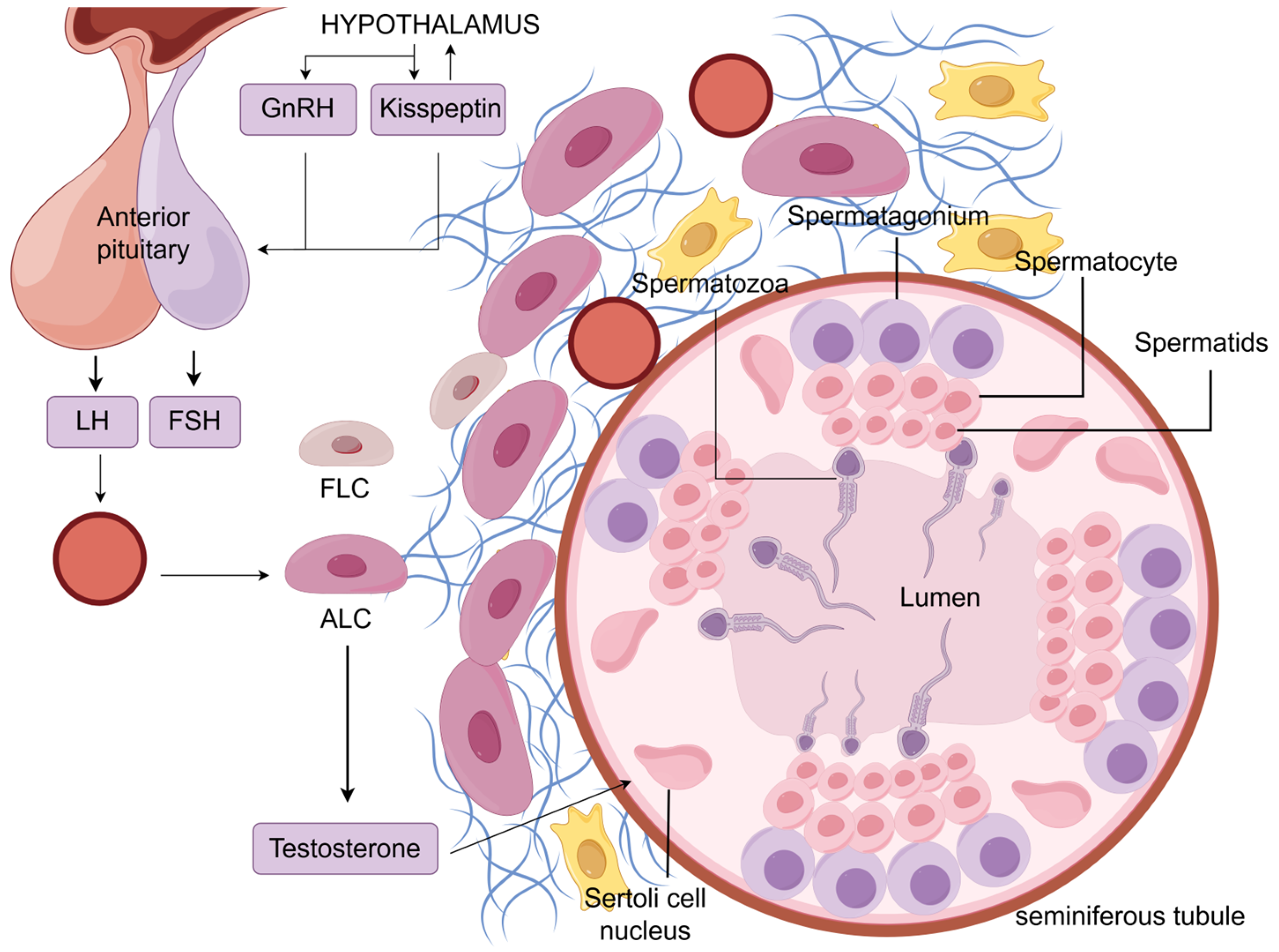
Figure 1. The endocrine regulation of spermatogenesis involving LH. The hypothalamus secretes GnRH and kisspeptin, which act on the pituitary gland, stimulating the synthesis and secretion of LH. LH travels through the bloodstream to the testes, where it acts on ALCs, stimulating the secretion of testosterone. Testosterone enters the seminiferous tubules and acts on Sertoli cells, promoting the differentiation of spermatogonia, spermatocytes, and spermatids into mature spermatozoa, which are released into the lumen of the seminiferous tubules. LCs are present in the testicular interstitium, a loose connective tissue containing various components such as blood vessels, collagen fibers, and LCs (only blood vessels, collagen fibers, and LCs are shown). LCs consist of FLCs and ALCs, but the amount of FLCs is small, and almost no testosterone is produced after birth. Figure support was provided by Figdraw (www.figdraw.com).
Ijms 26 03548 g001
Figure 2. Effects of LH on spermatogenesis, LCs, and sperm cells. LCs originate from fibroblasts or lymphatic endothelial cells. LH can promote testosterone synthesis in LCs, as well as their proliferation, differentiation, and regulation of the biological clock. Upon LH binding to LHR on the surface of ALCs, multiple pathways are initiated, leading to the synthesis of StAR protein located on the mitochondrial membrane. StAR protein facilitates the transfer of cholesterol into mitochondria, where it is converted into pregnenolone by CYP11A1. Pregnenolone undergoes conversion to dehydroepiandrosterone by CYP17A1, which further metabolizes into androstenedione by 3βHSD1 or into progesterone by 3βHSD2 before being converted into androstenedione by CYP17A1. Androstenedione is then synthesized into testosterone by 17βHDS3. Testosterone enters LCs and binds to androgen receptors (ARs), causing dissociation of heat shock proteins. Activated ARs translocate to the nucleus and bind to ARE elements, initiating gene transcription (classical pathway). Activated ARs can also activate MAPK kinase activity through EGFR activation, phosphorylating CREB protein to activate gene transcription (non-classical pathway). Through a series of pathways, this ultimately promotes spermatocyte meiosis, maturation, release, formation, and maintenance of the blood–testis barrier and the development of spermatogonial stem cells. LHR is also present on spermatocytes, suggesting that LH may regulate processes such as sperm energy acquisition, morphological changes, and motility. Figure support was provided by Figdraw (www.figdraw.com).
Ijms 26 03548 g002
Figure 3. LH regulates testosterone synthesis in LCs through multiple pathways. LH synthesized by the pituitary gland is transported to the testes via the bloodstream, where it binds to the G-protein-coupled receptor LHR on the surface of LCs. This activation leads to the stimulation of adenylate cyclase (AC) and Phospholipase C, resulting in the production of cAMP from ATP and the conversion of PIP2 to IP3 and DAG. cAMP activates PKA, which phosphorylates transcription factors such as CREB, leading to the expression of StAR protein or genes involved in testosterone synthesis. PKA can also directly contribute to the phosphorylation of StAR protein. Additionally, cAMP activates epidermal growth factor receptors (EGFRs) on the LC surface, initiating the MAPK signaling pathway through Ras, Raf, MEK, and ERK phosphorylation, further enhancing gene expression and StAR protein phosphorylation for increased transport capacity. DAG and IP3 activate PKC and IP3Rs on the endoplasmic reticulum, promoting calcium ion release. Calcium ions, in turn, activate CamK, inducing gene expression to facilitate testosterone synthesis. Transcription factor Foxo3 inhibits testosterone synthesis, but LH can suppress Foxo3 inhibition by activating Pi3K and downstream Akt. Excess testosterone promotes PDE-mediated cAMP degradation, activating AMPK to inhibit testosterone synthesis. Testosterone also inhibits its own synthesis by activating ROBO receptors to suppress Akt. These pathways cooperate to maintain dynamic equilibrium in testosterone synthesis. (The production of testosterone in LCs is shown in Figure 2.) Figure support was provided by Figdraw (www.figdraw.com).
Ijms 26 03548 g003
Table 1. Functional roles of LH in various processes of spermatogenesis.
RoleSignaling PathwayTargetsSpecies/Cell LinesReferences
Testosterone productionThe ERK pathwayStAR, Hsd3b6, Cyp17a1, and Hsd17b3Rat[100,101,102,106]
The JNK pathwayc-JunRat[95]
The ERK5 pathwayStAR and Nr4a1MA-10 LCs[108]
The calcium signaling pathwaySTAC3, CaMK, Dax-1MA-10 LCs, hamster[110,111,118,119]
The cGMP-PKG pathwayStARMouse, rat[124,125,126,127]
The PI3k/AKT pathwayFOXO3LC lines R2C[163]
The AMPK signaling pathway3βHSD, CYP17, and STARMouse, MA-10 LCs[128,129,130,131,132,133]
The Slit/Robo pathwayCREB, AKTMouse[134,135]
LCs proliferationThe ERK pathwayPCNA, D-cyclins D3Mouse, rat[71,72,73,74]
The PI3k/AKT pathwayMouse, rat[74]
LCs differentiationUnknownScarb1, Cyp11a1, Cyp17a1, and Srd5a1, Insl3Mouse, rat[68,69,77,78]
The circadian clock of LCsUnknownPer1/PER1, Dec1/2 and Rorb et al.Rat[18]
Sperm motilityUnknownUnknownHuman, Holstein bulls[19,159]
sperm concentrationUnknownUnknownHuman, Holstein bulls, rat[19,159]
Sperm morphologyUnknownUnknownRat[160]
Sperm capacityThe cAMP/PKA pathwayUnknownRat, human[20,21,162]
Disclaimer/Publisher’s Note: The statements, opinions and data contained in all publications are solely those of the individual author(s) and contributor(s) and not of MDPI and/or the editor(s). MDPI and/or the editor(s) disclaim responsibility for any injury to people or property resulting from any ideas, methods, instructions or products referred to in the content.