Review

The Current Roadmap of Lung Cancer Biology, Genomics and Racial Disparity

by
Enas S. Alsatari
1,2,
Kelly R. Smith
1,2,
Sapthala P. Loku Galappaththi
1,2,
Elba A. Turbat-Herrera
1,2 and
Santanu Dasgupta
1,2,3,*
1
Department of Pathology, Frederick P. Whiddon College of Medicine, University of South Alabama, Mobile, AL 36688, USA
2
Mitchell Cancer Institute, University of South Alabama, Mobile, AL 36604, USA
3
Department of Biochemistry and Molecular Biology, Frederick P. Whiddon College of Medicine, University of South Alabama, Mobile, AL 36688, USA
*
Author to whom correspondence should be addressed.
Int. J. Mol. Sci. 2025, 26(8), 3818; https://doi.org/10.3390/ijms26083818
Submission received: 20 January 2025 / Revised: 26 March 2025 / Accepted: 11 April 2025 / Published: 17 April 2025
(This article belongs to the Special Issue Advanced Molecular Research in Lung Diseases)

Abstract

:
Globally, lung cancer is the most prevalent cause of cancer-related death. There are two large histological groups of lung cancer: small-cell lung cancer (SCLC) and non-small-cell lung cancer (NSCLC). Based on histopathological and molecular features, adenocarcinoma (ADC) and squamous cell carcinoma (SCC) are the two major histologic subtypes of NSCLC. Various epidemiological and environmental factors are linked with an increased risk of lung cancer. However, these risk factors show disparities in patients with divergent racial and ethnic backgrounds. Interestingly, different populations were found to harbor distinct molecular features as evidenced by variations in genetic mutation profiles. Moreover, diverse histological and molecular progression patterns are identified in lung cancer, which could be crucial in improving diagnosis, prognosis, and therapeutic planning. In concert with a plethora of nuclear genetic alterations, mitochondrial alteration, epigenetic reprogramming, microbial dysbiosis, and immune alteration signatures have been identified in various lung cancer types. This review article provides a comprehensive overview of screening tests and the treatment strategies for NSCLC and SCLC, including surgery, radiation therapy, chemotherapy, targeted therapies, and immunotherapies. Through the unification of these diverse aspects, this review article aspires to a complete understanding of lung cancer’s genomics, biology, microbial landscapes, and racial disparity and seeks to understand the essential role of racial and ethnic factors in lung cancer occurrence and treatment.

1. Introduction

Lung cancer ranks as the second most prevalent malignancy, with an 11.4% incidence rate [1]. Over 230,000 new cases were detected in the United States in 2018, leading to more fatalities than all other cancers including breast, colon, and prostate cancer combined [2]. According to GLOBOCAN 2020 data, approximately 2.3 million new cases (11.4%) and almost 1.8 million deaths from lung cancer were recorded in 2020 [3]. Lung cancer is uncommon before the fifth decade of life, but its incidence rises with age [3]. In the U.S., lung cancer incidence among males continues to decline, while females showed an initial increase followed by a decline. It is particularly marked among younger women who have recently demonstrated higher incidence rates than males, notably for non-Hispanic Whites and Asians/Pacific Islanders [4]. Similarly, a study by Nolen et al. reported higher rates of lung cancer in the United States in young women than men of similar age, extending to those aged 50–54 [5]. A very recent study showed reductions in lung cancer mortality rates that have exceeded reductions in incidence, particularly among men (5.0% vs. 2.6% annually) and women (4.3% vs. 1.1% annually) [6]. On the other hand, the disparity in lung cancer incidence still exists among various racial and ethnic groups. The highest incidence rates and the slowest decline were seen in Native Americans, with various States, including Mississippi and Kentucky, continuing to experience mortality rates two to three times higher than most Western States due to historic smoking prevalence [6]. In addition, Cuban males show higher incidence rates among other Hispanic groups, whereas U.S.-born Black males show higher incidence rates than Caribbean-born Blacks [7]. Among females, US-born Blacks exhibit the highest incidence rates [7]. Nevertheless, lung cancer is the most common cause of cancer-related death in all parts of the world [8]. In 2020, lung cancer was responsible for around 1.8 million deaths, accounting for 18% of all cancer deaths. The age-standardized mortality rate (ASMR) was 18.0 per 100,000 (25.9 in men and 11.2 in women) [9,10]. Mortality exhibits substantial regional variation. The highest rates are seen in countries with high Human Development Index (HDI) scores, primarily from Europe and North America, while the lowest rates are noted among those mainly located in Sub-Saharan Africa [9].
In this review article, we summarize the current knowledge of lung cancer and racial disparities, focusing on various aspects, including genomics, biology, and microbial landscapes. We also presented divergent histopathological and molecular subtypes, epidemiology and risk factors, histopathological and molecular progression patterns, nuclear and mitochondrial genetic alterations, epigenetic alteration, immune system dysfunction, and microbiome dysbiosis associated with lung tumorigenesis.

2. Epidemiology and Risk Factors: An Ethnic and Ancestral View

2.1. Histological Subtypes of Lung Cancer

Lung cancer is divided into two major groups: small-cell lung cancer (SCLC) and non-small-cell lung cancer (NSCLC). SCLC is aggressive and has a high risk for distant metastasis at initial diagnosis [11] and accounts for 12% of all lung cancer cases [12]. NSCLC is, conversely, the most common group, representing 80% to 85% of the lung cancer cases [13]. Among the NSCLC, adenocarcinoma (LUAD) is the most common histologic subtype, accounting for 45% of all cases, followed by squamous cell carcinoma (LUSC) at 21% of cases, while 23% attributed to unclassified histologic subtypes [12]. Notably, LUAD is more common in never-smokes with a predominant EGFR gene mutation, whereas LUSC is more common among smokers with a predominant TP53 gene mutation [14,15].

2.2. Environmental and Lifestyle Risk Factors

Epidemiology and risk factors involve a complex interaction of environmental, genetic mutations, and lifestyle factors that contribute to susceptibility to lung cancer and its outcomes [16,17]. However, these interactions become more significant when considering ethnic and ancestor differences. Although smoking is the predominant cause of lung cancer, another study revealed that 10–25% of all lung cancer patients have never smoked [18]. This disparity underscores the need to investigate additional risk factors other than smoking, especially in populations where lung cancer is not related to smoking [19]. Cigarette smoking remains a major risk factor for lung cancer development. The initiation of smoking habits is mediated by peer pressure, family habits, and psychological distress [20,21]. Interestingly, Harrell et al. reported that demographic factors such as race, socioeconomic status, and pubertal development were significant predictors of early smoking initiation among schoolchildren [22]. Additionally, preventative measures for air pollution include techniques like urea-selective catalytic reduction (SCR), diesel particulate filters, and NOx storage-reduction catalysts approved to enhance air quality to avoid additional health effects from gaseous as well as particulate air pollution pollutants [23]. As a reason, there were substantial declines in lung cancer incidence in the USA from 2007 to 2018. On the other hand, there has been little change in rates among never-smokers, though rates increased significantly in Asian and Pacific Islander populations [24]. In Denmark, lung cancer trends are influenced by historical smoking patterns, where a decline in male smoking rates led to reduced incidence. In contrast, the prevalence of smoking in women remained stable for longer, contributing to a later increase in lung cancer incidence [25]. Existing evidence suggests that passive smoke is the cause of a significant proportion of lung cancer in women. For instance, Du et al. reported that passive smoking accounts for about 17.9% of lung cancer cases among never-smoking women, most of them exposed to household smoking [26]. Moreover, a study of Moroccan women showed that 75% of lung cancer cases were recorded in never-smokers, and LUAD was the most common subtype among passive smokers [27].
Zhu et al. reported that non-smoking people who drink tea ≥ 2 cups/day have a greater risk of lung cancer [28]. At the population level, cigarette smoking is the primary determinant of the occurrence of lung cancer [29]. Environmental factors increase the risk of developing lung cancer, such as air pollution, occupational exposure, secondhand smoke, and radiation exposure [30,31]. In China, a study by Liu et al. observed that occupational environment and meteorological conditions synergistically affect lung cancer development [32]. Furthermore, Chinese-style cooking increases lung cancer risk [33]. Moreover, long-term exposure to air pollutants such as PM2.5, NO2, and NOx significantly increases the risk of developing lung cancer [34]. The World Cancer Research Fund (WCRF) reported that drinking water with high concentrations of arsenic increases lung cancer risk, and the evidence was reported as “convincing” [35]. Additional interaction of these air pollutants with poor lifestyle and high genetic risk dramatically raises the likelihood of lung cancer occurrence [35]. Similarly, Huang et al. showed the same results [36]. However, predicting cancer associated with environmental factors like alcohol consumption and smoking can alter based on the variation in polymorphism of xenobiotic metabolizing enzymes (XME) genes [37]. Pettit et al. studied the genetic correlation between various traits and lung cancer risk, indicating a negative genetic correlation between lung cancer risk and some traits, including dietary behaviors, fitness metrics, educational attainment, and other psychosocial characteristics. On the contrary, the body mass index (BMI) showed a positive genetic correlation with the likelihood of lung cancer [38].
The relationship between lung cancer risk and dietary items like fruits, vegetables, micronutrients, phytochemicals, fat, and beverages has been studied. An increased intake of fruits, vegetables, and carotenoid-rich foods is associated with a reduced risk of developing lung cancer [35]. On the contrary, higher intake of retinol, red meat intake, processed meat intake, alcohol drinking, and dietary fat have been associated with an increased risk of lung cancer. However, no link has been reported between the phytochemical “bioflavonoid” and lung cancer risk.

2.3. Genetic and Racial Disparities and Lung Cancer Susceptibility

Lung cancer risk is influenced significantly by different racial and ethnic disparities. Individuals with African ancestry (AA) have higher mortality rates and incidence of lung cancer development at an earlier age compared to individuals with European ancestry (EA) due to disparities in preventive screening monitoring and treatment disparities [39,40]. In addition, there is a significant disparity in the metabolic pathways and how the body processes nicotine between AA and EA groups, as AA has lower levels of cotinine glucuronidation [41]. Non-Hispanic AA males show the highest rates of mortality and lung cancer incidence compared to all race-ethnicities [42,43]. Similarly, Primm et al. showed persistent disparities in NSCLC incidence between AA and EA men [44]. Interestingly, despite disparities in diagnosis and treatment, AA and Asian NSCLC patients demonstrate better outcomes for the same-stage cancer compared to EA patients [45]. The cause for disparities is genetic ancestry, as AA populations with LUSC have more genomic instability and aggressive molecular traits, while AA patients with LUAD have a higher frequency of PTPRT and JAK2 gene mutations [46,47]. Additionally, the Asian population demonstrates a higher frequency of STK11, TP53, and EGFR gene mutation [48], but they have longer survival rates and higher chemotherapy responses in comparison to EA patients [49]. Another study linked TP53, KRAS, and KEAP1 gene mutations with worse overall survival, whereas EGFR gene mutations are associated with a higher chance of survival [50]. Recent studies found that EA patients have higher mortality rates compared to Hispanics and Asians, and they have a higher susceptibility to lung cancer due to higher frequencies in smoking-related loci [51,52]. Many studies have revealed racial disparities in the genetic mutation profile of lung cancer patients. Compared to Japanese patients, EA-LUSC patients present a higher frequency of mutations in TP53, PIK3CA, KEAP1, and NFE2L2 genes [53]. On the contrary, EA-LUAD patients exhibit a significantly lower occurrence of EGFR mutation but an increased frequency of mutation in the PIK3CA, KEAP1, KRAS, TP53, BRAF, NF1, STK11, RBM10, and MET genes. Weiner and Winn reported a higher prevalence of EGFR gene mutation in the East Asian population and more predominant KRAS and STK11 gene alterations in EA and AA populations [54]. Generally, the disparities in survival rates between EA and AA populations are noticeable in patients who are young and have localized tumors [55]. The disparities also exist in histological subtype, stage, and tumor grade. Asian or Pacific Islander (API) exhibit a higher frequency of adenocarcinoma (ADC) compared to AA, EA, and American Indian/Alaska Native (AIAN) patients [56].

3. Current Detection and Treatment Modalities of Lung Cancer

NSCLC treatment varies depending on the tumor stage and overall health [57]. Surgery is a treatment option for early-stage lung cancer. There are four types of resections: wedge resection (removing the tumor and with normal tissue), lobectomy (removal of one whole lobe with the tumor), pneumonectomy (removal of a lung), and sleeve resection (removal of part of the bronchus). Following surgery, some patients may undergo chemotherapy and radiation therapy. Targeted therapy is used to attack cancer cells based on their specific characteristics, including monoclonal antibodies, tyrosine kinase inhibitors (TKIs), as well as mammalian target rapamycin (mTOR) inhibitors that include mTOR, and KRAS G12C inhibitors. By binding on specific sites, monoclonal antibodies can mark cancer cells for their destruction by the immune system. Furthermore, tyrosine kinase inhibitors target specific signals for cancer growth and spread, including specific EGFR tyrosine kinase inhibitors. Indeed, mTOR inhibitors block a protein called mTOR to keep cancer cells from growing and prevent the growth of new blood vessels. KRAS G12C Inhibitor is a drug that blocks transcription of KRAS p.G12C and halts the growth of cancerous cells. Immunotherapy treatments such as immune checkpoint inhibitors (ICIs), however, actually boost and restore natural defenses against cancer in patients. PD-1 and PDL-1 inhibitor therapy is a type of immunotherapy that prevents the binding of PD-1 protein on the surface of T cells with PD-L1 protein found on some types of cancer cells, allowing T cells to kill tumor cells [57]. Actually, the ICIs reported an objective response rate of around 42% in NSCLC patients, even higher among those with PD-L1 expression ≥ 50% [58]. Additionally, perioperative immunotherapy, primarily through PD-1/PD-L1 inhibitors, has made landmark strides in resectable NSCLC to establish a new standard of care for stages II to III due to the reduction in postoperative recurrence with an overall survival improvement [59]. The other approach to immunotherapy is a CTLA-4 inhibitor, which blocks CTLA-4 on T cells binding of B7 provided by antigen-presenting cells (APCs) so that T cells can kill the cancer cell [57]. Notably, both smokers and non-smokers with lung cancer benefit from immunotherapy [60]. Another treatment approach uses a laser beam (laser therapy) or drug and a specific type of laser light (photodynamic therapy); however, the drug is not effective until exposed to laser light in photodynamic therapy. Interestingly, fiberoptic tubes carry laser light to the site of cancer cells to activate the drug. Moreover, cryosurgery can freeze and destroy abnormal tissue, such as carcinoma in situ. Electric current is used to heat the needle or probe in electrocautery treatment to destroy abnormal tissues. A variety of treatment options exist for SCLC [57]. Surgery may not be the answer for SCLC, as it may happen bilaterally. Indeed, there is always the possibility of chemo and radiation following surgery. Systemic chemotherapy is a standard treatment for SCLC. In addition, external radiotherapy is given to SCLC patients, which is used as palliative therapy. Another effective option for SCLC treatment is immunotherapy drugs like mepolizumab and Durvalumb. A laser beam is also used to destroy SCLC cells. Interestingly, Personalized medicine in lung cancer, particularly NSCLC, involves tailoring treatment based on patient genomics. Such approaches improve treatment specificity, targeting genetic mutations that confer resistance and deliver nanoparticles to target NSCLC biomarkers [61].
Three lung cancer screening tests have been tested before the onset of symptoms: low-dose computed tomography (LDCT), chest X-ray, and sputum cytology [57]. A very low dose of radiation is used in LDCT to generate a sequence of very detailed pictures by X-ray machine. A chest X-ray uses an X-ray beam to capture images of organs and bones within the chest. Sputum cytology examines the sputum cells by using a microscope to identify abnormalities. These scanning tests are not sensitive enough to detect lung cancer early, as they may produce false negative or false positive results. Also, the chest is exposed to radiation in LDCT and chest X-ray tests. However, chest X-ray and sputum cytology are low-cost, and a combination of both is probably helpful in detecting early lung cancer in high-risk people [62]. CRISPR technology is also being used for biomarker identification in lung cancer patients to allow for better diagnosis and assess prognosis [63]. However, CRISPR-Cas9 is still in the early stages of development, lacking proven clinical protocols, and carries risks of incorrect editing [64].

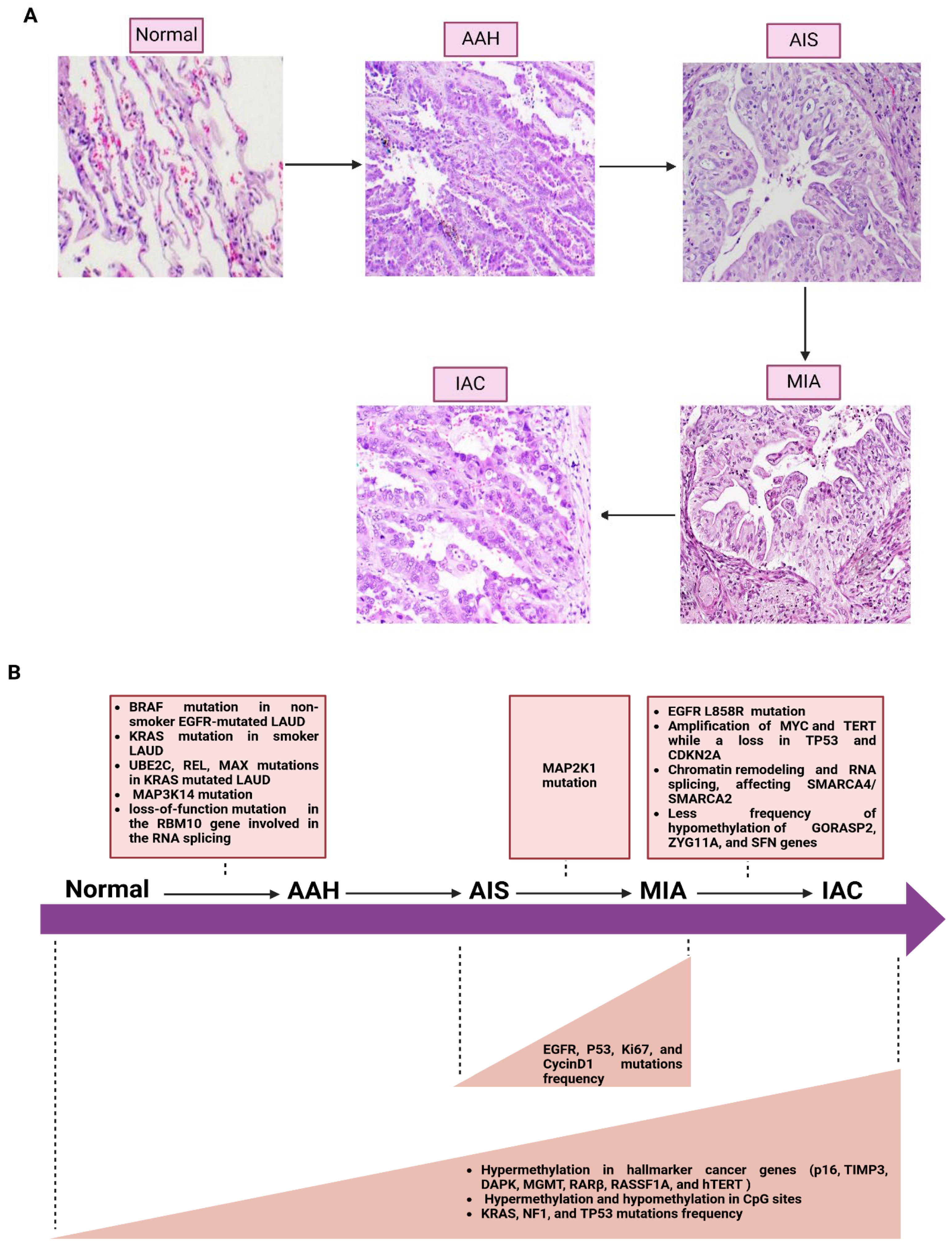
4. Molecular Progression of Lung Cancer

A more profound understanding of the molecular process continuum to lung cancer initiation and progression is urgently needed to improve patients’ prognosis and disease management. Lung cancer initiation is caused by genetic alterations that also drive and maintain lung cancer progression. These can be mutations, epigenetic variations, and alterations in non-coding RNAs or microRNAs. NSCLC is a multistep process ranging from normal epithelium to invasive carcinoma throughout histological stages. Normal epithelium first develops into hyperplasia, characterized by an abnormal proliferation of epithelial cells. Hyperplasia can also progress to metaplasia, during which epithelial cells convert into a different type of cell in response to inflammation or chronic irritation. In such situations, morphological and cell growth abnormalities may progress to dysplasia afterward. Epithelial dysplasia may progress to carcinoma in situ (CIS), a pre-invasive state where atypical cells remain bound by the basement membrane. Ultimately, these changes may progress to invasive carcinoma, where malignant cells cross through the basement membrane [65]. LUAD progresses in a systematic and sequential manner (Figure 1A). It starts from atypical adenomatous hyperplasia (AAH), a lesion derived from glandular cells in the epithelial tissue of peripheral airways, to adenocarcinoma in situ (AIS), then minimally invasive adenocarcinoma (MIA) before the next step into invasive adenocarcinoma (ADC or IAC) [66]. Early lung carcinoma includes AIS and MIA in the large bronchi. LUSC, which derives from squamous cells primarily in the large bronchi of the central airways, begins with the transformation of normal epithelium to hyperplasia (either mucous or basal). Metaplasia is followed by various degrees of dysplasia, leading finally to severe dysplasia, resulting in carcinoma situ and invasive cancer [67]. Pathways and premalignant lesions for LUSC and LUAD are significantly different. The development of LUSC is characterized by a series of molecular abnormalities that start with histologically normal epithelium and progress through various stages, including metaplastic and dysplastic changes, and ultimately culminate in LUSC. However, AAH is a significant premalignant lesion in the development of LUAD, AAH is implicated in the linear progression of the cells from the terminal respiratory unit to adenocarcinoma in situ (AIS) [68].
Several studies have focused on genetic and epigenetic changes associated with the histological progression of LUAD (Figure 1B). For example, Sivakumar et al. found BRAF mutation in 23% of AAH cases, predominantly LUAD cases co-occurring with EGFR [69]. However, KRAS mutations were limited to BRAF-mutant cases and revealed in 18% of AAH, all occurring among ever-smoker patients. Furthermore, the same investigation found (UBE2C, REL) and (MAX) were associated with KRAS-mutant AAHs [69]. Many studies have tried to identify whether genetic changes differ in specific states of LUAD. Nevertheless, AIS and early invasive adenocarcinoma share many common gene mutations together, but early invasive ADC exhibits significantly more mutations compared to AIS [70]. For instance, Jia et al. found significant differences in the genetic changes between AIS and MIA concerning EGFR mutation frequency, p53 and Ki67, and cyclinD1 expression level, notably higher in MIA than in AIS [71]. In addition, EGFR mutation is a significant subclone in AIS, MIA, and ADC but a minor one in AAH [72]. Other studies also obtained consistent results, representing the mutation frequency of EGFR increased from AIS to MIA and remained constant between MIA and IAC during stepwise progression [73]. Nevertheless, the AIS stage does not reveal ALK fusions or ROS1 mutation. In contrast, Haga et al. reported that EGFR mutations are similar in early and advanced adenocarcinoma stages, but EGFR mutation occurs in the early stage, causing an accumulation of many somatic mutations [74]. Many driver genes in early stages have also been identified in the previous literature reviews, such as MET (Y1021N, exon 14 splice site, exon 14 deletions), BRAF (A489_Q493del), KRAS (G12A, G12D), ALK (EML4-ALK) fusions, RET (KIF5B-RET), ERBB2 (V659E), and MAP2K1 (E102_I103del), with similarity to those in advanced stages [75,76,77,78,79]. This implies that the most essential somatic driver genes are present at an early stage. In addition, the KRAS, NF1, and TP53 mutation frequencies were significantly elevated from AIS to MIA and invasive ADC, supporting their role in cancer progression [70]. Gene mutations of MAP3K14, MAP2K1, and EGFR L858R were identified in AAH, MIA, and IAC, respectively [80]. In advanced stages of LUAD, additional oncogenic amplification and tumor suppressor deletion occurred, including a copy number increase in the regions of MYC and TERT while a loss in TP53 and CDKN2A [74].
Progressive epigenetic changes also occur in AAH and elevate it even further to LUAD, specifically promoter hypermethylation of hallmark cancer genes such as p16, TIMP3, DAPK, MGMT, RARβ, RASSF1A, and hTERT [81]. Nevertheless, the hypomethylation of GORASP2, ZYG11A, and SFN genes is much less frequent in invasive adenocarcinoma, which correlates with poorer patient outcomes [82]. Another study found that hypermethylation and hypomethylation of CpG sites increased in the later stages of lesions (AIS, MIA, and IAC) compared to AAH and normal lung tissue [83]. In addition, a high frequency of methylation heterogeneity and loci with distinct methylation were observed in later stages. Generally, the later-stage lesions have more epigenetic changes and fewer genetic alterations, specifically in DNA methylation, compared to early stages [83]. Alterations in chromatin remodeling and RNA splicing, involving the SWI/SNF chromatin-remodeling genes SMARCA4 and SMARCA2, are observed in advanced LUAD stages [74]. Nevertheless, the loss-of-function mutation in the RBM10 gene, which is involved in RNA splicing, occurs in the early stages of LUAD.

5. The Nuclear Genetic Alterations in Lung Cancer Subtypes and Their Racial Distribution

Lung cancer is classified into histological and molecular subtypes, which are of utmost importance for the decision-making process regarding treatment options and disease prognosis or diagnosis [1]. Indeed, both histopathological classification and molecular subtyping of lung cancer play essential roles in predicting the likelihood of bone metastasis [84]. Moreover, molecular subtyping is essential for targeted therapy. For instance, determining the differential expression of genes (DEGs) and microRNAs in lung cancer types may facilitate the treatment or diagnosis at earlier stages [85]. Wang et al. examined pathway enrichments and DEGs across SCLC and NSCLC (both SCC and ADC), highlighting the unique molecular responses to treatment in each cancer type [86]. The 2021 WHO classification for thoracic tumors also updated the molecular abnormalities associated with lung cancer, highlighting key driver mutations and their clinical relevance. EGFR mutations are common in Asians and non-smokers, particularly those with LUAD. Conversely, patients with a history of smoking often have KRAS mutations, more commonly seen in LUAD. ALK rearrangements are more prevalent in younger, non-smoking individuals with LUAD, while ROS1 fusions have emerged as a novel driver mutation in NSCLC. Additionally, emerging molecular targets such as RET mutations, though present in a small percentage of NSCLC cases, reflect the increasing appreciation for the heterogeneity of genetic alterations driving lung carcinogenesis [87].
Background racial disparities in the diagnosis, management, and outcome of lung cancer have persisted in healthcare for decades [88]. Thus, the death rate after lung cancer resection is affected by different races, and genetic changes would differ between ethnicities as well [89]. While disparities in human studies contribute to differences in tumor behavior and response to treatments, so do biological properties of lung cancer cells, such as heterogeneity and the interactions between the tumors and their microenvironment [90]. A further study conducted on NSCLC patients reported that the incidence of EGFR mutations is substantially greater among East Asian ancestry, while KRAS and STK11 alterations are more prevalent in EAs and AAs [91]. In contrast, James et al. showed that mutations in EGFR are more frequent in AA, while TP53 mutation is more common in Latin ancestries [92]. Lung disparity-related studies among AA men revealed that they have higher rates of lung cancer incidence and death compared with individuals from all other racial/ethnic groups, likely due to a combination of genetic, socioeconomic, and healthcare access factors [93,94].

5.1. The Nuclear Genetic Alterations in LUAD Subtype and Its Racial Distribution

The most common form of NSCLC is LUAD, and it encompasses several precursor lesions: AAH, AIS, MIA, and IAC (Figure 1A) [95]. Genome-wide mRNA expression profiles were generated for the four LUAD molecular subtypes, subtype 4 of which has the best prognosis and the higher rate of EGFR mutation. In contrast, subtypes 1 and 2 are related to immune-related processes and TP53 mutations, suggesting late-stage cancer; subtype 3 showed enrichment in cell cycle dysregulation, and subtype 4 had an extracellular matrix organization signature [96]. Additionally, 379 DEGs and 67 differential methylated sites have identified two clinically relevant subtypes within LUAD [97]. A recent study provided additional information about the mutations in distinct subtypes of lung cancer. Tlemsani et al. demonstrated that the clinical profiles of LUAD patients with NF1 deletions differed from those with NF1 mutations, consequently proposing molecular and clinical subclassifications for LUAD [98]. In addition, KEAP1/NRF2-mutant LUADs can be further grouped into three molecular subtypes, and the two variants of KEAP1/NEFR2-mutation handle distinct genetic, clinicopathologic, differentiation as well as immunological properties [99]. LUAD is primarily defined by multiple nuclear genetic defects such as EGFR, KRAS mutant, and ALK gene rearrangement, exhibiting significant variation among racial and ethnic populations (Table 1) [100,101]. A recent study showed that EA is positively associated with KRAS G12C mutation and negatively associated with EGFR mutation [102]. Similarly, Shi et al. found that mutation in KRAS G12C is the most common among EAs [101]. On the contrary, East Asians, Hispanic/Latino patients, and individuals with American Indigenous (AMR) ancestry showed the opposite [97,98]. Furthermore, though driver CTNNB1 mutations are rarely found in non-Hispanic White patients, they are confined to a subgroup of never-smoker non-Hispanic Asian individuals and, more specifically, enriched among those with East Asian ancestry [97]. Shi et al. found a higher rate of EGFR exon 21 L858R mutation, RET rearrangements, and ERBB2 amplifications in Asians than in other ethnic groups [101]. In addition, STK11 mutations are more common in EAs and AAs than in Asians. Ji et al. showed that ATM L2307F mutation is more frequent among Ashkenazi Jewish populations [103]. Meanwhile, EGFR and KRAS mutation frequencies were significantly correlated with genetic ancestry in LUAD patients from Latin America (LA), indicating a strong correlation between germline genetics and the development of mutations in LUAD [104]. Zhang et al. compared the frequency of EGFR and KRAS mutations among LUAD East Asian and EA patients, suggesting that common oncogenic driver mutations occur more frequently in East Asians with LUAD [105]. The most common driver is EGFR, followed by KRAS in East Asian patients, whereas the reverse is true for EA patients [104]. Unlike Japanese, KRAS, TP53, BRAF, PIK3CA, KEAP1, NF1, STK11, RBM10, and MET mutations were seen more frequently in EA patients [53]. On the other hand, Tunisian-LUAD patients were found to have a lower percentage of EGFR and KRAS mutations and ALK translocation compared with European or Asian series [106]. Research on tumor suppressor genes (TSGs), such as TP53, STK11, and MGA mutations, revealed a high prevalence of MGA in East Asians, whereas TP53 and STK11 mutations are more common in EAs [104]. Besides detecting the racial disparities of driver gene mutations, a recent study also found race-specific miRNA isoforms in EA and AA-LUAD patients, highlighting the racial disparities in miRNA expression profiles [107]. Indeed, novel targeted therapies and individualized care must be informed by race/ethnicity-specific lung cancer nuclear genomic alterations.

5.2. The Nuclear Genetic Alterations in LUSC Subtype and Its Racial Distribution

The second most prevalent NSCLC histological subtype is LUSC, which is commonly attributed to smoking and chronic inflammation [108,109]. LUSC provides complex mutational nuclear genetics with dramatic frequency heterogeneity among racial groups (Table 1). In India, 5.8% of patients with LUSC were found to harbor EGFR mutations, a higher percentage in comparison to EA patients [110]. EA has significantly more TP53 and PIK3CA, KEAP1, and NFE2L2 mutations than Japanese patients [53]. AA patients featured increased homologous recombination deficiency (HRD), higher rates of PTEN deletion, and KRAS amplification, suggesting that the higher prevalence of homologous recombination deficiency (HRD) is crucial for genomic instability in Blacks [46].

5.3. The Nuclear Genetic Alterations in Adenosquamous Carcinoma Subtype and Its Racial Distribution

The term adenosquamous carcinoma (ASC) refers to the presence of mutations associated with both adenocarcinoma and squamous cell carcinoma [111,112]. The nuclear genetic alterations in ASC exhibit significant variation among racial groups (Table 1). For instance, Vassela et al. found EGFR and PI3K pathway mutations to be the most frequent in ASC patients, while KRAS mutation occurs less frequently among EAs [112]. Wang et al. showed that TP53 and EGFR mutations are characteristic events in ASC patients, and CDKN2A, TERT, and LRP1B mutations also recurrently mutate [113]. Also, 64 gene fusions have been described, with ALK fusion being the most frequent, followed by CD74-ROS1 and ROS1-SYN3 fusions. A pilot study also found that the key driver mutations in ASC are EGFR and MET [114].

5.4. The Nuclear Genetic Alterations in SCLC and Its Racial Distribution

SCLC is the most aggressive type of carcinoma and has a poor prognosis [115]. Rudin et al. identified four molecular subtypes of SCLC, including ASCL1, YAP1, NEU-ROD1, and POU2F3, based on the level of expression of the following transcription factors: achaete-scute homolog1, yes-associated protein 1, neurogenic differentiation factor 1, and POU class2 homeobox 3, respectively. The four types are SCLC-A (ASCL1-dominant), SCLC-Y (YAP1-dominant), SCLC-P (POU2F3-dominant), and SCLC-N (NEUROD1-dominant) [116]. Ding et al. replicated the same categorization of four SCLC molecular subtypes [117]. Furthermore, the four SCLC molecular subtypes were identified based on distinct molecular and clinical features, particularly the finding that the endothelial-to-mesenchymal transition (EndMT) in the SCLC-I subtype is associated with platinum resistance and poor prognosis. Conversely, SCLC-A and SCLC-N subtypes were platinum-sensitive [118]. Curiously, Miyakawa et al. highlighted the critical role of super-enhancer-mediated miRNA expression regulation in determining SCLC molecular subtypes [119]. TP53/RB1 alteration and amplification of MYC family genes (MYC, MYCL, and MYCN) are prevalent in SCLCS [120]. Indeed, Pongor et al. have demonstrated that extrachromosomal DNA (ecDNA) contributes to MYC gene amplifications in SCLC [121]. In addition, other common mutations are identified in SCLC, including KMT2D, PTEN and NOTCH receptors, and CREBBP [120]. Indeed, different subtypes of SCLC are identified based on specific transcription factors transcription, such as ASCL1 (SCLC-A), NEUROD1 (SCLC-N), POUF23 (SCLC-P), YAP1 (SCLC-Y), and a recent acknowledged inflammatory/immune-related gene expression subtype SCLC-I subtype [120]. A comprehensive analysis of 3600 SCLC patients identified rare genetic subsets, including STK11-mutant tumors (1.7%) and TP53/RB1 wild-type tumors (5.5%) [122]. However, TP53/RB1 wild-type tumors did not demonstrate a tobacco mutational signature and exhibited alternate mechanisms of p53/Rb pathway inactivation (CDKN2A, CCND1, MDM2) and high human papillomavirus (HPV) positivity. Next-generation sequencing has identified gene mutations in SCLC, such as LRP1B, MAP3K13, MSH6, and SPEN [123]. Recent studies found significant variation in nuclear genetic alterations among different racial populations (Table 1). For example, the TP53 and RB1 gene mutations are identified as the most common mutations in Chinese patients with SCLC, and other mutations are detected as LRP1B, FAM135B, SPTA1, KMT2D, FAT1, and NOTCH3, emphasizing the ethnicity-dependent mutational profile in Chinese SCLC patients [124]. Similarly, another study in China revealed that TP53, RB1, and KMT2D are the most common mutations in Chinese patients with SCLC, and other novel genes (LRRK2, BRCA1, PTCH1, ARID2, and APC) are observed in 90% of these patients [125]. ERBB2 and CREBBP gene mutations were identified as the most prevalent genetic alterations in SCLC, followed by TP53 mutations [126]. However, the additional functionality of identification of this histological phenotype of lung cancer is not just at a genomic level but perhaps more effectively at transcriptomic levels, emphasizing that cell proliferation pathways (example: E2F, G2M, and MYC), upregulated in SCLC and large-cell neuroendocrine cancer as compared to LUAD [127]. In addition, Hu et al. reported that co-mutation of TP53 and RB1 and mutations in Wnt/Notch signaling pathways are more prone to be detected in EA patients with SCLC than in Chinese people [128].

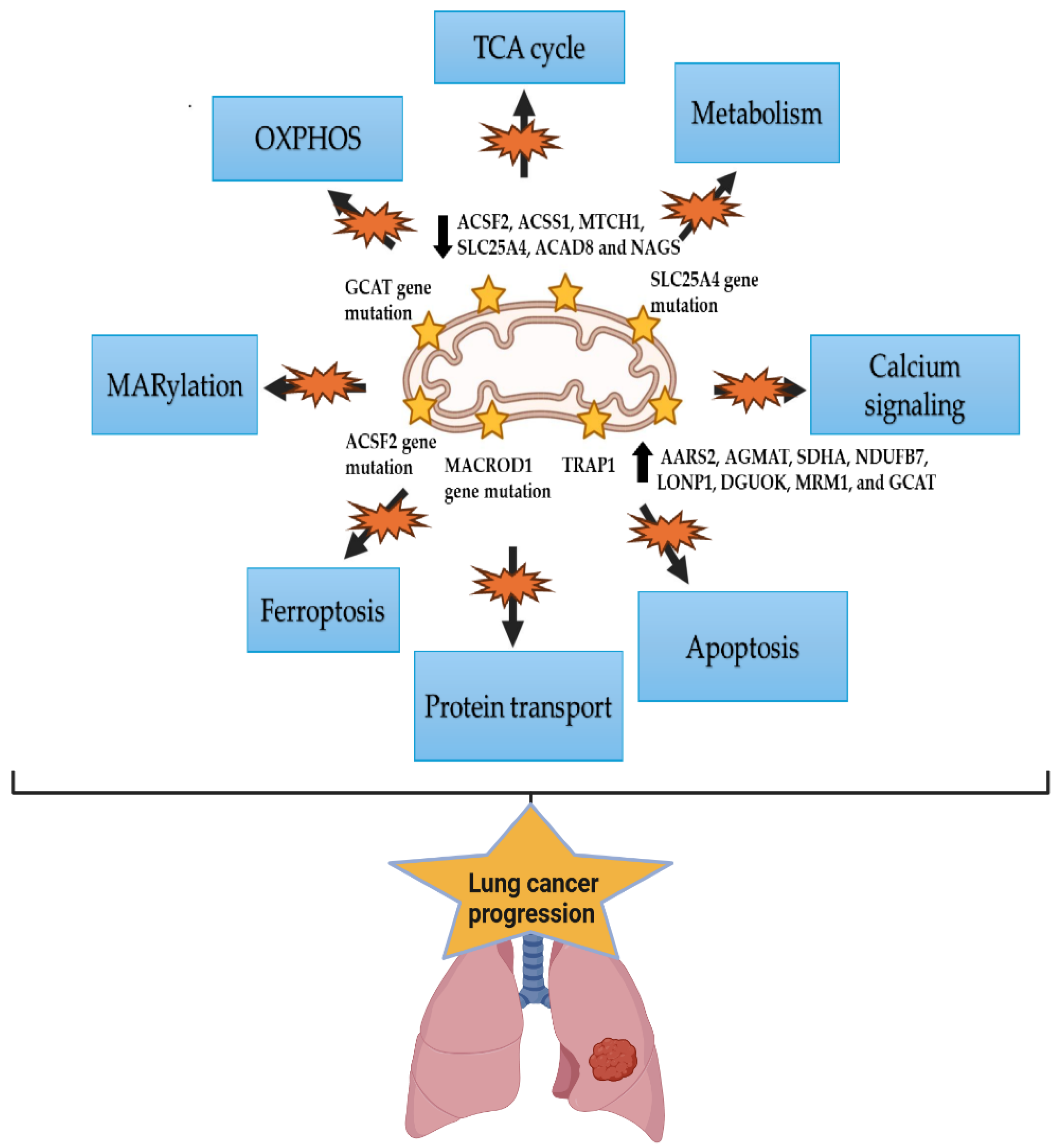
6. The Mitochondrial Alterations in Lung Cancer and Racial Disparity

Cancer cells undergo mitochondrial stress because of engagement in uncontrolled cell proliferation and produce ROS, causing damage to mitochondrial DNA (mtDNA) and mitochondrial proteins, including components of the oxidative phosphorylation (OXPHOS) family, consequently leading to mitochondrial dysfunction [129]. Furthermore, cancer cells activate the mitochondrial stress response to mitigate dysfunction of the mitochondria and aggregation of proteins, which ultimately stimulate the growth and progression of tumors (Figure 2). Mitochondrial genetic changes are also widely present in the occurrence of lung cancer with different ethnic characteristics [130]. Genomic and clinical studies have identified a strong correlation between mitochondrial genomic alterations and lung cancer development and prognosis, as evidenced by copy number variations in mitochondria-targeted genes such as SLC25A4, ACSF2, and MACROD1 in NSCLC [131]. Moreover, Yuan et al. observed that more than 5% of lung tumors have somatic transfer of mtDNA into the nucleus, contributing to somatic mutations [132]. Indeed, Hertweck et al. identified 40 mitochondrial-targeted genes and their genetic alterations in NSCLC, including LUAD and LUSC [130]. Altered mitochondrial genes play key roles in ferroptosis, protein transport, apoptosis, calcium signaling, metabolism, the TCA cycle, OXPHOS, and MARylation. In addition, specific alterations in genes (MACROD1, SLC25A4, ACSF2, and GCAT) are associated with poor survival in patients. Overexpression was observed for AARS2, AGMAT, SDHA, NDUFB7, LONP1, DGUOK, MRM1, and GCAT, and reduced expression of ACSF2, ACSS1, MTCH1, SLC25A4, ACAD8, and NAGS in both LUAD and LUSC. A pilot study showed that the mitochondrial heat shock protein TRAP1 induces cisplatin resistance in lung cancer cells and promotes ROS-dependent mitochondrial dysfunction, causing apoptosis inhibition [133]. Similar results were obtained by Kuchitsu et al. [134]. However, another study found that TRAP1 level is low in SCLC patients compared to NSCLC patients, suggesting using TRAP1 in combination with MSA and mad2 for better SCLC diagnosis [135]. Interestingly, mitochondrial alterations linked to lung cancer metastasis include a reduction in mitochondrial membrane potential and overall mitochondrial functionality. These changes were observed in metastatic lung tumor cells compared to the non-metastatic counterparts [136]. Most importantly, the clinical outcomes of early-stage ADC could be predicted by studying mitochondrial DNA (mtDNA), providing a reliable tool for improving patient care [137]. A higher relapse-free survival (RFS) was observed in patients with somatic mutations at the D-loop region, whereas a lower RFS was recorded in patients with respiratory complex (RC) IV and RCV gene mutations [138]. Potentially, the most mutated non-coding region in both somatic and germline mutations is the D-loop region. For protein-coding genes, CYTB and ND genes had the highest mutation frequency for germline and somatic mutations, respectively. Kazdal et al. found that a significant majority (90.6%) of somatic mtDNA mutations resulted in a non-synonymous amino acid change mapping to a protein-coding gene [139]. Similarly, Jin et al. found that the majority of mtDNA polymorphisms were indeed in protein-coding regions [140]. Moreover, 56 somatic mutations were detected in 60% of the lung cancer patients they analyzed, consisting of 48-point mutations, four single-nucleotide insertions, and four deletions. Interestingly, mtDNA mutations were found to be enriched in never-smoker NSCLC patients compared to current smokers, with a significant association observed between mtDNA and EGFR gene mutations [141]. Notably, the majority of the coding mtDNA mutations targeted RCI. In terms of racial disparities, the prevalence of mtDNA mutations was higher in the never-smoker Asian population compared to the current-smoker EA population. In another study, the airway mucosal biopsies obtained from follow-up NSCLC patients were histopathologically negative but exhibited multiple clonal mtDNA mutations consistent with those detected in the corresponding tumors [142].

7. The Epigenetic Alteration Patterns in Lung Cancer Patients with Various Ethnic and Racial Backgrounds

Significant racial disparities have been identified in lung cancer patients concerning epigenetic alterations. Different epigenetic modifications have been examined in NSCLC, such as histone modifications, non-coding RNA expression, and DNA methylation [143]. Indeed, DNA methylation has been investigated in lung cancer to understand its role in cancer initiation, progression, and outcome [144]. For instance, a study reported that the DNA hypermethylation of three cancer-related genes (MTHFR, RASSF1A, and CDKN2A) is influenced by tobacco smoking level or gender in lung cancer patients [145]. Understanding these epigenetic alterations in different racial backgrounds is crucial for developing tailored treatments and improving clinical outcomes among lung cancer patients [146].

8. The Microbiome Signature in Lung Cancer Subtypes and Racial Differences

Many laboratories have examined microbiome signatures in lung cancer (Table 2). Collectively, recent studies suggest that the human lung microbiome may contribute to lung cancer initiation and progression through various mechanisms, such as bacterial toxin-induced host genomic instability, inducing host inflammatory pathways, altering the local immune environment, the release of cancer-promoting microbial metabolites, and regulation of cancer-related signaling pathways in lung cells [147,148,149]. Nevertheless, a particular oral microbiota, Leptotrichia sp._oral_taxon_225, is associated with a reduced risk of developing lung cancer, suggesting its potential protective role [150]. Lung cancer patients have reduced microbial diversity compared to cancer-free individuals. Moreover, they show significant alterations in the abundance of specific bacteria [151,152]. For instance, the abundance of Actinobacteria phylum, Corynebacteriaceae, Halomonadaceae families, Corynebacterium, Lachnoanaerobaculum, and Halomonas genera were remarkably decreased in lung cancer individuals compared to healthy controls [153]. In fact, the racial/ethnic disparities in different subtypes of lung cancer may provide valuable insight for understanding pathogenesis and biomarkers from various microbiome studies [154].
Some specific microbial signatures were found to be associated with lung cancer patients, such as Enterococcus, Lactobacillus, Escherichia, Phylum TM7, Capnocytophaga, Blautia, Streptococcus, Neisseria, and Prevotella [155,156]. Another study suggested microbiota as potential biomarkers for predicting recurrence or metastasis (RM) in certain patients as significant differences have been observed for the presence of Acidovorax, Clostridioides, Succinimonas, and Shewanella between RM and non-RM groups [157]. Several studies also reported a significant difference in the abundance of 13 types of bacteria between LUAD and LUSC patients [158,159,160,161]. For instance, Jang et al. determined that Actinomyces graevenitzii is more common in LUSC. By contrast, Haemophilus parainfluenza, Neisseria subflava, Porphyromonas endodontics, Fusobacterium nucleatum, and Pseudomonas are more prevalent in LUAD [161]. In addition, specific bacterial genera, such as Acidovorax and Veillonella, were found to be potential biomarkers for detecting and discriminating LUSC from LUAD [162]. In LUAD, Streptococcus and Neisseria are the most prevalent, followed by Veillonella. Similarly, Streptococcus is the most common in LUSC, followed by Veillonella [156]. According to a recent study, Bacillus and Castellaniella were enriched in the microbiota of patients with LUAD, whereas Brucella was predominant during LUSC [163]. In addition, the microbiota diversity is higher in LUSC than in LUAD, particularly among heavy smokers and men, where Proteobacteria further discriminated between LUAD and LUSC [164]. Genus Thermus and Gram-positive bacteria are significantly more abundant in LUAD than in LUSC [165,166]. Additionally, five intratumoral microbiota, including Pseudoalteromonas, Luteibacter, Caldicellulosiruptor Loktanella, and Serratia, have been found to be altered from early to advanced stages of the LUADs [167].
Table 2. The microbiome signature and its significance in lung cancer. This table presents the specific abundance of various microbial species and their potential role in lung cancer.
MicrobiomeSignificanceCitations
Actinobacteria phylum, Corynebacteriaceae, Halomonadaceae families, Corynebacterium, Lachnoanaerobaculum, and Halomonas generaDecreased in lung cancer patients compared to control people[153]
Enterococcus, Lactobacillus, Escherichia, Phylum TM7, Capnocytophaga, Blautia, Streptococcus, Neisseria, and PrevotellaBacterial markers in lung cancer[155,156]
Acidovorax, Clostridioides, Succinimonas, and ShewanellaPrediction of recurrence or metastasis (RM) lung cancer tissue[157]
Actinomyces graevenitziiAbundant in LUSC[161]
Haemophilus parainfluenza, Neisseria subflava, Porphyromonas endodontics, Fusobacterium nucleatum, and PseudomonasAbundant in LUAD[161]
Acidovorax and VeillonellaDifferentiating between LUSC and LUAD[162]
Streptococcus and NeisseriaMost prevalent in LUAD[156]
StreptococcusMost prevalent in LUSC[156]
Bacillus and CastellaniellaEnriched in LUAD[163]
BrucellaEnriched in LUSC[163]
ProteobacteriaDiscriminated in LUAD and LUSC[164]
Thermus and Gram-positive bacteriaThe prevalence is higher in LUAD than in LUSC[165,166]
Leptotrichia sp._oral_taxon_225Reducing lung cancer risk in African Americans (AA)[150]

9. The Immune Alteration Signature in Lung Cancer

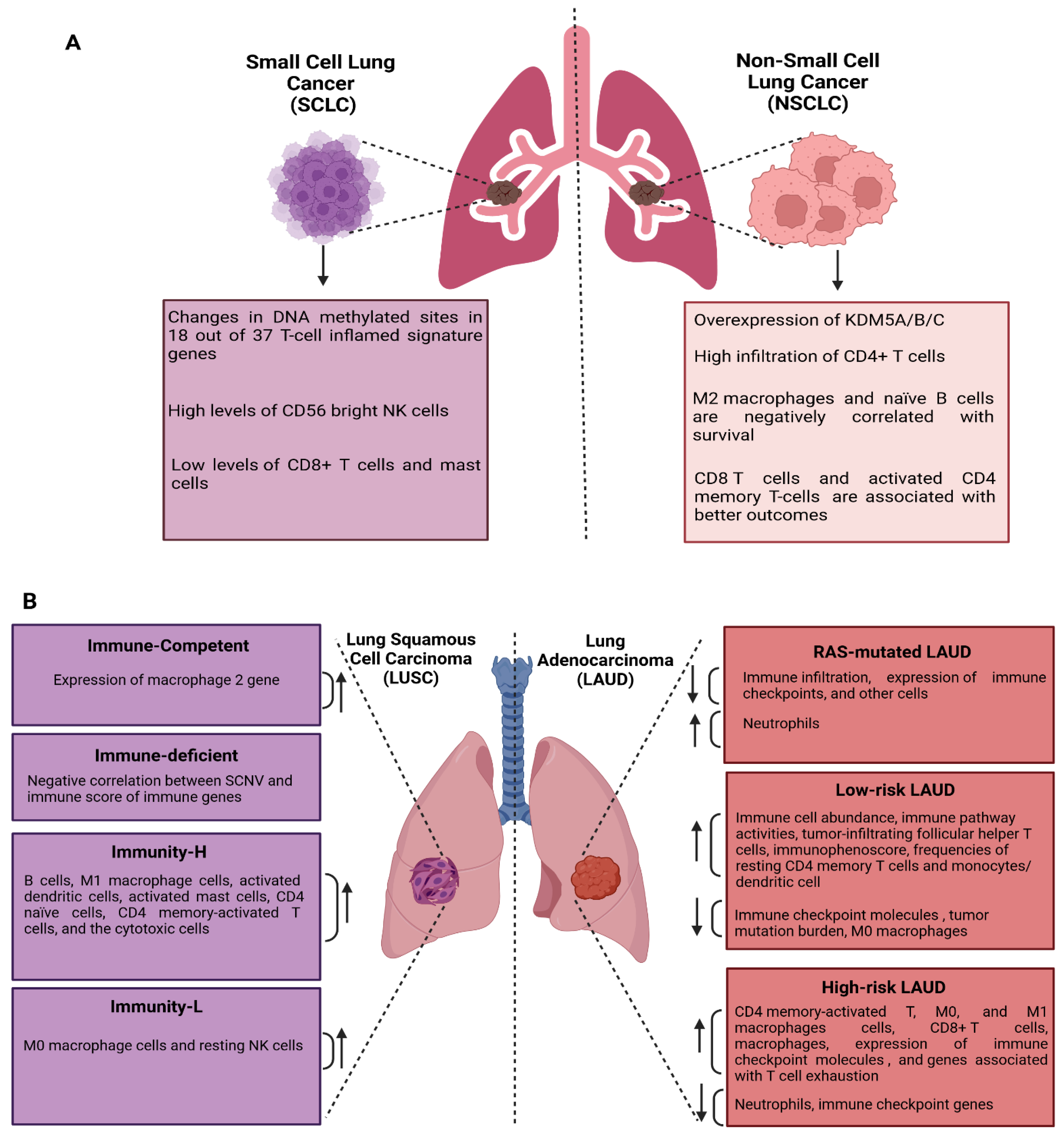
9.1. Immune Alteration Signatures in NSCLC and SCLC

The immune alteration signature in lung cancer is complex and multifaceted, requiring detailed investigation. In fact, immune-based identification differentiates lung cancer and controls patients by analyzing monocytic myeloid-derived suppressor cells (MDSCs), polymorphonuclear MDSCs, intermediate monocytes, and CD8+PD-1+ T cells [168]. Many immune alteration signatures have been reported in NSCLC (Figure 3A). For instance, overexpression of KDM5A/B/C is linked to increased infiltration of CD4+ T cells, especially regulatory T cells (Tregs) and Th17 cells [169]. Han et al. reported that high levels of M2 macrophages and naïve B cells correlate with poor survival, whereas CD8 T cells and activated CD4 memory T cells correlate with better outcomes in NSCLC [170]. Indeed, a study in NSCLC non-responders to immune checkpoint inhibitors (ICIs) identified an immune signature characterized by increased transcriptional activity in the NF-kB and STAT3 pathways, along with a higher level of CD4+ regulatory T cells, resident memory T cells, and TH17 cells [171]. In contrast, the ICI responders exhibit a higher abundance of activated CD8+ T cell subsets. In addition, many immune alteration signatures have been reported in SCLC (Figure 3A). Only 18 out of 37 T-cell inflamed signature genes were associated with changes in DNA methylated sites. This includes hypermethylation at CCL2, CD4, IFNG, and TNF, which may contribute to non-inflamed tumor microenvironment (TME) phenotypes and reduced efficacy of immune checkpoint inhibitors (ICIs) [172]. Furthermore, ten gene signatures (NR3C1, NR1D2, TANK, ARAF, HDGF, INHBE, LRSAM1, PLXNA1, PML, and SP1) were identified as predictors of overall survival of SCLC patients [173]. This signature is associated with increased immune cell infiltration, characterized by elevated levels of CD56 bright NK cells and reduced levels of CD8+ T cells and mast cells.

9.2. Immune Profiles of LUAD and LUSC

The LUAD and LUSC subtypes exhibit different immune alteration signatures (Figure 3B). The analysis of whole exosomes and transcriptomes of LUSC identified two types: immune competent or immune deficient subtypes. The immune-competent subtype exhibited high expression of M2 macrophage signature genes. In contrast, the immune-deficient subtype exhibited a negative correlation between somatic copy-number variation (SCNV) and immune score of immune genes [174]. Li et al. divided LUSC patients into high immunity (immunity-H) and low immunity (immunity-L) groups based on eight immune-related gene signatures, including C4BPB, FCAMR, GRAPL, MAP1LC3C, MGC2889, TRIM55, UGT1A1, and VIPR2. These signatures could predict overall survival and clinical characteristics [175]. Indeed, the immunity-H subgroup of LUSC showed a high prevalence of B cells, M1 macrophage cells, activated dendritic cells, activated mast cells, CD4 naïve cells, CD4 memory-activated T cells, and cytotoxic cells. On the other hand, M0 macrophage cells and resting NK cells are more prevalent in the immunity-L subgroup. The tumor immune microenvironment is vital in predicting the clinical outcomes of LUSC patients. Resting memory CD4 T cells, naive B cells, follicular helper T cells, and M2 macrophages were associated with the overall survival of these patients [176]. Similarly, one study has found T follicular helper cell initiative to be a prognostic signature for the survival of lung LUSC patients [177]. Moreover, the full-scale research on tumor microenvironment in LUSC identified an immune signature consisting of five genes, including filamin-C, Rho GTPase 1, interleukin 4-induced gene-1, transglutaminase 2, and prostaglandin I2 synthase, which are useful for predicting immunotherapy prediction [178]. The LUAD subtype also shows a distinct immune alteration signature (Figure 3B). For instance, a piolet study discovered 16 genes EREG, HPGDS, TSPAN32, ACSM5, SFTPD, SCN7A, CCR2, S100P, KLK12, MS4A1, INHA, HOXB9, CYP4B1, SPOCK1, STAP1, and ACAP1 can be utilized to forecast the prognosis of LUAD based on immune cell infiltration in tumor microenvironment (TME) [179]. Another study identified five immune-related genes as potential prognostic markers for LUAD, including PD1, PDL1, CTLA4, HHLA2, and VTCN1 [180]. Similarly, eight immune-related genes associated with prognosis in LUAD are S100A16, FGF2, IGKV4-1, CX3CR1, INHA, ANGPTL4, TNFRSF11A, and VIPR1 [181].
In addition, Song et al. discovered an immune-related gene signature for LUAD, including MAL, MS4A1, OAS1, and WFDC2 genes, which can distinguish patients at high or low risks [182]. RAS-mutated LUAD also exhibited decreased immune infiltration and reduced expression of immune checkpoints. In addition, a significant decrease in other cells was observed, such as B cells, CD8+ T cells, dendritic cells, natural killer cells, and macrophages [183]. RAS-mutated LUAD is associated with an increase in neutrophils, which impairs the activity of cytotoxic lymphocytes and antigen presentation. Chen et al. classified LUAD patients into high- and low-risk groups based on an immune-related lncRNA signature. Many low-risk patients exhibit higher levels of immune cell abundance, increased immune pathway activity, and lower immune checkpoint molecules than the high-risk patients [184]. Moreover, Li et al. classified LUAD patients into low and high-risk groups based on seven immune-related gene signatures, including CD1B, CHRNA6, CLEC12B, CLEC17A, CLNK, INHA, and SLC14A2, which could predict overall survival and clinical characteristics in these patients [175]. Moreover, CD4 memory-activated T cells and M0 and M1 macrophages associated with poor prognosis tended to distribute into LUAD patients with high-risk gene signatures. On the other hand, low-risk patients demonstrated higher frequencies of resting CD4 memory T cells and monocytes/dendritic cells, correlating with a good prognosis [185]. A similar study showed that LUAD patients with low-risk scores demonstrated better overall survival, elevated tumor-infiltrating follicular helper T cells, low levels of M0 macrophages, lower tumor mutation burden, and higher immunophenoscore [186]. Analyzing genes involved in cell death pathways also significantly stratified LUAD patients as high and low-risk groups [187]. Moreover, the high-risk group is characterized by increased infiltration of CD8+ T cells and macrophages, higher expression of immune checkpoint molecules (CD-274, PD-L1, and CTLA-4), and increased level of T cell exhaustion (HAVCR2, TIGIT, LAG3, PDCD1, CXCL13, and LYN). Additionally, neutrophil infiltration was lower. Two molecular subtypes of LUAD, iC1 and iC2, are identified by multi-omics analysis, with PTTG1, SLC2A1, and FAM83A as signatures for these subtypes [188]. The iC2 has a high Tumor Immune Dysfunction and Exclusion (TIDE) score, representing an immune-suppressive state characterized by elevated levels of CD8+, activated CD4+ cells, and PD-L1 expression, resulting in poor outcomes. The LUAD patients without EGFR, ALK, ROS1, and BRAF mutations are enriched with an immune-related prognostic model (IPM) based on three immune-related genes (PDE4B, RIPK2, and IFITM1) [189]. The IPM also identified the high-risk group by low expression in immune checkpoint genes (CTLA-4, PDCD1, HAVCR2, and TIGIT). Moreover, Immune checkpoint genes were also used to identify immune alteration signatures, composed of FCER2, CD200R1, RHOV, TNNT2, WT1, AHSG, and KRTAP5-8, to classify patients with LUAD into low and high-risk groups based on survival rate and immunotherapy responsiveness [190]. Additionally, a B cell signature combined with gene expression predicts the survival of LUAD patients. Overall survival is positively correlated with an increasing percentage of B cells [191]. A tumor microenvironment immune alteration signature including eight prognostic genes (ATAD5, CYP4F3, CYP4F12, ESPNL, FXYD2, GPX2, NLGN4Y, and SERPINC1) was found to be effectively predicting the overall survival of LUAD patients [192]. This eight-gene signature group has significantly poorer overall survival, characterized by higher levels of naive B cells, plasma cells, T cell follicular helper, M1 macrophage, and lower levels of T cells CD4 memory resting, monocytes, and resting dendritic cells.

10. Future Perspectives

Going forward, it is crucial to overcome the limitations of current lung cancer diagnostic tests, such as LDCT and chest X-rays, which suffer from low sensitivity and specificity. To this end, the development of non-invasive or minimally invasive biomarkers is of great necessity to improve early diagnosis and prognosis. For instance, liquid biopsy assay using blood, bodily fluids, or derived extracellular vesicles (EVs) bear promise for lung cancer detection [193]. In addition, circulating tumor cells (CTCs) and circulating tumor DNA (ctDNA) offer noninvasive methods for early diagnosis, monitoring treatment response, and prognostic evaluation [194]. Moreover, the research gap in microbiome analysis across different racial and ethnic groups must also be addressed in concert with genetic and epigenetic analysis. Along this path, determining the role of mitochondrial genetic and biological alterations in lung cancer and racial disparity is warranted, considering the essential role of mitochondria in human tumorigenesis. Additionally, little is known about how epigenetic changes (e.g., DNA methylation and histone modifications) differ between various racial and ethnic groups, which may aid in developing more tailored treatments. Although many studies have investigated nuclear genetic alterations in LUAD and LUSC among different ethnic groups, very little is known regarding the complete genotype of these genes in less-studied histologic subtypes such as ASC and SCLC subtypes. Closing these research gaps is crucial for better diagnostics, prognosis, and therapeutically improved outcomes. Application of emerging technologies such as spatial genomics, single-cell sequencing, characterization of circulating tumor cells and tumor cell DNA, CRISPR, and non-CRISPR-based genome editing tools in concert with artificial intelligence (AI) systems is likely to improve our understanding of lung cancer initiation and progression in various racial populations. The emerging application of AI in lung cancer screening and detection reduces radiologists’ workload, suggesting improved patient outcomes and supporting the integration of AI into routine clinical practice [195]. In parallel, the generation of race-specific organoid or animal model systems to understand the complex cellular and molecular interplay within the tumor microenvironment is warranted.
Globally, the overall reductions in lung cancer incidence are undermined by substantial disparities in marginalized individual populations, highlighting the urgent need to extend access to quality care [196]. Additionally, the benefits of recent treatment advances have not been equitably evident in different groups [44]. Solving those inequities requires public health policies to invest more in education, screening programs, and community outreach.

11. Conclusions

This review provides a comprehensive overview of the complexities that underlie lung cancer in terms of biology, genetics, and environmental influences, with particular reference to racial and ethnic disparities. It also illustrates the progress in understanding molecular and clinical characteristics of lung cancer and their histological subtypes, including LUAD and LUSC. On the other hand, it also highlights the considerable disparity in research on rarer subtypes of lung cancer, such as ASC and SCLC, particularly when it comes to divergent racial populations. It has been demonstrated that genetic mutations, epigenetic modifications, and microbiome dysbiosis contribute to race-specific differences in lung cancer initiation and progression and response to standard anti-cancer therapy. The review also highlights how screening technologies and other present diagnostic procedures are limited in terms of early detection, monitoring, and guiding therapeutics of lung cancer with precision. Minimizing these research gaps and enhancing diagnostic and prognostic accuracy is key to moving towards more effective, personalized treatments that benefit all patients, regardless of their race or ethnicity. These results reinforce the need for additional research and creativity in deconstructing lung cancer, with a specific eye on bridging outstanding survival gaps within cancer care.

Author Contributions

Conceptualization: E.S.A., S.P.L.G., K.R.S. and S.D.; supervision: S.D. and K.R.S.; resources: E.S.A., S.P.L.G., K.R.S., E.A.T.-H. and S.D.; writing—review and editing: E.S.A., K.R.S., S.P.L.G., E.A.T.-H. and S.D. All authors have read and agreed to the published version of the manuscript.

Funding

This research received no external funding.

Institutional Review Board Statement

Not applicable.

Informed Consent Statement

Not applicable.

Data Availability Statement

Not applicable.

Acknowledgments

The authors acknowledge the funding, excellent support, and resources from the University of South Alabama and the Mitchell Cancer Institute.

Conflicts of Interest

The authors declare no conflicts of interest.

References

  1. Sung, H.; Ferlay, J.; Siegel, R.L.; Laversanne, M.; Soerjomataram, I.; Jemal, A.; Bray, F. Global Cancer Statistics 2020: GLOBOCAN Estimates of Incidence and Mortality Worldwide for 36 Cancers in 185 Countries. CA Cancer J. Clin. 2021, 71, 209–249. [Google Scholar] [CrossRef] [PubMed]
  2. Siegel, R.L.; Miller, K.D.; Jemal, A. Cancer statistics, 2018. CA Cancer J. Clin. 2018, 68, 7–30. [Google Scholar] [CrossRef] [PubMed]
  3. Cao, W.; Chen, H.D.; Yu, Y.W.; Li, N.; Chen, W.Q. Changing profiles of cancer burden worldwide and in China: A secondary analysis of the global cancer statistics 2020. Chin. Med. J. 2021, 134, 783–791. [Google Scholar] [CrossRef] [PubMed]
  4. Fu, Y.; Liu, J.; Chen, Y.; Liu, Z.; Xia, H.; Xu, H. Gender disparities in lung cancer incidence in the United States during 2001–2019. Sci. Rep. 2023, 13, 12581. [Google Scholar] [CrossRef]
  5. Nolen, L. Lung Cancer Incidence in Young & Middle-Aged U.S. Women. Oncol. Times 2023, 45, 11–12. [Google Scholar]
  6. Kratzer, T.B.; Bandi, P.; Freedman, N.D.; Smith, R.A.; Travis, W.D.; Jemal, A.; Siegel, R.L. Lung cancer statistics, 2023. Cancer 2024, 130, 1330–1348. [Google Scholar] [CrossRef]
  7. Cranford, H.; Koru-Sengul, T.; Lopes, G.; Pinheiro, P. Lung Cancer Incidence by Detailed Race–Ethnicity. Cancers 2023, 15, 2164. [Google Scholar] [CrossRef]
  8. World Health Organization. Lung Cancer. Available online: http://globocan.iarc.fr/Pages/fact_sheets_cancer.aspx (accessed on 11 August 2018).
  9. Wéber, A.; Morgan, E.; Vignat, J.; Laversanne, M.; Pizzato, M.; Rumgay, H.; Singh, D.; Nagy, P.; Kenessey, I.; Soerjomataram, I.; et al. Lung cancer mortality in the wake of the changing smoking epidemic: A descriptive study of the global burden in 2020 and 2040. BMJ Open 2023, 13, e065303. [Google Scholar] [CrossRef]
  10. Sharma, R. Mapping of global, regional and national incidence, mortality and mortality-to-incidence ratio of lung cancer in 2020 and 2050. Int. J. Clin. Oncol. 2022, 27, 665–675. [Google Scholar] [CrossRef]
  11. Patil, S.; Tandel, N.; Bhangdiya, O. Case report: Small cell lung cancer presenting as the “sunray sign” in the chest radiograph and recurrent hemoptysis. BOHR Int. J. Cancer Res. 2022, 1, 26–31. [Google Scholar] [CrossRef]
  12. Centers for Disease Control and Prevention. Lung Cancer Types; U.S. Department of Health and Human Services: Atlanta, GA, USA, 2024. Available online: https://www.cdc.gov/united-states-cancer-statistics/publications/lung-cancer-types.html (accessed on 1 December 2024).
  13. American Cancer Society. Key Statistics for Lung Cancer; American Cancer Society: Atlanta, GA, USA, 2024; Available online: https://www.cancer.org/cancer/types/lung-cancer/about/key-statistics.html (accessed on 1 December 2024).
  14. Hasan, M.T.; Uddin, J.M.; Yasmin, M.T.; Chatterjee, M. Effect of Smoking Duration and Pack-Year History on Histological Subtypes of Lung Cancer. SAS J. Med. 2024, 10, 1338–1343. [Google Scholar] [CrossRef]
  15. Krishnamurthy, K.; Lindsey, A.M.; Estrada, C.A.; Martinez, C.C.; Cusnir, M.; Schwartz, M.; Sriganeshan, V.; Poppiti, R. Genomic landscape of squamous cell carcinoma-Different genetic pathways culminating in a common phenotype. Cancer Treat. Res. Commun. 2020, 25, 100238. [Google Scholar] [CrossRef] [PubMed]
  16. Marant Micallef, C.; Shield, K.D.; Baldi, I.; Charbotel, B.; Fervers, B.; Gilg Soit Ilg, A.; Guénel, P.; Olsson, A.; Rushton, L.; Hutchings, S.J.; et al. Occupational exposures and cancer: A review of agents and relative risk estimates. Occup. Environ. Med. 2018, 75, 604–614. [Google Scholar] [CrossRef] [PubMed]
  17. Bunjaku, J.; Lama, A.; Pesanayi, T.; Shatri, J.; Chamberlin, M.; Hoxha, I. Lung Cancer and Lifestyle Factors. Hematol. Oncol. Clin. N. Am. 2024, 38, 171–184. [Google Scholar] [CrossRef]
  18. Lee, Y.J.; Kim, J.H.; Kim, S.K.; Ha, S.J.; Mok, T.S.; Mitsudomi, T.; Cho, B.C. Lung cancer in never smokers: Change of a mindset in the molecular era. Lung Cancer 2011, 72, 9–15. [Google Scholar] [CrossRef]
  19. Samet, J.M.; Avila-Tang, E.; Boffetta, P.; Hannan, L.M.; Olivo-Marston, S.; Thun, M.J.; Rudin, C.M. Lung Cancer in Never Smokers: Clinical Epidemiology and Environmental Risk Factors. Clin. Cancer Res. 2009, 15, 5626–5645. [Google Scholar] [CrossRef]
  20. Tsai, Y.W.; Wen, Y.W.; Tsai, C.R.; Tsai, T.I. Peer Pressure, Psychological Distress and the Urge to Smoke. Int. J. Environ. Res. Public Health 2009, 6, 1799–1811. [Google Scholar] [CrossRef]
  21. Rozi, S.; Mahmud, S.; Lancaster, G.; Zahid, N. Peer Pressure and Family Smoking Habits Influence Smoking Uptake in Teenage Boys Attending School: Multilevel Modeling of Survey Data. Open J. Epidemiol. 2016, 6, 167–172. [Google Scholar] [CrossRef]
  22. Harrell, J.S.; Bangdiwala, S.I.; Deng, S.; Webb, J.P.; Bradley, C. Smoking initiation in youth. J. Adolesc. Health 1998, 23, 271–279. [Google Scholar] [CrossRef]
  23. Sharma, N.; Agarwal, A.K.; Eastwood, P.; Gupta, T.; Singh, A.P. (Eds.) Air Pollution and Control; Energy, Environment, and Sustainability; Springer: Singapore, 2018. [Google Scholar] [CrossRef]
  24. Sakoda, L.C.; Alabaster, A.; Sumner, E.T.; Gordon, N.P.; Quesenberry, C.P.; Velotta, J.B. Trends in Smoking-Specific Lung Cancer Incidence Rates Within a US Integrated Health System, 2007–2018. Chest 2023, 164, 785–795. [Google Scholar] [CrossRef]
  25. Borg, M.; Tønnesen, H.; Ibsen, R.; Hilberg, O.; Løkke, A. Lung cancer: A nationwide analysis of sex and age incidence trends from 1980 to 2022. Acta Oncol. 2024, 63, 526–531. [Google Scholar] [CrossRef] [PubMed]
  26. Du, Y.; Cui, X.; Sidorenkov, G.; Groen, H.J.M.; Vliegenthart, R.; Heuvelmans, M.A.; Liu, S.; Oudkerk, M.; De Bock, G.H. Lung cancer occurrence attributable to passive smoking among never smokers in China: A systematic review and meta-analysis. Transl. Lung Cancer Res. 2020, 9, 204–217. [Google Scholar] [CrossRef] [PubMed]
  27. Zakkouri, F.A.Z.; Saloua, O.; Halima, A.; Rachid, R.; Hind, M.; Hassan, E. Smoking, passive smoking and lung cancer cell types among women in Morocco: Analysis of epidemiological profiling of 101 cases. BMC Res. Notes 2015, 8, 530. [Google Scholar] [CrossRef]
  28. Zhu, J.; Smith-Warner, S.A.; Yu, D.; Zhang, X.; Blot, W.J.; Xiang, Y.; Sinha, R.; Park, Y.; Tsugane, S.; White, E.; et al. Associations of coffee and tea consumption with lung cancer risk. Int. J. Cancer 2021, 148, 2457–2470. [Google Scholar] [CrossRef]
  29. Malhotra, J.; Malvezzi, M.; Negri, E.; La Vecchia, C.; Boffetta, P. Risk factors for lung cancer worldwide. Eur. Respir. J. 2016, 48, 889–902. [Google Scholar] [CrossRef]
  30. Li, L.; Shao, M.; He, X.; Ren, S.; Tian, T. Risk of lung cancer due to external environmental factor and epidemiological data analysis. Math. Biosci. Eng. 2021, 18, 6079–6094. [Google Scholar] [CrossRef]
  31. Chaitanya Thandra, K.; Barsouk, A.; Saginala, K.; Sukumar Aluru, J.; Barsouk, A. Epidemiology of lung cancer. Współczesna Onkol. 2021, 25, 45–52. [Google Scholar] [CrossRef]
  32. Liu, Y.; Xu, Y.; Li, Y.; Wei, H. Identifying the Environmental Determinants of Lung Cancer: A Case Study of Henan, China. GeoHealth 2023, 7, e2023GH000794. [Google Scholar] [CrossRef]
  33. He, S.; Li, H.; Cao, M.; Sun, D.; Lei, L.; Li, N.; Peng, J.; Chen, W. Trends and risk factors of lung cancer in China. Chin. J. Cancer Res. 2020, 32, 683–694. [Google Scholar] [CrossRef]
  34. Liang, H.; Zhou, X.; Zhu, Y.; Li, D.; Jing, D.; Su, X.; Pan, P.; Liu, H.; Zhang, Y. Association of outdoor air pollution, lifestyle, genetic factors with the risk of lung cancer: A prospective cohort study. Environ. Res. 2023, 218, 114996. [Google Scholar] [CrossRef]
  35. World Cancer Research Fund/American Institute for Cancer Research. Diet, Nutrition, Physical Activity and Lung Cancer. Continuous Update Project Expert Report 2018. Available online: https://www.wcrf.org/wp-content/uploads/2024/10/lung-cancer-report.pdf (accessed on 11 June 2024).
  36. Huang, Y.; Zhu, M.; Ji, M.; Fan, J.; Xie, J.; Wei, X.; Jiang, X.; Xu, J.; Chen, L.; Yin, R.; et al. Air Pollution, Genetic Factors, and the Risk of Lung Cancer: A Prospective Study in the UK Biobank. Am. J. Respir. Crit. Care Med. 2021, 204, 817–825. [Google Scholar] [CrossRef] [PubMed]
  37. Ruwali, M.; Shukla, R. Interactions of Environmental Risk Factors and Genetic Variations: Association with Susceptibility to Cancer. In Environmental Microbiology and Biotechnology; Singh, A., Srivastava, S., Rathore, D., Pant, D., Eds.; Springer: Singapore, 2021; pp. 211–234. [Google Scholar] [CrossRef]
  38. Pettit, R.W.; Byun, J.; Han, Y.; Ostrom, Q.T.; Edelson, J.; Walsh, K.M.; Bondy, M.L.; Hung, R.J.; McKay, J.D.; Amos, C.I. The shared genetic architecture between epidemiological and behavioral traits with lung cancer. Sci. Rep. 2021, 11, 17559. [Google Scholar] [CrossRef] [PubMed]
  39. Duma, N.; Evans, N.; Mitchell, E. Disparities in lung cancer. J. Natl. Med. Assoc. 2023, 115 (Suppl. S2), S46–S53. [Google Scholar] [CrossRef] [PubMed]
  40. Harrison, S.; Judd, J.; Chin, S.; Ragin, C. Disparities in Lung Cancer Treatment. Curr. Oncol. Rep. 2022, 24, 241–248. [Google Scholar] [CrossRef]
  41. Dator, R.; Villalta, P.W.; Thomson, N.; Jensen, J.; Hatsukami, D.K.; Stepanov, I.; Warth, B.; Balbo, S. Metabolomics Profiles of Smokers from Two Ethnic Groups with Differing Lung Cancer Risk. Chem. Res. Toxicol. 2020, 33, 2087–2098. [Google Scholar] [CrossRef]
  42. Siegel, R.L.; Miller, K.D.; Fuchs, H.E.; Jemal, A. Cancer statistics, 2022. CA Cancer J. Clin. 2022, 72, 7–33. [Google Scholar] [CrossRef]
  43. Didier, A.J.; Roof, L.; Stevenson, J. Demographic Disparities in Lung Cancer Mortality and Trends in the United States From 1999 Through 2020: A Population-Based CDC Database Analysis. J. Natl. Compr. Cancer Netw. JNCCN 2024, 22, e247004. [Google Scholar] [CrossRef]
  44. Primm, K.M.; Zhao, H.; Hernandez, D.C.; Chang, S. Racial and Ethnic Trends and Disparities in NSCLC. JTO Clin. Res. Rep. 2022, 3, 100374. [Google Scholar] [CrossRef]
  45. Zhu, F.; Shogan, J.; Wang, H.; Ashamalla, H. Racial Disparities in Treatment and Outcome of Non-Small Cell Lung Cancer (NSCLC) Patients Across Different Facility Types. Int. J. Radiat. Oncol. 2022, 112, e16. [Google Scholar] [CrossRef]
  46. Sinha, S.; Mitchell, K.A.; Zingone, A.; Bowman, E.; Sinha, N.; Schäffer, A.A.; Lee, J.S.; Ruppin, E.; Ryan, B.M. Higher prevalence of homologous recombination deficiency in tumors from African Americans versus European Americans. Nat. Cancer 2020, 1, 112–121. [Google Scholar] [CrossRef]
  47. Mitchell, K.A.; Nichols, N.; Tang, W.; Walling, J.; Stevenson, H.; Pineda, M.; Stefanescu, R.; Edelman, D.C.; Girvin, A.T.; Zingone, A.; et al. Recurrent PTPRT/JAK2 mutations in lung adenocarcinoma among African Americans. Nat. Commun. 2019, 10, 5735. [Google Scholar] [CrossRef] [PubMed]
  48. Qian, J.; Nie, W.; Lu, J.; Zhang, L.; Zhang, Y.; Zhang, B.; Wang, S.; Hu, M.; Xu, J.; Lou, Y.; et al. Racial disparities in characteristics and prognosis in Asian versus white patients receiving atezolizumab: An ancillary analysis of POPLAR and OAK studies. Ann. Oncol. 2019, 30, ii48. [Google Scholar] [CrossRef]
  49. Kawaguchi, T.; Mack, P.C. Ethnic difference in lung cancer: An important issue in a globalized society. J. Thorac. Dis. 2020, 12, 3774–3775. [Google Scholar] [CrossRef]
  50. Kohan, A.; Kulanthaivelu, R.; Hinzpeter, R.; Liu, Z.A.; Ortega, C.; Leighl, N.; Metser, U.; Veit-Haibach, P. Disparity and Diversity in NSCLC Imaging and Genomics: Evaluation of a Mature, Multicenter Database. Cancers 2023, 15, 2096. [Google Scholar] [CrossRef]
  51. Dwyer, L.L.; Vadagam, P.; Vanderpoel, J.; Cohen, C.; Lewing, B.; Tkacz, J. Disparities in Lung Cancer: A Targeted Literature Review Examining Lung Cancer Screening, Diagnosis, Treatment, and Survival Outcomes in the United States. J. Racial Ethn. Health Disparities 2024, 11, 1489–1500. [Google Scholar] [CrossRef]
  52. Zhu, M.; Lv, J.; Huang, Y.; Ma, H.; Li, N.; Wei, X.; Ji, M.; Ma, Z.; Song, C.; Wang, C.; et al. Ethnic differences of genetic risk and smoking in lung cancer: Two prospective cohort studies. Int. J. Epidemiol. 2023, 52, 1815–1825. [Google Scholar] [CrossRef]
  53. Izumi, M.; Suzumura, T.; Ogawa, K.; Matsumoto, Y.; Sawa, K.; Yoshimoto, N.; Tani, Y.; Watanabe, T.; Kaneda, H.; Mitsuoka, S.; et al. Differences in molecular epidemiology of lung cancer among ethnicities (Asian vs. Caucasian). J. Thorac. Dis. 2020, 12, 3776–3784. [Google Scholar] [CrossRef]
  54. Weiner, G.J.; Winn, R.A. Disparate groups share cancer disparities. Trends Cancer 2022, 8, 283–285. [Google Scholar] [CrossRef]
  55. Brouwer, A.F.; Engle, J.M.; Jeon, J.; Meza, R. Sociodemographic Survival Disparities for Lung Cancer in the United States, 2000-2016. J. Natl. Cancer Inst. 2022, 114, 1492–1500. [Google Scholar] [CrossRef]
  56. Zeng, H.; Yuan, Z.; Zhang, G.; Li, W.; Guo, L.; Li, N.; Xue, Q.; Tan, F. Racial disparities in histological subtype, stage, tumor grade and cancer-specific survival in lung cancer. Transl. Lung Cancer Res. 2022, 11, 1348–1358. [Google Scholar] [CrossRef]
  57. National Cancer Institute. Non-Small Cell Lung Cancer Treatment (PDQ®)–Patient Version. Available online: https://www.cancer.gov/types/lung/patient/non-small-cell-lung-treatment-pdq (accessed on 25 August 2024).
  58. Islam, M.S.; Ahasan, M.N. Role of Immunotherapy in Non-Small Cell Lung Cancer (NSCLC). Sch. J. Appl. Med. Sci. 2024, 12, 1302–1308. [Google Scholar] [CrossRef]
  59. Zhang, R.; Zou, C.; Zeng, L.; Zhang, Y. Perioperative immunotherapy in nonsmall cell lung cancer. Curr. Opin. Oncol. 2024, 37, 40–47. [Google Scholar] [CrossRef] [PubMed]
  60. Luo, D.; Yang, D.; Cao, D.; Gong, Z.; He, F.; Hou, Y.; Lin, S. Effect of smoking status on immunotherapy for lung cancer: A systematic review and meta-analysis. Front. Oncol. 2024, 14, 1422160. [Google Scholar] [CrossRef] [PubMed]
  61. Dessai, A.; Nayak, U.Y.; Nayak, Y. Precision nanomedicine to treat non-small cell lung cancer. Life Sci. 2024, 346, 122614. [Google Scholar] [CrossRef]
  62. Felten, M.K.; Knoll, L.; Schikowsky, C.; Das, M.; Feldhaus, C.; Hering, K.G.; Böcking, A.; Kraus, T. Is it useful to combine sputum cytology and low-dose spiral computed tomography for early detection of lung cancer in formerly asbestos-exposed power industry workers? J. Occup. Med. Toxicol. 2014, 9, 14. [Google Scholar] [CrossRef]
  63. Qin, T. Application of CRISPR Technology for Early Detection of Lung Cancer. Highlights Sci. Eng. Technol. 2024, 102, 227–231. [Google Scholar] [CrossRef]
  64. Zhu, J. Targeted therapies for non-small cell lung cancer based on CRISPR-Cas9 technology. Theor. Nat. Sci. 2023, 17, 229–234. [Google Scholar] [CrossRef]
  65. Zhang, Y.; Deng, C.; Fu, F.; Ma, Z.; Wen, Z.; Ma, X.; Wang, S.; Li, Y.; Chen, H. Excellent Prognosis of Patients With Invasive Lung Adenocarcinomas During Surgery Misdiagnosed as Atypical Adenomatous Hyperplasia, Adenocarcinoma In Situ, or Minimally Invasive Adenocarcinoma by Frozen Section. Chest 2021, 159, 1265–1272. [Google Scholar] [CrossRef]
  66. Weichert, W.; Warth, A. Early lung cancer with lepidic pattern: Adenocarcinoma in situ, minimally invasive adenocarcinoma, and lepidic predominant adenocarcinoma. Curr. Opin. Pulm. Med. 2014, 20, 309–316. [Google Scholar] [CrossRef]
  67. Colby, T.V.; Wistuba, I.I.; Gazdar, A. Precursors to pulmonary neoplasia. Adv. Anat. Pathol. 1998, 5, 205–215. [Google Scholar] [CrossRef]
  68. Kadara, H.; Scheet, P.; Wistuba, I.I.; Spira, A.E. Early Events in the Molecular Pathogenesis of Lung Cancer. Cancer Prev. Res. 2016, 9, 518–527. [Google Scholar] [CrossRef] [PubMed]
  69. Sivakumar, S.; Lucas, F.A.S.; McDowell, T.L.; Lang, W.; Xu, L.; Fujimoto, J.; Zhang, J.; Futreal, P.A.; Fukuoka, J.; Yatabe, Y.; et al. Genomic Landscape of Atypical Adenomatous Hyperplasia Reveals Divergent Modes to Lung Adenocarcinoma. Cancer Res. 2017, 77, 6119–6130. [Google Scholar] [CrossRef]
  70. Qian, J.; Zhao, S.; Zou, Y.; Rahman, S.M.J.; Senosain, M.F.; Stricker, T.; Chen, H.; Powell, C.A.; Borczuk, A.C.; Massion, P.P. Genomic Underpinnings of Tumor Behavior in In Situ and Early Lung Adenocarcinoma. Am. J. Respir. Crit. Care Med. 2020, 201, 697–706. [Google Scholar] [CrossRef] [PubMed]
  71. Jia, M.; Yu, S.; Cao, L.; Sun, P.L.; Gao, H. Clinicopathologic Features and Genetic Alterations in Adenocarcinoma In Situ and Minimally Invasive Adenocarcinoma of the Lung: Long-Term Follow-Up Study of 121 Asian Patients. Ann. Surg. Oncol. 2020, 27, 3052–3063. [Google Scholar] [CrossRef]
  72. Hu, X.; Fujimoto, J.; Ying, L.; Fukuoka, J.; Ashizawa, K.; Sun, W.; Reuben, A.; Chow, C.-W.; McGranahan, N.; Chen, R.; et al. Multi-region exome sequencing reveals genomic evolution from preneoplasia to lung adenocarcinoma. Nat. Commun. 2019, 10, 2978. [Google Scholar] [CrossRef]
  73. Zhu, J.; Wang, W.; Xiong, Y.; Xu, S.; Chen, J.; Wen, M.; Zhao, Y.; Lei, J.; Jiang, T. Evolution of lung adenocarcinoma from preneoplasia to invasive adenocarcinoma. Cancer Med. 2023, 12, 5545–5557. [Google Scholar] [CrossRef]
  74. Haga, Y.; Sakamoto, Y.; Kajiya, K.; Kawai, H.; Oka, M.; Motoi, N.; Shirasawa, M.; Yotsukura, M.; Watanabe, S.-I.; Arai, M.; et al. Whole-genome sequencing reveals the molecular implications of the stepwise progression of lung adenocarcinoma. Nat. Commun. 2023, 14, 8375. [Google Scholar] [CrossRef]
  75. Dogan, S.; Shen, R.; Ang, D.C.; Johnson, M.L.; D’Angelo, S.P.; Paik, P.K.; Brzostowski, E.B.; Riely, G.J.; Kris, M.G.; Zakowski, M.F.; et al. Molecular Epidemiology of EGFR and KRAS Mutations in 3,026 Lung Adenocarcinomas: Higher Susceptibility of Women to Smoking-Related KRAS-Mutant Cancers. Clin. Cancer Res. 2012, 18, 6169–6177. [Google Scholar] [CrossRef]
  76. Sheikine, Y.; Pavlick, D.; Klempner, S.J.; Trabucco, S.E.; Chung, J.H.; Rosenzweig, M.; Wang, K.; Velcheti, V.; Frampton, G.M.; Peled, N.; et al. BRAF in Lung Cancers: Analysis of Patient Cases Reveals Recurrent BRAF Mutations, Fusions, Kinase Duplications, and Concurrent Alterations. JCO Precis. Oncol. 2018, 2, 1–15. [Google Scholar] [CrossRef]
  77. Ou, S.H.I.; Schrock, A.B.; Bocharov, E.V.; Klempner, S.J.; Haddad, C.K.; Steinecker, G.; Johnson, M.; Gitlitz, B.J.; Chung, J.; Campregher, P.V.; et al. HER2 Transmembrane Domain (TMD) Mutations (V659/G660) That Stabilize Homo- and Heterodimerization Are Rare Oncogenic Drivers in Lung Adenocarcinoma That Respond to Afatinib. J. Thorac. Oncol. 2017, 12, 446–457. [Google Scholar] [CrossRef]
  78. Arcila, M.E.; Drilon, A.; Sylvester, B.E.; Lovly, C.M.; Borsu, L.; Reva, B.; Kris, M.G.; Solit, D.B.; Ladanyi, M. MAP2K1 (MEK1) Mutations Define a Distinct Subset of Lung Adenocarcinoma Associated with Smoking. Clin. Cancer Res. 2015, 21, 1935–1943. [Google Scholar] [CrossRef] [PubMed]
  79. Tate, J.G.; Bamford, S.; Jubb, H.C.; Sondka, Z.; Beare, D.M.; Bindal, N.; Boutselakis, H.; Cole, C.G.; Creatore, C.; Dawson, E.; et al. COSMIC: The Catalogue Of Somatic Mutations In Cancer. Nucleic Acids Res. 2019, 47, D941–D947. [Google Scholar] [CrossRef]
  80. Song, J.; Xu, Y.; Yang, Z.; Liu, Y.; Zhang, P.; Wang, X.; Sun, C.; Guo, Y.; Qiu, S.; Shao, G.; et al. Coexistence of atypical adenomatous hyperplasia, minimally invasive adenocarcinoma and invasive adenocarcinoma: Gene mutation analysis. Thorac. Cancer 2021, 12, 693–698. [Google Scholar] [CrossRef]
  81. Licchesi, J.D.F.; Westra, W.H.; Hooker, C.M.; Herman, J.G. Promoter Hypermethylation of Hallmark Cancer Genes in Atypical Adenomatous Hyperplasia of the Lung. Clin. Cancer Res. 2008, 14, 2570–2578. [Google Scholar] [CrossRef]
  82. Husni, R.E.; Shiba-Ishii, A.; Nakagawa, T.; Dai, T.; Kim, Y.; Hong, J.; Sakashita, S.; Sakamoto, N.; Sato, Y.; Noguchi, M. DNA hypomethylation-related overexpression of SFN, GORASP2 and ZYG11A is a novel prognostic biomarker for early stage lung adenocarcinoma. Oncotarget 2019, 10, 1625–1636. [Google Scholar] [CrossRef]
  83. Hu, X.; Estecio, M.R.; Chen, R.; Reuben, A.; Wang, L.; Fujimoto, J.; Carrot-Zhang, J.; McGranahan, N.; Ying, L.; Fukuoka, J.; et al. Evolution of DNA Methylome from Precancerous Lesions to Invasive Lung Adenocarcinomas. Nat. Commun. 2021, 12, 687. [Google Scholar] [CrossRef]
  84. Jiang, M.; Chen, P.; Zhang, X.; Guo, X.; Gao, Q.; Ma, L.; Mei, W.; Zhang, J.; Zheng, J. Metabolic phenotypes, serum tumor markers, and histopathological subtypes in predicting bone metastasis: Analysis of 695 patients with lung cancer in China. Quant. Imaging Med. Surg. 2023, 13, 1642–1654. [Google Scholar] [CrossRef]
  85. Dong, A.; Wang, Z.W.; Ni, N.; Li, L.; Kong, X.Y. Similarity and difference of pathogenesis among lung cancer subtypes suggested by expression profile data. Pathol.-Res. Pract. 2021, 220, 153365. [Google Scholar] [CrossRef]
  86. Wang, L.; Pei, Y.; Li, S.; Zhang, S.; Yang, Y. Distinct Molecular Mechanisms Analysis of Three Lung Cancer Subtypes Based on Gene Expression Profiles. J. Comput. Biol. 2019, 26, 1140–1155. [Google Scholar] [CrossRef]
  87. Singh, G.; Singh, A.; Dave, R. An Update on WHO Classification of Thoracic Tumours 2021- Newly Described Entities and Terminologies. J. Clin. Diagn. Res. 2023. [Google Scholar] [CrossRef]
  88. Lutfi, W.; Martinez-Meehan, D.; Sultan, I.; Evans, N.; Dhupar, R.; Luketich, J.D.; Christie, N.A.; Okusanya, O.T. Racial disparities in local therapy for early stage non-small-cell lung cancer. J. Surg. Oncol. 2020, 122, 1815–1820. [Google Scholar] [CrossRef] [PubMed]
  89. Anand, S.; Vikramdeo, K.S.; Singh, S.; Singh, A.P.; Dasgupta, S. Racial disparities in the genetic landscape of lung cancer. Cancer Health Disparities 2022, 6, 210. [Google Scholar] [PubMed]
  90. Bhatia, A.; Sobti, R.C.; Sharma, S. Tumor Inflammatory Microenvironment in Lung Cancer: Heterogeneity and Implications. In Handbook of Oncobiology: From Basic to Clinical Sciences; Sobti, R.C., Ganguly, N.K., Kumar, R., Eds.; Springer Nature: Singapore, 2023; pp. 1–19. [Google Scholar] [CrossRef]
  91. Miura, K.; Shukuya, T.; Greenstein, R.; Kaplan, B.; Wakelee, H.; Kurokawa, K.; Furuta, K.; Kato, S.; Suh, J.; Sivakumar, S.; et al. Ancestry-, Sex-, and Age-Based Differences of Gene Alterations in NSCLC: From the Real-World Data of Cancer Genomic Profiling Tests. J. Natl. Compr. Cancer Netw. 2024, 22, e247021. [Google Scholar] [CrossRef] [PubMed]
  92. James, B.A.; Williams, J.L.; Nemesure, B. A systematic review of genetic ancestry as a risk factor for incidence of non-small cell lung cancer in the US. Front. Genet. 2023, 14, 1141058. [Google Scholar] [CrossRef]
  93. Theik, N.W.Y.; Uribe, C.C.; Alvarez, A.; Muminovic, M.; Raez, L.E. Diversity and Disparities in Lung Cancer Outcomes Among Minorities. Cancer J. 2023, 29, 323–327. [Google Scholar] [CrossRef]
  94. Siegel, R.L.; Miller, K.D.; Wagle, N.S.; Jemal, A. Cancer statistics, 2023. CA Cancer J. Clin. 2023, 73, 17–48. [Google Scholar] [CrossRef]
  95. Fang, W.; Zhang, G.; Yu, Y.; Chen, H.; Liu, H. Identification of pathological subtypes of early lung adenocarcinoma based on artificial intelligence parameters and CT signs. Biosci. Rep. 2022, 42, BSR20212416. [Google Scholar] [CrossRef]
  96. Hu, F.; Zhou, Y.; Wang, Q.; Yang, Z.; Shi, Y.; Chi, Q. Gene Expression Classification of Lung Adenocarcinoma into Molecular Subtypes. IEEE/ACM Trans. Comput. Biol. Bioinform. 2020, 17, 1187–1197. [Google Scholar] [CrossRef]
  97. Wang, S.; Liang, X.; Guo, R.; Gong, J.; Zhong, X.; Liu, Y.; Wang, D.; Hao, Y.; Hu, B. Identification of molecular subtypes in lung adenocarcinoma based on DNA methylation and gene expression profiling—A bioinformatic analysis. Ann. Transl. Med. 2022, 10, 882. [Google Scholar] [CrossRef]
  98. Tlemsani, C.; Pécuchet, N.; Gruber, A.; Laurendeau, I.; Danel, C.; Riquet, M.; Le Pimpec-Barthes, F.; Fabre, E.; Mansuet-Lupo, A.; Damotte, D.; et al. NF1 mutations identify molecular and clinical subtypes of lung adenocarcinomas. Cancer Med. 2019, 8, 4330–4337. [Google Scholar] [CrossRef]
  99. Cai, M.; Chen, M.; Ma, P.; Wu, J.; Lu, H.; Zhang, S.; Liu, J.; Zhao, X.; Zhuang, G.; Yu, Z.; et al. Clinicopathological, microenvironmental and genetic determinants of molecular subtypes in KEAP1/NRF2-mutant lung cancer. Int. J. Cancer 2019, 144, 788–801. [Google Scholar] [CrossRef] [PubMed]
  100. Herbst, R.S.; Morgensztern, D.; Boshoff, C. The biology and management of non-small cell lung cancer. Nature 2018, 553, 446–454. [Google Scholar] [CrossRef]
  101. Shi, H.; Seegobin, K.; Heng, F.; Zhou, K.; Chen, R.; Qin, H.; Manochakian, R.; Zhao, Y.; Lou, Y. Genomic landscape of lung adenocarcinomas in different races. Front. Oncol. 2022, 12, 946625. [Google Scholar] [CrossRef]
  102. Adib, E.; Nassar, A.H.; Abou Alaiwi, S.; Groha, S.; Akl, E.W.; Sholl, L.M.; Michael, K.S.; Awad, M.M.; Jänne, P.A.; Gusev, A.; et al. Variation in targetable genomic alterations in non-small cell lung cancer by genetic ancestry, sex, smoking history, and histology. Genome Med. 2022, 14, 39. [Google Scholar] [CrossRef]
  103. Ji, X.; Mukherjee, S.; Landi, M.T.; Bosse, Y.; Joubert, P.; Zhu, D.; Gorlov, I.; Xiao, X.; Han, Y.; Gorlova, O.; et al. Protein-altering germline mutations implicate novel genes related to lung cancer development. Nat. Commun. 2020, 11, 2220. [Google Scholar] [CrossRef]
  104. Carrot-Zhang, J.; Soca-Chafre, G.; Patterson, N.; Thorner, A.R.; Nag, A.; Watson, J.; Genovese, G.; Rodriguez, J.; Gelbard, M.K.; Corrales-Rodriguez, L.; et al. Genetic Ancestry Contributes to Somatic Mutations in Lung Cancers from Admixed Latin American Populations. Cancer Discov. 2021, 11, 591–598. [Google Scholar] [CrossRef]
  105. Zhang, Y.; Ma, Y.; Li, Y.; Shen, X.; Yu, Y.; Pan, Y.; Zhang, Y.; Zheng, D.; Zhao, Y.; Ye, T.; et al. Comparative analysis of co-occurring mutations of specific tumor suppressor genes in lung adenocarcinoma between Asian and Caucasian populations. J. Cancer Res. Clin. Oncol. 2019, 145, 747–757. [Google Scholar] [CrossRef]
  106. Dhieb, D.; Belguith, I.; Capelli, L.; Chiadini, E.; Canale, M.; Bravaccini, S.; Yangui, I.; Boudawara, O.; Jlidi, R.; Boudawara, T.; et al. Analysis of Genetic Alterations in Tunisian Patients with Lung Adenocarcinoma. Cells 2019, 8, 514. [Google Scholar] [CrossRef]
  107. Distefano, R.; Nigita, G.; Le, P.; Romano, G.; Acunzo, M.; Nana-Sinkam, P. Disparities in Lung Cancer: miRNA Isoform Characterization in Lung Adenocarcinoma. Cancers 2022, 14, 773. [Google Scholar] [CrossRef]
  108. Davidson, M.R.; Gazdar, A.F.; Clarke, B.E. The pivotal role of pathology in the management of lung cancer. J. Thorac. Dis. 2013, 5 (Suppl. S5), S463–S478. [Google Scholar]
  109. Bozinovski, S.; Vlahos, R.; Anthony, D.; McQualter, J.; Anderson, G.; Irving, L.; Steinfort, D. COPD and squamous cell lung cancer: Aberrant inflammation and immunity is the common link. Br. J. Pharmacol. 2016, 173, 635–648. [Google Scholar] [CrossRef] [PubMed]
  110. Joshi, A.; Mishra, R.; Desai, S.; Chandrani, P.; Kore, H.; Sunder, R.; Hait, S.; Iyer, P.; Trivedi, V.; Choughule, A.; et al. Molecular characterization of lung squamous cell carcinoma tumors reveals therapeutically relevant alterations. Oncotarget 2021, 12, 578–588. [Google Scholar] [CrossRef] [PubMed]
  111. Li, C.; Lu, H. Adenosquamous Carcinoma of the Lung. OncoTargets Ther. 2018, 11, 4829–4835. [Google Scholar] [CrossRef]
  112. Vassella, E.; Langsch, S.; Dettmer, M.S.; Schlup, C.; Neuenschwander, M.; Frattini, M.; Gugger, M.; Schäfer, S.C. Molecular profiling of lung adenosquamous carcinoma: Hybrid or genuine type? Oncotarget 2015, 6, 23905–23916. [Google Scholar] [CrossRef]
  113. Wang, H.; Liu, J.; Zhu, S.; Miao, K.; Li, Z.; Qi, X.; Huang, L.; Guo, L.; Wang, Y.; Cai, Y. Comprehensive analyses of genomic features and mutational signatures in adenosquamous carcinoma of the lung. Front. Oncol. 2022, 12, 945843. [Google Scholar] [CrossRef]
  114. Zhao, R.; Xu, Y.; Chen, Y.; Zhang, J.; Teng, F.; Liao, S.; Chen, S.; Wu, Q.; Xiang, C.; Pang, J.; et al. Clonal dynamics and Stereo-seq resolve origin and phenotypic plasticity of adenosquamous carcinoma. NPJ Precis. Oncol. 2023, 7, 80. [Google Scholar] [CrossRef]
  115. Gutiérrez, M.; Zamora, I.; Freeman, M.R.; Encío, I.J.; Rotinen, M. Actionable Driver Events in Small Cell Lung Cancer. Int. J. Mol. Sci. 2023, 25, 105. [Google Scholar] [CrossRef]
  116. Rudin, C.M.; Poirier, J.T.; Byers, L.A.; Dive, C.; Dowlati, A.; George, J.; Heymach, J.V.; Johnson, J.E.; Lehman, J.M.; MacPherson, D.; et al. Molecular subtypes of small cell lung cancer: A synthesis of human and mouse model data. Nat. Rev. Cancer 2019, 19, 289–297. [Google Scholar] [CrossRef]
  117. Ding, X.L.; Su, Y.G.; Yu, L.; Bai, Z.L.; Bai, X.H.; Chen, X.Z.; Yang, X.; Zhao, R.; He, J.-X.; Wang, Y.-Y. Clinical characteristics and patient outcomes of molecular subtypes of small cell lung cancer (SCLC). World J. Surg. Oncol. 2022, 20, 54. [Google Scholar] [CrossRef]
  118. Kim, J.; Kim, S.; Park, S.Y.; Lee, G.K.; Lim, K.Y.; Kim, J.Y.; Hwang, J.-A.; Yu, N.; Kang, E.H.; Hwang, M.; et al. Molecular Subtypes and Tumor Microenvironment Characteristics of Small-Cell Lung Cancer Associated with Platinum-Resistance. Cancers 2023, 15, 3568. [Google Scholar] [CrossRef]
  119. Miyakawa, K.; Miyashita, N.; Horie, M.; Terasaki, Y.; Tanaka, H.; Urushiyama, H.; Fukuda, K.; Okabe, Y.; Ishii, T.; Kuwahara, N.; et al. ASCL1 regulates super-enhancer-associated miRNAs to define molecular subtypes of small cell lung cancer. Cancer Sci. 2022, 113, 3932–3946. [Google Scholar] [CrossRef] [PubMed]
  120. Testa, U.; Pelosi, E.; Castelli, G. Genomic and Gene Expression Studies Helped to Define the Heterogeneity of Small-Cell Lung Cancer and Other Lung Neuroendocrine Tumors and to Identify New Therapeutic Targets. Onco 2022, 2, 186–244. [Google Scholar] [CrossRef]
  121. Pongor, L.S.; Schultz, C.W.; Rinaldi, L.; Wangsa, D.; Redon, C.E.; Takahashi, N.; Fialkoff, G.; Desai, P.; Zhang, Y.; Burkett, S.; et al. Extrachromosomal DNA Amplification Contributes to Small Cell Lung Cancer Heterogeneity and Is Associated with Worse Outcomes. Cancer Discov. 2023, 13, 928–949. [Google Scholar] [CrossRef]
  122. Sivakumar, S.; Moore, J.A.; Montesion, M.; Sharaf, R.; Lin, D.I.; Colón, C.I.; Fleishmann, Z.; Ebot, E.M.; Newberg, J.Y.; Mills, J.M.; et al. Integrative Analysis of a Large Real-World Cohort of Small Cell Lung Cancer Identifies Distinct Genetic Subtypes and Insights into Histologic Transformation. Cancer Discov. 2023, 13, 1572–1591. [Google Scholar] [CrossRef]
  123. Jiao, S.; Zhang, X.; Wang, D.; Fu, H.; Xia, Q. Genetic Alteration and Their Significance on Clinical Events in Small Cell Lung Cancer. Cancer Manag. Res. 2022, 14, 1493–1505. [Google Scholar] [CrossRef]
  124. Wang, Y.; Han, X.; Wang, X.; Sheng, W.; Chen, Z.; Shu, W.; Han, J.; Zhao, S.; Dai, Y.; Wang, K.; et al. Genomic based analyses reveal unique mutational profiling and identify prognostic biomarker for overall survival in Chinese small-cell lung cancer. Jpn. J. Clin. Oncol. 2019, 49, 1143–1150. [Google Scholar] [CrossRef]
  125. Jin, W.; Lei, Z.; Xu, S.; Fachen, Z.; Yixiang, Z.; Shilei, Z.; Tao, G.; Zhe, S.; Fengzhou, L.; Su, W.-H.; et al. Genetic Mutation Analysis in Small Cell Lung Cancer by a Novel NGS-Based Targeted Resequencing Gene Panel and Relation with Clinical Features. BioMed Res. Int. 2021, 2021, 3609028. [Google Scholar] [CrossRef]
  126. Zhou, M.; Fan, J.; Li, Z.; Li, P.; Sun, Y.; Yang, Y.; Zhou, X.; Wang, J.; Wang, Y.; Qi, H.; et al. Prognostic impact of tumor mutation burden and the mutation in KIAA1211 in small cell lung cancer. Respir. Res. 2019, 20, 248. [Google Scholar] [CrossRef]
  127. Tang, M.; Abbas, H.A.; Negrao, M.V.; Ramineni, M.; Hu, X.; Hubert, S.M.; Fujimoto, J.; Reuben, A.; Varghese, S.; Zhang, J.; et al. The Histologic Phenotype of Lung Cancers Is Associated with Transcriptomic Features Rather than Genomic Characteristics. Nat. Commun. 2021, 12, 7081. [Google Scholar] [CrossRef]
  128. Hu, J.; Wang, Y.; Zhang, Y.; Yu, Y.; Chen, H.; Liu, K.; Yao, M.; Wang, K.; Gu, W.; Shou, T. Comprehensive genomic profiling of small cell lung cancer in Chinese patients and the implications for therapeutic potential. Cancer Med. 2019, 8, 4338–4347. [Google Scholar] [CrossRef]
  129. O’Malley, J.; Kumar, R.; Inigo, J.; Yadava, N.; Chandra, D. Mitochondrial Stress Response and Cancer. Trends Cancer 2020, 6, 688–701. [Google Scholar] [CrossRef] [PubMed]
  130. Hertweck, K.L.; Vikramdeo, K.S.; Galeas, J.N.; Marbut, S.M.; Pramanik, P.; Yunus, F.; Singh, S.; Singh, A.P.; Dasgupta, S. Clinicopathological Significance of Unraveling Mitochondrial Pathway Alterations in Non-small-cell Lung Cancer. FASEB J. 2023, 37, e23018. [Google Scholar] [CrossRef] [PubMed]
  131. Ma, C.; Sun, X.; Shen, D.; Sun, Y.; Guan, N.; Qi, C. Ectopic expression of LAG-3 in non–small-cell lung cancer cells and its clinical significance. J. Clin. Lab. Anal. 2020, 34, e23244. [Google Scholar] [CrossRef] [PubMed]
  132. Yuan, Y.; Ju, Y.S.; Kim, Y.; Li, J.; Wang, Y.; Yoon, C.J.; Yang, Y.; Martincorena, I.; Creighton, C.J.; Weinstein, J.N.; et al. Comprehensive molecular characterization of mitochondrial genomes in human cancers. Nat. Genet. 2020, 52, 342–352. [Google Scholar] [CrossRef]
  133. Zhang, X.; Dong, Y.; Gao, M.; Hao, M.; Ren, H.; Guo, L.; Guo, H. Knockdown of TRAP1 promotes cisplatin-induced apoptosis by promoting the ROS-dependent mitochondrial dysfunction in lung cancer cells. Mol. Cell. Biochem. 2021, 476, 1075–1082. [Google Scholar] [CrossRef]
  134. Kuchitsu, Y.; Nagashio, R.; Igawa, S.; Kusuhara, S.; Tsuchiya, B.; Ichinoe, M.; Satoh, Y.; Naoki, K.; Murakumo, Y.; Saegusa, M.; et al. TRAP1 Is a Predictive Biomarker of Platinum-Based Adjuvant Chemotherapy Benefits in Patients with Resected Lung Adenocarcinoma. Biomed. Res. 2020, 41, 53–65. [Google Scholar] [CrossRef]
  135. Li, X.; Li, X.; Chen, S.; Wu, Y.; Liu, Y.; Hu, T.; Huang, J.; Yu, J.; Pei, Z.; Zeng, T.; et al. TRAP1 Shows Clinical Significance in the Early Diagnosis of Small Cell Lung Cancer. J. Inflamm. Res. 2021, 14, 2507–2514. [Google Scholar] [CrossRef]
  136. Chuang, C.H.; Dorsch, M.; Dujardin, P.; Silas, S.; Ueffing, K.; Hölken, J.M.; Yang, D.; Winslow, M.M.; Grüner, B.M. Altered Mitochondria Functionality Defines a Metastatic Cell State in Lung Cancer and Creates an Exploitable Vulnerability. Cancer Res. 2021, 81, 567–579. [Google Scholar] [CrossRef]
  137. Kim, M.Y.; Kim, H.; Sung, J.A.; Koh, J.; Cho, S.; Chung, D.H.; Jeon, Y.K.; Lee, S.D. Whole Mitochondrial Genome Analysis in Non–Small Cell Lung Carcinoma Reveals Unique Tumor-Specific Somatic Mutations. Arch. Pathol. Lab. Med. 2023, 147, 1268–1277. [Google Scholar] [CrossRef]
  138. Raghav, L.; Chang, Y.H.; Hsu, Y.C.; Li, Y.C.; Chen, C.Y.; Yang, T.Y.; Chen, K.-C.; Hsu, K.-H.; Tseng, J.-S.; Chuang, C.-Y.; et al. Landscape of Mitochondria Genome and Clinical Outcomes in Stage 1 Lung Adenocarcinoma. Cancers 2020, 12, 755. [Google Scholar] [CrossRef]
  139. Kazdal, D.; Harms, A.; Endris, V.; Penzel, R.; Kriegsmann, M.; Eichhorn, F.; Muley, T.; Stenzinger, A.; Pfarr, N.; Weichert, W.; et al. Prevalence of somatic mitochondrial mutations and spatial distribution of mitochondria in non-small cell lung cancer. Br. J. Cancer 2017, 117, 220–226. [Google Scholar] [CrossRef] [PubMed]
  140. Jin, X.; Zhang, J.; Gao, Y.; Ding, K.; Wang, N.; Zhou, D.; Jen, J.; Cheng, S. Relationship between mitochondrial DNA mutations and clinical characteristics in human lung cancer. Mitochondrion 2007, 7, 347–353. [Google Scholar] [CrossRef] [PubMed]
  141. Dasgupta, S.; Soudry, E.; Mukhopadhyay, N.; Shao, C.; Yee, J.; Lam, S.; Lam, W.; Zhang, W.; Gazdar, A.F.; Fisher, P.B.; et al. Mitochondrial DNA mutations in respiratory complex-I in never-smoker lung cancer patients contribute to lung cancer progression and associated with EGFR gene mutation. J. Cell. Physiol. 2012, 227, 2451–2460. [Google Scholar] [CrossRef]
  142. Dasgupta, S.; Yung, R.C.; Westra, W.H.; Rini, D.A.; Brandes, J.; Sidransky, D. Following Mitochondrial Footprints through a Long Mucosal Path to Lung Cancer. Toland, A.E.; editor. PLoS ONE 2009, 4, e6533. [Google Scholar] [CrossRef]
  143. Yang, S.; Huang, Y.; Zhao, Q. Epigenetic Alterations and Inflammation as Emerging Use for the Advancement of Treatment in Non-Small Cell Lung Cancer. Front. Immunol. 2022, 13, 878740. [Google Scholar] [CrossRef]
  144. Saldanha, S. (Ed.) Epigenetic Mechanisms in Cancer; Elsevier: Amsterdam, The Netherlands, 2018; Available online: https://linkinghub.elsevier.com/retrieve/pii/C20150062074 (accessed on 15 September 2024).
  145. Vaissière, T.; Hung, R.J.; Zaridze, D.; Moukeria, A.; Cuenin, C.; Fasolo, V.; Ferro, G.; Paliwal, A.; Hainaut, P.; Brennan, P.; et al. Quantitative Analysis of DNA Methylation Profiles in Lung Cancer Identifies Aberrant DNA Methylation of Specific Genes and Its Association with Gender and Cancer Risk Factors. Cancer Res. 2009, 69, 243–252. [Google Scholar] [CrossRef]
  146. Chao, Y.L.; Pecot, C.V. Targeting Epigenetics in Lung Cancer. Cold Spring Harb. Perspect. Med. 2021, 11, a038000. [Google Scholar] [CrossRef]
  147. Kovaleva, O.V.; Romashin, D.; Zborovskaya, I.B.; Davydov, M.M.; Shogenov, M.S.; Gratchev, A. Human Lung Microbiome on the Way to Cancer. J. Immunol. Res. 2019, 2019, 1394191. [Google Scholar] [CrossRef]
  148. Czarnecka-Chrebelska, K.H.; Kordiak, J.; Brzeziańska-Lasota, E.; Pastuszak-Lewandoska, D. Respiratory Tract Oncobiome in Lung Carcinogenesis: Where Are We Now? Cancers 2023, 15, 4935. [Google Scholar] [CrossRef]
  149. Zhou, Y.; Chen, T. Human microbiota: A crucial gatekeeper in lung cancer initiation, progression, and treatment. Med. Microecol. 2022, 13, 100055. [Google Scholar] [CrossRef]
  150. Shi, J.; Yang, Y.; Xie, H.; Wang, X.; Wu, J.; Long, J.; Courtney, R.; Shu, X.-O.; Zheng, W.; Blot, W.J.; et al. Association of oral microbiota with lung cancer risk in a low-income population in the Southeastern USA. Cancer Causes Control 2021, 32, 1423–1432. [Google Scholar] [CrossRef] [PubMed]
  151. Peters, B.A.; Hayes, R.B.; Goparaju, C.; Reid, C.; Pass, H.I.; Ahn, J. The Microbiome in Lung Cancer Tissue and Recurrence-Free Survival. Cancer Epidemiol. Biomarkers Prev. 2019, 28, 731–740. [Google Scholar] [CrossRef] [PubMed]
  152. Liu, B.; Li, Y.; Suo, L.; Zhang, W.; Cao, H.; Wang, R.; Luan, J.; Yu, X.; Dong, L.; Wang, W.; et al. Characterizing microbiota and metabolomics analysis to identify candidate biomarkers in lung cancer. Front. Oncol. 2022, 12, 1058436. [Google Scholar] [CrossRef] [PubMed]
  153. Najafi, S.; Abedini, F.; Azimzadeh Jamalkandi, S.; Shariati, P.; Ahmadi, A.; Gholami Fesharaki, M. The composition of lung microbiome in lung cancer: A systematic review and meta-analysis. BMC Microbiol. 2021, 21, 315. [Google Scholar] [CrossRef]
  154. Cheng, C.; Wang, Z.; Wang, J.; Ding, C.; Sun, C.; Liu, P.; Xu, X.; Liu, Y.; Chen, B.; Gu, B. Characterization of the lung microbiome and exploration of potential bacterial biomarkers for lung cancer. Transl. Lung Cancer Res. 2020, 9, 693–704. [Google Scholar] [CrossRef]
  155. Han, W.; Wang, N.; Han, M.; Liu, X.; Sun, T.; Xu, J. Identification of microbial markers associated with lung cancer based on multi-cohort 16 s rRNA analyses: A systematic review and meta-analysis. Cancer Med. 2023, 12, 19301–19319. [Google Scholar] [CrossRef]
  156. Ran, Z.; Liu, J.; Wang, F.; Xin, C.; Shen, X.; Zeng, S.; Song, Z.; Xiong, B. Analysis of Pulmonary Microbial Diversity in Patients with Advanced Lung Cancer Based on High-throughput Sequencing Technology. Zhongguo Fei Ai Za Zhi Chin. J. Lung Cancer 2020, 23, 1031–1038. [Google Scholar]
  157. Yuan, X.; Wang, Z.; Li, C.; Lv, K.; Tian, G.; Tang, M.; Ji, L.; Yang, J. Bacterial biomarkers capable of identifying recurrence or metastasis carry disease severity information for lung cancer. Front. Microbiol. 2022, 13, 1007831. [Google Scholar] [CrossRef]
  158. Liu, N.N.; Ma, Q.; Ge, Y.; Yi, C.X.; Wei, L.Q.; Tan, J.C.; Chu, Q.; Li, J.-Q.; Zhang, P.; Wang, H. Microbiome dysbiosis in lung cancer: From composition to therapy. NPJ Precis. Oncol. 2020, 4, 33. [Google Scholar] [CrossRef]
  159. Goto, T. Airway Microbiota as a Modulator of Lung Cancer. Int. J. Mol. Sci. 2020, 21, 3044. [Google Scholar] [CrossRef]
  160. Zheng, X.; Lu, X.; Hu, Y. Distinct respiratory microbiota associates with lung cancer clinicopathological characteristics. Front. Oncol. 2023, 13, 847182. [Google Scholar] [CrossRef] [PubMed]
  161. Jang, H.J.; Lee, E.; Cho, Y.J.; Lee, S.H. Subtype-Based Microbial Analysis in Non-small Cell Lung Cancer. Tuberc. Respir. Dis. 2023, 86, 294–303. [Google Scholar] [CrossRef] [PubMed]
  162. Leng, Q.; Holden, V.K.; Deepak, J.; Todd, N.W.; Jiang, F. Microbiota Biomarkers for Lung Cancer. Diagnostics 2021, 11, 407. [Google Scholar] [CrossRef] [PubMed]
  163. Sun, Y.; Liu, Y.; Li, J.; Tan, Y.; An, T.; Zhuo, M.; Pan, Z.; Ma, M.; Jia, B.; Zhang, H.; et al. Characterization of Lung and Oral Microbiomes in Lung Cancer Patients Using Culturomics and 16S rRNA Gene Sequencing. Li, D.; editor. Microbiol. Spectr. 2023, 11, e00314-23. [Google Scholar] [CrossRef]
  164. Gomes, S.; Cavadas, B.; Ferreira, J.C.; Marques, P.I.; Monteiro, C.; Sucena, M.; Sousa, C.; Vaz Rodrigues, L.; Teixeira, G.; Pinto, P.; et al. Profiling of lung microbiota discloses differences in adenocarcinoma and squamous cell carcinoma. Sci. Rep. 2019, 9, 12838. [Google Scholar] [CrossRef]
  165. Kovaleva, O.; Podlesnaya, P.; Rashidova, M.; Samoilova, D.; Petrenko, A.; Zborovskaya, I.; Mochalnikova, V.; Kataev, V.; Khlopko, Y.; Plotnikov, A.; et al. Lung Microbiome Differentially Impacts Survival of Patients with Non-Small Cell Lung Cancer Depending on Tumor Stroma Phenotype. Biomedicines 2020, 8, 349. [Google Scholar] [CrossRef]
  166. Huang, D.; Su, X.; Yuan, M.; Zhang, S.; He, J.; Deng, Q.; Qiu, W.; Dong, H.; Cai, S. The characterization of lung microbiome in lung cancer patients with different clinicopathology. Am. J. Cancer Res. 2019, 9, 2047–2063. [Google Scholar]
  167. Su, Y.; Li, S.; Sang, D.; Zhang, Y. The characteristics of intratumoral microbial community reflect the development of lung adenocarcinoma. Front. Microbiol. 2024, 15, 1353940. [Google Scholar] [CrossRef]
  168. Fortunato, O.; Huber, V.; Segale, M.; Cova, A.; Vallacchi, V.; Squarcina, P.; Rivoltini, L.; Suatoni, P.; Sozzi, G.; Pastorino, U.; et al. Development of a Molecular Blood-Based Immune Signature Classifier as Biomarker for Risks Assessment in Lung Cancer Screening. Cancer Epidemiol. Biomarkers Prev. 2022, 31, 2020–2029. [Google Scholar] [CrossRef]
  169. Hao, F. Systemic Profiling of KDM5 Subfamily Signature in Non-Small-Cell Lung Cancer. Int. J. Gen. Med. 2021, 14, 7259–7275. [Google Scholar] [CrossRef]
  170. Han, S.; Jiang, D.; Zhang, F.; Li, K.; Jiao, K.; Hu, J.; Song, H.; Ma, Q.-Y.; Wang, J. A new immune signature for survival prediction and immune checkpoint molecules in non-small cell lung cancer. Front. Oncol. 2023, 13, 1095313. [Google Scholar] [CrossRef] [PubMed]
  171. Kim, N.; Park, S.; Jo, A.; Eum, H.H.; Kim, H.K.; Lee, K.; Cho, J.H.; Ku, B.M.; Jung, H.A.; Sun, J.-M.; et al. Unveiling the Influence of Tumor and Immune Signatures on Immune Checkpoint Therapy in Advanced Lung Cancer. eLife 2024, 13, RP98366. [Google Scholar] [CrossRef] [PubMed]
  172. Almotawa, M.Z.; Albasha, S.A.; Alsaleh, A.A.; Alibrahim, N.N.; Radwan, Z.H.; Alhashem, Y.N.; Almatouq, J.; Alamoudi, M.K. Targeting Epigenetic Modification of T-Cell Inflamed Signature to Enhance Immune Checkpoint Inhibitors Response in Small Cell Lung Cancer. J. Pharmacol. Exp. Ther. 2023, 385, 435. [Google Scholar] [CrossRef]
  173. Xie, Q.; Chu, H.; Yi, J.; Yu, H.; Gu, T.; Guan, Y.; Liu, X.; Liang, J.; Li, Y.; Wang, J. Identification of a prognostic immune-related signature for small cell lung cancer. Cancer Med. 2021, 10, 9115–9128. [Google Scholar] [CrossRef]
  174. Seo, J.S.; Lee, J.W.; Kim, A.; Shin, J.Y.; Jung, Y.J.; Lee, S.B.; Kim, Y.H.; Park, S.; Lee, H.J.; Park, I.-K.; et al. Whole Exome and Transcriptome Analyses Integrated with Microenvironmental Immune Signatures of Lung Squamous Cell Carcinoma. Cancer Immunol. Res. 2018, 6, 848–859. [Google Scholar] [CrossRef]
  175. Li, N.; Wang, J.; Zhan, X. Identification of Immune-Related Gene Signatures in Lung Adenocarcinoma and Lung Squamous Cell Carcinoma. Front. Immunol. 2021, 12, 752643. [Google Scholar] [CrossRef]
  176. Chen, R.L.; Zhou, J.X.; Cao, Y.; Sun, L.L.; Su, S.; Deng, X.J.; Lin, J.-T.; Xiao, Z.-W.; Chen, Z.-Z.; Wang, S.-Y.; et al. Construction of a Prognostic Immune Signature for Squamous-Cell Lung Cancer to Predict Survival. Front. Immunol. 2020, 11, 1933. [Google Scholar] [CrossRef]
  177. Xu, F.; Zhang, H.; Chen, J.; Lin, L.; Chen, Y. Immune signature of T follicular helper cells predicts clinical prognostic and therapeutic impact in lung squamous cell carcinoma. Int. Immunopharmacol. 2020, 81, 105932. [Google Scholar] [CrossRef]
  178. Wu, J.; Xu, C.; Guan, X.; Ni, D.; Yang, X.; Yang, Z.; Wang, M. Comprehensive analysis of tumor microenvironment and identification of an immune signature to predict the prognosis and immunotherapeutic response in lung squamous cell carcinoma. Ann. Transl. Med. 2021, 9, 569. [Google Scholar] [CrossRef]
  179. Ma, C. A Novel Gene Signature based on Immune Cell Infiltration Landscape Predicts Prognosis in Lung Adenocarcinoma Patients. Curr. Med. Chem. 2024, 31, 6319–6335. [Google Scholar] [CrossRef]
  180. Wang, L.; Luo, X.; Cheng, C.; Amos, C.I.; Cai, G.; Xiao, F. A gene expression-based immune signature for lung adenocarcinoma prognosis. Cancer Immunol. Immunother. 2020, 69, 1881–1890. [Google Scholar] [CrossRef] [PubMed]
  181. Zhu, J.; Wang, M.; Hu, D. Identification of Prognostic Immune-Related Genes by Integrating mRNA Expression and Methylation in Lung Adenocarcinoma. Int. J. Genom. 2020, 2020, 9548632. [Google Scholar] [CrossRef] [PubMed]
  182. Song, C.; Guo, Z.; Yu, D.; Wang, Y.; Wang, Q.; Dong, Z.; Hu, W. A Prognostic Nomogram Combining Immune-Related Gene Signature and Clinical Factors Predicts Survival in Patients With Lung Adenocarcinoma. Front. Oncol. 2020, 10, 1300. [Google Scholar] [CrossRef] [PubMed]
  183. Peng, X.; Xia, Z.; Guo, Y.; Li, Y. Immune landscape and prognostic immune-related signature in KRAS-mutated lung adenocarcinoma. Aging 2023, 15, 4889–4905. [Google Scholar] [CrossRef]
  184. Chen, H.; Shen, W.; Ni, S.; Sang, M.; Wu, S.; Mu, Y.; Liu, K.; Li, N.; Zhu, L.; Xu, G. Construction of an immune-related lncRNA signature as a novel prognosis biomarker for LUAD. Aging 2021, 13, 20684–20697. [Google Scholar] [CrossRef]
  185. Chen, H.; Lin, R.; Lin, W.; Chen, Q.; Ye, D.; Li, J.; Feng, J.; Cheng, W.; Zhang, M.; Qi, Y. An immune gene signature to predict prognosis and immunotherapeutic response in lung adenocarcinoma. Sci. Rep. 2022, 12, 8230. [Google Scholar] [CrossRef]
  186. Yi, M.; Li, A.; Zhou, L.; Chu, Q.; Luo, S.; Wu, K. Immune signature-based risk stratification and prediction of immune checkpoint inhibitor’s efficacy for lung adenocarcinoma. Cancer Immunol. Immunother. 2021, 70, 1705–1719. [Google Scholar] [CrossRef]
  187. Ahluwalia, P.; Ahluwalia, M.; Mondal, A.K.; Sahajpal, N.; Kota, V.; Rojiani, M.V.; Rojiani, A.M.; Kolhe, R. Immunogenomic Gene Signature of Cell-Death Associated Genes with Prognostic Implications in Lung Cancer. Cancers 2021, 13, 155. [Google Scholar] [CrossRef]
  188. Huang, Z.; Li, B.; Guo, Y.; Wu, L.; Kou, F.; Yang, L. Signatures of Multi-Omics Reveal Distinct Tumor Immune Microenvironment Contributing to Immunotherapy in Lung Adenocarcinoma. Front. Immunol. 2021, 12, 723172. [Google Scholar] [CrossRef]
  189. Xu, J.Z.; Gong, C.; Xie, Z.F.; Zhao, H. Development of an Oncogenic Driver Alteration Associated Immune-Related Prognostic Model for Stage I-II Lung Adenocarcinoma. Front. Oncol. 2021, 10, 593022. [Google Scholar] [CrossRef]
  190. Hua, L.; Wu, J.; Ge, J.; Li, X.; You, B.; Wang, W.; Hu, B. Identification of lung adenocarcinoma subtypes and predictive signature for prognosis, immune features, and immunotherapy based on immune checkpoint genes. Front. Cell Dev. Biol. 2023, 11, 1060086. [Google Scholar] [CrossRef] [PubMed]
  191. Zhang, Y.; Yin, X.; Wang, Q.; Song, X.; Xia, W.; Mao, Q.; Chen, B.; Liang, Y.; Zhang, T.; Xu, L.; et al. A Novel Gene Expression Signature-Based on B-Cell Proportion to Predict Prognosis of Patients with Lung Adenocarcinoma. BMC Cancer 2021, 21, 1098. [Google Scholar] [CrossRef] [PubMed]
  192. Zhao, M.; Li, M.; Chen, Z.; Bian, Y.; Zheng, Y.; Hu, Z.; Liang, J.; Huang, Y.; Yin, J.; Zhan, C.; et al. Identification of immune-related gene signature predicting survival in the tumor microenvironment of lung adenocarcinoma. Immunogenetics 2020, 72, 455–465. [Google Scholar] [CrossRef] [PubMed]
  193. Khan, A.; Raza, F.; He, N. Nanoscale Extracellular Vesicle-Enabled Liquid Biopsy: Advances and Challenges for Lung Cancer Detection. Micromachines 2024, 15, 1181. [Google Scholar] [CrossRef]
  194. Ren, F.; Fei, Q.; Qiu, K.; Zhang, Y.; Zhang, H.; Sun, L. Liquid biopsy techniques and lung cancer: Diagnosis, monitoring and evaluation. J. Exp. Clin. Cancer Res. 2024, 43, 96. [Google Scholar] [CrossRef]
  195. Wu, Y.; Wu, F. AI-Enhanced CAD in Low-Dose CT: Balancing Accuracy, Efficiency, and Overdiagnosis in Lung Cancer Screening. Thorac. Cancer 2025, 16, e15499. [Google Scholar] [CrossRef]
  196. Bharmjeet; Das, A. Racial disparities in cancer care, an eyeopener for developing better global cancer management strategies. Cancer Rep. 2023, 6 (Suppl. S1), e1807. [Google Scholar] [CrossRef]
Figure 1. The progression model for lung adenocarcinoma (LUAD). (A) The LUAD initiates from atypical adenomatous hyperplasia (AAH), a lesion derived from glandular cells in the epithelial tissue of peripheral airways, to adenocarcinoma in situ (AIS), then minimally invasive adenocarcinoma (MIA) before the next step into invasive adenocarcinoma (IAC). (B) The BRAF mutation is related to EGFR-mutant AAHs, whereas UBE2C, REL, and MAX mutations are related to KRAS-mutant AAHs. Additionally, MAP3K14 mutation and loss-of-function mutation in the RBM10 gene are disposed of in AAH. The EGFR mutation frequency, p53 and Ki67, and cyclinD1 expression level are higher in MIA than in AIS. Moreover, MAP2K1 mutation is disposed of in MIA. In addition, the KRAS, NF1, and TP53 mutation frequencies, hypermethylation of hallmark cancer genes, hypermethylation, and hypomethylation of CpG sites are significantly elevated from AIS to MIA or IAC compared to AAH. The genetic and epigenetic changes associated with IAC are EGFR L858R mutation, amplification of MYC and TERT, loss of TP53 and CDKN2A, chromatin remodeling and RNA splicing, less frequency of hypomethylation of GORASP2, ZYG11A, and SFN genes. Figure (B) was created with https://www.biorender.com/ (accessed on 11 November 2024).
Ijms 26 03818 g001
Figure 2. Mitochondrial alterations and the associated functional reprogramming in lung cancer. Altered expressions of genes that predominantly function in mitochondria, including TRAP1, MACROD1, SLC25A4, ACSF2, GCAT, AARS2, AGMAT, SDHA, NDUFB7, LONP1, DGUOK, MRM1, and GCAT, ACSF2, ACSS1, MTCH1, SLC25A4, ACAD8, and NAGS, leads to the rewiring of various mitochondrial functional pathways and hence cellular processes. Consequently, these mitochondrial alterations stimulate lung cancer growth and progression. The figure was created with https://www.biorender.com/ (accessed on 13 November 2024).
Ijms 26 03818 g002
Figure 3. The immune alteration signatures in lung cancer. (A) The immune alteration signature in non-small-cell lung cancer (NSCLC) exhibits overexpression of KDM5A/B/C, higher infiltration of CD4+ T cells, M2 macrophages, and naïve B cells have been shown to negatively correlate with survival, whereas CD8 T cells and activated CD4 memory T cells are associated with improved outcomes. The immune alteration signature in small-cell lung cancer (SCLC) exhibits 18 out of 37 T-cell inflamed signature genes associated with changes in DNA methylated sites, high levels of CD56 bright NK cells, and diminished levels of CD8+ T cells and mast cells. (B) The immune alteration signatures differ among several lung adenocarcinoma (LUAD) subtypes, including Ras-mutated LUAD, low-risk LUAD, and high-risk LUAD. Additionally, squamous cell carcinoma (LUSC) shows different immune alteration signatures among several subtypes, including immune-competent, immune-deficient, high immunity (immunity-H), and low immunity (immunity-L). SCNV: somatic copy-number variation. The figure was created with https://www.biorender.com/ (accessed on 3rd November 2024).
Ijms 26 03818 g003
Table 1. The nuclear genetic alterations in different lung cancer types and their racial distribution. This table exhibits nuclear genetic changes in small-cell lung carcinoma and non-small-cell lung carcinoma with various histologic subtypes including squamous cell carcinoma, adenosquamous carcinoma, and lung adenocarcinoma.
Lung Cancer TypesRace InformationNuclear Genetic Alterations
Small-cell lung cancer
(SCLC)
ChineseTP53 and RB1 gene mutations are the most prevalent
LRP1B, FAM135B, SPTA1, KMT2D, FAT1, and NOTCH3
EACo-mutation of TP53 and RB1
Wnt and Notch signaling pathways mutations
Squamous cell carcinoma
(NSCLC)
EATP53, PIK3CA, KEAP1, and NFE2L2 mutations
IndianEGFR mutations
AAIncreased homologous recombination deficiency (HRD)
Higher rates of PTEN deletion and KRAS amplification
Adenosquamous carcinoma
(NSCLC)
EALess prevalent KRAS mutation
Adenocarcinoma
(NSCLC)
EAPositively associated with KRAS G12C mutation
Negatively associated with EGFR mutation
STK11 mutations
The common driver is KRAS, and the second is EGFR
TP53, BRAF, PIK3CA, KEAP1, NF1, STK11, RBM10, and MET mutations
East Asian, Hispanic/Latino, and American Indigenous (AMR)Negatively associated with KRAS G12C mutation
Positively associated with EGFR mutation
Never-smoker non-Hispanic Asian, specifically East Asian ancestryCTNNB1 driver mutations
AsianEGFR exon 21 L858R mutation
RET rearrangements
ERBB2 amplifications
AASTK11 mutations
LAEGFR and KRAS mutations
EA and AASpecific miRNA isoforms
Ashkenazi Jewish ATM L2307F mutation
TunisianReduced frequency of EGFR and KRAS mutations and ALK rearrangement
Disclaimer/Publisher’s Note: The statements, opinions and data contained in all publications are solely those of the individual author(s) and contributor(s) and not of MDPI and/or the editor(s). MDPI and/or the editor(s) disclaim responsibility for any injury to people or property resulting from any ideas, methods, instructions or products referred to in the content.

