Article

Significant Associations Between Blood Cell Counts and Plasma Cytokines, Chemokines, and Growth Factors

1
Department of Surgical Sciences, Uppsala University, Uppsala University Hospital, SE-751 85 Uppsala, Sweden
2
NOVA Medical School, New University of Lisbon, 1099-085 Lisbon, Portugal
3
Department of Medical Sciences, Uppsala University, Uppsala University Hospital, SE-751 85 Uppsala, Sweden
*
Author to whom correspondence should be addressed.
Int. J. Mol. Sci. 2025, 26(9), 4065; https://doi.org/10.3390/ijms26094065
Submission received: 11 March 2025 / Revised: 22 April 2025 / Accepted: 23 April 2025 / Published: 25 April 2025
(This article belongs to the Special Issue Cytokines in Inflammation and Health)

Abstract

:
The cytokine network plays a crucial role in regulating immune responses and facilitating intercellular communication. Cytokines are essential in numerous physiological and pathological processes. This study aimed to investigate associations between blood cell counts and a broad range of cytokines, chemokines, and growth factors. We included one hundred and sixty-five essentially healthy individuals in this study, which was approved by the Swedish Ethical Review Authority (Dnr 2015/378) and registered in EudraCT (2014-004235-39). Blood samples were collected for blood cell counts and analysis of cytokines, chemokines, and growth factors using the Proseek Multiplex Inflammation kit, Olink Bioscience, Uppsala, Sweden. Correlations between the different markers were calculated using Spearman rank correlations, adjusted for multiplicity and for multiple testing, with a significance threshold of p < 0.05. There were significant associations between platelet count and 23 cytokines, between white blood cell count and 8 cytokines, and between erythrocyte volume fractions and 19 cytokines. IL-6 had a central role within the cytokine network associated with platelets and was also associated with white blood cells and neutrophil cells. These findings emphasize the integrated nature of immune and hematological responses, where blood cell parameters are associated with systemic cytokine activity. The observed intercytokine associations, including cross-family interactions, may help to highlight regulatory pathways, providing potential targets for biomarker development and therapeutic intervention in immune-mediated conditions.

1. Introduction

Cytokines are signaling proteins secreted primarily by immune cells such as macrophages, T cells, dendritic cells, and other cell types in response to stimuli. These proteins play a critical role in regulating blood cell development, activation, and communication. The interaction between cytokines and blood cells is a cornerstone of immune system regulation and coordination. The interplay between blood cells and cytokine levels is an area of active research in immunology and systemic health. These associations provide insights into the interactions between the cytokine system and blood cells. The local and systemic inflammatory processes lead to cell activation, but the interactions between cytokines and blood cell counts are two-way communications [1,2,3].
High levels of white blood cells are often frequently associated with increased levels of pro-inflammatory cytokines (e.g., IL-1β, IL-6, TNF-α) in the blood [4,5]. This correlation indicates a systemic inflammatory response, where pro- and anti-inflammatory cytokines interrelate with a complex impact on the immune system and eventually cause multiple organ effects [6]. Elevated neutrophil counts, often seen in bacterial infections, can correspond to higher levels of cytokines, which attract more neutrophils to the site of infection or inflammation [7,8]. Neutrophil cells possess the ability to form neutrophil extracellular traps, which are released extracellularly and enable neutrophils to help counteract not only bacteria but also several other microbes, playing a crucial role in immune defense. However, excessive neutrophil extracellular entrapment may drive a massive inflammatory response and contribute to morbidity and mortality in many infectious diseases. Neutrophil extracellular traps also occur in noninfectious conditions and function as inflammatory mediators by degrading proinflammatory cytokines and chemokines [9,10,11].
Changes in lymphocyte counts, particularly T cells, can influence the levels of regulatory cytokines [12], which help to maintain immune balance and prevent excessive inflammation. Monocytes, which differentiate into macrophages and dendritic cells, are associated with cytokines like IL-1β, IL-6, and TNF-α [13]. Higher monocyte counts can lead to elevated levels of these cytokines, reflecting an active immune surveillance and response [14,15].
Even though leukocytes are the main type of cells associated with the cytokine network, erythrocytes are involved in both cytokine expression and signaling [16,17], as are platelets, which store and release a large number of both cytokines and chemokines [18,19,20]. Erythrocytes are involved in a multitude of actions, and apart from being oxygen carriers, erythrocytes may also act as cytokine reservoirs, enabling crosstalk with the immune system. Mature erythrocytes lack both nuclei and membrane-bound organelles and are thought to rely on their finite proteome throughout their ~120-day circulatory lifespan. Still, multiple lines for translation occur in mature erythrocytes, e.g., globin mRNAs are translated in mature erythrocytes, which are required for the maintenance of a normal level of globin proteins [21,22].
Cytokine families, their interactions, and their relations are crucial for the understanding of both physiological and pathophysiological reactions and events [23,24,25].
Systemic conditions such as diabetes, cardiovascular diseases, and autoimmune disorders often display altered blood cell counts and increased pro-inflammatory cytokine levels. For example, patients with diabetes may have elevated blood neutrophils and higher levels of IL-6 and TNF-α in saliva, indicating both systemic and local inflammation [26,27,28,29]. Psychological stress can also alter both blood cell counts and cytokine levels. Stress-induced leukocytosis can be accompanied by elevated levels of stress-related cytokines (e.g., IL-6) [30,31].
In summary, blood cell counts and plasma cytokine levels are interrelated, with changes in systemic immune parameters often mirrored by local cytokine production. These associations help in understanding the interplay between systemic and local immune responses and may be used in the diagnosis and monitoring of various health conditions. Although cytokines are a very crucial biological network, there is still a lack of understanding of the interactions between blood cells and the cytokine network. An important step in understanding the role of these biological mediators is to simultaneously study several cytokines to achieve a broader picture of the cytokine network rather than characterize individual cytokine responses. The aim of this study was to investigate the association between a broad range of cytokines, chemokines, growth factors, and enzymes in plasma with blood cell counts.

2. Results

2.1. Patient Characteristics

The cohort consisted of 165 individuals (53 males) with complete blood cell counts and PEA data. Mean age was 29 years, and interquartile range was 23–35 years.
The basic characteristics of the study cohort are presented in Table 1.
Numeric values for correlations and p-values between cytokines and related blood cells and blood cell markers are seen in Table 2. Analyses were performed by a proximity extension assay (PEA) (see Section 3 and Section 4.4).
The cytokines, chemokines, and growth factors analyzed by the Proseek Multiplex Inflammation kit are seen in Table S1.
Table S2 presents the correlations between measured cytokines and clinical variables. Statistically significant correlations were subsequently analyzed using multivariate adjustment (see Section 4.5).

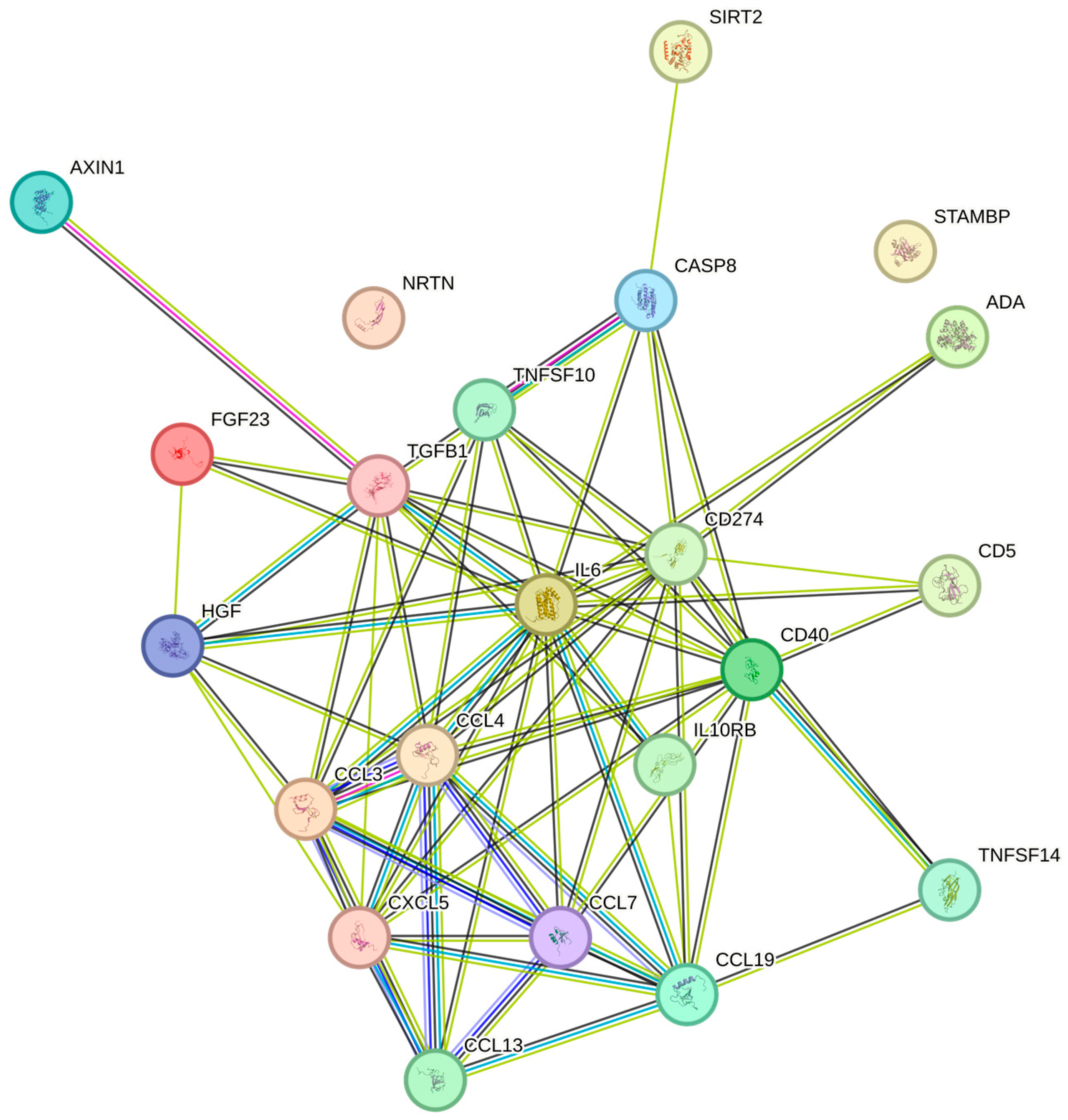
2.2. Correlations Between Platelet Count and Cytokine and Chemokine Levels

The platelet counts were significantly associated with FGF-23, IL-10RB, LAP TGF-beta-1, CCL19, SIRT2, IL6, VEGFA, AXIN1, CASP-8, CD40, PD-L1, CXCL5, MCP-3, STAMBP, CCL3, NRTN, HGF, CD5, TRAIL, CCL4, ADA. TNFSF14, and MCP-4 in increasing the Benjamini–Hochberg p-values order (Table 2). In the String plot for the cytokines that were significantly related to platelet counts, IL6 had a central role (Figure 1). The strongest associations with the platelet count were observed for MCP-3 and STAMBP (p = 0.052, both) and for CCL3 (p = 0.054), respectively.

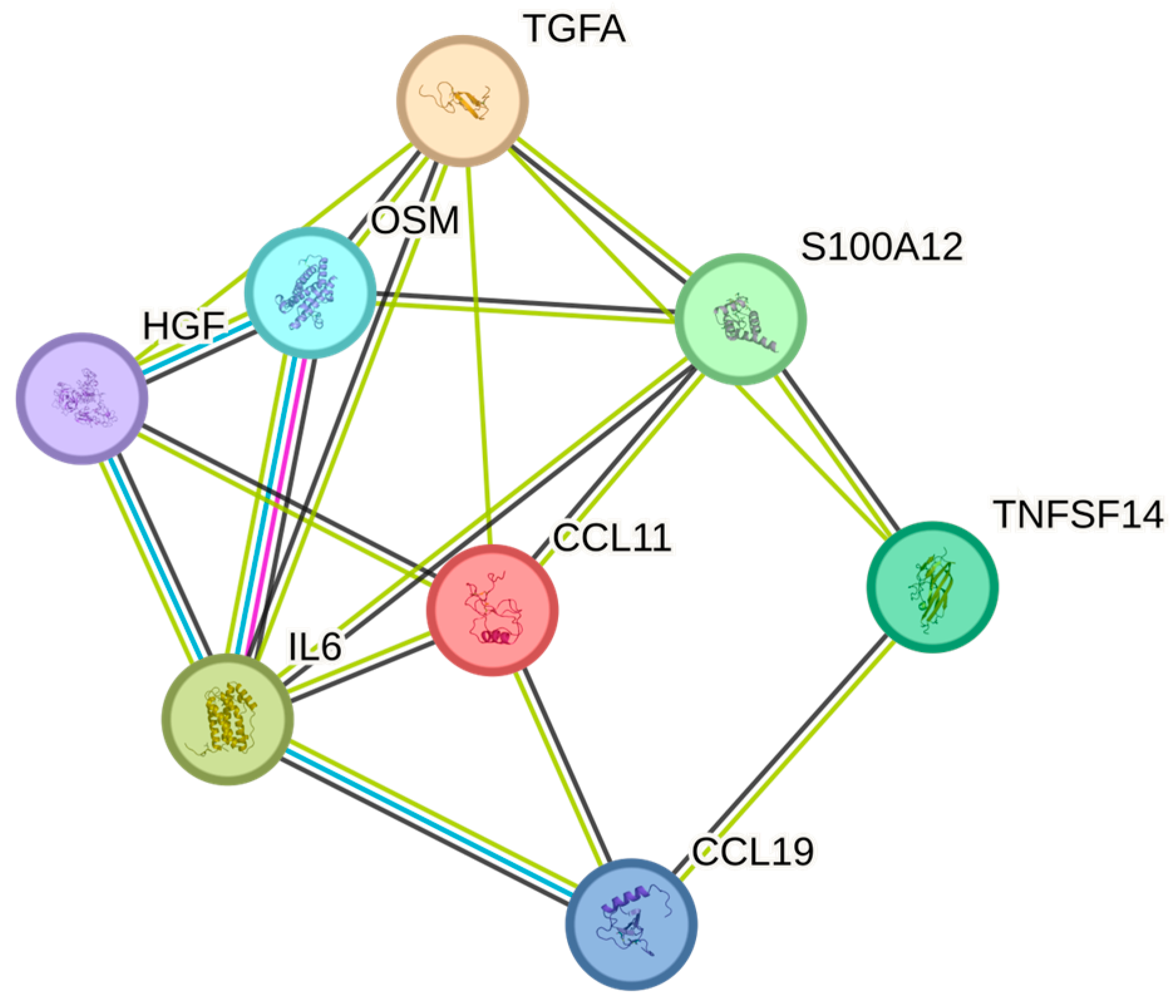
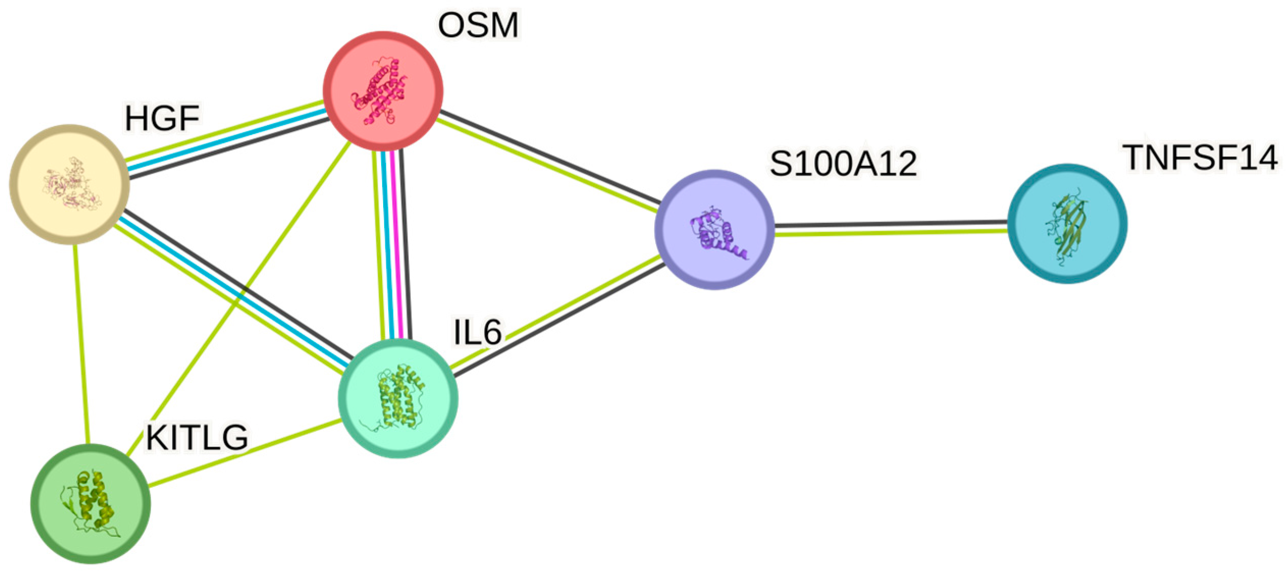
2.3. Correlations Between White Blood Cell Count and Cytokine Levels

The white blood cell counts were significantly associated with OSM, IL6, HGF, TNFSF14, EN-RAGE, TGF-alpha, CCL19, and CCL11in order of increasing p-values (Table 2). The greatest number of associations in the String plot was between OSM and IL6 (Figure 2). The strongest associations with the WBC were observed for OSM (p = 1.28 × 10⁻⁷), IL-6 (p = 0.002), and HGF (p = 0.017), respectively.
We included both the white blood cell count and the neutrophil cell count, as they are important clinical biomarkers. The white blood cell count comprised eight significant associations with cytokines, whereas the neutrophil count was associated with six out of these cytokines. TGF-alpha and CCL11 were not associated with the neutrophil count; otherwise, the associations that were found between both the white blood cell count and the neutrophil count, respectively, were in the same magnitude.
The neutrophil count showed similar associations as white blood cell counts and were significantly associated with OSM, IL6, TNFSF14, HGF, EN-RAGE, and SCF (Table 2). Similar to white blood cells, the most associations in the String plot were observed between OSM and IL6 (Figure 3). The strongest associations with the neutrophil cell count were observed for OSM (p = 4.73 × 10⁻⁹), IL-6 (p = 0.002), and TNFSF14 (p = 0.040), respectively.

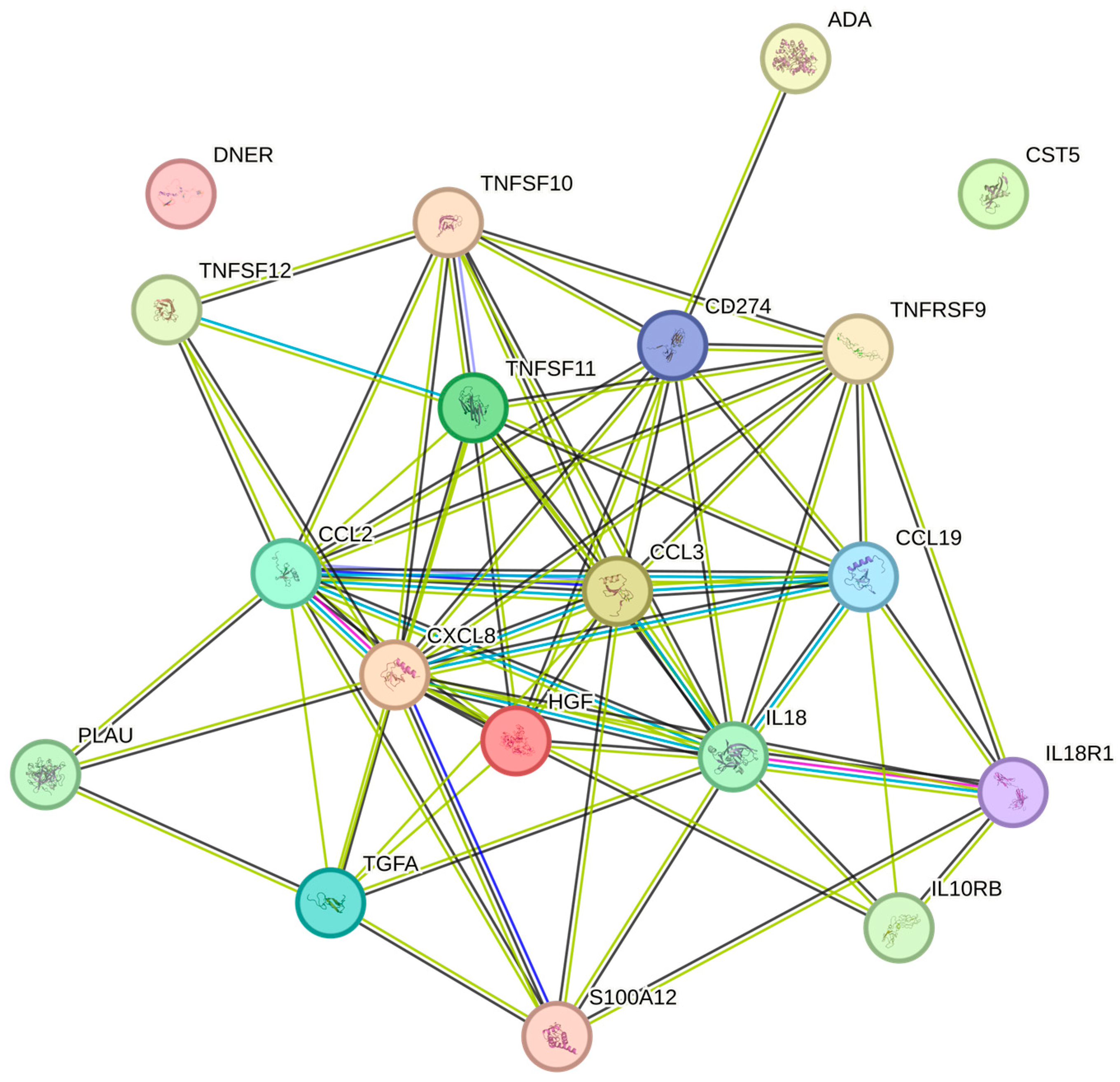
2.4. Correlations Between Cytokine Levels and Red Blood Cell Count

The red blood cell counts were significantly associated with TRAIL, IL-18R1, HGF, MCP-1, TRANCE, TNFRSF9, ADA, EN-RAGE, and CASP-8 (Table 2). The greatest number of associations in the String plot was noted between TNFSF10 and Casp-8 (Figure 4). The strongest associations with the red blood cell count were observed for TRAIL, IL-18R1, and HGF, respectively (p = 0.017, all).

2.5. Correlations Between Cytokine Levels and the Erythrocyte Volume Fraction

The erythrocyte volume fraction was significantly associated with TRAIL, MCP-1, HGF, TNFRSF9, ADA, DNER, CCL19, IL-10RB, IL18, TWEAK, CST5, PD-L1, CCL3, uPA, EN-RAGE, TRANCE, IL-18R1, TGF-alpha, and IL8 (Table 2). The most expressed associations were noted between TRAIL (p = 0.001), MCP-1 (p = 0.002), and HGF (p = 0.006), respectively (Figure 5).
The hemoglobin values were significantly associated with TRAIL, MCP-1, HGF, ADA, DNER, TNFRSF9, IL18, uPA, EN-RAGE, TWEAK, and TRANCE (Table 2).
There were also significant associations between MCHC and IL-12B and between MCV and IL-18R1.

3. Discussion

All studied PEA markers in this comparison have previously been proposed as either markers of inflammation or cardiovascular risk or both. The PEA assay unites high specificity, sensitivity, and low sample consumption and is therefore well suited for the detection of protein biomarkers [32]. We found several significant associations between blood cell parameters and cytokine levels, not only for the white blood cells but also for red blood cells and platelets. We also showed that within each cell type, there were significant associations between different cytokines, even when they did not belong to the same cytokine family (e.g., erythrocytes were associated with TNFSF9/10/11/12, IL-18, and CCL2), which were interrelated. It is noteworthy that the ten highest correlations between cell types and associated cytokine levels also had the lowest significance levels. There was no particular cell type, cytokine, or even cytokine family that constituted any link between these cells or cytokines. Since the most significant association was noted between the neutrophil count and OSM, followed by white blood cells and OSM, it is tempting to speculate that the main effect of white blood cells on OSM refers to neutrophils. We also noted associations, in decreasing order, between the number of white blood cells, neutrophil count, platelet count, and IL-6. We did not find any association between the platelet count and OSM, even though this cytokine belongs to the same family as IL-6 [33]. TRAIL, one of the cytokines with high correlation and low significance level, belonging to the TNF family [34], was, in decreasing order, significantly correlated to the erythrocyte volume fraction, hemoglobin, erythrocyte count, and, to some extent, also to the platelet count.
Our findings align with prior research. Several of the cytokines studied showed associations with platelet counts. FGF-23 has previously been shown to have a positive association with mean platelet volume [35]. Patients treated with rhuIL-10 developed decreased hemoglobin values and platelet counts [36]. Platelets were a major source of TGF-beta1 in the blood [37]. Regulation of protein acetylation by SIRT2 had an important role for platelet function [38]. IL-6 was shown to stimulate megakaryocytes to increase platelet production [39]. The interaction between CD40 and CD40L contributed to the binding of platelets to white blood cells [40]. Thus, there were several links between the platelet counts and the significantly associated cytokines in this study.
Cytokine–blood cell interactions have played a crucial role in developing therapeutic applications for various diseases, particularly in immunology, oncology, and inflammatory conditions. Some examples of these therapeutic applications are: Recombinant human erythropoietin (EPO) is used to treat anemia associated with chronic kidney disease and has improved the quality of life for kidney dialysis patients. G-CSF analogs (filgrastim) are used to treat neutropenia, especially in chemotherapy-induced neutropenia and congenital neutropenia, reducing the risk of infections. IL-6 inhibitors (tocilizumab, sarilumab) are used to treat rheumatoid arthritis, giant cell arteritis, and cytokine release syndrome from CAR-T cell therapy. TNF inhibitors (infliximab, adalimumab, etanercept) are used in treating autoimmune diseases like rheumatoid arthritis, Crohn’s disease, ulcerative colitis, psoriasis, and ankylosing spondylitis [41,42,43,44].

Limitations

This study has several limitations. Firstly, it was a single-site study. Secondly, due to geographic factors, the majority of the patients were Caucasians. Thirdly, those who were recruited were within a limited age span. Furthermore, the patients were not subjected to laboratory screening for co-existing diseases; instead, they were assessed clinically and deemed to be in good health. Thus, we cannot deduce to what extent, if any, either acute or chronic diseases may affect the associations between blood cell parameters, cytokines, chemokines, and growth factors. No protease inhibitor was used during sample collection, which means some degree of proteolysis may have occurred in the samples.

4. Materials and Methods

4.1. Population

This study included 165 essentially healthy patients, aged 18 to 44 years, weighing between 50 and 115 kg. All participants were previously referred to the Oral and Maxillofacial Surgery Clinic at Falun County Hospital, Sweden, for the surgical removal of an impacted lower third molar. Subjects with well-compensated systemic disease were accepted for inclusion in this study. The exclusion criteria were described in a previous publication [45].

4.2. Sampling Procedures

Blood samples were collected in vacutainer tubes while the patient was in the supine position. A peripheral venous catheter was used for sample collection. Complete blood cell counts were analyzed at the routine laboratory of Falun Hospital, Sweden. All blood samples were obtained prior to surgery. Plasma cytokine samples (EDTA) were frozen and stored at −80 °C until analysis.

4.3. Ethics

This study was conducted in accordance with the principles of the Helsinki Declaration [46]. It received approval from the Swedish Ethical Review Authority (Dnr 2015/378) and was registered in the European Union Regulating Authorities Clinical Trials Database (EudraCT) under number 2014-004235-39. Additionally, it is listed on ClinicalTrials.gov with the ID NCT04459377. All participants provided both oral and written informed consent before taking part in the study.

4.4. Proximity Extension Assay (PEA)

The proximity extension assay (PEA) was performed using the Proseek Multiplex Inflammation kit (Olink Bioscience, Uppsala, Sweden) [32]. In brief, 1 µL of plasma was combined with 3 µL of incubation mix, containing two probes (antibodies conjugated with unique DNA oligonucleotides). The mixture was incubated at 8 °C overnight.
Following incubation, 96 µL of extension mix, which included the PEA enzyme and PCR reagents, was added. The samples were then incubated at room temperature for 5 min before undergoing 17 cycles of DNA amplification in a thermal cycler.
A 96.96 Dynamic Array IFC (Fluidigm, South San Francisco, CA, USA) was prepared and primed according to the manufacturer’s guidelines. In a separate plate, 2.8 µL of the sample mixture was combined with 7.2 µL of detection mix, and 5 µL of this solution was loaded into the right side of the primed 96.96 Dynamic Array IFC. Unique primer pairs for each cytokine were loaded into the left side of the array. The protein expression analysis was then performed using the Fluidigm Biomark reader, following the Proseek protocol. Each Proseek kit quantified 92 biomarkers, resulting in a total analysis of 194 biomarkers in the study.

4.5. Statistical Analysis

Coefficients of variation were analyzed using Spearman rank correlations with Statistica (StatSoft (Version 14), Tulsa, OK, USA). Cytokine values that were above or below the highest and lowest standard points were assigned the values of these standards. To address the increased risk of false discoveries when analyzing a large number of correlations, p-values were adjusted for multiplicity using the false discovery rate (FDR) approach [47]. Adjusted p-values less than 0.10, corresponding to an expected false discovery rate of at most 10%, were considered significant. Results were adjusted for multiple testing, with a significance threshold of p < 0.05.

5. Conclusions

The observed associations between blood cell counts and circulating cytokines suggest potential applications in diagnostic and monitoring strategies for immune-mediated diseases and inflammatory conditions. For example, platelet count and IL-6, which showed strong associations in this study, have previously been linked to cardiovascular risk and inflammatory disorders [48]. Moreover, the identification of intercytokine interactions across different families may reflect coordinated inflammatory pathways that are relevant for disease progression, stratification, therapeutic response, and novel biomarker medicine approaches [49]. Future studies are warranted to validate these findings in disease cohorts and to explore their clinical utility in monitoring treatment response or predicting disease progression.

Supplementary Materials

The supporting information can be downloaded at: https://www.mdpi.com/article/10.3390/ijms26094065/s1.

Author Contributions

L.B.E.: conceptualization, methodology, investigation, data curation, formal analysis, visualization, writing—original draft, writing—review and editing, project administration, and resources. A.O.L.: conceptualization, methodology, formal analysis, writing—original draft, and writing—review and editing. M.B.E.: conceptualization, methodology, formal analysis, writing—original draft, and writing—review and editing. T.G.: conceptualization, methodology, formal analysis, writing—review and editing, and resources. All authors have read and agreed to the published version of the manuscript.

Funding

This work was supported by the Centre for Clinical Research, Dalarna, Uppsala-Örebro Regional Research Council and Public Dental Care Dalarna, Sweden.

Institutional Review Board Statement

The study was conducted in accordance with the ethical principles originating from the 1975 Declaration of Helsinki, which was revised in 2013 [40], and was consistent with ICH/GCP E6 (R2) guidelines and received approval from the Swedish Ethical Review Authority on 2 December 2015 (Dnr 2015/378). The European Union Regulating Authorities Clinical Trials Database (EudraCT) number (2014-004235-39) was obtained on 29 September 2014. Furthermore, it was registered on ClinicalTrials.gov under the ID NCT04459377 on 8 July 2020.

Informed Consent Statement

All patients signed the informed consent form at screening before any study-specific procedures commenced.

Data Availability Statement

Under Swedish Law, the authors cannot share the data used in this study and cannot conduct any further research other than what is specified in the ethical permissions application. For inquiries about the data, researchers should first contact the owner of the database, the University of Uppsala, Sweden. Please contact the corresponding author with requests for and assistance with data. If the university approves the request, researchers can submit an application to the Regional Ethical Review Board for the specific research question that the researcher wants to examine.

Acknowledgments

We are grateful to Charina Brännström for skilled technical assistance.

Conflicts of Interest

The authors declare no conflicts of interest.

References

  1. Hegde, R.; Awan, K.H. Effects of periodontal disease on systemic health. Dis. Mon. 2019, 65, 185–192. [Google Scholar] [CrossRef]
  2. Plachokova, A.S.; Andreu-Sánchez, S.; Noz, M.P.; Fu, J.; Riksen, N.P. Oral Microbiome in Relation to Periodontitis Severity and Systemic Inflammation. Int. J. Mol. Sci. 2021, 22, 5876. [Google Scholar] [CrossRef]
  3. Bayani, M.; Pourali, M.; Keivan, M. Possible interaction between visfatin, periodontal infection, and other systemic diseases: A brief review of literature. Eur. J. Dent. 2017, 11, 407–410. [Google Scholar] [CrossRef] [PubMed]
  4. Liu, C.; Chu, D.; Kalantar-Zadeh, K.; George, J.; Young, H.A.; Liu, G. Cytokines: From Clinical Significance to Quantification. Adv. Sci. 2021, 8, e2004433. [Google Scholar] [CrossRef] [PubMed]
  5. Fajgenbaum, D.C.; June, C.H. Cytokine Storm. N. Engl. J. Med. 2020, 383, 2255–2273. [Google Scholar] [CrossRef] [PubMed]
  6. Jaffer, U.; Wade, R.G.; Gourlay, T. Cytokines in the systemic inflammatory response syndrome: A review. HSR Proc. Intensive Care Cardiovasc. Anesth. 2010, 2, 161–175. [Google Scholar]
  7. Brinkmann, V.; Reichard, U.; Goosmann, C.; Fauler, B.; Uhlemann, Y.; Weiss, D.S.; Weinrauch, Y.; Zychlinsky, A. Neutrophil extracellular traps kill bacteria. Science 2004, 303, 1532–1535. [Google Scholar] [CrossRef]
  8. Branzk, N.; Lubojemska, A.; Hardison, S.E.; Wang, Q.; Gutierrez, M.G.; Brown, G.D.; Papayannopoulos, V. Neutrophils sense microbe size and selectively release neutrophil extracellular traps in response to large pathogens. Nat. Immunol. 2014, 15, 1017–1025. [Google Scholar] [CrossRef]
  9. Meier, A.; Sakoulas, G.; Nizet, V.; Ulloa, E.R. Neutrophil Extracellular Traps: An Emerging Therapeutic Target to Improve Infectious Disease Outcomes. J. Infect. Dis. 2024, 230, 514–521. [Google Scholar] [CrossRef]
  10. Jorch, S.K.; Kubes, P. An emerging role for neutrophil extracellular traps in noninfectious disease. Nat. Med. 2017, 23, 279–287. [Google Scholar] [CrossRef]
  11. Wang, H.; Kim, S.J.; Lei, Y.; Wang, S.; Wang, H.; Huang, H.; Zhang, H.; Tsung, A. Neutrophil extracellular traps in homeostasis and disease. Signal Transduct. Target. Ther. 2024, 9, 235. [Google Scholar]
  12. Roy, S.; Rizvi, Z.A.; Awasthi, A. Metabolic Checkpoints in Differentiation of Helper T Cells in Tissue Inflammation. Front. Immunol. 2018, 9, 3036. [Google Scholar] [CrossRef]
  13. Boyette, L.B.; Macedo, C.; Hadi, K.; Elinoff, B.D.; Walters, J.T.; Ramaswami, B.; Chalasani, G.; Taboas, J.M.; Lakkis, F.G.; Metes, D.M. Phenotype, function, and differentiation potential of human monocyte subsets. PLoS ONE 2017, 12, e0176460. [Google Scholar] [CrossRef] [PubMed]
  14. Austermann, J.; Roth, J.; Barczyk-Kahlert, K. The Good and the Bad: Monocytes’ and Macrophages’ Diverse Functions in Inflammation. Cells 2022, 11, 1979. [Google Scholar] [CrossRef] [PubMed]
  15. Arango Duque, G.; Descoteaux, A. Macrophage cytokines: Involvement in immunity and infectious diseases. Front. Immunol. 2014, 5, 491. [Google Scholar] [CrossRef]
  16. Antonelli, A.; Scarpa, E.S.; Magnani, M. Human Red Blood Cells Modulate Cytokine Expression in Monocytes/Macrophages Under Anoxic Conditions. Front. Physiol. 2021, 12, 632682. [Google Scholar] [CrossRef] [PubMed]
  17. Karsten, E.; Herbert, B.R. The emerging role of red blood cells in cytokine signalling and modulating immune cells. Blood Rev. 2020, 41, 100644. [Google Scholar] [CrossRef] [PubMed]
  18. Bakogiannis, C.; Sachse, M.; Stamatelopoulos, K.; Stellos, K. Platelet-derived chemokines in inflammation and atherosclerosis. Cytokine 2019, 122, 154157. [Google Scholar] [CrossRef]
  19. Maouia, A.; Rebetz, J.; Kapur, R.; Semple, J.W. The Immune Nature of Platelets Revisited. Transfus. Med. Rev. 2020, 34, 209–220. [Google Scholar] [CrossRef]
  20. Habets, K.L.; Huizinga, T.W.; Toes, R.E. Platelets and autoimmunity. Eur. J. Clin. Investig. 2013, 43, 746–757. [Google Scholar] [CrossRef]
  21. Anastasiadi, A.T.; Arvaniti, V.Z.; Hudson, K.E.; Kriebardis, A.G.; Stathopoulos, C.; D’Alessandro, A.; Spitalnik, S.L.; Tzounakas, V.L. Exploring unconventional attributes of red blood cells and their potential applications in biomedicine. Protein Cell 2024, 15, 315–330. [Google Scholar] [CrossRef] [PubMed]
  22. Kumar, S.D.; Kar, D.; Akhtar, M.N.; Willard, B.; Roy, D.; Hussain, T.; Rajyaguru, P.I.; Eswarappa, S.M. Evidence for low-level translation in human erythrocytes. Mol. Biol. Cell 2022, 33, br21. [Google Scholar] [CrossRef]
  23. Kaczmarski, R.S.; Mufti, G.J. The cytokine receptor superfamily. Blood Rev. 1991, 5, 193–203. [Google Scholar] [CrossRef]
  24. Kelso, A. Cytokines and their receptors: An overview. Ther. Drug Monit. 2000, 22, 40–43. [Google Scholar] [CrossRef] [PubMed]
  25. Dinarello, C.A. Historical insights into cytokines. Eur. J. Immunol. 2007, 37, S34–S45. [Google Scholar] [CrossRef] [PubMed]
  26. Rathnayake, N.; Akerman, S.; Klinge, B.; Lundegren, N.; Jansson, H.; Tryselius, Y.; Sorsa, T.; Gustafsson, A. Salivary biomarkers for detection of systemic diseases. PLoS ONE 2013, 8, e61356. [Google Scholar] [CrossRef]
  27. Balmasova, I.P.; Lomakin, Y.A.; Babaev, E.A.; Tsarev, V.N.; Gabibov, A.G.; Smirnov, I.V.; Knorre, V.D.; Ovchinnikova, L.A.; Gnuchev, N.V.; Khurs, E.N.; et al. “Shielding” of Cytokine Induction by the Periodontal Microbiome in Patients with Periodontitis Associated with Type 2 Diabetes Mellitus. Acta Naturae 2019, 11, 79–87. [Google Scholar] [CrossRef]
  28. Galhardo, L.F.; Ruivo, G.F.; de Oliveira, L.D.; Parize, G.; Santos, S.; Pallos, D.; Leão, M.V.P. Inflammatory markers in saliva for diagnosis of sepsis of hospitalizes patients. Eur. J. Clin. Investig. 2020, 50, e13219. [Google Scholar] [CrossRef]
  29. Chen, I.L.; Huang, H.C.; Ou-Yang, M.C.; Chen, F.S.; Chung, M.Y.; Chen, C.C. A novel method to detect bacterial infection in premature infants: Using a combination of inflammatory markers in blood and saliva. J. Microbiol. Immunol. Infect. 2020, 53, 892–899. [Google Scholar] [CrossRef]
  30. Wang, Z.; Mandel, H.; Levingston, C.A.; Young, M.R.I. An exploratory approach demonstrating immune skewing and a loss of coordination among cytokines in plasma and saliva of Veterans with combat-related PTSD. Hum. Immunol. 2016, 77, 652–657. [Google Scholar] [CrossRef]
  31. von Majewski, K.; Kraus, O.; Rhein, C.; Lieb, M.; Erim, Y.; Rohleder, N. Acute stress responses of autonomous nervous system, HPA axis, and inflammatory system in posttraumatic stress disorder. Transl. Psychiatry 2023, 13, 36. [Google Scholar] [CrossRef] [PubMed]
  32. Assarsson, E.; Lundberg, M.; Holmquist, G.; Björkesten, J.; Thorsen, S.B.; Ekman, D.; Eriksson, A.; Rennel Dickens, E.; Ohlsson, S.; Edfeldt, G.; et al. Homogenous 96-plex PEA immunoassay exhibiting high sensitivity, specificity, and excellent scalability. PLoS ONE 2014, 9, e95192. [Google Scholar] [CrossRef] [PubMed]
  33. Rose, T.M.; Bruce, A.G. Oncostatin M is a member of a cytokine family that includes leukemia-inhibitory factor, granulocyte colony-stimulating factor, and interleukin 6. Proc. Natl. Acad. Sci. USA 1991, 88, 8641–8645. [Google Scholar] [CrossRef]
  34. MacFarlane, M.; Ahmad, M.; Srinivasula, S.M.; Fernandes-Alnemri, T.; Cohen, G.M.; Alnemri, E.S. Identification and molecular cloning of two novel receptors for the cytotoxic ligand TRAIL. J. Biol. Chem. 1997, 272, 25417–25420. [Google Scholar] [CrossRef]
  35. Takeda, Y.; Fujita, S.; Ikemoto, T.; Okada, Y.; Sohmiya, K.; Hoshiga, M.; Ishizaka, N. The relationship of fibroblast growth factors 21 and 23 and α-Klotho with platelet activity measured by platelet volume indices. Clin. Chem. Lab. Med. 2015, 53, 1569–1574. [Google Scholar] [CrossRef] [PubMed]
  36. Sosman, J.A.; Verma, A.; Moss, S.; Sorokin, P.; Blend, M.; Bradlow, B.; Chachlani, N.; Cutler, D.; Sabo, R.; Nelson, M.; et al. Interleukin 10-induced thrombocytopenia in normal healthy adult volunteers: Evidence for decreased platelet production. Br. J. Haematol. 2000, 111, 104–111. [Google Scholar]
  37. Blakytny, R.; Ludlow, A.; Martin, G.E.; Ireland, G.; Lund, L.R.; Ferguson, M.W.; Brunner, G. Latent TGF-beta1 activation by platelets. J. Cell Physiol. 2004, 199, 67–76. [Google Scholar] [CrossRef]
  38. Moscardó, A.; Vallés, J.; Latorre, A.; Jover, R.; Santos, M.T. The histone deacetylase sirtuin 2 is a new player in the regulation of platelet function. J. Thromb. Haemost. 2015, 13, 1335–1344. [Google Scholar] [CrossRef]
  39. Senchenkova, E.Y.; Komoto, S.; Russell, J.; Almeida-Paula, L.D.; Yan, L.S.; Zhang, S.; Granger, D.N. Interleukin-6 mediates the platelet abnormalities and thrombogenesis associated with experimental colitis. Am. J. Pathol. 2013, 183, 173–181. [Google Scholar] [CrossRef]
  40. Sonmez, O.; Sonmez, M. Role of platelets in immune system and inflammation. Porto Biomed. J. 2017, 2, 311–314. [Google Scholar] [CrossRef]
  41. Leone, G.M.; Mangano, K.; Petralia, M.C.; Nicoletti, F.; Fagone, P. Past, Present and (Foreseeable) Future of Biological Anti-TNF Alpha Therapy. J. Clin. Med. 2023, 12, 1630. [Google Scholar] [CrossRef] [PubMed]
  42. Kishimoto, T. Discovery of IL-6 and Development of Anti-IL-6R Antibody. Keio J. Med. 2019, 68, 96. [Google Scholar] [CrossRef]
  43. Dale, D.C.; Crawford, J.; Klippel, Z.; Reiner, M.; Osslund, T.; Fan, E.; Morrow, P.K.; Allcott, K.; Lyman, G.H. A systematic literature review of the efficacy, effectiveness, and safety of filgrastim. Support. Care Cancer 2018, 26, 7–20. [Google Scholar] [CrossRef] [PubMed]
  44. Bunn, H.F. Erythropoietin. Cold Spring Harb. Perspect. Med. 2013, 3, a011619. [Google Scholar] [CrossRef] [PubMed]
  45. Eriksson, L.B.; Gordh, T.; Karlsten, R.; LoMartire, R.; Thor, A.; Tegelberg, Å. Intravenous S-ketamine’s analgesic efficacy in third molar surgery. A randomized placebo-controlled double-blind clinical trial. Br. J. Pain. 2024, 18, 197–208. [Google Scholar] [CrossRef]
  46. WMA Declaration of Helsinki—Ethical Principles for Medical Research Involving Human Subjects. Available online: https://www.wma.net/policies-post/wma-declaration-of-helsinki-ethical-principles-for-medical-research-involving-human-subjects/ (accessed on 23 March 2022).
  47. Benjamini, Y.; Hochberg, Y. Controlling the False Discovery Rate: A Practical and Powerful Approach to Multiple Testing. J. R. Stat. Soc. Ser. B (Methodol.) 2018, 57, 289–300. [Google Scholar] [CrossRef]
  48. Ridker, P.M.; Everett, B.M.; Thuren, T.; MacFadyen, J.G.; Chang, W.H.; Ballantyne, C.; Fonseca, F.; Nicolau, J.; Koenig, W.; Anker, S.D.; et al. Antiinflammatory Therapy with Canakinumab for Atherosclerotic Disease. N. Engl. J. Med. 2017, 377, 1119–1131. [Google Scholar] [CrossRef]
  49. Tanaka, T.; Narazaki, M.; Kishimoto, T. IL-6 in inflammation, immunity, and disease. Cold Spring Harb. Perspect. Biol. 2014, 6, a016295. [Google Scholar] [CrossRef]
Figure 1. The figure represents a network of interactions among cytokines, demonstrating significant associations between circulating cytokine levels and platelets. The nodes (cytokines) are connected by edges of varying thickness, indicating interaction strength.
Ijms 26 04065 g001
Figure 2. The figure represents a network of interactions among cytokines, demonstrating significant associations between circulating cytokine levels and white blood cells. The nodes (cytokines) are connected by edges of varying thickness, indicating interaction strength.
Ijms 26 04065 g002
Figure 3. The figure represents a network of interactions among cytokines, demonstrating significant associations between circulating cytokine levels and neutrophils. The nodes (cytokines) are connected by edges of varying thickness, indicating interaction strength.
Ijms 26 04065 g003
Figure 4. The figure represents a network of interactions among cytokines, demonstrating significant associations between circulating cytokine levels and the erythrocyte count. The nodes (cytokines) are connected by edges of varying thickness, indicating interaction strength.
Ijms 26 04065 g004
Figure 5. The figure represents a network of interactions among cytokines, demonstrating significant associations between circulating cytokine levels and the erythrocyte volume fraction. The nodes (cytokines) are connected by edges of varying thickness, indicating interaction strength.
Ijms 26 04065 g005
Table 1. Number, weight (kg), and height (cm).
Valid NGeometric MeanMinimumMaximumStd. Dev.CV
Weight16573481111317
Height1651711531958.65
BMI1652515414.417
hsCRP1650.80.2102.0250
Hb1651379416712.19
RBC1655460.419
EVF1650.410.140.480.0410
Platelets1652381004305221
WBC1655.73.413.11.831
Neutrophils1653.10.59.51.442
Abbreviations: Body mass index (BMI; kg × m−2), high sensitivity C-reactive protein (hsCRP; mg × L−1), hemoglobin (Hb; g × L−1), erythrocyte count (RBC; 1012 × L−1), erythrocyte volume fraction (EVF; %), platelet count (platelets; 109 × L−1), white blood cell count (WBC; 109 × L−1), and neutrophil count (neutrophils; 109 × L−1). Standard deviation (Std. Dev.), CV (coefficients of variation).
Table 2. Cytokines, chemokines, and growth factors and their relations to cell types and red blood cell parameters, determined by both Benjamini–Hochberg p-values and Spearman rank correlations.
Blood Cell MarkerCytokineUniProt IDNSpearman Rank CorrelationsBenjamini–Hochberg p-Value
EVFTRAILP505911650.3490.001
EVFMCP-1P135001650.3210.002
EVFHGFP142101650.3040.006
EVFTNFRSF9Q070111650.2590.022
EVFADAP008131650.2510.031
EVFDNERQ8NFT81650.2360.046
EVFCCL19Q997311650.230.051
EVFIL-10RBQ083341650.2290.051
EVFIL18Q141161650.2280.051
EVFTWEAKO435081650.2240.053
EVFCST5P283251650.2170.061
EVFPD-L1Q9NZQ71650.2130.067
EVFCCL3P101471650.2120.069
EVFuPAP007491650.2110.069
EVFEN-RAGEP805111650.2050.076
EVFTRANCEO147881650.2050.076
EVFIL-18R1Q134781650.2020.081
EVFTGF-alphaP011351650.1980.088
EVFIL8P101451650.1960.092
HbTRAILP505911650.3370.001
HbMCP-1P135001650.2910.011
HbHGFP142101650.2760.017
HbADAP008131650.2330.049
HbDNERQ8NFT81650.2320.049
HbTNFRSF9Q070111650.2290.051
HbIL18Q141161650.2050.076
HbuPAP007491650.2000.084
HbEN-RAGEP805111650.1990.087
HbTWEAKO435081650.1960.092
HbTRANCEO147881650.1930.099
MCHCIL-12BP29460165−0.2140.066
MCVIL-18R1Q13478165−0.2290.051
NeutrophOSMP137251650.5050
NeutrophIL6P052311650.3270.002
NeutrophTNFSF14O435571650.2410.040
NeutrophHGFP142101650.2320.049
NeutrophEN-RAGEP805111650.2140.067
NeutrophSCFP21583165−0.2040.079
PltFGF-23Q9GZV91650.2820.016
PltIL-10RBQ083341650.2690.017
PltLAP TGF-beta-1P011371650.2690.017
PltCCL19Q997311650.2630.021
PltSIRT2Q8IXJ61650.2610.022
PltIL6P052311650.260.022
PltVEGFAP156921650.2570.024
PltAXIN1O151691650.2440.037
PltCASP-8Q147901650.2390.042
PltCD40P259421650.2310.050
PltPD-L1Q9NZQ71650.2270.051
PltCXCL5P428301650.2270.051
PltMCP-3P800981650.2250.052
PltSTAMBPO956301650.2250.052
PltCCL3P101471650.2230.054
PltNRTNQ997481650.2230.054
PltHGFP142101650.2180.059
PltCD5P061271650.2160.063
PltTRAILP505911650.2140.067
PltCCL4P132361650.2100.070
PltADAP008131650.2100.071
PltTNFSF14O435571650.2080.072
PltMCP-4Q996161650.1930.099
RBCTRAILP505911650.2770.017
RBCIL-18R1Q134781650.2700.017
RBCHGFP142101650.2700.017
RBCMCP-1P135001650.2400.042
RBCTRANCEO147881650.2270.051
RBCTNFRSF9Q070111650.2240.053
RBCADAP008131650.2110.069
RBCEN-RAGEP805111650.2090.071
RBCCASP-8Q147901650.1990.086
WBCOSMP137251650.4630
WBCIL6P052311650.3250.002
WBCHGFP142101650.2770.017
WBCTNFSF14O435571650.2650.020
WBCEN-RAGEP805111650.2220.054
WBCTGF-alphaP011351650.2120.069
WBCCCL19Q997311650.2030.079
WBCCCL11P51671165−0.1970.090
Abbreviations: erythrocyte count (RBC; 1012 × L−1), erythrocyte volume fraction (EVF; %), hemoglobin (Hb; g × L−1), mean corpuscular hemoglobin (MCHC; g × L−1), mean corpuscular volume (MCV; fl, 10−15), erythrocyte volume fraction white blood cell count (WBC; 109 × L−1), neutrophil count (Neutroph; 109 × L−1), and platelet count (Plt; 109 × L−1).
Disclaimer/Publisher’s Note: The statements, opinions and data contained in all publications are solely those of the individual author(s) and contributor(s) and not of MDPI and/or the editor(s). MDPI and/or the editor(s) disclaim responsibility for any injury to people or property resulting from any ideas, methods, instructions or products referred to in the content.

