Abstract
Based on the hypothesis that arsenic exposure results in toxicity and mitogenecity, this study examined the dose-response of arsenic in established human cell lines of keratinocytes (HaCaT), melanocytes (1675), dendritic cells (THP-1/A23187), dermal fibroblasts (CRL1904), microvascular endothelial cells (HMEC), monocytes (THP-1), and T cells (Jurkat). Cytotoxicity was determined by incubating THP-1, THP-1+ A23187 and JKT cells in RPMI 1640, 1675 in Vitacell, HMEC in EBM, and dermal fibroblasts and HaCaT in DMEM with 10% fetal bovine serum, 1% streptomycin and penicillin for 72 hrs in 96-well microtiter plates, at 37oC in a 5% CO2 incubator with different concentrations of arsenic using fluorescein diacetate (FDA). Cell proliferation in 96-well plates was determined in cultured cells starved by prior incubation for 24 hrs in 1% FBS and exposed for 72 hours, using the 96 cell titer proliferation solution (Promega) assay. Cytotoxicity assays yielded LD50s of 9 μg/mL for HaCaT, 1.5 μg/mL for CRL 1675, 1.5 μg/mL for dendritic cells, 37 μg/mL for dermal fibroblasts, 0.48 μg/mL for HMEC, 50 μg/mL for THP-1 cells and 50 μg/mL for JKT-T cells. The peak proliferation was observed at 6 μg/mL for HaCaT and THP-1 cells, 0.19 μg/mL for CRL 1675, dendritic cells, and HMEC, and 1.5 μg/mL for dermal fibroblasts and Jurkat T cells. These results show that arsenic is toxic at high doses to keratinocytes, fibroblasts, monocytes and T cells, and toxic at lower doses to melanocytes, microvascular endothelial cells and dendritic cells. Proliferation studies showed sub-lethal doses of arsenic to be mitogenic.
Introduction
Arsenic occurs naturally in the environment and is a natural contaminant of water. Everyone is exposed to low levels of arsenic (especially inorganic arsenic) because it is always present in soil, water, food and air. The presence of elevated concentrations of arsenic in the environment has over the past 20 years given rise to increasing concern due to mounting evidence of adverse human health effects. Arsenic exposure has been associated with the development of cancer (particularly of the skin, lung and bladder, prostate, kidney and liver) [1]. While low dose ingestion does not have immediate fatal consequences, studies have shown that prolonged arsenic exposure significantly increases the risk of contracting various forms of cancer. Studies have shown that inorganic arsenic can increase the risk of skin cancer. The World Health Organization (WHO), the Department of Health and Human Services (DHHS), and the U. S. Environmental Protection Agency (EPA) have determined that inorganic arsenic is a human carcinogen [2]. Cancer does not occur immediately, and usually takes several years to develop [3].
Over the past several years, skin and internal cancers have become a main human health concern arising from arsenic. Following long-term exposure, the first changes usually observed in the skin include pigmentation changes and then hyperkeratosis. Chronic exposure to arsenic frequently results in skin, lung, bladder, and kidney cancer [4].
The skin is made up of the epidermis and the dermis. The epidermis is the most outermost layer of the skin and provides the first barrier of protection from the invasion of foreign substances into the body. The principal cells of the epidermis are the keratinocytes. Melanocytes migrate to the basal layer of the epidermis and reside there at a ratio of 10 keratinocytes to 1 melanocyte. Dendritic cells are antigen presenting cells that are capable of stimulating T cells. The ratio for melanocyte: basal keratinocyte is 1:10 [5]. Langerhan cells are the dendritic cells of the skin. THP-1 cells acquire the characteristics of dendritic cells (DC) in the presence of calcium ionophore A23187 [6].
The dermis is produced largely by fibroblasts. The dermis supports the vascular network to supply the avascular epidermis with immune surveillance provided mainly by monocytes/macrophages and T cells.
To understand the toxicity of arsenic on the skin, it is important to study the individual contributions made by the cellular elements of the skin. Since arsenic is a foreign element to the body it is imperative that we incorporate the contributions made by the immune cells which are the trigger for any responsiveness to foreign stimuli. Oxidative damage by ischemia-reperfusion injury may also be a trigger involved in the pathogenesis of arsenic induced toxicity/carcinogenecity. Therefore, the main parameter to be investigated in carcinogenesis is proliferation while evaluating the toxic nature of arsenic, cell cytotoxicity must be employed.
In this research, we investigated cell proliferation and cytotoxicity in established human cell lines represented by keratinocytes (HaCaT), melanocytes (1675), dendritic cells (THP-1+A23187), dermal fibroblasts (CRL1904), microvascular endothelial cells (HMEC), monocytes (THP-1) and T cells (Jurkat).
Materials and Methods
Chemicals and Cell Culture
Arsenic trioxide (As) with a 99.99% purity was purchased from Fisher Scientific (Suwanee, GA) and used throughout the experiments. Media and chemicals for cell culture were purchased from Gibco (Grand Island, NY). Fetal bovine serum was obtained from Hyclone Laboratories (Utah). HaCaT cell line was obtained from Dr. N. Fusenig (Division of Differentiation and Carcinogenesis in vitro, at the German Cancer Research Center). THP-1, skin dermal, melanocytes (1675), Jurkat T cell line (TIB152), dermal fibroblast CRL1904, and THP-1 TIB202 were obtained from American Type Culture Collection (Rockville, MD). Dermal microvascular endothelial cells from Center for Disease Control (CDC) (Atlanta, GA). The cells were cultured in a humidified atmosphere with 5% CO2 at 37o C.
The standard growth medium was prepared according to specific cell lines. For adherent cells (HaCaT), Dulbecco’s minimum essential medium (DMEM) was used; Iscove’s DMEM for dermal fibroblasts; Vitacell minimum essential media (VMEM) for melanocytes; endothelial cell basal medium (EBM) for microvascular endothelial cells (HMEC-1); and RPMI 1640 medium for non-adherent cells (THP-1, dendritic cells and Jurkat T cells). Media were made complete by adding 10% fetal bovine serum (FBS) and 1% 100X Fungizone (penicillin/streptomyocin).
Cell Treatment
Cells were counted and resuspended in media. Cells were starved 24 hrs for proliferation in 1% FBS. For the cytotoxicity assay, cells were resuspended in 10% FBS with no serum starvation. The cell count for proliferation assay was 2,500 cells/well and 20,000 cells/well for cytotoxicity assay. An aliquot of 100 µL of cell suspension was placed in each well of 3-96 well plates. Various concentrations of As were used to treat the cells. Plates were incubated for 24, 48 and 72 hrs.
Cell Treatment for Proliferation
In column 1, 100µL of media containing cells was added. In columns 2 –12, 100µL of the different concentrations of As were added such that the final starting concentration of As was 0.37 μg/mL and ending with 200 μg/mL in row 12. After 72 hrs incubation, 20 µL of the 96 Cell Titer Solution was added, cells incubated for 1 and 4 hours at 37º C in humidified 5% CO2 atmosphere and absorbance read at 490 nm. The total volume of the reaction mixture was 120 µl for the proliferation assay.
Cell Treatment for Cytotoxicity
In column1, 100µL of media containing cells were added. In columns 2 –12, 100µL of the different concentrations of As were added such that the final starting concentration of As was 0.37 μg/mL and ending with 200 μg/mL in row 12. After 72 hrs incubation, cells were centrifuged and washed twice with PBS. In the final wash, 20 µL of supernatant remained. An aliquot of 100 µL of Fluorescein diacetate (FDA; 10ng/mL) was added, plates incubated 35 minutes and read using a Floroskan II with filters set at emission wavelength of 485 nm and excitation of 538 nm. The total volume of the reaction mixture was 120 µL for the cytotoxicity assay.
Results
Data obtained from the cytotoxicity assay yielded LD50 values of 9 μg/mL for keratinocytes, 1.5 μg/mL for CRL 1675, and dendritic cells, 37 μg/mL for dermal fibroblasts, 0.48 μg/mL for HMEC, and 50 μg/mL for Jurkat T cells and monocytes. The peaks of cell proliferation were observed at 6 μg/mL for HaCaT cells, 0.19 μg/mL for CRL 1675 and dendritic cells, 1.5 μg/mL for dermal fibroblasts, 6.0 μg/mL for THP-1, 0.19 μg/mL for HMEC, and 1.5 μg/mL for Jurkat T cells. Table 1 is a compilation of median doses for cell lethality (LD50s) and peak proliferation-related doses in different cell lines.

Table 1.
Cytotoxic and proliferation-related doses of arsenic trioxide in various cell lines.
| Cell Types | Cytotoxicity (LD50) | Peak Proliferation Dose |
| Keratinocytes (HaCaT cells) | 9 μg/mL (45.5µM) | 6 μg/mL (30µM) |
| Melanocytes (CRL 1675) | 1.5 μg/mL (7.6µM) | 0.19 μg/mL (0.95µM) |
| Dendritic cells | 1.5 μg/mL (7.6µM) | 0.19 μg/mL (0.95µM) |
| Dermal fibroblasts (CRL1904) | 37 μg/mL (187µM) | 1.5 μg/mL (7.6µM) |
| Microvascular endothelial cells (HMEC-1) | 0.48μg/mL (2.4µM) | 0.19 μg/mL (0.95µM) |
| Monocytes (TIB202) | 50μg/mL (252.7µM) | 6 μg/mL (30µM) |
Cytotoxicity Assay
Figure 1 shows the cytotoxic effects of arsenic trioxide on HaCaT cells. These data indicate a strong dose-response relationship with regard to the cytotoxicity of arsenic. As indicated in this figure, there was a gradual decrease in the viability of HaCaT cells, with increasing doses of arsenic trioxide. A similar trend was observed with all the other tested cell lines. Following 72 hrs of exposure the doses of arsenic trioxide required to cause 50% reduction in cell viability were computed to be 9 μg/mL for HaCaT cells, 1.5 μg/mL for CRL 1675 and dendritic cells, 37 μg/mL for dermal fibroblasts, 0.48 μg/mL for HMEC, and 50 μg/mL for both Jurkat T cells and monocytes.
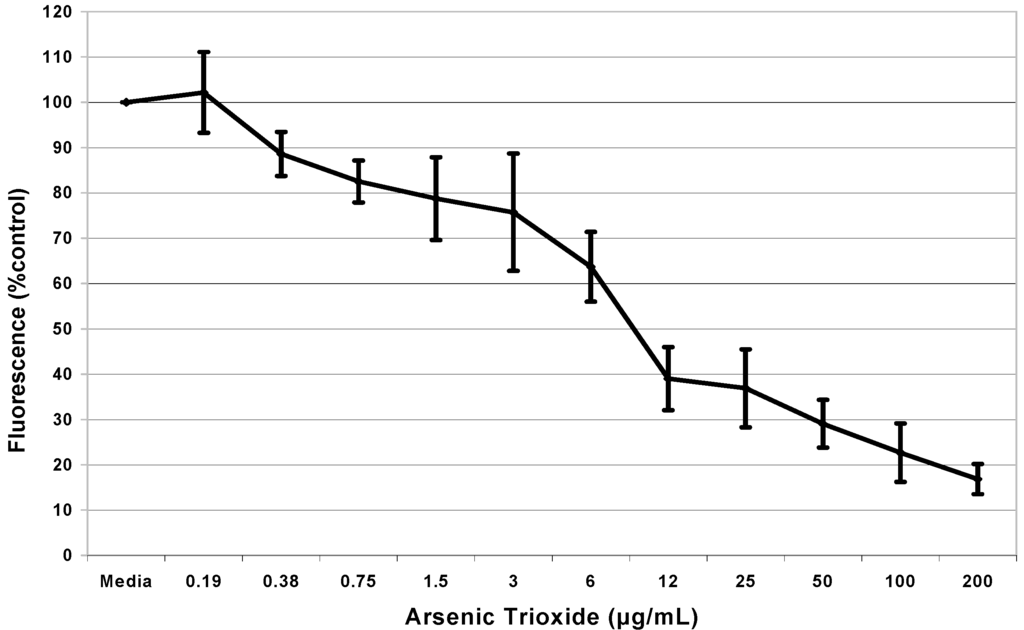
Figure 1.
Cytotoxicity of arsenic trioxide to HaCaT cells. Cells were seeded in 96 well plates (20,000 cells / well) and different doses of arsenic trioxide were added in triplicate samples. Each point represents the mean of three samples expressed as percent control.
Figure 1.
Cytotoxicity of arsenic trioxide to HaCaT cells. Cells were seeded in 96 well plates (20,000 cells / well) and different doses of arsenic trioxide were added in triplicate samples. Each point represents the mean of three samples expressed as percent control.

Cell Proliferation Assay
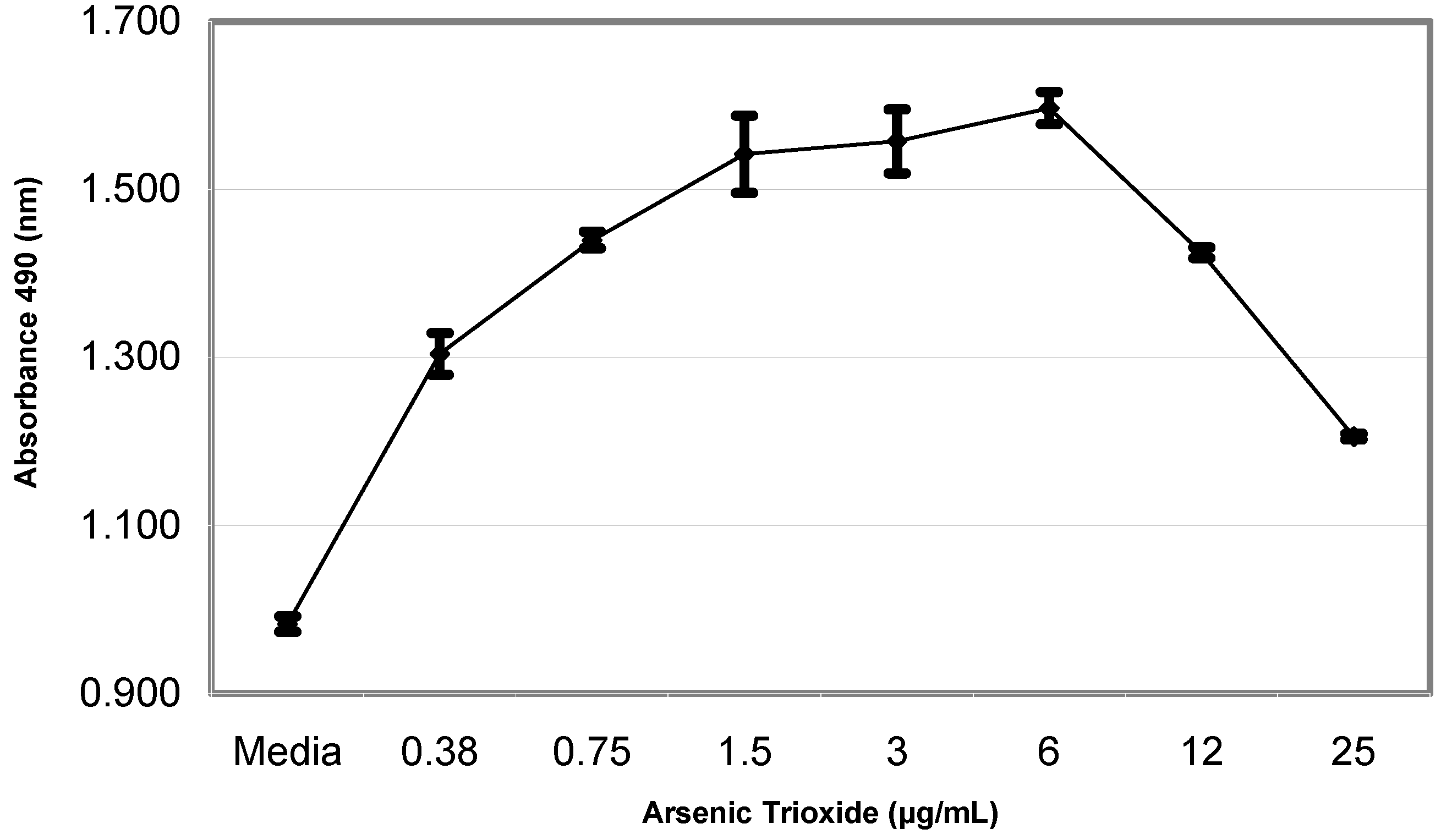
Figure 2 presents the data obtained from the proliferation assay of arsenic trioxide on HaCaT cells. Data presented in this figure also indicate a strong dose-response relationship with regard to the mitogenic effect of arsenic trioxide, in the dose range of 0-6 μg/mL. The peak proliferation was observed at the 6 μg/mL dose level. Tests with other cell lines showed peak proliferations at 6 μg/mL for THP-1 cells, 0.19 μg/mL for melanocytes, dendritic cells and microvascular endothelial cells, and 1.5 μg/mL for both dermal fibroblasts and Jurkat T-cells.
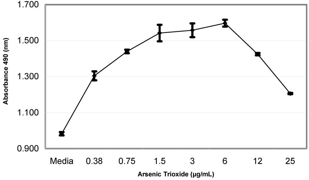
Figure 2.
Proliferation of HaCaT cells exposed to arsenic trioxide. Cells were seeded in 96 well plates (2,500 cells / well). Arsenic-treated and control cells were rendered quiescent and stimulated with different doses of arsenic trioxide in triplicates. Each point represents the mean of three samples.
Discussion
Disturbance of the balance between cell proliferation and cell death can result in cancer. Proliferation assays measure cellular growth or increase after treatment while cytotoxicity assays measure the negative effects of a substance, arsenic, on cells. Cytotoxicity has been defined as the cell killing property of a chemical compound independent from the mechanism of death.
In the present study, it was demonstrated that different doses of arsenic trioxide induce proliferation and cytotoxicity in different human cell lines. Data obtained from this research clearly demonstrate that under similar exposure conditions, the mitogenicity and cytotoxicity of arsenic is highly dose-dependent; indicating a somewhat biphasic response that encompasses cell proliferation at lower doses of exposure, and cytotoxicity at higher does of exposure.
Arsenic has been shown to cause skin lesions. The skin is the first barrier of defense in disease control. However, arsenic poisoning comes from within the body through mostly the ingestion of contaminated water. Absorption of arsenic through the skin is minimal. It was assumed that the amount of arsenic needed to cause cytotoxicity would be higher in dermal fibroblasts than in keratinocytes. Following ingestion, arsenic should affect the inner most cells first, and it is anticipated that the dose of arsenic lowers as it reaches the outer layer of skin. In our parallel study of dermal fibroblasts and keratinocytes, we found that the LD50 for dermal fibroblasts was 37 μg/mL (187µM) compared to only 9 μg/mL (45.5µM) for keratinocytes, thus showing that As is less toxic to dermal fibroblasts than keratinocytes.
Arsenic exhibits a clear predilection for the skin [7]. Several studies have been done to show arsenic increases proliferation in keratinocytes, dermal fibroblasts and Jurkat T-cells. Chiang et al. reported that continuous exposure to low levels of arsenite increased cell growth, and cell density in HaCaT cells [8]. It was reported by Chen et al. that arsenic alters epidermal keratinocyte differentiation processes, induces overexpression of growth factors, and enhances proliferation of human keratinocytes [9]. Bae et al. studied the effect of sodium meta-arsenite on HaCaT cells and found that the LD50 was 4.8µM [10]. Keratinocytes treated with sodium arsenite have been shown to significantly increase in cell proliferation by showing an increase in cell numbers, and c-myc gene expression [11]. Studies have shown that exposure to arsenic induces genes involved in a variety of cellular processes that include proliferation and genetic recombination. Arsenic may stimulate keratinocyte proliferation by different mechanisms directly by affecting gene expression or indirectly by affecting cytokine regulators [11]. Vega et al. found that trivalent arsenicals induced cell proliferation at concentrations of 0.001 to 0.01 µM, and inhibited cell proliferation at high concentrations (>0.5 µM) [12]. In our study, 6 μg/mL arsenic trioxide was the corresponding peak proliferation-related dose for HaCaT cells.
It has also been reported that 1µmoL of arsenic trioxide caused reduced proliferation in Jurkat cells [13]. Our studies found that Jurkat T-cells (TIB 152) had a peak proliferation at 1.5 μg/mL (7.6µM). Several studies have used other monocytic cell lines, for example U937, NB4, HL60, but no data was found for THP-1. Jia et al. found that 0.1 to 0.5 µM/L As2O3 inhibited cell proliferation in RPMI 8226 and U266 cell lines. We found that THP-1 cells had a peak proliferation at 6 μg/mL arsenic trioxide [13]. Arsenic-exposed C3H 10T1/2 fibroblasts have been shown to undergo cell proliferation [14]. We found the peak proliferation of dermal fibroblast to occur at 1.5 μg/mL (7.6µM ) arsenic trioxide.
Several studies have addressed cytotoxicity of arsenic to keratinocytes, dermal fibroblasts, monocytes and Jurkat T-cells. Bae et al. showed that the mean LD50 for HaCaT cells, when exposed to arsenic for over 24 hours, was 4.8µM [10]. Our studies found the LD50 for HaCaT to be 9 μg/mL (45.5µM). It has been shown that when using 0.5 to 1µM of arsenic trioxide, apoptosis was induced in the monocytic cells line NB4 [15,16]. Cytotoxicity studies of two multiple myeloma (MM) derived cell lines, RPMI 8226 and U266, found that 2.0 µM/L As2O3 induced cell apoptosis, and 1.0 µM/L As2O3 inhibited proliferation resulting in a weak degree of apoptosis induction. These results showed that As2O3 exerts apoptosis-inducing and growth-inhibiting effects on MM derived cells [17].
Jurkat T-cells were shown not to be in a state of apoptosis (a form of cytotoxicity) when treated with low concentrations (1µmol/l) of arsenic trioxide. The amount of arsenic trioxide needed to induce apoptosis in Jurkat T-cells was a concentration of 5µM up to a 35 day incubation [13]. We found that the LD50 for Jurkat T-cells was 50 μg/mL (252.7µM) on 72 h incubation.
Sodium arsenite concentrations of less or equal to 6 µM were shown to produce no cytotoxicity to the fibroblasts C3H 10T1/2 following 48 hrs exposure [14]. Toxicity study of sodium arsenite to human fibroblasts, WI 138, has determined an LD50 of approximately 1.85µM [18]. Dermal fibroblast CRL1904, in our study, had a LD50 of 37ppm (187µM).
When comparing our results to data from other studies on arsenic toxicity to monocytes, T-cells, dermal fibroblasts, and keratinocytes, it was shown that our doses were much higher than those previously reported. No data was found in the literature regarding the mitogenicity as well as the cytotoxicity of arsenic on demal microvascular endothelial cells, melanocytes or the dendritic cells. Therefore, the consideration of epidermal and dermal layers of the skin in the assessment of cellular effects of arsenic on skin cells is highly significant. Further studies must be done to determine the disparities between our observations and those of others.
Acknowledgments
This research was financially supported in part by a grant from the National Institutes of Health (Grant No. 1G12RR13459) through the RCMI-Center for Environmental Health, and in part by a grant from the United States Department of Education (Grant No. PO31B990006) to Jackson State University, under the Title-III Graduate Education Program.
References
- Agency for Toxic Substances and Disease Registry (ATSDR). Toxfacts for Arsenic (Update); U.S. Department of Health and Human Services, Public Health Service: Atlanta, GA, 2000. online at http://www.atsdr.cdc.gov/tfacts2.html.
- U.S. Environmental Protection Agency. Integrated Risk Information System (IRIS) on Arsine; Environmental Criteria and Assessment Office, Office of Health and Environmental Assessment, Office of Research and Development: Cincinnati, OH, 1993.
- World Health Organization (WHO). Arsenic in Drinking Water. Fact sheet No 210; Geneva, Switzerland, 2001. [Google Scholar]
- Gandolfi, A. J. Molecular Effects of Low Level Exposure to Arsenic. 2000; superfund-info@pharmacy.arizona.edu. [Google Scholar]
- Quinby, G. Melanocytes and Melanogenesis. Case Western University, 2000. On line at: http://dermed.cwru.edu/residents/melanocyte.htm.
- Waclavicek, M.; Berer, A.; Oehler, L.; Stockl, J.; Schloegl, E.; Majdic, O.; Knapp, W. Calcium ionophore: a single reagent for the differentiation of primary human acute myelogenous leukaemia cells towards dendritic cells. Br. J. Haematol. 2001, 114(2), 466–73. [Google Scholar] [CrossRef]
- Ali, A.; Lock, J. Physiological and toxicological changes in the skin resulting from the action and interaction of metal ions. Crit. Rev. Toxicol. 1995, 25, 397–462. [Google Scholar] [CrossRef] [PubMed]
- Chiang, M. C.; Yih, L. H.; Haung, R. N.; Peck, K.; Lee, T. C. Tumor formation of immortalized HaCaT cells in nude mice by long term exposure to sodium arsenite at non toxic doses. Toxicology 2001, 164, 95. [Google Scholar]
- Chen, C-S. J.; Siegel, D. M. Arsenical keratosis. eMedicine J. 2001, 2(6). [Google Scholar]
- Bae, D. S.; Gennings, C.; Carter, W. H., Jr.; Yang, R. S.; Campain, J. A. Toxicological interactions among arsenic, cadmium, chromium, and lead in human keratinocytes. Toxicol. Sci. 2001, 63, 132–142. [Google Scholar] [CrossRef] [PubMed]
- Germolec, D.R.; Yoshida, T.; Gaido, K.; Wilmer, J. L.; Simeonova, P. P.; Kayama, F.; Burleson, F.; Dong, W.; Lange, R. W.; Luster, M. I. Arsenic induces overexpression of growth factors in human keratinocytes. Toxicol. Appl. Pharmacol. 1996, 141, 308–318. [Google Scholar] [CrossRef] [PubMed]
- Vega, L.; Styblo, M; Patterson, R.; Cullen, W.; Wang, C.; Germolec, D. Differential effects of trivalent and pentavalent arsenicals on cell proliferation and cytokine secretion in normal human epidermal keratinocytes. Toxicol. Appl. Pharmacol. 2001, 172(3), 225–232. [Google Scholar]
- Rojewski, M. T.; Baldus, C.; Knauf, W.; Thiel, E.; Schrezenmeier, H. Dual effects of arsenic trioxide (As2O3) on non-acute promyelocytic leukaemia myeloid cell lines: induction of apoptosis and inhibition of proliferation. Br. J. Haematol. 2002, 116, 555–563. [Google Scholar]
- Trouba, K. J.; Glanzer, J. G.; Vorce, R. L. Wild-type and Ras-transformed fibroblasts display differential mitogenic responses to transient sodium arsenite exposure. Toxicol. Sci. 1999, 50, 72–81. [Google Scholar] [CrossRef] [PubMed]
- Chen, G. Q.; Zhu, J.; Shi, X. G.; Ni, J. H.; Zhong, H. J.; Si, G. Y.; Jin, X. L.; Tang, W.; Li, X. S.; Xong, S. M.; Shen, Z. X.; Sun, G. L.; Ma, J.; Zhang, P.; Zhang, T. D.; Gazin, C.; Naoe, T.; Chen, S. J.; Wang, Z. Y.; Chen, Z. In vitro studies on cellular and molecular mechanisms of arsenic trioxide (As2O3) in the treatment of acute promyelocytic leukemia: As2O3 induces NB4 cell apoptosis with downregulation of Bcl-2 expression and modulation of PML-RAR alpha/PML proteins. Blood 1996, 88, 1052–1061. [Google Scholar] [Green Version]
- Jing, Y.; Dai, J.; Chalmers-Redman, R. M.; Tatton, W. G.; Waxman, S. Arsenic trioxide selectively induces acute promyelocytic leukemia cell apoptosis via a hydrogen peroxide-dependent pathway. Blood 1999, 94, 2102–2111. [Google Scholar] [PubMed]
- Jia, P.; Chen, G.; Huang, X.; Cai, X.; Yang, J.; Wang, L.; Zhou, Y.; Shen, Y.; Zhou, L.; Yu, Y.; Chen, S.; Zhang, X.; Wang, Z. Arsenic trioxide induces multiple myeloma cell apoptosis via disruption of mitochondrial transmembrane potentials and activation of caspase-3. Chin. Med. J. (Engl) 2001, 114, 19–24. [Google Scholar]
- Vogt, B.; Rossman, T. Effects of arsenite on p53, p21 and cyclin D expression in normal human fibroblasts a possible mechanism for arsenite's comutagenicity. Mutat. Res. 2001, 478, 159–168. [Google Scholar] [CrossRef] [PubMed]
© 2003 by MDPI (http://www.mdpi.org).