Article

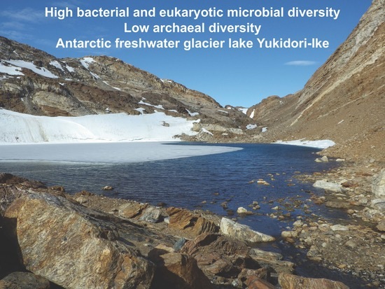
Community Structures of Bacteria, Archaea, and Eukaryotic Microbes in the Freshwater Glacier Lake Yukidori-Ike in Langhovde, East Antarctica

1
Department of Science and Engineering for Sustainable Innovation, Faculty of Science and Engineering, Soka University, 1-236 Tangi-machi, Hachioji, Tokyo 192-8577, Japan
2
Ehime Prefectural Science Museum, 2133-2 Ojoin, Niihama, Ehime 792-0060, Japan
3
Astrobiology Center, National Institutes of Natural Science, 2-21-1 Mitaka, Tokyo 181-8588, Japan
4
National Institute of Polar Research, 10-3 Midori-cho, Tachikawa, Tokyo 190-8518, Japan
5
SOKENDAI (The Graduate University for Advanced Studies), 10–3 Midori–cho, Tachikawa, Tokyo 190-8518, Japan
*
Author to whom correspondence should be addressed.
Diversity 2019, 11(7), 105; https://doi.org/10.3390/d11070105
Submission received: 3 June 2019 / Revised: 2 July 2019 / Accepted: 4 July 2019 / Published: 6 July 2019

Abstract

:
Since most studies about community structures of microorganisms in Antarctic terrestrial lakes using molecular biological tools are mainly focused on bacteria, limited information is available about archaeal and eukaryotic microbial diversity. In this study, the biodiversity of microorganisms belonging to all three domains in a typical Antarctic freshwater glacier lake (Yukidori-Ike) was revealed using small subunit ribosomal RNA (SSU rRNA) clone library analysis. The bacterial clones were grouped into 102 operational taxonomic units (OTUs) and showed significant biodiversity. Betaproteobacteria were most frequently detected, followed by Cyanobacteria, Bacteroidetes, and Firmicutes as major lineages. In contrast to the bacterial diversity, much lower archaeal diversity, consisting of only two OTUs of methanogens, was observed. In the eukaryotic microbial community consisting of 20 OTUs, Tardigradal DNA was remarkably frequently detected. Genera affiliated with the phyla Ciliophora, Cryptomycota, Chlorophyta, Bacillariophyta, and Apusozoa were also detected. The biodiversity and species compositions of the whole microbial community of Lake Yukidori-Ike are similar to those of freshwater environments in temperate regions but are different from saline lakes in Antarctica, indicating that the salinity seems to affect the microbial composition more than the temperature.

Graphical Abstract

1. Introduction

Approximately 98% of the Antarctic continent is permanently covered in ice, and only 0.4% of the ice-free area, the so-called “Antarctic Oases”, experiences temporary ice melts during summer [1]. The ice-free areas are colonized by various microorganisms, including bacteria, archaea, yeasts, filamentous fungi, algae, and protozoa. Previous studies on microorganisms in Antarctic lakes using molecular biological tools have revealed significant bacterial diversity, particularly among bacteria [2,3,4,5,6,7]. For example, Bowman et al. [3] investigated diverse bacterial communities in Antarctic saline lakes located in Vestfold Hills, Eastern Antarctica, using 16S rRNA clone library analysis. They found that Proteobacteria, Cyanobacteria, and Bacteroidetes were the dominant bacterial phyla and that archaeal diversity was remarkably low. In a later study by our group [5], we also conducted 16S rRNA clone library analysis in an Antarctic meromictic lake located in Langhovde, Eastern Antarctica. That study also revealed high bacterial diversity, particularly of Alpha-, Delta-, and Gammaproteobacteria, and limited archaeal diversity. However, most of these studies were conducted in marine relic saline lakes, and only limited information is available on the prokaryotic community structure in Antarctic freshwater lakes. The diversity and molecular phylogeny of eukaryotic microbes in Antarctic lakes have been much less well investigated than those of the prokaryotes.
Langhovde (69°14′ S, 39°40′ E) is one of the ice-free areas located approximately 30 km south of Japanese Syowa Station, Soya Coast (the east coast of Lützow-Holm Bay) in East Antarctica. In this area, the Yukidori Valley is recognized as having high biodiversity and is designated as an Antarctic Specially Protected Area (ASPA) No. 141 to protect the fragile and typical continental Antarctic fell field ecosystem and its component species, some of which are endemic to Antarctica, from human activity in Antarctica. Long-term monitoring programs have been conducted at this site [8,9]. Lake Yukidori-Ike, the sampling site of this study, is a freshwater glacial lake located in the middle of Yukidori Valley (Figure 1a). The maximum depth and area of the lake are 8.6 m and approximately 0.041 km2, respectively. The bottom of the lake is covered by photosynthetic benthos [10]. The Snow Petrel (Pagodroma nivea; “Yukidori” is the Japanese name for the Snow Petrel), a seabird, inhabits the Yukidori Valley.
In the present study, we attempted to analyze the whole microbial community of Lake Yukidori-Ike using SSU rRNA clone library analysis, as a model microbiome of Antarctic freshwater glacier lakes. High-throughput DNA sequencing (formerly called next-generation sequencing) has become a major tool for biodiversity analyses because of its high comprehensiveness and low cost in processing a large number of samples. In this study, however, we chose SSU rRNA clone library analyses using Sanger sequencing method to identify the microbes at the species level using longer data sequences and to compare the results with those of previous studies.

2. Materials and Methods

2.1. Study Site and Sampling

Lake Yukidori-Ike (Figure 1a) is located in central Langhovde on the east coast of Lutzow-Holm Bay, Antarctica (69°14′26” S, 39°45′23” E). The altitude, maximum depth, and area of Lake Yukidori-Ike are 125, 8.6, and 0.041 km2, respectively [1]. Water samples, including surface sediment, of approximately 80 mL were collected from three points on the lakeshore both on December 26 and 31, 2012, during the 54th Japanese Antarctic Research Expedition. The sediment mainly consisted of sandstone in diameters of approximately 0.5–5 mm. Each of the three points were 20–40 cm in depth (Figure 1b). The water temperature and pH were 5.4 °C and 8.4 on December 26, and 10.5 °C and 8.1 on December 31. The samples were taken by using autoclaved stainless ladles and put into 100-mL sterile plastic tubes. The algal mat inhabiting places on the bottom of the lake was avoided for sampling, but small amounts of algal debris might be included. The samples in the tubes were kept in freezing conditions until the DNA extraction experiments. A mixture of equal parts of the six samples was used for the extraction of environmental DNA.

2.2. Nutrient Analysis

Each water sample was filtered through a membrane filter with a 0.22-μm pore size. The concentrations of nitrate (NO3), nitrite (NO2), ammonium (NH4), phosphate (PO4), and silicate (SiO2) in the filtrate were analyzed using a nutrient auto-analyzer (SWAAT, BLTEC, Osaka, Japan).

2.3. Bacterial 16S rRNA Clone Library Analysis

Environmental DNA was extracted from 5.0 g of the mixed lake sample using Ultraclean Soil DNA Kit Mega Prep (Qiagen, Hilden, Germany). The extracted environmental DNA was used for the polymerase chain reaction (PCR) amplification of the bacterial 16S rRNA gene using a bacterial universal primer set: forward primer 5′-AGAGTTTGATCCTGGCTCAG-3′ (B27F) and reverse primer 5′-GGYTACCTTGTTACGACTT-3′ (U1492R) [11]. Each 50 μL PCR reaction mixture consisted of ~10 ng of environmental DNA, 0.5 μmol·L−1 of each primer, and 25 μL of 2 × PCR Master Mix (EmeraldAmp, Takara-bio, Kusatsu, Japan). PCR amplification was conducted by initial denaturation at 94 °C for 3 min, followed by 30 cycles of 30 s at 94 °C, 30 s at 58 °C, and 100 s at 72 °C, and a final extension at 72 °C for 5 min. The amplified PCR product was purified from agarose gel using an illustra GFX PCR Purification Kit (GE Healthcare, Chicago, IL). After clean-up, the purification product was checked by electrophoresis on 1% agarose gel with ethidium bromide staining before cloning. The PCR product was cloned into pT7 blue vector (Merck, Darmstadt, Germany), and the recombinant plasmids were transformed into Escherichia coli DH5α cells. The transformants were plated on Luria–Bertani medium (LB) plates containing 100 μg·mL−1 ampicillin, 40 μg·mL−1 X-gal, and 0.5 mM Isopropyl β-D-1-thiogalactopyranoside (IPTG). Randomly picked white colonies were subcultured in 100 μL of LB medium containing 100 μg·mL−1 ampicillin in 96-well plates at 37 °C overnight. The inserted 16S rRNA gene was amplified by PCR using 1 μL of the culture as a template with primers 5′-TAATACGACTTCACTATAGGG-3′ (T7P-F) and 5′-GTTTTCCCAGTCACGACGT-3′ (T7U-R). The PCR program was as follows: initial denaturation at 94 °C for 3 min; followed by 35 cycles of 30 s at 94 °C, 30 s at 51 °C, and 2 min at 72 °C; and a final extension at 72 °C for 5 min. Approximately 800 bp of the 5′-region of each 16S rRNA clone was sequenced.

2.4. Archaeal 16S rRNA Clone Library Analysis

All procedures for the archaeal 16S rRNA clone library analysis were the same as those for bacterial 16S rRNA clone library analysis except for the PCR conditions. The first PCR for archaeal 16S rRNA genes was conducted using archaeal forward primer 5’-TCYGGTTGATCCTGCCRG-3′ (Ar4f) [12] and reverse primer 5’-YCCGGCGTTGAVTCCAATT-3′ (Ar958r) [11]. Then, nested PCR was further conducted using archaeal forward primer Ar4f and reverse primer 5’- CCCGCCAATTCCTTTAAGTTTC-3′ (Ar9r) [13]. Both PCR stages were conducted by initial denaturation at 94 °C for 3 min; followed by 30 cycles of 30 s at 94 °C, 30 s at 58 °C, and 100 s at 72 °C; and a final extension at 72 °C for 5 min.

2.5. Eukaryotic 18S rRNA Clone Library Analysis

All procedures for the eukaryotic 18S rRNA clone library analysis were the same as those for bacterial and archaeal 16S rRNA clone library analysis except for the PCR conditions. The eukaryotic 18S rRNA gene was amplified with eukaryotic forward primer 5′-GAAACTGCGAATGGCTC-3′ (EK-82F) and reverse primer 5′-CYGCAGGTTCACCTAC-3′ (EK-1520R) [14]. Then, nested PCR was performed with the same forward primer and reverse primer 5′-CACCTACGGAAACCTTGTTA-3′ (EK-1498R) [15]. The first PCR and nested PCR conditions comprised denaturation at 94 °C for 3 min, 10 cycles of touch-down PCR (denaturation at 94 °C for 30 s, a 30-s annealing step at decreasing temperature from 65 to 55 °C employing a 1 °C decrease with each cycle, and extension at 72 °C for 2 min), 20 additional cycles at 55 °C annealing temperature, and final extension at 72 °C for 5 min.

2.6. Phylogenetic Analysis

Sanger DNA sequencing of the SSU rRNA genes was conducted by Eurofins Genomics K.K. (Tokyo, Japan). The obtained sequences were grouped into operational taxonomic units (OTUs) using 98% sequence similarity. To identify individual clones, the obtained 16S rRNA genes were submitted to an EzTaxon search, and 18S rRNA gene sequences were submitted to the Basic Local Alignment Search Tool (BLAST). After the removal of chimeric sequences manually, the SSU rRNA gene sequences of a representative clone were aligned using Clustal W alignment program as implemented in the MEGA10 software [16], with related species published in the DDBJ/EMBL/GenBank database. The neighbor-joining tree including bootstrap probabilities (1000 samplings) was reconstructed using the MEGA10 software (gap/missing data treatment: complete deletion, Kimura 2-parameter substitution model).

2.7. Coverage and Diversity Indices

The homologous coverage (C) was determined by the following equation: C = 1 − (N/n), where N is the number of OTUs and n is the total number of analyzed clones [17]. The Shannon–Wiener index and Chao1 were calculated using Estimate S software [18].

2.8. Nucleotide Sequences

The nucleotide sequences of the representative clones a reported in this study are available in the DDBJ/EMBL/GenBank database under the accession numbers: Bacteria; LC489022-LC489131, Archaea; LC489132 and LC489133, and Eukarya; LC489002-LC489021.

3. Results

3.1. Nutrients

The concentrations of NO3, NO2, NH4, and SiO2 in Lake Yukidori-Ike were 5.18, 0.13, 1.27, and 25.6 µM, respectively. The PO4 concentration was below the detection limit (0.03 µM).

3.2. Bacterial Community

A total of 160 bacterial clones was grouped into 101 OTUs. These OTUs were phylogenetically classified into nine phyla, 19 classes, 34 orders, 49 families, and 61 genera (Table 1). The Shannon–Wiener index and Chao1 of the bacterial community were 4.5 and 302, respectively. The homologous coverage value was 0.37.
The most frequently detected bacterial phylum in Lake Yukidori-Ike was Proteobacteria, which accounted for 56% of total bacterial clones and consisted of 63 OTUs (Figure 2). These OTUs were classified into four classes (clonal frequencies within the Proteobacteria): Alphaproteobacteria (26%), Betaproteobacteria (58%), Deltaproteobacteria (10%), and Gammaproteobacteria (4%). The Alphaproteobacterial clones consisted of 18 OTUs. The phylogenetic analysis indicated that 12 of the 18 OTUs were classified into any of five described families: Sphingomonadaceae, Rhodobacteraceae, Micropepsaceae, Hyphomicrobiaceae, and Caulobacteraceae. The other six OTUs could not be affiliated with any known families and were classified into two unknown groups. The family Sphingomonadaceae was most frequently detected and diverse within the Alphaproteobacteria.
The Betaproteobacteria were the most diverse in Lake Yukidori-Ike and consisted of 33 OTUs. 15 of the 33 OTUs were classified into any of the five described families: Comamonadaceae, Oxalobacteraceae, Burkholderiaceae, Oxalobacteraceae, Rhodocyclaceae, and Methylophilaceae. However, the other 18 OTUs could not be affiliated with any known families and were classified into three unknown groups. The family Comamonadaceae, consisting of four genera and eight OTUs, was most frequently detected and diverse in the Betaproteobacteria. Particularly, Polaromonas spp. and Rhodoferax spp., consisting of six OTUs together, accounted for 29% of the Betaproteobacterial clones. Delta- and Gammaproteobacteria were less frequently detected than the Alpha- and Betaproteobacteria and consisted of 10 OTUs. Only four of these OTUs were identified at the family level; this was not possible for the others. There were two OTUs (YK1B-121, YK1B-197) that were unable to classify into any described classes in the Proteobacteria (Figure 3).
The second most frequently detected bacterial phylum was Cyanobacteria, which accounted for 18% of the total bacterial clones and consisted of nine OTUs. These OTUs were classified into six families—Gomontiellaceae, Oscillatoriales, Leptolyngbyaceae, Pseudanabaenaceae, Chamaesiphonaceae, and Nostocales—showing clonal frequencies of 41, 3, 10, 7, 3, 3, and 10%, respectively, within the Cyanobacteria. The third most frequently detected bacterial phylum was Bacteroidetes, which accounted for 10% of bacterial clones, followed by the phyla Firmicutes (6%), Acidobacteria (6%), Ignavibacteria (2%), Chlorobi (1%), Actinobacteria (1%), and Armatimonadetes (1%) (Figure 2).

3.3. Archaeal Community

A total of 72 archaeal clones were grouped into only two OTUs. These OTUs were phylogenetically classified into one order, two families, and two genera (Table 2). The Shannon–Wiener index and Chao1 were 0.011 and 2, respectively. The homologous coverage value was 0.97. One OTU (YK1A-009), which accounted for 97.2% of the archaeal community, was identified as the methanogenic euryarchaeon Methanosarcina subterranea based on a nucleotide sequence similarity of 99.0%. The other OTU (YK1A-058) was also identified as a methanogenic euryarchaeon, Methanosaeta sp. The nucleotide sequence of OTU YK1A-058 showed 98.1% similarity with that of Methanosaeta concilii. No other archaeal phyla were detected.

3.4. Eukaryotic Community

A total of 90 eukaryotic clones were grouped into 20 OTUs. These OTUs were phylogenetically classified into seven phyla, 12 classes, 13 orders, 16 families, and 18 genera including unclassified taxa (Table 3). The Shannon–Wiener index and Chao1 were 2.6 and 28, respectively. The homologous coverage value was 0.79.
The most frequently detected phylum was Tardigrada, which accounted for 53% of eukaryotic clones and was classified into class Eutardigrada (Figure 4). Most of the Tardigrada clones were identified as Diphascon sp., which accounted for 44% of all the eukaryotic clones. The other Eutardigrada was identified as Acutuncus sp.
The second most frequently detected eukaryotic phylum was Cryptomycota (fungi), which accounted for 19% of all the eukaryotic clones and consisted of three OTUs. The third most frequently detected phylum was Ciliophora, which accounted for 14% of eukaryotic clones and consisted of six OTUs. These OTUs were classified into five families—Oxytrichidae, Vorticellidae, Phlasteridae, Spathidiidae, and Chilodonellidae—showing clonal frequencies of 62, 15, 8, 8, and 8%, respectively, within the phylum Ciliophora. The most frequently detected OTU was identified as Onychodromopsis flexilis. The next frequently detected phylum was Chlorophyta (8%), followed by the phyla Bacillariophyta (2%), Apusozoa (2%), and Bicosocidae (no rank in the phyla level, 1%) (Figure 4).

4. Discussion

The concentrations of NO3, NO2, NH4, and SiO2 in Lake Yukidori-Ike indicated that this lake is oligotrophic in type. In particular, the PO4 concentration was below the detection limit, suggesting that the phosphate concentration may limit the biological production in this lake.
The biodiversity of bacteria was remarkably higher than those of archaea and eukarya (Figure 5, Figure 6 and Figure 7). The Shannon–Wiener index and estimated number of bacterial species (Chao1 value) were comparable to those in aquatic environments in temperate or tropical regions. The previous studies regarding bacterial diversity in Antarctic lakes also reported high bacterial diversity. These previous studies and the current study used the Sanger sequencing method to reveal the SSU rRNA clones and are not so comprehensive when compared to analysis using a high-throughput sequencing method. Therefore, most of the detected clones may actually be derived from proliferating dominant organisms in the Antarctic lakes.
In Lake Yukidori-Ike, Betaproteobacteria was the most dominant and diverse class in the bacterial community (Figure 5). This was also observed in some previous studies conducted in Arctic freshwater environments [19,20,21,22]. The bacterial community structures found in Antarctic saline lakes were different to those in Lake Yukidori-Ike. In the saline lakes, Alphaproteobacteria were usually dominant [2,3,4,5,6], and some clones were found to be common with those revealed in the sea samples. Most saline lakes located in Antarctic coastal areas were formed by separation from the ocean during the uplift of the continent after the last glacial period [23]. For example, the bacterial community structure was different from that of Lake Nurume-Ike [5], also located in the Langhovde ice-free area and known as a meromictic saline lake. In Lake Nurume-Ike, Alphaproteobacteria were dominant and Betaproteobacteria were not detected. In the community structure of Cyanobacteria, the classes Oscillatoriales and Synechococcales were dominant in Lake Yukidori-Ike, whereas the class Oscillatoriales was not detected in Lake Nurume-Ike. These contrasts are observed not only in Antarctica but also in freshwater and saline lakes in temperate areas. These results suggest that the salinity affects the structure of bacterial communities more than the temperature.
The archaeal diversity was much lower than the bacterial diversity in Lake Yukidori-Ike. Only two archaeal OTUs were detected (Figure 8). One, sharing 97% of archaeal clones, was identified as the methanogenic euryarchaeon Methanosarcina subterranea. The type strain of this species has been isolated from groundwater in the northernmost part of Japan. They are neither psychrophilic nor psychrotolerant archaea and may be a little different physiologically to the species detected in our study. The other OTU was also identified as a methanogenic euryarchaeon, Methanosaeta sp. The closest described species was M. concilii, but the nucleotide sequence similarity between them was 98.2%, which is below the species threshold value of 98.65% [24]. The methanogen is known to be strictly anaerobe. On the other hand, the “microenvironments” in which the micro-anaerobic zones exist, are everywhere in the aerobic samples including the aerobic sediment sample used in this study. For example, methanogens belonging to the families Methanobacteriaceae and Methanosarcinaceae were detected in the aerobic sample from Antarctic cryoconite holes [25].
Our knowledge about the diversity and community structures of eukaryotic microbes in Antarctic lakes is currently limited. Unrein et al. [26] reported the composition of planktonic eukaryotes of coastal Antarctic lakes using denaturing gradient gel electrophoresis (DGGE). They frequently detected some clones affiliated with Chrysophyceae, but this group was not detected in Lake Yukidori-Ike. This difference may be due to the different salinity levels of these lakes. The eukaryotic community structure in Lake Yukidori-Ike has common characteristics with that in the Antarctic moss pillars inhabiting an Antarctic freshwater lake, Hotoke-Ike [27]. In the Antarctic moss pillar, the first, second, and third most abundant phyla were Fungal phyla, Tardigrada, and Ciliophora, respectively. These groups were also dominant in Lake Yukidori-Ike.
Two OTUs of Tardigrada were detected in this study. The most abundant OTU (YK1E-100) shared 84% of Tardigradal clones and showed 98.3% 18S rRNA gene sequence similarity with Diphascon pingue. This OTU also showed 99.5% similarity with an environmental clone isolated from an Antarctic moss pillar in Lake Hotoke-Ike, located in another ice-free area in Skarvsnes [27]. These results indicate that OTU YK1E-100 was derived from the same species that was found in Lake Hotoke-Ike but a different species from D. pingue. The other Tardigrada OTU, YK1E-099, showed 96.9% similarity with Acutuncus antarcticus, which is one of the most widespread Antarctic tardigrade species [28]. A. antarcticus is also reported as the most common and dominant species in both terrestrial and lake environments in Antarctica [29,30,31]. However, this species seemed much less dominant than Diphascon sp. in Lake Yukidori-Ike.
Most of the eukaryotic clones that were affiliated with the phyla Tardigrada, Ciliiophora, Bacillariophyta, Chlorophyta, and Apusozoa were identified at the genus level (Figure 9). However, the fungal clones could not be classified at the class level. Because the 18S rRNA gene sequences of nearly all fungal species are available in the DNA database, the fungal clones in Lake Yukidori-Ike may be derived from novel uncultured species.
In this study, many OTUs could be identified only in higher taxonomic levels such as order, class, and phylum. This is probably not caused by methods used in this study. For bacterial and archaeal 16S rRNA genes, we used 700–800 bp including highly variable regions V1, V2, V3, V4, which is more variable than the 3′ regions [32]. Also, for eukaryotic 18S rRNA genes, data containing highly variable regions V1, V2, V3 [33] was used for BLAST. The possible reasons for the presence of many unidentified OTUs are (1) many uncultured microbes exist in this lake as well as other environments, (2) many eukaryal 18S sequences are not available in the DNA database.

5. Conclusions

The bacterial, archaeal, and eukaryotic microbial community structures of the Antarctic freshwater lake Yukidori-Ike were revealed using SSU rRNA clone library analysis. The bacterial and eukaryotic microbial communities consisted of remarkably diverse species in contrast to the archaeal community. The results of the present study provide novel information about the microbial diversity of Antarctic freshwater lakes and help us to understand the living organisms in the cryosphere. This study was conducted during summer, and different microbial communities may appear in the winter season. The roles of microbial communities should also be investigated using functional genes to understand the ecology of these Antarctic lakes.

Author Contributions

Conceptualization, N.K.; Data curation, A.C. and N.K.; Formal analysis, A.C.; Funding acquisition, S.I.; Project administration, S.I.; Resources, N.K., A.K. and M.K.; Supervision, N.K.; Writing—original draft, A.C.; Writing—review and editing, N.K. and S.I.

Funding

This study is a part of the Science Program of Japanese Antarctic Research Expedition (JARE). It was supported by National Institute of Polar Research (NIPR) under Ministry of Education, Culture, Sports, Science and Technology (MEXT).

Acknowledgments

We thank the expedition members of JARE 54 for support in sample collection.

Conflicts of Interest

The authors declare no conflict of interest.

References

  1. Kimura, S.; Ban, S.; Imura, S.; Kudoh, S.; Matsuzaki, M. Limnological characteristics of vertical structure in the lakes of Syowa oasis, East Antarctica. Polar Sci. 2010, 3, 262–271. [Google Scholar] [CrossRef]
  2. Franzmann, P.D. Examination of Antarctic prokaryotic diversity through molecular comparisons. Biodivers. Conserv. 1996, 5, 1295–1305. [Google Scholar] [CrossRef]
  3. Bowman, J.P.; McCammon, S.A.; Rea, S.M.; McMeekin, T.A. The microbial composition of three limnologically disparate hypersaline Antarctic lakes. FEMS Microbiol. Lett. 2000, 183, 81–88. [Google Scholar] [CrossRef] [PubMed]
  4. Sjöling, S.; Cowan, D.A. High 16S rDNA bacterial diversity in glacial meltwater lake sediment, Bratina Island, Antarctica. Extremophiles 2003, 7, 275–282. [Google Scholar] [CrossRef] [PubMed] [Green Version]
  5. Kurosawa, N.; Sato, S.; Kawarabayasi, Y.; Imura, S.; Naganuma, T. Archaeal and bacterial community structures in the anoxic sediment of Antarctic meromictic lake Nurume-Ike. Polar Sci. 2010, 4, 421–429. [Google Scholar] [CrossRef] [Green Version]
  6. Michaud, L.; Caruso, C.; Mangano, S.; Interdonato, F.; Bruni, V.; Giudice, A.L. Predominance of Flavobacterium, Pseudomonas, and Polaromonas within the prokaryotic community of freshwater shallow lakes in the northern Victoria Land, East Antarctica. FEMS Microbiol. Ecol. 2012, 82, 391–404. [Google Scholar] [CrossRef] [PubMed]
  7. Bowman, J.S. Identification of Microbial Dark Matter in Antarctic Environments. Front. Microbiol. 2018, 9, 3165. [Google Scholar] [CrossRef]
  8. Kanda, H.; Inoue, M.; Mochida, Y.; Sugawara, H.; Ino, Y.; Ohtani, S.; Ohyama, Y. Biological studies on ecosystems in the Yukidori Valley, Langhovde, East Antarctica. Antarct. Rec. 1990, 34, 76–93. [Google Scholar]
  9. Kanda, H.; Inoue, M. Ecological monitoring of moss and lichen vegetation in the Syowa Station area, Antarctica. Mem. NIPR Symp. Polar Biol. 1994, 7, 221–231. [Google Scholar]
  10. Tanabe, Y.; Yasui, S.; Osono, T.; Uchida, M.; Kudoh, S.; Yamamuro, M. Abundant deposits of nutrients inside lakebeds of Antarctic oligotrophic lakes. Polar Biol. 2017, 40, 603–613. [Google Scholar] [CrossRef]
  11. Weisburg, W.G.; Barns, S.M.; Pelletier, D.A.; Lane, D.J. 16S ribosomal DNA amplification for phylogenetic study. J. Bacteriol. 1991, 173, 697–703. [Google Scholar] [CrossRef] [PubMed] [Green Version]
  12. Hershberger, K.L.; Barns, S.M.; Reysenbach, A.L.; Dawson, S.C.; Pace, N.R. Wide diversity of Crenarchaeota. Nature 1996, 384, 420. [Google Scholar] [CrossRef] [PubMed]
  13. Jurgens, G.; Glöckner, F.O.; Amann, R.; Saano, A.; Montonen, L.; Likolammi, M.; Münster, U. Identification of novel Archaea in bacterioplankton of a boreal forest lake by phylogenetic analysis and fluorescent in situ hybridization. FEMS Microbiol. Ecol. 2000, 34, 45–56. [Google Scholar] [CrossRef]
  14. López-García, P.; Rodriguez-Valera, F.; Pedrós-Alió, C.; Moreira, D. Unexpected diversity of small eukaryotes in deep-sea Antarctic plankton. Nature 2001, 409, 603–607. [Google Scholar] [CrossRef] [PubMed]
  15. López-García, P.; Philippe, H.; Gail, F.; Moreira, D. Autochthonous eukaryotic diversity in hydrothermal sediment and experimental microcolonizers at the Mid-Atlantic Ridge. Proc. Natl. Acad. Sci. USA 2003, 100, 697–702. [Google Scholar] [CrossRef] [PubMed] [Green Version]
  16. Kumar, S.; Stecher, G.; Li, M.; Knyaz, C.; Tamura, K. MEGA X: Molecular Evolutionary Genetics Analysis across computing platforms. Mol. Biol. Evol. 2018, 35, 1547–1549. [Google Scholar] [CrossRef] [PubMed]
  17. Good, I.J. The population frequencies of species and the estimation of population parameters. Biometrika 1953, 40, 237–264. [Google Scholar] [CrossRef]
  18. Colwell, R.K. EstimateS: Statistical Estimation of Species Richness and Shared Species from Samples; Version 9; User’s Guide and application [WWW document] 2013. Available online: http://purl.oclc.org/estimates (accessed on 2 June 2019).
  19. Zwart, G.; Crump, B.C.; Kamst-van Agterveld, M.P.; Hagen, F.; Han, S.K. Typical freshwater bacteria: An analysis of available 16S rRNA gene sequences from plankton of lakes and rivers. Aquat. Microb. Ecol. 2002, 28, 141–155. [Google Scholar] [CrossRef]
  20. Garneau, M.È.; Vicent, W.F.; Alonso-Sáez, L.; Gratton, Y.; Lovejoy, C. Prokaryotic community structure and heterotrophic production in a river-influenced coastal arctic ecosystem. Aquat. Microb. Ecol. 2006, 42, 27–40. [Google Scholar] [CrossRef] [Green Version]
  21. Cheng, S.M.; Foght, J.M. Cultivation-independent and-dependent characterization of bacteria resident beneath John Evans Glacier. FEMS Microbiol. Ecol. 2007, 59, 318–330. [Google Scholar] [CrossRef]
  22. Galand, P.E.; Lovejoy, C.; Pouliot, J.; Garneau, M.È.; Vincent, W.F. Microbial community diversity and heterotrophic production in a coastal Arctic ecosystem: A stamukhi lake and its source waters. Limnol. Oceanogr. 2008, 53, 813–823. [Google Scholar] [CrossRef] [Green Version]
  23. Pienitz, R.; Doran, P.T.; Lamoureux, S.F. Origin and geomorphology of lakes in the polar regions. In Polar Lakes and Rivers: Limnology of Arctic and Antarctic Aquatic Ecosystems; Vincent, W.F., Laybourn-Parry, J., Eds.; Oxford University Press: Oxford, UK, 2008; pp. 25–41. [Google Scholar]
  24. Kim, M.; Oh, H.S.; Park, S.C.; Chun, J. Towards a taxonomic coherence between average nucleotide identity and 16S rRNA gene sequence similarity for species demarcation of prokaryotes. Int. J. Syst. Evol. Microbiol. 2014, 64, 346–351. [Google Scholar] [CrossRef] [PubMed]
  25. Cameron, K.A.; Hodson, A.J.; Osborn, A.M. Structure and diversity of bacterial, eukaryotic and archaeal communities in glacial cryoconite holes from the Arctic and the Antarctic. FEMS Microbiol. Ecol. 2012, 82, 254–267. [Google Scholar] [CrossRef] [PubMed] [Green Version]
  26. Unrein, F.; Izaguirre, I.; Massana, R.; Balagué, V.; Gasol, J.M. Nanoplankton assemblages in maritime Antarctic lakes: Characterisation and molecular fingerprinting comparison. Aquat. Microb. Ecol. 2005, 40, 269–282. [Google Scholar] [CrossRef]
  27. Nakai, R.; Abe, T.; Baba, T.; Imura, S.; Kagoshima, H.; Kanda, H.; Naganuma, T. Eukaryotic phylotypes in aquatic moss pillars inhabiting a freshwater lake in East Antarctica, based on 18S rRNA gene analysis. Polar Biol. 2012, 35, 1495–1504. [Google Scholar] [CrossRef] [Green Version]
  28. Velasco-Castrillón, A.; Gibson, J.A.E.; Stevens, M.I. A review of current Antarctic limno-terrestrial microfauna. Polar Biol. 2014, 37, 1517–1531. [Google Scholar] [CrossRef]
  29. McInnes, S.J. Tardigrades from Signy Island, South Orkney Islands, with particular reference to freshwater species. J. Nat. Hist. 1995, 29, 1419–1445. [Google Scholar] [CrossRef]
  30. Gibson, J.A.E.; Comer, L.; Agius, J.T.; McInnes, S.J.; Marley, N.J. Tardigrade eggs and exuviae in Antarctic lake sediments: Insights into Holocene dynamics and origins of the fauna. J. Limnol. 2007, 66, 65–71. [Google Scholar] [CrossRef]
  31. Tsujimoto, M.; McInnes, S.J.; Convey, P.; Imura, S. Preliminary description of tardigrade species diversity and distribution pattern around coastal Syowa Station and inland Sør Rondane Mountains, Dronning Maud Land, East Antarctica. Polar Biol. 2014, 37, 1361–1367. [Google Scholar] [CrossRef]
  32. Yang, B.; Wang, Y.; Qian, P.Y. Sensitivity and correlation of hypervariable regions in 16S rRNA genes in phylogenetic analysis. BMC Bioinform. 2016, 17, 135. [Google Scholar] [CrossRef]
  33. Hadziavdic, K.; Lekang, K.; Lanzen, A.; Jonassen, I.; Thompson, E.M.; Troedsson, C. Characterization of the 18S rRNA gene for designing universal eukaryote specific primers. PLoS ONE 2014, 9, e87624. [Google Scholar] [CrossRef] [PubMed]
Figure 1. Lake Yukidori-Ike (a) and a sampling point (b).
Diversity 11 00105 g001
Figure 2. Community structure of bacterial clones at the phyla level. The number in parenthesis is the number of operational taxonomic units (OTUs) belonging to each phylum.
Diversity 11 00105 g002
Figure 3. Community structure of archaeal clones at the family level. The number in parenthesis is the number of OTUs belonging to each family. Both families are affiliated with the phylum Euryarchaeota.
Diversity 11 00105 g003
Figure 4. Community structure of eukaryal clones at the phyla level. The number in parenthesis is the number of OTUs belonging to each phylum. The family Bicosocidae has no phylum name.
Diversity 11 00105 g004
Figure 5. Phylogenetic tree of Beta- and Gamma-proteobacteria clones. The DNA database accession numbers for described species are in parentheses. The bar indicates evolutionally distance. The bootstrap value of each node is indicated.
Diversity 11 00105 g005
Figure 6. Phylogenetic tree of Alpha- and Delta-proteobacteria, Chloroflexi and Acidobacteria clones. The DNA database accession numbers for described species are in parentheses. The bar indicates evolutionally distance. The bootstrap value of each node is indicated.
Diversity 11 00105 g006
Figure 7. Phylogenetic tree of Firmicutes, Ignavibacteria, Bacteroidetes, Cyanobacteria, Actinobacteria and Armatimonadetea clones. The DNA database accession numbers for described species are in parentheses. The bar indicates evolutionally distance. The bootstrap value of each node is indicated.
Diversity 11 00105 g007
Figure 8. Phylogenetic tree of archaeal clones. The DNA database accession numbers for described species are in parentheses. The bar indicates evolutionally distance. The bootstrap value of each node is indicated.
Diversity 11 00105 g008
Figure 9. Phylogenetic tree of eukaryal clones. The DNA database accession numbers for described species are in parentheses. The bar indicates evolutionally distance. The bootstrap value of each node is indicated.
Diversity 11 00105 g009
Table 1. List of bacterial clones.
PhylumClassOderFamilyGenusNumber of ClonesNumber of OTUs
Proteobacteriaα-ProteobacteriaSphingomonadalesSphingomonadaceaePolymorphobacter32
Novosphingobium21
Sphingomonas11
Sandarakinorhabdus11
Sphingopyxis11
RhodobacteralesRhodobacteraceaeGemmobacter21
Seohaeicola11
RhodospirillalesNo rankReyranella11
MicropepsalesMicropepsaceaeUnclassified11
RhizobialesHyphomicrobiaceaePedomicrobium11
CaulobacteralesCaulobacteraceaePhenylobacterium11
UnclassifiedUnclassified22
UnclassifiedUnclassifiedUnclassified64
β-ProteobacteriaBurkholderialesComamonadaceaePolaromonas83
Rhodoferax73
Hydrogenophaga11
Unclassified11
No rankMethylibium11
BurkholderiaceaeRalstonia21
OxalobacteraceaeUndibacterium63
UnclassifiedUnclassified65
RhodocyclalesRhodocyclaceaePropionivibrio31
NitrosomonadalesMethylophilaceaeMethylobacillus11
UnclassifiedUnclassified87
NeisserialesNeisseriaceaeNeisseria11
UnclassifiedUnclassifiedUnclassified75
δ-ProteobacteriaMyxococcalesPolyangiaceaeByssovorax11
NannocystaceaeUnclassified21
UnclassifiedUnclassified33
DesulfuromonadalesGeobacteraceaeGeobacter11
UnclassifiedUnclassified22
γ-ProteobacteriaXanthomonadalesRhodanobacteraceaeUnclassified31
UnclassifiedUnclassified11
UnclassifiedUnclassifiedUnclassifiedUnclassified22
CyanobacteriaCyanobacteriaOscillatorialesGomontiellaceaeCrinalium121
OscillatoriaceaePhormidium11
SynechococcalesLeptolyngbyaceaeLeptolyngbya32
Phormidesmis21
Unclassified61
PseudanabaenaceaeLimnothrix11
ChamaesiphonaceaeChamaesiphon11
NostocalesNostocaceaeNostoc31
BacteroidetesCytophagiaCytophagalesCytophagaceaeArcicella11
UnclassifiedUnclassified33
ChitinophagiaChitinophagalesChitinophagaceaeFerruginibacter11
Terrimonas31
Unclassified33
UnclassifiedUnclassifiedUnclassified63
FirmicutesTissierelliaTissierellalesPeptoniphilaceaeAnaerococcus32
UnclassifiedUnclassified11
ClostridiaClostridialesRuminococcaceaeAcetivibrio11
ClostridiaceaeUnclassified11
BacilliBacillalesStaphylococcaceaeStaphylococcus31
NegativicutesVeillonellalesVeillonellaceaeVeillonella11
AcidobacteriaBlastocatelliaBlastocatellalesBlastocatellaceaeAridibacter52
UnclassifiedUnclassifiedUnclassifiedUnclassified43
IgnavibacteriaUnclassifiedUnclassifiedUnclassifiedUnclassified22
ChlorobiUnclassifiedUnclassifiedUnclassifiedUnclassified11
ActinobacteriaActinobacteriaMicrococcalesMicrobacteriaceaeMicrobacterium11
ArmatimonadetesChthonomonadetesUnclassifiedUnclassifiedUnclassified11
Total160101
Table 2. List of archaeal clones.
PhylumClassOderFamilyGenusNumber of ClonesNumber of OTUs
EuryarchaeotaMethanomicrobiaMethanosarcinalesMethanosarcinaceaeMethanosarcina701
MethanosaetaceaeMethanosaeta21
Total722
Table 3. List of eukaryotic clones.
PhylumClassOderFamilyGenusNumber of ClonesNumber of OTUs
TardigradaEutardigradaParachelaHypsibiidaeDiphascon401
Acutuncus81
CiliophoraStichotrichiaSporadotrichidaOxytrichidaeOnychodromopsis61
Rigidocortex21
OligohymenophoreaPeritrichiaVorticellidaeVorticellides21
PhilasteridaPhilasteridaeUnclassified11
LitostomateaHaptoridaSpathidiidaeSpathidium11
PhyllopharyngiaChlamydodontidaChilodonellidaePhascolodon11
BacillariophytaBacillariophyceaeBacillariophycidaeNaviculalesHumidophila21
no rankno rankBicosoecidaBicosoecidaeUnclassified11
ChlorophytaChlorophyceaeChlamydomonadalesHaematococcaceaeChlorogonium31
ChlamydomonadaceaeOogamochlamys11
ActinochloridaceaeMacrochloris11
UnclassifiedUnclassified22
Cryptomycota (Fungi)UnclassifiedUnclassifiedUnclassifiedUnclassified111
UnclassifiedUnclassifiedUnclassifiedUnclassified51
UnclassifiedUnclassifiedUnclassifiedUnclassified11
Apusozoano rankno rankApusomonadidaeApusomonas22
Total9020

