Article

Comparative Plastome Analyses of Ephedra przewalskii and E. monosperma (Ephedraceae)

1
Key Laboratory of Adaptation and Evolution of Plateau Biota, Northwest Institute of Plateau Biology & Institute of Sanjiangyuan National Park, Chinese Academy of Sciences, Xining 810008, China
2
University of Chinese Academy of Sciences, Beijing 100039, China
3
School of Pharmacy, Weifang Medical University, Weifang 261053, China
4
Qinghai Provincial Key Laboratory of Crop Molecular Breeding, Xining 810008, China
*
Author to whom correspondence should be addressed.
Diversity 2022, 14(10), 792; https://doi.org/10.3390/d14100792
Submission received: 25 July 2022 / Revised: 18 September 2022 / Accepted: 20 September 2022 / Published: 24 September 2022
(This article belongs to the Special Issue Evolutionary History of Plants on the Qinghai-Tibetan Plateau)

Abstract

:
Ephedra species were erect, branching shrubs found in desert or arid regions worldwide as the source of ephedrine alkaloids. In this study, the complete chloroplast genome of Ephedra przewalskii and E. monosperma on the Qinghai-Tibet Plateau were sequenced, assembled, and annotated. Compared with the other four published Ephedra species, the chloroplast genomes of Ephedra species were highly conservative, with a quadripartite structure. The length of the chloroplast genome was 109,569 bp in E. przewalskii with 36.6% GC and 109,604 bp in E. monosperma with 36.6% GC. We detected 118 genes in both Ephedra species, including 73 PCGs, 37 tRNA genes, and eight rRNA genes. Among them, the ndh family genes were lost, which could be used to study the phylogeny and genetic diversity of the genus Ephedra, combined with multiple highly variable intergenic spacer (IGS) regions. Codon usage preference of Ephedra species was weak. The ratio of non-synonymous substitutions and synonymous substitutions was low, showing that the PCGs of Ephedra may be under the pressure of purifying selection. ML and BI analysis showed similar phylogenetic topologies. Ephedra species clustered together in a well-supported monophyletic clade. E. przewalskii and E. monosperma were not gathered in one clade, consistent with the classification system by Flora of China. This study reveals differences in the chloroplast genomes of Ephedra, providing valuable and abundant data for the phylogenetic analysis and species identification of Ephedra.

1. Introduction

The genus Ephedra (Ephedraceae) is mainly distributed in desert and arid regions, with approximately 40 species worldwide. 14 species of Ephedra are distributed in China [1]. Potential divergence factors of Ephedra include the uplift of the Qinghai-Tibetan Plateau (QTP) and the Asian aridification [2]. Since Linnaeus established Ephedra in 1753, several scientists have held diverse views on classification system revisions within this genus [3,4,5,6,7].
The species of Ephedra are known for their ecological and medicinal values. Due to a well-developed root system with drought-and cold-resistant characteristics, it can be used in sand fixation and soil conservation programs. It has also long been an important medicinal plant in China. Containing a plethora of chemical components, it can be used to treat a variety of diseases including cold, asthma, hay fever, and urticaria [1,8]. In addition, Ephedra is a good source of ephedrine alkaloids that can be used to make weight-loss medicine and illicit drugs such as methamphetamine in Western countries [9]. Besides, extracts of E. sinica may be useful in the treatment of COVID-19. [10].
E. przewalskii has a much lower ephedrine content than other Ephedra species. However, it contains synthesis pathways for stilbene, diarylheptanoid, and other medicinal components. Stilbene has a variety of biological activities, including disease-resistance, anti-oxidation, anti-tumor, and anti-inflammatory activity. Also, the diarylheptanoid has anti-tumor activity [11,12]. Direct ionization mass spectrometry or ITS2 barcodes are commonly used to identify Ephedra species [13,14]. Studies of Ephedra have concentrated on transcriptome data mining, the medicinal value of its chemical compounds, and the classification of morphological characteristics [11,12,13,14]. Despite numerous previous studies, it is difficult to distinguish Ephedra species based on morphological features [15]. However, several studies, most of which did reveal the phylogenetic relationship between the Ephedra species used chloroplast DNA fragments or nucleus DNA (ITS sequences) [16,17]. Also, some other studies employed complete plastid genome sequences. These studies provided new insights and ideas for dealing with the phylogenetic issues of Ephedra [18,19].
The chloroplast, having a small genome and being inherited uniparentally, is an essential organelle for photosynthesis [20]. Fairly conservative in their structure and sequence plastomes have evolved into an effective tool in plant evolutionary and systematic studies [21,22]. Most land plants have a chloroplast genome that is 120 to 160 kb in size. It has a quadripartite structure, consisting of two single-copy regions (LSC, SSC) and two inverted repeat regions (IRa, IRb) [23]. According to literature records, the ancient cyanophyte endosymbiosis had chloroplasts with a number of functional genes. However, there have been gene loss or transfer events during the evolution of chloroplasts, such as the absence of ndh (NADH dehydrogenase) family genes [24]. This was observed in the other species [25,26]. With the rapid advancement of sequencing technology and its decreasing cost, the demand for chloroplast genomes sequencing has been increasing. Some medicinal plants, including Dipterygium glaucum, Cleome chrysantha, Bupleurum sikangense, and Ephedra equisetina have been sequenced with massive chloroplast genome data obtained [25,27,28]. Moreover, chloroplast genome sequences have been widely used in phylogenetic and population genetic studies [29,30,31].
In this study, we sequenced and annotated the chloroplast genomes of E. przewalskii and E. monosperma. We also carefully compared them with the other published chloroplast genomes from Ephedra (E. intermedia, E. equisetina, E. foeminea, and E. sinica) to detect the differences in the chloroplast genome. The analysis of chloroplast genome structure, long repeats, short repeats, codon preference, prediction of potential RNA editing sites, and analysis of the adaptive evolution by selective pressure analysis of protein-coding genes contributes to a better understanding of the differences in the chloroplast genome of Ephedra species. It provides valuable and abundant data for the phylogenetic analysis and species identification of Ephedra.

2. Materials and Methods

2.1. DNA Extraction and Sequencing

Fresh leaves of E. przewalskii and E. monosperma were sampled in Mangai (Geographic coordinates: 38°25′ N, 90°48′ E; Altitude: 2594 m) and Xinghai (Geographic coordinates: 35°21′ N, 99°13′ E; Altitude: 2594 m), Qinghai Province, P. R. China, respectively (Table S1). The fresh leaves were cleaned with 75% alcohol and ddH2O, quickly placed in liquid nitrogen, then transferred to –80 °C for storage after returning to the laboratory. Voucher specimens (E. przewalskii: QXA160729005; E. monosperma: Chensl-0514) were deposited in the Qinghai-Tibetan Plateau Museum of Biology (HNWP). Total genomic DNA was extracted from fresh leaf tissue by the modified cetyltrimethylammonium bromide (CTAB) method [32]. Qubit Fluorometer (Thermo Fisher, Asheville, NC, USA) was used to estimate DNA concentration. Quality analysis of extracted DNA was evaluated using agarose gel electrophoresis and completed library preparation, following the manufacturer’s instructions. E. przewalskii and E.monosperma were sequenced on Novaseq 6000 platform (Illumina Inc., San Diego, CA, USA) with 150 bp paired-end (PE) sequencing.

2.2. Genome Assembly and Annotation

Raw data were filtered using Trimmomatic v. 0.33 [33] and FastQC v. 0.11.8 [34] by discarding low-quality reads, shorter reads, and adapters. High-quality reads (clean reads) were assembled with the default parameters by using SPAdes v3.13.1 [35] and NOVOPlasty v3.2, with E. equisetina (MH161420) as the reference genome [36]. Online software GeSeq [37] annotated the complete chloroplast genome of two Ephedra species with reference genomes (E. monosperma, Genbank: NC_054357 and E. equisetina, Genbank: MH161420). After manually reviewing, the GenBank files were submitted to GB2sequin to obtain the original sequin files. Sequin software v16.0 was used to check sequin files by adjusting the position of the intron and exon. The circular gene map of the chloroplast genome was drawn by Organellar GenomeDRAW (OGDRAW) [38], and the final cp genomes of E. przewalskii and E. monosperma were submitted to the GenBank [39]. Available online: https://www.ncbi.nlm.nih.gov (accessed on 3 January 2021) (Accession number: E. przewalskii MZ567015 and E. monosperma OK505605).

2.3. Comparative Plastomics in Ephedra

We used the online software BLAST (https://blast.ncbi.nlm.nih.gov/Blast.cgi, accessed on 10 January 2021) to determine cp genome sequences with more than 95% coverage and a length was more than 100 kb. Finally, four other Ephedra species were selected to conduct comparative genomic studies, including E. intermedia (NC_044772.1), E. equisetina (MH161420), E. foeminea (NC_029347), and E.sinica (NC_044773). E. przewalskii (MZ567015) and E. monosperma (OK505605) plastome sequences were compared with the above-mentioned Ephedra cp genomes to visualize their similarities and differences using mVISTA online software with Shuffle-LAGAN mode, with E. przewalskii as the reference [40]. IRscope was used to compare LSC (Large Single Copy), IRb (Inverted repeat), SSC (Small Single Copy), and IRa (Inverted repeat) regions of these six complete cp genomes with default parameters, illustrating the contraction and expansion for IR/SC regions [41]. The GC content of the six species was conducted by MEGAX 11.0 [42].
The Microsatellite identification software (MISA) were used for simple sequence repeats (SSRs) analysis, with search parameters: 1 = 10; 2 = 5; 3 = 4; 4 = 3; 5 = 3; 6 = 3 for mono-, di-, tri-, tetra-, penta-, and hexanucleotide sequences, respectively [43]. We used REPuter software to identify forward (F), reverse (R), complementary (C), and palindromic (P) repeat sequences with a minimal length of 30 bp, Hamming distance of 3, and 90% sequence identity [44].
The sequences of six Ephedra complete cp genomes were aligned using MAFFT v7 [45]. Then, we used aligned results to calculate the nucleotide variability (Pi) using a sliding window analysis in DnaSP v6, with a window length of 600 bp and a step size of 200 bp [46]. We chose three Ephedra species (E. przewalskii, E. monosperma, E. intermedia), Gnetum luofuense, and Cycas szechuanensis to study evolutionary selection pressure. Ka Ks_ Calculator v2.0 was used to obtain the ratio of non-synonymous to synonymous rates (Ka/Ks) for shared protein-coding genes (PCGs) of these five species, with genetic code table: 11 bacterial and plant plastid code, the way of calculation: NG [47]. Moreover, we used the PREP online tool (http://prep.unl.edu/, accessed on 30 June 2021) to predict the RNA editing sites for the PCGs of two Ephedra (E. przewalskii, E. monosperma) species.

2.4. Codon Usage

Relative synonymous codon usage (RSCU) of six Ephedra species (E. przewalskii, E. monosperma, E. intermedia, E. equisetina, E. foeminea, and E. sinica) for protein-coding genes was calculated by using CodonW v 1.4.2 (http://codonw.sourceforge.net/, accessed on 12 October 2021). Moreover, to determine the level of usage bias of synonymous codons for six Ephedra species, we calculated the various indices. It included CAI (codon adaptation index), CBI (codon bias index), ENc (Effective number of codons), GC3s (GC content of the synonymous third codons), T3s (Synonymous third codon thymine content), C3s (Synonymous third codon cytosine content), A3s (Synonymous third codon adenosine content), and G3s (Homonymous third codon guanine content).

2.5. Phylogenetic Profiling

We utilized 14 sequences of Ephedra cp genomes from NCBI to conduct phylogenetic analysis on concatenated sequences of 68 PCGs, with Cycas szechuanensis (NC_042668.1) as the outgroup (Table S2). PhyloSuite v1.2.2 was used to extract PCGs of these cp genomes [48]. MAFFT v7 was used to align sequences for CDS (Coding Sequence) and manually adjusted the aligned sequences using MEGA v11.0 [42]. ModelFinder was used to find the best-fitting models in IQ-TREE v1.6.12 [49]. We used IQ-TREE v1.6.12 to reconstruct the Maximum likelihood (ML) tree with the GTR + F + G4 model and MrBayes v3.2.6 in PhyloSuite v1.2.2 to reconstruct the Bayesian inference (BI) tree for the GTR + G + F model, with two parallel runs and 1,000,000 generations [48,50]. Sampling trees every 100 generations, and discarding the first 25% generation (burn-in = 25%) of preheated trees. The branch support analysis was conducted using Ultrafast bootstrap and 5000 bootstrap replications.

3. Results

3.1. Ephedra Chloroplast Genome Features

A total of 10 Gb sequencing data were obtained using Novaseq 6000. All chloroplast genome sequences for Ephedra species showed a highly conservative circular structure with four regions, including Large Single Copy (LSC), Small Single Copy (SSC), and two copies of the Inverted Repeat Region (IRa, IRb) (Figure 1). The length of the complete chloroplast genomes in E. monosperma (109,604 bp) was longer than E. przewalskii (109,569 bp), and the remaining four full-length cp genomes ranged from 109,550 bp in E. sinica to 109,667 bp in E. intermedia (Table 1). All six Ephedra cp genomes were divergent by only 8117 bp in size. The longest length of the LSC region was 60,027 bp in E. foeminea, and the shortest was 59,936 bp in E. intermedia. The lengths of SSC and IR regions ranged from 8078 bp in E. equisetina to 8247 bp in E. intermedia, and from 20,731 bp in E. przewalskii to 20,753 bp in E. monosperma, respectively.
The overall GC content of six Ephedra species varied from 36.6% to 36.7%, in which LSC and SSC regions ranged from 34.1% to 34.2% and from 27.3% to 27.9%, respectively. The IR regions possessed a GC content of 42% in all Ephedra species (Table 1). Six Ephedra species were identical in gene order and content. A total of 118 genes were annotated, including 73 protein-coding genes, 37 tRNA genes, and eight rRNA genes. Nineteen genes located in IR regions were duplicated, whereas others were unique. The LSC regions contained 58 PCGs and 20 tRNA. The IR regions contained 7 PCGs, 8 tRNA, and four rRNA. The SSC regions had four PCGs, and one tRNA in six Ephedra species (Table 2). The ycf3 gene included two introns. The ycf3 and rps12 gene contained three exons, and the remaining ten genes contained two exons. The rps12 gene was a trans-splicing gene, whose exons were split between LSC and IR regions (Table 3).

3.2. Repeat Sequences and SSR Analysis

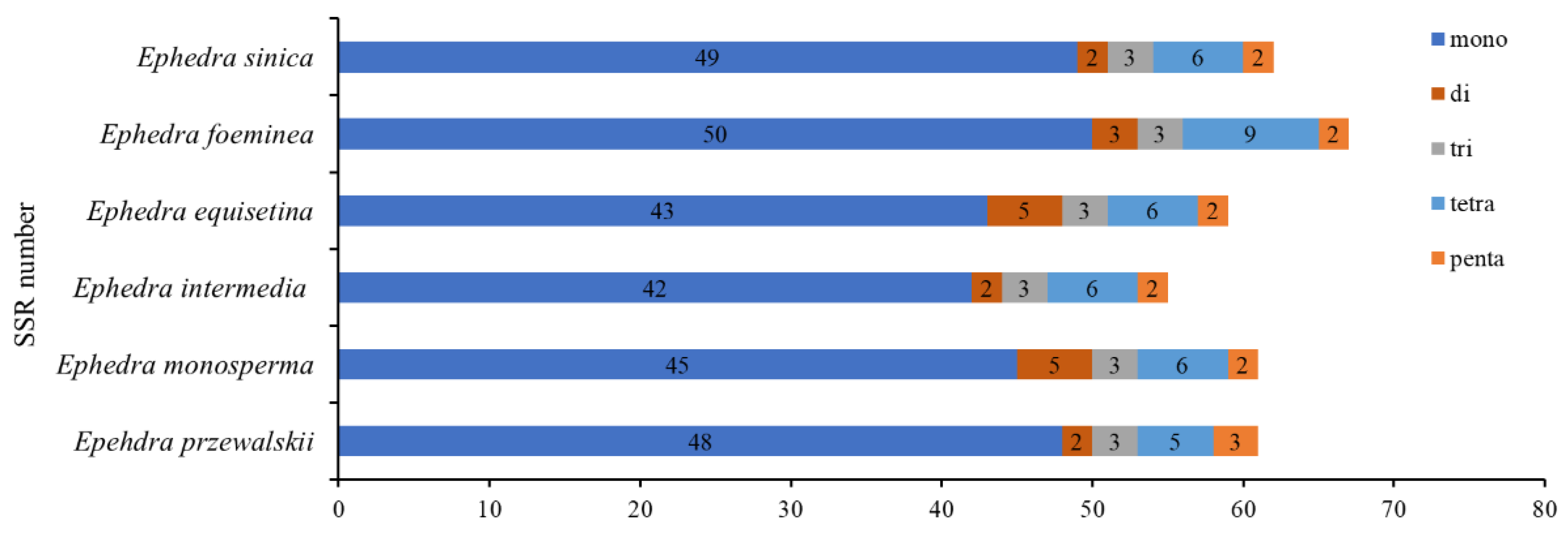
We analyzed six cp genome sequences for short repeats (SSRs, simple sequence repeats). The result indicated mononucleotide was the most abundant repeat type, but no hexanucleotide was found in Ephedra. 61, 61, 55, 59, 67, and 62 SSRs were detected in E. przewalskii, E. monosperma, E. intermedia, E. equisetina, E. foeminea, and E. sinica, respectively (Figure 2). Among these SSRs, there was the most mononucleotide with the number in E. foeminea (50) and the least in E. intermedia (42). Besides, we detected SSR in various regions of cp genomes, including LSC, SSC, IR, CDS, rRNA, tRNA, and IGS (Intergenic spacers) (Figure S1). SSRs were more abundant in IGS regions than in other regions, but in rRNA regions, they were the least abundant. The mononucleotide repeat analysis results were presented in six species (E. przewalskii, E. monosperma, E. intermedia, E. equisetina, E. foeminea, E. sinica): the highest number was poly A/T, ranging from 40 to 47, and the lowest was poly C/G, varying from one to three. Only one di-nucleotide (AT/TA) and tri-nucleotide (ATA/TTA), five tetra-nucleotide (AGGT/ATTG, CAAA/TTCT, ATAA/ATCT, ATAG/AATA, and CTAC/CTAT), two pentanucleotide (TTTTA/TTTTC, ATAAA/AAGAA) were discovered in each cp genomes, while ATTTC was merely in E. przewalskii (Figure S2).
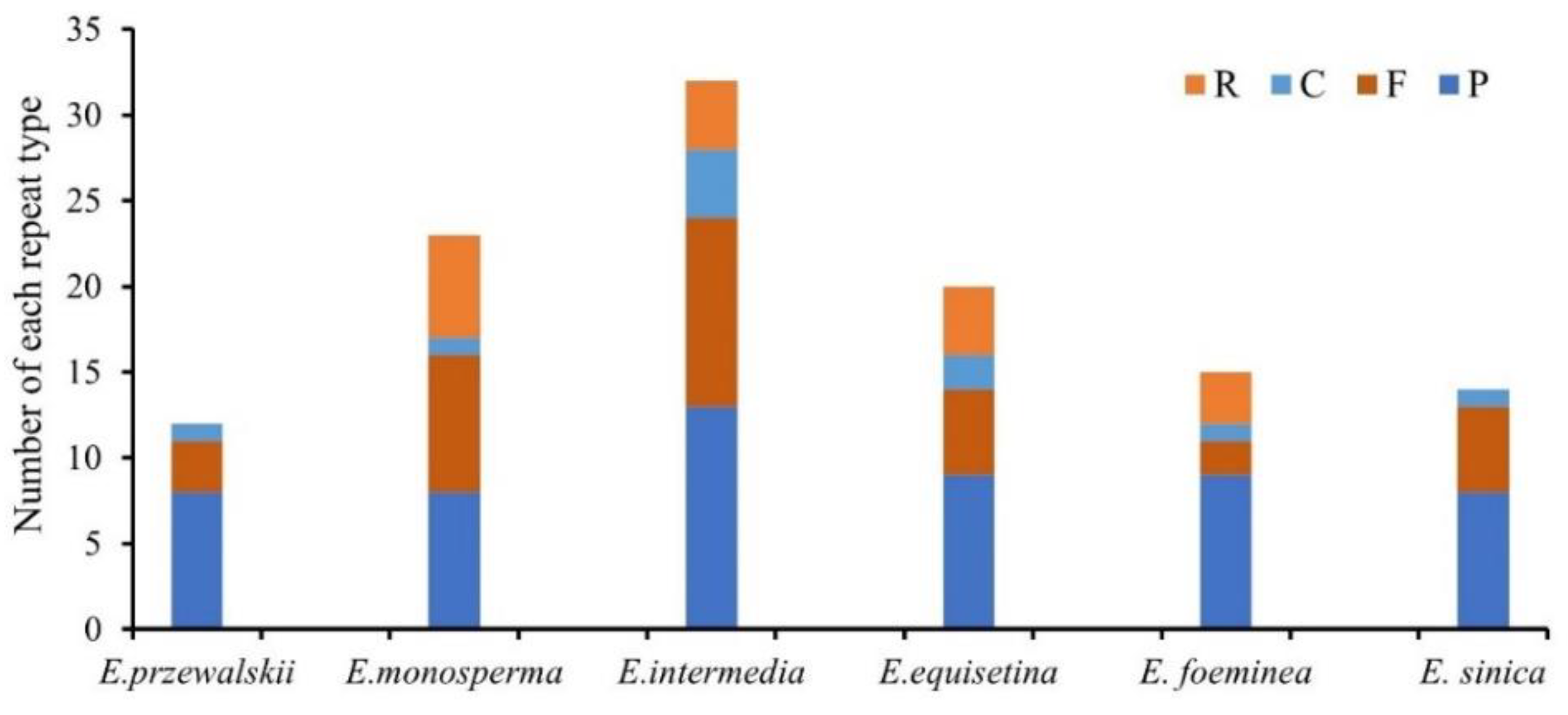
Also, we conducted the long repeats analysis that detected 125 non-overlapped repeats (54 palindromic, 34 forward, 19 complement, and 18 reverse repeats) in six Ephedra cp genomes (Figure 3). The palindromic repeats were the most common in these genomes, and the number varies from seven to eight. No reverse repeat was detected in E. przewalskii, whereas seven reverse repeats were found in E. monosperma. All four types of repeats were more abundant in the LSC region than in the SSC and IR regions. Moreover, we identified the length statistics of long repeat sequences in different size ranges for these cp genomes (Figure S3).

3.3. Codon Usage

We used 73 shared PCGs to analyze the codon usage bias and to calculate the relative synonymous codon usage (RSCU) value by codonW in all six cp genomes (Figure 4). There were twenty Amino acids and 64 codons, including three stop codons UAA, UAG, and UGA. There was only one codon in Methionine (Met) and Tryptophan (Trp). This study illustrated that there were 22,897 codons in E. przewalskii, 27,414 codons in E. monosperma, 27,631 codons in E. intermedia, 27,625 codons in E. equisetina, 27,620 codons in E. foeminea, and 27,623 codons in E. sinica encoded 73 PCGs, respectively. Moreover, in the six cp genomes, Leucine (Leu) was the most frequent amino acid with the codon number ranging from 2038 to 3603. Trp was the least frequent amino acid with the codon numbers varying from 371 to 490. As in Figure 4, the results of RSCU values indicated slight differences among E. przewalskii and E. monosperma. The RSCU value of 30 codons was more significant than 1 with A/T endings. Other codons were less than 1 with G/C endings, and the value was equal to 1 with only one codon.
The results exhibited various indices in the usage bias of synonymous codons for six Ephedra species, including CAI, CBI, ENc, GC3s, T3s, C3s, A3s, and G3s (Table S3). Within six Ephedra species, the ENc ranged from 46.19 to 54.63, and the GC3s varied from 22.3% to 34.1%. CAI values were 16.6–17.5%.

3.4. Divergence in Six Ephedra Chloroplast Genome

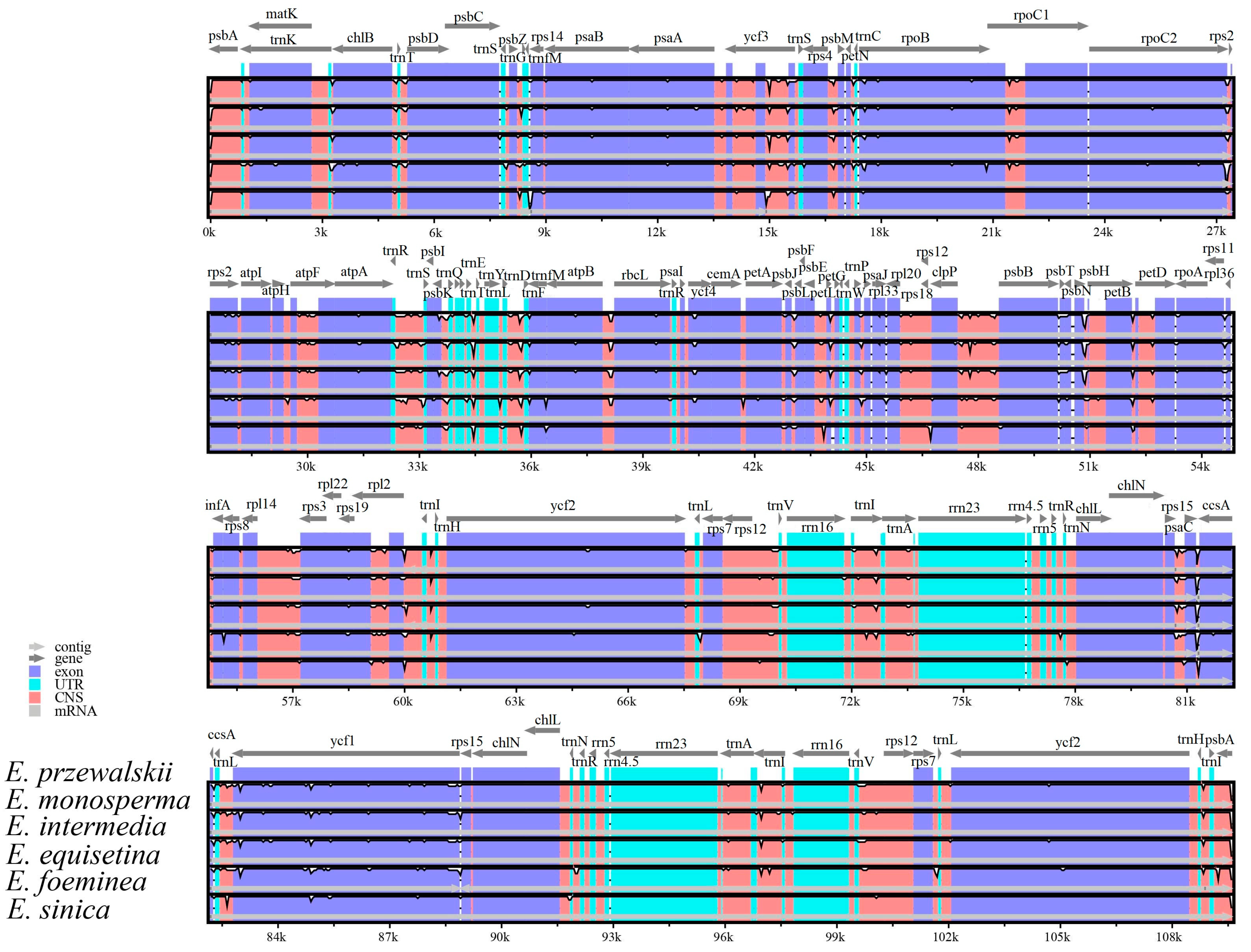
Results of mVISTA revealed, that non-coding regions were less conserved than protein-coding regions (Figure 5). The LSC and SSC regions were more divergent than the IRs regions, and the rRNA gene was highly conserved.
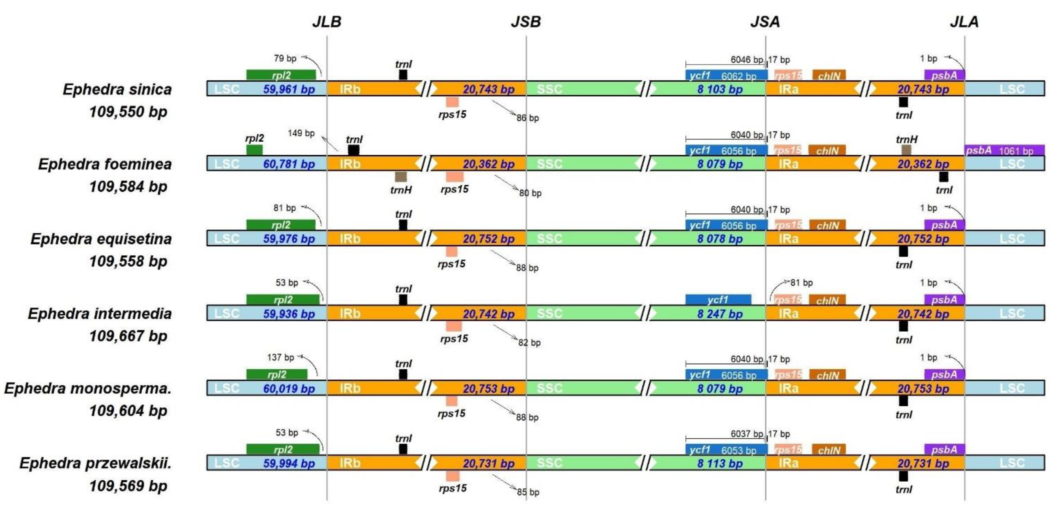
Also, we compared the boundaries of LSC, SSC, and IR among six cp genomes with IRscope tools. The result exhibited that six Ephedra species had little variations of IR/LSC and IR/SSC junction position and characteristics (Figure 6). In five Ephedra species, the rpl2 gene entirely existed in the LSC region. It was 53 to 149 bp away from the LSC/IRb junction regions, except that E. foeminea had an extremely short length of the rpl2 gene. Three genes (trnI, rps15, and chlN) were entirely situated at IR (IRa, IRb) regions, and the rps15 gene was 80 to 88 bp away from the IRb/SSC junction regions. The psbA gene was located in the IRa region of the other Ephedra species, but it moved to the LSC region in E. foeminea. The trnH gene only appeared in the IR region of E. foeminea. The ycf1 gene spanned through the IRa/SSC junction regions and ranged from 6053 to 6062 bp in length. This gene extended by 17 bp of the same length into the IRa regions in five Ephedra species. Due to the contraction of the ycf1 gene in E. intermedia, the length was shorter than its whole length (6056 bp)
We detected the nucleotide polymorphisms in the complete chloroplast genomes sequence of six Ephedra species (Figure S4). The pi value ranged from 0.0000 to 0.018. We found the intergenic spacers with high sequence variability (Pi value > 0.009), including trnF-GAA_ trnfM-CAU (0.01389), trnfM-CAU_atpE (0.01189), rpl36_infA (0.11), psbC_trnS-UGA (0.01056), trnS-UGA_psbZ (0.00967), trnfM-CAU_rps14 (0.00933). All the above-mentioned intergenic spacers were located in the LSC region. The Pi value (Pi value > 0.009) in the sequence of ccsA, ycf1, psaC, atpE and rpoB gene was 0.018, 0.01656, 0.01089, 0.01, and 0.00933, respectively. Most of them were found in the SSC region.

3.5. Evolutionary Rates in Protein-Coding Genes of Ephedra species

We used 68 PCGs from five cp genomes (E. przewalskii, E. monosperma, E. intermedia, Gnetum luofuense, Cycas szechuanensis), with Cycas szechuanensis as the reference. The final results were the average values of non-synonymous nucleotide substitutions (Ka), synonymous nucleotide substitutions (Ks), and Ka/Ks for 68 PCGs, respectively (Table S4). Among the Ks value of genes, 0.0005 in ycf2 and 0.063 in psbT were the smallest and the largest value, respectively. All PCGs showed a lower Ka value. The smallest Ka value was 0.0002 in psaB, and the largest was 0.007 in rps12 and rps14. The Ka/Ks values of 68 PCGs were less than 1, meaning that the synonymous substitutions rates were higher than the non-synonymous nucleotide substitutions. It exhibited that all of them undergo purifying selection, ranging from 0 to 0.97. The largest Ka/Ks value was in rpoA, and the smallest was in cemA and chlB.

3.6. Predicted RNA Editing Sites for E. przewalskii and E. monosperma

We predicted the RNA editing sites for E. przewalskii and E. monosperma (Figure 7; Table S5). We found 57, and 56 predicted RNA editing sites in the 15 PCGs of E. przewalskii and E. monosperma, respectively. Among these PCGs, the RNA Polymerase group had the highest number of predicted RNA editing sites. Specifically, the genes rpoB, rpoC2, rpoC1, and rpoA possessed fourteen, eleven, nine, and two RNA editing sites, respectively. All predicted editing sites were C to U transitions, and the most frequent amino acid conversions were from proline to serine.

3.7. Phylogenetic Inference

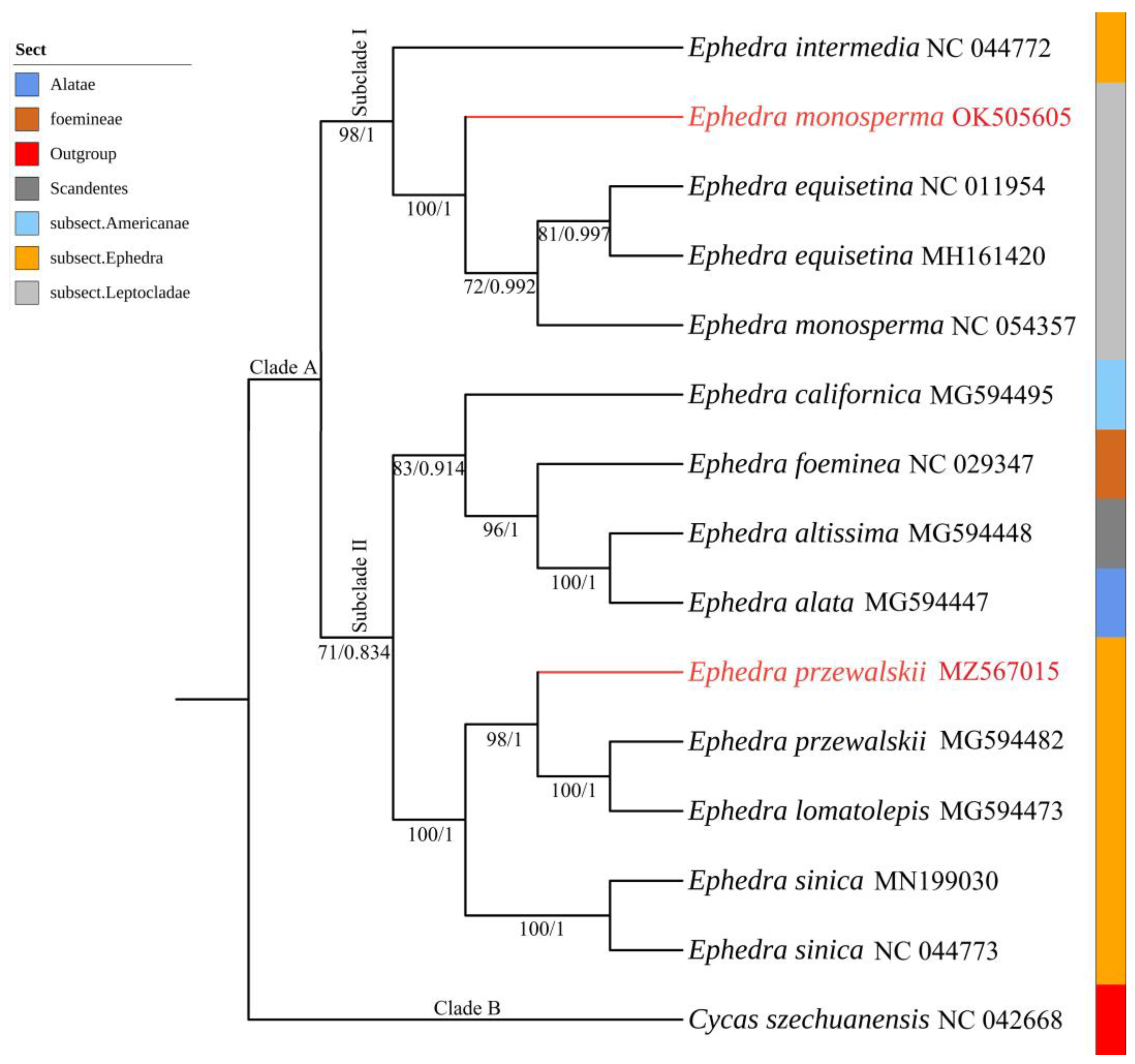
We inferred the phylogenetic relationship of 14 cp genomes sequences of Ephedra and observed the same tree topology in maximum likelihood (ML) and Bayesian inference (BI) analysis (Table S2; Figure 8). The maximum likelihood of bootstrap support (MLBS) and bayesian posterior probability (BPP) were high for each lineage. The first monophyletic Clade (Clade A) included all Ephedra species (E. przewalskii, E. monosperma, E. intermedia, E. equisetina, E. foeminea, E. sinica, E. alata, E. altissima, E. lomatolepis, E. californica). A sister group to the second Clade (Clade B), included Cycas szechuanensis from Cycadaceae. However, E. przewalskii (MZ567015) and E. monosperma (OK505605) were not gathered in one clade, consistent with the classification system by Flora of China [1]. Specifically, Clade A included subclade I (E. monosperma, E. intermedia, E. equisetina) and subclade II (E. californica, E. foeminea, E. altissima, E. alata, E. przewalskii, E. lomatolepis, E. sinica). In subclade I, E. intermedia (NC_044772, subsect. Ephedra) was sister to E. monosperma (OK505605), two E. equisetina (NC_011954, MH161420), and one E. monosperma (NC_054357) (subsect. Leptocladae) (MLBS = 98, BPP = 1). In subclade II, E. californica (MG594495, subsect. Americanae), E. foeminea (NC_029347, sect. foemineae), E. altissima (MG594448, sect. Scandentes), and E. alata (MG594447, sect. Alatae) were sister to two E. przewalskii (MZ567015, MG594482), E. lomatolepis (MG594473), and two E. sinica (MN199030, NC_044773) (subsect. Ephedra) (MLBS = 71, BPP = 0.834).

4. Discussion

4.1. Genome Feature in Ephedra

With the advancement of sequencing technology and cost reduction, chloroplast genome research has been continuously growing, with an increasing number of chloroplast genomes published in public databases, deepening people’s understanding and knowledge of plastid genomes [51]. These abundant genomic data paved the way for plant phylogenetic analysis [52,53]. The chloroplast genome had many advantages, such as maternal inheritance, highly conserved, abundant gene composition, etc. [52]. It has evolved into one of the most effective phylogenetic studies and molecular taxonomy tools [52]. The genomic information was extremely valuable in terms of species origin, evolution, and species relationships, and it has solved numerous phylogenetic problems [54,55]. As a traditional Chinese herbal medicine, Ephedra had crucial medicinal value [56]. We revealed the phylogenetic relationship between ten Ephedra species based on shared chloroplast PCGs, providing valuable phylogenetic information for this genus. At the same time, conducting comparative genomics and evolutionary analysis can provide abundant data from the plastid genome for species identification of Ephedra.
High AT base content found in the cp genomes of six Ephedra species was also reported in gymnosperms [25]. The GC content ranged from 36.6% to 36.7%, with that of IR regions significantly higher than that of the SC regions in the six species (Table 1). There were several examples consistent with previous studies [53,57,58]. Gene loss, duplication, and transfer between chloroplast and nuclear genomes, reliable sources of evolution as they are, also occurred in this study [59]. These events also occurred in this study. It may offer helpful information on evolutionary studies for Ephedra and whole gymnosperm plants.
Ndh (NADH dehydrogenase subunit) encoded the subunits of the proton-pumping NADH and played a significant role in green plants [60]. The Ephedra species in this study had lost all the ndh family genes, consistent with the result of the same genus and related genus, Welwitschia [25,26]. Also, we believed that there might be two reasons for the loss of ndh genes in the Ephedra species. First, this type of gene did not play any role in the evolution of Ephedra and was eliminated after selection. Secondly, the absence of ndh genes in some gymnosperms, including Pinaceae and Gnetales, was related to the living conditions in the Mesozoic era, including high temperature and carbon dioxide concentration [61,62,63]. However, there was no relevant evidence to prove that the missing ndh functional genes might be transferred to the nucleus to become a nuclear gene due to the high mutation rate. The absence of genes did not appear to affect the photosynthetic function of green plants [64,65]. Evidence has shown that Welwitschia plants mainly rely on glyceric acid to metabolize CAM for photosynthesis due to the loss event of the ndh genes [60]. Therefore, we assumed that Ephedra also depends on this metabolism to replace the function of the ndh genes. Losing genes can be a standard feature of the Ephedra species to clarify its phylogenetic relationship. Further studies with increasing samples are necessary to provide more information for the evolutionary analysis of Ephedra. The gene matK was one of the fastest evolving genes in the cp genome and located in the intron of trnK-UUU [66]. This gene was broadly used in phylogenetic studies within families, and inter-genera [67,68,69,70]. This gene had a 0.751 Ka/Ks value in this study, indicating it experienced purifying selection.

4.2. Comparison of Genomes for Ephedra

Six cp genomes were carefully compared to determine their identity and divergence. In mVISTA, the plot could visualize the sequence identity of six Ephedra species (Figure 2). The coding region of six cp genomes was more conservative than the non-coding region, congruent with other studies [71]. We found 31 non-coding highly variable regions, and nine PCGs were also highly variable. These highly variable regions and genes might be molecular markers for Ephedra species identification and population genetic study. There were subtle divergences in the LSC/IRS/SSC boundary gene distribution in the six Ephedra species. The psbA gene moved to the LSC region in E. foeminea, its length was longer than other species. This gene was a complete gene in E. foeminea. Compared with other Ephedra species, E. foeminea’s IR region and rpl2 gene in length were shorter, with its IR region showing noticeable contraction changes. The rpl2 gene was a pseudogene in E. foeminea. The ycf1 gene was entirely located in the SSC region of E. intermedia, it was a pseudogene in this species. These results can be used as one of the features to distinguish Ephedra species.
Nucleotide diversity was a proposed measure, to express the degree of nucleotide polymorphism in a population [72]. We analyzed the sequence variation of six complete cp genomes. The IR regions were found to be more conserved than the SC regions. The nucleotide polymorphisms in the SC region were greater than that in the IR region. The sequence of the ccsA gene with the highest Pi value (Pi = 0.018) was located in the SSC region. The trnF-GAA_trnfM-CAU intergenic spacer showed the highest variation (Pi = 0.01389) and was located in the LSC region.
SSRs are highly variable genetic markers and could be used for species identification, population genetics analyses, or evolutionary biology studies [73,74]. Our SSRs analysis revealed that the single-base repeat type (A/T) variation was the most abundant, implying that there were more replications in six Ephedra species. SSRs were mainly found in the LSC and the IGS, while fewer SSRs were situated in the IR region. This result has been observed in other plants [75]. These results may be used to study genetic diversity for Ephedra species. The number and distribution of the four types of long repeats differed lightly between the six Ephedra species. All species had complement, forward, and palindromic repeats. The reverse repeat was also identified in all Ephedra species except for E. przewalskii and E. sinica.

4.3. Codon Usage Bias Analysis

Understanding codon usage bias might reveal the effects of long-term evolution on the plant genome [76]. Due to the combined effects of gene selection, mutation, and drift in the long-term evolution process, most species had various Codon Usage Bias [77]. Interestingly, we found that codon usage preference mostly ends with AT, consistent with the determination result of AT-rich base content. CAI could estimate gene expression levels as an essential indicator of species codon usage preference [78,79]. These values were all-around 0.175, indicating a relatively low codon usage preference. The parameter ENc could quantify codon usage bias with a greater than 46 value in Ephedra species, indicating that codon preference was weak [80]. Similar results were observed for other species [81]. In general, these indicators analysis found that codon usage preferences of Ephedra species were not strong. Still, this study was limited to these few parameters, which were insufficient to explain the preference strength of Ephedra species. Therefore, the sample of Ephedra should be increased for a more specific analysis of codons usage bias in future studies.
Since the discovery of the first RNA editing event in trypanosome mitochondria [82], many studies on RNA editing have been published [83,84]. RNA editing events also existed in plants, which affect plants’ growth, development, and response to various stresses [85,86,87,88]. For most of the chloroplast genomes of gymnosperms and angiosperms, only a few dozen RNA editing sites could be predicted [89]. Similarly, we observed only 56–57 RNA editing sites in the cp genomes of E. przewalskii and E. monosperma. The most frequent type of RNA editing was from C to U conversion in a variety of plants such as thale cress (Arabidopsis thaliana) [90], grape (Vitis vinifera) [91], tobacco (Nicotiana tabacum) [92], as well as this two Ephedra species. Our study indicated that the most frequent amino acid conversion was proline to serine in the two Ephedra species, similar to the results in other studies [56,59]. Moreover, all the DNA-dependent RNA polymerase genes involved the most predicted RNA editing sites. These results are published for the first time and can offer a novel insight into future RNA editing studies for E. przewalskii and E. monosperma.

4.4. Evolution Analysis

Analysis of the non-synonymous and synonymous substitutions ratio has become a significant element of molecular evolution studies [93]. It was widely utilized to determine selection pressure for PCGs [94]. The Ka/Ks value of 68 PCGs was less than 1, meaning non-synonymous substitutions were less than synonymous substitutions. This often results in harmful traits for non-synonymous substitutions that would face elimination [95]. These PCGs were subject to purifying selection, similar to that in other studies [27,96,97]. The DNA-dependent RNA polymerase function gene group of E. przewalskii and E. monosperma showed a high Ka/Ks value, especially the rpoA with the highest value (0.97). This illustrated that these genes had a faster evolution rate than other functional groups of genes.

4.5. Phylogenetic Analysis

Many scholars have explored the phylogenetic relationship of Ephedra, mainly based on RAPD markers, nrDNA, and cpDNA sequences [11,16,17,98]. This study also utilized the concatenated sequences of chloroplast PCGs to construct phylogenetic trees based on different methods (ML/BI), with high bootstrap value and Bayesian posterior probability. All Ephedra species clustered together in a well-supported monophyletic clade, congruing with previous studies [19,20]. In our study, E. przewalskii and E. monosperma did not cluster together, consistent with the previously reported phylogenetic trees based on molecular data [18,20,99]. Moreover, from the phylogenetic tree’s topological structure, it could be observed that Ephedra species from different countries are nested together. The Ephedra species (E. californica, E. altissima, E. alata) from North America, Africa, and Kazakhstan were all nested in the sister branches of China species (E. foeminea), consistent with previous findings [99]. In summary, we reported the phylogenetic relationship of the ten Ephedra species based on the concatenated sequences of PCGs. It offers valuable information for the phylogeny of the genus Ephedra.

5. Conclusions

Our study reported the complete chloroplast genome of E. przewalskii and E. monosperma. We also revealed the slight differences in cp genome characterization, the number of long repeats and SSRs, codon usage bias, and RNA editing sites. Moreover, all PCGs in Ephedra species were affected by purifying selection. Also, we revealed the phylogenetic relationship of ten Ephedra species with high maximum likelihood bootstrap support and Bayesian posterior probability. These data provide valuable and abundant information for the phylogenetic analysis and species identification of the medical plant, Ephedra.

Supplementary Materials

The following supporting information can be downloaded at: https://www.mdpi.com/article/10.3390/d14100792/s1. Figure S1: The number of short sequence repeats (SSRs) in LSC, SSC, IR, CDS, rRNA, tRNA, and IGS in the cp genomes of six Ephedra species; Figure S2: The frequency of each SSR in the cp genomes of six Ephedra species. The order of six columns of each SSR is E. przewalskii, E. monosperma, E. intermedia, E. equisetina, E. foeminea, E. sinica, respectively; Figure S3: The number of four types of long repeats with different lengths ranges in the cp genomes of six Ephedra species; R: reverse repeats; C: complement repeats; F: forward repeats; P: palindromic repeats. Repeats with different lengths for six cp genomes are shown in different colors; Figure S4: The results of the nucleotide variability (Pi) values in the six Ephedra species. The x-axis is the position of the midpoint of the window. The y-axis is the nucleotide diversity (Pi) values of each window; Table S1: Specimen information of E. przewalskii and E. monosperma; Table S2: The cp genome sequences used in the phylogenetic analysis; Table S3: Codon usage of protein-coding genes in chloroplast genomes of six Ephedra species (Unit: %); Table S4: The Ka/Ks ratio of 68 protein-coding genes of five cp genomes (E. przewalskii, E. monosperma, E. intermedia, Gnetum luofuense, Cycas szechuanensis), with C. szechuanensis as the reference; Table S5: The number of predicted RNA editing sites in the cp genomes of E. przewalskii and E. monosperma.

Author Contributions

Conceptualization, S.H., M.X. and J.Y.; methodology, S.H. and M.X.; software S.H. and J.Y.; validation, S.H., J.Y. and F.Z.; formal analysis, S.H.; investigation, M.X., J.Y., H.X. and F.Z.; resources, M.X., J.Y., H.X. and F.Z.; data curation, S.H., H.X. and Y.H.; writing—original draft preparation S.H. and F.Z.; writing—review and editing, all authors; visualization, S.H. and H.X.; supervision, F.Z.; project administration, F.Z.; funding acquisition, F.Z. All authors have read and agreed to the published version of the manuscript.

Funding

This research was funded by the Second Tibetan Plateau Scientific Expedition and Research (STEP) Program (2019QZKK0502), Construction Project for Innovation Platform of Qinghai Province (2022-ZJ-Y04).

Institutional Review Board Statement

Not applicable.

Data Availability Statement

All data generated or analyzed during this study are included in this published article.

Acknowledgments

The authors are most grateful to anonymous reviewers for their helpful suggestions and comments.

Conflicts of Interest

The authors declare no conflict of interest.

References

  1. Cheng, W.J.; Fu, L.K. Gymnospermae. Flora of China; Science Press: Beijing, China, 1978; Volume 7, pp. 471–489. [Google Scholar]
  2. Qin, A.L.; Wang, M.M.; Cun, Y.Z.; Yang, F.S.; Wang, S.S.; Ran, J.H.; Wang, X.Q. Phylogeographic evidence for a link of species divergence of Ephedra in the Qinghai-Tibetan Plateau and adjacent regions to the Miocene Asian aridification. PLoS ONE 2013, 8, e56243. [Google Scholar] [CrossRef] [PubMed]
  3. Meyer, C.A. Versuch einer Monographie der Gattung Ephedra. Mem. Akad. Imper. Sci. St. Petersburg. Ser.6 (Sci. Nat.) 1846, 5, 225–297. (In German) [Google Scholar]
  4. Stapf, O. Die Arten der Gattung Ephedra. Denkschr. Math.-Nat. Kl. Akad Wiss. Wien, lvi. 1889, 56, 35. [Google Scholar]
  5. Shen, G.M. Distribution and evolution of the genus Ephedra in China. Arid. Zone Res. 1993, 10, 39–48. [Google Scholar]
  6. Yang, Y. Systematics and Evolution of Ephedra L. (Ephedraceae) from China. Chin. Acad. Sci. 2002, 1–231. Available online: https://cir.nii.ac.jp/crid/1572824500456924160 (accessed on 3 January 2021).
  7. Vishal, S.; Harihara, V.; Mehendale, M. Ephedra. In Encyclopedia of Toxicology, 2nd ed.; Philip, W., Ed.; Elsevier: Amsterdam, The Netherlands, 2005; pp. 223–228. [Google Scholar]
  8. Barker, W.D.; Antia, U. A study of the use of Ephedra in the manufacture of methamphetamine. Forensic Sci. Int. 2007, 166, 102–109. [Google Scholar] [CrossRef]
  9. Haller, C.A.; Benowitz, N.L.; Jacob, P. Hemodynamic effects of ephedra-free weight-loss supplements in humans. Am. J. Med. 2005, 118, 998–1003. [Google Scholar] [CrossRef]
  10. Mei, J.; Zhou, Y.; Yang, X.; Zhang, F.; Liu, X.; Yu, B. Active components in Ephedra sinica Stapf disrupt the interaction between ACE2 and SARS-CoV-2 RBD: Potent COVID-19 therapeutic agents. J. Ethnopharmacol. 2021, 278, 114303. [Google Scholar] [CrossRef]
  11. Jiang, H.Y.; Li, W.; Li, S.; Shi, L.N.; Gao, Y.H.; Wu, J.L. Relationship analysis of five species in the genus Ephedra L. by RAPD. J. Gansu Agric. Univ. 2006, 41, 49–52. [Google Scholar]
  12. Deng, N.; Shi, S.Q.; Chang, E.M.; Liu, J.F.; Lan, Q.; Jiang, Z.P. Transcriptomic Analysis of Germinated Seeds of Ephedra przewalskii. J. Northeast. For. Univ. 2015, 43, 28–32. [Google Scholar]
  13. Xin, G.-Z.; Hu, B.; Shi, Z.-Q.; Zheng, J.-Y.; Wang, L.; Chang, W.-Q.; Li, P.; Yao, Z.-P.; Liu, L.-F. A direct ionization mass spectrometry-based approach for differentiation of medicinal Ephedra species. J. Pharm. Biomed. Anal. 2016, 117, 492–498. [Google Scholar] [CrossRef] [PubMed]
  14. Zheng, Y.; Gao, H.; Song, M.; Lin, Y.; Fan, J.; Liu, X. Identification of plant materials containing ephedrine alkaloids based on DNA barcoding and TaqMan real-time PCR assay. Acta Physiol. Plant. 2021, 4311, 143. [Google Scholar] [CrossRef]
  15. Rydin, C.; Pedersen, K.R.; Friis, E.M. On the evolutionary history of Ephedra: Cretaceous fossils and extant molecules. Proc. Natl. Acad. Sci. USA 2004, 101, 16571–16576. [Google Scholar] [CrossRef] [PubMed]
  16. Long, C.; Kakiuchi, N.; Takahashi, A.; Komatsu, K.; Cai, S.; Mikage, M. Phylogenetic analysis of the DNA sequence of the non-coding region of nuclear ribosomal DNA and chloroplast of Ephedra plants in China. Planta Med. 2004, 70, 1080–1084. [Google Scholar] [CrossRef] [PubMed]
  17. Huang, J.; Giannasi, D.E.; Price, R.A. Phylogenetic relationships in Ephedra (Ephedraceae) inferred from chloroplast and nuclear DNA sequences. Mol. Phylogenet Evol. 2005, 35, 48–59. [Google Scholar] [CrossRef] [PubMed]
  18. Chen, X.; Cui, Y.; Nie, L.; Hu, H.; Xu, Z.; Sun, W.; Gao, T.; Song, J.; Yao, H. Identification and Phylogenetic Analysis of the Complete Chloroplast Genomes of Three Ephedra Herbs Containing Ephedrine. Biomed. Res. Int. 2019, 2019, 5921725. [Google Scholar] [CrossRef] [PubMed] [Green Version]
  19. Rydin, C.; Blokzijl, R.; Thureborn, O.; Wikström, N. Node ages, relationships, and phylogenomic incongruence in an ancient gymnosperm lineage—Phylogeny of Ephedra revisited. Taxon 2021, 70, 701–719. [Google Scholar] [CrossRef]
  20. Moore, M.J.; Soltis, P.S.; Bell, C.D.; Burleigh, J.G.; Soltis, D.E. Phylogenetic analysis of 83 plastid genes further resolves the early diversification of eudicots. Proc. Natl. Acad. Sci. USA 2010, 107, 4623–4628. [Google Scholar] [CrossRef] [PubMed]
  21. Samigullin, T.H.; Logacheva, M.D.; Terenteva, E.I.; Degtjareva, G.V.; Vallejo-Roman, C.M. The plastid genome of Seseli montanum: Complete sequence and comparison with plastomes of other members of the Apiaceae family. Biochemistry 2016, 81, 981–985. [Google Scholar] [CrossRef]
  22. Androsiuk, P.; Jastrzębski, J.P.; Paukszto, Ł.; Makowczenko, K.; Okorski, A.; Pszczółkowska, A.; Chwedorzewska, K.J.; Górecki, R.; Giełwanowska, I. Evolutionary dynamics of the chloroplast genome sequences of six Colobanthus species. Sci. Rep. 2020, 10, 11522. [Google Scholar] [CrossRef] [PubMed]
  23. Ravi, V.; Khurana, J.P.; Tyagi, A.K.; Khurana, P. An update on chloroplast genomes. Syst. Evol. 2008, 271, 101–122. [Google Scholar] [CrossRef]
  24. Zhang, X.; Sun, Y.; Landis, J.B.; Lv, Z.; Shen, J.; Zhang, H.; Lin, N.; Li, L.; Sun, J.; Deng, T.; et al. Plastome phylogenomic study of Gentianeae (Gentianaceae): Widespread gene tree discordance and its association with evolutionary rate heterogeneity of plastid genes. BMC Plant Biol. 2020, 20, 340. [Google Scholar] [CrossRef] [PubMed]
  25. Wu, C.S.; Lai, Y.T.; Lin, C.P.; Wang, Y.N.; Chaw, S.M. Evolution of reduced and compact chloroplast genomes (cpDNAs) in gnetophytes: Selection toward a lower-cost strategy. Mol. Phylogenet Evol. 2009, 52, 115–124. [Google Scholar] [CrossRef] [PubMed]
  26. McCoy, S.R.; Kuehl, J.V.; Boore, J.L.; Raubeson, L.A. The complete plastid genome sequence of Welwitschia mirabilis: An unusually compact plastome with accelerated divergence rates. BMC Evol. Biol. 2008, 8, 130. [Google Scholar] [CrossRef] [PubMed]
  27. Alzahrani, D.; Albokhari, E.; Yaradua, S.; Abba, A. Complete chloroplast genome sequences of Dipterygium glaucum and Cleome chrysantha and other Cleomaceae Species, comparative analysis and phylogenetic relationships. Saudi J. Biol. Sci. 2021, 28, 2476–2490. [Google Scholar] [CrossRef] [PubMed]
  28. Xie, X.; Huang, R.; Li, F.; Tian, E.; Li, C.; Chao, Z. Phylogenetic position of Bupleurum sikangense inferred from the complete chloroplast genome sequence. Gene 2021, 798, 145801. [Google Scholar] [CrossRef] [PubMed]
  29. Thomson, A.M.; Vargas, O.M.; Dick, C.W. Complete plastome sequence from Bertholletia excelsa and 23 related species yield informative markers for Lecythidaceae. Appl. Plant Sci. 2018, 6, e01151. [Google Scholar] [CrossRef]
  30. Sun, C.; Chen, F.; Teng, N.; Xu, Y.; Dai, Z. Comparative analysis of the complete chloroplast genome of seven Nymphaea species. Aquat. Bot. 2021, 170, 103353. [Google Scholar] [CrossRef]
  31. Khan, G.; Zhang, F.; Gao, Q.; Fu, P.C.; Xing, R.; Wang, J.; Liu, H.; Chen, S. Molecular phylogeography and intraspecific divergence of Spiraea alpina (Rosaceae) distributed in the Qinghai-Tibetan Plateau and adjacent regions inferred from nrDNA. Biochem. Syst. Ecol. 2014, 57, 278–286. [Google Scholar] [CrossRef]
  32. Doyle, J.J.; Doyle, J.L. A rapid DNA isolation procedure for small quantities of fresh leaf tissue. Phytochem. Bullet. 1987, 19, 11–15. [Google Scholar]
  33. Bolger, A.M.; Lohse, M.; Usadel, B. Trimmomatic: A flexible trimmer for Illumina sequence data. Bioinformatics 2014, 30, 2114–2120. [Google Scholar] [CrossRef]
  34. Simon, A. FastQC: A Quality Control Tool for High Throughput Sequence Data; Babraham Bioinformatics, Babraham Institute: Cambridge, UK, 2010. [Google Scholar]
  35. Bankevich, A.; Nurk, S.; Antipov, D.; Gurevich, A.A.; Dvorkin, M.; Kulikov, A.S.; Lesin, V.M.; Nikolenko, S.I.; Pham, S.; Prjibelski, A.D.; et al. SPAdes: A New Genome Assembly Algorithm and Its Applications to Single-Cell Sequencing. J. Comput. Biol. 2012, 19, 455–477. [Google Scholar] [CrossRef]
  36. Nicolas, D.; Patrick, M.; Guillaume, S. NOVOPlasty: De novo assembly of organelle genomes from whole genome data. Nucleic Acids Res. 2016, 4, e18. [Google Scholar]
  37. Tillich, M.; Lehwark, P.; Pellizzer, T.; Ulbricht-Jones, E.S.; Fischer, A.; Bock, R.; Greiner, S. GeSeq—Versatile and accurate annotation of organelle genomes. Nucleic Acids Res. 2017, 45, W6–W11. [Google Scholar] [CrossRef] [PubMed]
  38. Lohse, M.; Drechsel, O.; Bock, R. OrganellarGenomeDRAW (OGDRAW): A tool for the easy generation of high-quality custom graphical maps of plastid and mitochondrial genomes. Curr. Genet. 2007, 52, 267–274. [Google Scholar] [CrossRef] [PubMed]
  39. Benson, D.A.; Cavanaugh, M.; Clark, K.; Karsch-Mizrachi, I.; Lipman, D.J.; Ostell, J.; Sayers, E.W. GenBank. Nucleic Acids Res. 2013, 41, D36–D42. [Google Scholar] [CrossRef] [PubMed]
  40. Frazer, K.A.; Pachter, L.; Poliakov, A.; Rubin, E.M.; Dubchak, I. VISTA: Computational tools for comparative genomics. Nucleic Acids Res. 2004, 32, W273–W279. [Google Scholar] [CrossRef] [PubMed]
  41. Ali, A.; Jaakko, H.; Peter, P. IRscope: An online program to visualize the junction sites of chloroplast genomes. Bioinformatics 2018, 34, 3030–3031. [Google Scholar]
  42. Kumar, S.; Stecher, G.; Li, M.; Knyaz, C.; Tamura, K. MEGA X: Molecular Evolutionary Genetics Analysis across computing platforms. Mol. Biol. Evol. 2018, 35, 1547–1549. [Google Scholar] [CrossRef]
  43. Sebastian, B.; Thomas, T.; Thomas, M.; Uwe, S.; Martin, M. MISA-web: A web server for microsatellite prediction. Bioinformatics 2017, 33, 2583–2585. [Google Scholar]
  44. Kurtz, S.; Choudhuri, J.V.; Ohlebusch, E.; Schleiermacher, C.; Stoye, J.; Giegerich, R. REPuter: The manifold applications of repeat analysis on a genomic scale. Nucleic Acids Res. 2001, 29, 4633–4642. [Google Scholar] [CrossRef] [PubMed]
  45. Katoh, K.; Rozewicki, J.; Yamada, K.D. MAFFT online service: Multiple sequence alignment, interactive sequence choice and visualization. Brief. Bioinform. 2019, 20, 1160–1166. [Google Scholar] [CrossRef] [PubMed]
  46. Rozas, J.; Ferrer-Mata, A.; Sánchez-DelBarrio, J.C.; Guirao-Rico, S.; Librado, P.; Ramos-Onsins, S.E.; Sánchez-Gracia, A. DnaSP 6: DNA Sequence Polymorphism Analysis of Large Data Sets. Mol. Biol. Evol. 2017, 34, 3299–3302. [Google Scholar] [CrossRef] [PubMed]
  47. Wang, D.P.; Zhang, Y.B.; Zhang, Z.; Zhu, J.; Yu, J. KaKs_Calculator 2.0: A Toolkit Incorporating Gamma-Series Methods and Sliding Window Strategies. Genom. Proteom. Bioinform. 2010, 8, 77–80. [Google Scholar] [CrossRef]
  48. Zhang, D.; Gao, F.; Jakovlić, I.; Zou, H.; Zhang, J.; Li, W.X.; Wang, G.T. PhyloSuite: An integrated and scalable desktop platform for streamlined molecular sequence data management and evolutionary phylogenetics studies. Mol. Ecol. Resour. 2020, 20, 348–355. [Google Scholar] [CrossRef]
  49. Minh, B.Q.; Schmidt, H.A.; Chernomor, O.; Schrempf, D.; Woodhams, M.D.; von Haeseler, A.; Lanfear, R. IQ-TREE 2: New Models and Efficient Methods for Phylogenetic Inference in the Genomic Era. Mol. Biol. Evol. 2020, 37, 1530–1534. [Google Scholar] [CrossRef]
  50. Ronquist, F.; Huelsenbeck, J.P. MrBayes 3: Bayesian phylogenetic inference under mixed models. Bioinformatics 2003, 19, 1572–1574. [Google Scholar] [CrossRef]
  51. Armbruster, U.; Pesaresi, P.; Pribil, M.; Hertle, A.; Leister, D. Update on chloroplast research: New tools, new topics, and new trends. Mol. Plant. 2011, 4, 1–16. [Google Scholar] [CrossRef]
  52. Khan, A.; Khan, I.A.; Asif, H.; Azim, M.K. Current trends in chloroplast genome research. Afr. J. Biotechnol. 2010, 9, 3494–3500. [Google Scholar]
  53. Huang, Q.; Liu, Z.X.; Wang, C.; Jing, M.Y.; Liu, J.Q.; Zhou, W.; Kai, G.Y. The Complete Chloroplast Genome Sequences of Anisodus Acutangulus and a Comparison with Other Solanaceae Species. Clin. Complement. Med. Pharmacol. 2021, 1, 100002. [Google Scholar] [CrossRef]
  54. Zhang, Y.; Yu, J.; Xia, M.; Chi, X.; Khan, G.; Chen, S.; Zhang, F. Plastome Sequencing Reveals Phylogenetic Relationships among Comastoma and Related Taxa (Gentianaceae) from the Qinghai-Tibetan Plateau. Ecol. Evol. 2021, 11, 16034–16046. [Google Scholar] [CrossRef] [PubMed]
  55. Huang, Y.; Wang, J.; Yang, Y.; Fan, C.; Chen, J. Phylogenomic Analysis and Dynamic Evolution of Chloroplast Genomes in Salicaceae. Front. Plant Sci. 2017, 8, 1050. [Google Scholar] [CrossRef] [PubMed]
  56. González-Juárez, D.E.; Escobedo-Moratilla, A.; Flores, J.; Hidalgo-Figueroa, S.; Martínez-Tagüeña, N.; Morales-Jiménez, J.; Muñiz-Ramírez, A.; Pastor-Palacios, G.; Pérez-Miranda, S.; Ramírez-Hernández, A.; et al. A Review of the Ephedra genus: Distribution, Ecology, Ethnobotany, Phytochemistry and Pharmacological Properties. Molecules 2020, 25, 3283. [Google Scholar] [CrossRef] [PubMed]
  57. Shahzadi, I.; Abdullah, M.F.; Ali, Z.; Ahmed, I.; Mirza, B. Chloroplast genome sequences of Artemisia maritima and Artemisia absinthium: Comparative analyses, mutational hotspots in genus Artemisia and phylogeny in family Asteraceae. Genomics 2020, 112, 1454–1463. [Google Scholar] [CrossRef] [PubMed]
  58. Wei, F.; Tang, D.; Wei, K.; Qin, F.; Li, L.; Lin, Y.; Zhu, Y.; Khan, A.; Kashif, M.H.; Miao, J. The complete chloroplast genome sequence of the medicinal plant Sophora Tonkinensis. Sci. Rep. 2020, 10, 12473. [Google Scholar] [CrossRef]
  59. Xiong, A.-S.; Peng, R.-H.; Zhuang, J.; Gao, F.; Zhu, B.; Fu, X.-Y.; Xue, Y.; Jin, X.-F.; Tian, Y.-S.; Zhao, W.; et al. Gene duplication, transfer, and evolution in the chloroplast genome. Biotechnol. Adv. 2009, 27, 340–347. [Google Scholar] [CrossRef]
  60. Shikanai, T. Chloroplast NDH: A different enzyme with a structure similar to that of respiratory NADH dehydrogenase. Biochim. Biophys. Acta. 2016, 1857, 1015–1022. [Google Scholar] [CrossRef]
  61. Wakasugi, T.; Tsudzuki, J.; Ito, S.; Nakashima, K.; Tsudzuki, T.; Sugiura, M. Loss of all ndh genes as determined by sequencing the entire chloroplast genome of the black pine Pinus thunbergii. Proc. Natl. Acad. Sci. USA 1994, 91, 9794–9798. [Google Scholar] [CrossRef]
  62. Ranade, S.S.; García-Gil, M.R.; Rosselló, J.A. Non-functional plastid ndh gene fragments are present in the nuclear genome of Norway spruce (Picea abies L. Karsch): Insights from in silico analysis of nuclear and organellar genomes. Mol. Genet. Genomics. 2016, 291, 935–941. [Google Scholar] [CrossRef]
  63. Braukmann, T.W.A.; Kuzmina, M.; Stefanović, S. Loss of all plastid ndh genes in Gnetales and conifers: Extent and evolutionary significance for the seed plant phylogeny. Curr. Genet. 2009, 55, 323–337. [Google Scholar] [CrossRef]
  64. Ruhlman, T.A.; Chang, W.J.; Chen, J.J.; Huang, Y.T.; Chan, M.T.; Zhang, J.; Liao, D.C.; Blazier, J.C.; Jin, X.; Shih, M.C.; et al. NDH expression marks major transitions in plant evolution and reveals coordinate intracellular gene loss. BMC Plant Biol. 2015, 15, 100. [Google Scholar] [CrossRef] [PubMed] [Green Version]
  65. Lin, C.; Chen, J.J.; Chiu, C.; Hsiao, H.C.; Yang, C.; Jin, X.; Leebens-Mack, J.; Depamphilis, C.W.; Huang, Y.; Chang, W.; et al. Concomitant loss of NDH complex-related genes within chloroplast and nuclear genomes in some orchids. Plant J. 2017, 90, 994–1006. [Google Scholar] [CrossRef] [PubMed]
  66. Hilu, K.W.; Liang, H.P. The matK gene: Sequence variation and application in plant systematics. Am. J. Bot. 1997, 84, 830–839. [Google Scholar] [CrossRef]
  67. Plunkett, G.M.; Soltis, D.E.; Soltis, P.S. Clarification of the relationship between Apiaceae and Araliaceae based on matK and rbcL sequence data. Am. J. Bot. 1997, 84, 565–580. [Google Scholar] [CrossRef] [PubMed]
  68. Cbol Plant Working Group. A DNA barcode for land plants. Proc. Natl. Acad. Sci. USA 2009, 106, 12794–12797. [Google Scholar] [CrossRef]
  69. Steven, G.N.; Subramanyam, R. Testing plant barcoding in a sister species complex of pantropical Acacia (Mimosoideae, Fabaceae. Mol. Ecol. Resour. 2009, 9 (Suppl. S1), 172–180. [Google Scholar] [CrossRef]
  70. Wang, T.; Liu, J.; Guo, Y.; Yuan, N. Phylogenetic relationship among local legumes in Jiangsu Province based on analyses of matK gene and ITS sequence. J. Nanjing Agric. Univ. 2017, 40, 795–803. [Google Scholar]
  71. Nie, X.; Lv, S.; Zhang, Y.; Du, X.; Wang, L.; Biradar, S.S.; Tan, X.; Wan, F.; Weining, S. Complete Chloroplast Genome Sequence of a Major Invasive Species, Crofton Weed (Ageratina adenophora). PLoS ONE 2012, 7, e36869. [Google Scholar] [CrossRef]
  72. Nei, M.; Li, W.H. Mathematical Model for Studying Genetic Variation in Terms of Restriction Endonucleases. Proc. Natl. Acad. Sci. USA 1979, 76, 5269–5273. [Google Scholar] [CrossRef]
  73. Tuler, A.C.; Carrijo, T.T.; Nóia, L.R.; Ferreira, A.; Peixoto, A.L.; Ferreira, M.F.D.S. SSR markers: A tool for species identification in Psidium (Myrtaceae). Mol. Biol. Rep. 2015, 42, 1501–1513. [Google Scholar] [CrossRef]
  74. Mohamed, A. El-Esawi.SSR analysis of genetic diversity and structure of the germplasm of faba bean (Vicia faba L.). Comptes Rendus Biol. 2017, 340, 474–480. [Google Scholar]
  75. Bi, Y.; Zhang, M.F.; Xue, J.; Dong, R.; Du, Y.P.; Zhang, X.H. Chloroplast genomic resources for phylogeny and DNA barcoding: A case study on Fritillaria. Sci. Rep. 2018, 8, 1184. [Google Scholar] [CrossRef] [PubMed]
  76. Duan, H.; Zhang, Q.; Wang, C.; Li, F.; Tian, F.; Lu, Y.; Hu, Y.; Yang, H.; Cui, G. Analysis of codon usage patterns of the chloroplast genome in Delphinium grandiflorum L. reveals a preference for AT-ending codons as a result of major selection constraints. Peer J. 2021, 9, e10787. [Google Scholar] [CrossRef] [PubMed]
  77. Ma, L.N.; Cui, P.; Zhu, J.; Zhang, Z.H.; Zhang, Z. Translational selection in human: More pronounced in housekeeping genes. Biol Direct 2014, 9, 17. [Google Scholar] [CrossRef]
  78. Sharp, P.M.; Li, W.H. The codon adaptation index—A measure of directional synonymous codon usage bias, and its potential applications. Nucleic Acids Res. 1987, 15, 1281–1295. [Google Scholar] [CrossRef]
  79. Supriyo, C.; Prosenjit, P.; Mazumder, T.H. Codon Usage Bias Prefers AT Bases in Coding Sequences Among the Essential Genes of Haemophilus influenzae. Not. Sci. Biol. 2014, 6, 417–421. [Google Scholar] [CrossRef]
  80. Wright, F. The ’effective number of codons’ used in a gene. Gene 1990, 87, 23–29. [Google Scholar] [CrossRef]
  81. Jiang, S.Z.; Lian, H.; Xiong, Y.F.; Zhang, S.; Chen, S.P. Analysis of Codon Bias in Chloroplast Genome of Castanopsis carlesii. Mol. Plant Breed. 2021, 26, 1–12. [Google Scholar]
  82. Benne, R. RNA-editing in trypanosome mitochondria. Biochim. Biophys. Acta. 1989, 1007, 131–139. [Google Scholar] [CrossRef]
  83. Bock, R.; Kössel, H.; Maliga, P. Introduction of a heterologous editing site into the tobacco plastid genome: The lack of RNA editing leads to a mutant phenotype. EMBO J. 1994, 13, 4623–4628. [Google Scholar] [CrossRef]
  84. Liu, H.; Wang, Q.; He, Y.; Chen, L.; Hao, C.; Jiang, C.; Li, Y.; Dai, Y.; Kang, Z.; Xu, J.R. Genome-wide A-to-I RNA editing in fungi independent of ADAR enzymes. Genome Res. 2016, 26, 499–509. [Google Scholar] [CrossRef]
  85. Gualberto, J.M.; LaMattina, L.; Bonnard, G.; Weil, J.H.; Grienenberger, J.-M. RNA editing in wheat mitochondria results in the conservation of protein sequences. Nature 1989, 341, 660–662. [Google Scholar] [CrossRef]
  86. Wakasugi, T.; Hirose, T.; Horihata, M.; Tsudzuki, T.; Kössel, H.; Sugiura, M. Creation of a novel protein-coding region at the RNA level in black pine chloroplasts: The pattern of RNA editing in the gymnosperm chloroplast is different from that in angiosperms. Proc. Natl. Acad. Sci. USA 1996, 93, 8766–8770. [Google Scholar] [CrossRef]
  87. Wang, W.; Yu, H.; Wang, J.; Lei, W.; Gao, J.; Qiu, X.; Wang, J. The Complete Chloroplast Genome Sequences of the Medicinal Plant Forsythia suspensa (Oleaceae). Int. J. Mol. Sci. 2017, 18, 2288. [Google Scholar] [CrossRef] [PubMed] [Green Version]
  88. Kumbhar, F.; Nie, X.; Xing, G.; Zhao, X.; Lin, Y.; Wang, S.; Weining, S. Identification and characterisation of RNA editing sites in chloroplast transcripts of einkorn wheat (Triticum monococcum). Ann. Appl. Biol. 2018, 172, 197–207. [Google Scholar] [CrossRef]
  89. Mower, J.P. The PREP suite: Predictive RNA editors for plant mitochondrial genes, chloroplast genes and user-defined alignments. Nucleic Acids Res. 2009, 37, W253–W259. [Google Scholar] [CrossRef] [PubMed]
  90. Giegé, P.; Brennicke, A. RNA editing in Arabidopsis mitochondria effects 441 C to U changes in ORFs. Proc. Natl. Acad. Sci. USA 1999, 96, 15324–15329. [Google Scholar] [CrossRef] [PubMed]
  91. Picardi, E.; Horner, D.S.; Chiara, M.; Schiavon, R.; Valle, G.; Pesole, G. Large-scale detection and analysis of RNA editing in grape mtDNA by RNA deep-sequencing. Nucleic Acids Res. 2010, 38, 4755–4767. [Google Scholar] [CrossRef] [PubMed]
  92. Grimes, B.T.; Sisay, A.K.; Carroll, H.D.; Cahoon, A.B. Deep sequencing of the tobacco mitochondrial transcriptome reveals expressed ORFs and numerous editing sites out-side coding regions. BMC Geno. 2014, 15, 31. [Google Scholar]
  93. Nei, M.; Kumar, S. Molecular Evolution and Phylogenetics; Oxford University Press: New York, NY, USA, 2000. [Google Scholar]
  94. Yin, K.; Zhang, Y.; Li, Y.; Du, F. Different natural selection pressures on the atpF gene in evergreen sclerophyllous and deciduous oak species: Evidence from comparative analysis of the complete chloroplast genome of Quercus aquifolioides with other oak species. Int. J. Mol. Sci. 2018, 19, 1042. [Google Scholar] [CrossRef] [PubMed]
  95. Yang, Z.H. Computational Molecular Evolution; Oxford University Press: Oxford, UK, 2006; p. 284. [Google Scholar]
  96. Azarin, K.; Usatov, A.; Makarenko, M.; Khachumov, V.; Gavrilova, V. Comparative analysis of chloroplast genomes of seven perennial Helianthus species. Gene 2021, 774, 145418. [Google Scholar] [CrossRef] [PubMed]
  97. Huang, R.; Xie, X.; Li, F.; Tian, E.; Chao, Z. Chloroplast genomes of two Mediterranean Bupleurum species and the phylogenetic relationship inferred from combined analysis with East Asian species. Planta 2021, 253, 81. [Google Scholar] [CrossRef] [PubMed]
  98. Rydin, C.; Pedersen, K.R.; Crane, P.R.; Friis, E.M. Former diversity of Ephedra (Gnetales): Evidence from Early Cretaceous seeds from Portugal and North America. Ann. Bot. 2006, 98, 123–140. [Google Scholar] [CrossRef] [PubMed]
  99. Rydin, C.; Korall, P. Evolutionary Relationships in Ephedra (Gnetales), with Implications for Seed Plant Phylogeny. Int. J. Plant Sci. 2009, 170, 1031–1043. [Google Scholar] [CrossRef] [Green Version]
Figure 1. Chloroplast genome maps of E. przewalskii and E. monosperma. The positive coding genes were on the outside of the ring, whereas the reverse coding genes were on the inside of the ring. The darker gray in the inner circle corresponds to GC content, whereas the lighter gray corresponds to AT content. The different groups of genes were shown in different colors.
Diversity 14 00792 g001
Figure 2. The type and amounts of simple sequences repeat (SSRs) in the cp genomes of six Ephedra species.
Diversity 14 00792 g002
Figure 3. The types of long repeats and their number in the cp genome of six Ephedra species. R: reverse repeats; C: complement repeats; F: forward repeats; P: palindromic repeats. The number of four types of long repeats were shown in different colors.
Diversity 14 00792 g003
Figure 4. Codon usage in 73 protein-coding genes of the cp genomes of six Ephedra species. The order of six columns of every amino acid is E. przewalskii, E. monosperma, E. intermedia, E. equisetina, E. foeminea, and E. sinica, respectively.
Diversity 14 00792 g004
Figure 5. Comparison of the chloroplast genome of E. przewalskii, E. monosperma, E. intermedia, E. equisetina, E. foeminea, E. sinica, with E. przewalskii as the reference by mVISTA tool.
Diversity 14 00792 g005
Figure 6. Comparisons of LSC, SSC, and IR region junctions among the six Ephedra cp genomes. The numbers above genes represent the distance between the gene and the junction.
Diversity 14 00792 g006
Figure 7. The number of predicted RNA editing sites in the cp genomes of E. przewalskii and E. monosperma. The X-axis presents the PCGs with predicted editing sites. Y-axis presents the number of predicted editing sites for each PCGs.
Diversity 14 00792 g007
Figure 8. Phylogenetic relationship of two Ephedra species with related species based on the CDS shared by all cp genomes sequence. E. przewalskii (MZ567015) and E. monosperma (OK505605) were not gathered in one clade, and they were marked by the red color. Trees obtained with ML (Maximum Likelihood) and BI (Bayesian inference) methods have identical topology, therefore number/number above the branch means ML bootstrap support (MLBS)/Bayesian posterior probability (BPP).
Diversity 14 00792 g008
Table 1. Comparison of the chloroplast genome features of six Ephedra species.
SpeciesE. przewalskiiE. monospermaE. intermediaE. equisetinaE. foemineaE. sinica
Accession NumberMZ567015OK505605NC_044772.1MH161420NC_029347NC_044773
Genome size (bp)109,569109,604109,667109,558109,584109,550
LSC length (bp)59,99460,01959,93659,97660,02759,961
SSC length (bp)811380798247807880798103
IR length (bp)20,73120,75320,74220,75220,73920,743
Overall GC content (%)36.636.636.636.636.736.7
GC content in LSC (%) 34.234.234.234.234.134.2
GC content in SSC (%)27.627.927.327.527.727.9
GC content in IR (%)424242.1424242
Total number of genes118118118118118118
Protein-coding genes737373737373
tRNA genes373737373737
rRNA genes888888
Duplicated genes191918181818
Table 2. List of annotated genes in six Ephedra cp genomes.
Category of GenesGroup of GeneGene IDs
Self-replicationRibosomal RNA genesrrn23d,i; rrn16d,i; rrn5d,i; rrn4.5d,i
Transfer RNA genestrnY-GUAl; trnW-CCAl; trnV-GACd,i; trnT-UGUl;
trnT-GGUl; trnS-UGA l; trnS-GGAl; trnS-GCUl;
trnR-UCUl; trnR-CCGl; trnR-ACGd,i; trnQ-UUG
l; trnP-UGGl; trnN-GUUd,i; trnL-UAAl; trnL-CAAd,i;
trnL-AUGl; trnK-UUU *,l; trnI-GAU d,*,i; trnI-CAU d,i;
trnH-GUG d,i; trnG-UCCv l; trnfM-CAU d; trnF-GAA l;
trnE-UUC l; trnD-GUC l; trnC-GCA l; trnA-UGC d,*,i;
trnL-UAG s
Small subunit of ribosomerps19l; rps18l; rps15d,i; rps14l; rps12d,**,l&i; rps11l; rps8
l; rps7d,i; rps4l; rps3l; rps2l
Large subunit of ribosomerpl36l; rpl33l; rpl22l; rpl20l; rpl14l; rpl2 *,l
DNA-dependent RNA polymeraserpoC2l; rpoC1 *,l; rpoB l; rpoA l
Genes for PhotosynthesisSubunits of photosystem IpsaAl; psaBl; psaCs; psaIl; psaJl
Subunits of photosystem IIpsbA *,i,l; psbB l; psbC l; psbD l; psbE l; psbF l; psbH l; psbI l; psbJ l; psbK l; psbL l; psbM l; psbN l; psbT l; psbZ l; psbN **,l
Subunits of Cytochrome b/f complexpetAl; petBl; petD *,l; petG l; petL l; petN l
Subunits of ATP synthaseatpAl; atpBl; atpEl; atpF *,l; atpH l; atpI l
Large subunit of RUBISCOrbcLl
MaturasematKl
Other genesEnvelope membrane proteincemAl
Photochlorophyllide reductase subunit B/L/NchlBl; chlLd,i; chlNd,i
C-type cytochrome synthesis geneccsAs
ProteaseclpPl
Translational initiation factorinfAl
Genes of unknown functionConserved open reading framesycf1s; ycf2d,i
Assembly/stability of photosystem Iycf3 **,l; ycf4l
Note: d Gene with copies, *,** Gene with one intron/ two introns, l,s,i Gene located in LSC/SSC/IR region, l&i Gene was a trans-splicing gene, whose exons could be found in the LSC and IR regions.
Table 3. Exon and intron length of six cp genomes.
GeneE. przewalskiiE. monospermaE. intermediaE. equisetinaE. foemineaE. sinica
Exon I/Intron IExon II/Intron IIExon ⅢExon I/Intron IExon II/Intron IIExon ⅢExon I/Intron IExon II/Intron IIExon ⅢExon I/Intron IExon II/Intron IIExon ⅢExon I/Intron IExon II/Intron IIExon ⅢExon I/Intron IExon II/Intron IIExon Ⅲ
rps12 *113/-31/-231113/-31/-231113/-31/-231113/-31/-231113/-31/-231113/-31/-231
rpl2439/499360/--439/499360/--439/493360/--439/499360/--439/497360/--439/494360/--
rpl16395/6048/--395/6088--395/6038/--395/6038/--395/6048/--395/6028/--
rpoC1457/5751629/--457/5811629/--457/5801629/--457/5811629/--457/5811629/--457/5741629/--
petB5/517641/--5/517641/--5/517641/--5/517641/--5/518641/--5/517641/--
petD7/521474/--7/527474/--7/525477/--7/524477/--7/524477/--7/518477/--
atpF143/588410/--143/584410/--143/585410/--143/584410/--144/590409/--143/589410/--
ycf3154/661225/636125154/661225/615125152/658227/633125152/659227/615125152/654227/636125152/655227/641125
trnK-UUU34/229837/--34/229837/--34/229837/--34/229837/--34/229437/--34/229837/--
trnI-GAU35/74935/--35/76135/--34/75736/--34/76136/--34/75536/--34/74936/--
trnA-UGC35/76139/--35/75939/--35/75839/--35/75939/--35/76239/--35/76039/--
trnL-UAA34/29149/--34/29149/--34/29149/--34/29149/--34/29049/--34/29149/--
Note: (*): indicated the rps12 gene was a trans-splicing gene, and consist of three exons. These exons were split between LSC and IR regions; the number indicates exon and intron length (bp); (-): suggested the absence of intron or gene in the species.
Publisher’s Note: MDPI stays neutral with regard to jurisdictional claims in published maps and institutional affiliations.