Article

Euchlorocystis marina sp. nov. (Oocystaceae, Trebouxiophyceae), a New Species of Green Algae from a Seawater Shrimp Culture Pond

College of Fisheries, Guangdong Ocean University, Zhanjiang 524088, China
*
Author to whom correspondence should be addressed.
These authors have contributed equally to this work and share first authorship.
Diversity 2022, 14(2), 119; https://doi.org/10.3390/d14020119
Submission received: 28 January 2022 / Revised: 1 February 2022 / Accepted: 2 February 2022 / Published: 8 February 2022
(This article belongs to the Special Issue Biodiversity of Marine Microbes II)

Abstract

:
Oocystaceae is a cosmopolitan family of green algae with distinct morphology and ultrastructure. Most of the reported species in this family are freshwater species, and there are few marine species reported. In this study, we describe a new marine species of Oocystaceae, Euchlorocystis marina sp. nov. based on material collected from a seawater shrimp culture pond in Zhanjiang, China. An integrative approach, including phylogenetic analyses of 18S rDNA, light microscopy, and transmission electron microscopy, was used for the taxonomic study of the strains. Morphological observation results showed that it has a multilayer thick cell wall, multiple pyrenoids in the chloroplast, and usually 2–16 cells forming a colony in the extended mother cell wall. These features are morphologically similar to the genus Euchlorocystis and are distinguished from other taxa of the family Oocystaceae. The 18S rDNA phylogenetic trees revealed that the strains and Euchlorocystis subsalina formed an independent clade in Oocystaceae with robust support. However, horseshoe-shaped chloroplasts and rounder cells morphologically distinguished it from Euchlorocystis subsalina. Apart from the morphology, the direct comparison of sequences also supported that they were distinct species. The discovery and description of the new species enriches the marine species record of the family Oocystaceae.

1. Introduction

Oocystaceae is a cosmopolitan family of green algae with distinct morphology and ultrastructure. It has formed unique and complex biological characteristics and ecological environment adaptability in the course of long-term evolution. Some Oocystaceae species, owing to their high nitrogen and phosphorus removal ability [1,2], high CO2-tolerance [3], simple growth requirements, and adaptability to various environmental conditions [4], have shown great potential in improving the aquaculture water environment [1,4,5], wastewater treatment [2,6], carbon emissions [3], and biofuel [7].
Oocystaceae are widely distributed, with records and reports all over the world. The early morphological classification assigned the Oocystaceae family to the Chlorophyceae class [8,9], but molecular studies confirmed that this family was a monophyletic group in the Trebouxiophyceae class [10,11,12,13]. Štenclová et al. [14] conducted a molecular phylogenetic study on 54 Oocystaceae species in 2017, and divided Oocystaceae into three subfamilies and five clades. Recently, five new genera were defined in the family Oocystaceae, including Planctonemopsis [15], Quadricoccopsis [16], Euchlorocystis, Densicystis [17], and Reticulocystis [18]. Currently, 96 genera and 304 species of Oocystaceae have been recorded in AlgaeBase [19]. However, few marine species of this family have been recorded.
As a new member of the Oocystaceae family, the genus Euchlorocystis was defined by Liu et al. [17] in 2018. The genus Euchlorocystis has multiple pyrenoids, distinguishing it from Oocystis. It also has a multilayered cell wall and trough-shaped chloroplast, which is different from the Oonephris genus. In addition, the cell size of Euchlorocystis and the number of chloroplasts per cell were used as the main morphological markers that distinguish the genus Eremosphaera. Currently, only one taxonomically accepted species of Euchlorocystis is listed in AlgaeBase [19]. In this study, we isolated a new strain of algae from a mariculture pond of shrimp in Zhanjiang, Guangdong Province, China, and successfully cultured it in the laboratory. Morphological and phylogenetic analyses identified this strain as a new species, Euchlorocystis marina sp. nov., belonging to the Oocystaceae (Trebouxiophyceae, Chlorophyta). The results will increase the understanding of the genus Euchlorocystis and enrich records of marine species of Oocystaceae.

2. Results

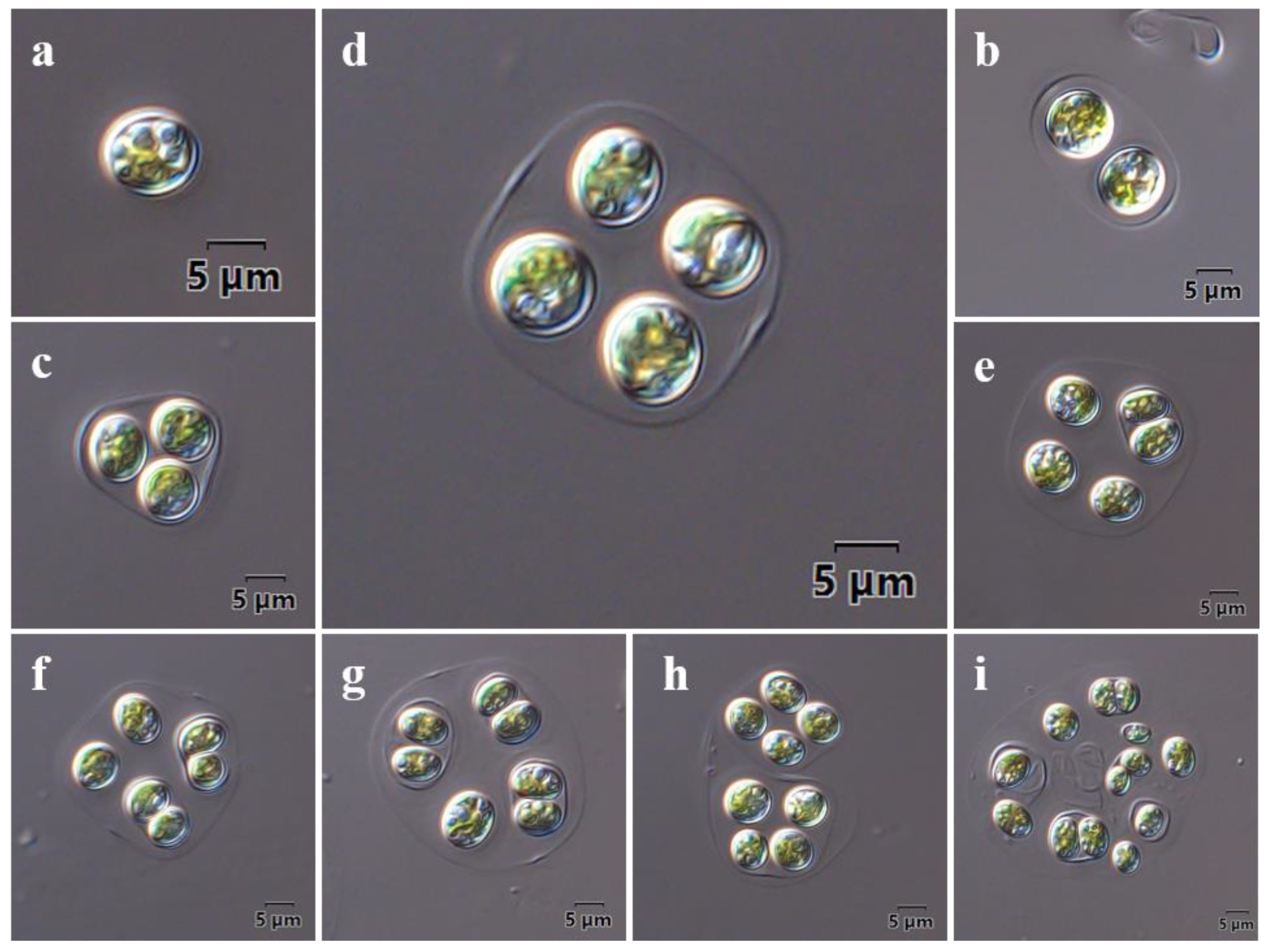
2.1. Morphological Observations

Cells are solitary or in 2–16 cell colonies within a thin, hyaline mucilaginous envelope or within an expanded mother cell wall (Figure 1). Cells are round, oval, or slightly reniform, ranging in size from 11.3 to 16.6 μm long and 6.3 to 10.3 μm wide. Cell walls are thick, sometimes stratified (Figure 1b,c), and smooth, without thickening polars. Mother cell walls are extended according to the number and arrangement of daughter cells and are usually an irregularly round shape, with or without tapered thickening ends (Figure 1b,d). The number and shape of chloroplasts are difficult to discern on the microscopic level. When observed with TEM, a horseshoe-shaped chloroplast was observed, which occupied most of the cell volume (Figure 2a,d). Asexual reproduction was by 2–4 autospores released by a rupture of the mother cell wall. These autospores could be propagated again independently inside the mother cell (Figure 1e,g). The asynchronous cell division led to the size difference between the daughter cells (Figure 1i) and the formation of multilayer mother cell walls (Figure 2e,f).
When observed with TEM, 1–4 pyrenoids were observed in the mature cells of Euchlorocystis marina (Figure 2a,d). The pyrenoid was surrounded by a starch sheath composed of 2–4 starch plates, and the pyrenoid matrix was homogenous (Figure 2a,d). However, in some young cells, pyrenoids were not observed (Figure 2b,c).

2.2. Molecular Phylogeny

Sequencing of the 18S rDNA of the new strain produced a 1720 bp sequence, introns were excluded. The final 18S rDNA sequences alignment of 29 taxa with 1633 base positions were used for the phylogenetic tree construction, and Prasiola mexicana (Prasiolaceae) was chosen for the outgroup. The ML and NJ analyses yielded a similar topology and only the ML tree was presented in Figure 3. The 18S rDNA phylogenetic trees consistently recovered that the newly isolated strain and Euchlorocystis subsalina formed an independent clade with high support (99/100 for ML/NJ) (Figure 3). Direct comparison showed that the 18S rDNA of the new strain had 41 differences (97.5% identity) to Euchlorocystis subsalina (Table 1).

3. Discussion

Members of the family Oocystaceae are common in freshwater, but they are rarely found in seawater. Currently, only a few marine species in this group are taxonomically accepted, such as Oocystis submarina, Oocystis marina, and Euchlorocystis subsalina. Some other species, due to their extensive adaptation, are also reported to exist in saline or semi-saline conditions [1,20,21,22,23,24], but they are taxonomically considered freshwater species. In this study, a new member of this family was found in a mariculture pond of shrimp in China. An integrative taxonomic study including morphological and phylogenetic analyses identified it as a new species, Euchlorocystis marina sp. nov., belonging to the genus Euchlorocystis (Oocystaceae).
The genus Euchlorocystis was originally erected by Liu et al. [17] in 2018. It has morphology similar to the well-known genus Oocystis. As a type of the family Oocystaceae, Oocystis has always been controversial in taxonomy. Komárek and Fott [8] listed 28 species with an additional 16 species designated as incompletely described or difficult to identify. Hindák [25] assigned to the genus Oocystis only those forms without a pyrenoid and listed eight species for the genus. Those forms included in the genus Oocystis with a pyrenoid were classified in the genus Oocystella [26]. The chloroplasts of Euchlorocystis mature cells have multiple pyrenoids, a feature that easily distinguishes them from Oocystis, Oocystella, and other Oocystaceae members without or with one pyrenoid. Oonephris species were described as having a central pyrenoid and sometimes more than one pyrenoid within the chloroplast [8]. According to Liu et al. [17], Euchlorocystis was morphologically different from Oonephris in terms of chloroplast shape and layered cell walls. We believe that the asynchronous cell division present in Euchlorocystis is also a distinctive feature to distinguish from Oonephris. Eremosphaera were also described as having more than one pyrenoid [8]. Liu et al. [17] suggested that Euchlorocystis and Eremosphaera could be distinguished by cell size and the number of chloroplasts per cell. In addition, the number of cells within colonies and whether the division of daughter cells within the mother cell is synchronized can also distinguish Euchlorocystis from Eremosphaera. In conclusion, the asynchronous cell division may be one of the most distinctive features for distinguishing Euchlorocystis from other taxa of the family Oocystaceae.
Euchlorocystis subsalina was originally described by Liu et al. [17] based on the materials collected from a semi-saline lake, Qinghai Lake. It is a type of the genus Euchlorocystis and is the only taxonomically accepted Euchlorocystis species reported currently. The new species presented here, Euchlorocystis marina, was collected from a seawater shrimp culture pond in Zhanjiang, China. It has a multilayer thick cell wall, multiple pyrenoids in the chloroplast, and usually 2–16 cells forming a colony in the extended mother cell wall. These features are morphologically similar to the genus Euchlorocystis, and are distinguished from other taxa of the family Oocystaceae. Most importantly, however, the 18S rDNA phylogenetic trees revealed that Euchlorocystis marina and Euchlorocystis subsalina formed an independent clade in Oocystaceae with robust support. However, horseshoe-shaped chloroplasts and rounder cells morphologically distinguish it from Euchlorocystis subsalina (Table 2). Apart from the morphology, the molecular results of this study also supports that they are distinct species. The discovery of Euchlorocystis marina may imply more hidden species of the genus Euchlorocystis that have yet to be discovered. More new species within this genus should be collected and sequenced in the future.
Taxonomic assessment
Euchlorocystis marina Dong, Li & Huang, sp. nov. (Figure 1a–i)
Diagnosis: Cells round, oval, or slightly reniform, ranging in size from 6.9–12.3 μm long and 4.3–10.7 μm wide. Cell wall thick, sometimes stratified, without thickening polars. Mother cell walls were usually an irregularly round shape, with or without tapered thickening ends. Single horseshoe-shaped chloroplast with 1–4 pyrenoids within mature cells. Asexual reproduction by 2–4 autospores. Daughter cells have asynchronous cell division.
Holotype: Formaldehyde-fixed material was stored at the Algae Resource Development and Culture Environment Ecological Remediation Laboratory of Guangdong Ocean University, Zhanjiang, China, as specimen No. HXH1. The population was partially illustrated here in LM (Figure 1a–i) and TEM (Figure 2a–f).
Reference strain: A living culture was deposited in the Algae Resource Development and Culture Environment Ecological Remediation Laboratory of Guangdong Ocean University, Zhanjiang, China, as specimen GDOU-406.
Type locality: Xuwen seawater shrimp culture pond (20°88′ N, 109°74′ E; salinity 29‰; water temperature = 30.7 °C; pH = 8.12) in Zhanjiang, China. Water samples were collected in August 2016.
Etymology: The species was named for its habitat of seawater.

4. Materials and Methods

4.1. Strains and Culture Conditions

Euchlorocystis marina was sampled at a seawater shrimp culture pond (20°88′ N, 109°74′ E, salinity 29‰, strain GDOU-406) in Zhanjiang, Guangdong Province, China, and maintained in the Algae Resource Development and Culture Environment Ecological Remediation Laboratory of Guangdong Ocean University (Zhanjiang, China). The strain was isolated using the serial dilution pipetting technique (Hoshaw and Rosowski, 1973) until single colonies were obtained. Then, dividual colonies were cultivated in a seawater medium (Zhanshui 107–13) at pH = 8.0 with the following nutrient compositions: KH2PO4 (8 mg·L−1), NaNO3 (80 mg·L−1), ferric citrate (50 mg·L−1). The cultures were carried out in a 500 mL Erlenmeyer flask containing 300 mL medium at 25±1 °C and 30 μmol·m−2·s−1 of continuous light (T8 LED lamps, white light).

4.2. Morphological Observation

Microscopic observation and photographing of algal cells were performed using an OLYMPUS BX53 Light microscope.
For ultrastructure, algal cells were collected by centrifugation (3000 rpm, 5 min) and washed three times with PBS buffer. Then, the cells were fixed with 2.5% glutaraldehyde solution for 2 h at 4 °C, and post-fixed with 1% OsO4 in 0.1 M phosphate buffer for 2 h at 4 °C. The fixed materials were dehydrated with a graded acetone series and embedded in Spurr resin. Uranium acetate and lead citrate were used to stain the final sections. The samples’ slice preparation and staining were performed by Guangdong Medical University (Zhanjiang, China). The algal slices were examined and photos taken by a JEOL JEM-1400 transmission electron microscope.

4.3. Molecular Analyses

Total genomic DNA of algae was extracted with the DNeasy Plant Mini Kit (Qiagen, Germany) following the manufacturer’s protocol. The PCR cycling conditions were as follows: 5 min initial denaturation at 95 °C; 35 cycles of denaturation at 95 °C for 30 s, annealing at 55 °C for 30 s, and extension at 72 °C for 40 s; a final extension of 10 min at 72 °C. The 18S rDNA was amplificated using Echl18SF (ACTGTGAAACTGCGAATGG) and Echl18SR (TAGGTGGGAGGGTTTAGG) primers. The PCR products were sequenced by Shanghai SaiHeng Biotechnology Co., Ltd., Shanghai, China. The assembled sequences were submitted to GenBank under the accession number OM413748.
The new 18S rDNA sequences were aligned with gene sequences downloaded from GenBank of 28 representative species. Phylogenies were estimated by Clustalx 1.8 and Mega 5.0 software with maximum likelihood (ML) and neighbor-joining (NJ) methods. Kimura 2-parameter model was selected as best-fit model. Bootstrap analysis was performed with 1000 replicates of the dataset to estimate statistical reliability.

Author Contributions

Data curation, F.L. and M.D.; methodology, F.L., N.Z., Y.Z. and Q.L. (Qianru Li); investigation, F.L., M.D., Z.Q., Q.L. (Qingsheng Lian) and J.L.; project administration, C.L. and X.H.; resources, C.L. and X.H.; writing—original draft, F.L. and M.D.; writing—review and editing, F.L. All authors have read and agreed to the published version of the manuscript.

Funding

This research was mainly supported by the National Key R & D Plan “Blue Granary Science and Technology Innovation” (grant number 2020YFD0902005) and the Guangdong Provincial Key Area Research and Development Plan Projects (grant number 2020B0202010009); National Natural Science Foundation of China (grant number 32102796); Natural Science Foundation of Guangdong Province (grant number 2020A1515110086); Program for Scientific Research Start-up Funds of Guangdong Ocean University (grant number 060302022102 and R18020).

Institutional Review Board Statement

Not applicable.

Data Availability Statement

The strain used in this study was collected from a seawater shrimp culture pond (20°88′ N, 109°74′ E; salinity 29‰; water temperature = 30.7 °C; pH = 8.12) in Xuwen, Zhanjiang, China. A living culture and formaldehyde-fixed material were stored at the Algae Resource Development and Culture Environment Ecological Remediation Laboratory of Guangdong Ocean University, Zhanjiang, China. The 18S rDNA sequences of this strain has been submitted to the GenBank under the accession number OM413748.

Conflicts of Interest

The authors declare no conflict of interest.

References

  1. Liu, M.; Huang, X.H.; Zhang, R.; Gu, B.H. Uptake of urea nitrogen by Oocystis borgei in prawn (Litopenaeus vannamei) aquaculture ponds. Bull. Environ. Contam. Toxicol. 2018, 101, 586–591. [Google Scholar] [CrossRef]
  2. Ajala, S.O.; Alexander, M.L. Assessment of Chlorella vulgaris, Scenedesmus obliquus, and Oocystis minuta for removal of sulfate, nitrate, and phosphate in wastewater. Int. J. Energy Environ. Eng. 2020, 11, 311–326. [Google Scholar] [CrossRef] [Green Version]
  3. Takeuchi, T.; Utsunomiya, K.; Kobayashi, K.; Owada, M.; Karube, I. Carbon dioxide fixation by a unicellular green alga Oocystis sp. J. Biotechnol. 1992, 25, 261–267. [Google Scholar] [CrossRef]
  4. Huang, X.H.; Li, C.L.; Liu, C.W.; Zeng, D.S. Study on the ecological factors of Oocystis borgei. J. Zhanjiang Ocean. Univ. 2002, 22, 8–12. (In Chinese) [Google Scholar]
  5. Wang, X.Q.; Zhang, Y.L.; Li, C.L.; Huang, X.H.; Li, F.; Wang, X.Y.; Li, G. Allelopathic effect of Oocystis borgei culture on microcystis aeruginosa. Environ. Technol. 2020, 1–10. [Google Scholar] [CrossRef] [PubMed]
  6. Chuka-ogwude, D.; Ogbonna, J.; Borowitzka, M.A.; Moheimani, N.R. Screening, acclimation and ammonia tolerance of microalgae grown in food waste digestate. J. Appl. Phycol. 2020, 32, 3775–3785. [Google Scholar] [CrossRef]
  7. Vidyashankar, S.; Venugopal, K.S.; Swarnalatha, G.V.; Kavitha, M.D.; Chauhan, V.S.; Ravi, R.; Bansal, A.K.; Singh, R.; Pande, A.; Ravishankar, G.A.; et al. Characterization of fatty acids and hydrocarbons of Chlorophycean microalgae towards their use as biofuel source. Biomass Bioenergy 2015, 77, 75–91. [Google Scholar] [CrossRef]
  8. Komárek, J.; Fott, B. Chlorophyceae (Grünalgen) Ordnung: Chlorococcales. In Das Phytoplankton des Süßwassers: Systematik und Biologie; Huber-Pestalozzi, G., Ed.; Schweizerbart’sche Verlagsbuchhandlung: Stuttgart, Germany, 1983; Volume 7, pp. 1–1044. [Google Scholar]
  9. Silva, T.G.; Štenclová, L.; Archanjo, N.C.P.; Bagatini, I.L. Revised phylogenetic position of Nephrocytium nägeli (Sphaeropleales, Chlorophyceae), with the description of Nephrocytiaceae fam. nov. and Nephrocytium vieirae sp. nov. Taxon 2021, 70, 917–930. [Google Scholar] [CrossRef]
  10. Hepperle, D.; Hegewald, E.; Krienitz, L. Phylogenetic position of the Oocystaceae (Chlorophyta). J. Phycol. 2000, 36, 590–595. [Google Scholar] [CrossRef]
  11. Pažoutová, M.; Škaloud, P.; Nemjová, K. Phylogenetic position of Ooplanctella planoconvexa, gen. et comb. nova and Echinocoleum elegans (Oocystaceae, Trebouxiophyceae, Chlorophyta). Fottea 2010, 10, 75–82. [Google Scholar] [CrossRef] [Green Version]
  12. Krienitz, L.; Bock, C. Elongatocystis ecballocystiformis gen. et comb. nov., and some reflections on systematics of Oocystaceae (Trebouxiophyceae, Chlorophyta). Fottea 2011, 11, 271–278. [Google Scholar] [CrossRef] [Green Version]
  13. Xia, S.; Zhu, H.; Cheng, Y.Y.; Liu, G.X.; Hu, Z.Y. Phylogenetic position of Ecballocystis and Ecballocystopsis (Chlorophyta). Fottea 2013, 13, 65–75. [Google Scholar] [CrossRef] [Green Version]
  14. Štenclová, L.; Fučíková, K.; Kaštovský, J.; Pažoutová, M. Molecular and morphological delimitation and generic classification of the family Oocystaceae (Trebouxiophyceae, Chlorophyta). J. Phycol. 2017, 53, 1263–1282. [Google Scholar] [CrossRef] [PubMed]
  15. Liu, X.; Zhu, H.; Liu, B.; Liu, G.; Hu, Z. Classification of Planctonema-like algae, including a new genus Planctonemopsis gen. nov., a new species Planctonema gelatinosum sp. nov. and a reinstated genus Psephonema (Trebouxiophyceae, Chlorophyta). J. Phycol. 2017, 53, 869–879. [Google Scholar] [CrossRef] [PubMed]
  16. Liu, X.; Zhu, H.; Song, H.; Liu, B.; Wang, Q.; Liu, G.; Hu, Z. Quadricoccopsis gen. nov., a new genus of Quadricoccus-like algae in Oocystaceae from China (Trebouxiophyceae, Chlorophyta). Fottea 2018, 18, 189–199. [Google Scholar] [CrossRef]
  17. Liu, X.; Zhu, H.; Song, H.; Wang, Q.; Xiong, X.; Wu, C.; Liu, G.; Hu, Z. Euchlorocystis gen. nov. and Densicystis gen. nov., two new genera of Oocystaceae algae from high-altitude semi-saline habitat (Trebouxiophyceae, Chlorophyta). J. Eukaryot. Microbiol. 2018, 65, 200–210. [Google Scholar] [CrossRef]
  18. Liu, X.; Wang, Q.; Zhu, H.; Liu, B.; Rindi, F.; Liu, G.; Xie, S.; Hu, Z. Reticulocystis yunnanense gen. et sp. nov., a new member of freshwater Oocystaceae algae (Trebouxiophyceae, Chlorophyta). Eur. J. Phycol. 2020, 55, 507–516. [Google Scholar] [CrossRef]
  19. Guiry, M.D.; Guiry, G.M. AlgaeBase. World-Wide Electronic Publication, National University of Ireland, Galway. Available online: https://www.algaebase.org (accessed on 25 October 2021).
  20. Campos, H.; Soto, D.; Parra, O.; Steffen, W.; Aguero, G. Limnological studies of Amarga lagoon, Chile: A saline lake in Patagonian South America. Int. J. Salt Lake Res. 1995, 4, 301–314. [Google Scholar] [CrossRef]
  21. Wen, Z.; Zhi-Hui, H. Biological and ecological features of inland saline waters in North Hebei, China. Int. J. Salt Lake Res. 1999, 8, 267–285. [Google Scholar] [CrossRef]
  22. Ramírez-Olvera, M.A.; Alcocer, J.; Merino-Ibarra, M.; Lugo, A. Nutrient limitation in a tropical saline lake: A microcosm experiment. Hydrobiologia 2009, 626, 5–13. [Google Scholar] [CrossRef]
  23. Chen, X.; Yang, J.; Chen, Y.; Hou, H. Seasonal dynamics of phytoplankton and its relationship with environmental factors of a Chinese Lake. Pol. J. Environ. Stud. 2016, 25, 1427–1433. [Google Scholar] [CrossRef]
  24. Huang, X.; Li, X.; Wang, Y.; Zhou, M. Effects of environmental factors on the uptake rates of dissolved nitrogen by a salt-water green alga (Oocystis borgei Snow). Bull. Environ. Contam. Toxicol. 2012, 89, 905–909. [Google Scholar] [CrossRef] [PubMed]
  25. Hindák, F. Studies of the Chlorococcal algae (Chlorophyceae) IV. In Biologické Pracé; Ruzicka, M., Ed.; Veda: Bratislava, Slovakia, 1988; Volume 34, pp. 1–263. [Google Scholar]
  26. Stoyneva, M.P.; Cocquyt, C.; Gärtner, G.; Vyverman, W. Oocystis lacustris CHOD. (Chlorophyta, Trebouxiophyceae) in Lake Tanganyika (Africa). Linz. Biol. Beitr. 2007, 39, 571–632. [Google Scholar]
Figure 1. Light microscopy of Euchlorocystis marina. (a) Solitary cell; (b) 2-celled colony; (c) 3-celled colony; (d) 4-celled colony; (e) 5-celled colony; (f) 6-celled colony; (g) 7-celled colony; (h) 8-celled colony; (i) 13-celled colony.
Diversity 14 00119 g001
Figure 2. Transmission electron microscopy of Euchlorocystis marina sp. nov. (a,d) Mature cells; (b,c) Young cells; (e,f) Ultrastructure of multilayer mother cell wall. (CW = cell wall; C = chloroplast; P = pyrenoid; S = starch grains; ST = starch sheath; N = nucleus). The mother cell walls are indicated by black arrow.
Diversity 14 00119 g002
Figure 3. Maximum likelihood tree of 18S rDNA sequences. Bootstrap support from ML and NJ posterior probabilities are presented on the nodes. The sequence obtained in this study is shaded gray. The cell morphology picture of Euchlorocystis subsalina was from Liu et al. [17].
Diversity 14 00119 g003
Table 1. Direct comparison of 18S rDNA sequences of Euchlorocystis marina (bold) with some species of the Oocystaceae family. Numbers below the diagonal indicate nucleotide substitutions between a pair of sequences and those above the diagonal are percent identities. Black boxed area indicates the high sequence similarities between the two closest related species.
Positions Compared: 1633.Accession NumberEremo. viridisEcball. dichotomusOocystis. marinaEuchloro. subsalinaEuchloro. marinaOocystella. oogamaOonephris. obesa
Eremosphaera viridisAF38715490.0%94.6%94.5%92.6%93.9%87.6%
Ecballocystopsis dichotomusJX01818416390.4%90.3%88.1%90.6%82.7%
Oocystis
marina
MF1007848815796.7%94.7%95.8%86.6%
Euchlorocystis subsalinaMF100785901585497.5%95.8%86.8%
Euchlorocystis marinaOM413748121194874194.1%84.6%
Oocystella
oogama
KM02008010015469699686.5%
Oonephris
obesa
KY006558202283219216251220
Table 2. Morphological comparison between Euchlorocystis subsalina and Euchlorocystis marina.
Morphology CharacterEuchlorocystis subsalinaEuchlorocystis marina
Cell shapeOval to elongated elliptical with round ends round and no thickeningsRound, oval, or slightly reniform without thickening polars
Cell size11.3–16.6 μm long and 6.3–10.3 μm wide6.9–12.3 μm long and 4.3–10.7 μm wide
Cell arrangementSolitary, 2–16 cell coloniesSolitary, 2–16 cell colonies
Mucilage envelopment Lemma- to square-shapeUsually irregular round shape, with or without gelled and thickened poles.
Cell wallThick, layered.Thick, layered.
Chloroplasts numberSingleSingle
Chloroplast shapeWide trough shape, parietalHorseshoe-shaped
Pyrenoids number per chloroplast2–61–4, within mature cells
Cell reproductionPropagation by 2–4 autosporesAsexual reproduction by 2–4 autospores; There exist the asynchronous division in cell colonies
Reference[17]This study
Publisher’s Note: MDPI stays neutral with regard to jurisdictional claims in published maps and institutional affiliations.

