Article

Trichotorquatus salinus sp. nov. (Oculatellaceae, Cyanobacteria) from a Saltern of Gomso, Republic of Korea

1
Department of Life Science, College of Natural Science, Kyonggi University, Suwon 16227, Republic of Korea
2
Protist Research Team, Microbial Research Department, Nakdonggang National Institute of Biological Resources, Sangju 37242, Republic of Korea
3
Division of Life Science, Gyeongsang National University, Jinju 52828, Republic of Korea
*
Authors to whom correspondence should be addressed.
Diversity 2023, 15(1), 65; https://doi.org/10.3390/d15010065
Submission received: 28 November 2022 / Revised: 23 December 2022 / Accepted: 28 December 2022 / Published: 5 January 2023

Abstract

:
Six strains of subaerial cyanobacteria were isolated from a Gomso saltern in the Republic of Korea, all of which were studied using morphological and molecular traits. Trichotorquatus salinus sp. nov. was studied using a light microscope (LM), transmission electron microscope (TEM), 16S rRNA, 16S–23S ITS region, and ecological data. T. salinus is a thin and simple filament with a false branch and a firm collar sheath. The phylogenetic analyses of 16S rRNA revealed that Trichotorquatus formed a monophyletic lineage and the strains of T. salinus formed a distinct clade among the species in the genus Trichotorquatus. In the statistical analysis, the inter-species genetic distance of the five species of Trichotorquatus, including T. salinus, is shown to be greater than the distance of the previously reported species of Trichotorquatus. Additionally, 16S–23S ITS gene sequences between T. salinus and four species of Trichotorquatus showed dissimilarities of 55.3–59.4%. In the secondary structure of 16S–23S ITS region (type 2 operon), D1–D1′, Box-B, and V3 helix of T. salinus were different from the other taxa in the genus Trichotorquatus. These results demonstrate that T. salinus sp. nov. has unique morphological, ecological, and molecular traits. Therefore, we propose that T. salinus sp. nov. is a novel species belonging to the genus Trichotorquatus.

1. Introduction

The order of Synechococcales includes coccoid and bacilloid unicellular and colonial genera, even the Pseudanabaenales of simple filamentous form, making it more complicated because genera having various forms are mixed [1]. Mai et al. [1] have suggested that the group formerly classified as Leptolyngbyaceae could be divided into four family-level clades through the phylogenetic analysis of 16S rRNA gene sequences. Two of the four family-level clades were previously recognized families Leptolyngbyaceae and Prochlorotrichaceae. The remaining two families were newly named as the Oculatellaceae and Trichocoleaceae. There are six newly identified genera in the family Oculatellaceae, including the genus Trichotorquatus by Pietrasiak and Johansen [2], and fourteen new species were newly described in these genera [1]. Moreover, recent studies by Strunecký et al. [3] have included the family Oculatellaceae in the new order Oculatellales.
The genus Trichotorquatus of the family Oculatellaceae is one of the problematic groups in cyanobacteria taxonomy [1,4] because the genus Trichotorquatus is morphologically very similar to Leptolyngbya sensu stricto [2]. The morphological taxonomic keys of Trichotorquatus are indistinguishable from several genera, such as Drouetiella by Mai, Johansen, and Pietrasiak [1]; Plectolyngbya by Taton, Wilmotte, Šmarda, Elster, and Komárek [5]; and Tildeniella by Mai, Johansen, and Pietrasiak [1] of the family Leptolyngbyaceae, a thin filamentous cyanobacterium which has been previously reported [1,5]. Moreover, except for the type species, Trichotorquatus maritimus by Pietrasiak, Reeve, and Johansen [2], which has slight morphological differences (e.g., wider trichomes with more robust, firmer, and more widened sheaths than other species), the other species are cryptic species that are morphologically indiscernible from each other [2]. Because of this morphological ambiguity, Trichotorquatus species were distinguished from each other through molecular analysis and distinctive ecological types, as in other recent studies, applying the polyphasic approach to taxonomy [6,7,8]. For instance, although Trichotorquatus andrei by Pietrasiak, and Johansen [2] and Trichotorquatus ladouxae by Pietrasiak and Johansen [2] were morphologically and genetically (16S rRNA) similar, they were designated as different species based on their distinctive ecological characteristics [2]. Moreover, novel genera and species, such as Alkalinema by Vaz et al. [9]; Cephalothrix by Malone et al. [10]; Halotia by Genuário et al. [11]; Monilinema by Malone, Genuário, Vaz, Fiore, and Sant’Anna [12]; and Pantanalinema by Vaz et al. [9], have been identified using ecology data [9,10,11,12].
Cyanobacteria form a dense mat that are adapted in saline lakes, salt ponds, high salinity lagoons, and saline biocrusts [13,14,15,16]. There is a high possibility of discovering new taxa that are adapted in unique high-salinity environments [13,16,17]. Compared to other environments, relatively few studies have investigated the diversity and distribution of cyanobacteria in saline–alkaline habitats [12,18,19,20]. Therefore, exploring new species in a hypersaline environment is worthwhile in order to decipher the patterns of diversity.
In this study, the unialgal cultures were established by isolating six strains of thin filamentous cyanobacteria living on salt piles in Gomso saltern in the Republic of Korea. Morphological traits were measured by using the LM and TEM. The phylogenetic analyses using 16S rRNA and ITS regions were conducted, and the secondary structures were compared to the previously reported species. In this context, the Korean Trichotorquatus species had unique genetic and ecological features compared to its close relatives. Therefore, we propose a new species belonging to the genus Trichotorquatus, family Oculatellaceae.

2. Materials and Methods

2.1. Sample Collections and Cultures

A natural sample was collected from the subaerophytes (tile surface) in Gomso saltern of the Republic of Korea on October 18, 2020 (35°35′44.8″ N 126°36′44.1″ E) (Figure 1). These subaerophytic cyanobacteria were obtained by scraping the tile surface with a soft brush or sterilized spatula [21]. The collected sample was kept at 4 °C and transported to the lab for deposition at Algal Culture Collection of Kyonggi University (ACKU).
For unialgal culture, a single trichome was separated in a 24-well plate (SPL, Pocheon, Republic of Korea) with a Pasteur’s pipette (Hilgenberg GmbH, Mansfeld, Germany) under a light microscope (LM). After one to two weeks of culture on the 24-well plate, the unialgal cultured trichome was tested for contamination and moved to a 50 mL cell culture flask (SPL, Pocheon, Republic of Korea) for mass culture [22]. BG-11 liquid medium was used for unialgal and mass culture. Synthetic culture was carried out at a temperature of 20–25 °C, a photoperiod of 16h:8h (light:dark), and an illumination of 25 μmol photons m−2 s−1. To observe the morphology of colony cohesion, the culture was placed in a shaking incubator. In the process of the mass culture, a diluted oligotroph of 1 × 10⁻¹ BG-11 medium was used to observe the trichomes, the variety of cells, and the mucilaginous sheath.
The reference strain for species has been deposited at Freshwater Bioresources Culture Collection (FBCC, https://fbp.nnibr.re.kr/fbcc/, accessed on 28 August 2021) at the Nakdonggang National Institute of Biological Resources (NNIBR) of the Republic of Korea, with an accession number of FBCC-A1483–1488. The inoculated subculture of the reference strain (FBCC-A1483) was preserved in 4% (v/v) formaldehyde and deposited in the Herbarium at the National Institute of Biological Resources (KB, https://species.nibr.go.kr/, accessed on 22 September 2022) (culture aliquot NIBRCY0000001649).

2.2. Morphological Analysis and Characterization

The collected subaerophytic environmental sample was treated with BG-11 liquid medium for hydration and observed with a light microscope under 100–1000 × magnifications (Olympus BX53) (Olympus, Tokyo, Japan). A unialgal cultured strain was photographed under 100–1000 × magnifications (Olympus UC-90) (Olympus, Tokyo, Japan) and the ultrastructure of the cell was observed and photographed with a transmission electron microscope (TEM). The samples required for the TEM photograph were fixed with 2% glutaraldehyde and 2% paraformaldehyde in phosphate buffer (pH 7.4) at 4 °C for 1 h and then postfixed with 2% osmium tetroxide and 3% potassium hexacyanoferrate at 4 °C for 40 min and were dehydrated in a graded series of ethanol and embedded into LR white resin. Afterwards, the Ultra-thin (80 nm) sample sections were prepared using an Ultra-cut microtome (Leica Co., Greenwood Village, CO, USA) and placed on a coated square copper grid. The final samples were stained with uranyl acetate and lead citrate. The TEM images were taken at 120 kV using a field emission electron microscope (JEM-2100F) (Jeol, Tokyo, Japan) coupled with a OneView camera (Gatan, Pleasanton, CA, USA) at the Korean Basic Science Institute (KBSI), Chuncheon, Republic of Korea [23].
For the identification of cyanobacteria, we used the taxonomic classification system of the cyanobacteria of Komárek et al. [3], referred to the AlgaeBase on 23 August 2022 [24] and compared with results of Pietrasiak et al. [2].

2.3. DNA Extraction, PCR, and Sequencing

Exponential phase cells were harvested via centrifugation (10,080× g). Pellets were then used for gDNA extraction using an i-genomic Plant DNA Extraction Mini Kit (iNtRON, Daejeon, Republic of Korea), according to the manufacturer’s instructions.
The PCR reactions of 16S–23S rRNA genes were conducted using a set of bacterial universal forward primer (27F) and a previously designed 23S reverse primer (CY-23R600) [25]. PCR amplification was carried out using a Maxime™ i-Stars Taq PCR premix (iNtRON, Daejeon, Republic of Korea) in a total volume of 20 μL, including 17 μL of sterile distilled water, 1 μL of each primer (10 pmoles), and 1 μL of template DNA. The gDNA was amplified in a Mastercycler gradient (Eppendorf, Hamburg, Germany) using the following cycling conditions: 95 °C for 5 min followed by 35 cycles of 94 °C for 20 s, 55 °C for 30 s, and 72 °C for 90 s, with a final extension at 72 °C for 10 min. Positive and negative controls were used for all amplification reactions. Resulting PCR products were subjected to electrophoresis in a 1.0% agarose gel and visualized under ultraviolet light on a transilluminator. Amplified PCR products were purified with a MEGAquick-spin™ Plus Fragment DNA Purification Kit (iNtRON, Daejeon, Republic of Korea) and sent to a commercial Sanger sequencing service (Macrogen, Seoul, Republic of Korea). DNA sequencing reactions were run with an ABI PRISM® BigDye™ Terminator Cycle Sequencing Ready Reaction Kit (PE Biosystems, Foster City, CA, USA). Primers used for sequencing included bacterial universal primers 27F, 518F, 800R, 1492R, 1241F [26], 23S30R [27], and CY-23R600. Sequencing was performed with an automated DNA sequencer (Model 3730XL) (Applied Biosystems, Foster City, CA, USA). All newly determined 16S rRNA and 16S–23S ITS gene sequences were deposited in GenBank (NCBI, https://www.ncbi.nlm.nih.gov, accessed on 30 September 2022) under accession number of OP058661–OP058666.

2.4. Phylogenetic Analyses and Molecular Distance Analyses

The phylogenetic analyses of 16S rRNA and 16S–23S ITS regions of six strains were conducted using software MEGA X [28], including the sequences of closely related taxa obtained from GenBank. Maximum Likelihood (ML) analyses of the 16S rRNA region was conducted using the program RAxML 7.0.3 with the default GTRGAMMA model [29]. Furthermore, 200 independent tree inferences were used to identify the best tree. ML bootstrap values were determined using 1000 replicates. Bayesian Inference (BI) was conducted using MrBayes v.3.2.7 [30] with the default GTR + G + I model to determine the best available model for the data from each region. Four independent Markov Chain Monte Carlo (MCMC) runs were performed, and 4,000,000 generations were carried out with a sampling frequency of one per 100 generations. After this analysis, the first 23,250 trees were eliminated as burn-in. The point was identified by the average standard deviation of split frequencies (<0.007) between runs. Then, the consensus tree was constructed.
For the 16S–23S ITS region, alignment was conducted using ClustalW with consideration of conserved domains. Because our strains had similar sequences with type 2 operon [2], only strains with type 2 operon were used for alignment. An unrooted tree was obtained by conducting a BI analysis with 2,000,000 generations, discarding the first 9100 samples whose average standard deviation of split frequencies between runs was greater than 0.007. The average standard deviation of split frequencies was 0.004445, and the average potential scale reduction factor (PSRF) was 1.000 (the maximum PSRF for parameters = 1.002). Moreover, Maximum Parsimony (MP) was carried out using MEGA X, and the bootstrap value was found by repeating the process × 1000 times. The MP tree was obtained using the Subtree–Pruning–Regrafting (SPR) algorithm. Phylogenetic trees were visualized in FigTree 1.4.4 (GitHub) and re-drawn in Adobe Illustrator CS5.1 (Adobe Systems, San Jose, CA, USA). Bayesian posterior probabilities (BPP > 0.50) and ML or MP bootstrap value (>50%) were indicated at each branch node in the Bayesian tree. Molecular genetic divergences among Trichotorquatus species were calculated as pairwise genetic distance (p-distance) with the Kimura 2-parameter model in MEGA X.

2.5. ITS Structure Analyses

The secondary structure of the 16S–23S ITS region was inferred by considering the study of Pietrasiak et al. [2]. All tRNA genes were predicted using the tRNAscan-SE On-line [31]. Putative secondary structures were constructed using a web-based software Mfold [32] and re-drawn in PseudoViewer3 [33] for easy comparison with available structures from relative taxa.

3. Results

3.1. Taxonomic Treatment and Morphological Characterization

Based on the results of polyphasic analysis, we propose the following description of the new cyanobacterial taxon under the provisions of the ICN (International Code of Nomenclature for algae, fungi, and plants) [34].
Trichotorquatus salinus N. Lee, D. Kim, A. Lim, and O. Lee sp. nov. (Figure 2 and Figure 3).
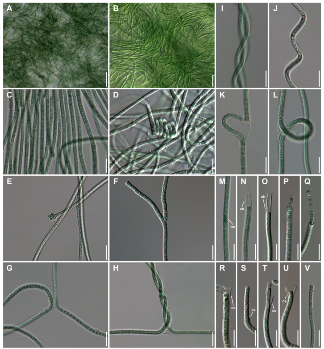
Description: Filaments unbranched, Scytonema-type or Tolypothrix-type false branching sometimes observed, with a single trichome in each sheath, 2.1–3.4 μm wide. Sheath typically thin, sometimes frayed, firm and rigid, sometimes absent, 0.1–0.9 μm thick when present, often has a within bulging, long or short collar. Trichomes distinctly constricted at the crosswalls, fragmenting at necridia or at simple separating cell divisions, regularly or loosely spiral, 1.9–2.9 μm wide. Cells usually isodiametric, sometimes longer than wide, with thylakoids either parietal or in strand-like fascicles extending longitudinally through the centroplasm, 2.0–3.0 μm long. Apical cell blue-green, rarely tangled, 2.2–4.3 μm long. Necridia often in rows, forming sections up to 5 μm long. Hormogonia short, typically under 10 cells long.
Diagnosis: Species show morphological resemblance to the species of genus Trichotorquatus, but distinguishable differences are evident (Table 1), and they also differ from other species of genus Trichotorquatus by 16S rRNA gene sequences. Furthermore, the distinctive structures of D1–D1′, Box-B, and V3 helices from the 16S–23S rRNA ITS region are evidence of a new species.
Etymology: salinus, “sal-” means salt, living in the hypersaline water in saltern. Specific name according to the character of the habitat.
Holotype (designated here): A formaldehyde fixed specimen, NIBRCY0000001649 in the Herbarium at the National Institute of Biological Resources (KB), from reference strain FBCC-A1483.
Reference strain: FBCC-A1483.
Additional strains: FBCC-A1484, FBCC-A145, FBCC-A146, FBCC-A147, and FBCC-A1488.
Type locality: Gomso saltern of the Republic of Korea (35°35′44.8″ N 126°36′44.1″ E).
Habitat: On tile, sometimes slightly covered by shallow layers of water (in saline, >50‰).
Gene sequences: 16S rRNA, 16S–23S ITS, and 23S rRNA gene sequences with the GenBank accession numbers of OP058661–OP058666, respectively.

3.2. 16S rRNA and Phylogenetic Affiliation of Trichotorquatus salinus

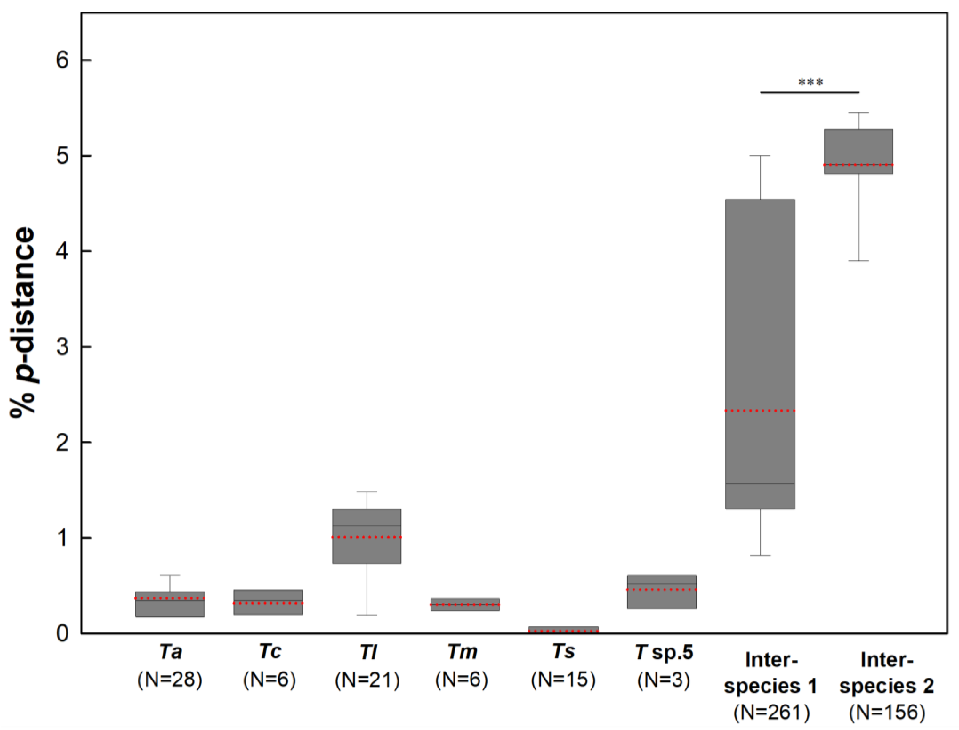
Sequences of 16S rRNA of T. salinus were 1404 bp for FBCC-A1483, FBCC-A1484, FBCC-A1486, FBCC-A1487, and FBCC-A1488, but 1405 bp for FBCC-A1485. Except for the 1 bp-insertion in strain FBCC-A1485, other sequences were identical. When compared to other species in the genus Trichotorquatus using the p-distance score calculated with the Kimura 2-parameter model, the genetic diversity among strains of T. salinus was considerably lower than that of other Trichotorquatus species (Figure 4). The average % p-distance of the 16S rRNA gene sequences in T. salinus was 0.024, while those in T. andrei, Trichotorquatus coquimbo by Osorio-Santos, Pietrasiak, and Johansen [2], T. ladouxae, T. maritimus, and Trichotorquatus sp. 5 were 0.371, 0.317, 1.007, 0.303, and 0.461, respectively (Figure 4). Average % p-distances among species T. andrei, T. coquimbo, T. ladouxae, T. maritimus, and Trichotorquatus sp. 5 (i.e., inter-species 1) were significantly lower than those between T. salinus and other Trichotorquatus species (i.e., inter-species 2) (t-test, p < 0.001, Figure 4). The mean % p-distance in the inter-species 1 was 2.332, while that in the inter-species 2 was 4.906 (Figure 4). Additionally, the percent similarity [100 × (1−p)] of the 16S rRNA region of T. salinus with other Trichotorquatus species ranged from 94.5% to 96.3%.
In the phylogenetic tree based on 16S rRNA gene sequences, the Trichotorquatus species, including T. salinus formed a monophyletic clade with highly supported values (BPP = 1.00 and ML bootstrap = 100%) (Figure 5). T. salinus strains formed a distinct clade with Trichotorquatus sp. 5 strains, while the other T. andrei, T. coquimbo, T. ladouxae, and T. maritimus formed a clade together. Moreover, a clade containing T. salinus strains was clearly divergent from the clade consisting of Trichotorquatus sp. 5 strains.

3.3. Characterization of the 16S–23S ITS Region of Trichotorquatus salinus

The complete 16S–23S ITS region of T. salinus FBCC-A1483 was 289 bp (46.0% GC). In the ITS region, tRNAAla and tRNAIle were not present in the species. V2 helices were not found either. The sequences of the 16S–23S ITS region of the other four strains of T. salinus were identical to those of FBCC-A1483 but not FBCC-A1485. Our T. salinus strains shared the start sequence of the 23S rRNA gene (5′-GGTCAAGCGAATAGT-3′) with other species known to have type 2 operon.
The predicted 16S–23S ITS region secondary structure included D1–D1′, Box-B, and V3 helices. It was compared with the genus Trichotorquatus previously studied by Pietrasiak et al. [2] (Figure 6 and Figure 7).
The total base number of D1–D1′ helix from T. salinus FBCC-A1483 was 76 bases. It was slightly shorter than other species belonging to the genus Trichotorquatus. The D1–D1′ helix of the T. salinus FBCC-A1483 showed structures different from other species in the genus Trichotorquatus. The basal stem of T. salinus FBCC-A1483 consisted of 5-bp helix (5′-GACCU-AGGUC-3′). It was the same as that of T. andrei CMT-3SWIN-NPC29, T. ladouxae WJT40-NPBG3, or T. maritimus SMER-A, but different from that of T. coquimbo ATA2-1-KO25A (5′-GACCUU-AAGGUC-3′). In addition, a 4-bp terminal hairpin (5′-GCAA-3′) of T. salinus FBCC-A1483 showed differences from all species belonging to genus Trichotorquatus.
The total length of Box-B helix was 44 bases. The basal stem consisted of 5-bp helix (5′-AGCAA-UUGCU-3′) in the T. salinus FBCC-A1483. The Box-B helix of T. salinus FBCC-A1483 was longer than those of other species (36 bp). T. salinus FBCC-A1483 showed two mid-internal loops at 6–9/36–39 and 16–18/27–29 with a 4-bp terminal hairpin (5′-GCGA-3′). However, the terminal hairpin (5′-GCAA-3′) of T. salinus FBCC-A1485 was different from the other five strains. Nevertheless, the secondary structure of Box-B helix was the same for all six strains of T. salinus. The length and configuration of terminal hairpins within the same genus also varied.
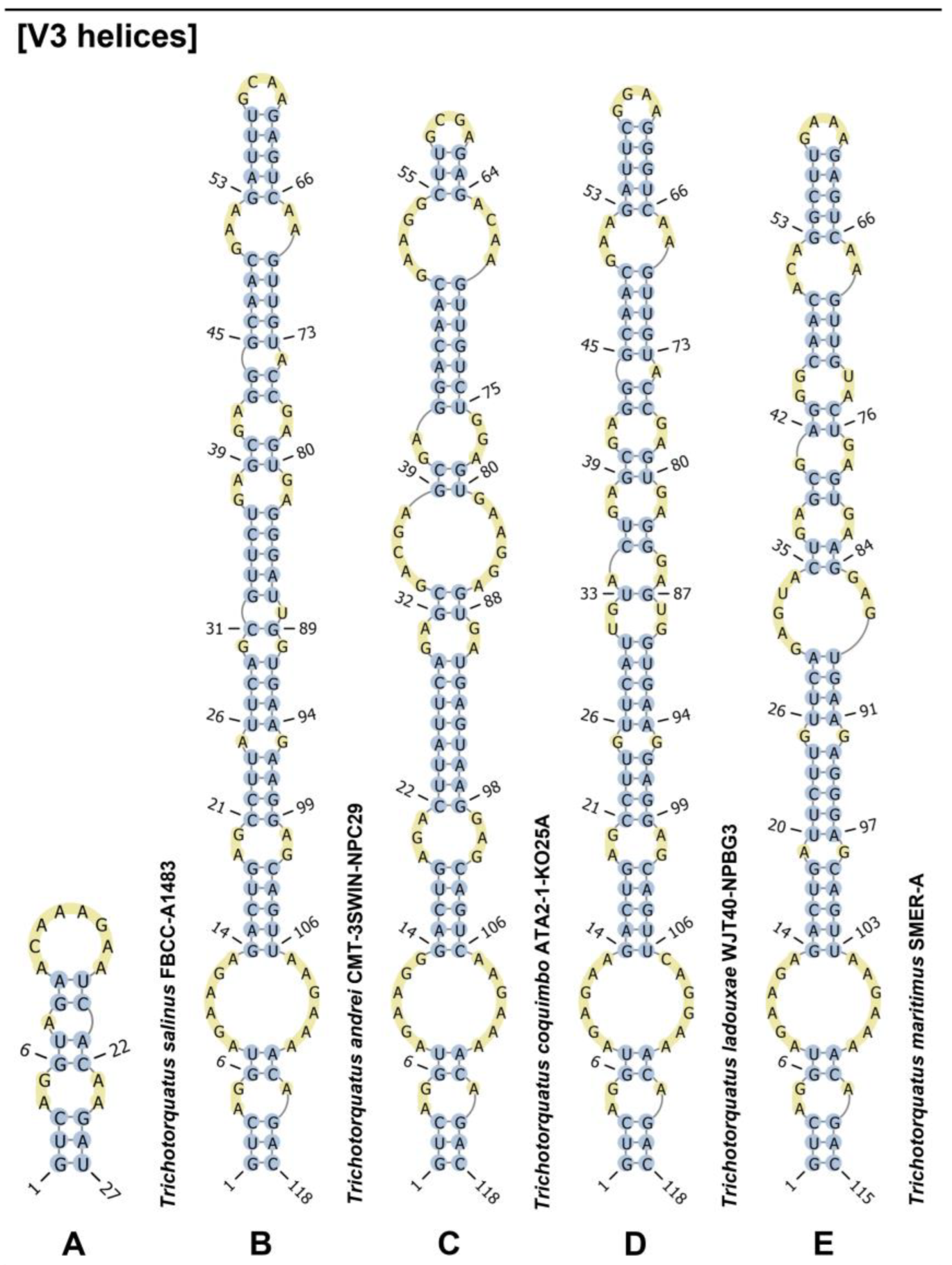
The V3 helix of the T. salinus FBCC-A1483 showed considerable differences in structure compared to that of its relative species. The total length of V3 helix was 27 bases. The basal stem consisted of 3-bp helix (5′-GUC-GAU-3′) in the T. salinus FBCC-A1483. The V3 helix of T. salinus FBCC-A1483 was shorter than those of other species (115–118 bp). All species compared with T. salinus FBCC-A1483 showed the same basal stem (5′-GUC-GAU-3′). On the other hand, the first mid-internal loops at 4–5/23–24 in the T. salinus FBCC-A1483 were different from other species.
The phylogenetic tree based on the ITS region in orthologous type 2 operon showed a clear difference for each species. Bayesian and MP analyses revealed that each species formed a distinct clade, making the species easily distinguishable (Figure 8). All separated clades were supported with high values of BPP and MP bootstrap (Figure 8, BPP > 0.91 and MP bootstrap > 97%).
In addition, the sequences of the 16S–23S ITS region of T. salinus were considerably different from those of other Trichotorquatus species. The percent dissimilarities of T. salinus ranged from 55.3% to 59.4%, whereas those among other Trichotorquatus species ranged from 12.16% to 30.37% (Table 2). Although all five species had more than 12% of 16S–23S ITS region dissimilarities among species, the dissimilarities of these T. salinus (0.0–0.3%) were less than 1.8% (Table 2).

4. Discussion

Cyanobacteria are widespread in a diversity of environments, such as freshwater, seawater, and brackish waters, as well as soils, plants, and rocks, and can also be distributed in more extreme environments, such as thermal springs or salt fields [14,35]. This ability to colonize almost every habitat on earth is considered a result of adapting to a wide range of environmental conditions coupled with their long evolution [36]. The vast adaptability of cyanobacteria to diverse environments leads to species diversity [37,38,39]. Thus, the ecological features that cyanobacteria inhabit are important to cyanobacterial speciation [40,41]. Trichotorquatus salinus, in this study, was isolated from the substrate of black tiles in cyanobacterial mats in Korean saltern, where it inhabited a saturated salinity with 50 or more. However, type species T. maritimus was collected from the biological soil crust around coastal sage scrub [2]. The other three species were also isolated from desert soils [2], suggesting that they also lived in extreme environments. Although in the same extreme environment, there is a significant difference between saltern and subaerophyte resistance that T. salinus has a unique ecology. Therefore, species belonging to the genus Trichotorquatus are either halophilic or subaerophytic species and may be highly adapted to harsh environments.
T. salinus morphologically belonged to the genus Trichotorquatus because it had the same characteristics as the collar sheath of the genus Trichotorquatus (Figure 2). All previously described Trichotorquatus species have thin filamentous trichomes, making it difficult to differentiate them from each other. In the present study, T. salinus was additionally shown to be false branched (Scytonema-type) along with the previously reported false branched (Tolypothrix-type) genus Trichotorquatus. Otherwise, however, T. salinus was a cryptic species that was difficult to morphologically distinguish from previously reported species in the genus Trichotorquatus. This feature can be seen in genera with Leptolyngbya by Anagnostidis and Komárek [42] morphotypes, such as genera Oculatella by Zammit et al. [43]; Nodosilinea by Perkerson and Casamatta [44]; Pinocchia by Dvořák, Jahodářová, and Hašler [45]; and Pantanalinema [9,44,45,46,47,48].
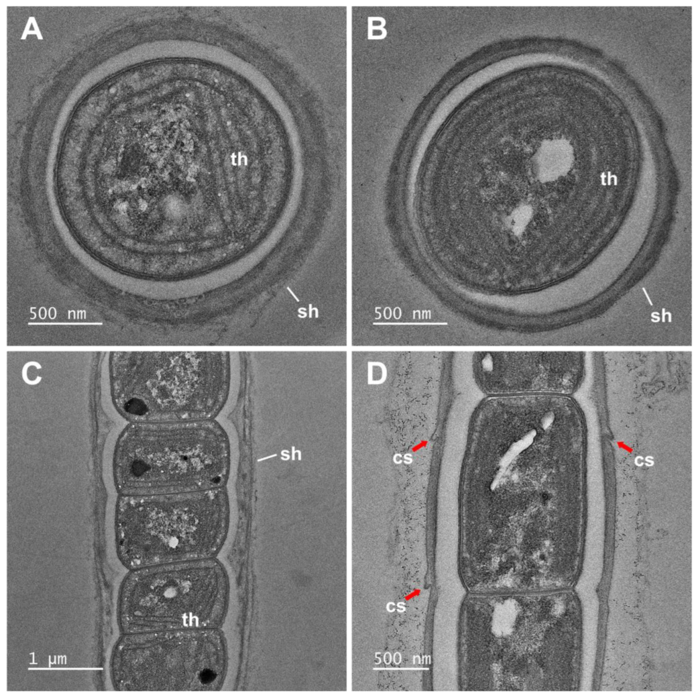
In addition, the ultrastructure of the genus Trichotorquatus has characteristics (single filaments with collar sheath) that can be seen in LM. However, since the ultrastructure of the previously reported genus Trichotorquatus has not been identified, the ultrastructure of T. salius can be helpful for future research.
Based on the results of the molecular analyses of the 16S region, T. salinus was clearly distinguished from other previously reported Trichotorquatus species. The genetic diversity in the 16S rRNA gene sequences of T. salinus was significantly lower compared to that of other Trichotorquatus species. A striking disparity was shown when comparing the difference in the genetic diversity among previously reported five Trichotorquatus species (inter-species 1) and between T. salinus and the other five species (inter-species 2) (Figure 4). These genetic diversities of T. salinus were also clearly shown in the phylogenetic tree (Figure 5). Moreover, the genetic similarities of T. salinus (94.5–96.3%) to other species were below the current species threshold of 98.65–98.7% for the 16S rRNA region [49,50]. Therefore, the 16S rRNA gene sequences of T. salinus well support T. salinus as a new species.
Recently, the species has been classified by comparing the secondary structure of the ITS region in genera Drouetiella, Oculatella, Pegethrix by Mai, Johansen, and Bohunická [1], and Tildeniella included in the family Oculatellaceae [1]. Likewise, Pietrasiak et al. [2] classified four species of the genus Trichotorquatus by comparing their ITS regions. The genus Trichotorquatus has three types of operons. Korean T. salinus lacks all tRNAs (Ala, Ile). The starting nucleotide sequence corresponds to type 2 operon starting with guanine (G). Therefore, we compared type 2 operon in the ITS region of T. salinus (5′-GGTCAAGCGAATAGT-3′) to the four previously studied species of the genus Trichotorquatus, except for Trichotorquatus sp. 5 (WJT32-NPBGA), which only has type 3 operon. Although the starting nucleotide of 23S rRNA gene of T. salinus (GG) was the same as other species with a type 2 operon (5′-GGTCAAGWTAATAAG-3′), the middle sequences (CG) were similar to those of other species (HK) with a type 3 operon (5′-AGTCAAGHKAATAAG-3′) [2]. Moreover, the latter sequences of T. salinus (GT) were recognizable from those (AG) with type 2 or 3 operons. In the secondary structures, the Box-B helix was most similar to other species with a type 2 operon, although bases (44 bp) of T. salinus were longer than those of the other four species (36 bp). Conversely, the D1–D1′ helix showed the shortest bases (76 bp) compared to the other four species (80–101 bp). The most significant difference was that the bases of V3 helix of the existing genus Trichotorquatus were 115–118 bp, whereas those of T. salinus were only 27 bp. Unique V3 structure and the beginning 23S rRNA sequences of T. salinus could be used as a molecular classification key for T. salinus in the genus Trichotorquatus with other species.
Along with ITS region’s secondary structure, the phylogenetic tree based on 16S–23S ITS region revealed that T. salinus was a distinct species (Figure 8). In comparison with Trichotorquatus species with a type 2 operon, T. salinus formed a separated clade similar to other species reported in a previous study [2]. Moreover, the percent dissimilarity of ITS regions among type 2 operon species showed strong evidence that T. salinus could be a newly recognized taxon (Table 2). The comparison of ITS region is worthwhile when classifying a new species [1,2,51,52,53].
Considering all these results (unique morphological, ecological, and molecular traits), this study suggests that Trichotorquatus salinus is a unique and novel species within the genus Trichotorquatus.

Supplementary Materials

The following supporting information can be downloaded at: https://www.mdpi.com/article/10.3390/d15010065/s1, Table S1: List of the genus Trichotorquatus species and GenBank accession numbers used in pairwise genetic distance.

Author Contributions

Conceptualization, N.-J.L.; methodology, N.-J.L. and D.-H.K.; validation, N.-J.L., D.-H.K., A.S.L. and O.-M.L.; formal analysis, N.-J.L.; investigation, N.-J.L. and D.-H.K.; resources, N.-J.L.; data curation, N.-J.L.; writing—original draft preparation, N.-J.L.; writing—review and editing, N.-J.L.; visualization, N.-J.L. and D.-H.K.; supervision, N.-J.L., A.S.L. and O.-M.L.; project administration, A.S.L. and O.-M.L.; funding acquisition, J.-H.K. All authors have read and agreed to the published version of the manuscript.

Funding

This work was supported by the Korea Environment Industry and Technology Institute (KEITI) through the project to make multi-ministerial national biological research resources more advanced, funded by Korea Ministry of Environment (MOE) (2021003420004), a grant from the National Institute of Biological Resources (NIBR), funded by MOE of the Republic of Korea (NIBR202203204), and Basic Science Research Program through the National Research Foundation (NRF) grants funded by the Ministry of Education (NRF-2021R1I1A3048458).

Institutional Review Board Statement

Not applicable.

Data Availability Statement

Not applicable.

Acknowledgments

We wish to thank Michael Guiry of AlgaeBase for advising on the scientific names and etymology.

Conflicts of Interest

The authors declare no conflict of interest.

References

  1. Mai, T.; Johansen, J.R.; Pietrasiak, N.; Bohunická, M.; Martin, M.P. Revision of the Synechococcales (Cyanobacteria) through recognition of four families including Oculatellaceae fam. nov. and Trichocoleaceae fam. nov. and six new genera containing 14 species. Phytotaxa 2018, 365, 1–59. [Google Scholar] [CrossRef] [Green Version]
  2. Pietrasiak, N.; Reeve, S.; Osorio-Santos, K.; Lipson, D.A.; Johansen, J.R. Trichotorquatus gen. nov.—A new genus of soil cyanobacteria discovered from American drylands. J. Phycol. 2021, 57, 886–902. [Google Scholar] [CrossRef]
  3. Strunecký, O.; Ivanova, A.P.; Mareš, J. An updated classification of cyanobacterial orders and families based on phylogenomic and polyphasic analysis. J. Phycol. 2022; accepted. [Google Scholar] [CrossRef]
  4. Komárek, J.; Kaštovský, J.; Mareš, J.; Johansen, J.R. Taxonomic classification of cyanoprokaryotes (cyanobacterial genera) 2014, using a polyphasic approach. Preslia 2014, 86, 295–335. [Google Scholar]
  5. Taton, A.; Wilmotte, A.; Šmarda, J.; Elster, J.; Komárek, J. Plectolyngbya hodgsonii: A novel filamentous cyanobacterium from Antarctic lakes. Polar Biol. 2011, 34, 181–191. [Google Scholar] [CrossRef]
  6. Hentschke, G.S.; Ramos, V.; Pinheiro, Â.; Barreiro, A.; Costa, M.S.; Rego, A.; Brule, S.; Vasconcelos, V.M.; Leão, P.N. Zarconia navalis gen. nov., sp. nov., Romeriopsis navalis gen. nov., sp. nov. and Romeriopsis marina sp. nov., isolated from inter-and subtidal environments from northern Portugal. Int. J. Syst. Evol. Microbiol. 2022, 72, 005552. [Google Scholar] [CrossRef]
  7. Rasouli-Dogaheh, S.; Komárek, J.; Chatchawan, T.; Hauer, T. Thainema gen. nov. (Leptolyngbyaceae, Synechococcales): A new genus of simple trichal cyanobacteria isolated from a solar saltern environment in Thailand. PLoS ONE 2022, 17, e0261682. [Google Scholar] [CrossRef] [PubMed]
  8. Tawong, W.; Pongcharoen, P.; Nishimura, T.; Saijuntha, W. Siamcapillus rubidus gen. et sp. nov. (Oculatellaceae), a novel filamentous cyanobacterium from Thailand based on molecular and morphological analyses. Phytotaxa 2022, 558, 33–52. [Google Scholar] [CrossRef]
  9. Vaz, M.G.M.V.; Genuario, D.B.; Andreote, A.P.D.; Malone, C.F.S.; Sant’Anna, C.L.; Barbiero, L.; Fiore, M.F. Pantanalinema gen. nov. and Alkalinema gen. nov.: Novel pseudanabaenacean genera (Cyanobacteria) isolated from saline–alkaline lakes. Int. J. Syst. Evol. Microbiol. 2015, 65, 298–308. [Google Scholar] [CrossRef]
  10. Malone, C.F.D.S.; Rigonato, J.; Laughinghouse IV, H.D.; Schmidt, E.C.; Bouzon, Z.L.; Wilmotte, A.; Fiore, M.F.; Sant’Anna, C.L. Cephalothrix gen. nov. (Cyanobacteria): Towards an intraspecific phylogenetic evaluation by multilocus analyses. Int. J. Syst. Evol. Microbiol. 2015, 65, 2993–3007. [Google Scholar] [CrossRef]
  11. Genuário, D.B.; Vaz, M.G.M.V.; Hentschke, G.S.; Sant’Anna, C.L.; Fiore, M.F. Halotia gen. nov., a phylogenetically and physiologically coherent cyanobacterial genus isolated from marine coastal environments. Int. J. Syst. Evol. Microbiol. 2015, 65, 663–675. [Google Scholar] [CrossRef] [PubMed]
  12. Malone, C.F.D.S.; Genuário, D.B.; Vaz, M.G.M.V.; Fiore, M.F.; Sant’Anna, C.L. Monilinema gen. nov., a homocytous genus (Cyanobacteria, Leptolyngbyaceae) from saline–alkaline lakes of Pantanal wetlands, Brazil. J. Phycol. 2021, 57, 473–483. [Google Scholar] [CrossRef] [PubMed]
  13. Jungblut, A.D.; Hawes, I.; Mountfort, D.; Hitzfeld, B.; Dietrich, D.R.; Burns, B.P.; Neilan, B.A. Diversity within cyanobacterial mat communities in variable salinity meltwater ponds of McMurdo Ice Shelf, Antarctica. Environ. Microbiol. 2005, 7, 519–529. [Google Scholar] [CrossRef] [PubMed] [Green Version]
  14. Oren, A. Halophilic microbial communities and their environments. Curr. Opin. Biotechnol. 2015, 33, 119–124. [Google Scholar] [CrossRef]
  15. Saccò, M.; White, N.E.; Harrod, C.; Salazar, G.; Aguilar, P.; Cubillos, C.F.; Meredith, K.; Baxter, B.K.; Oren, A.; Anufriieva, E.; et al. Salt to conserve: A review on the ecology and preservation of hypersaline ecosystems. Biol. Rev. 2021, 96, 2828–2850. [Google Scholar] [CrossRef]
  16. Sommer, V.; Mikhailyuk, T.; Glaser, K.; Karsten, U. Uncovering unique green algae and cyanobacteria isolated from biocrusts in highly saline potash tailing pile habitats, using an integrative approach. Microorganisms 2020, 8, 1667. [Google Scholar] [CrossRef]
  17. Patel, H.M.; Rastogi, R.P.; Trivedi, U.; Madamwar, D. Cyanobacterial diversity in mat sample obtained from hypersaline desert, Rann of Kachchh. 3 Biotech 2019, 9, 304. [Google Scholar] [CrossRef]
  18. Mourão, G.H.; Ishii, I.H.; Campos, Z. Alguns fatores limnológicos relacionados com a ictiofauna de baías e salinas do Pantanal da Nhecolândia, Mato Grosso do Sul, Brasil. Acta Limnol. Bras. 1988, 11, 181–198. [Google Scholar]
  19. Cellamare, M.; Duval, C.; Drelin, Y.; Djediat, C.; Touibi, N.; Agogué, H.; Leboulanger, C.; Ader, M.; Bernard, C. Characterization of phototrophic microorganisms and description of new cyanobacteria isolated from the saline-alkaline crater-lake Dziani Dzaha (Mayotte, Indian Ocean). FEMS Microbiol. Ecol. 2018, 94, fiy108. [Google Scholar] [CrossRef]
  20. Shalygin, S.; Pietrasiak, N.; Gomez, F.; Mlewski, C.; Gerard, E.; Johansen, J.R. Rivularia halophila sp. nov. (Nostocales, Cyanobacteria): The first species of Rivularia described with the modern polyphasic approach. Eur. J. Phycol. 2018, 53, 537–548. [Google Scholar] [CrossRef]
  21. Kiel, G.; Gaylarde, C.C. Bacterial diversity in biofilms on external surfaces of historic buildings in Porto Alegre. World J. Microbiol. Biotechnol. 2006, 22, 293–297. [Google Scholar] [CrossRef]
  22. Lee, N.-J.; Bang, S.-D.; Kim, T.; Ki, J.-S.; Lee, O.-M. Pseudoaliinostoc sejongens gen. & sp. nov. (Nostocales, Cyanobacteria) from floodplain soil of the Geum River in Korea based on polyphasic approach. Phytotaxa 2021, 479, 55–70. [Google Scholar] [CrossRef]
  23. Kim, J.-H.; Jeong, M.S.; Kim, D.-Y.; Her, S.; Wie, M.-B. Zinc oxide nanoparticles induce lipoxygenase-mediated apoptosis and necrosis in human neuroblastoma SH-SY5Y cells. Neurochem. Int. 2015, 90, 204–214. [Google Scholar] [CrossRef]
  24. Guiry, M.D.; Guiry, G.M. AlgaeBase. World-Wide Electronic Publication, National University of Ireland, Galway. 2022. Available online: www.algaebase.org (accessed on 23 August 2022).
  25. Lee, N.-J.; Seo, Y.; Ki, J.-S.; Lee, O.-M. Morphology and molecular description of Wilmottia koreana sp. nov. (Oscillatoriales, Cyanobacteria) isolated from the Republic of Korea. Phytotaxa 2020, 447, 237–251. [Google Scholar] [CrossRef]
  26. Neilan, B.A.; Jacobs, D.; Therese, D.D.; Blackall, L.L.; Hawkins, P.R.; Cox, P.T.; Goodman, A.E. rRNA sequences and evolutionary relationships among toxic and nontoxic cyanobacteria of the genus Microcystis. Int. J. Syst. Evol. Microbiol. 1997, 47, 693–697. [Google Scholar] [CrossRef]
  27. Taton, A.; Grubisic, S.; Brambilla, E.; De Wit, R.; Wilmotte, A. Cyanobacterial diversity in natural and artificial microbial mats of Lake Fryxell (McMurdo Dry Valleys, Antarctica): A morphological and molecular approach. Appl. Environ. Microbiol. 2003, 69, 5157–5169. [Google Scholar] [CrossRef] [Green Version]
  28. Kumar, S.; Stecher, G.; Li, M.; Knyaz, C.; Tamura, K. MEGA X: Molecular evolutionary genetics analysis across computing platforms. Mol. Biol. Evol. 2018, 35, 1547–1549. [Google Scholar] [CrossRef]
  29. Stamatakis, A. RAxML-VI-HPC: Maximum likelihood-based phylogenetic analyses with thousands of taxa and mixed models. Bioinformatics 2006, 22, 2688–2690. [Google Scholar] [CrossRef] [Green Version]
  30. Ronquist, F.; Teslenko, M.; Van Der Mark, P.; Ayres, D.L.; Darling, A.; Höhna, S.; Larget, B.; Liu, L.; Suchard, M.A.; Huelsenbeck, J.P. MrBayes 3.2: Efficient Bayesian phylogenetic inference and model choice across a large model space. Syst. Biol. 2012, 61, 539–542. [Google Scholar] [CrossRef] [Green Version]
  31. Lowe, T.M.; Chan, P.P. tRNAscan-SE On-line: Integrating search and context for analysis of transfer RNA genes. Nucleic Acids Res. 2016, 44, W54–W57. [Google Scholar] [CrossRef]
  32. Zuker, M. Mfold web server for nucleic acid folding and hybridization prediction. Nucleic Acids Res. 2003, 31, 3406–3415. [Google Scholar] [CrossRef] [PubMed]
  33. Byun, Y.; Han, K. PseudoViewer3: Generating planar drawings of large-scale RNA structures with pseudoknots. Bioinformatics 2009, 25, 1435–1437. [Google Scholar] [CrossRef] [PubMed] [Green Version]
  34. Turland, N.J.; Wiersema, J.H.; Barrie, F.R.; Greuter, W.; Hawksworth, D.L.; Herendeen, P.S.; Knapp, S.; Kusber, W.-H.; Li, D.-Z.; Marhold, K.; et al. International Code of Nomenclature for Algae, Fungi, and Plants (Shenzhen Code) Adopted by the Nineteenth International Botanical Congress Shenzhen, China, July 2017; Regnum Vegetabile 159; Koeltz Botanical Books: Glashütten, Germany, 2018; pp. 1–254. [Google Scholar]
  35. Bravakos, P.; Kotoulas, G.; Skaraki, K.; Pantazidou, A.; Economou-Amilli, A. A polyphasic taxonomic approach in isolated strains of Cyanobacteria from thermal springs of Greece. Mol. Phylogenet. Evol. 2016, 98, 147–160. [Google Scholar] [CrossRef] [PubMed]
  36. Jung, P.; Sommer, V.; Karsten, U.; Lakatos, M. Salty Twins: Salt-Tolerance of Terrestrial Cyanocohniella Strains (Cyanobacteria) and Description of C. rudolphia sp. nov. Point towards a Marine Origin of the Genus and Terrestrial Long Distance Dispersal Patterns. Microorganisms 2022, 10, 968. [Google Scholar] [CrossRef] [PubMed]
  37. Sant’Anna, C.L.; Azevedo, M.T.P.; Kaštovský, J.; Komárek, J. Two form-genera of aerophytic heterocytous cyanobacteria from Brasilian rainy forest “Mata Atlântica”. Fottea 2010, 10, 217–228. [Google Scholar] [CrossRef] [Green Version]
  38. Beck, C.; Knoop, H.; Axmann, I.M.; Steuer, R. The diversity of cyanobacterial metabolism: Genome analysis of multiple phototrophic microorganisms. BMC Genom. 2012, 13, 56. [Google Scholar] [CrossRef] [Green Version]
  39. Whitton, B.A. Ecology of Cyanobacteria II: Their Diversity in Space and Time; Springer Science & Business Media: Berlin, Germany, 2012; pp. 1–760. [Google Scholar]
  40. Rahman, O.; Pfitzenmaier, M.; Pester, O.; Morath, S.; Cummings, S.P.; Hartung, T.; Sutcliffe, I.C. Macroamphiphilic components of thermophilic actinomycetes: Identification of lipoteichoic acid in Thermobifida fusca. J. Bacteriol. Res. 2009, 191, 152–160. [Google Scholar] [CrossRef] [Green Version]
  41. Prabha, R.; Singh, D.P.; Somvanshi, P.; Rai, A. Functional profiling of cyanobacterial genomes and its role in ecological adaptations. Genom. Data 2016, 9, 89–94. [Google Scholar] [CrossRef] [Green Version]
  42. Anagnostidis, K.; Komárek, J. Modern approach to the classification system of cyanophytes. 3-Oscillatoriales. Arch. Hydrobiol. 1988, 80, 327–472. [Google Scholar]
  43. Zammit, G.; Billi, D.; Albertano, P. The subaerophytic cyanobacterium Oculatella subterranea (Oscillatoriales, Cyanophyceae) gen. et sp. nov.: A cytomorphological and molecular description. Eur. J. Phycol. 2012, 47, 341–354. [Google Scholar] [CrossRef] [Green Version]
  44. Perkerson, R.B.; Johansen, J.R.; Kovácik, L.; Brand, J.; Kaštovský, J.; Casamatta, D.A. A unique pseudanabaenalean (Cyanobacteria) genus Nodosilinea gen. nov. based on morphological and molecular data. J. Phycol. 2011, 47, 1397–1412. [Google Scholar] [CrossRef] [PubMed]
  45. Dvořák, P.; Jahodářová, E.; Hašler, P.; Gusev, E.; Poulíčková, A. A new tropical cyanobacterium Pinocchia polymorpha gen. et sp. nov. derived from the genus Pseudanabaena. Fottea 2015, 15, 113–120. [Google Scholar] [CrossRef] [Green Version]
  46. Komárek, J.; Anagnostidis, K. Cyanoprokaryota Teil 2: Oscillatoriales. In Süsswasserflora von Mitteleuropa Band 19/2; Büdel, B., Krienitz, L., Gärtner, G., Schagerl, M., Eds.; Elsevier: Heidelberg, Germany, 2005; pp. 1–759. [Google Scholar]
  47. Komárek, J. A polyphasic approach for the taxonomy of cyanobacteria: Principles and applications. Eur. J. Phycol. 2016, 51, 346–353. [Google Scholar] [CrossRef]
  48. Becerra-Absalón, I.; Johansen, J.R.; Osorio-Santos, K.; Montejano, G. Two new Oculatella (Oculatellaceae, Cyanobacteria) species in soil crusts from tropical semi–arid uplands of México. Fottea 2020, 20, 160–170. [Google Scholar] [CrossRef]
  49. Kim, M.; Oh, H.-S.; Park, S.-C.; Chun, J. Towards a taxonomic coherence between average nucleotide identity and 16S rRNA gene sequence similarity for species demarcation of prokaryotes. Int. J. Syst. Evol. Microbiol. 2014, 64, 346–351. [Google Scholar] [CrossRef]
  50. Yarza, P.; Yilmaz, P.; Pruesse, E.; Glöckner, F.O.; Ludwig, W.; Schleifer, K.-H.; Whitman, W.B.; Euzéby, J.; Amann, R.; Rosselló-Móra, R. Uniting the classification of cultured and uncultured bacteria and archaea using 16S rRNA gene sequences. Nat. Rev. Microbiol. 2014, 12, 635–645. [Google Scholar] [CrossRef]
  51. Mikhailyuk, T.; Vinogradova, O.; Holzinger, A.; Glaser, K.; Akimov, Y.; Karsten, U. Timaviella dunensis sp. nov. from sand dunes of the Baltic Sea, Germany, and emendation of Timaviella edaphica (Elenkin) OM Vynogr. & Mikhailyuk (Synechococcales, Cyanobacteria) based on an integrative approach. Phytotaxa 2022, 532, 192–208. [Google Scholar] [CrossRef]
  52. Tawong, W.; Pongcharoen, P.; Pongpadung, P.; Ponza, S.; Saijuntha, W. Amazonocrinis thailandica sp. nov. (Nostocales, Cyanobacteria), a novel species of the previously monotypic Amazonocrinis genus from Thailand. Algae 2022, 37, 1–14. [Google Scholar] [CrossRef]
  53. Osorio-Santos, K.; Pietrasiak, N.; Bohunická, M.; Miscoe, L.H.; Kováčik, L.; Martin, M.P.; Johansen, J.R. Seven new species of Oculatella (Pseudanabaenales, Cyanobacteria): Taxonomically recognizing cryptic diversification. Eur. J. Phycol. 2014, 49, 450–470. [Google Scholar] [CrossRef]
Figure 1. Map showing site in the Gomso saltern of the Republic of Korea. (A) The red arrow indicates a sample site; (B) Collections from the substrate of black tile in cyanobacterial mats (red circle) (in saline, >50).
Diversity 15 00065 g001
Figure 2. Light microscopic photographs of Trichotorquatus salinus, the culture sample from the reference strains (FBCC-A1483). (A,B) Entangled growth of the arrangement of trichomes; (C) Parallel trichomes; (D) Coiled trichomes; (E) Tangled apical cell; (F) Tolypothrix-type of false branched; (G,H) Scytonema-type of false branched; (I,J) Trichomes regularly or loosely spiralis; (K,L) Trichome broke out of the sheath (sh); (M) Trichome with necridia cell in rows; (N,O) Bulging within a sheath; (PU) Collar sheath (cs) near trichome apices or a telescoping and persistent after trichome escapes from sheath; (V) Rounded apical cell. Scale bars, 50 μm (A); 20 μm (B); 10 μm (CV).
Diversity 15 00065 g002
Figure 3. Ultrastructure of Trichotorquatus salinus FBCC-A1483. (A,B) Cross section of the trichome, circular in shape; (C,D) Longitudinal section of the trichome; (AC) Parietal position of thylakoids (th) and single filaments with sheaths (sh); (D) Red arrows showing collar sheath (cs).
Diversity 15 00065 g003
Figure 4. Nucleotide divergences of the genus Trichotorquatus 16S rRNA gene sequences (datasets used in Table S1) based on corrected pairwise genetic distance (p-distance), including T. salinus. Genetic distances between each paired sequences were calculated by the Kimura 2-parameter model, where a total of six Trichotorquatus species (32 sequences) were compared. Bar heights indicate % p-distance measured for intra-species [e.g., T. andrei (Ta), T. coquimbo (Tc), T. ladouxae (Tl), T. maritimus (Tm), T. salinus (Ts), Trichotorquatus sp. 5 (Tsp.5)], inter-species 1 (e.g., Ta vs. Tc, Ta vs. Tl, Ta vs. Tm, Ta vs. Tsp.5, Tc vs. Tl, Tc vs. Tm, Tc vs. Tsp.5, Tl vs. Tm, Tl vs. Tsp.5, Tm vs. Tsp.5) and inter-species 2 (Ta vs. Ts, Tc vs. Ts, Tl vs. Ts, Tm vs. Ts, Ts vs. Tsp.5). The borders of the box show the first and third quartiles. The black solid and red dotted lines indicate the median and mean values, respectively (t-test, *** p < 0.001).
Diversity 15 00065 g004
Figure 5. Consensus Bayesian tree based on species in the family Oculatellaceae with closely related unidentified taxa and Gloeobacter violaceus as an outgroup. Our strains are marked in bold. The branch lengths are proportional to the number of character changes. Posterior probabilities (left) and ML bootstrap values (right) were presented on the node. Posterior probabilities ≥ 0.5 are shown.
Diversity 15 00065 g005
Figure 6. Secondary structures for the D1–D1ʹ and Box-B (type 2 operon) in the conserved regions of the 16S–23S internal transcribed spacer region. (A,F) Trichotorquatus salinus FBCC-A1483; (B,G) T. andrei CMT-3SWIN-NPC29; (C,H) T. coquimbo ATA2-1-KO25A; (D,I) T. ladouxae WJT40-NPBG3; (E,J) T. maritimus SMER-A. These structures were drawn considering Pietrasiak et al. [2].
Diversity 15 00065 g006
Figure 7. Secondary structures for the V3 (type 2 operon) in the conserved regions of the 16S–23S internal transcribed spacer region. (A) Trichotorquatus salinus FBCC-A1483; (B) T. andrei CMT-3SWIN-NPC29; (C) T. coquimbo ATA2-1-KO25A; (D) T. ladouxae WJT40-NPBG3; (E) T. maritimus SMER-A. These structures were drawn considering Pietrasiak et al. [2].
Diversity 15 00065 g007
Figure 8. Bayesian Inference analysis of 16S–23S internal transcribed spacer gene sequences of orthologous type 2 operon, with posterior probabilities (left) and Maximum parsimony bootstrap values (right) on the node.
Diversity 15 00065 g008
Table 1. Morphological characteristics of genus Trichotorquatus as described by Pietrasiak et al. [2].
SpeciesFilamentsSheathTrichomesApical CellNecridiaHormogonia
Trichotorquatus salinusFalse branched
(Scytonema-type, Tolypothrix-type)
2.1–3.4 μm wide
Sometimes absent
Long or short collar
Within bulging
0.1–0.9 μm thick
Distinctly constricted
Regularly or loosely spiral
1.9–2.9 μm wide
Blue-green
Rarely tangled
2.3–4.3 μm long
Rows
Sections up to 5 μm long
Short
Under 10 cells long
T. andreiUnbranched
1.6–5.6 μm wide
Sometimes absent
0.2–3.0 μm thick
Distinctly constricted
1.0–2.8 μm wide
Yellowish
1.4–6.3 μm long
Rows
Sections up to 11 μm long
Short
4–8 cells long
T. coquimboUnbranched
2.2–3.6 μm wide
Sometimes absent
0.1–0.6 μm thick
Rarely short collar
Distinctly constricted
1.8–3.4 μm wide
Pale yellowish green
1.6–5.0 μm long
Rows
Sections up to 4.9 μm long
Short
4–10 cells long
T. ladouxaeMostly unbranched
2.0–7.0 μm wide
Sometimes absent
0.2–0.3 μm thick
Distinctly constricted
2.0–3.6 μm wide
Blue-green
1.8–4.6 μm long
Rows
Sections up to 12 μm long
Short
Under 10 cells long
T. maritimus TMostly unbranched
2.2–7.3 μm wide
Sometimes absent
Frayed collar
0.2–2.4 μm thick
Distinctly constricted
2.1–4.3 μm wide
Blue-green
2.2–32 μm long
Rows
Sections up to 32 μm long
Very short
1–6 cells long
Trichotorquatussp. 5Unbranched
2.0–4.0 μm wide
Sometimes absent
0.2–0.6 μm thick
Indistinctly constricted
2.0–2.8 μm wide
Blue-green-Short
Under 10 cells long
T: type species, -: no data available.
Table 2. Percent dissimilarity of 16S–23S internal transcribed spacer region among the species that have the type 2 operon. Comparisons among strains in the same species are reported in bold font.
T. salinusT. andreiT. coquimboT. ladouxaeT. maritimus
T. salinus0.00–0.30%
T. andrei55.31–57.34%0.00–1.33%
T. coquimbo58.52–59.25%23.35–24.63%0.21%
T. ladouxae56.66–58.52%12.16–13.22%29.57–30.37%0.00–1.80%
T. maritimus59.37%13.00–14.36%20.66–21.28%15.62–16.72%0.00–0.22%
Disclaimer/Publisher’s Note: The statements, opinions and data contained in all publications are solely those of the individual author(s) and contributor(s) and not of MDPI and/or the editor(s). MDPI and/or the editor(s) disclaim responsibility for any injury to people or property resulting from any ideas, methods, instructions or products referred to in the content.