Article

Genetic Structure of Juvenile Stages of Phocanema bulbosum (Nematoda, Chromadorea: Anisakidae) Parasitizing Commercial Fish, Atlantic Cod Gadus morhua, and American Plaice Hippoglossoides platessoides in the Barents Sea

by
Ilya I. Gordeev
1,2,*,
Yuri I. Bakay
3,
Marina Yu. Kalashnikova
3,
Andrey D. Logvinenko
2,
Olga R. Emelianova
4,5 and
Sergey G. Sokolov
6
1
Pacific Salmon Department, Russian Federal Research Institute of Fisheries and Oceanography, Okruzhnoy Proyezd 19, 105187 Moscow, Russia
2
Department of Invertebrate Zoology, Faculty of Biology, Lomonosov Moscow State University, Leninskie Gory 1/12, 119234 Moscow, Russia
3
Laboratory of Aquaculture and Hydrobionts Diseases, Polar Branch of the Russian Federal Research Institute of Fisheries and Oceanography, Akademika Knipovicha Srt. 6, 183038 Murmansk, Russia
4
Molecular Genetics Department, Russian Federal Research Institute of Fisheries and Oceanography, Okruzhnoy Proyezd 19, 105187 Moscow, Russia
5
Department of Biological Evolution, Faculty of Biology, Lomonosov Moscow State University, Leninskie Gory 1/12, 119234 Moscow, Russia
6
Center of Parasitology, A.N. Severtsov Institute of Ecology and Evolution, Russian Academy of Sciences, Leninskiy Pros. 33, 119071 Moscow, Russia
*
Author to whom correspondence should be addressed.
Diversity 2023, 15(10), 1036; https://doi.org/10.3390/d15101036
Submission received: 28 July 2023 / Revised: 20 September 2023 / Accepted: 21 September 2023 / Published: 26 September 2023
(This article belongs to the Special Issue Diversity of Macroparasites in Marine Fishes—2nd Edition)

Abstract

:
Atlantic cod Gadus morhua and American plaice Hippoglossoides platessoides are two of the most commercially valuable species in the Barents Sea (FAO Area 27). They are considered as an important but neglected source of zoonotic risk associated with nematodes from the genus Phocanema. The abundance of Phocanema spp. in a fish host individual in the Barents Sea may be quite high, which is convenient for studying the genetic structure of its populations. A total of 69 third-stage juveniles of Phocanema spp. were isolated from the liver, the mesentery, and the musculature of G. morhua and H. platessoides and genotyped by the mtDNA Cox2 gene. Almost all these juveniles (68) were molecularly identified as P. bulbosum. The mtDNA Cox2 gene was also used to reveal the haplotype diversity and the genetic structure of P. bulbosum. A comparison of the specimens examined in this study with each other and with the haplotypes previously identified by us in the White Sea showed that there were no significant differences between the groups from different hosts and from different catch areas.

1. Introduction

The genus Phocanema (Myers, 1959) is a small group of anisakid nematodes comprising five nominal species: Phocanema azarasi (Yamaguti and Arima, 1942), Phocanema bulbosum (Cobb, 1888), Phocanema cattani (George-Nascimento and Urrutia, 2000), Phocanema decipiens (Krabbe, 1878) sensu stricto, and Phocanema krabbei (Paggi, Mattiucci, Gibson, Berland, Nascetti, Cianchi and Bullini, 2000) [1,2].
Phocanema spp. uses pinnipeds as definitive hosts, and marine crustaceans and fishes as intermediate and paratenic hosts, respectively [3]. However, it is hardly appropriate to consider crustaceans in the life cycle of this nematode, as well as other anasakids, as an intermediate host, since the juveniles of these parasites do not have any development in them until the next (higher) stage. The embryonic and initial stages of postembryonic development of Phocanema spp., culminating in the formation of the third-stage juvenile, take place in the egg. The third-stage juvenile emerging from the egg is swallowed by benthic crustaceans and, moving along trophic chains, accumulates in fish [4,5]. Third-stage juveniles of these nematodes cause an important fish-borne zoonosis (e.g., [6,7,8,9,10,11,12,13]).
Phocanema bulbosum was originally described as Ascaris bulbosa by Cobb in 1888 [14], but A. bulbosa is considered a synonym of Ascaris decipiens Krabbe, 1878 (=Phocanema decipiens) as recognized later by many authors [1,15,16]. Paggi et al. [17], Deco et al. [18], and Mattiucci et al. [19] showed that P. decipiens (as member of Pseudoterranova Mozgovoi, 1951 in these authors) is a species complex consisting of at least five species with a clear genetic differentiation. These species were provisionally designated with letters (A, B, C, etc.). Mattiucci et al. [19] resurrected the name Pseudoterranova bulbosa for Pseudoterranova decipiens C. However, the monophyly of the genus Pseudoterranova is not supported by recent research [2,20]. Bao et al. [2] proposed the resurrection of the genus Phocanema, with Ph. decipiens as the type species, to encompass P. bulbosum and several other species. The definitive hosts of P. bulbosum are Erignathus barbatus (Erxleben, 1777), Halichoerus grypus, Pusa hispida, and probably Monachus monachus (e.g., [16,17,18,19,21]. Third-stage juveniles of P. bulbosum were discovered in pleuronectid, gadiid, macrourid, sebastid, and cottid fishes (e.g., [16,22,23]. This nematode has a broad geographic distribution in temperate, subarctic, and arctic seas of the Northern Hemisphere (e.g., [17,19,21,23,24,25,26]. The data of [27] also indicate that P. bulbosum might be present in the Mediterranean Sea. In the Barents Sea, it was recorded in E. barbatus and various fishes caught in the northern and western areas [2,17,24,28,29,30]. There are no records of P. bulbosum in the southern and eastern areas of this sea.
The population genetic structure (the distribution of genetic variation in time and space) affects the response of a species to selection pressures, and so shapes its evolution [31]. Studying the population genetics of parasites provides insights into their infection dynamics [32] and the ways it affects the entire community [33]. Taxonomic issues based on morphology can be elucidated with the help of genetic methods, which advance cladistics to a level inaccessible to morphology, including the allocation of cryptic species, as shown, in particular, for nematodes [34]. Patterns of the population genetic structure of a parasite can provide information on the present and past migrations of their hosts [35,36,37]. Among the molecular markers in use, the Cox2 mitochondrial gene is one of the best suited to assess the population genetic structure and the phylogeography of anisakids species (e.g., [36,37,38,39]). However, only a few sequences of Cox2 gene are available for P. bulbosum [30,40].
The aims of our study were to genetically identify the third-stage juveniles of Phocanema spp. from the Atlantic cod G. morhua and the American plaice H. platessoides caught in the southern and the eastern parts of the Barents Sea and to assess the genetic diversity and the population genetic structure of P. bulbosum in the region. The overall aim of the study was to advance the knowledge of the distribution and population structure of the Phocanema species infecting commercial fish in the Barents Sea through the use of genetic analysis.

2. Materials and Methods

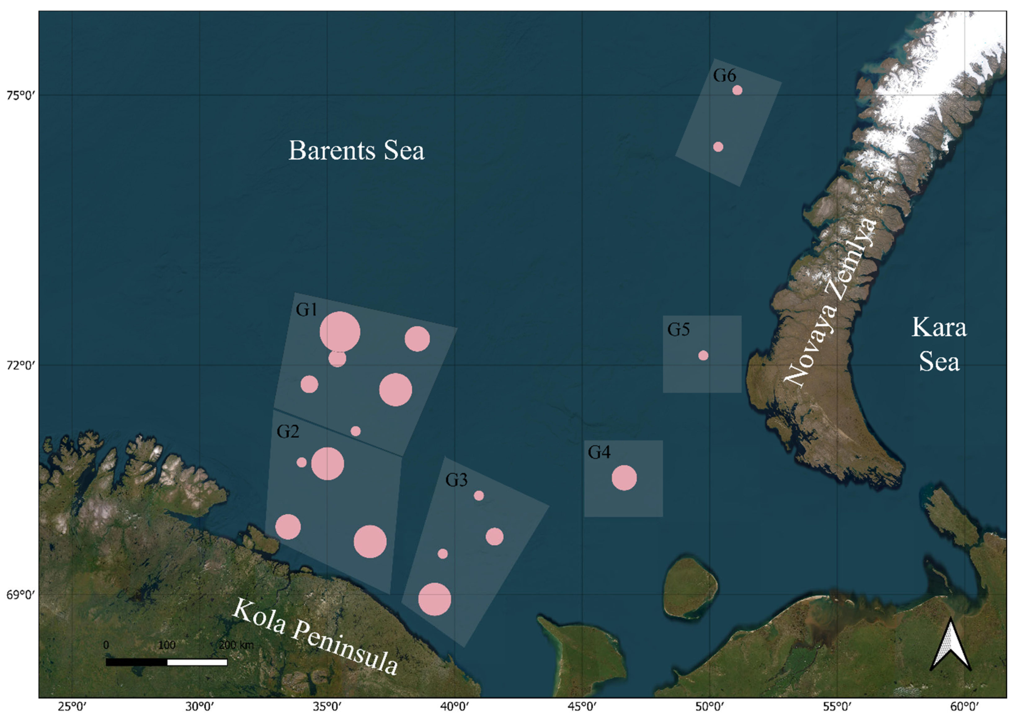
2.1. Sampling and Morphological Examination

The host fishes were caught by the bottom trawl in the period from 26 February 2021 to 17 February 2022 in areas of the Barents Sea within the Russian Exclusive Economic Zone (Figure 1). In total, 225 specimens of Gadus morhua (TL 22–122 cm, mean 90.46 ± 3.08; weight 178–11, 150 g, mean 7172 ± 471.61; male/female ratio 1:3.91) and 375 specimens of Hippoglossoides platessoides (TL 21–47 cm, mean 35.92 ± 0.86; weight 72–1126, mean 490.18 ± 38.10; male/female ratio 1:4.42) were examined. The fishes were caught from RV “Vilnius” under permissions no. 512021030001NI and no. 512022030007NI of 18 February 2021 and 17 January 2022, accordingly, issued by the Federal Agency for Fisheries. Parasitological dissection was performed according to the standard methodology [41,42]. The juveniles of Phocanema spp. were fixed with 96% ethanol and stored at −20 °C. Parasitological indices (prevalence, intensity, and mean intensity) were calculated in accordance with [43].
For the purpose of this study, the catch locations were divided into 6 groups (Figure 1, G1–G6), and these groups were compared in order to reveal a geographical trend, if any, in the distribution of the haplotypes in the study area.
Figure 1. Sampling map. Pink points are sampling locations. Point size represents the number of Phocanema bulbosum larvae collected at the location (from 1 to 21). G1–G6 are geographical groups of locations corresponding to the haplotype structure in Figure 2.
Diversity 15 01036 g001

2.2. DNA Extraction, Amplification, and Sequencing

Total DNA was extracted separately from each of the 69 third-stage juveniles of Phocanema using PALL™ AcroPrep 96-well purification plates by PALL Corp. following a protocol by [44]. The DNA samples were used as a template for amplification of partial cytochrome c oxidase subunit II (Cox2) (~671 bp). Cox2 of Phocanema juveniles were amplified by a polymerase chain reaction (PCR) with primers 210 (5′CACCAACTCTTAAAATTATC3′) and 211 (5’TTTTCTAGTTATAGATTGRTTYAT3′) described by [36]. PCRs were carried out in a 25-µL reaction volume (5 µL of 5× HotTaq Red buffer (Eurogen Lab, Moscow, Russia), 0.5 µL of HotTaq polymerase (Eurogen Lab, Moscow, Russia), 0.5 µL of dNTP (50 µM stock), 0.3 µL of each primer (10 µM stock), 1 µL of genomic DNA, and 17.4 µL of sterile water). Amplification was performed according to the following protocol: 3 min denaturation at 94 °C, 34 cycles of 30 s at 94 °C, 60 s at 46 °C, and 1.5 min at 72 °C, and 10 min elongation at 72 °C. The negative control was amplified using both primers.
Both strands of each amplicon were sequenced with the BigDye Terminator v3.1 sequencing kit (Applied Biosystems. Waltham, MA, USA) and NovaDye Terminator sequencing kit (Thermo Fisher, Waltham, MA, USA). Sequencing reactions were analyzed by capillary electrophoresis on ABI 3500 Genetic Analyser (Thermo Fisher, Waltham, MA, USA) or Nanophore-05 (Syntol, Moscow, Russia) at the Core Centrum of Koltsov Institute of Developmental Biology (Moscow, Russia). All newly obtained sequences were deposited in GenBank (NCBI) (Table 1). Raw reads for each gene were assembled and checked for improper base-calling using GeneiousPro 10.0.9 (Biomatters, Auckland, New Zealand) and the sites identified in this way were further modified.
The obtained sequences of Cox2 gene were processed using Geneious 8.1.8 software package [45], ClustalW alignment. Estimates of evolutionary divergence (p-distances) were made with the use of MEGA XI software (ver. 11.0.11) [46].
Table 1. List of the Cox2 sequences obtained in this study.
GenBank Accession NumberHostLocality *
OQ731840–OQ731844, OQ731846, OQ731848, OQ731849, OQ731867Atlantic cod Gadus morhuaG1
OQ731832–OQ731839G2
OQ731845,
OQ731850–OQ731866
G3
OQ731847G5
OQ731868–OQ731874
OQ731882–OQ731887
American plaice Hippoglossoides platessoidesG1
OQ731888–OQ731896G2
OQ731875,
OQ731897–OQ731899
G3
OQ731876–OQ731879G4
OQ731880, OQ731881G6
* following Figure 1.

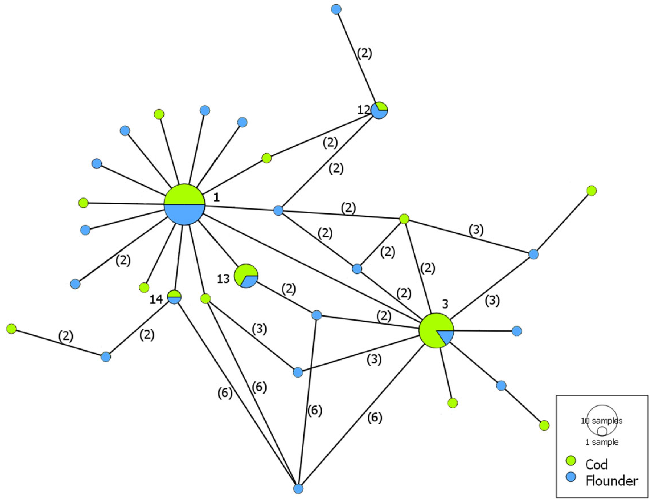
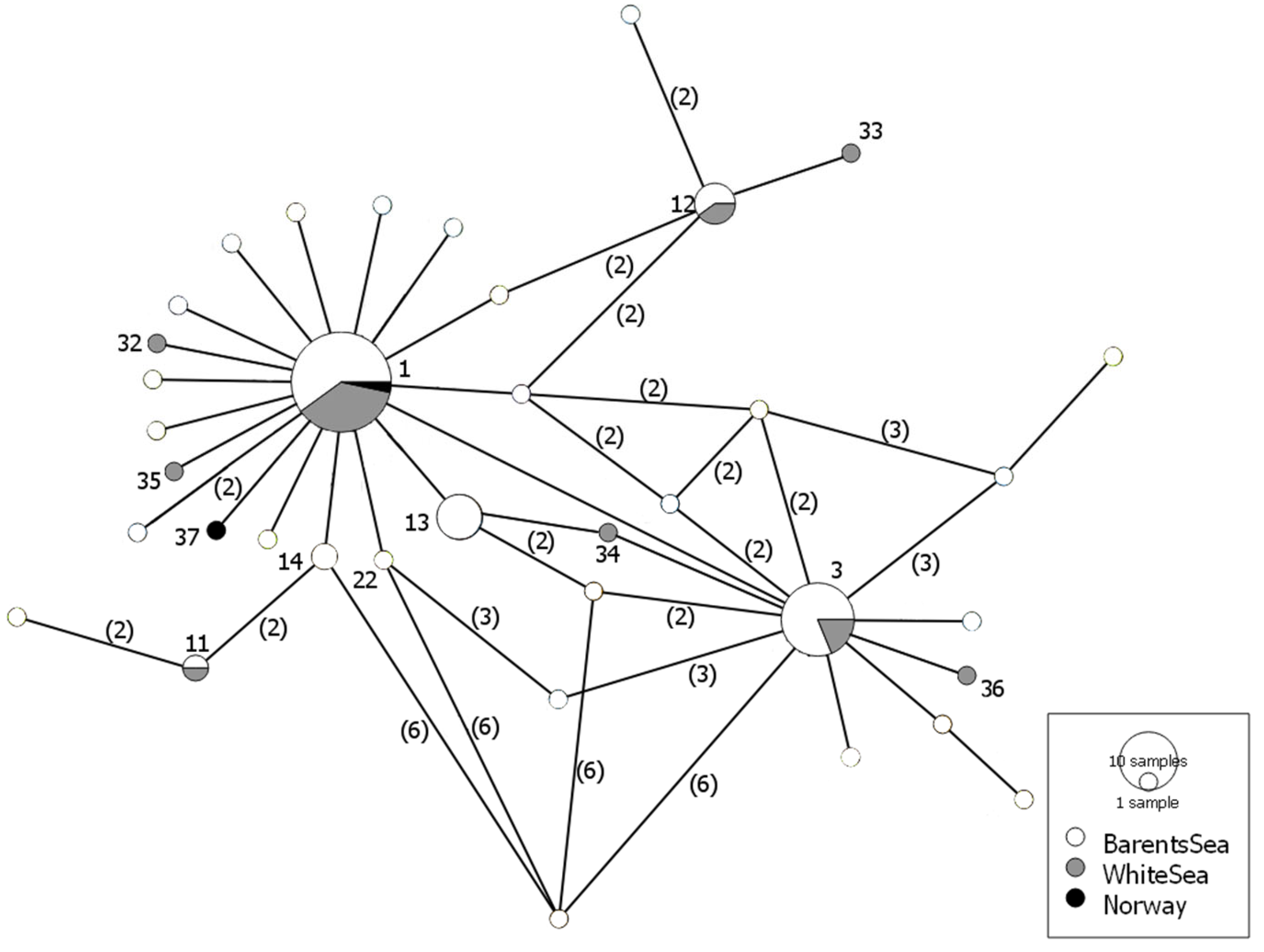
2.3. Genetic Diversity and Haplotype Analysis

The nucleotide sequences were translated into the format suitable for constructing a median-joining haplotype network in the PopArt program [47]. The genetic differentiation index (FST) and p-value were calculated using the Arlequin 3.5.1.3 program [48], and a FaBox 1.41 converter was used to convert the fasta file into the format required for calculation [49]. The average number of nucleotide substitutions (K), the number of polymorphic sites (S), the number of haplotypes (h), haplotypic diversity (Hd), and nucleotide diversity (Pi) in each sample and across all the samples were analyzed in DnaSP 5.10.01 software package [50]. Tajima’s neutrality tests [51] were performed using Arlequin v.3.5.2.2 [48] with 1000 non-parametric permutations (p  =  0.05). Sequences of mtDNA Cox2 genes from the waters of Norway OP418114-OP418115 [30] and from the White Sea previously obtained by us (OQ274151-OQ274172) were retrieved from GenBank (NCBI).

3. Results

Third-stage juveniles of Phocanema spp. localized in the liver, mesentery, and less commonly, in the muscles of the fish. Parasitological indices are given in Table 2.
The sequences of almost all juveniles (68 out of 69) genotyped in this study were molecularly similar (p-distance ≤ 0.021%) with those of P. bulbosum available in GenBank (NCBI), namely, with the sequences deposited under numbers HM147280, KU558720, OP418114, and OP418115. At the same time, one juvenile from our material (OQ731831) was molecularly identical with P. decipiens sensu stricto (OK338713 and KU558723).
The alignment of all P. bulbosum sequences (474 bp) contained 36 variable sites (S), which resulted in 31 haplotypes. Genetic diversity indices for localities and hosts are shown in Table 3. The overall value of haplotype diversity (Hd) was 0.890, that of nucleotide diversity (Pi) was 0.00516, while the value of the average number of nucleotide differences (K) was 2.430. The genetic diversity indices calculated for P. bulbosum from each locality showed a similar haplotype diversity, which ranged between 0.801 for G1 and 1.000 for G6. No trends in the changes in Hd and Pi were revealed. Neutrality test, Tajima’s D, showed that negative values were statistically significant (p-value < 0.05) for all divisions (Table 3), except for samples G2 and G4. Statistically significant negative values may indicate the effects of the purifying selection on the gene and/or that populations underwent recent expansion.
The value of the FST between P. bulbosum samples obtained from the Atlantic cod G. morhua and the American plaice Hippoglossoides platessoides in the Barents Sea sorted by hosts was negative. The values of the FST between P. bulbosum samples sorted by locality were also negative in all cases except pair G1–G2 (FST = 0.033, p > 0.05). Thus, in both cases of sorting, the samples are not statistically different.
Table 3. Genetic characteristics of Phocanema bulbosum samples in the Barents Sea (N—number of sequences, S—number of polymorphic sites, h—number of haplotypes; Hd—haplotypic diversity, m—error of mean, σ—standard deviation of Hd, K—number of nucleotide substitutions, Pi—nucleotide diversity, D—Tajima’s neutrality test, and p-value—Tajima’s neutrality test p-value). Significant p-values (p < 0.05) are highlighted in bold.
SamplesNShHd ± mσKPiDp-Value
G12219120.801 ± 0.0070.0882.2990.005−2.0670.010
G2171290.890 ± 0.0030.0542.3820.005−1.2340.109
G32219140.922 ± 0.0020.0452.8610.006−1.6680.018
G44430.833 ± 0.0490.2222.0000.004−0.7800.187
G51N/A1N/AN/AN/AN/AN/AN/A
G62121.000 ± 0.2500.51.0000.0020.0001.000
Cod3620150.846 ± 0.0020.0421.9650.004−1.9960.011
Flounder3229210.921 ± 0.0010.0402.9720.006−2.0920.005
All6836310.890 ± 0.0010.0272.4300.005−2.159<0.00001
Neither the grouping by geographic localities (Figure 2) nor the grouping by the host (Figure 3) revealed any trends in the distribution of P. bulbosum juveniles in the Barents Sea. Four haplotypes were present in two or more sampling areas (G1–G6). One of them (OQ731840 and identical ones, Figure 3 No.1) was found in all the six sampling areas (Figure 2). Another one (OQ731832 and identical ones, Figure 3 No. 3) was found in G1–3. The third one (OQ731834 and identical ones, Figure 3 No. 13) was found in G2–4. The fourth one (OQ731867 and identical ones, Figure 3 No. 12) was found in G1 and G3. The network revealed 27 haplotypes that were found only in a single sampling area.
Figure 2. Median-joining haplotype network of Phocanema bulbosum Cox2 gene sequences obtained from the Atlantic cod Gadus morhua and the American plaice Hippoglossoides platessoides in the Barents Sea sorted by geographic locations within the boundaries of the Russian Exclusive Economic Zone (Table 1, Figure 1). Circle size represents the frequency of the haplotype. Hatch marks show the number of mutations distinguishing the haplotypes.
Diversity 15 01036 g002
Figure 3. Median-joining haplotype network of Phocanema bulbosum Cox2 gene sequences obtained from muscular sections of the Atlantic cod Gadus morhua and the American plaice Hippoglossoides platessoides in the Barents Sea sorted by hosts. Circle size represents the frequency of the haplotype. Hatch marks show the number of mutations distinguishing the haplotypes.
Diversity 15 01036 g003
Five haplotypes were shared by both hosts (Figure 3). Haplotype OQ731840 and identical ones (Figure 4, No. 1) together with OQ731832 and identical ones (Figure 4, No. 3) were the most frequent (12 and 9 sequences, respectively). Ten haplotypes were present only in the Atlantic cod, while 16 haplotypes were present only in the American plaice (Figure 4).
Only two sequences of the Cox2 gene of P. bulbosum specimens from the Barents Sea were available in GenBank before this study: OP418114 and OP418115. They belong to specimens collected from the Atlantic cod. Sequence OP418114 matches our haplotype number 1 (OQ731840 and identical ones) (Figure 4, ‘Norway’). Sequence OP418115 has no match and forms a separate haplotype (one substitution).

4. Discussion

There were no records of P. bulbosum juveniles in fish from the Russian waters of the Barents Sea before our study, although Phocanema spp. was the dominant group of helminths in terms of occurrence in the Atlantic cod and the American plaice in all previous parasitological research in this area. Our finding of this species was undoubtedly due to the use of improved identification methods. It is obvious that P. bulbosum was previously reported from the Russian waters of the Barents Sea as P. decipiens sensu lato [52,53,54,55,56].
The Atlantic cod and the American plaice are common and widespread fishes in the Barents Sea, occurring, on average, in 78–93% of bottom trawl catches in most of this area all year round [57]. The cod makes an extended autumn–winter migration from the southern, eastern, central, and northwestern regions to the southwestern part of the sea, where most of its mature individuals spawn off the northwestern coast of Norway in March–April. During the summer months, it returns to the feeding areas [58]. The American plaice, on the contrary, makes only minor local migrations, being distributed all year round over most of the Barents Sea.
The diet of cod juveniles (age 1–2 years, TL up to 24 cm) is dominated by Hyperiidae, Euphausiidae, and the northern prawn (shrimp) Pandalus borealis. As it grows, the cod starts to consume more small pelagic fish such as capelin, Mallotus villosus (Müller, 1776), polar cod Boreogadus saida (Lepechin, 1774), and herring Clupea spp. and juvenile bottom fish such as the Atlantic cod, haddock Melanogrammus aeglefinus (Linnaeus, 1758), Sebastes spp., American plaice, etc. [57].
The diet of juvenile H. platessoides (TL up to 15 cm) consists of benthic organisms such as echinoderms (mainly Ophiuroidea) and annelids, and less often, Euphausiidae, Parathemisto sp. and Mysidacea. Larger individuals switch to the predominant consumption of various fish (mainly capelin, polar cod, and Atlantic cod fry), and less often, shrimps (Pandalidae) and bivalve molluscs. At an older age (length more than 30 cm), in addition to the food objects mentioned above, H. platessoides also consumes fishery waste such as trimmings and homogenized residues from the processing of fish and invertebrates on fishing and factory vessels [57].
Juveniles of the genus Phocanema commonly parasitize bottom fish, which become infected while feeding on bottom crustaceans (amphipods, copepods, mysids, and isopods), polychaetes, and molluscs as well as other fish infected with this nematode (its intermediate or paratenic hosts) [59,60,61,62,63]. This explains the fact that H. platessoides and G. morhua are paratenic hosts in the life cycle of these nematodes. Significantly higher infection with P. bulbosum in the American plaice in the southwestern part of the Barents Sea (prevalence 15.60% vs. 6.40% in the north-east) may be due to its active consumption of fishing waste in this area. The absence of geographical differences in the infection of cod with this nematode is due to its migratory activity: Russian and Norwegian sampling sites are located on the cod spawning migration routes from the southern part of the Barents Sea.
Levsen et al. [30] studied infection of the Atlantic cod, the saithe, Pollachius virens (Linnaeus, 1758), and the haddock M. aeglefinus with P. bulbosum in the Barents Sea, namely, at ‘Helmsøybanken’ bank (approximately 71°N 25°E) off West-Finnmark, Norway. According to these authors, the mean prevalence of this parasite with the Atlantic cod infection in 2019 was 37.7%, while the maximal intensity never exceeded 14, being 1.3 ± 2.7 on average. These values are much greater than those recorded in our study (Table 2). Though these differences could be due to methodology, e.g., the use of a hydraulic pressing device and 366 nm UV-light by Levsen et al. [30], it cannot be ruled out that they reflect real differences in infection between sampling sites. Najda et al. [24] also presented data on the infection of G. morhua by P. bulbosum in the Barents Sea with comparable mean intensity of infection (1.2 ind.), but without genetic data on P. bulbosum.
Sequences of P. bulbosum that we collected earlier from the White Sea cod, Gadus morhua marisalbi (Derjugin, 1920), the navaga Eleginus navaga (Walbaum, 1792), and the shorthorn sculpin, Myoxocephalus scorpius (Linnaeus, 1758) in the White Sea (Velikaya Salma Straight, Lomonosov Moscow State University White Sea Biological Station) were deposed in GenBank with accession numbers OQ274151-OQ274172. Despite the obvious isolation caused by a low depth of the White Sea Throat (app. 30 m) and freshening of the White Sea, especially in the Dvina Bay and the Onega Bay, where salinity could be 8–12‰, we see no clear differentiation between the seas (Figure 4). Three haplotypes were present in both seas. The network revealed five haplotypes that were found only in the White Sea, twenty four haplotypes found only in the Barents Sea, and one only in Norwegian waters.
The genetic diversity indices calculated for mtDNA Cox2 sequences of P. bulbosum (Table 3) from all sampling localities in the Barents Sea within the boundaries of the Russian Exclusive Economic Zone (G1-G6) showed a high haplotype diversity (Hd) but a low nucleotide diversity (Pi). A single haplotype (OQ731840 and identical ones, Figure 4, No. 1) clearly dominated, being distributed in all six localities in the Barents Sea (Figure 1) as well as in the White Sea (Figure 4, No. 1). It indicates a clear connection between the samples of P. bulbosum from the Barents Sea and from the White Sea. Moreover, the distribution of this haplotype seems even wider since it corresponds to the haplotype (OP418114) of P. bulbosum from the waters of Norway (Figure 4). A high frequency of this haplotype and its wide geographic and host range might mean that it is the ancestral haplotype of this species.

5. Conclusions

This is the first study to reveal the genetic diversity of P. bulbosum. Our molecular data on the Cox2 gene and the comparison of the available sequences from the White Sea and from the waters of Norway showed the genetic homogeneity of third-stage juveniles of P. bulbosum and the absence of differences between geographic locations and host species. Using different genetic markers to assess genetic variation of Phocanema spp. led to the discovery of a similar picture of low genetic distances between conspecific populations, even though they were collected thousands of kilometers apart, indicating high levels of gene flow. Further studies involving more sequenced specimens and host species might reveal some patterns in the distribution of P. bulbosum larvae in Russian waters. However, given the unity and scale of the marine ecosystem, the current picture is quite consistent with the modern concepts of the distribution of generalist helminths in marine ichthyocenoses. This study provides useful information about the genetic diversity of these parasites in the Barents Sea.

Author Contributions

Conceptualization, genetics, and writing—original draft preparation, I.I.G.; genetics, A.D.L. and O.R.E.; sample collection, Y.I.B. and M.Y.K.; writing—editing, I.I.G., S.G.S. and O.R.E. All authors have read and agreed to the published version of the manuscript.

Funding

The research for this paper did not receive any specific grant from funding agencies in the public, commercial or not-for-profit sectors.

Institutional Review Board Statement

Not applicable.

Informed Consent Statement

Not applicable.

Data Availability Statement

All data generated or analysed during this study are included in this published article.

Acknowledgments

We would like to thank Nikolay Mugue, Anna Barmintseva, and Lubov Mugue (Molecular Genetics Department of the Russian Federal Research Institute of Fisheries and Oceanography) for their help with the genetic studies and valuable advice.

Conflicts of Interest

The authors declare no conflict of interest.

References

  1. Myers, B.J. Phocanema, a new genus for the anisakid nematode of seals. Can. J. Zool. 1959, 37, 459–465. [Google Scholar] [CrossRef]
  2. Bao, M.; Giulietti, L.; Levsen, A.; Karlsbakk, E. Resurrection of genus Phocanema Myers, 1959, as a genus independent from Pseudoterranova Mozgovoĭ, 1953, for nematode species (Anisakidae) parasitic in pinnipeds and cetaceans, respectively. Parasitol. Int. 2023, 97, 102794. [Google Scholar] [CrossRef] [PubMed]
  3. Klimpel, S.; Palm, H.W. Anisakid Nematode (Ascaridoidea) Life Cycles and Distribution: Increasing Zoonotic Potential in the Time of Climate Change? In Progress in Parasitology. Parasitology Research Monographs; Mehlhorn, H., Ed.; Springer: Berlin/Heidelberg, Germany, 2011; Volume 2. [Google Scholar] [CrossRef]
  4. Bowen, W.D. Population biology of sealworm (Pseudoterranova decipiens) in relation to its intermediate and seal hosts. Can. Bull. Fish. Aquat. Sci. 1990, 222, 306. [Google Scholar]
  5. Køie, M.; Berland, B.; Burt, M.D. Development to third-stage larvae occurs in the eggs of Anisakis simplex and Pseudotetranova decipiens (Nematoda, Ascaridoidea, Anisakidae). Can. J. Fish. Aquat. Sci. 1995, 52, 134–139. [Google Scholar] [CrossRef]
  6. Ko, R.C. Fish-borne parasitic zoonoses. In Fish Diseases and Disorders, Vol. 1: Protozoan and Metazoan Infections; Woo, P.T.K., Ed.; CABI Publishing: Oxford, UK, 1995; pp. 631–671. [Google Scholar]
  7. Arizono, N.; Miura, T.; Yamada, M.; Tegoshi, T.; Onishi, K. Human infection with Pseudoterranova azarasi roundworm. Emerg. Infect. Dis. 2011, 17, 555–556. [Google Scholar] [CrossRef] [PubMed]
  8. Brunet, J.; Pesson, B.; Royant, M.; Lemoine, J.-P.; Pfaff, A.W.; Abou-Bacar, A.; Yera, H.; Fréalle, E.; Dupouy-Camet, J.; Merino-Espinosa, G.; et al. Molecular diagnosis of Pseudoterranova decipiens s.s. in human, France. BMC Infect. Dis. 2017, 17, 397. [Google Scholar] [CrossRef] [PubMed]
  9. Cavallero, S.; Scribano, D.; D’Amelio, S. First case report of invasive pseudoterranoviasis in Italy. Parasitol. Int. 2016, 65, 488–490. [Google Scholar] [CrossRef] [PubMed]
  10. Menghi, C.I.; Gatta, C.L.; Arias, L.E.; Santoni, G.; Nicola, F.; Smayevsky, J.; Degese, M.F.; Krivokapich, S.J. Human infection with Pseudoterranova cattani by ingestion of “ceviche” in Buenos Aires, Argentina. Rev. Argent. Microbiol. 2020, 52, 118–120. [Google Scholar] [CrossRef]
  11. Pinel, C.; Beaudevin, M.; Chermette, R.; Grillot, R.; Ambroise-Thomas, P. Gastric anisakidosis due to Pseudoterranova decipiens larva. Lancet 1996, 347, 1829. [Google Scholar] [CrossRef]
  12. Torres, P.; Jercic, M.I.; Weitz, J.C.; Dobrew, E.K.; Mercado, R.A. Human Pseudoterranovosis, an emerging infection in Chile. J. Parasitol. 2007, 93, 440–443. [Google Scholar] [CrossRef]
  13. Suzuki, S.; Bandoh, N.; Goto, T.; Uemura, A.; Sasaki, M.; Harabuchi, Y. Severe laryngeal edema caused by Pseudoterranova species. Medicine 2021, 100, e24456. [Google Scholar] [CrossRef] [PubMed]
  14. Cobb, N.A. Neue parasitische Nematoden. In Beitrage zur Fauna Spitzbergens; Kukenthal, W., Ed.; Archiv für Naturgeschichte: Berlin, Germany, 1888; Volume 55, pp. 149–159. [Google Scholar]
  15. Stiles, C.W.; Hassall, A. Internal Parasites of the Fur Seal. In The Fur Seals and Fur Seal Islands of the North Pacific Ocean; Report on Fur Seal Investigations; Government Printing Office: Washington, DC, USA, 1899; pp. 99–177. [Google Scholar]
  16. Baylis, H.A. On the ascarids parasitic in seals, with special reference to the genus Contracaecum. Parasitology 1937, 29, 121–130. [Google Scholar] [CrossRef]
  17. Paggi, L.; Nascetti, G.; Cianchi, R.; Orecchia, P.; Mattiucci, S.; D’amelio, S.; Berland, B.; Brattey, J.; Smith, J.W.; Bullini, L. Genetic evidence for three species within Pseudoterranova decipiens (Nematoda, Ascaridida, Ascaridoidea) in the north Atlantic and Norwegian and Barents seas. Int. J. Parasitol. 1991, 21, 195–212. [Google Scholar] [CrossRef] [PubMed]
  18. Di Deco, M.A.; Orecchia, P.; Paggi, L.; Petrarca, V. Morphometric stepwise discriminant analysis of three genetically identified species within Pseudoterranova decipiens (Krabbe, 1878) (Nematoda: Ascaridida). Syst. Parasitol. 1994, 29, 81–88. [Google Scholar] [CrossRef]
  19. Mattiucci, S.; Paggi, L.; Nascetti, G.; Ishikura, H.; Kikuchi, K.; Sato, N.; Cianchi, R.; Bullini, L. Allozyme and morphological identification of shape Anisakis, Contracaecum and Pseudoterranova from Japanese waters (Nematoda, Ascaridoidea). Syst. Parasitol. 1998, 40, 81–92. [Google Scholar] [CrossRef]
  20. Takano, T.; Sata, N. Multigene phylogenetic analysis reveals non-monophyly of Anisakis s.l. and Pseudoterranova (Nematoda: Anisakidae). Parasitol. Int. 2022, 91, 102631. [Google Scholar] [CrossRef] [PubMed]
  21. Brattey, J.; Davidson, W.S. Genetic variation within Pseudoterranova decipiens (Nematoda: Ascaridoidea) from Canadian Atlantic marine fishes and seals: Characterization by RFLP analysis of genomic DNA. Can. J. Fish. Aquat. Sci. 1996, 53, 333–341. [Google Scholar] [CrossRef]
  22. Karpiej, K.; Simard, M.; Pufall, E.; Rokicki, J. Anisakids (Nematoda: Anisakidae) from ringed seal, Pusa hispida, and bearded seal, Erignathus barbatus (Mammalia: Pinnipedia) from Nunavut region. J. Mar. Biol. Assoc. UK 2014, 94, 1237–1241. [Google Scholar] [CrossRef]
  23. Paoletti, M.; Mattiucci, S.; Colantoni, A.; Levsen, A.; Gay, M.; Nascetti, G. Species-specific Real Time-PCR primers/probe systems to identify fish parasites of the genera Anisakis, Pseudoterranova and Hysterothylacium (Nematoda: Ascaridoidea). Fish. Res. 2018, 202, 38–48. [Google Scholar] [CrossRef]
  24. Najda, K.; Kijewska, A.; Kijewski, T.; Plauška, K.; Rokicki, J. Distribution of ascaridoid nematodes (Nematoda: Chromadorea: Ascaridoidea) in fish from the Barents Sea. Oceanol. Hydrobiol. Stud. 2018, 47, 128–139. [Google Scholar] [CrossRef]
  25. Paggi, L.; Mattiucci, S.; Ishikura, H.; Kikuchi, K.; Sato, N.; Nascetti, G.; Cianchi, R.; Bullini, L. Molecular genetics in anisakid nematodes from the Pacific Boreal Region. In Host Response to International Parasitic Zoonoses; Springer: Tokyo, Japan, 1998; pp. 83–107. [Google Scholar]
  26. Najda, K.; Simard, M.; Osewska, J.; Dziekońska-Rynko, J.; Dzido, J.; Rokicki, J. Anisakidae in beluga whales Delphinapterus leucas from Hudson Bay and Hudson Strait. Dis. Aquat. Org. 2015, 115, 9–14. [Google Scholar] [CrossRef] [PubMed]
  27. Koitsanou, E.; Sarantopoulou, J.; Komnenou, A.; Exadactylos, A.; Dendrinos, P.; Papadopoulos, E.; Gkafas, G.A. First Report of the Parasitic Nematode Pseudoterranova spp. Found in Mediterranean Monk Seal (Monachus monachus) in Greece: Conservation Implications. Conservation 2022, 2, 122–133. [Google Scholar] [CrossRef]
  28. Bristow, G.A.; Berland, B. On the ecology and distribution of Pseudoterranova decipiens C (Nematoda: Anisakidae) in an intermediate host, Hippoglossoides platessoides, in northern Norwegian waters. Int. J. Parasitol. 1992, 22, 203–208. [Google Scholar] [CrossRef] [PubMed]
  29. Karpiej, K.; Dzido, J.; Rokicki, J.; Kijewska, A. Anisakid Nematodes of Greenland halibut Reinhardtius hippoglossoides from the Barents Sea. J. Parasitol. 2013, 99, 650–654. [Google Scholar] [CrossRef] [PubMed]
  30. Levsen, A.; Cipriani, P.; Palomba, M.; Giulietti, L.; Storesund, J.E.; Bao, M. Anisakid parasites (Nematoda: Anisakidae) in 3 commercially important gadid fish species from the southern Barents Sea, with emphasis on key infection drivers and spatial distribution within the hosts. Parasitology 2022, 149, 1942–1957. [Google Scholar] [CrossRef] [PubMed]
  31. Gilabert, A.; Wasmuth, J.D. Unravelling parasitic nematode natural history using population genetics. Trends Parasitol. 2013, 29, 438–448. [Google Scholar] [CrossRef] [PubMed]
  32. Criscione, C.D.; Poulin, R.; Blouin, M.S. Molecular ecology of parasites: Elucidating ecological and microevolutionary processes. Mol. Ecol. 2005, 14, 2247–2257. [Google Scholar] [CrossRef]
  33. Frainer, A.; McKie, B.G.; Amundsen, P.-A.; Lafferty, K.D. Parasitism and the biodiversity-functioning relationship. Trends Ecol. Evol. 2018, 33, 260–268. [Google Scholar] [CrossRef]
  34. Blouin, M.S. Molecular prospecting for cryptic species of nematodes: Mitochondrial DNA versus internal transcribed spacer. Int. J. Parasitol. 2002, 32, 527–531. [Google Scholar] [CrossRef]
  35. Nieberding, C.; Morand, S.; Libois, R.; Michaux, J.R. A parasite reveals cryptic phylogeographic history of its host. Proc. R. Soc. London Ser. B 2004, 271, 2559–2568. [Google Scholar] [CrossRef]
  36. Baldwin, R.E.; Rew, M.B.; Johansson, M.L.; Banks, M.A.; Jacobson, K.C. Population structure of three species of Anisakis nematodes recovered from Pacific sardines (Sardinops sagax) distributed throughout the California current system. J. Parasitol. 2011, 97, 545–554. [Google Scholar] [CrossRef] [PubMed]
  37. Mattiucci, S.; Giulietti, L.; Paoletti, M.; Cipriani, P.; Gay, M.; Levsen, A.; Klapper, R.; Karl, H.; Bao, M.; Pierce, G.J.; et al. Population genetic structure of the parasite Anisakis simplex (s.s.) collected in Clupea harengus L. from North East Atlantic fishing grounds. Fish. Res. 2018, 202, 103–111. [Google Scholar] [CrossRef]
  38. Valentini, A.; Mattiucci, S.; Bondanelli, P.; Webb, S.C.; Mignucci-Giannone, A.A.; Colom-Llavina, M.M.; Nascetti, G. Genetic relationships among Anisakis species (Nematoda: Anisakidae) inferred from mitochondrial cox2 sequences, and comparison with allozyme data. J. Parasitol. 2006, 92, 156–166. [Google Scholar] [CrossRef] [PubMed]
  39. Ramilo, A.; Rodríguez, H.; Pascual, S.; González, Á.F.; Abollo, E. Population genetic structure of Anisakis simplex infecting the European hake from north east Atlantic fishing grounds. Animals 2023, 13, 197. [Google Scholar] [CrossRef] [PubMed]
  40. Liu, G.H.; Nadler, S.A.; Liu, S.S.; Podolska, M.; D’Amelio, S.; Shao, R.; Gasser, R.B.; Zhu, X.Q. Mitochondrial phylogenomics yields strongly supported hypotheses for ascaridomorph nematodes. Sci. Rep. 2016, 6, 39248. [Google Scholar] [CrossRef] [PubMed]
  41. Bykhovskaya-Pavlovskaya, I.E. Parasites of Fishes. The Manual; Nauka: Saint Petersburg, Russia, 1985; 124p. [Google Scholar]
  42. Klimpel, S.; Kuhn, T.; Münster, J.; Dörge, D.D.; Klapper, R.; Kochmann, J. Parasites of Marine Fish and Cephalopods; Springer: Cham, Switzerland, 2019; 169p. [Google Scholar]
  43. Bush, A.O.; Lafferty, K.D.; Lotz, J.M.; Shostak, A.W. Parasitology meets ecology on its own terms: Margolis et al. revisited. J. Parasitol. 1997, 83, 575–583. [Google Scholar] [CrossRef] [PubMed]
  44. Ivanova, N.V.; Zemlak, T.S.; Hanner, R.H.; Hebert, P.D. Universal primer cocktails for fish DNA barcoding. Mol. Ecol. Notes 2007, 7, 544–548. [Google Scholar] [CrossRef]
  45. Kearse, M.; Moir, R.; Wilson, A.; Stones-Havas, S.; Cheung, M.; Sturrock, S.; Buxton, S.; Cooper, A.; Markowitz, S.; Duran, C.; et al. Geneious Basic: An integrated and extendable desktop software platform for the organization and analysis of sequence data. Bioinformatics 2012, 28, 1647–1649. [Google Scholar] [CrossRef]
  46. Kumar, S.; Stecher, G.; Li, M.; Knyaz, C.; Tamura, K. MEGA X: Molecular evolutionary genetics analysis across computing platforms. Mol. Biol. Evol. 2018, 35, 1547–1549. [Google Scholar] [CrossRef]
  47. Leigh, J.W.; Bryant, D. POPART: Full-feature software for haplotype network construction. Methods Ecol. Evol. 2015, 6, 1110–1116. [Google Scholar] [CrossRef]
  48. Excoffier, L.; Lischer, H.E. Arlequin suite ver 35: A new series of programs to perform population genetics analyses under Linux and windows. Mol. Ecol. Res. 2010, 10, 564–567. [Google Scholar] [CrossRef] [PubMed]
  49. Villesen, P. FaBox: An online toolbox for FASTA sequences. Mol. Ecol. Notes 2007, 7, 965–968. [Google Scholar] [CrossRef]
  50. Librado, P.; Rozas, J. DnaSP v5: A software for comprehensive analysis of DNA polymorphism data. Bioinformatics 2009, 25, 1451–1452. [Google Scholar] [CrossRef] [PubMed]
  51. Tajima, F. Statistical method for testing the neutral mutation hypothesis by DNA polymorphism. Genetics 1989, 123, 585–595. [Google Scholar] [CrossRef]
  52. Karasev, A. On infestation of cod (Gadus morhua morhua L.) with nematodes of Anisakidae Skrjabin et Karokhin, 1945 in the Barents Sea. Int. Counc. Explor. Sea CM 1980, 24, 10. [Google Scholar]
  53. Karasev, A. Using of parasites as biological tags when studying intraspecies structure of cod in the coastal areas of Russia and Norway. Bull. Scand. Soc. Parasitol. 1994, 4, 17. [Google Scholar]
  54. Karasev, A.B. Catalog of Fish Parasites in the Barents Sea; PINRO Publishing House: Murmansk, Russia, 2003; 150p. [Google Scholar]
  55. Karasev, A.; Bakai, Y. Infection of the Barents Sea cod Gadus morhua and redfish Sebastes mentella with larval anisakidae nematodes: Long-term data. Bull. Scand. Soc. Parasitol. 1994, 4, 11–12. [Google Scholar]
  56. Karasev, A.B.; Bakai, Y.I.; Kalashnikova, M.Y.; Bessonov, A.A. Parasitological Monitoring of Commercial Fish in the Barents Sea: History, Results, Economic Significance; PINRO Publishing House: Murmansk, Russia, 2022; 44p. [Google Scholar]
  57. Dolgov, А.V. Composition, Formation and Trophic Structure of the Barents Sea Fish Communities; PINRO Publishing House: Murmansk, Russia, 2016; 336p. (In Russian) [Google Scholar]
  58. Yaragina, N.; Aglen, A.; Sokolov, K. Cod. In The Barents Sea: Ecosystem, Resources, Management. Half a Century of Russian-Norwegian Cooperation; Jakobsen, T., Ozhigin, V.K., Eds.; Tapir Academic Press: Trondheim, Norway, 2011; Chapter 5.4; pp. 225–270. [Google Scholar]
  59. Biørge, A. An isopod as intermediate host of cod-worm. Fiskeridir. Scr. Ser. Havunders. 1979, 16, 561–565. [Google Scholar]
  60. Marcogliese, D. Neomysis americana (Crustacea: Mysidacea) as an intermediate host for sealworm, Pseudoterranova decipiens (Nematoda: Ascaridoidea), and spirurid nematodes (Ascaridoidea). Can. J. Fish Aquat. Sci. 1992, 49, 513–515. [Google Scholar] [CrossRef]
  61. Martell, D.; MacClelland, G. Transmission of Pseudoterranova decipiens (Nematoda: Ascaridoidea) via bentic macrofauna to sympatric flatfishes (Hippoglossoides platessoides, Pleuronectes ferrugineus, P. americanus) on Sable Island Bank, Canada. Mar. Biol. 1995, 122, 129–135. [Google Scholar] [CrossRef]
  62. Hauksson, E. Diet and sealworm infections of short spinde sea scorpions (Myoxocephalus scorpius) and Atlantic catfish (Anarhichas lupus) in Icelandic waters. Bull. Scand. Soc. Parasitol. 1999, 9, 35–36. [Google Scholar]
  63. Hemmingsen, W.; McKenzie, K. The parasite fauna of the Atlantic cod, Gadus morhua L. Adv. Mar. Biol. 2001, 40, 1–80. [Google Scholar]
Figure 4. Median-joining haplotype network of Phocanema bulbosum sorted by the Barents Sea (Russian waters, this study), the White Sea (our previous studies), and the waters of Norway [30].
Diversity 15 01036 g004
Table 2. Occurrence of Phocanema spp. third-stage juveniles in two studied fish species.
LocalitiesAtlantic Cod Gadus morhuaAmerican Plaice Hippoglossoides platessoides
nPrevalence, %Intensity, min–maxMean
Intensity
nPrevalence, %Intensity,
Min–Max
Mean
Intensity
G1-G31504.71–31.5725015.61–61.62
G4-G6754.01–21.661256.41–21.25
Total2254.41–31.6037512.51–61.55
Disclaimer/Publisher’s Note: The statements, opinions and data contained in all publications are solely those of the individual author(s) and contributor(s) and not of MDPI and/or the editor(s). MDPI and/or the editor(s) disclaim responsibility for any injury to people or property resulting from any ideas, methods, instructions or products referred to in the content.

