Article

First Report of Diseases and Compromised Health Conditions on Hard Corals around Rodrigues Island, Southwest Indian Ocean

by
Shakeel Yavan Jogee
1,
Shivam Gopalsing
1,
Sruti Jeetun
1,
Melanie Ricot
1,
Nawsheen Taleb-Hossenkhan
1,
Sushma Mattan-Moorgawa
1,2,
Deepeeka Kaullysing
1,2,
Diah Permata Wijayanti
3,
Beatriz Estela Casareto
4,
Yoshimi Suzuki
4 and
Ranjeet Bhagooli
1,2,3,5,6,*
1
Department of Biosciences and Ocean Studies, Faculty of Science & Pole of Research Excellence in Sustainable Marine Biodiversity, University of Mauritius, Réduit 80837, Mauritius
2
The Biodiversity and Environment Institute, Réduit 80837, Mauritius
3
Department of Marine Science, Faculty of Fisheries and Marine Science, Diponegoro University, Jalan Prof. Soedarto SH, Kampus Tembalang, Semarang 50275, Indonesia
4
Graduate School of Science and Technology, Shizuoka University, Suruga-ku, Shizuoka 422-8529, Japan
5
The Society of Biology (Mauritius), Réduit 80837, Mauritius
6
Institute of Oceanography and Environment (INOS), Universiti Malaysia Terengganu, Kuala Nerus, Kuala Terengganu 21030, Malaysia
*
Author to whom correspondence should be addressed.
Diversity 2023, 15(10), 1086; https://doi.org/10.3390/d15101086
Submission received: 30 August 2023 / Revised: 12 October 2023 / Accepted: 13 October 2023 / Published: 15 October 2023
(This article belongs to the Special Issue Indo-Pacific Coral Diseases: Diversity, Impacts and Solutions)

Abstract

:
Coral diseases represent a prominent menace to coral reefs and to the associated ecological services they provide to the surrounding coastal communities. Studies on diseases and compromised health conditions (CHCs) on hard corals in the Southwest Indian Ocean (SWIO) are scarce, and their consequences are often overlooked. This study aimed to establish the baseline prevalence of diseases and CHC of hard corals around Rodrigues Island. Coral disease and CHC prevalence were visually assessed using 2 m × 50 m belt transects at eight sites around the island. This is the first report of four coral diseases, namely White Plague (WP), White Syndrome (WS), Black Band (BB), and Growth Anomalies (GA), and two CHCs, two forms of Pink Pigmentation Responses (PPR)—Pink Patches (PP) and Pink Line Syndrome (PLS)—observed on six genera of hard corals from the island of Rodrigues. PP on Fungia (15.92 ± 5.65%), followed by the WS on Montipora (4.67 ± 3.72%) and GA on Gardineroseris (4.16 ± 4.16%), so far unreported from the SWIO, were the most prevalent around the island. The least prevalent disease was BB on Montipora (0.13 ± 0.13%). Although the overall disease and CHC prevalence for Rodrigues Island (0.98 ± 0.30%) were much lower than the surrounding islands in the SWIO, the observations of these diseases and CHCs on hard corals and relevant environmental parameters warrant further in-depth characterization to better inform coral reefs management and conservation actions.

1. Introduction

Coral reefs represent one of the most productive and ecologically diverse marine tropical ecosystems on the planet. However, coral reefs are also prone to a plethora of abiotic and biotic stressors that constantly challenge the resilience and subsistence of such ecosystems, especially in a globally warming ocean [1]. Coral diseases are one such group of biotic stressors that have been impacting coral reefs through significant mortalities and coral community structure modifications in the last few decades [2,3]. Back in 1965, during the early stages of coral disease research, only a handful of coral diseases, mostly occurring as isolated cases, including Growth Anomalies, Black Band Disease, White Band Disease, and White Plague Type I, were known. Since then, more than 40 coral diseases have been recorded on more than 200 scleractinian species of coral hosts and from over 75 countries [4].
The coral reefs of the Caribbean, often dubbed as a ‘disease hotspot’ [1,5,6], have been suffering largely from the impacts of coral diseases since the late 1970s and early 1980s, with over 80% of the population of keystone species, such as Acropora palmata and Acropora cervicornis, being decimated by the White Band disease [7]. These mass coral mortalities that were associated with the coral diseases led to the rapid shift from coral-dominated to algal-dominated or weedy coral-dominated (such as Porites or Agaricia) reefs in the Caribbean region and contributed to the loss of the characteristic zonation patterns innate to that region [8,9]. Although comparatively less studied than the Caribbean, the Indo-Pacific tropical coral reefs are also not spared from the damaging impacts of coral diseases with several reef-building species of Acropora, Montipora, and Porites [10,11,12,13,14,15,16] reported with signs of widespread coral diseases. The current disease prevalence level in that region, along with other stressors of coral health such as increased fluctuations in light and sea surface temperatures [16,17], also indicate that this region is at risk of community structure changes [11,18]. Several recent studies have also reported the occurrence and rising prevalence of coral diseases from the northern Indian Ocean region [14,15,17,19,20]. Recently, around 20 coral diseases affecting 16 coral taxa were also recorded from the Red Sea, with the most common one being the Acropora White Syndrome disease [17].
In addition to coral diseases, other compromised health conditions (CHCs) in hard corals, such as Pigmentation Responses, sediment damage, unexplained bleaching, sponge or cyanobacterial growth, and trematode infestation [21,22] also threaten the coral reef ecosystem as they can be a major contributor of coral mortality and inducing factor for coral diseases [23]. CHCs can be characterized as morphologically different tissues compared to healthy-looking coral tissues, which are often associated with obvious signs of recent mortality, evidenced by a partially white, dead, denuded coral skeleton [22]. Physiological stress, such as sediment damage, mainly affects the metabolism and growth rate of coral by causing changes in the coral reproductive capabilities, fertilization success rate, and larval settlement success rate [21,24,25]. These changes, linked with coral habit loss and the faster-growing rate of competitive macroalgae, can cause a reef to shift from a coral-dominated to an algae-dominated one [25]. Another CHC, Pink Pigmentation Response, has also been observed to affect the health of massive Porites corals by inducing changes in the antioxidant activity, cellular integrity, microbiome composition [26], and photo-physiological responses [27]. Based on the different lesion-related morphological characteristics, Pink Pigmentation Responses may occur in the form of Pink Lines, Pink Spots, or Pink Patches [26].
The Western Indian Ocean, known as a region of high marine biodiversity and endemism [28], has some records of coral diseases from Mauritius Island [29,30], Saya de Malha [31], Madagascar [32], Maldives [33,34], Chagos Archipelago [35], Kenya [36], Reunion Island, South Africa, and Mayotte [37], with the most common diseases being the White Syndromes, Skeletal Eroding Band, Brown Band, and Growth Anomalies, and with the most affected host being the Acroporidae and Poritidae [33,37,38]. However, the status of coral diseases in several other ecologically and economically important coral reef ecosystems from this region remains unchartered.
The coral reef ecosystem in Rodrigues Island has around 130 species of scleractinian corals [39], with a mid–high recruitment rate of <40 recruits/m2 [40]. This relatively mid–high recruitment rate is explained by the reduced spatial variability in the taxonomic diversity of coral around Rodrigues Island [40]. The corals of Rodrigues Island suffered from the 2002 coral bleaching event, which caused high coral mortality rates in Acropora cytherea (100%), Acropora abrotanoides (>75%), and digitate Acropora colonies (>50%). Even though the bleaching event was not widespread, certain sites were severely affected, with coral mortality reaching up to 75% [41]. High sea surface temperatures in 2016 caused another coral bleaching event that caused a 38.76% to 93.26% mortality rate in the reef-building corals of Rodrigues Island [42]. As a result of this event, the mean live coral cover dropped from 35% in 2010 to just 15% in 2016 across several sites around Rodrigues Island [42].
Apart from coral bleaching, common stressors for the coral reefs of Rodrigues Island include sedimentation, sea level rise, cyclones, eutrophication from agricultural and urban runoff, artisanal reef fishing, and climate change [38,43,44]. The economy of Rodrigues Island revolves mostly around wild-capture fisheries and tourism, two primordial economic sectors for the island’s economy, which are strongly dependent on the status of the coral reefs. In order to restore and maintain the economic productivity of the island from the previous coral bleaching events, several coral reef management and conservation strategies have been implemented for the island of Rodrigues [45].
Despite the fragility of the coral reef ecosystem of Rodrigues Island and its associated economic and cultural importance, very little is known about the status of one of the most lethal stressors of coral health—diseases and CHCs on hard corals—and not much is known about the effectiveness of these management measures for coral disease. This study, thus, aimed at uncovering the status and prevalence of diseases and CHCs on hard corals around the island of Rodrigues from the Southwest Indian Ocean. The interactions of the prevalence of disease and CHCs on hard corals prevalence with environmental and ecological factors, such as live coral cover, coral colony density, coral colony size, and depth, were assessed.

2. Materials and Methods
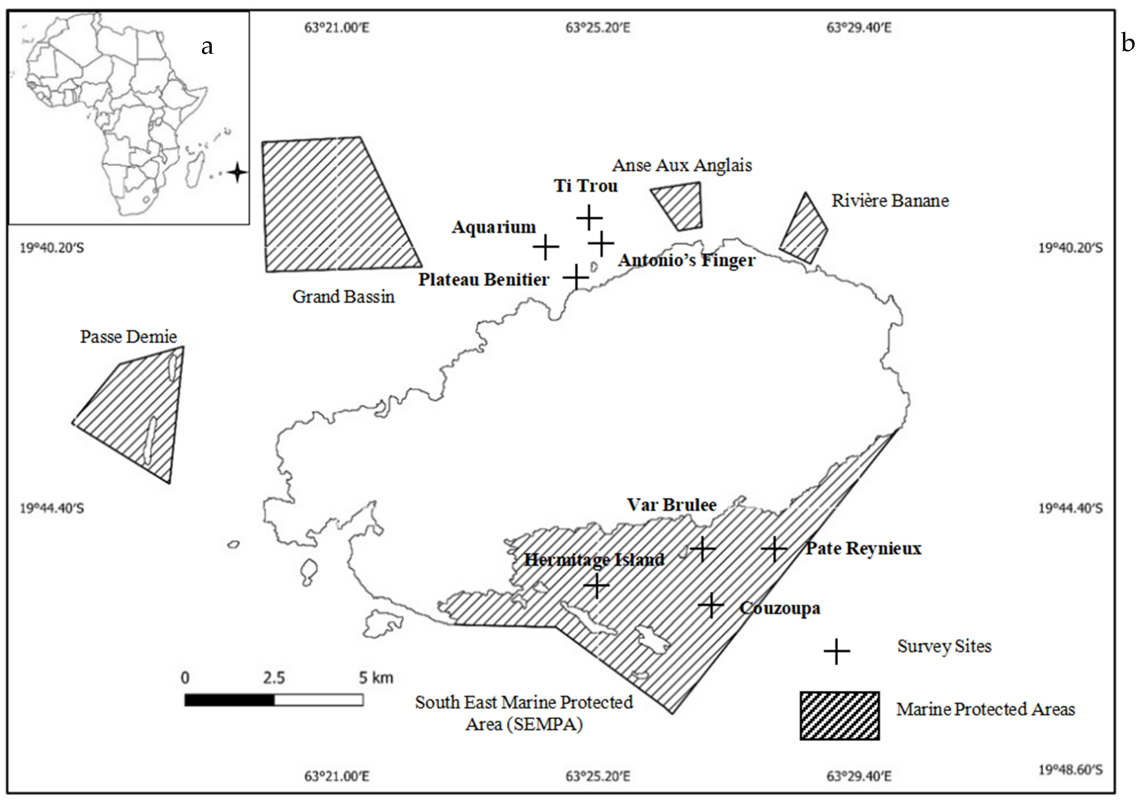
2.1. Study Site

Rodrigues Island (19°42′59.99″ S, 63°24′59.99″ E) (Figure 1) is an outer autonomous island of the Republic of Mauritius that forms part of the Mascarene Islands. The island is situated in the southwestern part of the Indian Ocean (Figure 1a) at around 560 km to the east of the island of Mauritius. The island measures 18 km in length and 6.5 km in width, with a total land area of 108 km2. The island is surrounded by a large fringing reef, with an estimated area of 230.6 km2 [43,44]. Several smaller islets and two sand cays can be found inside the island’s lagoon. The reef flats in Rodrigues Island, which typically range from 50 m to 2 km in width, can be divided into 4 sections: (i) a compact reef flat, measuring between 50 and 300 m in width; (ii) fragmented reef flats with scattered coral colonies separated by meandering channels; (iii) a zone of coral-built alignments that run perpendicular to the reef edge and is separated by shallow grooves; and (iv) micro-atoll in sheltered zones [46,47,48]. There are also four marine reserves (Passe Demie, Grand Bassin, Anse aux Anglais, and Riviere Banane) and one multiple-use marine protected area (South East Marine Protected Area (SEMPA) around the island (Figure 1).
This study was conducted from 17 to 20 December 2020, where the average highest sea surface temperature was 28.7 °C (seatemperature.info, 2020), and the lowest mean sea surface temperature was 26.17 °C (seatemperature.info, 2020). Eight study sites (Table 1), namely Aquarium, Plateau Benitier, Ti Trou, Antonio’s Finger, Hermitage Island, Pate Reynieux Couzoupa, and Var Brulee, were chosen around the Island. Hermitage, Couzoupa, Pate Reynieux, and Var Brulee were situated in the SEMPA, while all the other sites were located in a non-protected area in the northeast of Rodrigues Island.

2.2. Disease and CHC Prevalence on Hard Corals

To assess disease and CHC prevalence on hard corals at each site, three 2 × 50 m belt transects were placed randomly with a minimum interval of 20 m between each. All colonies that were found inside the belt transect or those with 50% of their area inside the margins of the transect were counted. All the coral colonies were then identified in situ to their genus level [49]. Coral diseases were identified from their lesion characteristics using Underwater Cards for assessing coral diseases [21]. CHCs such as the Pigmentation Responses were classified as Pink Pigmentation Response (PPR), which is morphologically further characterized as Pink Patches (PP), Pink Spots (PS), or Pink Line Syndromes (PLS). Underwater photographs of diseases or CHCs on coral colonies were also taken using the underwater cameras Tough TG-6 (Olympus, Amazon, Japan) and GoPro (Hero 8, USA). All diseased and CHC colonies found inside the margins of the belt transect were counted, and the same was performed for the healthy-looking colonies as well. In order to estimate disease prevalence, the number of diseased and CHC colonies was divided by the total number of colonies. Other diseases or CHC observations outside the belt transect were also reported in this study, but the prevalence was not recorded. Table 2 lists the diseases or CHCs observed outside the belt transect in parentheses. Coral disease and CHC prevalence for each affected taxon (taxon-specific) were also estimated by dividing the number of affected corals from one genus by the total number of colonies observed for that specific genus. The mean disease and CHC prevalence per site were calculated by averaging the prevalence values from the three belt transects per site. Surveys for disease and CHC prevalence were conducted by both snorkeling and SCUBA diving at different depth ranges: 1–3 m (D1), 4–6 m (D2), 7–9 m (D3), and 10–12 m (D4).

2.3. Benthic Cover Assessment

At each site, the live coral cover was quantified in situ through visual surveys along triplicate 50 m line transects using the line-intercept transect (LIT) method [50]. The coral density and coral colony size were assessed along triplicates of 2 m × 50 m belt transects. The coral colony size (in cm) was estimated along the transect tape and recorded as the highest linear coral colony diameter. The colony size was estimated within different size class ranges: 0–10, 11–20, 21–30, 31–40, 41–50, and >50 cm.

2.4. Statistical Analysis

All data were transformed to meet the assumption of normality with log10 transformation. A one-way ANOVA was performed to test for significant differences (α = 0.05) in the disease or CHCs prevalence on hard corals between the sites surveyed around the island of Rodrigues. A two-way ANOVA was used to test for significant differences (α = 0.05) between depth and disease/CHC prevalence and between the conservation status and prevalence of coral diseases and CHCs. To assess the relationship between the live coral cover and colony density or coral disease/CHC prevalence around Rodrigues Island, Pearson’s Correlation test was performed. All statistical analyses were performed on MS Excel, SPSS (IBM Version 21), and R Statistical Package (Version 4.1.1). A Principal Component Analysis (PCA) was constructed using OriginPro (version 10.0.5.157) to show the distribution of coral genera and coral diseases/CHCs across the different survey sites.

3. Results

3.1. Coral Community Structure

The highest live coral cover (72.87 ± 8.02%) and coral colony density (1.74 ± 0.26 colonies/m2) were recorded at Pate Reynieux (Figure 2). The lowest mean live coral cover was recorded at Plateau Benitier at 7.3%, and the least number of coral colonies per unit area was recorded at Antonio’s Finger (0.15 ± 0.12 colonies/m2). Var Brulee had the second-highest mean live coral cover, followed by Couzoupa, Hermitage Island, Ti Trou, Antonio’s Finger, and Aquarium. The second-highest coral colony density was recorded at Hermitage Island, followed by Couzoupa, Var Brulee, Ti Trou, Plateau Benitier, and Aquarium.
Sixteen diseased/CHC colonies were observed from Couzoupa in the 11–20, 21–30, 41–50, and>50 cm size classes. Seventeen colonies were found at Pate Reynieux in the 0–10, 11–20, 21–30, 30–41, and 41–50 cm size classes. Sixteen colonies were reported from Hermitage Island in the 0–10 cm size classes. Eight diseased colonies from Ti Trou in the 0–10, 11–20, 21–30, and>50 cm size classes, two colonies from Aquarium in the >50 cm size class, and one at Plateau Benitier in the >50 cm size class were found (Figure 3).
The most dominant (27%) coral genus at Couzoupa was the branching Millepora, followed by Montipora (26%) (mostly large colonies of Montipora aequituberlata). At Pate Reynieux, the most dominant genus was the solitary Fungia corals (28%). Branching colonies of Acropora were most dominant at Var Brulee (51%). Pavona colonies, mostly of small size, were most dominant (78%) at Hermitage Island. At the non-protected sites, Favites, branching Acropora, and Pocillopora were most dominant at Plateau Benitier (14%), Ti Trou (39%), and Antonio’s Finger (43%), respectively. An equal dominant proportion (21%) of coral colonies were observed of Platygyra and Favites at the Aquarium.

3.2. Coral Disease Occurrence and Prevalence

Benthic surveys conducted at eight sites around the island of Rodrigues revealed the presence of four coral diseases and two CHCs on six different genera of scleractinian corals (Figure 4). The diseases observed were Growth Anomalies, Black Band disease, White Plague, and White Syndrome. The CHCs recorded were two forms of PPR, namely, Pink Patches and Pink Line Syndrome. A total of 1662 coral colonies were recorded during our survey, and out of them, 122 colonies exhibited signs of coral diseases. A total of 120 diseased/CHC coral colonies were found within the transect, and 1 colony of massive Porites with signs of Growth Anomalies and another colony with Pink Line Syndrome were observed outside the margins of the belt transect. A total of 16 diseased colonies were observed from Couzoupa, 17 colonies from Pate Reynieux, 16 colonies from Hermitage Island, 8 from Ti Trou, 2 colonies from Aquarium, and 1 diseased colony from Plateau Benitier.
Disease and CHC prevalence recorded around Rodrigues Island ranged from 3.63% PPR to 0.023% Black Band disease (Figure 4a). Porites Growth Anomalies and Pink Line Syndrome were also observed on Porites lobata and P. lutea, respectively, at Plateau Benitier. Since they were not found within the belt transects, their prevalence was not reported. The prevalence of all the coral diseases and CHCs across all genera observed within the transects is reported in Table 2. The highest disease/CHC-specific prevalence (13.86 ± 2.47%) was reported at Pate Reynieux for the Pink Patch on solitary Fungia colonies (Figure 4b and Figure 5). The lowest disease/CHC-specific prevalence (0.19 ± 0.19%) was reported for the Black Band disease and White Syndrome on Montipora aequituberlata from Pate Reynieux.
PPR (3.63 ± 1.33%) was the most prevalent around the island of Rodrigues, followed by the White Syndrome disease (0.90 ± 0.36%). The least prevalent disease observed around Rodrigues Island was Black Band disease (0.023 ± 0.12%). In terms of taxonomic classification, the PP (15.92 ± 5.65%) on Fungia, followed by White Syndrome on plating Montipora (4.67 ± 3.72%) were most prevalent (Table 2, Figure 4a). Black Band disease on foliose Montipora was the least prevalent taxon-specific disease for Rodrigues Island. With the exception of Black Band disease, all the other diseases were observed in both deep and shallow waters.
At Couzoupa, White Syndrome disease was observed on the Montipora aequituberlata at a prevalence of 0.89 ± 0.46% (Figure 4b). White plague disease on fire coral Millepora tenella was observed at the highest prevalence of 2.82 ± 1.83% (Figure 4b and Figure 5), and Pink Patches were observed on a few Fungia corals at a prevalence of 1.03 ± 0.22% (Figure 4b). Growth Anomalies were observed on one Gardineroseris planulata colony at a prevalence of 0.26 ± 0.26% (Figure 4b). At Pate Reynieux, Montipora coral was observed with White Syndrome, White Plague, and Black Band at respective prevalences of 0.19 ± 0.19%, 0.22 ± 0.22%, and 0.19 ± 0.19% (Figure 4b). Pink Patches were also observed on Fungia at a prevalence of 13.89 ± 2.48% (Figure 4b). At Hermitage Island, only Pink Patches on Fungia were observed at a prevalence of 9.63 ± 7.07%. At Plateau Benitier, Pink Patches were observed on massive Porites lutea at a prevalence of 0.98 ± 0.98% (Figure 4b). At Ti Trou, White Syndrome disease was observed on Montipora aequituberlata at a prevalence of 2.48 ± 1.30% and Pink Patches on Fungia at a prevalence of 3.51 ± 2.82% (Figure 4b). At Aquarium, White Syndrome on Goniastrea stelligera was observed at a prevalence of 3.60 ± 1.87% (Figure 4b). No disease lesions were observed on any coral colony at Antonio’s Finger and Var Brulee.
No significant difference (p > 0.05) in the coral disease or CHC prevalence among the sites surveyed was observed around Rodrigues Island. Although different diseases and CHCs on hard corals were observed in the protected areas and the non-protected areas, no significant difference (p > 0.05) was recorded in coral disease prevalence between the sites surveyed in protected and non-protected areas. Similarly, the different depth ranges surveyed around Rodrigues Island did not significantly (p > 0.05) affect the coral disease prevalence recorded.
A weak positive correlation (r = 0.390, p < 0.05) was noted between the live coral cover and the prevalence of coral diseases around Rodrigues Island. A weak negative correlation (r= −0.141, p < 0.05) was observed between coral colony density and coral disease prevalence.

4. Discussion

This study around the island of Rodrigues represents the first report on the occurrence of four coral diseases, including White Plague, White Syndrome, Black Band, and Growth Anomalies, and 2 CHCs in the form of Pink Pigmentation Responses (Pink Patches and Pink Line Syndrome) on six coral genera namely Montipora, Goniastrea, Millepora, Porites, Gardineroseris, and Fungia. All the observed diseases and CHCs, and most of the affected hosts in this study, have been reported in several other areas of the Indian Ocean. Closest to the island of Rodrigues, the corals around Mauritius Island have been reported with signs of diseases including White Plague-like, White Band-like, Brown Band, Skeletal Eroding Band, Growth Anomalies, and CHCs-like Pink Pigmentation Response [29]. The most prevalent disease around Mauritius Island was White Plague (20.33 ± 3.33%), which affected the most dominant reef-building coral species, Acropora muricata. White Syndrome was also reported in the Saya de Malha banks [31]. White Syndrome disease on Montipora, Growth Anomalies on massive Porites, and Pink Line Syndrome were also reported from two other islands located in the Southwest Indian Ocean, namely, Réunion and Maldives [37]. White Syndrome disease (1.0 ± 1.4%), along with Porites White Patch Syndrome (PWPS) (1.0 ± 0.4%), were reported to be most prevalent in Mayotte, whereas Réunion Island was mostly affected by the PWPS (2.3 ± 2.0%), followed by the Pink Line Syndrome (2.0 ± 3.9%). Other locations in the Indian Ocean including the Republic of Maldives [33], Chagos Archipelago [35], Madagascar [32], South Africa [37], and Southern India (Gulf of Mannar) [51,52,53] were observed to have similar coral diseases. The only unreported coral genus with disease from the Southwest Indian Ocean is Gardineroseris, which was observed with Growth Anomalies around Rodrigues Island.
The overall disease prevalence for Rodrigues Island (0.98 ± 0.30%) was much lower than not only the surrounding islands of Réunion (7.5%), Mayotte (2.7%), Maldives (<2%), and the Chagos Archipelago (5.2%) [37] but also than those reported for the Indo-Pacific and the Caribbean regions. However, it is noteworthy that Clark in reported no or little signs of diseases on the reefs of Rodrigues [54]. Since then, global environmental change and anthropogenic influences have greatly increased and impacted coral reefs, including coral disease dynamics [1]. The present study found four coral diseases and two CHCs around Rodrigues Island, most probably because of the prevailing more pronounced climate change-driven ocean warming and/or anthropogenic-related marine environmental degradation than decades ago [41].
The lack of spatial heterogeneity in the prevalence of coral diseases and CHCs among the surveyed sites around Rodrigues Island may be due to homogeneous patterns of susceptible coral host genera. The presence of almost similar environmental factors, as a consequence of the small size of the island, can also help explain the lack of significant variation in the levels of prevalence among the studied sites.
However, the occurrence of certain diseases and their associated prevalence levels at a specific site may possibly be explained at least partially by environmental and/or ecological factors at that site. Several studies have reported that the prevalence of coral diseases increases with the availability of susceptible coral hosts. Bruno and colleagues reported high coral cover (>50%) as an important driver of White Syndromes [55]. Aeby and colleagues also positively correlated the prevalence of Acroporid and Porites Growth Anomalies with host density [12].
A low prevalence of Montipora White Syndrome was recorded from Couzoupa and Pate Reynieux and on Goniastrea from Aquarium. Since White Syndrome is a pathogenic and infectious disease, the abundance of susceptible coral hosts, which play an important role in the occurrence of this disease [55,56], was observed at the sites. A large number of M. aequituberculata colonies occurring at high density were recorded at Couzoupa, highlighting the high host density required for the occurrence and transmission of White Syndrome. Sussman and colleagues [57] highlighted that the causative pathogen of White Syndrome can easily pass from one coral colony to another through the water column. The large size of the M. aequituberculata colonies at Couzoupa, coupled with its foliose morphology, may also explain the prevalence of White Syndrome in that site since large colony size provides increased surface area for pathogen attack [58,59]. The large size of the M. aequituberculata colonies at Couzoupa also makes them more prone to physical damage and injuries. These injuries may facilitate the infection of opportunistic pathogens, such as those involved in causing White Syndromes. The high species diversity at Couzoupa and Pate Reynieux can also explain the prevalence of White Syndrome since the abundance of potential vectors of this disease is also high [12].
White Plague is another coral disease that is characterized by a sharp line of bright white tissue that separates the healthy part of the coral from the bare, rapidly algae-colonized skeleton [60]. Very few coral diseases have been reported on Millepora corals; only Skeletal Eroding Band has been reported on Millepora alcicornis [61]. The prevalence of White Plague, a pathogenic and highly infectious disease, may be explained by the high abundance of the coral host, Millepora tenella, at Couzoupa and Montipora at Pate Reynieux. Advanced studies are required in the Rodrigues waters at the pathogen or microbiome level to shed further light on the White Plague coral disease characteristics.
While the occurrence of Growth Anomalies at Couzoupa on Gardineroseris planulata and on Porites lobata from Plateau Benitier cannot be explained by host density, the large colony size for both the Porites and Gardineroseris colonies can help explain the occurrence of this disease. The high sediment loads and the associated turbidity at Plateau Benitier can also help explain the calicoblastic neoplasm on Porites colonies, as reported by Peters and colleagues [62] for the coral Acropora palmata.
As a highly infectious coral disease [63,64], Black Band disease observations at Pate Reynieux may be explained by the high coral cover and density recorded at that site. This disease is seasonal and is more virulent during the summer months [65,66], thus explaining our observation of this disease on Montipora coral at one of our study sites surveyed in the summer month of December. Black Band commonly affects Montipora corals and has even been observed on some Montipora corals from Hawai’i for at least 10 years [67].
In this study, the low prevalence of PPR at Plateau Benitier may be explained by the occurrence of extremely low host density, as only one colony of Porites lutea was observed at Plateau Benitier. Pink Pigmentation is not considered an infectious disease, and environmental factors influence the occurrence of this response. An example of environmental stress at Plateau Benitier is the high level of sedimentation and turbidity associated with river runoff in the bay of Port Mathurin and in the vicinity of Anse aux Anglais. Pollock and colleagues [68] assessed the prevalence of several coral diseases and CHCs in an offshore dredging area in northwest Australia. The study found strong positive correlations between the prevalence of PPR and the number of days the coral colonies were exposed to the sediment plume associated with the dredging activities. Similarly, this may possibly explain the occurrence of PPR at Plateau Benitier. However, Stoddart and colleagues [69] did not find any association between disease prevalence and dredging activities and even criticized the use of coral disease prevalence as an indicator of the impact of dredging.
Plateau Benitier is a popular and well-frequented diving site in the north of Rodrigues. As a high-use diving site, physical injuries on coral colonies by divers are not uncommon. SCUBA diving can also be another physical stress leading to the occurrence of PPR. Lamb and colleagues [58] also associated the prevalence of PPR with high-use SCUBA diving sites at Koh Tao, Thailand. PPR in the form of Pink Patches was found on Fungia, at the highest prevalence at Pate Reynieux in this study. The occurrence of this disease on this host is not uncommon and has been reported by other studies from the Indian Ocean [51,52,53]. The presence of Pink Patches at Pate Reynieux may be explained by the accumulation of sediments on Fungia colonies due to the shallow depth of water and the possible re-suspension of sediments by wave action. Although a high density of Fungia colonies was observed over there, Pink Patches are not transmissible and cannot help explain its high occurrence. However, for PPR, it is noteworthy that an important factor is strong illumination. In shallow areas, this may be the case; therefore, the coral produces pink chromoproteins to protect themselves from strong irradiance damage through the formation of reactive oxygen species (ROS), and thus, high irradiance environmental parameters may be more related to sedimentation. Further investigations along these lines are warranted to thoroughly understand the occurrence of PPR in these coral genera around Rodrigues Island.
The status of protection of marine areas around Rodrigues Island did not have any influence on the prevalence of coral diseases and/or CHCs. A similar observation was made by Page and colleagues [70], who reported no effect of marine protected areas on coral disease prevalence. For a marine protected area to differently influence the dynamics of coral diseases or CHCs than a non-protected area, the presence of susceptible hosts and vectors of coral diseases must also be different between the protected and non-protected areas. Contributing factors such as increased coral colony size, high abundance of reef fishes and invertebrates, and the high availability and density of coral hosts are characteristics features of marine protected areas, which are often linked to changes in coral disease prevalence [70,71]. The different depth ranges surveyed also did not affect the prevalence of coral diseases and CHCs. While some studies have reported mostly negative associations [12,72] between depth and coral disease prevalence, other studies have found no relationship between these two parameters [71,73]. The lack of significant difference in the prevalence levels among the studied depth categories may be explained by the different populations of coral host species observed at each depth range.
In this study, a weak positive correlation was observed between the live coral cover and the prevalence of coral diseases or CHCs around Rodrigues Island. A similar observation was made by other studies [12,55,74], and the main explanation for such an observation is the reduced distance between coral hosts. A higher coral cover implies shorter distances between coral hosts and increased likelihood and ease of infection by coral pathogens [75]. Higher coral cover also enhances the reef’s structural complexity, thus increasing the abundance of other potential vectors of coral disease or CHCs and, at the same time, increasing the transmission of pathogens. A high colony density also increases the availability of coral hosts and facilitates the transmission of coral pathogens from one colony to another. Such observation has been reported in several studies [12,18,76]. In this study, however, a negative correlation between the density of coral colonies and the prevalence of coral disease or CHCs was recorded. This may be explained by the non-infectious nature of the most prevalent CHCs (Pink Patches) around Rodrigues Island.

5. Conclusions

This study aimed to provide baseline information on the occurrence, distribution, prevalence, and characterization of diseases and compromised health conditions on hard corals around the island of Rodrigues. Four diseases, namely, White Syndromes, White Plague, Black Band, Growth Anomalies, and two CHCs, including Pink Patch and Pink Line Syndrome, were recorded on six coral genera, Montipora, Porites, Goniastrea, Gardineroseris, Fungia, and Millepora were recorded. Diseases and CHCs on hard corals were observed at six (out of the eight) surveyed sites, with three of the affected sites being located in the Southeast Marine Protected Area—one of the largest regional marine protected areas. Although the overall coral diseases and CHC prevalence around Rodrigues Island are much lower than other islands in the Indian Ocean, the uniqueness and vulnerability of the island’s coral reefs, characterized by a self-seeding, low-rate recruitment pattern, and past coral bleaching events, which caused mass coral mortalities and altered the coral community structure, highlights its fragility to stressors of coral health.

Author Contributions

R.B., S.Y.J., S.M.-M., D.K., B.E.C. and Y.S.: Conceptualization. S.Y.J., R.B., S.G., S.J., M.R., S.M.-M. and D.K.: Methodology, Data Collection, and Analyses. S.Y.J. and R.B.: Writing—Original Draft Preparation. S.Y.J., R.B., S.G., S.J., M.R., N.T.-H., S.M.-M., B.E.C., Y.S., D.K. and D.P.W.: Writing—Reviewing and Editing. All authors have read and agreed to the published version of the manuscript.

Funding

This research was funded by the Higher Education Commission (Mauritius) grant numbers T0105 and T0721 And The APC was waived by MDPI.

Institutional Review Board Statement

Not applicable.

Data Availability Statement

The data presented in this study are available on request from the corresponding author.

Acknowledgments

The authors would like to thank the University of Mauritius for the logistical support to conduct this study. The authors are also grateful to the Department for Continental Shelf, Maritime Zones Administration and Exploration (DCSMZAE) and the Outer Islands Development Cooperation (OIDC) under the Prime Minister’s Office, the Albion Fisheries Research Centre under the Ministry of Blue Economy, Marine Resources, Fisheries and Shipping, and the Rodrigues Regional Assembly (RRA) in Rodrigues for granting necessary permits and authorization to conduct marine ecological surveys and collect coral samples. R.B., S.M.-M., D.K. and S.Y.J. are grateful to the Higher Education Commission (Mauritius) for research grant (T0105 and T0721) to conduct this study.

Conflicts of Interest

The authors declare no conflict of interest.

References

  1. Harvell, D.; Jordán-Dahlgren, E.; Merkel, S.; Rosenberg, E.; Raymundo, L.; Smith, G.; Weil, E.; Willis, B. Coral Disease, Environmental Drivers, and the Balance between Coral and Microbial Associates. Oceanography 2007, 20, 172–195. [Google Scholar] [CrossRef]
  2. Weil, E. Coral Reef Diseases in the Wider Caribbean. In Coral Health and Disease; Rosenberg, E., Loya, Y., Eds.; Springer: Berlin/Heidelberg, Germany, 2004; pp. 35–68. ISBN 978-3-642-05863-9. [Google Scholar]
  3. Weil, E.; Smith, G.; Gil-Agudelo, D.L. Status and Progress in Coral Reef Disease Research. Dis. Aquat. Organ. 2006, 69, 1–7. [Google Scholar] [CrossRef]
  4. Woodley, C.M.; Downs, C.A.; Bruckner, A.W.; Porter, J.W.; Galloway, S.B. Diseases of Coral; John Wiley & Sons: Hoboken, NJ, USA, 2016; ISBN 978-0-8138-2411-6. [Google Scholar]
  5. Harvell, C.D.; Kim, K.; Burkholder, J.M.; Colwell, R.R.; Epstein, P.R.; Grimes, D.J.; Hofmann, E.E.; Lipp, E.K.; Osterhaus, A.D.M.E.; Overstreet, R.M.; et al. Emerging Marine Diseases-Climate Links and Anthropogenic Factors. Science 1999, 285, 1505–1510. [Google Scholar] [CrossRef]
  6. Green, E.; Bruckner, A. The Significance of Coral Disease Epizootiology for Coral Reef Conservation. Biol. Conserv. 2000, 96, 347–361. [Google Scholar] [CrossRef]
  7. Cramer, K.L.; Jackson, J.B.C.; Donovan, M.K.; Greenstein, B.J.; Korpanty, C.A.; Cook, G.M.; Pandolfi, J.M. Widespread Loss of Caribbean Acroporid Corals Was Underway before Coral Bleaching and Disease Outbreaks. Sci. Adv. 2020, 6, eaax9395. [Google Scholar] [CrossRef] [PubMed]
  8. Aronson, R.; Precht, W.; Aronson, R.B.; Precht, W.F. White-Band Disease and the Changing Face of Caribbean Coral Reefs. Hydrobiologia 2001, 460, 25–38. [Google Scholar] [CrossRef]
  9. Cramer, K.L.; Donovan, M.K.; Jackson, J.B.C.; Greenstein, B.J.; Korpanty, C.A.; Cook, G.M.; Pandolfi, J.M. The Transformation of Caribbean Coral Communities since Humans. Ecol. Evol. 2021, 11, 10098–10118. [Google Scholar] [CrossRef]
  10. Raymundo, L.J.; Rosell, K.B.; Reboton, C.T.; Kaczmarsky, L. Coral Diseases on Philippine Reefs: Genus Porites Is a Dominant Host. Dis. Aquat. Organ. 2005, 64, 181–191. [Google Scholar] [CrossRef] [PubMed]
  11. Sato, Y.; Bourne, D.G.; Willis, B.L. Dynamics of Seasonal Outbreaks of Black Band Disease in an Assemblage of Montipora Species at Pelorus Island (Great Barrier Reef, Australia). Proc. Biol. Sci. 2009, 276, 2795–2803. [Google Scholar] [CrossRef] [PubMed]
  12. Aeby, G.S.; Williams, G.J.; Franklin, E.C.; Kenyon, J.; Cox, E.F.; Coles, S.; Work, T.M. Patterns of Coral Disease across the Hawaiian Archipelago: Relating Disease to Environment. PLoS ONE 2011, 6, e20370. [Google Scholar] [CrossRef]
  13. Weil, E.; Irikawa, A.; Casareto, B.; Suzuki, Y. Extended Geographic Distribution of Several Indo-Pacific Coral Reef Diseases. Dis. Aquat. Org. 2012, 98, 163–170. [Google Scholar] [CrossRef] [PubMed]
  14. Das, R.R.; Sreeraj, C.R.; Mohan, G.; Simon, N.T.; Ramachandran, P.; Ramachandran, R.; Krishnan, P.; Kumar, D.S.V. Evidence of Coral Diseases, Phase Shift, and Stressors in the Atolls of Lakshadweep Islands, Arabian Sea—With Geographical Notes on Their Occurrence within the Indian EEZ and Contiguous International Waters. Diversity 2023, 15, 382. [Google Scholar] [CrossRef]
  15. Bharath, M.S.; Chandran, R.; Aeby, G.S.; Senthilkumaran, R.; Ramkumaran, K.; Thanappan, V.P.P.; Chaudhury, N.R.; Satyanarayana, C. First Report of Yellow-Banded Tissue Loss Disease on Coral Reefs Outside the Arabian/Persian Gulf. Dis. Aquat. Org. 2023, 153, 1–8. [Google Scholar] [CrossRef]
  16. Burke, S.; Pottier, P.; Lagisz, M.; Macartney, E.L.; Ainsworth, T.; Drobniak, S.M.; Nakagawa, S. The Impact of Rising Temperatures on the Prevalence of Coral Diseases and Its Predictability: A Global Meta-Analysis. Ecol. Lett. 2023, 26, 1466–1481. [Google Scholar] [CrossRef] [PubMed]
  17. Aeby, G.S.; Shore, A.; Jensen, T.; Ziegler, M.; Work, T.; Voolstra, C.R. A Comparative Baseline of Coral Disease in Three Regions along the Saudi Arabian Coast of the Central Red Sea. PLoS ONE 2021, 16, e0246854. [Google Scholar] [CrossRef]
  18. Myers, R.L.; Raymundo, L.J. Coral Disease in Micronesian Reefs: A Link between Disease Prevalence and Host Abundance. Dis. Aquat. Organ. 2009, 87, 97–104. [Google Scholar] [CrossRef]
  19. Mohamed, A.; Sweet, M. Current Knowledge of Coral Diseases Present Within the Red Sea. In Oceanographic and Biological Aspects of the Red Sea. Springer Oceanography; Springer: Cham, Switzerland, 2019; pp. 387–400. ISBN 978-3-319-99416-1. [Google Scholar]
  20. Hazraty-Kari, S.; Tavakoli-Kolour, P.; Das, R.R.; Farhadi, M.; Barkhordari-Ahmadi, A.; Yahyavi, M.; Rezai, H. Baseline Assessment of Coral Diseases in an Environmentally Extreme Environment of the Northern Persian Gulf. Mar. Pollut. Bull. 2021, 171, 112707. [Google Scholar] [CrossRef]
  21. Beeden, R.; Willis, B.L.; Raymundo, L.; Page, C.; Weil, E. Underwater Cards for Assessing Coral Health on Indo-Pacific Reefs. Coral Reef Targeted Research and Capacity Building for Management Program; Currie Communications: Melbourne, VIC, Australia, 2008. [Google Scholar]
  22. Weil, E.; Hooten, A.J. Underwater Cards for Assessing Coral Health on Caribbean Coral Reefs; GEF-CRTR-Currie Communications: Melbourne, VIC, Australia, 2008. [Google Scholar]
  23. Aldyza, N. Afkar Analisis Genus Dan Penyakit Karang Di Perairan Pulau Tuan Kecamatan Peukan Bada Kabupaten Aceh Besar. BIOTIK J. Ilm. Biol. Teknol. Dan Kependidikan 2017, 3, 107–115. [Google Scholar] [CrossRef]
  24. Hadi, T.A. Impacts of sedimentation on stony corals. Oseana 2017, 42, 45–58. [Google Scholar] [CrossRef]
  25. Box, S.J.; Mumby, P.J. Effect of Macroalgal Competition on Growth and Survival of Juvenile Caribbean Corals. Mar. Ecol. Prog. Ser. 2007, 342, 139–149. [Google Scholar] [CrossRef]
  26. Yucharoen, M. Pink Pigmentation Response during Recovery Period after Coral Bleaching. Master’s Thesis, Shizuoka University, Shizuoka, Japan, 2016. [Google Scholar]
  27. Jogee, S.Y.; Jeetun, S.; Ricot, M.; Taleb-Hossenkhan, N.; Mattan-Moorgawa, S.; Kaullysing, D.; Riemann, P.; Blanc, L.; Casareto, B.E.; Suzuki, Y.; et al. Photo-Physiology of Healthy-Looking and Diseased/Health-Compromised Hard Corals from Mauritius Island, Western Indian Ocean. Indo Pac. J. Ocean. Life 2023, 7, 27–37. [Google Scholar] [CrossRef]
  28. McClanahan, T.; Munbodhe, V.; Naggea, J.; Muthiga, N.; Bhagooli, R. Rare coral and reef fish species status, possible extinctions, and associated environmental perceptions in Mauritius. Conserv. Sci. Pract. 2021, 3, e527. [Google Scholar] [CrossRef]
  29. Bhagooli, R.; Mattan-Moorgawa, S.; Kaullysing, D.; Taleb-Hossenkhan, N. A first field report of coral diseases around Mauritius Island, Western Indian Ocean. West. Indian Ocean. J. Mar. Sci. 2017, 1/2, 71–72. [Google Scholar]
  30. Mattan-Moorgawa, S.M.; Kaullysing, D.; Taleb-Hossenkhan, N.; Rughooputh, S.D.D.V.; Bhagooli, R. Photophysiology of in hospite zooxanthellae in diseased and non-diseased scleractinian corals from Belle Mare, Mauritius. WIOJMS 2017, 1/2, 1–12. [Google Scholar]
  31. Bhagooli, R.; Jogee, S.; Kaullysing, D.; Ramah, S. First report of White Syndrome on branching Acropora at Saya de Malha, Mascarene Plateau. West. Indian Ocean. J. Mar. Sci. 2021, 2, 189–192. [Google Scholar] [CrossRef]
  32. Sheridan, C.; Baele, J.M.; Kushmaro, A.; Fréjaville, Y.; Eeckhaut, I. Terrestrial Runoff Influences White Syndrome Prevalence in SW Madagascar. Mar. Environ. Res. 2014, 101, 44–51. [Google Scholar] [CrossRef] [PubMed]
  33. Montano, S.; Strona, G.; Seveso, D.; Galli, P. First Report of Coral Diseases in the Republic of Maldives. Dis. Aquat. Org. 2012, 101, 159–165. [Google Scholar] [CrossRef] [PubMed]
  34. Montano, S.; Strona, G.; Seveso, D.; Maggioni, D.; Galli, P. Widespread Occurrence of Coral Diseases in the Central Maldives. Mar. Freshw. Res. 2015, 67, 1253–1262. [Google Scholar] [CrossRef]
  35. Sheppard, C.R.C.; Ateweberhan, M.; Bowen, B.W.; Carr, P.; Chen, C.A.; Clubbe, C.; Craig, M.T.; Ebinghaus, R.; Eble, J.; Fitzsimmons, N.; et al. Reefs and Islands of the Chagos Archipelago, Indian Ocean: Why It Is the World’s Largest No-Take Marine Protected Area. Aquat. Conserv. 2012, 22, 232–261. [Google Scholar] [CrossRef]
  36. McClanahan, T.; Weil, E.; Maina, J. Strong Relationship between Coral Bleaching and Growth Anomalies in Massive Porites. Glob. Change Biol. 2009, 15, 1804–1816. [Google Scholar] [CrossRef]
  37. Séré, M.; Chabanet, P.; Turquet, J.; Quod, J.-P.; Schleyer, M. Identification and Prevalence of Coral Diseases Observed on Three Western Indian Ocean (WIO) Coral Reefs. Dis. Aquat. Org. 2015, 114, 249–261. [Google Scholar] [CrossRef]
  38. Bhagooli, R.; Mattan-Moorgawa, S.; Kaullysing, D.; Chumun, P.K.; Klaus, R.; Munbodhe, V. Status and sustainability of reefs and shorelines of the Republic of Mauritius. In Sustainable Development Goals; Parsad, R., Ed.; Star Publication Pvt. Ltd.: New Delhi, India, 2021; pp. 142–177. [Google Scholar]
  39. Fenner, D.; Clark, T.H.; Turner, J.R.; Chapman, B. A checklist of the corals of the island state of Rodrigues, Mauritius. J. Nat. Hist. 2004, 38, 3091–3102. [Google Scholar] [CrossRef]
  40. Jouval, F.; Latreille, A.C.; Bureau, S.; Adjeroud, M.; Penin, L. Multiscale Variability in Coral Recruitment in the Mascarene Islands: From Centimetric to Geographical Scale. PLoS ONE 2019, 14, e0214163. [Google Scholar] [CrossRef] [PubMed]
  41. Hardman, E.R.; Meunier, S.; Turner, J.R.; Lynch, T.L.; Klaus, R. The extent of coral bleaching in Rodrigues. J. Nat. Hist. 2004, 38, 3077–3089. [Google Scholar] [CrossRef]
  42. Klaus, R. Assessing the impact of the 2015–2016 coral bleaching in Rodrigues (Republic of Mauritius). In Proceedings of the Western Indian Ocean Marine Science Association Symposium (WIOMSA) 2017, Dar es Salaam, Tanzania, 30 October–4 November 2017. [Google Scholar]
  43. Turner, J.; Klaus, R. Coral Reefs of the Mascarenes, Western Indian Ocean: One Contribution of 24 to a Discussion Meeting “Atmosphere-Ocean-Ecology Dynamics in the Western Indian Ocean”. Philos. Trans. R. Soc. A Math. Phys. Eng. Sci. 2005, 363, 229–250. [Google Scholar] [CrossRef]
  44. Bhagooli, R.; Kaullysing, D. Seas of Mauritius, Chapter 16. In World Seas: An Environmental Evaluation Volume II: The Indian Ocean to the Pacific, 2nd ed.; Sheppard CCR; Elsevier: Dordrecht, The Netherlands, 2019; pp. 253–277. [Google Scholar]
  45. Pasnin, O.; Klaus, R.; Attwood, C. Marine Systematic Conservation Plan for Rodrigues Island: Western Indian Ocean. Ocean. Coast. Manag. 2016, 130, 213–220. [Google Scholar] [CrossRef]
  46. Montaggioni, L. Coral reefs and quaternary shorelines in the Mascarene archipelago, Indian Ocean. In Proceedings of the Second International Coral Reef Symposium, Brisbane, Australia, 22 June–2 July 1974; Volume 1, pp. 579–593. [Google Scholar]
  47. Faure, G. Etude comparative des récifs coralliens de l’archipel des Mascareignes (Océan Indien). In Biologie Marine et Exploitation des Ressources de l’Océan Indien Occidental; ORSTOM: Paris, France, 1976; pp. 153–177. [Google Scholar]
  48. Montaggioni, L.F.; Faure, G. Récifs Coralliens des Mascareignes: Océan Indien; Université française de l’Océan indien, Centre universitaire de la Réunion, 1980. Available online: https://www.eaureunion.fr/fileadmin/user_upload/Etudes/ETUDE_01127.PDF (accessed on 12 October 2023).
  49. Veron, J.E.N. Corals of the World; Australian Institute of Marine Science: Townsville, QLD, Australia, 2000; pp. 1–3.
  50. English, S.; Wilkinson, C.; Baker, V. Survey Manual for Tropical Marine Resources; Australian Institute of Marine Science: Cape Cleveland, QLD, Australia, 1997; No. 333.952 S9.
  51. Thinesh, T.; Gilbert, M.; Patterson, E. Coral Disease Prevalence in Mandapam Group of Islands, Gulf of Mannar, Southeastern India. Indian J. Geo-Mar. Sci. 2009, 38, 444–450. [Google Scholar]
  52. Thinesh, T.; Mathews, G.; Edward, J.K.P. Coral Disease Prevalence in the Palk Bay, Southeastern India—with Special Emphasis to Black Band. Indian J. Geo-Mar. Sci. 2011, 40, 813–820. [Google Scholar]
  53. Thangaradjou, T.; Machendiranathan, M.; Ranith, R.; Senthilnathan, L.; Sasamal, S.K.; Choudhury, S.B. Coral Disease Prevalence in Gulf of Mannar and Lakshadweep Islands. Indian J. Geo-Mar. Sci. 2016, 45, 1755–1762. [Google Scholar]
  54. Clark, T.H. The Status of the Coral Reefs in Rodrigues, Report to the Shoals of Capricorn Programme. 2001; 27p.
  55. Aeby, G.S.; Callahan, S.; Cox, E.F.; Runyon, C.; Smith, A.; Stanton, F.G.; Ushijima, B.; Work, T.M. Emerging Coral Diseases in Kāne’ohe Bay, O’ahu, Hawai’i (USA): Two Major Disease Outbreaks of Acute Montipora White Syndrome. Dis. Aquat. Organ. 2016, 119, 189–198. [Google Scholar] [CrossRef]
  56. Bruno, J.F.; Selig, E.R.; Casey, K.S.; Page, C.A.; Willis, B.L.; Harvell, C.D.; Sweatman, H.; Melendy, A.M. Thermal Stress and Coral Cover as Drivers of Coral Disease Outbreaks. PLoS Biol. 2007, 5, e124. [Google Scholar] [CrossRef]
  57. Sussman, M.; Willis, B.L.; Victor, S.; Bourne, D.G. Coral Pathogens Identified for White Syndrome (WS) Epizootics in the Indo-Pacific. PLoS ONE 2008, 3, e2393. [Google Scholar] [CrossRef] [PubMed]
  58. Lamb, J.B.; True, J.D.; Piromvaragorn, S.; Willis, B.L. Scuba Diving Damage and Intensity of Tourist Activities Increases Coral Disease Prevalence. Biol. Conserv. 2014, 178, 88–96. [Google Scholar] [CrossRef]
  59. Greene, A.; Donahue, M.J.; Caldwell, J.M.; Heron, S.F.; Geiger, E.; Raymundo, L.J. Coral Disease Time Series Highlight Size-Dependent Risk and Other Drivers of White Syndrome in a Multi-Species Model. Front. Mar. Sci. 2020, 7, 601469. [Google Scholar] [CrossRef]
  60. Richardson, L.; Smith, G.; Ritchie, K.; Carlton, R. Integrating Microbiological, Microsensor, Molecular, and Physiologic Techniques in the Study of Coral Disease Pathogenesis. Hydrobiologia 2001, 460, 71–89. [Google Scholar] [CrossRef]
  61. Page, C.; Willis, B. Distribution, Host Range and Large-Scale Spatial Variability in Black Band Disease Prevalence on the Great Barrier Reef, Australia. Dis. Aquat. Org. 2006, 69, 41–51. [Google Scholar] [CrossRef] [PubMed]
  62. Peters, E.C.; Halas, J.C.; McCarty, H.B. Calicoblastic Neoplasms in Acropora Palmata, with a Review of Reports on Anomalies of Growth and Form in Corals. J. Natl. Cancer Inst. 1986, 76, 895–912. [Google Scholar]
  63. Richardson, L.L. Black Band Disease. In Coral Health and Disease; Rosenberg, E., Loya, Y., Eds.; Springer: Berlin/Heidelberg, Germany, 2004; pp. 325–336. ISBN 978-3-662-06414-6. [Google Scholar]
  64. Edmunds, P. Extent and effect of black band disease on a carribean reef. Coral Reefs 1991, 10, 161–165. [Google Scholar] [CrossRef]
  65. Kuta, K.G.; Richardson, L. Ecological Aspects of Black Band Disease of Corals: Relationships between Disease Incidence and Environmental Factors. Coral Reefs 2002, 21, 393–398. [Google Scholar] [CrossRef]
  66. Zvuloni, A.; Artzy-Randrup, Y.; Stone, L.; Kramarsky-Winter, E.; Barkan, R.; Loya, Y. Spatio-Temporal Transmission Patterns of Black-Band Disease in a Coral Community. PLoS ONE 2009, 4, e4993. [Google Scholar] [CrossRef]
  67. Aeby, G.S.; Work, T.M.; Runyon, C.M.; Shore-Maggio, A.; Ushijima, B.; Videau, P.; Beurmann, S.; Callahan, S.M. First Record of Black Band Disease in the Hawaiian Archipelago: Response, Outbreak Status, Virulence, and a Method of Treatment. PLoS ONE 2015, 10, e0120853. [Google Scholar] [CrossRef]
  68. Pollock, F.J.; Lamb, J.B.; Field, S.N.; Heron, S.F.; Schaffelke, B.; Shedrawi, G.; Bourne, D.G.; Willis, B.L. Sediment and Turbidity Associated with Offshore Dredging Increase Coral Disease Prevalence on Nearby Reefs. PLoS ONE 2014, 9, e102498. [Google Scholar] [CrossRef] [PubMed]
  69. Stoddart, J.; Jones, R.; Page, C.; Marnane, M.; Lestang, P.; Elsdon, T. No Effect of Dredging on the Prevalence of Coral Disease Detected during a Large Dredging Program. Mar. Pollut. Bull. 2019, 140, 353–363. [Google Scholar] [CrossRef] [PubMed]
  70. Page, C.A.; Baker, D.M.; Harvell, C.D.; Golbuu, Y.; Raymundo, L.; Neale, S.J.; Rosell, K.B.; Rypien, K.L.; Andras, J.P.; Willis, B.L. Influence of Marine Reserves on Coral Disease Prevalence. Dis. Aquat. Org. 2009, 87, 135–150. [Google Scholar] [CrossRef] [PubMed]
  71. Raymundo, L.J.; Halford, A.R.; Maypa, A.P.; Kerr, A.M. Functionally Diverse Reef-Fish Communities Ameliorate Coral Disease. Proc. Natl. Acad. Sci. USA 2009, 106, 17067–17070. [Google Scholar] [CrossRef]
  72. Cróquer, A.; Weil, E. Changes in Caribbean Coral Disease Prevalence after the 2005 Bleaching Event. Dis. Aquat. Org. 2009, 87, 33–43. [Google Scholar] [CrossRef]
  73. Borger, J.L.; Steiner, S.C.C. The Spatial and Temporal Dynamics of Coral Diseases in Dominica, West Indies. Bull. Mar. Sci. 2005, 77, 137–154. [Google Scholar]
  74. Selig, E.R.; Drew Harvell, C.; Bruno, J.F.; Willis, B.L.; Page, C.A.; Casey, K.S.; Sweatman, H. Analyzing the Relationship Between Ocean Temperature Anomalies and Coral Disease Outbreaks at Broad Spatial Scales. In Coral Reefs and Climate Change: Science and Management; American Geophysical Union (AGU): Washington, DC, USA, 2006; pp. 111–128. ISBN 978-1-118-66615-9. [Google Scholar]
  75. Hughes, T.P.; Baird, A.H.; Bellwood, D.R.; Card, M.; Connolly, S.R.; Folke, C.; Grosberg, R.; Hoegh-Guldberg, O.; Jackson, J.B.C.; Kleypas, J.; et al. Climate Change, Human Impacts, and the Resilience of Coral Reefs. Science 2003, 301, 929–933. [Google Scholar] [CrossRef] [PubMed]
  76. Hobbs, J.-P.A.; Frisch, A.J. Coral Disease in the Indian Ocean: Taxonomic Susceptibility, Spatial Distribution and the Role of Host Density on the Prevalence of White Syndrome. Dis. Aquat. Org. 2010, 89, 1–8. [Google Scholar] [CrossRef]
Figure 1. (a) Location of Rodrigues Island (star) in the Indian Ocean; (b) Rodrigues Island, indicating the 8 survey sites with demarcation of the marine protected areas (grids).
Diversity 15 01086 g001
Figure 2. Live coral cover and coral density at all survey sites around Rodrigues Island. Bars represent Mean ± SD.
Diversity 15 01086 g002
Figure 3. Size class frequency distribution of coral colonies at the study sites. Black dots represent the number of diseased/CHC colonies.
Diversity 15 01086 g003
Figure 4. (a) Disease/CHC-specific and (b) taxon-specific prevalence at each study site. Bars represent mean ± SE.
Diversity 15 01086 g004
Figure 5. PCA biplot of the taxonomic distribution of hard genera and disease/CHC prevalence on hard corals at the study sites surveyed around Rodrigues Island.
Diversity 15 01086 g005
Table 1. Geographic location, depth, and protection status of the sites surveyed around Rodrigues Island.
SiteGeographic LocationDepth (m)Depth Category (D1–D4)Protected/Non-Protected
Plateau Benitier19°40′11.60″ S, 63°25′44.90″ E6–9D3Non-protected
Ti Trou19°40′0.00″ S, 63°26′14.00″ E6–9D3Non-protected
Aquarium19°40′21.0″ S, 63°28′36.0″ E6–9D3Non-protected
Antonio’s Finger19°40′11.4″ S, 63°25′47.4″ E6–9D3Non-protected
Couzoupa19°45′32.4″ S, 63°27′48.5″ E9–12D4Protected
Pate Reynieux19°44′44.00″ S, 63°28′43.00″ E1–3D1Protected
Hermitage Island19°44′59.0″ S, 63°20′40.0″ E1–3D1Protected
Var Brule19°44′44.00″ S, 63°28′43.00″ E1–3D1Protected
Table 2. Disease/CHC-specific and taxon-specific prevalence around the Island of Rodrigues. Diseased colonies or colonies with CHCs are listed in between parentheses. “-“ represents not observed. “n” represents number of observed colonies.
GenusnDisease/CHC ObservedNumber of Affected ColoniesMean Overall Prevalence (%) ± SE
Disease/CHC-SpecificTaxon-Specific
Pocillopora62----
Branching Acropora248----
Montipora281WS80.90 ± 0.344.67 ± 3.72
WP10.38 ± 0.270.23 ± 0.23
BBD10.023 ± 0.0230.13 ± 0.13
Gardineroseris2GA10.03 ± 0.164.17 ± 4.17
Pachyseris4----
Pavona248----
Fungia262PP973.63 ± 1.3315.92 ± 5.65
Goniopora3----
Massive Porites17PP13.63 ± 1.334.17 ± 4.17
GA(1)--
PLS(1)--
Echinopora36----
Goniastrea36WS20.90 ± 0.341.89 ± 1.31
Favites81----
Dipsastrea23----
Platygyra189----
Lobophyllia5----
Millepora238WP100.38 ± 0.271.10 ± 0.89
Disclaimer/Publisher’s Note: The statements, opinions and data contained in all publications are solely those of the individual author(s) and contributor(s) and not of MDPI and/or the editor(s). MDPI and/or the editor(s) disclaim responsibility for any injury to people or property resulting from any ideas, methods, instructions or products referred to in the content.

