Article

Limnonema gen. nov. (Aerosakkonemataceae, Cyanobacteria): Two Novel Species from Republic of Korea Characterized by Morphological and Molecular Analyses

Department of Life Science, College of Natural Science, Kyonggi University, Suwon 16227, Republic of Korea
*
Author to whom correspondence should be addressed.
Diversity 2023, 15(12), 1174; https://doi.org/10.3390/d15121174
Submission received: 14 October 2023 / Revised: 21 November 2023 / Accepted: 22 November 2023 / Published: 27 November 2023

Abstract

:
In this study, 18 strains of cyanobacteria were isolated from seven sites, including the Han River and Anseong Stream in Republic of Korea, and we propose these isolated strains as Limnonema gen. nov., belonging to the Aerosakkonemataceae family of the Oscillatoriales order, and also, as L. hangangris sp. nov. and L. anseonga sp. nov. These strains were identified based on morphological data using a light microscope and a transmission electron microscope, and molecular data using 16S rRNA and 16S–23S ITS gene sequences. The genus Limnonema was mainly collected as planktons, and some L. hangangris (ACKU-695–697) appeared as epilithic cyanobacteria. The genus Limnonema showed filamentous trichomes, intracellular gas vacuoles, and irregular thylakoids arrangement, which was distinct from genera Cephalothrix, Microseira, and Potamosiphon belonging to the family Aerosakkonemataceae. Moreover, the cell widths of genus Limnonema were narrower than those of genus Aerosakkonema, which is the type genus of the family Aerosakkonemataceae, and L. anseonga contained more cells with wider widths than those of L. hangangris. In the 16S rRNA gene sequence phylogeny, genus Limnonema belonged to the family Aerosakkonemataceae and was distinguished from its close relatives, genera Aerosakkonema and Cephalothrix, and L. hangangris and L. anseonga formed different branches. In 16S rRNA gene sequence similarity, genus Limnonema showed 95.4–95.9% and 93.6–94.4% similarity with genera Aerosakkonema and Cephalothrix, respectively, and L. hangangris and L. anseonga showed 97.6–97.7% similarity between each other. In the 16S–23S ITS secondary structure, the D1–D1′, Box-B, and V3 helices of genus Limnonema were distinguished from genera belonging to the family Aerosakkonemataceae, and the V3 helices of L. hangangris and L. anseonga were also different from each other.

1. Introduction

Species belonging to the order Oscillatoriales are simple filamentous cyanobacteria that lack true branching and specialized cells such as heterocyte or akinete [1]. Morphological characters, such as the presence or absence of sheaths and false branching, have been used to classify genera included in the order Oscillatoriales, but these traditional taxonomic criteria are not sufficient to define taxa because they deal with unstable features [1,2,3]. Therefore, recent classification of cyanobacteria has been performed using a polyphasic approach that adds intracellular ultrastructural analyses using an electron microscope and molecular analyses using 16S rRNA gene sequences to the traditional analyses method that includes morphological analyses with a light microscope [4,5,6,7,8].
Through polyphasic research, a classification system for cyanobacteria consisting of a total of 8 orders and 45 families was proposed, in which the order Oscillatoriales included 7 families (Borziaceae, Coleofasciculaceae, Cyanothecaceae, Gomontiellaceae, Homoeotrichaceae, Microcoleaceae, and Oscillatoriaceae) based on molecular phylogeny based on intracellular thylakoids arrangement and protein base sequence [7]. Since then, the classification system of cyanobacteria has continued to develop, and in a recent study, a new classification system consisting of a total of 20 orders and 54 families was proposed using overall cyanobacteria phylogenetic analyses based on 16S rRNA gene sequences; and with such, the order Oscillatoriales came to include families Oscillatoriaceae, Microcoleaceae, and Aerosakkonemataceae [9]. However, the family Aerosakkonemataceae was not well supported phylogenetically, unlike the families Oscillatoriaceae and Microcoleaceae, and was provisionally placed in the order Oscillatoriales due to its morphological similarity to species belonging to the family Microcoleaceae, and was considered to require further research in the future [9].
The family Aerosakkonemataceae has been studied since 2012, and a total of four genera and seven species have been reported to date [10], which include genera Aerosakkonema reported by Thu and Watanabe, Cephalothrix reported by da Silva Malone et al., Microseira reported by McGregor and Sendall, and Potamosiphon reported by McGregor and Sendall [9]. It was reported that the genus Aerosakkonema appeared in reservoirs of tropical regions [11]; the genus Cephalothrix appeared in various environments, such as freshwater and alkaline lakes and lake soil shoreline [12,13]; the genus Microseira appeared in freshwater or the rocks of that freshwater in subtropical regions [14,15]; and the genus Potamosiphon appeared in coastal streams [16]. The family Aerosakkonemataceae has filamentous trichomes, its apical cells may thicken or have calyptra, its cell length is shorter than the width, and its thylakoids arrangement is generally irregular. A. funiforme, reported by Thu and Watanabe, C. komarekiana reported by da Silva Malone et al., and C. lacustris reported by da Silva Malone et al., all belonging to this family, have gas vacuoles [11,12,13,14,15,16]. Gas vacuoles are intracellular, hollow, cylindrical vesicular protein complexes (such as GvpA or GvpC) that help in the suspension of cyanobacteria [11] and appear to be closely associated with ecological niches, but have also been found in cyanobacteria inhabiting a variety of environments [17]. As an example, the genus Desertifilum reported by Dadheech and Krienitz has gas vacuoles but was reported to be found in desert crusts [18].
In this study, 18 strains of planktonic and epilithic cyanobacteria were isolated from surface water and benthic gravel at seven sites, including the Han River and Anseong Stream in Republic of Korea, and these cyanobacteria were identified via morphological observation using a light microscope (LM) and a transmission electron microscope (TEM) and molecular analyses using 16S rRNA, 16S–23S ITS gene sequences. As a result of this study, we propose these cyanobacteria as one new genus and two new species belonging to the family Aerosakkonemataceae.

2. Materials and Methods

2.1. Sample Collection and Culture Conditions

The natural samples were collected from August 2021 to August 2022, and cyanobacteria were isolated from 8 sites of river, reservoir, swamp, and stream. (Table 1; Figure 1). The planktonic cyanobacteria were collected using a plankton net with a mesh diameter of 25 μm [19], and the epilithic cyanobacteria were obtained via scraping the gravel surface with a soft brush or sterilized spatula [20]. The collected samples were sealed up in an icebox at 4 °C and transported to the Algal Culture Collection of Kyonggi University-Cyanobacteria (ACKU-CY) laboratory.
For the unialgal culture, only one trichome was picked using a Pasteur’s pipette under a light microscope and then transferred to a 12-well plate (SPL, Pocheon, Republic of Korea) containing BG-11 medium [21]. After 2–3 weeks, the unialgal cultured cyanobacteria was moved to a 50 mL cell culture flask (SPL, Pocheon, Republic of Korea) filled with BG-11 medium for mass culture. The culture was carried out at a temperature of 20–25 °C, a photoperiod of 16 h:8 h (light–dark), and illumination of 25 μmol/m2s [22].

2.2. Morphological and and Ultrastructure Analyses

To observe and photograph the cultured cyanobacteria, a light microscope (Olympus BX53, Olympus, Tokyo, Japan) was used, through which they were observed at magnifications of 100–1000× and photographed at magnifications of 200 and 1000× (Olympus UC-90, Olympus, Tokyo, Japan).
The intracellular ultrastructure of the cyanobacteria strains ACKU-684 and ACKU-700 clone A was observed and photographed using a transmission electron microscope (TEM). Samples were collected using centrifugation and washed with 0.1M cacodylate buffer. Fixation of samples was carried out for 1 h in phosphate buffer (pH 7.4) containing glutaraldehyde (2%) and paraformaldehyde (2%) at 4 °C, and then they were further fixed for 40 min in osmium tetroxide (2%) and potassium hexacyanoferrate (3%) at 4 °C. Samples were then dehydrated in 50%, 60%, 70%, 80%, 90%, and 100% ethanol and embedded in LR white resin. After the embedded samples were cut into 80 nm sections, using an Ultra-cut microtome (Leica Co., Greenwood Village, CO, USA), the samples were placed on a coated copper grid. Lastly, the samples were stained with uranyl acetate and lead citrate, and TEM images were taken with a transmission electron microscope (JEM-2100F, Jeol, Tokyo, Japan) connected to a OneView camera (Gatan, Pleasanton, CA, USA) at the Korean Basic Science Institute (KBSI), Chuncheon, Republic of Korea [23].
For morphological identification of cyanobacteria, we referred to Komárek et al. [7] and AlgaeBase [10] and compared them with Thu et al. [11], da Silva Malone et al. [12], Strunecký et al. [13], McGregor and Sendall [14], Geng et al. [15], and McGregor et al. [16].

2.3. Molecular Methods

For genomic DNA (gDNA) extraction, cultured cells were centrifuged at 10,080× g for 5–10 min, and genomic DNA of cells was extracted using an i-genomic Plant DNA Extraction Mini Kit (iNtRON, Seongnam, Republic of Korea) according to the manufacturer’s protocol.
Forward primer (27F, 5′-AGAGTTTGATCCTGGCTCAG-3′) [24] and reverse primer (CY-23R600, 5′-CGGCTCATTCTTCAACAGGCAC-3′) [22] were used to amplify the 16S-23S rRNA gene sequences. The PCR amplification was carried out in 20 μL volumes containing 17 μL of sterile distilled water, 1 μL of the extracted gDNA, and, respectively, 1 μL of primers, 27F1 and 23S600R (10 pmol, 1 μL), using MaximeTM PCR PreMix Kit (iNtRON, Republic of Korea). PCR reaction was performed using the Mastercycler® NEXUS GRADIENT 6331 model (Eppendorf, Germany). The PCR reaction was initially denatured at 94 °C for 5 min, followed by 35 repeated cycles at 94 °C for 20 s, 55 °C for 30 s and 72 °C for 90 s, with a final extension at 72 °C for 10 min [22]. The PCR amplicons were subjected to electrophoresis with 1.0% agarose gel (Laboratorios Conda, Madrid, Spain) and visualized under ultraviolet light with a transilluminator. They were then purified with a MEGAquick-spinTM Plus DNA Purification Kit (iNtRON, Seongnam, Republic of Korea), and sequenced at Bionics Co., Ltd. (Seoul, Republic of Korea) for sequencing. As sequencing primers, bacterial universal primers 27F, 1241F, 1492R [22], and 23S30R [25] were used. Editing and contig assembly of rRNA sequence fragments were carried out using DNASTAR Lasergene® SeqMan ProTM ver.7.1.0 (DNASTAR Inc., Madison, WI, USA). All newly determined 16S rRNA and 16S–23S ITS gene sequences were deposited in GenBank, under accession No. OR644266–OR644275 and OR644698–OR644705 (https://www.ncbi.nlm.nih.gov, accessed on 7 October 2023).
A pair of published universal primers for gvpA gene, GVPA-5′ (GCTCTAGAY-MGIATYYTIGAYAARGG) and GVPA-3′ (CGGAATTCIGCRTAYTTIARRTAIGT) were used to amplify gvpA genes [11]. The PCR tube consisted of a total of 20 μL, including 17 μL of sterile distilled water, 1 μL of the extracted gDNA, and, respectively, 1 μL of primers, GVPA-5′ and GVPA-3′ (10 pmol, 1 μL). The PCR amplification was set with an initial denaturation step at 94 °C for 3 min, followed by 35 cycles of 94 °C for 1 min, 50 °C for 1 min, and 72 °C for 30 s. The PCR amplicons were subjected to electrophoresis with 1.5% agarose gel and visualized under ultraviolet light with a transilluminator. Like 16S rRNA gene sequences, amplified PCR products were purified with a MEGAquick-spin™ Plus DNA Purification Kit and sent to Bionics Co., Ltd. for sequencing. Also, the analyzed gvpA genes were deposited in GenBank, under accession No. OR640711 and OR640712 (https://www.ncbi.nlm.nih.gov, accessed on 6 October 2023).

2.4. Phylogenetic and Molecular Distance Analyses

For phylogenetic analysis, a dataset of 139 16S rRNA gene sequences was constructed, which consists of species and other relatives obtained from GenBank (Table S1), referring to Strunecký et al. [9]. After multiple sequence alignment using the web-based software MAFFT version 7 [26], manual editing was performed using BioEdit ver.7.2.5 [27]. Gloeobacter violaceus by Rippka et al. and Pseudanabaena catenata by Lauterborn were used as outgroups, and orders other than the order Oscillatoriales (Chroococcales, Coleofasciculales, Gomontiellales) were also included in the analyses. For phylogenetic identification of cyanobacteria, we used the cyanobacteria classification system of Strunecký et al. [9], also referring to AlgaeBase [10].
In this study, phylogenetic and statistical supports for monophyletic nodes were inferred from the Maximum Likelihood (ML) method using RAxML ver. 7.0.3 [28], and the Bayesian Inference (BI) using MrBayes ver. 3.1.2 [29]. ML analysis was performed on the 16S rRNA gene sequence matrix (1565 alignment sites) using the GTR + G model, which is a general time reversible model with rate heterogeneity. In the analyses, ‘-f a’ option was applied and 1000 replicates for bootstrap replications were performed. BI analysis was implemented with the same dataset and the GTR + G + I model, and the model was inferred through MEGA X [30]. The Markov chain Monte Carlo (MCMC) process was set to have 4 chains. A total of 5 million generations were carried out with a sampling frequency of 1 per 100 generations. After these analyses, the first 15,000 trees were deleted as burn-in, and a consensus tree was constructed. Phylogenetic trees were visualized in TreeView ver 1.6.6. [31] and redrawn in the Adobe illustrator CS6 (Adobe Systems, San Jose, CA, USA). Bootstrap proportions (>50%) in ML and Bayesian posterior probabilities (>0.50) were indicated at each branch node in the ML tree.
In addition, the molecular distance was analyzed for comparing our strains and the genera Aerosakkonema and Cephalothrix, which formed the same clade of the above phylogenetic analyses. The 16S rRNA gene sequences similarity of the aligned sequences (1088 bp) was calculated using BioEdit ver.7.2.5, and genetic distances were estimated using the Kimura 2-parameter model in MEGA X [30].
2.5. 16S–23S rRNA Internal Transcribed Spacer (ITS) Structure Analyses
The secondary structure of 16S–23S rRNA ITS region was inferred with reference to Malone et al. [12]. The secondary structure estimation was made using the web-based software Mfold version 2.3 [32] and re-drawn in PseudoViewer3 [33] for comparison with secondary structures of related taxa.

3. Results

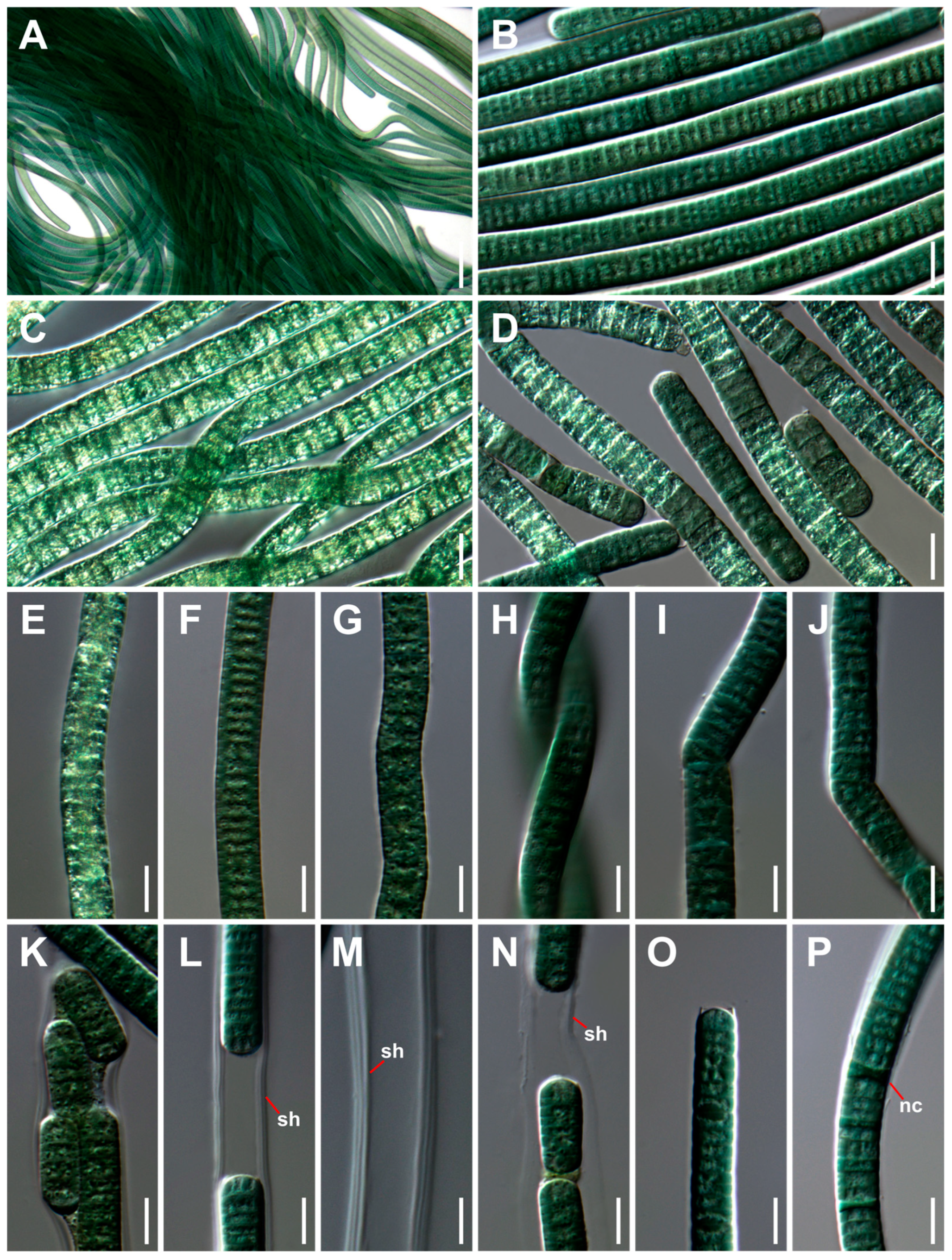
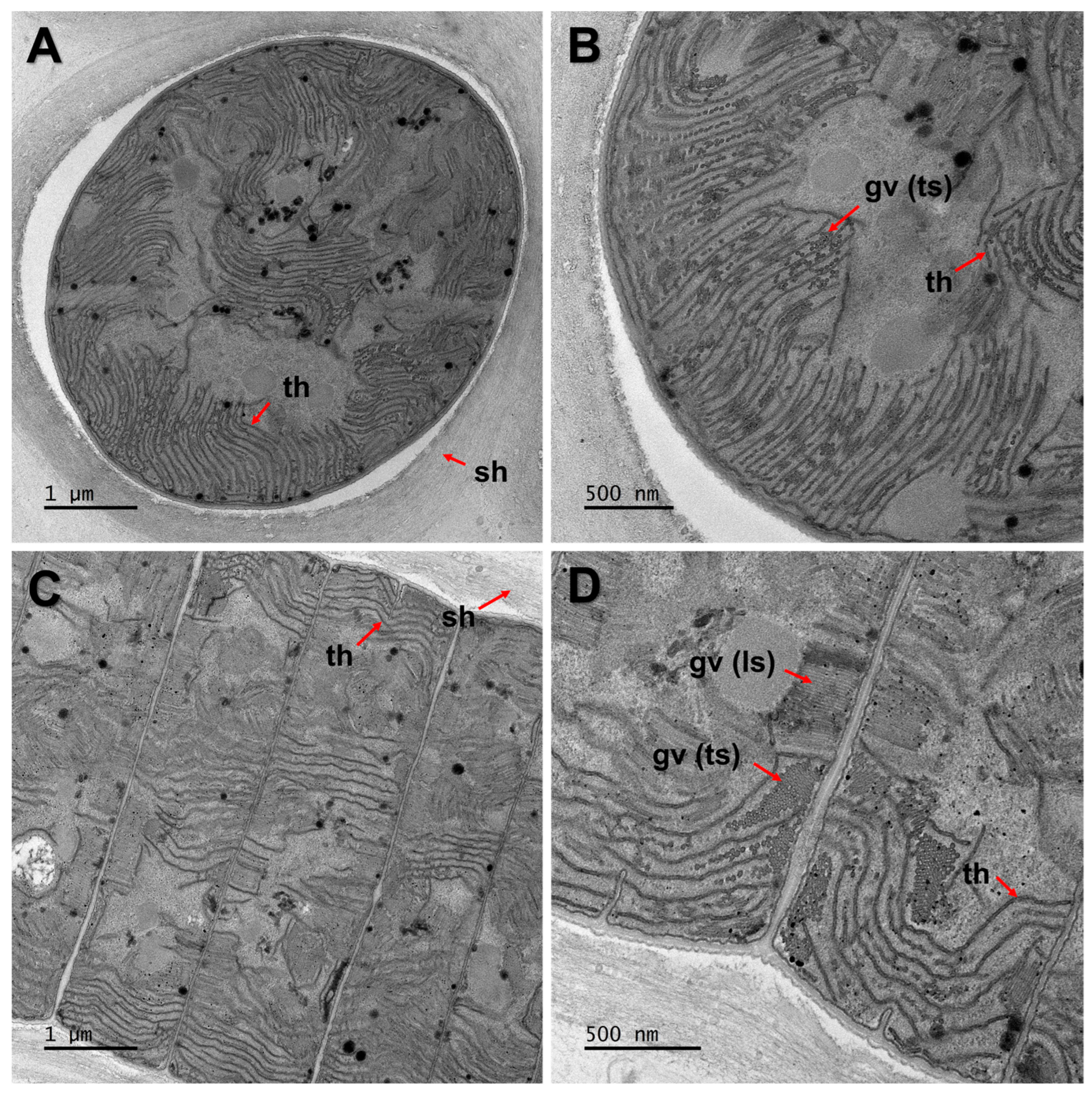
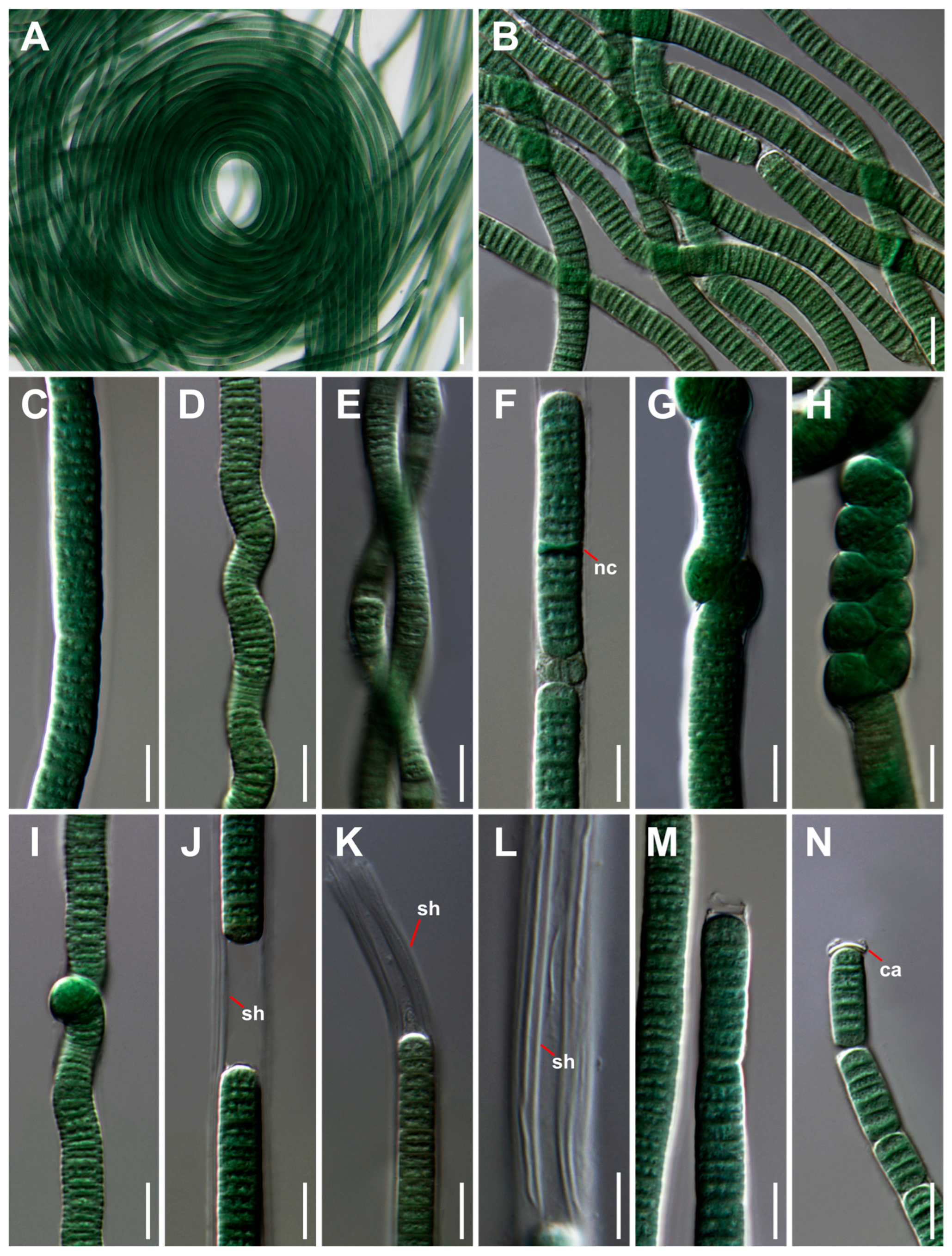
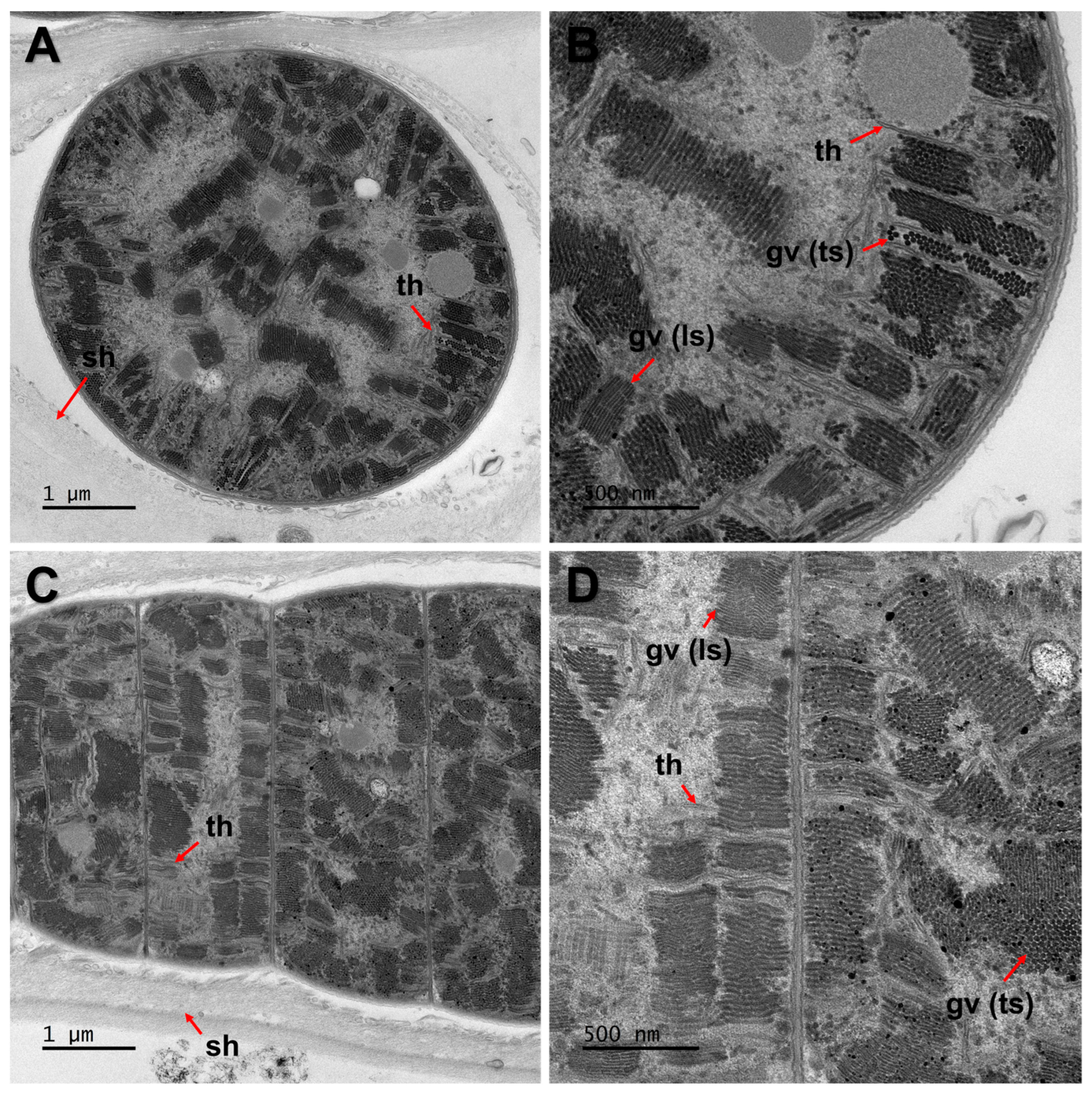
3.1. Taxonomic Treatment and Morphological Characterization

On the basis of the results of polyphasic analyses, we propose the following descriptions of new cyanobacteria taxa under the provisions of the ICN (International Code of Nomenclature for algae, fungi, and plants) [34].
Order: Oscillatoriales Schaffner 1922.
Family: Aerosakkonemataceae Strunecký and Mareš 2023.
Genus: Limnonema J. Song, S. Kim, and O. Lee gen. nov.
Description: The filament is blue-green, solitary, or in fine mat and cluster. The sheath is firm and thick and longitudinally lamellated. The trichome is straight, slightly curved, wavy, rarely twisted, sometimes bent into an angular shape or tightly twisted within sheath, not or slightly constricted at the cross walls, and not attenuated towards the end cells. The cells are discoid, usually shorter than they are wide, with aerotopes. The apical cells are round and rarely with calyptra. The arrangement of thylakoids is irregular. Reproduction occurs by necridic cells.
Diagnosis: The genus Limnonema has a sheath, gas vacuoles, and an irregular arrangement of thylakoids, but the genus Aerosakkonema has no sheath. Also, the genera Microseira and Potamosiphon have no gas vacuoles, and the genus Cephalothrix has a radial or parietal arrangement of thylakoids. In terms of cell width and length, the genus Limnonema differs from species belonging to the family Aerosakkonemataceae (Table 2).
Etymology: The generic name “Limnonema” is Greek, “Limno” means water or lake, and “Nema” means thread, indicating cell shape or characteristics.
Type species: Limnonema hangangris J. Song, S. Kim, and O. Lee sp. nov.
Limnonema hangangris J. Song, S. Kim, and O. Lee sp. nov. (Figure 2 and Figure 3).
Description: The filament is blue-green, solitary or in the form fine mat and cluster. The sheath is firm and thick, longitudinally lamellated, and very rarely diffluent. The trichome is straight, slightly curved, wavy, rarely twisted into a rope or bent into an angular shape, not or slightly constricted at the cross walls, and not attenuated towards the end cells. The cells are discoid, usually shorter than they are wide (6.0–7.9 width, 2.2–2.5 length), with aerotopes. Aerotopes are well visible using LM and TEM, scattered inside the cells. The apical cells are round and without calyptra. The thylakoids are arranged irregularly across the whole protoplast. Reproduction occurs via the fragmentation of trichomes by necridic cells.
Etymology: The specific epithet “hangangris” means that this species first appeared in the Han River, Republic of Korea.
Holotype: A formaldehyde fixed specimen, NIBRCY0000001648 in the Herbarium at the National Institute of Biological Resources, from cultured strain ACKU-684.
Reference strain: ACKU-684
Additional strains: ACKU-684–ACKU-699
Type locality: The water surface of the Han River in Seoul, Republic of Korea (35°36′25.8″ N, 126°41′25.5″ E)
Habitat: It inhabits the surface or gravel of freshwater in the temperate region as planktonic or epilithic cyanobacteria.
Gene sequences: GenBank Accession number of OR644266–OR644274, OR644698–OR644704 (16S rRNA, 16S–23S ITS, and 23S rRNA gene sequences), and OR640711 (gvpA gene).
Limnonema anseonga J. Song, S. Kim, and O. Lee sp. nov. (Figure 4 and Figure 5).
Description: The filament is blue-green, solitary, or in the form mat and cluster. The sheath is firm and thick and longitudinally lamellated. The trichome is straight, slightly curved, wavy, sometimes tightly twisted within the sheath, not or slightly constricted at the cross walls, and not attenuated towards the end cells. The cells are discoid, usually shorter than they are wide (6.4–9.5 width, 2.1–2.5 length), with aerotopes. The aerotopes are well visible using only TEM, scattered inside the cells. The apical cells are round and rarely with calyptra. The thylakoids are arranged irregularly across the whole protoplast. Reproduction occurs via the fragmentation of trichomes by necridic cells.
Etymology: The specific epithet “ anseonga “ means that this species first appeared in the Anseong Stream, Pyeongtaek, Republic of Korea.
Holotype: A formaldehyde fixed specimen, NIBRCY0000001647 in the Herbarium at the National Institute of Biological Resources, from cultured strain ACKU-700 clone A.
Reference strain: ACKU-700 clone A
Additional strain: ACKU-700 clone B
Type locality: The water surface of the Anseong Stream in Pyeongtaek, Gyeonggi-do, Republic of Korea (36°58′36.6″ N, 126°58′41.4″ E)
Habitat: They inhabit the surface of rivers as a plankton in the temperate region.
Gene sequences: GenBank Accession number of OR644275, OR644705 (16S rRNA, 16S–23S ITS, and 23S rRNA gene sequences), and OR640712 (gvpA gene).

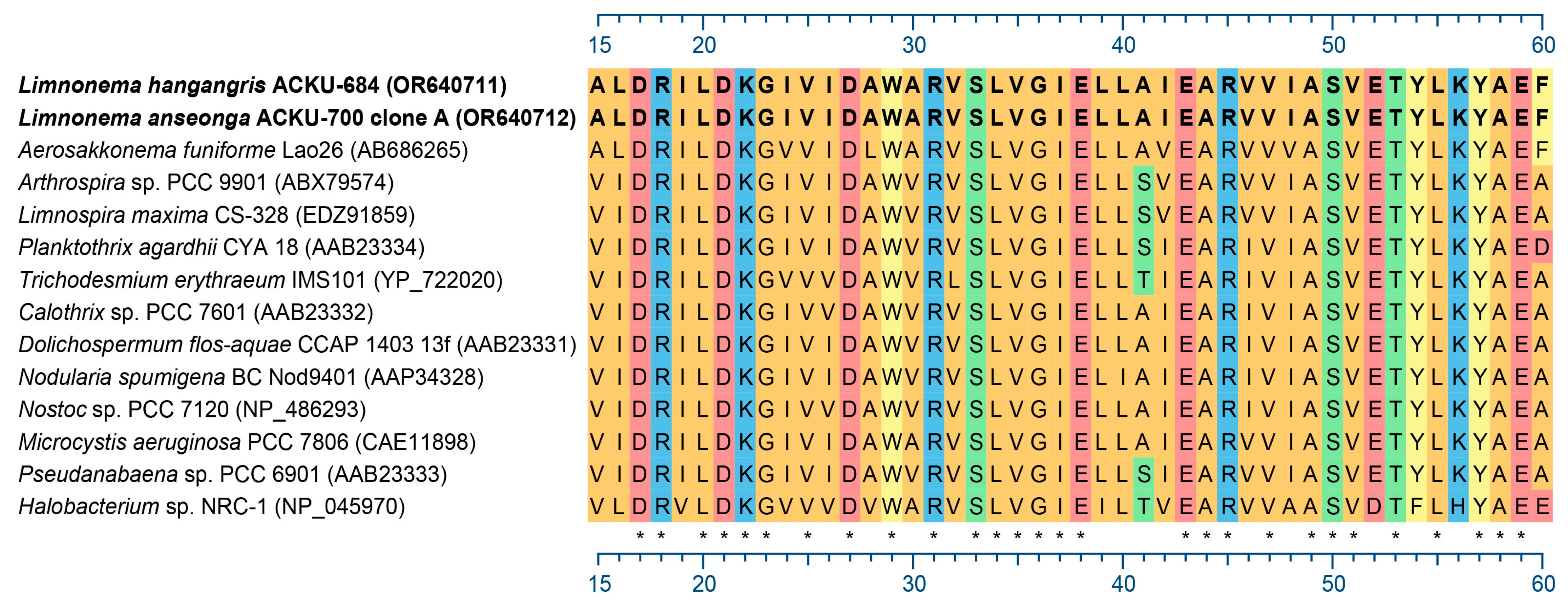
3.2. Detection of Gas Vesicle Protein (gvp)

As a result of PCR analyses for the gvpA gene, both species of Limnonema showed a single band. Sequencing and subsequent BLASTX (http://blast.ncbi.nlm.nih.gov/Blast.cgi, accessed on 3 August 2023) homology searches for L. hangangris (ACKU-684) and L. anseonga (ACKU-700 clone A) confirmed that PCR amplicons from those strains encoded a portion of the GvpA protein with the highest amino acid sequence similarity to GvpA from Microcystis aeruginosa PCC 7806. Also, the GvpA amino acid sequences of L. hangangris and L. anseonga were aligned with the GvpA amino acid sequences of other cyanobacteria and archaea with gas vacuoles (Figure 6).

3.3. 16S rRNA Gene Sequences Phylogenetic Tree and Molecular Distance

The phylogenetic tree was analyzed for the partial 16S rRNA gene sequences (1565 bp), and 139 sequences including outgroups were used. The pattern of the tree was based on the Maximum Likelihood (ML) method, but the tree according to the Bayesian Inference (BI) analyses also showed almost the same branching pattern as the ML tree. As a result of the analyses, the family Aerosakkonemataceae of the order Oscillatoriales was clustered separately from the families Oscillatoriaceae and Microcoleaceae of the order Oscillatoriales (Figure 7). The genus Limnonema was included in the family Aerosakkonemataceae along with the genera Aerosakkonema, Cephalothrix, Microseira, and Potamosiphon, each genus formed a different clade, and L. hangangris and L. anseonga also branched from each other (Figure 8).
16S rRNA gene sequences similarity and genetic distances were analyzed for a partial 16S rRNA gene sequence (1088 bp), which included 17 sequences from the molecularly similar genera Aerosakkonema, Cephalothrix, Microseira, Potamosiphon, and Limnnonema. As a result of the analyses, L. hangangris showed a similarity of 94.3–95.5% and genetic distances of 4.49–5.86 with species belonging to the genera Aerosakkonema and Cephalothrix; L. anseonga showed a similarity of 93.6–95.9% and genetic distances of 4.08–6.67 with species belonging to the genera Aerosakkonema and Cephalothrix. Also, the molecular distance between L. hangangris and L. anseonga showed a similarity of 97.6–97.7% and genetic distances of 2.25–2.34 (Table 3).

3.4. Secondary Structure of 16S–23S ITS Region

The inferred 16S–23S ITS secondary structures included the D1–D1′, Box-B, and V3 helices and were compared to 16S–23S ITS secondary structures of Aerosakkonema funiforme ACKU-621, Cephalothrix komarekiana CCIBt3277, Microseira wollei NIES-4236, and Potamosiphon australiensis FHC0914.02 (Figure 9).
The D1–D1′ helices of L. hangangris (three strains including ACKU-684) had a total length of 67 bp, consisted of a 4 bp basal stem (5′-GACC–GGUC-3′), a 0:6 base large unilateral bulge, two small unilateral bulges, a large loop (5:3 base bilateral bulge), and a 6 bp terminal loop (5′-AAAAAC-3′; Figure 9A–C). Also, the D1–D1′ helix of L. anseonga (ACKU-700 clone A) had a total length of 65 bp, consisted of a 5 bp basal stem (5′-GACCU–AGGUC-3′), a 1:7 base large bilateral bulge, two small unilateral bulges, a 6:0 base large unilateral bulge, and a 5 bp terminal loop (5′-GUUCG-3′; Figure 9D).
The Box-B helices of L. hangangris had a total length of 37 bp, consisted of a 4 bp basal stem (5′-AGCA–UGCU-3′), 2 small loops, and a 6 bp terminal loop (5′-AUAAAC-3′; Figure 9I–K). Also, the Box-B helix of L. anseonga had a total length of 37 bp, consisted of a 4 bp basal stem (5′-AGCA–UGCU-3′), two small loops, and a 6 bp terminal loop (5′-AAAAAC-3′; Figure 9L).
The V3 helices of L. hangangris had a total length of 59 or 62 bp, consisted of a 3 bp basal stem (5′-GUC–GAC-3′), a large loop (4:5 base bilateral bulge), two or three small loops, and a 4 or 7 bp terminal loop (5′-UAUU-3′ or 5′-UAUUAUU-3′; Figure 9Q–S). Also, the V3 helix of L. anseonga had a total length of 42 bp, consisted of a 3 bp basal stem (5′-GUC–GAC-3′), a large loop (4:5 base bilateral bulge), and a 7 bp terminal loop (5′-UUUUGAU-3′; Figure 9T).

4. Discussion

The genus Limnonema proposed in this study belongs to the family Aerosakkonemataceae, which has four genera including genus Aerosakkonema [9]. The genus Limnonema was found in freshwater of temperate regions, and although most of the L. hangangris and L. anseonga strains appeared as plankton, some of the L. hangangris strains (ACKU-695–697) appeared as epilithic cyanobacteria as well (Table 1). Similarly, most species in the family Aerosakkonemataceae appeared as plankton, but Cephalothrix alaskaensis by Strunecký et al. and Microseira minor by Geng and Yu of the family Aerosakkonemataceae appeared as epilithic cyanobacteria [11,12,13,14,15,16].
As the cell width of genus Limnonema is 6.0–9.5 μm (Table 2), and the cell width of genus Aerosakkonema is 11.7–16.6 μm [11], the genus Limnonema classified as having a narrower width than the genus Aerosakkonema, and L. anseonga contained more cells with wider widths than those of L. hangangris (Table 2). The thylakoids arrangement of the genera Limnonema and Aerosakkonema was irregular (Table 2; Figure 3 and Figure 5) [11], but that of C. alaskaensis was parietal, and that of C. komarekiana was radial [12,13]. Until recently, thylakoids arrangement has been useful for classifying cyanobacteria at the family level [7], but it appears to be very diverse in the family Aerosakkonemataceae. Also, TEM images showed that the genus Limnonema had many gas vacuoles (Figure 3 and Figure 5), like the genera Aerosakkonema and Cephalothrix [11,12].
GvpA of the genus Limnonema detected using PCR analyses of the gvpA genes showed the highest amino acid sequence similarity with Microcystis aeruginosa by (Kützing) Kützing of another order in terms of sequencing and subsequent homology searches (Figure 6). This is consistent with studies showing that the characteristic of having gas vacuoles does not provide strong phylogenetic information [11]. However, this experiment on the gvpA gene is meaningful in confirming whether the gas vacuoles observed using LM and TEM are also detected as genes.
In a recent study, the family Aerosakkonemataceae was classified in the order Oscillatoriales, but was clustered separately in the phylogenetic analyses based on 16S rRNA gene sequences [9]. The same result was shown in the analyses including 18 strains of Genus Limnonema, and the monophyly of the family Aerosakkonemataceae was supported by very high values (99% MLB/1.00 BPP) in all phylogenetic analyses (ML and BI) (Figure 7). Also, the genus Limnonema within the family Aerosakkonemataceae showed very high support values (100% MLB/1.00 BPP) and was clustered separately from other genera (Aerosakkonema, Cephalothrix, Microseira, and Potamosiphon), and the two species of the genus Limnonema also branched from each other with values of 100% MLB and 1.00 BPP, showing that they are different species (Figure 8). Additionally, the genus Limnonema species showed 16S rRNA gene sequences similarity of 95.4–95.9% with genus Aerosakkonema and 93.6–94.4% with genus Cephalothrix, confirming that it is a different genus [35]. The 16S rRNA gene sequences similarity between the two new species of genus Limnonema was 97.6–97.7%, indicating that they are different species (Table 3) [36].
As a result of comparing ITS secondary structures (D1–D1′, Box-B, and V3) between the two species of genus Limnonema and A. funiforme, C. komarekiana, M. wollei, and P. australiensis, D1–D1′ and Box-B helices showed overall structural differences due to the differences in length and number of bulges and loops, and there was almost no structural difference between the two species of the genus Limnonema (Figure 9A–P). In the V3 helices structure, all six species differed in total length, location, number, and length of the bulge and loop; moreover, the lengths and structures of the basal stem and terminal loop were also shown to be different (Figure 9Q–X).
Based on the morphological and phylogenetic criteria, the Korean cyanobacteria strains in this study were proposed as Limnonema gen. nov., Limnonema hangangris sp. nov., and Limnonema anseonga sp. nov. within the family Aerosakkonemataceae, in the order Oscillatoriales.

Supplementary Materials

The following are available online at https://www.mdpi.com/article/10.3390/d15121174/s1, Table S1: Sequences used in the 16S rRNA gene sequences analysis. The sequences were obtained from GenBank and include GenBank accessions.

Author Contributions

Conceptualization, J.-H.S.; methodology, J.-H.S. and H.-R.W.; validation, J.-H.S., S.-W.K., N.-J.L., D.-H.K., and O.-M.L.; formal analysis, J.-H.S.; investigation, J.-H.S.; resources, J.-H.S.; data curation, J.-H.S.; writing—original draft preparation, J.-H.S.; writing—review and editing, J.-H.S.; visualization, J.-H.S.; supervision, J.-H.S. and O.-M.L.; project administration, O.-M.L.; funding acquisition, O.-M.L. All authors have read and agreed to the published version of the manuscript.

Funding

This work was supported by a grant from the National Institute of Biological Resources (NIBR) (NIBR202203204) and Korea Environment Industry & Technology Institute (KEITI) through the project to make multi-ministerial national biological research resources more advanced (2021003420004), funded by Korea Ministry of Environment (MOE).

Institutional Review Board Statement

Not applicable.

Data Availability Statement

Data are contained within the article and Supplementary Materials.

Acknowledgments

We wish to thank the Korean Basic Science Institute (KBSI) of Chuncheon for taking the TEM images.

Conflicts of Interest

The authors declare no conflict of interest.

References

  1. Rippka, R.; Deruelles, J.; Waterbury, J.B.; Herdman, M.; Stanier, R.Y. Generic Assignments, Strain Histories and Properties of Pure Cultures of Cyanobacteria. J. Gen. Microbiol. 1979, 111, 1–61. [Google Scholar] [CrossRef]
  2. Casamatta, D.A.; Vis, M.L.; Sheath, R.G. Cryptic species in cyanobacterial systematics: A case study of Phormidium retzii (Oscillatoriales) using RAPD molecular markers and 16S rDNA sequence data. Aquat. Bot. 2003, 77, 295–309. [Google Scholar] [CrossRef]
  3. Dvořák, P.; Jahodářová, E.; Hašler, P.; Gusev, E.; Poulíčková, A. A new tropical cyanobacterium Pinocchia polymorpha gen. et sp. nov. derived from the genus Pseudanabaena. Fottea 2015, 15, 113–120. [Google Scholar] [CrossRef]
  4. Castenholz, R.W. Species usage, concept, and evolution in the cyanobacteria (blue-green algae). J. Phycol. 1992, 28, 737–745. [Google Scholar] [CrossRef]
  5. Komárek, J. The modern classification of cyanoprokaryotes (Cyanobacteria). Oceanol. Hidrobiol. Stud. 2005, 34, 5–17. [Google Scholar]
  6. Komárek, J.; Anagnostidis, K. Cyanoprokaryota Teil 2: Oscillatoriales. In Süsswasserflora von Mitteleuropa Band 19/2; Büdel, B., Krienitz, L., Gärtner, G., Schagerl, M., Eds.; Elsevier: Heidelberg, Germany, 2005; pp. 1–759. [Google Scholar]
  7. Komárek, J.; Kaštovský, J.; Mareš, J.; Johansen, J.R. Taxonomic classification of cyanoprokaryotes (cyanobacterial genera) 2014, using a polyphasic approach. Preslia 2014, 86, 295–335. [Google Scholar]
  8. Komárek, J. A polyphasic approach for the taxonomy of cyanobacteria: Principles and applications. Eur. J. Phycol. 2016, 51, 346–353. [Google Scholar] [CrossRef]
  9. Strunecky, O.; Ivanova, A.P.; Mares, J. An updated classification of cyanobacterial orders and families based on phylogenomic and polyphasic analysis. J. Phycol. 2023, 59, 12–51. [Google Scholar] [CrossRef]
  10. Guiry, M.D.; Guiry, G.M. AlgaeBase. World-Wide Electronic Publication, National University of Ireland, Galway. 2023. Available online: www.algaebase.org (accessed on 14 October 2023).
  11. Thu, N.K.; Tanabe, Y.; Yoshida, M.; Matsuura, H.; Watanabe, M.M. Aerosakkonema funiformegen. et sp. nov. (Oscillatoriales), a new gas-vacuolated oscillatorioid cyanobacterium isolated from a mesotrophic reservoir. Phycologia 2012, 51, 672–683. [Google Scholar] [CrossRef]
  12. da Silva Malone, C.F.; Rigonato, J.; Laughinghouse, H.D.; Schmidt, E.C.; Bouzon, Z.L.; Wilmotte, A.; Fiore, M.F.; Sant’Anna, C.L. Cephalothrix gen. nov. (Cyanobacteria): Towards an intraspecific phylogenetic evaluation by multilocus analyses. Int. J. Syst. Evol. Microbiol. 2015, 65, 2993–3007. [Google Scholar] [CrossRef]
  13. Strunecky, O.; Raabova, L.; Bernardova, A.; Ivanova, A.P.; Semanova, A.; Crossley, J.; Kaftan, D. Diversity of cyanobacteria at the Alaska North Slope with description of two new genera: Gibliniella and Shackletoniella. FEMS Microbiol. Ecol. 2020, 96, 1–60. [Google Scholar] [CrossRef] [PubMed]
  14. McGregor, G.B.; Sendall, B.C. Phylogeny and toxicology of Lyngbya wollei (Cyanobacteria, Oscillatoriales) from north-eastern Australia, with a description of Microseira gen. nov. J. Phycol. 2015, 51, 109–119. [Google Scholar] [CrossRef]
  15. Geng, R.; Li, W.; Chao, A.; Guo, X.; Li, H.; Yu, G.; Li, R. Establishment of a New Filamentous Cyanobacterial Genus, Microcoleusiopsis gen. nov. (Microcoleaceae, Cyanobacteria), from Benthic Mats in Open Channel, Jiangxi Province, China. Diversity 2021, 13, 178–190. [Google Scholar] [CrossRef]
  16. McGregor, G.B.; Sendall, B.C. Potamosiphon australiensis gen. nov., sp nov. (Oscillatoriales), a new filamentous cyanobacterium from subtropical north-eastern Australia. Phytotaxa 2019, 387, 77–93. [Google Scholar] [CrossRef]
  17. Sciuto, K.; Moro, I. Cyanobacteria: The bright and dark sides of a charming group. Biodivers. Conserv. 2015, 24, 711–738. [Google Scholar] [CrossRef]
  18. Dadheech, P.K.; Abed, R.M.M.; Mahmoud, H.; Mohan, M.K.; Krienitz, L. Polyphasic characterization of cyanobacteria isolated from desert crusts, and the description of Desertifilum tharensegen. et sp. nov. (Oscillatoriales). Phycologia 2012, 51, 260–270. [Google Scholar] [CrossRef]
  19. Sournia, A. Phytoplankton Manual; UNESCO: France, Paris, 1978; pp. 1–337. [Google Scholar]
  20. Kiel, G.; Gaylarde, C.C. Bacterial diversity in biofilms on external surfaces of historic buildings in Porto Alegre. World J. Microbiol. Biotechnol. 2006, 22, 293–297. [Google Scholar] [CrossRef]
  21. Stanier, R.Y.; Kunisawa, R.; Mandel, M.C.B.G.; Cohen-Bazire, G. Purification and properties of unicellular blue-green algae (order Chroococcales). Bacteriol. Rev. 1971, 35, 171–205. [Google Scholar] [CrossRef]
  22. Lee, N.-J.; Seo, Y.; Ki, J.-S.; Lee, O.-M. A study of newly recorded genus and species for aerial cyanobacteria Wilmottia murrayi (Oscillatoriales, Cyanobacteria) in Korea. Korean J. Environ. Biol. 2019, 37, 260–267. [Google Scholar] [CrossRef]
  23. Kim, J.-H.; Jeong, M.S.; Kim, D.-Y.; Her, S.; Wie, M.-B. Zinc oxide nanoparticles induce lipoxygenase-mediated apoptosis and necrosis in human neuroblastoma SH-SY5Y cells. Neurochem. Int. 2015, 90, 204–214. [Google Scholar] [CrossRef]
  24. Neilan, B.A.; Jacobs, D.; Blackall, L.L.; Hawkins, P.R.; Cox, P.T.; Goodman, A.E. rRNA sequences and evolutionary relationships among toxic and nontoxic cyanobacteria of the genus Microcystis. Int. J. Syst. Evol. Microbiol. 1997, 47, 693–697. [Google Scholar] [CrossRef] [PubMed]
  25. Taton, A.; Grubisic, S.; Brambilla, E.; De Wit, R.; Wilmotte, A. Cyanobacterial diversity in natural and artificial microbial mats of Lake Fryxell (McMurdo Dry Valleys, Antarctica): A morphological and molecular approach. Appl. Environ. Microbiol. 2003, 69, 5157–5169. [Google Scholar] [CrossRef] [PubMed]
  26. Katoh, K.; Standley, D.M. MAFFT multiple sequence alignment software version 7: Improvements in performance and usability. Mol. Biol. Evol. 2013, 30, 772–780. [Google Scholar] [CrossRef] [PubMed]
  27. Hall, T. BioEdit: A user-friendly biological sequence alignment editor and analysis program for Windows 95/98/NT. Nucleic Acids. Symp. Ser. 1999, 41, 95–98. [Google Scholar]
  28. Stamatakis, A. RAxML-VI-HPC: Maximum likelihood-based phylogenetic analyses with thousands of taxa and mixed models. Bioinformatics 2006, 22, 2688–2690. [Google Scholar] [CrossRef] [PubMed]
  29. Huelsenbeck, J.P.; Ronquist, F. MRBAYES: Bayesian inference of phylogenetic trees. Bioinformatics 2001, 17, 754–755. [Google Scholar] [CrossRef] [PubMed]
  30. Kumar, S.; Stecher, G.; Li, M.; Knyaz, C.; Tamura, K. MEGA X: Molecular evolutionary genetics analysis across computing platforms. Mol. Biol. Evol. 2018, 35, 1547–1549. [Google Scholar] [CrossRef] [PubMed]
  31. Page, R.D.M. Tree View: An application to display phylogenetic trees on personal computers. Bioinformatics 1996, 12, 357–358. [Google Scholar] [CrossRef]
  32. Zuker, M. Mfold web server for nucleic acid folding and hybridization prediction. Nucleic Acids Res. 2003, 31, 3406–3415. [Google Scholar] [CrossRef]
  33. Byun, Y.; Han, K. PseudoViewer3: Generating planar drawings of large-scale RNA structures with pseudoknots. Bioinformatics 2009, 25, 1435–1437. [Google Scholar] [CrossRef]
  34. Turland, N.J.; Wiersema, J.H.; Barrie, F.R.; Greuter, W.; Hawksworth, D.L.; Herendeen, P.S.; Knapp, S.; Kusber, W.-H.; Li, D.-Z.; Marhold, K.; et al. International Code of Nomenclature for Algae, Fungi, and Plants (Shenzhen Code) Adopted by the Nineteenth International Botanical Congress Shenzhen, China, 17 July 2017; Regnum Vegetabile 159; Koeltz Botanical Books: Glashütten, Germany, 2018; pp. 1–254. [Google Scholar] [CrossRef]
  35. Stackebrandt, E.; Goebel, B.M. Taxonomic note: A place for DNA-DNA reassociation and 16S rRNA sequence analysis in the present species definition in bacteriology. Int. J. Syst. Evol. Microbiol. 1994, 44, 846–849. [Google Scholar] [CrossRef]
  36. Kim, M.; Oh, H.-S.; Park, S.-C.; Chun, J. Towards a taxonomic coherence between average nucleotide identity and 16S rRNA gene sequence similarity for species demarcation of prokaryotes. Int. J. Syst. Evol. Microbiol. 2014, 64, 346–351. [Google Scholar] [CrossRef] [PubMed]
Figure 1. Map showing sampling sites. (A) The circles are sampling sites; (BH) the habitat view of a collection sites.
Diversity 15 01174 g001
Figure 2. Microphotographs of Limnonema hangangris cultured. (A,B) Arrangement of filament in the colony; (CE) gas vacuoles scattered in the trichome; (F,G) morphologies of trichomes; (H) twisted trichomes; (I,J) trichome bent into an angular shape; (K) dividing trichome; (L,M) longitudinal lamellations in the sheath (sh); (N) diffluent sheath; (O) morphology of apical cell; (P) necridic cell (nc) in the trichome. Scale bars, 50 μm (A); 10 μm (BP).
Diversity 15 01174 g002
Figure 3. Transmission electron micrographs of Limnonema hangangris ACKU-684. (A,B) Cross section of the trichome; (C,D) longitudinal section of the trichome; (A,C) irregular thylakoids (th) arrangement and sheath (sh); (B,D) view of gas vacuoles (gv) in longitudinal section (ls) and transverse section (ts).
Diversity 15 01174 g003
Figure 4. Microphotographs of Limnonema anseonga cultured. (A,B) arrangement of filament in the colony; (C,D) morphologies of trichomes; (E) twisted trichomes; (F) necridic cell (nc) in the trichome; (GI) tightly twisted trichomes within a sheath; (JL) longitudinal lamellations in the sheath (sh); (M) morphology of apical cell; (N) apical cell with calyptra (ca). Scale bars, 50 μm (A); 10 μm (BN).
Diversity 15 01174 g004
Figure 5. Transmission electron micrographs of Limnonema anseonga ACKU-700 clone A. (A,B) Cross section of the trichome; (C,D) longitudinal section of the trichome; (A,C) irregular thylakoids (th) arrangement and sheath (sh); (B,D) view of gas vacuoles (gv) in longitudinal section (ls) and transverse section (ts).
Diversity 15 01174 g005
Figure 6. Amino acid sequence alignment of gas vesicle protein GvpA of L. hangangris ACKU-684, L. anseonga ACKU-700 clone A, and other gas-vacuolated cyanobacteria. Asterisks indicate the same amino acids occurred in all strains. Numbering above the alignment is based on the amino acid sequence of GvpAI of Microcystis aeruginosa PCC 7806.
Diversity 15 01174 g006
Figure 7. 16S rRNA gene sequences phylogenetic tree of cyanobacteria. This tree was drawn with reference to Strunecký et al. [9] and was inferred from 1565 bp of the 16 rRNA gene sequences alignment using an ML method. Additional BI analyses generated similar topology to the ML tree. The first numbers at the nodes display ML bootstrap support (MLB; >50%). The second numbers at the nodes display Bayesian posterior probability (BPP; >0.50). Sequences determined in this work are indicated in bold, and branch lengths are proportional to the scale given. * = type species.
Diversity 15 01174 g007
Figure 8. The phylogenetic tree showing only the family Aerosakkonemataceae in Figure 7. This tree was also drawn based on 16S rRNA gene sequences. * = type strain.
Diversity 15 01174 g008
Figure 9. Secondary helix structures of the conserved D1-D1′, Box-B, and V3 sequences in the 16S-23S internal transcribed spacer (ITS) regions. (A,I,Q) L. hangangris ACKU-684; (B,J,R) L. hangangris ACKU-690; (C,K,S) L. hangangris ACKU-699; (D,L,T) L. anseonga ACKU-700 clone A; (E,M,U) A. funiforme ACKU-621; (F,N,V) C. komarekiana CCIBt3277; (G,O,W) Microseira wollei NIES-4236; (H,P,X) P. australiensis FHC0914.02. These structures are drawn with reference to Malone et al. [12].
Diversity 15 01174 g009
Table 1. Locations and occurrence of isolated cyanobacteria strains.
StrainSiteLocationGPSOccurrenceTemperaturepH
Limnonema hangangris
ACKU-684Site 1Han River, Seoul, Republic of Korea37°31′10.2″ N 127°05′27.2″ EPlankton on the surface of river26.3 °C7.6
ACKU-685
ACKU-686
ACKU-687
ACKU-688
ACKU-689Site 2Yeonjoong Reservoir, Daedong-ri, Julpo-myeon, Buan, Jeollabuk-do, Republic of Korea35°36′25.8″ N 126°41′25.5″ EPlankton on the surface of reservoir29.2 °C7.0
ACKU-694
ACKU-690Site 3Pungjeong Reservoir, Nokjin-ri, Nam-myeon, Jangseong, Jeollanam-do, Republic of Korea35°14′54.7″ N 126°47′47.9″ EPlankton on the surface of reservoir29.1 °C7.1
ACKU-691
ACKU-693
ACKU-692Site 4Sanmak Reservoir, Deungim-dong, Gwangsan-gu, Gwangju, Jeollanam-do, Republic of Korea35°11′34.6″ N 126°46′03.7″ EPlankton on the surface of reservoir32.4 °C8.5
ACKU-699Site 5Beopsu Jilnal Swamp, Ugeo-ri, Beopsu-myeon, Haman, Gyeongsangnam-do, Republic of Korea35°19′11.7″ N 128°21′00.2″ EPlankton on the surface of reservoir in the swamp20.1 °C6.9
ACKU-698Site 6-1Daepyeong Reservoir, Daesong-ri, Beopsu-myeon, Haman, Gyeongsangnam-do, Republic of Korea35°20′25.0″ N 128°20′04.7″ EPlankton on the surface of reservoir21.6 °C7.0
ACKU-695Site 6-2Epilithic cyanobacteria of gravel at the bottom of reservoir
ACKU-696
ACKU-697
Limnonema anseonga
ACKU-700 clone ASite 7Anseong Stream, Dodu-ri, Paengseong-eup, Pyeongtaek, Gyeonggi-do, Republic of Korea36°58′36.6″ N 126°58′41.4″ EPlankton on the surface of stream25.9 °C8.2
ACKU-700 clone B
Table 2. Morphology and ecology comparison between two new species and similar genera (Aerosakkonema, Cephalothrix, Microseira, and Potamosiphon).
Apical CellCalyptraSheathGas VacuolesThylakoidsCell Size (μm)Habitat/Distribution
WidthLength
Limnonema hangangris ACKU-684RoundedNot observedThick, longitudinally lamellatedObservedIrregular6.0–7.92.2–2.5Surface or gravel of freshwater/Korea
L. anseonga ACKU-700 clone ARoundedRarely observedThick, longitudinally lamellatedObservedIrregular6.4–9.52.1–2.5Surface of freshwater/Korea
Aerosakkonema funiforme Lao26Rounded or flattened-roundedNot observedNot observedObservedIrregular11.7–16.62.6–4.1Reservoir/Laos
Cephalothrix alaskaensis L30RoundedNot observedHyaline, firm, sometimes loosening at the end of filamentsNot observedParietal5.0–6.51.8–2.0Water soil shoreline/Alaska
C. komarekiana CCIBt3277Strongly capitateNot observedAttached to trichome or wideObservedRadial4.8–6.62.0–3.4Alkaline lake/Brazilian
C. lacustris CCIBt3261Strongly capitateObserved (conical)Attached to trichomeObservedND5.6–6.72.2–3.1Freshwater pond/Brazilian
Microseira wollei YC0404RoundedNot observedColorless, hyaline, firm, lamellatedNot observedND30–484.0–9.0Catchments/Australia
M. minor CHAB 4136RoundedNot observedColorless, hyaline, firm, lamellatedNot observedIrregular9.0–13.33.1–7.7Rock surface in freshwater river/China
Potamosiphon australiensis FHC0914RoundedNot observedColourless, sometimes lamellatedNot observedIrregular15.7–17.81.7–2.5Benthic in freshwater coastal streams/Australia
ND = No data.
Table 3. 16S rRNA gene sequences similarity and genetic distance (p-distance) of two species of the genus Limnonema from species belonging to the genera Aerosakkonema, Cephalothrix, Microseira, and Potamosiphon.
No.Species and Strain<1><2><3><4><5><6><7><8><9><10><11><12><13><14><15><16><17>
DNA similarity (%)
<1>Limnonema hangangris ACKU-684 10010010010099.999.997.795.595.595.594.494.494.391.891.992.9
<2>L. hangangris ACKU-6890.00 10010010099.999.997.795.595.595.594.494.494.391.891.992.9
<3>L. hangangris ACKU-6900.000.00 10010099.999.997.795.595.595.594.494.494.391.891.992.9
<4>L. hangangris ACKU-6920.000.000.00 10099.999.997.795.595.595.594.494.494.391.891.992.9
<5>L. hangangris ACKU-6950.000.000.000.00 99.999.997.795.595.595.594.494.494.391.891.992.9
<6>L. hangangris ACKU-6980.090.090.090.090.09 10097.695.495.495.494.394.494.391.891.992.9
<7>L. hangangris ACKU-6990.090.090.090.090.090.00 97.695.495.495.494.394.494.391.891.992.9
<8>L. anseonga ACKU-700 clone A2.252.252.252.252.252.342.34 95.995.995.994.093.693.691.691.993.7
<9>Aerosakkonema funiforme Lao264.464.464.464.464.464.564.564.08 10010095.895.095.192.492.994.2
<10>A. funiforme Lao284.464.464.464.464.464.564.564.080.00 10095.895.095.192.492.994.2
<11>A. funiforme ACKU-6214.464.464.464.464.464.564.564.080.000.00 95.895.095.192.492.994.2
<12>Cephalothrix alaskaensis L305.665.665.665.665.665.765.766.164.184.184.18 97.497.691.792.093.1
<13>C. komarekiana CCIBt32775.765.765.765.765.765.765.766.665.065.065.062.53 98.992.191.992.8
<14>C. lacustris CCIBt32615.865.865.865.865.865.865.866.674.964.964.962.251.02 92.291.792.6
<15>Microseira wollei YC04048.618.618.618.618.618.618.618.917.887.887.888.718.298.19 94.794.2
<16>M. minor CHAB 4136.01-48.398.398.398.398.398.298.298.297.167.167.168.218.428.635.25 95.0
<17>Potamosiphon australiensis FHC0914.027.477.477.477.477.477.477.476.555.855.855.857.067.577.785.954.96
p-distance (%)
Disclaimer/Publisher’s Note: The statements, opinions and data contained in all publications are solely those of the individual author(s) and contributor(s) and not of MDPI and/or the editor(s). MDPI and/or the editor(s) disclaim responsibility for any injury to people or property resulting from any ideas, methods, instructions or products referred to in the content.

