
Effect of Various Mentha sp. Extracts on the Growth of Trichoderma viride and Chaetomium globusom on Agar Medium and Pine Wood

 , ,
, ,  , and
, and
Abstract
:1. Introduction
2. Materials and Methods
2.1. Materials
2.2. Methods
2.2.1. Assessment of Fungicidal Properties
2.2.2. Assessment of the Degree of Fouling of the Impregnated Wood Surface
- (A)
- Determination of the area of the upper surface of the sample as 100%:
- (B)
- Determination of the area of overgrowth of the upper surface of the sample by mycelium:
2.2.3. Growth of Fungi on Plant Material
2.2.4. GC-MS Analysis
- Temperature: 50 °C maintained for 5 min, increase of 13 °C/min to 200 °C maintained for 5 min, increase of 13 °C/min to 300 °C maintained 10 min;
- Flow: 0.8 mL/min;
- Temperature of the injection: 250 °C;
- Detector voltage: 0.8 kV;
- Carrier gas: helium 5.0 (PGNiG, Warsaw, Poland);
- Injection mode: direct;
- Ion source temperature: 200 °C;
- Interface temperature: 200 °C.
3. Results and Discussion
4. Conclusions
Author Contributions
Funding
Institutional Review Board Statement
Data Availability Statement
Conflicts of Interest
References
- Faria, J.M.S.; Barbosa, P.; Vieira, P.; Vicente, C.S.L.; Figueiredo, A.C.; Mota, M. Phytochemicals as biopesticides against the pinewood nematode Bursaphelenchus xylophilus: A Review on essential oils and their volatiles. Plants 2021, 10, 2614. [Google Scholar] [CrossRef] [PubMed]
- Woźniak, M. Antifungal Agents in Wood Protection—A Review. Molecules 2022, 27, 6392. [Google Scholar] [CrossRef] [PubMed]
- Elgharbawy, A.A.M.; Samsudin, N.; Benbelgacem, F.F.; Hashim, Y.Z.H.-Y.; Salleh, H.M.; Santhanam, J. Phytochemicals with antifungal properties, Cure from nature. Malays. J. Microbiol. 2020, 16, 2231–7538. [Google Scholar] [CrossRef]
- Asamoah, A.; Frimpong-Mensah, K.; Antwi-Boasiako, C. Efficacy of Erythropleumsuaveolens(potrodom) and Distemonanthusbenthamianus (bonsamdua) water extractives on the durability of five Ghanaian less used timber species of varying perviousness and retentiveness. J. Ind. Acad. Wood Sci. 2014, 11, 72–81. [Google Scholar] [CrossRef]
- Betlej, I.; Andres, B.; Krajewski, K. Evaluation of fungicidal effects of post-culture medium of selected mold fungi and bacteria in relation to Basidiomycetes fungi, causing wood destruction. BioResources 2020, 15, 2471–2482. [Google Scholar] [CrossRef]
- Broda, M. Natural compounds for wood protection against fungi—A review. Molecules 2020, 25, 3538. [Google Scholar] [CrossRef]
- Abanikannda, J.P.; Adetogun, A.C.; Mukhtar, R.B. Evaluation of honey bee propolis as wood preservative using weight loss. Sci. World J. 2020, 5, 45–47. [Google Scholar]
- Betlej, I.; Andres, B.; Szadkowska, D.; Krajewski, K.; Ościłowska, A. Fungicidal properties of the medium from SCOBY microorganism cultivation in saturated wood against Coniophora puteana fungus. BioResources 2021, 16, 1287–1295. [Google Scholar] [CrossRef]
- Betlej, I.; Krajewski, K. Application of wood destroying fungi in biotechnological processes. Ochr. Przed Koroz. 2012, 55, 32–35. [Google Scholar]
- Tascioglu, C.; Yalçın, M.; Şen, S.; Akcay, C. Antifungal properties of some plant extracts used as wood preservatives. Int. Biodeterior. Biodegrad. 2013, 85, 23–28. [Google Scholar] [CrossRef]
- Sumthong, P.; Romero-González, R.R.; Verpoorte, R. Identification of anti-wood rot compounds in teak (Tectona grandis L.f.) sawdust extract. J. Wood Chem. Technol. 2008, 28, 247–260. [Google Scholar] [CrossRef]
- Chittenden, C.; Singh, T. Antifungal activity of essential oils against wood degrading fungi and their applications as wood preservatives. Int. Wood Prod. J. 2011, 2, 44–48. [Google Scholar] [CrossRef]
- Kedia, A.; Prakash, B.; Mishra, P.K.; Chanotiya, C.S.; Dubey, N.C. Antifungal, antiaflatoxigenic, and insecticidal efficacy of spearmint (Mentha spicata L.) essential oil. Int. Biodeterior. Biodegrad. 2014, 89, 29–36. [Google Scholar] [CrossRef]
- Ngo-Mback, M.N.L.; Famewo, E.B.; Mubarak Ali, D.; Eke, P.; Thajuddin, N.; Afolayan, A.J.; Jazet Dongmo, P.M.; FekamBoyom, F. An investigation of chemical composition and antimicrobial activity of essential oils extracted from Aeollanthus and Plectranthus species. Biocatal. Agric. Biotechnol. 2019, 22, 101412. [Google Scholar] [CrossRef]
- Cheng, S.S.; Lin, C.-Y.; Gu, H.-J.; Chang, S.-T. Antifungal activities and chemical composition of wood and leaf essential oils from Cunnin ghamiakonishii. J. Wood Chem. Technol. 2011, 31, 204–217. [Google Scholar] [CrossRef]
- Kwaśniewska-Sip, P.; Cofta, G.; Nowak, P.B. Resistance of fungal growth on Scots pine treated with caffeine. Int. Biodeterior. Biodegrad. 2018, 132, 178–184. [Google Scholar] [CrossRef]
- Kwaśniewska-Sip, P.; Woźniak, M.; Jankowski, W.; Ratajczak, I.; Cofta, G. Chemical changes of wood treated with caffeine. Materials 2021, 14, 497. [Google Scholar] [CrossRef]
- Ratajczak, I.; Woźniak, M.; Kwaśniewska-Sip, P.; Szentner, K.; Cofta, G.; Mazela, B. Chemical characterization of wood treated with a formulation based on propolis, caffeine and organosilanes. Eur. J. Wood Wood Prod. 2018, 76, 775–781. [Google Scholar] [CrossRef] [Green Version]
- Tomak, E.D.; Gonultas, O. The wood preservative potentials of valonia, chestnut, tara and sulphited oak tannins. J. Wood Chem. Technol. 2018, 38, 13–197. [Google Scholar] [CrossRef]
- Hussain, A.; Shrivastav, A.; Jain, S.K. Antifungal activity of essential oils against local wood degrading cellulolytic filamentous fungi. Adv. Biores. 2013, 4, 161–167. [Google Scholar]
- Pánek, M.; Reinprecht, L.; Hulla, M. Ten essential oils for beech wood protection—Efficacy against wood-destroying fungi and moulds, and effect on wood discoloration. BioResources 2014, 9, 5588–5603. [Google Scholar] [CrossRef] [Green Version]
- Bahmani, M.; Schmidt, O. Plant essential oils for environment-friendly protection of wood objects against fungi. Maderas Cienc. Tecnol. 2018, 20, 325–332. [Google Scholar] [CrossRef]
- Yang, V.W.; Clausen, C.A. Antifungal effect of essential oils on southern yellow pine. Int. Biodeterior. Biodegrad. 2007, 59, 302–306. [Google Scholar] [CrossRef]
- Chang, T.-C.; Chang, S.-T.; Cheng, S.-S. Antioxidant activities of ethanolic extract and lyoniresinol from bark of Zelkova serrata. J. Wood Chem. Technol. 2022, 42, 265–273. [Google Scholar] [CrossRef]
- Bhardwaj, S.K.; Singla, S.K.; Bhardwaj, R.K. Evaluation of plant extracts as antifungal agents against wood rotting fungi Coriolus versicolor (L.: Fr.) Quelet. J. Ind. Acad. Wood Sci. 2012, 9, 62–65. [Google Scholar] [CrossRef]
- Kamatou, G.P.P.; Vermaak, I.; Viljoen, A.M.; Lawrence, B.M. Menthol: A simple monoterpene with remarkable biological properties. Phytochemistry 2013, 96, 15–25. [Google Scholar] [CrossRef]
- Di Pasqua, R.; Mamone, G.; Ferranti, P.; Ercolini, D.; Mauriello, G. Changes in the proteome of Salmonella enterica serovar Thompson as stress adaptation to sublethal concentrations of thymol. Proteomics 2010, 10, 1040–1049. [Google Scholar] [CrossRef]
- Hafedh, H.; Fethi, B.A.; Mejdi, S.; Emira, N.; Amina, B. Effect of Mentha longifolia L. ssp. longifolia essential oil on the morphology of four pathogenic bacteria visualized by atomic force microscopy. Afr. J. Microbiol. Res. 2010, 4, 1122–1127. [Google Scholar]
- Kiełtyka-Dadasiewicz, A.; Jabłońska-Trypuć, A.; Taraseviciene, Z.; Kubat-Sikorska, A. Characteristics and functional properties of mint’s raw materials. Pol. J. Commod. Sci. 2016, 1, 93–105. [Google Scholar] [CrossRef]
- Shahbazi, Y. Chemical composition and in vitro antibacterial activity of Mentha spicata essential oil against common food-borne pathogenic bacteria. J. Pathog. 2015, 2015, 916305. [Google Scholar] [CrossRef]
- Singh, R.; Shushin, M.A.M.; Belkheir, A. Antibacterial and antioxidant activities of Mentha piperita L. Arab. J. Chem. 2015, 8, 322–328. [Google Scholar] [CrossRef] [Green Version]
- Soković, M.D.; Vukojević, J.; Marin, P.D.; Brkić, D.D.; Vajs, V.; van Griensven, L.J.L.D. Chemical composition of essential oils of Thymus and Mentha species and their antifungal activities. Molecules 2009, 14, 238–249. [Google Scholar] [CrossRef]
- Moghtader, M. In vitro antifungal effects of the essential oil of Mentha piperita L. and its comparison with synthetic menthol on Aspergillus niger. Afr. J. Plant Sci. 2013, 7, 521–527. [Google Scholar] [CrossRef] [Green Version]
- Ludwiczuk, A.; Kiełtyka-Dadasiewicz, A.; Sawicki, R.; Golus, J.; Ginalska, G. Essential oils of some Mentha species and cultivars, their chemistry and bacteriostatic activity. Nat. Prod. Commun. 2016, 11, 1015–1018. [Google Scholar] [CrossRef] [PubMed]
- Souza, M.A.A.; Lemos, M.J.; Brito, D.M.C.; Fernandes, M.S.; Castro, R.N.; Souza, S.R. Production and quality of menthol mint essential oil and antifungal and antigerminative activity. Am. J. Plant Sci. 2014, 5, 3311–3318. [Google Scholar] [CrossRef] [Green Version]
- Ejaz, R.; Malik, S.; Ahmad, M.; Ali, H.; Choudhry, S. Anti-biofilm potential of menthol purified from Mentha piperita L. (mint). Biol. Clin. Sci. Res. J. 2020, 2020, 37. [Google Scholar] [CrossRef]
- Satoh, M.; Kusumoto, N.; Matsui, N.; Makino, R.; Hashida, K.; Arai, D.; Iiduka, Y.; Ashitani, T. Antitermitic and antifungal properties of enantiopure linalool and furanoid linalool oxide confirmed in Lindera umbellata var. membranacea. J. Wood Chem. Technol. 2022, 42, 37–45. [Google Scholar] [CrossRef]
- Saharkhiz, M.J.; Motamedi, M.; Zomorodian, K.; Pakshir, K.; Miri, R.; Hemyari, K. Chemical composition, antifungal and antibiofilm activities of the essential oil of Mentha piperita L. Int. Sch. Res. Not. 2012, 2012, 718645. [Google Scholar] [CrossRef] [Green Version]
- Škrinjar, M.M.; Mandić, A.I.; Mišan, A.Č.; Sakač, M.B.; Šarić, L.Ć.; Zec, M.M. Effect of mint (Mentha piperita L.) and caraway (Carum carvi L.) on the growth of some toxigenic Aspergillus species and aflatoxin B1 production. Zb. Matice Srp. Prir. Nauk. 2009, 116, 131–139. [Google Scholar] [CrossRef]
- Tullio, V.; Roana, J.; Scales, D.; Mandras, N. Evaluation of the antifungal activity of Mentha x piperita (Lamiaceae) of pancalieri (Turin, Italy) essential oil and its synergistic interaction with azoles. Molecules 2019, 24, 3148. [Google Scholar] [CrossRef] [Green Version]
- Ali, J.; Hussain, A.; Rehman, S.; Khan, F.A.; Sher, M. Antifungal potential of Mentha piperita leaves and stem extracts against phytopathogenic fungi. Spec. J. Biol. Sci. 2017, 3, 38–43. [Google Scholar]
- Şen, S.; Yalçın, M. Activity of commercial still waters from volatile oils production against wood decay fungi. Maderas Cienc. Tecnol. 2010, 12, 127–133. [Google Scholar] [CrossRef]
- Verma, R.K.; Chaurasia, L.; Kumar, M. Antifungal activity of essential oils against selected building fungi. Indian J. Nat. Prod. Resour. 2011, 2, 448–451. [Google Scholar]
- Perveen, K.; Bokahri, N.A. Management of Alternaria leaf blight in tomato plants by mentha essential oil. Plant Prot. Sci. 2020, 56, 191–196. [Google Scholar] [CrossRef]
- Panda, P.; Aiko, V.; Mehta, A. Effect of aqueous extracts of Mentha arvensis (mint) and Piper betle (betel) on growth and citrinin production from toxigenic Penicillium citrinum. J. Food Sci. Technol. 2015, 55, 3466–3474. [Google Scholar] [CrossRef] [Green Version]
- El-Said, M.A.; Hassan, R.G. Evaluation of the antimicrobial activity of aqueous extract of mint leaves and basil leaves for using in water purification. Egypt J. Appl. Sci. 2021, 36, 41–50. [Google Scholar]
- Prado, J.M.; Leal, P.F.; Meireles, A.A. Comparison of manufacturing cost of thyme extract obtained by supercritical fluid extraction and steam distillation. In Proceedings of the 9th International Symposium on Supercritical Fluids, New trends in Supercritical Fluids: Energy, Materials, Processing, Arcachon, France, 18–20 May 2009. [Google Scholar]
- Veggi, P.C.; Prado, I.M.; Vaz, N.; Prado, J.M.; Meireles, M.A.A. Manufacturing cost of extracts from jackfruit (Artocarpus heterophyllus) leaves obtained via supercritical technology and solvent extraction. In Proceedings of the 9th International Symposium on Supercritical Fluids, New trends in Supercritical Fluids: Energy, Materials, Processing, Arcachon, France, 18–20 May 2009. [Google Scholar]
- Borysiuk, P.; Krajewski, K.; Auriga, A.; Auriga, R.; Betlej, I.; Rybak, K.; Nowacka, M.; Boruszewski, P. PLA Biocomposites: Evaluation of resistance to mold. Polymers 2022, 14, 157. [Google Scholar] [CrossRef]
- Górski, R.; Dorna, H.; Rosińska, A.; Szopińska, D.; Kałużewicz, A. Effects of essential oils on in vitro growth of fungi Cladobotryum dendroides and Mycogone perniciosa infecting button mushroom. Ecol. Chem. Eng. S. 2021, 28, 411–427. [Google Scholar] [CrossRef]
- Ali, H.M.; Abo Elgat, W.A.A.; EL-Hefny, M.; Salem, M.Z.M.; Taha, A.S.; Al Farraj, D.A.; Elshikh, M.S.; Hatamleh, A.A.; Abdel-Salam, E.M. New approach for using of Mentha longifolia L. and Citrus reticulata L. essential oils as wood-biofungicides: GC-MS, SEM, and MNDO quantum chemical studies. Materials 2021, 14, 1361. [Google Scholar] [CrossRef]
- El-Mohamedy, R.S.R. Plant essential oils for controlling plant 9 pathogenic fungi. In Volatiles and Food Security. Role of Volatiles in Agro-Ecosystems; Choudhary, D.K., Sharma, A.K., Agarwal, P., Varma, A., Tuteja, N., Eds.; Springer: Singapore, 2017; pp. 171–198. [Google Scholar]
- Mohammadhosseini, M.; Venditti, A.; Mahdavi, B. Characterization of essential oils and volatiles from the aerial parts of Mentha pulegium L. (Lamiaceae) using microwave-assisted hydrodistillation (MAHD) and headspace solid phase microextraction (HS-SPME) in combination with GC-MS. Nat. Prod. Res. 2021, 30, 338–342. [Google Scholar] [CrossRef]


| Mentha spp. | Concentration of Mint Extracts in Growth Medium (mL/100 mL) | Day of Observation | F emp. | F 0.05 | ||
|---|---|---|---|---|---|---|
| 2 | 4 | 6 | ||||
| Growth Diameter of Mycelium (mm) | Tukey’s Test | |||||
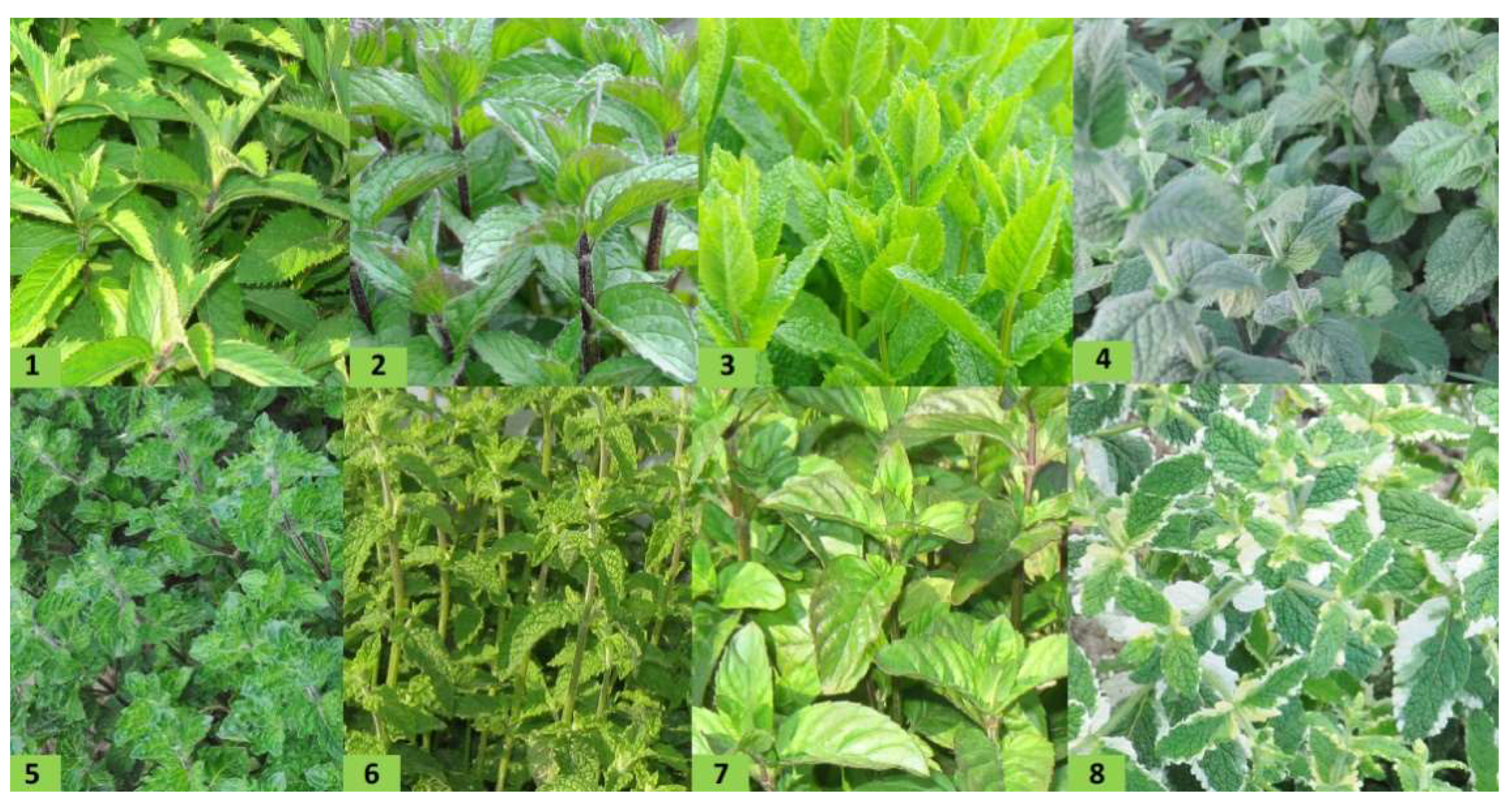
| (No. 1) M. × piperita ‘Swiss’ | statistics F | 1.0 | 3.11 | |||
| 5 | 75.5 | 90.0 | - | a | ||
| 10 | 55.3 | 90.0 | - | a | ||
| 15 | 56.8 | 90.0 | - | a | ||
| 20 | 55.3 | 90.0 | - | a | ||
| 40 | 46.7 | 90.0 | - | a | ||
| 60 | 37.5 | 90.0 | - | a | ||
| (No. 2) M. × piperita ‘Multimentha’ | statistics F | 8.41 | 3.11 | |||
| 5 | 76.0 | 90.0 | - | a | ||
| 10 | 55.3 | 71.7 | - | b | ||
| 15 | 56.8 | 71.8 | - | b | ||
| 20 | 70.7 | 87.2 | - | ab | ||
| 40 | 67.3 | 83.0 | - | ab | ||
| 60 | 62.8 | 75.2 | - | ab | ||
| (No. 3) M. spicata ‘Morocco’ | statistics F | 99.99 | 3.11 | |||
| 5 | 90.0 | - | - | a | ||
| 10 | 78.8 | - | - | b | ||
| 15 | 70.0 | - | - | bcd | ||
| 20 | 70.3 | - | - | abc | ||
| 40 | 63.5 | - | - | cd | ||
| 60 | 45.8 | - | - | e | ||
| (No. 4) M. rotundifolia | statistics F | 1.0 | 3.11 | |||
| 5 | 77.5 | 90.0 | - | a | ||
| 10 | 68.8 | 90.0 | - | a | ||
| 15 | 70.2 | 90.0 | - | a | ||
| 20 | 65.8 | 90.0 | - | a | ||
| 40 | 65.2 | 90.0 | - | a | ||
| 60 | 53.0 | 90.0 | - | a | ||
| (No. 5) M. spicata ‘Crispa’ | statistics F | 101.7 | 3.11 | |||
| 5 | 90.0 | - | - | a | ||
| 10 | 79.2 | - | - | b | ||
| 15 | 69.2 | - | - | c | ||
| 20 | 68.7 | - | - | cd | ||
| 40 | 59.5 | - | - | d | ||
| 60 | 44.8 | - | - | e | ||
| (No. 6) M. × piperita ‘Almira’ | statistics F | 1146.4 | 3.11 | |||
| 5 | 29.2 | 90.0 | - | a | ||
| 10 | 14.3 | 75.2 | - | b | ||
| 15 | 6.7 | 49.7 | - | c | ||
| 20 | 0.0 | 31.5 | - | d | ||
| 40 | 0.0 | 9.8 | - | e | ||
| 60 | 0.0 | 0.0 | - | f | ||
| (No. 7) M. × piperita ‘Granada’ | statistics F | 1.0 | 3.11 | |||
| 5 | 29.2 | 90.0 | - | a | ||
| 10 | 33.0 | 90.0 | - | a | ||
| 15 | 20.5 | 90.0 | - | a | ||
| 20 | 32.2 | 90.0 | - | a | ||
| 40 | 26.5 | 90.0 | - | a | ||
| 60 | 17.8 | 86.8 | - | a | ||
| (No. 8) M. suaveolens ‘Variegata’ | statistics F | 115.3 | 3.11 | |||
| 5 | 87.8 | 90.0 | - | a | ||
| 10 | 66.8 | 90.0 | - | a | ||
| 15 | 66.7 | 90.0 | - | a | ||
| 20 | 59.7 | 90.0 | - | a | ||
| 40 | 47.7 | 90.0 | - | a | ||
| 60 | 28.2 | 74.5 | - | b | ||
| Control | - | 77.8 | 90 | |||
| Mentha spp. | Concentration of Mint Extracts in Growth Medium (mL/100 mL) | Day of Observation | F emp. | F 0.05 | ||
|---|---|---|---|---|---|---|
| 2 | 4 | 6 | ||||
| Growth Diameter of Mycelium (mm) | Tukey’s Test | |||||
(No. 1) M. × piperita ‘Swiss’ | statistics F | 2.93 | 3.11 | |||
| 5 | 32.2 | 72.8 | 90.0 | a | ||
| 10 | 28.0 | 63.5 | 90.0 | a | ||
| 15 | 24.8 | 58.3 | 90.0 | a | ||
| 20 | 25.0 | 58.3 | 90.0 | a | ||
| 40 | 21.5 | 51.5 | 90.0 | a | ||
| 60 | 25.2 | 51.3 | 90.0 | a | ||
| (No. 2) M. × piperita ‘Multimentha’ | statistics F | 4.63 | 3.11 | |||
| 5 | 27.5 | 69.8 | 90.0 | a | ||
| 10 | 29.5 | 68.0 | 90.0 | a | ||
| 15 | 26.2 | 60.0 | 83.8 | a | ||
| 20 | 24.8 | 60.0 | 86.8 | a | ||
| 40 | 25.0 | 55.2 | 81.3 | a | ||
| 60 | 21.5 | 48.2 | 69.4 | a | ||
| (No. 3) M. spicata ‘Morocco’ | statistics F | 85.65 | 3.11 | |||
| 5 | 27.5 | 69.8 | 90.0 | a | ||
| 10 | 28.8 | 68.0 | 90.0 | a | ||
| 15 | 28.7 | 68.2 | 90.0 | a | ||
| 20 | 23.5 | 49.3 | 78.0 | ab | ||
| 40 | 19.8 | 44.0 | 73.5 | b | ||
| 60 | 15.8 | 22.5 | 37.7 | a | ||
| (No. 4) M. rotundifolia | statistics F | 12.80 | 3.11 | |||
| 5 | 22.2 | 67.3 | 90.0 | a | ||
| 10 | 25.7 | 71.8 | 90.0 | a | ||
| 15 | 27.7 | 72.5 | 90.0 | a | ||
| 20 | 26.2 | 70.7 | 90.0 | a | ||
| 40 | 20.8 | 55.5 | 89.3 | a | ||
| 60 | 18.5 | 33.8 | 42.7 | b | ||
| (No. 5) M. spicata ‘Crispa’ | statistics F | 11.26 | 3.11 | |||
| 5 | 26.8 | 63.0 | 90.0 | a | ||
| 10 | 28.2 | 57.7 | 90.0 | a | ||
| 15 | 29.0 | 56.8 | 90.0 | a | ||
| 20 | 25.2 | 52.5 | 86.2 | a | ||
| 40 | 21.3 | 45.3 | 79.5 | ab | ||
| 60 | 16.7 | 32.2 | 56.0 | b | ||
| (No. 6) M. × piperita ‘Almira’ | statistics F | 209.9 | 3.11 | |||
| 5 | 22.7 | 63.7 | 90.0 | a | ||
| 10 | 13.3 | 30.3 | 62.0 | b | ||
| 15 | 9.8 | 23.7 | 44.2 | c | ||
| 20 | 6.7 | 17.0 | 27.0 | d | ||
| 40 | 3.5 | 9.0 | 14.8 | e | ||
| 60 | 0.0 | 0.0 | 0.0 | f | ||
| (No. 7) M. × piperita ‘Granada’ | statistics F | 8.85 | 3.11 | |||
| 5 | 22.7 | 63.7 | 90.0 | a | ||
| 10 | 24.7 | 64.0 | 86.2 | a | ||
| 15 | 23.8 | 64.7 | 90.0 | a | ||
| 20 | 22.2 | 62.0 | 90.0 | a | ||
| 40 | 20.8 | 54.3 | 90.0 | a | ||
| 60 | 16.3 | 36.3 | 62.0 | b | ||
| (No. 8) M. suaveolens ‘Variegata’ | statistics F | 46.67 | 3.11 | |||
| 5 | 22.7 | 63.7 | 90.0 | a | ||
| 10 | 24.7 | 64.0 | 86.2 | abc | ||
| 15 | 23.8 | 64.7 | 90.0 | ab | ||
| 20 | 22.2 | 62.0 | 90.0 | bcd | ||
| 40 | 20.8 | 54.3 | 90.0 | d | ||
| 60 | 16.3 | 36.3 | 62.0 | e | ||
| Control | - | 27.9 | 63.0 | 90 | ||
| Compound | Class * | Concentration (%) Calculated Relative to the Area of Peaks Identified | |||
|---|---|---|---|---|---|
| M. spicata ‘Morocco’ (No. 3) | M. spicata ‘Crispa’ (No. 5) | M. piperita ‘Almira’ (No. 6) | M. suaveolens ‘Variegata’ (No. 8) | ||
| Limonene | MH | 1.26 | 3.42 | - | - |
| Beta-terpineol, acetate | COM | 0.35 | - | - | - |
| Carvone derivative | COM | 30.77 | 27.55 | 20.9 | 34.6 |
| Levomenthol | COM | 3.26 | 5.11 | 5.89 | 48.98 |
| Citronellal | COM | - | - | 3.04 | - |
| d-Menthol | COM | 43.76 | 53.45 | - | 1.86 |
| Citronellyl n-butyrate | COS | - | - | 42.63 | - |
| Isopulegol | COM | - | - | 2.46 | - |
| p-Menthan-1-ol | COM | 4.34 | - | - | - |
| Trans-2-Pinanol | COM | 0.54 | 0.27 | 1.31 | - |
| Cis-3-Hexenyl valerate | COM | 0.19 | 0.11 | - | - |
| (.+/−.)-Pulegone | COM | 0.33 | - | - | - |
| Piperitone | COM | 0.61 | 0.83 | - | 0.58 |
| Citronellol acetate | COM | 9.62 | - | - | - |
| Methyl acetate | COM | 0.36 | 5.85 | - | 4.73 |
| Carane | MH | - | - | 6.74 | - |
| Caryophyllene oxide | COS | 0.23 | 1.36 | 0.61 | - |
| p-Vinylguaiacol | NH | - | 0.34 | - | - |
| d-Verbenone | COS | - | 0.51 | - | - |
| .alpha.-Bourbonene | SH | - | - | - | 0.23 |
| (−)-.beta.-Bourbonene | SH | 1.73 | 0.28 | - | - |
| 3-Cyclopenten-1-one, 2-hydroxy-3-(3-methyl-2-butenyl)- | COM | - | - | 0.88 | - |
| Isocaryophyllene | SH | - | - | - | 1.0 |
| 2-Methylisoborneol | COM | - | - | 2.28 | - |
| Caryophyllene | MH | 0.18 | 1.17 | - | - |
| (S)-8-Hydroxy-p-menth-1-en-6-one | COM | - | - | 0.74 | - |
| (S)-cis-Verbenol | COM | - | - | - | 0.36 |
| Humulene epoxide 2 | COS | - | - | 1.78 | - |
| .alpha.-Bisabolol | COS | - | - | 1.84 | - |
| Valeric acid, octyl ester | COM | 2.08 | - | - | - |
| .alpha.-Farnesene | SH | 0.18 | - | - | - |
| Isocarveol | COM | 0.14 | - | - | - |
Disclaimer/Publisher’s Note: The statements, opinions and data contained in all publications are solely those of the individual author(s) and contributor(s) and not of MDPI and/or the editor(s). MDPI and/or the editor(s) disclaim responsibility for any injury to people or property resulting from any ideas, methods, instructions or products referred to in the content. |
© 2023 by the authors. Licensee MDPI, Basel, Switzerland. This article is an open access article distributed under the terms and conditions of the Creative Commons Attribution (CC BY) license (https://creativecommons.org/licenses/by/4.0/).
Share and Cite
Betlej, I.; Andres, B.; Krajewski, K.; Kiełtyka-Dadasiewicz, A.; Szadkowska, D.; Zawadzki, J. Effect of Various Mentha sp. Extracts on the Growth of Trichoderma viride and Chaetomium globusom on Agar Medium and Pine Wood. Diversity 2023, 15, 152. https://doi.org/10.3390/d15020152
Betlej I, Andres B, Krajewski K, Kiełtyka-Dadasiewicz A, Szadkowska D, Zawadzki J. Effect of Various Mentha sp. Extracts on the Growth of Trichoderma viride and Chaetomium globusom on Agar Medium and Pine Wood. Diversity. 2023; 15(2):152. https://doi.org/10.3390/d15020152
Chicago/Turabian StyleBetlej, Izabela, Bogusław Andres, Krzysztof Krajewski, Anna Kiełtyka-Dadasiewicz, Dominika Szadkowska, and Janusz Zawadzki. 2023. "Effect of Various Mentha sp. Extracts on the Growth of Trichoderma viride and Chaetomium globusom on Agar Medium and Pine Wood" Diversity 15, no. 2: 152. https://doi.org/10.3390/d15020152
APA StyleBetlej, I., Andres, B., Krajewski, K., Kiełtyka-Dadasiewicz, A., Szadkowska, D., & Zawadzki, J. (2023). Effect of Various Mentha sp. Extracts on the Growth of Trichoderma viride and Chaetomium globusom on Agar Medium and Pine Wood. Diversity, 15(2), 152. https://doi.org/10.3390/d15020152