Abstract
A greater understanding of the relationship between native orchids and their mycorrhizal symbionts is needed to ensure more effective orchid conservation strategies. A protocol for symbiotic seed germination and seedling development was developed for E. geminiflorum. Mature seeds were collected from a naturally occurring orchid population in Ecuador. Putative mycorrhizal fungi isolated from other native orchid species were used to screen their ability to facilitate germination and seedling development in vitro in either a 0/24 h or 12/12 h light/dark photoperiod at 20 °C. The mycorrhizal fungus Tulasnella calospora (UAMH 9824) isolated from Spiranthes brevilabris in Florida, USA, was also included in this study. Sterilization treatments using 0.3%, 0.5% sodium hypochlorite/ethanol or 2% calcium hypochlorite were tested for their effectiveness as sterilant and their subsequent effects on seed germination percentage. Effective surface seed sterilization was achieved with either 0.5% NaClO/ethanol or 2% calcium hypochlorite. However, significantly higher percentages of germinated embryos developed into protocorms when NaOCl solutions were used compared to the other treatments. Seed germination occurred in both photoperiods tested; however, delayed germination was observed under complete darkness. Seeds of E. geminiflorum germinated without fungal inoculation; however, co-culture with Tulasnella strains improved germination significantly. Seedling development was only observed when seeds were cultured in asymbiotic medium or co-cultured with T. caloscopa (UAMH 9824). Significantly longer seedlings were obtained when T. calospora was present in the culture compared with seedlings cultured in asymbiotic medium. The establishment of mycorrhizal associations was confirmed by the presence of pelotons in the roots of E. geminiflorum seedlings.
1. Introduction
Orchidaceae is one of the most diverse and largest families of flowering plants, with more than 27,000 species [1,2]. Orchids are globally distributed; they can be found on all continents except Antarctica, with the tropics being the region with the greatest orchid diversity [3,4]. Specifically, the tropical Andes is considered the largest plant biodiversity hotspot worldwide [5], with orchids being one of the main contributors to species richness in this area [6]. A clear example of this extraordinary orchid diversity is found in Ecuador, the smallest country in Andean South America, where more than 4200 orchid species have been reported [7,8]. Most of these orchid species (3214) occur in the ecosystem of the Andean forest, located 1000 to 2500 m.a.s.l. [9].
The genus Epidendrum is the second-largest orchid genus in Ecuador. The genus is comprised of 1200 species, of which 453 occur naturally in Ecuador [7]. Epidendrum species are distributed throughout tropical America, from the sea level up to 3700 m.a.s.l. These species can be found in most habitats in Ecuador, but the highest diversity and abundance are in wet cloud Andean forests [10]. Epidendrum geminiflorum Kunth is a native orchid species distributed in Venezuela, Guianas, Colombia, Ecuador, and Peru [11,12]. In Ecuador, the species can be found in the coastal, Andean, and Amazonian regions, from 1000 to 3500 m.a.s.l [11]. Plants are epiphytic, sympodial, 20–50 cm in height, characterized by cane-like stems, with coriaceous but flexible leaves, and resupinate green to greenish flowers without fragrance (Figure 1) [12]. Epidendrum geminiflorum is very common and widely distributed; therefore, its conservation status is qualified as “less concerned” (LC) [12,13]. However, orchids that are common today may not be common tomorrow.
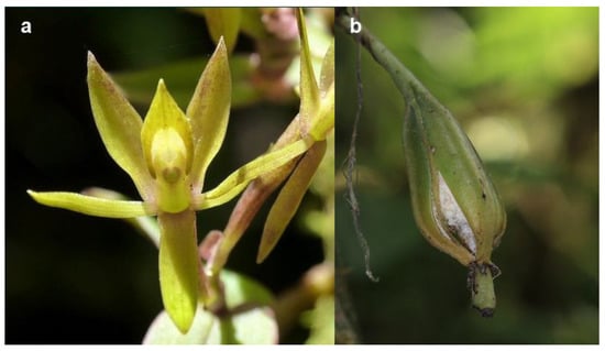
Figure 1.
(a) Flower of Epidendrum geminiflorum and (b) open seed capsule collected in Cuyuja, Ecuador. Photo by Andreas Kay with permission.
Several biological and ecological factors have been attributed to the high diversity and worldwide distribution of Orchidaceae [14]. Mycorrhizal associations developed during seed germination and seedling establishment in situ are likely to have a significant effect on orchid distribution and population structure [15,16,17]. Orchid seeds are the smallest produced in flowering plants. They are produced in large numbers, with small undifferentiated embryos containing minimal nutrient reserves [3,18]. Therefore, orchid seeds fully depend on the establishment of mycorrhizal associations for enhanced nutrients and water during the germination and early seedling stages [19]. Adult plants may develop different fungal associations or may no longer require such associations [20].
Epiphytic and terrestrial green (photosynthetic) orchids usually associate with fungal species from Ceratobasidiaceae, Sebacinales, and Tulasnellaceae in the Basidiomycota taxa [15,21]. Many of these mycorrhizal fungi are widespread and occur in a wide variety of habitats [17]. The degree of specificity for their mycorrhizal fungi varies with orchid species. Epiphytic and terrestrial orchids may be associated with a very narrow or very wide range of mycorrhizal fungi [22,23]. Patterns of specificity towards mycorrhizal associates throughout the orchid life cycle have been described mostly for terrestrial orchid species. The seed of four terrestrial orchid species germinate and develop into protocorms when co-cultured with many fungal isolates; however, only a subset of compatible fungi support the development of more advanced orchid seedling stages [24].
Reductions in the diversity and distribution of orchid populations are often the result of anthropogenic activities [14,25]. Many endangered orchid species occur in the neotropics, a region recognized for its species richness and high levels of endemism but with high rates of habitat loss [5]. Illegal collection also threatens many orchid species with extinction. In Ecuador, the collection of wild species for commercial use in local and international markets is the second major threat to the conservation of orchids [26]. The potential effects of climate change on the long-term survival of orchid populations are difficult to predict due to the complexity of the obligated interactions with other sympatric organisms for orchid survival [25].
Despite the extraordinary diversity and abundance of epiphytic orchid species in the Andean forest [27,28], most information regarding the mycorrhizal association during seed germination and seedling establishment is well documented for terrestrial orchids in temperate zones [15]. In Ecuador, research on orchid mycorrhizal associations has been focused on the isolation and identification of endophytes isolated from orchid species from the Andean cloud forest of south Ecuador [22,23,29,30]. Successful germination and seedling development using symbiotic seed culture have been obtained only on Epidendrum secundum Jacq., a native terrestrial Ecuadorian orchid species using fungal species: Ceratobasidum sp. and Sebacina vermifera, isolated from native orchid species located in the southern Andes of Ecuador [31].
Symbiotic seed germination and seedling development involve the co-culture of orchid seeds with their fungal associates, which promote the establishment of the mycorrhizal association in vitro under sterile and controlled environmental conditions [32]. Physiological compatibility, patterns of in vitro fungal specificity, dependency, and environmental factors that control germination and seedling development, can be studied using this approach [24]. Stewart and Kane [33] reported that mycobionts isolated from Spiranthes floridana promoted the high percent germination of Spiranthes brevilabris; however, subsequent early seedling development was only supported by a fungus isolated from the same species. These results suggested that the high degree of mycobiont specificity observed under in vitro culture conditions may be linked to the rarity of this Florida orchid species in situ. Symbiotic seed culture can also be used as an ex situ conservation strategy for the effective sexual propagation of plant material that may reduce collection pressure on wild populations [25]. The production of plant material with compatible symbiotic fungi can be used in restoration projects to ensure the long-term survival of the populations [32,34].
Factors that affect seed germination and seedling development, mycorrhizal diversity, and the degree of specificity and dependency of orchids on certain fungi species need to be experimentally evaluated. This scientific information will be critical to developing effective strategies for orchid conservation and the restoration of native orchid species from Ecuador [14,20,35]. Consequently, the objectives of the present study are to: (1) evaluate the effects of photoperiods on symbiotic seed germination and seedling development in E. geminiflorum; (2) document the morphological development from seed to the mature seedling stage; and (3) screen putative mycorrhizal fungi isolated from native Ecuadorean orchid species for their ability to facilitate seed germination and seedling development in vitro.
2. Materials and Methods
2.1. Sample Collection Permits
Permits to collect and transport seed capsules and root pieces for research purposes were obtained from Ministerio del Ambient del Ecuador (MAE). All these permits had the institutional support of Instituto Nacional de Biodiversidad (INABIO) through a contract (Contrato Marco MAE-DNB-CM-2016-0045) of this research institution with MAE, which allowed the collection of native flora and research that includes DNA analysis. All regulations were followed to comply with the legal collection of the native orchids. Orchid seeds were imported into the United States under USDA/APHIS permit number P37-16-0-5568. For the import of fungal mycelia to the USA, a permit to move live plant pests, noxious weeds, and soil (P526P-19-02945) was obtained from the US Department of Agriculture (USDA).
2.2. Seed Collection
Recently dehisced mature capsules of Epidendrum geminiflorum Kunth were collected from a small native orchid population located in Cuyuja (S 00.42885 W 078.01733, 2616 m.a.s.l.), Napo Province, in September 2018. Capsules were placed in sterile paper bags and then transported to the laboratory in Ecuador. Seeds were removed from the capsules and stored in loosely capped glass vials over CaSO4 desiccant inside sealed 50 mL Falcon® tubes in the dark at 22 ± 2 °C until shipping to the in vitro plant culture laboratory located at the University of Florida, Gainesville, Florida, USA. Seeds were dried for 10 weeks after collection before being stored at −10 °C in the dark until used for experimentation. A vial with seeds was also maintained at 22 ± 2 °C in the dark for 7 additional weeks to test seed viability loss over time under these storage conditions.
2.3. Seed Size and Weight
Seed weights were estimated as follows: three samples of dry seed, taken from cold storage, were weighed on an ultra-microbalance instrument (Model No. XP2U Mettler-Toledo LLC, Columbus, OH, USA). Seeds were then rinsed with water to facilitate counting over a black cloth using a stereomicroscope. The weight of each sample was divided by the number of seeds per sample. Estimated seed weight was used to calculate the solution volume required to deliver uniform 60 μL aliquots, each containing ca. 100 sterile seeds, to ensure similar numbers of seeds per sub-replicate in each experiment.
2.4. Seed Viability Testing
Percentage viability was tested using triphenyl tetrazolium chloride (TTC) staining before being stored, after 7 wks under storage conditions (−10 °C in the dark and at 0% RH) and 17 wks at 22 ± 2 °C in the dark and 0% relative humidity after field collection. Seed samples (1.5 mL microtubes containing 10 mg seed) were preconditioned in 1 mL 10% sucrose solution for 24 h at 22 ± 2 °C. Seeds were then rinsed twice with deionized distilled (dd) water; 1 mL 1% TTC solution was added to the microtubes, and samples were incubated in the dark at 36 °C for 24 h. To avoid embryo discoloration, the TTC solution was replaced with dd water and samples were stored at 4 °C until their evaluation using light microscopy. Seeds with rose-red stained embryos were considered viable, while white embryos were recorded as non-viable [36,37].
2.5. Seed Sterilant Screening
Different hypochlorite solutions and sterilization time combinations were evaluated for their effectiveness on orchid seed surface sterilization and their subsequent effects on percent seed germination. Approximately 5 mg of seeds contained in sterile 1.5 mL microtubes were surface sterilized with either 0.3%, 0.5% sodium hypochlorite/ethanol or 2% calcium hypochlorite solutions for 1, 3, and 5 min. The sodium hypochlorite (NaOCl)/ethanol solutions were prepared by adding 250 μL 100% ethanol and 250 (0.3% v/v) or 416 μL (0.5% v/v) 6.0% sodium hypochlorite (Clorox© Bleach), and then diluting the solution to 5 mL with sterile dd water. To prepare a saturated Ca(OCl)2 solution, 307 mg powder (65% available chlorine) was dissolved in 10 mL sterile dd water. This gave a final solution that provided 2% available chlorine. Sterile filter paper (Whatman ®, Qualitative 1, Cat# 1001-125) was used to filter the solution immediately before use. Once the sterilization times were completed under constant agitation, the solutions were removed by placing a sterile pipette tip into the bottom of the microtube. Seeds were then rinsed three times for 1 min each with sterile dd water. The control treatment consisted of no sterilization and three rinses for 1 min each with sterile dd water.
Surface-sterilized seeds were sown on Petri dishes (100 × 15 mm) containing 30 mL Orchid Seed Sowing Medium (PhytoTechnology Laboratories, Shawnee Mission, KS, Cat# P723). Three 60 μL aliquots of sterile seeds in sterile dd water were pipetted and placed in 3 different sections on the surface of the germination media. Seeds were then evenly spread using a sterile inoculating loop. Four Petri dishes per treatment were sealed with one layer of sealing film (PhytoTechnology Laboratories, Cat# A003) and incubated for 4 wks at 20 °C under a 12 h-light/12 h-dark photoperiod in a Percival I-35LL growth chamber (Percival Scientific, Boone, IA, USA). Light was provided by cool-white florescent tubes (General Electric F20T12/CW) at 35 μMm−2s−1 (photosynthetic active radiation, PAR).
2.6. Symbiotic Seed Germination
Fungal isolation and molecular identification. Field collection of roots from young or adult plants and seedlings, when available, was made from native orchid populations located in the northern tropical Andes in Ecuador. Collections were made in May 2016, July 2017, and August 2018. At most, three active growing roots or seedlings were detached from the tree bark using a sterile scalpel and placed in sterile plastic bags. Fungal isolation from mature roots of E. germiniflorum was not possible due to the absence of fungal pelotons. Likewise, no young plants or seedlings were observed in the population. Samples were transported over ice in an insulated container to the Idgen Molecular Diagnostic Laboratory located in Quito, Ecuador. Isolation of root endophytes from root cortical tissue was performed following standard methods described by Zettler et al. [38] and Zettler and Corey [34]. Briefly, roots were rinsed with sterile distilled (d) water to remove bark debris and then surface-sterilized in a solution containing 6% NaClO:98% ethanol:d water (8:5:87) for 1 min followed by two 1-min rinses with sterile distilled (d) water.
Roots were cut into 1 cm long segments starting at the tip. Each segment was placed into a separate sterile Petri dish with 200 μL sterile d water. Using a sterile scalpel and forceps, root segments were chopped to break open the cortical cells harboring fungal pelotons. Aliquots (25 mL) of sterile warm fungal isolation medium (FIM) [39], supplemented with 10 mg/L streptomycin sulfate antibiotic, were slowly poured into individual Petri dishes. Dishes were then gently swirled to disperse the cortical tissue homogenate throughout the medium. Plates were sealed with one layer of sealing film and incubated for 24–48 h at 22 ± 2 °C. Using a stereo microscope, plates were observed for actively growing hyphae emerging from pelotons. Individual pelotons with hyphal tips were subcultured into separate Petri dishes containing 25 mL ½X potato dextrose agar (PDA, PhytoTechnology Laboratories, Cat# P772). Plates were sealed and incubated again at 22 ± 2 °C. After 24–72 h, pelotons with short hyphal tips or hyphal tips alone were transferred to fresh 1⁄2 PDA on slants in glass tubes. Culture tubes were sealed with a layer of sealing film and incubated at 23 ± 2 °C for at least 48 h and then stored at 4 °C until their transportation to the in vitro culture laboratory located at the University of Florida, Gainesville, Florida, USA.
Fungal cultures in tubes were subcultured into Petri dishes containing 25 mL 1⁄2 PDA. Pure cultures with morphological features assigned to the form-genus Rhizoctonia [39] were kept for further identification to the genus level using Sanger sequencing of the ribosomal DNA internal transcribed spacer (ITS) sequences [40]. Pure cultures of putative mycorrhizal fungi were transferred to fresh 1⁄2 PDA slants in glass tubes maintained in the dark at 10 °C and subcultured every 4 months. These cultures are permanently stored at the University of Alberta Microfungus Herbarium in Toronto, Canada (UAMH). (Table 1).

Table 1.
Putative orchid mycorrhizal fungi, isolated from different orchid species from Ecuador and the United States, used as symbiotic treatments to germinate sterile seeds of E. geminiflorum.
Symbiotic seed culture. Sterile seeds of E. geminiflorum were co-cultured with fungal strains isolated from different native Ecuadorean orchid species (Table 1) and one Tulasnella calospora strain (University of Alberta Microfungus Herbarium UAMH 9824) isolated from a root of Spiranthes brevilabris in Levy County, Florida, USA [41]. Fungal isolates, stored in the dark at 10 °C, were subcultured onto fresh 1⁄2 PDA for 2 wks in the case of Tulasnella and 2–3 days for Ceratobasidium strains to obtain actively growing mycelia prior to experiment initiation.
The symbiotic seed germination medium used was oatmeal agar (OMA), which was prepared with 2.5 g/L oat flour and 7 g/L TC agar (PhytoTechnology Laboratories, Cat# A296). The medium was sterilized by autoclaving at 117.7 kPa and 121 °C for 40 min. After solidification of 30 mL OMA in each Petri dish (100 × 15 mm), a triangle-shaped cellophane membrane (BIO-RAD, Cat# 1650922) was placed over the surface of the medium to improve visualization of the growing hyphae. A 1 × 4 cm black filter paper strip (Thomas Scientific, Cat# 4740C10) was placed parallel to each side of the triangle to support the sterile seeds (Figure 2c). Black filter paper instead of standard white paper facilitates seeing the light-colored seeds more clearly.
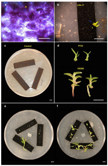
Figure 2.
Symbiotic seed culture of E. geminiflorum. (a) Peloton (Pl) formation in root cortical tissue of E. geminiflorum seedlings co-cultured with US266. (b) Seedling (blue arrow) growing with LPA1T after 42 wks. (c) Control symbiotic plate with a cellophane membrane and black filter paper strips on solidified agar OMA. (d) Seedlings growing on P723 or co-cultured with US266. (e) Seedling co-cultured with US266 after 42 wks under complete darkness for 6 wks and (f) 42 wks under 12 h-light photoperiod. Scale bars = 500 µm (a) and 5 mm (b–f).
Seeds were removed from cold storage and maintained at 22 ± 2 °C for at least 4 h before sterilization. Three seed samples weighing ca. 20 mg each were surface-sterilized using a 0.5% sodium hypochlorite/ethanol solution for 3 min, followed by three 1-min rinses with sterile dd water. After sterilization, dd water was added to the seeds to obtain a solution containing 100 seeds per 60 µL aliquot. Three aliquots were pipetted out and placed over the surface of the black filter paper strips. A 1 × 1 × 0.5 cm3 PDA block containing the mycelium from each isolate was inoculated over the cellophane membrane. A control treatment consisted of OMA without fungal inoculation. Based on a preliminary asymbiotic media germination screening, P723 was included as the asymbiotic treatment.
Petri dishes were sealed with one layer of sealing film and incubated under both 0/24 h and 12/12 h light/dark photoperiods at 20 °C in a Percival I-35LL growth chamber (Percival Scientific, Boone, IA); light was provided by cool-white florescent tubes (General Electric F20T12/CW) at 35 μMm−2s−1 PAR. Plates were examined for germination after 2 wks. Germination and seedling development stages (Table 2) were recorded every 2 wks using a stereomicroscope. After 6 wks of culture, Petri dishes under continuous dark treatment were unwrapped from the aluminum foil and exposed to a 12-h photoperiod for the remainder of the experiment. Roots from seedlings produced on the symbiotic media were finely sliced and stained using an aqueous 0.5% Toluidine Blue staining solution for 24 h to evaluate the presence of fungal pelotons in the cortical tissue using a microscope.
Table 2.
Seed germination and developmental stages in Epidendrum geminiflorum adapted from Stewart and Zettler [42].
2.7. Experimental Design and Statistical Analysis
Treatment effects were evaluated using a completely randomized experimental design. Each factor or combination of factors consisted of 7 replicates with 3 sub-replicates (Figure 2). The experimental unit consisted of a Petri dish containing 30 mL culture medium with three areas, each containing a filter paper strip with ca. the same number of seeds on the surface of each strip (Figure 2c). These areas were considered sub-replicates. The number of seeds in each developmental stage was recorded at each data collection time. All experiments were repeated once. Percent data were arcsine-transformed prior to the analysis using a multi-factor ANOVA by each data collection point. Mean separation was assessed using Tukey’s methods at the α = 0.05 significant level. Statistical analyses were completed with JMP Pro15 (Manufacture, SAS Institute Inc., Cary, NC, USA). Survival analysis was performed using the survival package in R studio 3.2.5. Time-to-event data, in this case, the time from sowing to germination (Stage 2), were analyzed. A Kaplan–Meier estimator was used to estimate the survival function with 95% confidence intervals. Log-rank tests were performed to evaluate differences between survival curves at the α = 0.05 significant level. Multiple pair-wise comparisons were performed using a log-rank test with Bonferroni correction to adjust p-values at the α = 0.05 significance level.
3. Results
3.1. Seed Viability Testing
Mature seeds of E. geminiflorum collected from a native population in Ecuador exhibited high viability based on TTC staining (Figure 3a). Viability testing on seeds that were dried over CaSO4 desiccant at 22 ± 2 °C in the dark for 10 wks before being stored at 10 °C averaged 96.3%. Storage temperature (10 °C) did not affect seed viability as 99.1% seed viability was observed following seed storage at −10 °C in the dark for 7 wks. Even seeds maintained for 17 wks at 22 ± 2 °C in the dark over a desiccant exhibited high viability (93.3%).
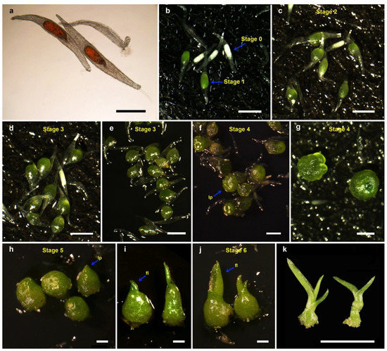
Figure 3.
Asymbiotic seed germination and early seedling development of E. geminiflorum on P723 or co-cultured with US266 under 12 h-dark/12 h-light at 20 °C (a) Viable TTC-stained seed. (b) Stage 0 and imbibed seeds (Stage 1). (c) Germinated seeds after 14 days culture on US266. (d,e) Globular green Stage 3 protocorms. (f,g) Protocorms with leaf primordia (lp) formation. (h) Seedlings with elongated leaf primordia (lp) (Stage 5). (i,j) Formation of first leaf (lf). (k) Seedlings with larger leaves growing on P723 at 42 wks. Scale bars = 500 μm (a–f) and 5 mm (k).
3.2. Seed Sterilant Screening
Visual bacterial or fungal contamination was observed in plates that were sown with seeds that were surface sterilized with 0.3% NaOCl/ethanol solution (50% contamination) as well as seeds that were not sterilized (control; 75% contamination). Effective surface sterilization was achieved with 2% Ca(OCl)2 or 0.5% NaOCl/ethanol solutions during all exposure times tested. No visible contamination was observed in plates sown with seeds surface-sterilized using these treatments. No significant differences were observed in seed germination among all treatments, including the control (Figure 4A). However, significantly higher percentages of germinated embryos developed into Stage 3 protocorms (Figure 3e) when seeds were surface-sterilized with NaOCl solutions compared with 2% Ca(OCl)2 or in the control treatment (Figure 4B).
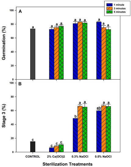
Figure 4.
Effects of seed surface sterilization treatments on seed germination (A) and protocorm formation (B) of Epidendrum geminiflorum cultured on P723 under 24 h-dark/0 h-light at 20 °C for 2 wks. Histobars with the same letter are not significantly different (α = 0.05).
3.3. Seed Germination and Seedling Development
The morphological development from seed to early seedling was documented for E. geminiflorum (Figure 3). Dry seeds weighed ca. 1.32 μg each. A high percentage of seeds (46.2%) did not contain embryos. Embryos swelled and changed from white to light green color (Stage 1, Figure 3b) as water imbibition occurred, regardless of photoperiod. High germination percentages of seeds with visible embryos were observed (~88%). However, seedling development was extremely limited. Seeds germinated regardless of photoperiod, with no significant differences in cumulative seed germination between both photoperiods tested at the end of the study. However, significantly faster germination was observed under the 12 h-dark/12 h-light photoperiod compared to the 24 h-dark/0 h-light photoperiod (Figure 5). At least 50% germination (median germination) was observed at 28 days in plates incubated under a 12 h-light photoperiod compared with 56 days in plates in complete darkness for 42 days (Figure 5). Seeds in plates that were incubated under complete darkness continued to germinate after exposure to a 12 h-light photoperiod after 42 days of culture.
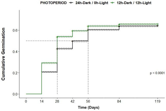
Figure 5.
Cumulative germination function for Epidendrum geminiflorum seeds cultured under different photoperiods at 20 °C for 119 days, with 95% confidence intervals.
The delayed effect of darkness on germination was observed across all symbiotic treatments except in P723 and US266, where germination rates were not significantly different between photoperiods (Figure 6). However, in seeds of E. geminiflorum germinated in the absence of fungal isolates (58.6–61.6%), significantly higher germination rates were observed when seeds were co-cultured with Tulasnella strains either from orchid species from Ecuador (LPA2T, LPA,3T, LPA1T, LPA4T) or from Florida, USA (US266). Cumulative germination percentages at the end of the study were significantly lower in LPA2T, LPA3T, LPA1T, and LPA4T compared with US266. In addition, faster germination was observed in the presence of US266, where more than 50% of the seeds germinated in only 14 days (median germination) after sowing compared with 42 days in LPA1T, LPA2T, and LPA3T and 56 days in CM4T (Figure 6). Faster germination was observed in seeds co-cultured with Ceratobasidium ER2C compared with the control treatment; median germination was reached at 56 days compared with 84 days in the symbiotic control. Germination rates were significantly reduced in the presence of the Ceratobasidium strain MC4C compared with the symbiotic control and the other treatments. The highest germination percentages were observed when seeds were sown on P723. Maximum germination for seeds with visible embryos (87.9%) was observed on P723 at 119 days (Figure 6).
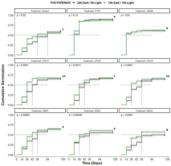
Figure 6.
Cumulative proportion of germinated Epidendrum geminiflorum seeds co-cultured with different fungal strains under two photoperiods at 20 °C. Within each treatment, significant differences between photoperiods were not observed, with p-values greater than 0.05 according to a log-rank test at the α = 0.05 significant level. Curves with the same letters were not significantly different according to a multiple pair-wise comparison log-rank test of the treatment effect, with Bonferroni correction to adjust p-values to = 0.05 significance.
Globular protocorms were observed after 2 wks of culture only in P723 and US266 (Figure 7). Significantly higher percentages of Stage 3 protocorms were obtained in P723 compared to seeds co-cultured with US266. Significantly higher percentages of Stage 3 protocorms were observed under a 12 h-light photoperiod than under complete darkness at 6 wks (Figure 7). Germinated embryos with LPA1T or LPA2T eventually developed into Stage 3 protocorms by 6 wks but with significantly lower percentages compared with P723 or US266 (Figure 7). Prior to the formation of leaf primordia in the anterior region of the embryos, a color change from dark green to light green or yellowing color in the area was observed and was documented as Stage 4 protocorms for this orchid species (Figure 3f–g). Stage 4 protocorms were only observed in P723 under both photoperiods and with US266 only under the 12 h photoperiod (Figure 8). Uniform and significantly higher percentages of Stage 4 protocorms were produced in P723 (Figure 3f) than with US266. However, larger Stage 4 protocorms were observed with US266 (Figure 3g). Elongation of leaf primordia and further development was only observed with US266 for the 12 h-light photoperiod during the duration of the experiment (17 wks) (Figure 8). However, after 42 wks of culture, seedlings were observed in P723 (Figure 3k) regardless of the photoperiod. Very few seedlings were observed in LP1T (Figure 2b) or US266 when seeds were germinated under complete darkness for 6 wks (Figure 2e). Larger seedlings were produced in the presence of US266 compared with P723 (Figure 2d). Pelotons were observed after the staining of seedling root cortical cells cultured in the presence of US266 (Figure 2a). This confirmed that the interaction between seeds of E. geminiflorum and the US266 fungal strain resulted in a mycorrhizal association that supported seedling development in E. geminiflorum.
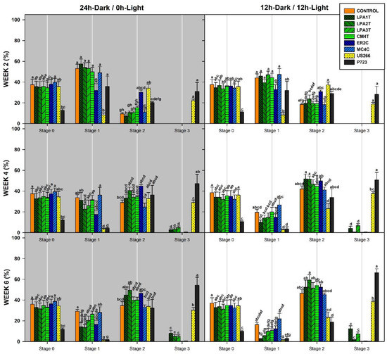
Figure 7.
Comparative effects of photoperiod and symbiotic treatments on seed germination and early seedling development stages of Epidendrum geminiflorum incubated at 20 °C for 2, 4, and 6 wks. Shaded areas indicate 24 h-Dark/0 h-Light photoperiod Histobars with the same letter are not significantly different (α = 0.05).
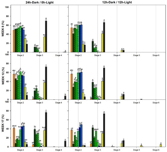
Figure 8.
Comparative effects of photoperiod and symbiotic treatments on seed germination and early seedling development stages of Epidendrum geminiflorum incubated at 20 °C for 8, 12, and 16 wks. Histobars with the same letter are not significantly different (α = 0.05).
4. Discussion
Our results demonstrated that E. geminiflorum seedlings can be produced in vitro on asymbiotic media (P723) or through symbiotic seed culture with T. calospora (US266-UAMH 9824) isolated from S. brevilabris, an endangered terrestrial orchid native to Florida, USA [41]. After 42 weeks, very few seedlings in early developmental stages were also produced in OMA with a Tulasnella strain isolated from a native population of L. acarina, located in the same location where E. geminiflorum seeds were collected. To the best of our knowledge, this study represents the first report on the in vitro seed germination and early seedling development of E. geminiflorum. Morphological developmental stages, from seed to early seedling development, were also documented for the first time for this native species collected in the northern Andes of Ecuador.
Open capsules were collected from an individual in a small patch of the Andean forest located on a private property in Cuyuja, Ecuador. Small numbers of individuals at the collection site reflected the scattered distribution, with a few individuals restricted to small areas, a typical characteristic described for populations of epiphytic orchids of the neotropics [43]. In addition, the absence of local seedling recruitment may be the result of the altered or degraded habitat where seeds were collected. Favorable conditions for germination and seedling establishment may no longer be present at the site, leading to an eventual population decline [20]. The possible senile nature of this population may have contributed to the difficulty of isolating compatible fungal associates of this orchid.
The viability of seeds is highly variable within orchid species [3]. It is commonly known that if seeds are allowed to dry and then stored at low temperatures (0–10 °C), seeds may remain viable for long periods of time. However, if they are maintained at ambient temperatures, they will rapidly lose their viability [3]. However, this seems not to be the case for E. geminiflorum seeds, which exhibited 93.2% viability after being stored for 17 wks at 22 ± 2 °C in the dark. Despite the high percentage of seeds with no embryos, a high viability of seeds with visible embryos (~96%) was observed, and the maximum germination obtained in P723 (87.0–88.8%) did not differ from the seed viability estimated with the TTC staining test. Consequently, TTC seed viability can be used as a reliable test to estimate the seed viability and germination potential of seed samples of this species. This could be valuable for germination trials for preservation purposes [36].
Sterilization treatments are used under in vitro culture conditions to eliminate cultivable bacterial or fungal contamination from orchid seed surfaces. Since compounds that block water imbibition, such as suberin, are eliminated with hypochlorite solutions, sterilization has also been used as a scarification treatment to improve germination percentages [44,45]. Our sterilant screening showed that reliable seed sterilization was possible either with 2% Ca(OCl)2 or 0.5% NaOCl solutions, with either no detrimental or positive effect on germination percentages. However, faster development into advanced protocorm stages was observed when the seeds were sterilized with NaOCl solutions. Faster protocorm development could probably also ensure faster and higher seedling development. Therefore, the use of a 0.5% sodium hypochlorite/ethanol solution is recommended as a sterilant for E. geminiflorum seeds.
Symbiotic seed culture can result in higher germination percentages compared with those obtained with asymbiotic germination in some orchid species [46]. Our results showed the opposite response. Significantly lower germination was observed when seeds were co-cultured with fungal strains compared with that on P723 medium (asymbiotic control). The required nutrients that may be available in P723 for the optimal germination of E. geminiflorum may not have been sufficient to provide for any of the fungi tested. Seeds of E. geminiflorum were also able to germinate (Stage 2) in the symbiotic control in the absence of nutrients or readily available carbohydrates. Other orchid seeds, such as Cynorkis purpurea (Thouars) Kraez from Madagascar, were also able to germinate in OMA without fungal inoculation [46]. The presence of Tulasnella strains significantly improved germination percentages over the control treatment once fungal mycelia reached the E. geminiflorum seeds on the filter paper. Tulasnella calospora (US266) grows faster than the other Tulasnella strains isolated from native orchid species from Ecuador, which possibly resulted in earlier and higher germination rates with US266 compared to the other Tulasnella strains. Only one Ceratobasidium isolate, ER2C, significantly increased germination rates compared with the control. However, embryos germinated with Ceratobasidium strains displayed browning on one side. This browning may be the result of localized decomposition by the fungus, which is not considered a typical characteristic of symbiotic culture [47]. Tulasnella calospora (UAMH 9824) has been recognized for its ability to promote successful germination and seedling development in a variety of orchid species from different geographical locations after decades of subculturing [48]. These include Epidendrum species such as E. nocturnum, an epiphytic orchid native to Florida, USA [49], and other genera: Habenaria repens, a semi-aquatic orchid from Florida [42], and Platanthera holochila, a terrestrial endangered orchid from Hawaii [50].
Responses to light during germination and seedling development stages vary within orchid species [19,44]. Inhibition of germination under illumination has been reported in both terrestrial and epiphytic species [51]. Our result showed that light is not a requirement for the germination of E. geminiflorum seeds as the seeds germinated under both 12 h-light and 24 h-dark photoperiods; however, light significantly promotes faster germination. After the exposure of seeds maintained under complete darkness conditions prior to a 12 h-light photoperiod, they continued to germinate until reaching cumulative germination, with no significant differences between photoperiod treatments at 126 days. This confirmed the ability of E. geminiflorum to germinate regardless of light conditions. Seeds of the Florida native E. nocturnum from south Florida cultured in OMA with T. calospora (US266-UAMH 9824) also germinated (52–68%), with no significant differences between dark and light treatments [49].
Although the seed germination of E. geminiflorum was not affected by photoperiod, the presence of light during symbiotic seed germination (US266) was critical for subsequent seedling development. Non-green protocorms produced under complete darkness failed to develop into the seedling stages. Immediate exposure of germinated embryos to light conditions will probably result in successful seedling establishment on site. Epiphyte position in tree canopies allows them to have access to light, which may be an important requirement to be met during the earlier developmental stages of E. geminiflorum.
Temperature has also been reported as an important environmental factor that influences the germination and seedling establishment of orchid species [19,20,44]. Preliminary asymbiotic seed germination studies (data not shown) performed with E. geminiflorum showed that seedling development was not possible at 23 °C, regardless of the availability of nutrients in the media. Constant temperatures higher than 20 °C are not present at the collection site (Cuyuja, Ecudor, 2616 m.a.s.l.) [52]. Orchid species located at this altitudinal gradient may be sensitive to small increases in average temperature. Prediction models suggest a significant increase in temperature between 2 and 7 °C in the tropical Andean region toward the end of the 21st century [53]. Between 2000 and 3500 m.a.s.l., observations and projections both suggested stronger warming scenarios in the western slope of the Andes [53]. Future environmental conditions may not be favorable for the seedling establishment of this orchid species at its original locations. Given the wide altitudinal distribution (1000–3500 m.a.s.l.) of E. geminiflorum [11], ecological studies including seeds from different geographic locations are needed to determine if observed responses to temperature may be the result of ecotypic differentiation under local environmental conditions. Biomass allocation and corm formation timing during in vitro seed germination studies have been presented as evidence of ecotypic differentiation between different Calopogon tuberosus var. tuberosus populations collected from Michigan, South Carolina, and Florida. Seedlings from higher latitudinal populations tended to allocate biomass faster to their underground structures (corms) than plants from lower latitudinal populations [54]. Scientific information from these studies will be critical for the selection of plant material adapted to restoration target sites. This may ensure the long-term survival of the restored populations [55].
The presence of compatible mycorrhizal fungi during seed germination and seedling establishment are critical for the survival of orchid species in situ [20]. Degrees of specificity towards fungal associates may change throughout the life cycle in the same orchid species. Some terrestrial orchids seem to be associated with a great number of diverse fungi during germination, but only a subset of these was able support seedling development [24]. A similar pattern of specificity was observed in E. geminiflorum in this study, where seed germination was supported with all fungal strains tested, but advanced seedling development was only observed with one fungus (US266-UAMH 9824).
Early seedling development of E. geminiflorum in situ may require specific fungi different than those screened in our study. These fungi may be expected to facilitate higher percentages of seedling production than those observed with US266. Symbiotic germination studies with fungi associated with E. geminiflorum would be critical to determine the levels of specificity toward fungal associates of this orchid species, which may result in higher seedling production than that obtained in this study. Although T. calospora (US266; US266-UAMH 9824) is considered ubiquitous, the release of orchid seedlings with this exotic fungus into the wild could pose an ecological risk to other orchid species, considering its foreign origin (Florida) [49]. The survival of seedlings to ex vitro conditions needs to be evaluated for this orchid species, with the aim of developing an efficient protocol to produce suitable plant materials that ensure successful restoration or conservation projects.
Author Contributions
All authors significantly contributed to various aspects of the study. Plant field locations and the identification of the plants from which seed capsules and root samples were collected were determined by L.E.B. L.W.Z. provided training in the isolation, culture, and identification of mycorrhizal fungi and symbiotic seed culture techniques. Experimental designs, interpretation of results, and discussion were provided by P.H.Q.-L., M.E.K. and L.W.Z. The first draft of the manuscript was written by P.H.Q.-L. and all authors have commented on previous versions of the manuscript. All authors have read and agreed to the published version of the manuscript.
Funding
This research was partially funded by the Ecuadorian government through the SENESCYT Scholarship Program.
Institutional Review Board Statement
Not applicable.
Informed Consent Statement
Not applicable.
Data Availability Statement
Data sharing is not applicable to this article.
Acknowledgments
This paper and the research behind it would not have been possible without the financial support of the Scholarship Program SENESCYT from the Ecuadorian Government. We would like to acknowledge the institutional support provided by Universidad de las Americas (UDLA), Jardin Botanico de Quito (JBQ), Instituto Nacional de Bio-diversidad (INABIO), and Ministerio del Ambiente de Ecuador (MAE). We would like to also acknowledge Francisco Flores and Patricia Garrido, owners of the Molecular Diagnostic Laboratory IDGen in Ecuador, for allowing us to use their facilities to isolate mycorrhizal fungi during field collections in Ecuador.
Conflicts of Interest
The authors declare no conflict of interest.
References
- Chase, M.W.; Cameron, K.M.; Freudenstein, J.V.; Pridgeon, A.M.; Salazar, G.; Van den Berg, C.; Schuiteman, A. An updated classification of Orchidaceae. Bot. J. Linn. Soc. 2015, 177, 151–174. [Google Scholar] [CrossRef]
- The Plant List. Available online: www.theplantlist.org (accessed on 23 September 2020).
- Arditti, J. Factors affecting the germination of orchid seeds. Bot. Rev. 1967, 33, 1–97. [Google Scholar] [CrossRef]
- Dressler, R.L. Phylogeny and Classification of the Orchid Family; Cambridge University Press: Melbourne, VIC, Australia, 1993. [Google Scholar]
- Myers, N.; Mittermeier, R.A.; Mittermeier, C.G.; da Fonseca, G.A.B.; Kent, F. Biodiversity hotspots for conservation priorities. Nature 2000, 403, 853–858. [Google Scholar] [CrossRef]
- Richter, M.; Diertl, K.H.; Emck, P.; Peters, T.; Beck, E. Reasons for an outstanding plant diversity in the tropical Andes of Southern Ecuador. Landsc. Online 2009, 12, 1–35. [Google Scholar] [CrossRef]
- Dodson, C.H. Why are there so many orchid species? Lankesteriana 2003, 7, 99–103. [Google Scholar] [CrossRef]
- Endara, L. Orchidaceae. Libro Rojo de las Plantas Endémicas del Ecuador, 2nd ed.; Leon-Yanez, S., Valencia, R., Pitman, N., Endara, L., Ulloa-Ulloa, C., Navarrete, H., Eds.; Publicaciones del Herbario QCA, Pontificia Universidad Católica del Ecuador: Quito, Ecuador, 2011; ISBN 978-9942-03-393-2. [Google Scholar]
- Leon-Yanez, S.; Valencia, R.; Pitman, N.; Endara, L.; Ulloa-Ulloa, C.; Navarrete, H. Libro Rojo de Las Plantas Endémicas Del Ecuador, 2nd ed.; Publicaciones del Herbario QCA, Pontificia Universidad Católica del Ecuador: Quito, Ecuador, 2011; ISBN 978-9942-03-393-2. [Google Scholar]
- Dodson, C.H. Native Ecuadorian Orchids; Imprenta Mariscal Quito: Quito, Ecuador, 2001; Volume II, pp. 268–297. ISBN 978-997-841-923-6. [Google Scholar]
- Tropicos. Missouri Botanical Garden. Available online: http://www.tropicos.org (accessed on 23 September 2020).
- Hágsater, E.; Santiago, E. (Eds.) Species new & old in Epidendrum. In The Genus Epidendrum; Icones Orchidacearum; Instituto Chinoin: Cuidade de Mexico, Mexico, 2019; Volume 17, pp. 1701–1756. ISSN 0188-4018. [Google Scholar]
- Romand-Monnier, F. Epidendrum geminiflorum. The IUCN Red List of Threatened Species 2013. Available online: https://doi.org/10.2305/IUCN.UK.2013-1.RLTS.T44393548A44402271.en (accessed on 20 October 2020).
- Swarts, N.D.; Dixon, K.W. Terrestrial orchid conservation in the age of extinction. Ann. Bot. 2009, 104, 543–556. [Google Scholar] [CrossRef]
- Rasmussen, H.N. Recent developments in the study of orchid mycorrhiza. Plant Soil 2002, 244, 149–163. [Google Scholar] [CrossRef]
- Waterman, R.J.; Bidartondo, M.I. Deception above, deception below: Linking pollination and mycorrhizal biology of orchid. J. Exp. Bot. 2008, 59, 1085–1096. [Google Scholar] [CrossRef]
- McCormick, M.K.; Jacquemyn, H. What constrains the distribution of orchid populations? New Phytol. 2014, 202, 392–400. [Google Scholar] [CrossRef]
- Arditti, J.; Ghani, A.K.A. Tansley Review No. 110 Numerical and physiological properties of orchid seeds and their biological implications. New Phytol. 2000, 145, 367–421. [Google Scholar] [CrossRef]
- Rasmussen, H.N. Terrestrial Orchids from Seed to Mycotrophic Plant; Cambridge University Press: Cambridge, UK, 1995; ISBN 978-0521048811. [Google Scholar]
- Rasmussen, H.N.; Dixon, K.W.; Jersáková, J.; Těšitelová, T. Germination and seedling establishment in orchids: A complex of requirements. Ann. Bot. 2015, 166, 391–402. [Google Scholar] [CrossRef]
- Dearnaley, J.D.W.; Martos, F.; Selosse, M. Orchid Mycorrhizas: Molecular Ecology, Physiology, Evolution and Conservation Aspects. In Fungal Associations, 2nd ed.; Hock, B., Ed.; Springer: Berlin/Heidelberg, Germany, 2012; pp. 207–230. ISBN 978-3-642-30825-3. [Google Scholar]
- Suárez, J.P.; Weiß, M.; Abele, A.; Garnica, S.; Oberwinkler, F.; Kottke, I. Diverse Tulasnelloid fungi form mycorrhizas with epiphytic orchids in an Andean cloud forest. Mycol. Res. 2006, 110, 1257–1270. [Google Scholar] [CrossRef] [PubMed]
- Novotná, A.; Benitez, A.; Herrera, P.; Cruz, C.; Filipczyková, E.; Suárez, J.P. High diversity of root-associated fungi isolated from three epiphytic orchids in southern Ecuador. Mycoscience 2018, 59, 24–32. [Google Scholar] [CrossRef]
- Bonnardeaux, Y.; Brundrett, M.; Batty, A.; Dixon, K.; Koch, J.; Sivasithamparam, K. Diversity of mycorrhizal fungi of terrestrial orchids: Compatibility webs, brief encounters, lasting relationships, and alien invasions. Mycol. Res. 2007, 111, 51–61. [Google Scholar] [CrossRef] [PubMed]
- Seaton, P.T.; Hu, H.; Perner, H.; Pritchard, H.W. Ex situ conservation of orchids in a warming world. Bot. Rev. 2010, 76, 193–203. [Google Scholar] [CrossRef]
- Cuoco, L.B.; Cronan, J.B. Orchidaceae: Using a globalized commodity to promote conservation and sustainable economic development in Southern Ecuador. J. Sustain. For. 2009, 28, 799–824. [Google Scholar] [CrossRef]
- Gentry, A.H.; Dodson, C. Diversity and Biogeography of Neotropical vascular epiphytes. Ann. Mo. Bot. Gard. 1987, 74, 205–233. [Google Scholar] [CrossRef]
- Kreft, H.; Koster, N.; Kuper, W.; Nieder, J.; Barthlott, W. Diversity and biogeography of vascular epiphytes in Western Amazonia, Yasunı, Ecuador. J. Biogeogr. 2004, 31, 1463–1476. [Google Scholar] [CrossRef]
- Suárez, J.P.; Weiß, M.; Abele, A.; Oberwinkler, F.; Kottke, I. Members of Sebacinales subgroup B form mycorrhizae with epiphytic orchids in a neotropical mountain rain forest. Mycol. Prog. 2008, 7, 75–85. [Google Scholar] [CrossRef]
- Riofrío, M.L.; Cruz, D.; Torres, E.; De la Cruz, M.; Iriondo, J.M.; Suarez, J.P. Mycorrhizal preferences and fine spatial structure of the epiphytic orchid Epidendrum rhopalostele. Am. J. Bot. 2013, 100, 2339–2348. [Google Scholar] [CrossRef]
- Durán-López, M.E.; Caroca-Cáceres, R.; Jahreis, K.; Narváez-Vera, M.; Ansaloni, R.; Cazar, M.E. The micorryzal fungi Ceratobasidium sp. and Sebacina vermifera promote seed germination and seedling development of the terrestrial orchid Epidendrum secundum Jacq. S. Afr. J Bot. 2019, 125, 54–61. [Google Scholar] [CrossRef]
- Zettler, L.W. Terrestrial orchid conservation by symbiotic seed germination: Techniques and perspectives. Selbyana 1997, 18, 188–194. [Google Scholar]
- Stewart, S.L.; Kane, M.E. Symbiotic seed germination and evidence for in vitro mycobiont specificity in Spiranthes brevilabris (Orchidaceae) and its implications for species-level conservation. Vitr. Cell Dev. Biol. 2007, 43, 178–186. [Google Scholar] [CrossRef]
- Zettler, L.W.; Corey, L.L. Orchid Mycorrhizal Fungi: Isolation and Identification Techniques. In Orchid Propagation: From Laboratories to Greenhouses—Methods and Protocols; Lee, Y.I., Yeung, E.T., Eds.; Springer Protocols Handbooks; Humana Press: New York, NY, USA, 2018; ISBN 978-149-397-771-0. [Google Scholar]
- Fay, M.F. Orchid conservation: How can we meet the challenges in the twenty-first century? Bot. Stud. 2018, 5, 16. [Google Scholar] [CrossRef]
- Hosomi, S.T.; Santos, R.; Custodio, C.; Seaton, P.; Marks, T.; Machado-Neto, N.B. Preconditioning Cattleya seeds to improve the efficacy of the tetrazolium test for viability. Seed Sci. Technol. 2011, 39, 178–189. [Google Scholar] [CrossRef]
- Hosomi, S.T.; Custódio, C.C.; Seaton, P.T.; Marks, T.R.; Machado-Neto, N.B. Improved assessment of viability and germination of Cattleya (Orchidaceae) seeds following storage. Vitr. Cell Dev. Biol. 2012, 48, 127–136. [Google Scholar] [CrossRef]
- Zettler, L.W.; Sharma, J.; Rasmussen, F. Mycorrhizal diversity. In Orchid Conservation; Dixon, K.W., Kell, S.P., Barrett, R.L., Cribb, P.J., Eds.; Natural History Publications (Borneo): Kota Kinabalu, Sabah, 2003; pp. 205–226. ISBN 983-812-078-2. [Google Scholar]
- Clements, M.A.; Muir, H.; Cribb, P.J. A preliminary report on the symbiotic germination of European terrestrial orchids. Kew. Bull. 1986, 41, 437–445. [Google Scholar] [CrossRef]
- Otero, J.T.; Ackerman, J.D.; Bayman, P. Diversity and host specificity of endophytic Rhizoctonia-like fungi from tropical orchids. Am. J. Bot. 2002, 89, 1852–1858. [Google Scholar] [CrossRef]
- Stewart, S.L.; Zettler, L.W.; Minso, J.; Brown, P.M. Symbiotic Germination and Reintroduction of Spiranthes brevilabris Lindley, an endangered orchid native to Florida. Selbyana 2003, 24, 64–70. [Google Scholar]
- Stewart, S.L.; Zettler, L.W. Symbiotic germination of three semi-aquatic rein orchids (Habenaria repens, H. quinquiseta, H. macroceratitis) from Florida. Aquat. Bot. 2002, 72, 25–35. [Google Scholar] [CrossRef]
- Miesel, J.E.; Kaufmann, R.S.; Pupulin, F. Orchid of Tropical America; Comstock Publishing Associates: London, UK, 2014; ISBN 978-080-147-768-3. [Google Scholar]
- Kauth, P.J.; Dutra, D.; Johnson, T.R.; Stewart, S.L.; Kane, M.E.; Vendrame, W. Techniques and Applications of In Vitro Orchid Seed Germination. In Floriculture, Ornamental and Plant Biotechnology: Advances and Topical Issues; Teixeira da Silva, J.A., Ed.; Global Science Books: City, UK, 2008; Volume 5, pp. 375–391. ISBN 978-490-331-312-2. [Google Scholar]
- Swarts, N.D.; Dixon, K.W. Conservation Methods for Terrestrial Orchids; J. Ross Publishing: Fort Lauderdale, FL, USA, 2017; ISBN 978-160-427-123-2. [Google Scholar]
- Rafter, M.; Yokoya, K.; Schofield, E.J.; Zettler, L.W.; Sarasan, V. Nonspecific symbiotic germination of Cynorkis purpurea (Thouars) Kraezl., a habitat-specific terrestrial orchid from the Central Highlands of Madagascar. Mycorrhiza 2016, 26, 541–552. [Google Scholar] [CrossRef] [PubMed]
- Rasmussen, H.N.; Rasmussen, F.N. Seedling mycorrhiza: A discussion of origin and evolution in Orchidaceae. Bot. J. Linn. Soc. 2014, 175, 313–327. [Google Scholar] [CrossRef]
- Zettler, L.W.; Dvorak, C. Tulasnella calospora (UAMH 9824) retains its effectiveness at facilitating orchid seed germination after two decades of subculturing. Bot. Stud. 2021, 62, 14. [Google Scholar] [CrossRef] [PubMed]
- Zettler, L.W.; Pouler, S.B.; McDonald, K.I.; Stewart, S.L. Conservation-driven propagation of an epiphytic orchid (Epidendrum nocturnum) with a mycorrhizal fungus. HortScience 2007, 42, 135–139. [Google Scholar] [CrossRef]
- Zettler, L.W.; Perlman, S.; Dennis, D.J.; Hopkins, S.E.; Poulter, S.B. Symbiotic germination of a Federally endangered Hawaiian endemic, Platanthera holochila (Orchidaceae), Using a mycobiont from Florida: A conservation dilemma. Selbyana 2005, 26, 269–276. [Google Scholar]
- Dutra, D.; Kane, M.E.; Richardson, L. Asymbiotic seed germination and in vitro seedlings development of Cyrtopodium punctatum: A propagation protocol for and endangered Florida native orchid. Plant Cell Tiss. Organ Cult. 2009, 96, 235–243. [Google Scholar] [CrossRef]
- Meteorogisk Institutt Web. Available online: https://www.yr.no/en/forecast/graph/2-10793584/Ecuador/Napo/Cant%C3%B3n%20Quijos/Cuyuja (accessed on 13 July 2018).
- Urrutia, R.; Vuille, M. Climate change projections for the tropical Andes using a regional climate model: Temperature and precipitation simulations for the end of the 21st century. J. Geophys. Res. 2009, 114, 1–15. [Google Scholar] [CrossRef]
- Kauth, P.J.; Kane, M.E. In vitro ecology of Calopogon tuberosus var. tuberosus (Orchidaceae) seedlings from distant populations: Implications for assessing ecotypic differentiation. J. Torrey. Bot. Soc. 2009, 136, 433–444. [Google Scholar] [CrossRef]
- McKay, J.K.; Christian, C.E.; Harrison, S.; Rice, K.J. “How local is local?”—A review of practical and conceptual issues in the genetics of restoration. Restor. Ecol. 2005, 13, 432–440. [Google Scholar] [CrossRef]


Disclaimer/Publisher’s Note: The statements, opinions and data contained in all publications are solely those of the individual author(s) and contributor(s) and not of MDPI and/or the editor(s). MDPI and/or the editor(s) disclaim responsibility for any injury to people or property resulting from any ideas, methods, instructions or products referred to in the content. |
© 2023 by the authors. Licensee MDPI, Basel, Switzerland. This article is an open access article distributed under the terms and conditions of the Creative Commons Attribution (CC BY) license (https://creativecommons.org/licenses/by/4.0/).