Article

Living Taxa and Their Importance in Understanding the Extinct Diversity: A Look at Polypterid Pinnules

1
Departamento de Zoologia, Instituto de Biologia-IBRAG, Universidade do Estado do Rio de Janeiro, Rio de Janeiro 20550-900, RJ, Brazil
2
UMR 8067, Département Adaptations du Vivant, Muséum National d’Histoire Naturelle, 75005 Paris, France
3
PALEVOPRIM, UMR CNRS-UP 7262, Université de Poitiers, 86073 Poitiers, France
*
Author to whom correspondence should be addressed.
Diversity 2023, 15(4), 517; https://doi.org/10.3390/d15040517
Submission received: 31 January 2023 / Revised: 28 March 2023 / Accepted: 30 March 2023 / Published: 3 April 2023
(This article belongs to the Special Issue Evolution and Diversity of Fishes in Deep Time)

Abstract

:
Pinnules are the peculiar, impaired spines that carry each of the numerous finlets that constitute the dorsal fins of polypterid fishes (Cladistia and Polypteriformes). Previous studies, including a recent detailed paper on the comparative analysis of the morphology of pinnules in most of the extant species (genera Polypterus and Erpetoichthys), suggest that they display unique characteristics that allow for species identification. Since most of the polypterid fossil records are composed of scales that lack specific characteristics and isolated pinnules, this work aims to test their taxonomic relevance before comparing the fossil pinnule morphologies across the fossil records in order to evaluate polypterid paleodiversity. Therefore, we describe the intra-individual and intra-specific morphological variations of the pinnules in the extant species Polypterus bichir. Furthermore, we compared it with the various morphologies described in the Polypteridae family. We report intra-individual variability related to the anteroposterior axis of the dorsal fin. We also report morphological differences in the pinnules among specimens that overlap those anticipated among different species, concluding that the pinnule morphology cannot support taxonomic purposes in polypterid fishes in their current state.

1. Introduction

The dorsal fin of all Polypteriformes is divided into several finlets [1,2,3]. Each finlet is composed of a central spine (called a pinnule), a lepidotrichia partially fused to the pinnule and divided into secondary rami, and a membrane connecting a finlet to the anterior part of the next finlet (Figure 1) [4,5]. Pinnules are only found in Polypteriformes. They are the most unique and distinguished characteristic of the group, and they represent a considerable part of the known fossils of the group, usually found isolated and disarticulated (e.g., [2,4,6,7,8]). The first isolated fossil pinnules were reported in 1988 for Polypterus sp. [6], and a wealth of material is described as such in Cenozoic outcrops from Africa (e.g., [9,10,11,12,13,14,15,16,17,18,19,20]).
In 1996, isolated material was reported from the Coniacian in Niger, and in 1997, more pinnules were described from the Cenomanian in Sudan, allowing the description of new extinct taxa. All these fossils equate to a total of six genera that comprise 17 species, of which two are Polypterus species [2,4,6,7,8]. Since pinnules are an exclusive characteristic of Polypteriformes [1,21,22], fossil pinnules are confidently assigned to polypteriform fish. Conversely, diagnostic values for distinguishing species appear fragile, and only two critical works regarding the validity of pinnules as taxonomic units in extant fish have been published so far. In 2018, Coelho et al. [23] described the intraindividual morphological variation relative to the position on the dorsal fin in five specimens from four different species. They pointed out that the descriptions of fossil species based on pinnule morphology do not consider individual variations, and thus, the isolated material described as different species might belong to the same taxon. In 2020, Meunier and Gayet [5] described the pinnule morphology and its variation in 217 specimens from eleven extant species. They concluded that intraspecific variation is not sufficient to prevent identification and species designation. We suspect that the contradiction between the conclusions of these two studies is at least partly due to different accounts for the intra-individual and intra-specific variations as well as the methodology, with a priori assignation to species in one case but not in the other. The separation of the invalid subspecies of Polypterus bichir from Meunier and Gayet’s research hints at the disregard of this variation. Therefore, a comprehensive study of extant species is still needed to determine the utilization of isolated pinnules to identify species of the fossils and to estimate the paleodiversity of the Polypteriformes, which is fundamental to elucidating questions about their diversification and their paleobiogeographical history [24].
The recognition of vertebrate fossil species is based on morpho-anatomical criteria typically applied to skeletal elements. When studying modern members of a specific group, analyzing anatomical differences between species is crucial for recognizing a fossil species and identifying the possibility of intra-specific and intra-individual variabilities, which can be particularly challenging when the characteristic concerns a repeated element of the skeleton. Considering intra-individual variability to establish fossil species seems even more critical when several taxa of the same species are described at the same site. The description of several fossil species of polypteriform fish in Cretaceous localities, based solely on the morphology of the pinnules (the spines of their fins), appears to be rather ambiguous in this respect. Therefore, evaluating the intra- and inter-individual variations of the features used to classify these fossil species as extant polypteriform species is a prerequisite for any further study of their paleodiversity.
With that aim, we proposed a qualitative analysis of intraspecific variabilities of the pinnule morphology. We described the intra-individual and inter-individual morphologies of the pinnules of P. bichir in five large specimens from the same population. We then discussed the reliability of the features claimed to allow the specific assignation of isolated pinnules by Meunier and Gayet [5], along with their use in the fossil.

2. Materials and Methods

The pinnules used in this work were removed from five adult specimens of the well-known type species P. bichir (PO1, PO2, PO3, PO5, and PO6), ranging in length from 52 cm to 59 cm in standard length, collected at the N’Djamena (Chad) fish market in 2004 and housed in the collections of the Université de Poitiers (CVCU).
All the pinnules were soaked in a solution of oxygenated water for at least one day, and then they were rinsed and brushed to remove soft tissues.
The images were obtained using the LasX optical microscope (Leica Microsystems, Wetzlar, Germany).
The anatomical nomenclature used in this study is based on that of Meunier and Gayet [5] (Figure 1).

3. Results

PO1 (Figure A1, Figure A6, Figure A11 and Figure A16). The specimen had 16 pinnules with a general trapezoidal shape; the head of the pinnule is wider than the body, except for the 1st pinnule, which has a rectangular shape with the body as wide as the articular head. The 2nd to 6th pinnules have a narrower distance between both basal processes, whereas this distance grows on the 7th to 16th pinnules. The anterior processes are slightly to moderately developed in all the pinnules. The median processes begin less developed and curve slightly upward (1st to 8th) and then become a little more prominent and present two projections oriented downward (9th to 16th). The basal processes also become more prominent towards the last pinnules (this pattern repeats itself in the other specimens). The lateral processes are slightly to moderately developed and absent in the last pinnule. The posterior processes are prominent in all the pinnules. The basal foramen grows larger in an antero-posterior orientation. The glenoid cavity also grows larger in the antero-posterior axis, where the last pinnule is more ’open’, given its position merging into the caudal fin. The median process is higher than the ventral projection of the basal processes on the 1st to 6th and 13th to 16th pinnules, and at the same height on the 7th to 12th pinnules.
PO2 (Figure A2, Figure A7, Figure A12 and Figure A17). The specimen had 15 pinnules with a general square shape except for the 12th, 13th, 14th, and 15th, which present a more trapezoidal shape; the head of the pinnule is wider than the body. The 1st to 13th pinnules present flat median processes (except for the 3rd, which presents a curved process), and the 14th and 15th pinnules present two projections similar to the last pinnule of PO1. The anterior processes, lateral processes, posterior processes, basal foramen, and glenoid cavity present themselves as in PO1. In all the pinnules, the median process is slightly above the ventral projection of the basal processes.
PO3 (Figure A3, Figure A8, Figure A13 and Figure A18). The specimen had 14 pinnules with a general trapezoidal shape, except for the 1st, 2nd, and 3rd pinnules, which present a squarer shape; the head of the pinnule is wider than the body. The specimen has less prominent median processes than PO2, where the 1st to 12th pinnules have the processes curved slightly upwards or flat; the 13th and 14th pinnules present the two projections as in PO2 and PO3. The lateral processes are slightly developed in all but the last pinnule, where they are absent. The anterior processes, posterior processes, basal foramen, and glenoid cavity present themselves as in PO1 and PO2. The median process is the same height as the ventral projection of the basal processes, except for the 9th to 14th pinnules.
PO5 (Figure A4, Figure A9, Figure A14 and Figure A19). The specimen had 15 pinnules with a general trapezoidal shape; the head of the pinnule is as wide as its body. The median processes on the 1st to 10th pinnules are curved upward and then become flat on the 11th to 13th pinnules, and the last two (the 14th and 15th) are similar to those of the other specimens. The lateral processes are slightly developed except for the 14th and 15th pinnules, which are absent. The anterior processes, posterior processes, basal foramen, and glenoid cavity present themselves as in PO1, PO2, and PO3. The median process is higher than the ventral projection of the basal processes, except for the 1st and 9th pinnules.
PO6 (Figure A5, Figure A10, Figure A15 and Figure A20). The specimen had 15 pinnules with a general square shape except for the 13th, 14th, and 15th pinnules, which present a more or less trapezoidal shape. The head of the pinnule is as wide as its body and presents a generally rounded base. All the pinnules present less individualized processes except for the posterior processes, which are considerably prominent. The median processes are curved on the 1st to 7th and flat on the 8th to 13th, and they present the two projections on the 14th and 15th pinnules. The lateral processes are slightly developed or absent in several pinnules. The basal foramen and the glenoid cavity become larger in an antero-posterior orientation, but not as much as is observed in the other specimens. The anterior processes are similar to what is observed in the other specimens. The median process is higher than the ventral projection of the basal processes on the 1st to 7th and 15th pinnules and at the same height on the 8th to 14th pinnules.

4. Discussion

Intra- and inter-individual variability of the pinnule shape in P. bichir and its value to distinguish extant species.
In all the specimens, there is a similar trend in the changes in pinnule shape along the dorsal fin. For the first time, we observed in P. bichir, independent of the number of pinnules, the following: (i) the basal processes become more prominent and more separated, with the last pinnules becoming more open; (ii) the glenoid cavity gets larger; (iii) the basal foramen also gets larger; and (iv) the median process begins less developed and slightly curved upwards or flat and becomes more prominent while presenting two projections oriented downward.
However, besides this common pattern, there are differences among the specimens: PO2 and PO6 present predominantly the head of the pinnule with a general square shape, whereas PO1, PO3, and PO5 present a predominantly trapezoidal head shape; PO1 to PO5 have the head of the pinnule wider than the body, except PO6, wherein the head is as wide as the body; and PO1 to PO5 present well-individualized processes, except PO6, which presents less-individualized processes (except for the posterior processes). The ventral projection of the basal processes with respect to the median process also varies on each specimen; however, it does not seem to have any relation to the antero-posterior axis.
As proposed by Meunier and Gayet, the sum of intra- and inter-individual variations produces a rather wide range of morphologies that overlap with the feature distribution among the extant species ([5] in Appendix 2).
First, three features differed in Meunier and Gayet’s paper (their features 8, 13, and 19 in [5] Appendix 2) among the now known invalid subspecies of P. bichir [22]. This indicates that these three features have no specific value. However, they could have been characteristics to distinguish populations. Nevertheless, in each large Chadian specimen (that would belong to the subspecies P. bichir bichir), we observe that the ‘basal processes distance’ (their character 8) depends on the width of the glenoid cavity and appears to be more related to the position on the dorsal fin than to the species since the posteriormost pinnules have a wider glenoid cavity given its position merging into the caudal fin. This trend on the posteriormost pinnules is also observed in P. delhezi and P. endlicheri ([23] in Figure 6I,J), although distant basal processes were considered characteristics for P. ansorgii, P. bichir, and P. endlicherii, according to Meunier and Gayet [5]. Finally, we observe different distances when we consider isolated P. bichir pinnules in our sample (Figure 2). The ’concavity below the transversal pad’ (their character 13) also appears variable in our sample, as does the ventral extension of the ganoid cover onto the head of the pinnule (Figure 2). At last, while Meunier and Gayet [5] predict a smooth ganoid cover in P. bichir bichir and P. bichir lapradei as well as ridges in P. b. katangae only, the Chadian bichirs display ridges (Figure S6–S10). Indeed, the ornamentation more closely resembles the pattern described for P. weeksii (considered diagnostic for the species) than the ornamentation described for P. bichir and its subspecies ([5]—Figure 13).
Moreover, for several other characters established by Meunier and Gayet ([5] in Appendix 2) to distinguish Polypterus species, we observe different character states in P. bichir’s pinnules from Chad, therefore, invalidating their use. The ’posterior and basal processes’ (their character 1) are either clearly distant, as expected, or weakly separated (Figure 3). We also found that the ’shape of posterior processes’ (their character 1) is either sharp and narrow, as expected, or round and wide (Figure 3), and both states of character can be found in each specimen. Nevertheless, it is difficult to clearly separate these two shapes in our sample due to the intermediate shapes found in some pinnules. We found that the ’position of the basal process’ according to the basal line (their character 12) is on the basal line for our sample instead of above the basal line as expected (Figure 2). The ’shape of the articular head’ (their character 15) is either trapezoidal, as expected, or square in some specimens (Figure 3), and one might even be triangular in outline. The ’shape of the basal foramen’ (their character 17) varies in the same specimen, being round in some pinnules and long and oval in others (Figure 3).
For some other characters, we found the state described by Gayet and Meunier for P. bichir in most but not all the Chadian specimens. In most of the specimens, the ’orientation of posterior processes’ (their character 4) is lateral, but this position is not that clear and might be interpreted as ventral (Figure 3). We also observe a ’strong concavity below posterior processes’ (their character 5) or at least a concavity in certain specimens (Figure 3), and the ’orientation of basal processes’ (their character 7) appears rather lateral than ventral in a few specimens (e.g., Figure 3).
For some other characters, we find it difficult to identify the state of the character from the descriptions and illustrations alone (their characters are 3, 6, 9, 10, 11, 14, 16, 18, 22, and 23). Additionally, we did not analyze the groove description since most of the distal parts of the pinnules are lacking in the fossil. Thus, these features (their characters 20 and 21) are of little interest to us.
Indeed, the characters related to the shape of the head of the pinnule, the degree of individualization of the processes, and the ganoin ornamentation on the anterior face of the pinnule appear strongly affected by interindividual variation, while the overall shape of the processes seems more reliable, according to our observations on inter-individual variation in P. bichir.
Although polypterid pinnules exhibit both intra-specific and intra-individual variations, it appears that some morphologies encountered in modern species are sufficiently distinct, enabling successful distinguishment between corresponding species and the identification of an isolated remnant in a fossil outcrop. For instance, the pinnules of Erpetoichthys calabaricus exhibit a unique morphology, characterized by a general round shape, posterior processes that are round and in a central position, and lateral processes that are triangular, elongated, and oriented upwards. Similarly, the pinnules of Polypterus endlicherii can be distinguished from those of other Polypterus species based on features such as the median and posterior processes being at the same level as the basal line, the posterior face of the articular head being flattened, and the upper limit of the basal foramen usually being lower than the upper limit of the lateral processes [5,23]. This indicates that the use of pinnules may still be useful in the identification of species. However, it is important to note that the definition of these features may need to be improved and made more explicit, ideally with quantification of the shape variation in at least one species.
Implications for the validity of the fossil species.
The fossil pinnules can be categorized into two types: asymmetrical and symmetrical. Asymmetrical pinnules are exclusively found in fossils, and two genera and three species have been described exclusively based on them: Inbecetemia torta, Inbecetemia tortissima, and Nagaia extrema [7,8]. According to Meunier and Gayet [5], these pinnules likely belong to the paired fins of a Serenoichthys-like polypterid rather than its dorsal fin. They might thus belong to the same species as certain symmetrical spines described from the same outcrop. However, the diagnostic value of these pinnules can only be evaluated by the descriptions of articulated specimens that exhibit them.
Symmetrical fossil pinnules can be identified as belonging to the dorsal fin. However, currently there are fourteen fossil species that have been described based on symmetrical pinnule morphology, including two species of Polypterus and twelve species attributing to four extinct genera. These species are: Bartschichthys arnoulti, Bartschichthys napatensis, Bartschichthys tubularis, Polypterus dageti, Polypterus sudanensis, Saharichthys africanus, Saharichthys nigeriensis, Sainthilairia beccusiformes, Sainthilairia elongata, Sainthilairia falciformis, Sainthilairia grandis, Sainthilairia intermedia, Sudania gracilis, and Sudania oblonga [7,8]. It is important to note that the disarticulated pinnula of the South American species Dagetella sudamericana could not have its processes recognized and described due to its poor conservation state [25].
The diagnostic characteristics proposed to identify these fossil taxa based on the morphology of their symmetrical pinnules include a range of characteristics observed on the pinnules of P. bichir and variables at an inter- or intra-individual level [Table 1]. We recommend referring to the original descriptions [7,8] to have a complete identification since the species are not described based on the characters discussed here.
For instance, the shape of the head of the pinnule is used to define the genera Bartschichthys (inverse trapezoid head of the pinnule) and Sudania (wider than the high rectangular head of the pinnule), and also to distinguish certain species in each genus [Table 1]. The ganoin cover extension is also used to distinguish species among the genus Bartschichthys, and P. dageti is characterized by the presence of two ganoin plates with no ridges. S. grandis is defined based on an open glenoid cavity [Table 1]. Finally, the definition of most extinct genera and species includes a given degree of individualization of the processes [Table 1], which was found to be variable within the extant species P. bichir.
Conversely, other characters used to diagnose fossil taxa based on their symmetrical pinnules appear to be relatively stable in P. bichir. They mostly concern the shape of the posterior processes in the fossil [Table 1]. They are described as “bullet-like” in Bartschichthys, round in B. arnoulti, long and tubular in P. sudanensis, round in S. africanus and S. nigeriensis, curved in Sainthilairia, “scythe-shaped” for S. falciformis, and round again in S. gracilis. Moreover, S. oblonga is defined based on an oblong basal process [7,8]. However, whenever certain fossil species display a remarkable shape in their processes, we find that they would certainly benefit from a more detailed and, if possible, quantitative description.
Furthermore, some fossil genera and species display alveolar bony tissue at their posterior and lateral processes, which can be easily observed in ([7]—Figure 3a–e and [8]—Figures 7 and 8). This feature is used for diagnostic purposes in Saharichthys and Sainthilairia (and also in Inbecetemia, assumed here to correspond to paired fins of another species). Such tissue is lacking in extant polypterid pinnules. Consequently, the presence of this trait is characteristic of extinct fish, but it is unclear whether this trait is subject to intraspecific variation or not.
Overall, a few reliable characters remain in the process of identifying extinct nominal species based on the symmetrical pinnule morphology. They require a global reassessment, including a quantitative redefinition and evaluation of their variability in fossil assemblages. The fossil record of extinct taxa mainly consists of Late Cretaceous materials from Nigerian (In Becetem), Sudanese (Wadi Milk Formation), and Moroccan (Kem Kem beds) localities, with certain species being identified from multiple localities (e.g., [7,8]). This may indicate that some species had a large geographical distribution and coexisted with each other and with species with a narrower distribution, as observed with extant taxa [22]. However, since it is possible that several nominal taxa represent the same species, geographical co-occurrence could also be used to discuss such a hypothesis.

5. Conclusions

The pinnule morphology is known to depend on the location. We have shown that there is a strong interindividual variation that is independent from growth (our specimens are roughly the same size). Moreover, we show that this variation hinders, or at least limits, the use of certain characters for identification, among which most were used by Meunier and Gayet [5] for extant species, including P. bichir. As well as in the extant specimens, our results put on hold the utilization of these characters in fossil taxa and thus question the validity of the fossil species erected based on pinnule morphology [4,7,8]. It appears that in these later papers, some intra-individual and intraspecific variation might have been interpreted as taxonomical variation (see Grande [26] for a discussion on the different levels of morphological variation).
In this paper, we followed a rather qualificative way of describing the pinnules, with the main aim being the estimation of diagnostic characters’ values. However, we are now convinced that only a quantification of the morphologies (e.g., geometric morphometrics) together with a systematic exploration of the intra- and inter-individual variations would allow us to properly define diagnostic characters applicable to isolated fossil pinnules. Hence, moving forward, a revision of the fossil taxa will be feasible for accurately estimating the past diversity of the polypteriforms. Moreover, this might allow to elucidate the probable pattern in the morphological change along the antero-posterior axis of the dorsal fin of P. bichir and their common traits among polypterid species.

Author Contributions

Conceptualization, M.V.C., P.M.B. and O.O.; methodology, M.V.C. and O.O.; validation, M.V.C., C.C., P.M.B. and O.O.; formal analysis, M.V.C.; investigation, M.V.C.; data curation, M.V.C. and O.O.; writing—original draft preparation, M.V.C.; writing—review and editing, M.V.C., C.C., P.M.B. and O.O.; project administration, M.V.C.; funding acquisition, O.O. All authors have read and agreed to the published version of the manuscript.

Funding

M.V.C. was funded by Coordenação de Aperfeiçoamento de Pessoal de Nível Superior—Brasil (CAPESPROEX—Finance Code 001), CNPq (SWE 202812/2019-7), and FAPERJ (E-26/203.217/2022). C.C. was funded by FAPERJ (E-26/200.605/2022 and E-26/210.369/2022). P.M.B. was funded by CNPq (305118/2021-8) and FAPERJ (E-26/201.172/2022). O.O. was funded by the CNRS-Université de Poitiers.

Institutional Review Board Statement

Not applicable.

Data Availability Statement

The data that support this work are available from the corresponding authors upon reasonable request. All the data used in this work is available in the appendix.

Acknowledgments

We thank the CNAR and Chadian authorities for the collection agreement and Mahamat Adoum, who collected and prepared the fish for conservation. We also thank Jérôme Surault for the assistance with obtaining the images. And we also thank the anonymous reviewers and the editors for their valuable comments.

Conflicts of Interest

The authors declare no conflict of interest. The funders had no role in the design of the study, in the collection, analysis, or interpretation of data, in the writing of the manuscript or in the decision to publish the results.

Appendix A

Figure A1. Microscopic images of the posterior view of the pinnules of P. bichir PO1 (ap) correspond to the 1st, 2nd, 3rd, 4th, 5th, 6th, 7th, 8th, 9th, 10th, 11th, 12th, 13th, 14th, 15th, and 16th pinnules, respectively. Scale bars = 1 mm.
Diversity 15 00517 g0a1
Figure A2. Microscopic images of the posterior view of the pinnules of P. bichir PO2. The images (ao) correspond to the 1st, 2nd, 3rd, 4th, 5th, 6th, 7th, 8th, 9th, 10th, 11th, 12th, 13th, 14th, and 15th pinnules, respectively. Scale bars = 1 mm.
Diversity 15 00517 g0a2
Figure A3. Microscopic images of the posterior view of the pinnules of P. bichir PO3. The images (an) correspond to the 1st, 2nd, 3rd, 4th, 5th, 6th, 7th, 8th, 9th, 10th, 11th, 12th, 13th, and 14th pinnules, respectively. Scale bars = 1 mm.
Diversity 15 00517 g0a3
Figure A4. Microscopic images of the posterior view of the pinnules of P. bichir PO5. The images (ao) correspond to the 1st, 2nd, 3rd, 4th, 5th, 6th, 7th, 8th, 9th, 10th, 11th, 12th, 13th, 14th, and 15th pinnules, respectively. Scale bars = 1 mm.
Diversity 15 00517 g0a4
Figure A5. Microscopic images of the posterior view of the pinnules of P. bichir PO6. The images (ao) correspond to the 1st, 2nd, 3rd, 4th, 5th, 6th, 7th, 8th, 9th, 10th, 11th, 12th, 13th, 14th, and 15th pinnules, respectively. Scale bars = 1 mm.
Diversity 15 00517 g0a5
Figure A6. Microscopic images of the anterior view of the pinnules of P. bichir PO1. The images (ap) correspond to the 1st, 2nd, 3rd, 4th, 5th, 6th, 7th, 8th, 9th, 10th, 11th, 12th, 13th, 14th, 15th, and 16th pinnules, respectively. Scale bars = 1 mm.
Diversity 15 00517 g0a6
Figure A7. Microscopic images of the anterior view of the pinnules of P. bichir PO2. The images (ao) correspond to the 1st, 2nd, 3rd, 4th, 5th, 6th, 7th, 8th, 9th, 10th, 11th, 12th, 13th, 14th, and 15th pinnules, respectively. Scale bars = 1 mm.
Diversity 15 00517 g0a7
Figure A8. Microscopic images of the anterior view of the pinnules of P. bichir PO3. The images (an) correspond to the 1st, 2nd, 3rd, 4th, 5th, 6th, 7th, 8th, 9th, 10th, 11th, 12th, 13th, and 14th pinnules, respectively. Scale bars = 1 mm.
Diversity 15 00517 g0a8
Figure A9. Microscopic images of the anterior view of the pinnules of P. bichir PO5. The images (ao) correspond to the 1st, 2nd, 3rd, 4th, 5th, 6th, 7th, 8th, 9th, 10th, 11th, 12th, 13th, 14th, and 15th pinnules, respectively. Scale bars = 1 mm.
Diversity 15 00517 g0a9
Figure A10. Microscopic images of the anterior view of the pinnules of P. bichir PO6. The images (ao) correspond to the 1st, 2nd, 3rd, 4th, 5th, 6th, 7th, 8th, 9th, 10th, 11th, 12th, 13th, 14th, and 15th pinnules, respectively. Scale bars = 1 mm.
Diversity 15 00517 g0a10
Figure A11. Microscopic images of the lateral left view of the pinnules of P. bichir PO1. The images (ap) correspond to the 1st, 2nd, 3rd, 4th, 5th, 6th, 7th, 8th, 9th, 10th, 11th, 12th, 13th, 14th, 15th, and 16th pinnules, respectively. Scale = 1 mm.
Diversity 15 00517 g0a11
Figure A12. Microscopic images of the lateral left view of the pinnules of P. bichir PO2. The images (ao) correspond to the 1st, 2nd, 3rd, 4th, 5th, 6th, 7th, 8th, 9th, 10th, 11th, 12th, 13th, 14th, and 15th pinnules, respectively. Scale = 1 mm.
Diversity 15 00517 g0a12
Figure A13. Microscopic images of the lateral left view of the pinnules of P. bichir PO3. The images (an) correspond to the 1st, 2nd, 3rd, 4th, 5th, 6th, 7th, 8th, 9th, 10th, 11th, 12th, 13th, and 14th pinnules, respectively. Scale = 1 mm.
Diversity 15 00517 g0a13
Figure A14. Microscopic images of the lateral left view of the pinnules of P. bichir PO5. The images (ao) correspond to the 1st, 2nd, 3rd, 4th, 5th, 6th, 7th, 8th, 9th, 10th, 11th, 12th, 13th, 14th, and 15th pinnules, respectively. Scale = 1 mm.
Diversity 15 00517 g0a14
Figure A15. Microscopic images of the lateral left view of the pinnules of P. bichir PO6. The images (an) correspond to the 1st, 2nd, 3rd, 4th, 5th, 6th, 7th, 8th, 9th, 10th, 11th, 12th, 13th, 14th, and 15th pinnules, respectively. Scale = 1 mm.
Diversity 15 00517 g0a15
Figure A16. Microscopic images of the lateral right view of the pinnules of P. bichir PO1. The images (ap) correspond to the 1st, 2nd, 3rd, 4th, 5th, 6th, 7th, 8th, 9th, 10th, 11th, 12th, 13th, 14th, 15th, and 16th pinnules, respectively. Scale = 1 mm.
Diversity 15 00517 g0a16
Figure A17. Microscopic images of the lateral right view of the pinnules of P. bichir PO2. The images (ao) correspond to the 1st, 2nd, 3rd, 4th, 5th, 6th, 7th, 8th, 9th, 10th, 11th, 12th, 13th, 14th, and 15th pinnules, respectively. Scale = 1 mm.
Diversity 15 00517 g0a17
Figure A18. Microscopic images of the lateral right view of the pinnules of P. bichir PO3. The images (an) correspond to the 1st, 2nd, 3rd, 4th, 5th, 6th, 7th, 8th, 9th, 10th, 11th, 12th, 13th, and 14th pinnules, respectively. Scale = 1 mm.
Diversity 15 00517 g0a18
Figure A19. Microscopic images of the lateral right view of the pinnules of P. bichir PO5. The images (ao) correspond to the 1st, 2nd, 3rd, 4th, 5th, 6th, 7th, 8th, 9th, 10th, 11th, 12th, 13th, 14th, and 15th pinnules, respectively. Scale = 1 mm.
Diversity 15 00517 g0a19
Figure A20. Microscopic images of the lateral right view of the pinnules of P. bichir PO6. The images (ao) correspond to the 1st, 2nd, 3rd, 4th, 5th, 6th, 7th, 8th, 9th, 10th, 11th, 12th, 13th, 14th, and 15th pinnules, respectively. Scale = 1 mm.
Diversity 15 00517 g0a20

References

  1. Lacépède, B.G.E. Histoire Naturelle des Poissons; Massan: Paris, France, 1803; Volume 5, p. 803. [Google Scholar]
  2. Daget, J.; Gayet, M.; Meunier, F.J.; Sire, J.Y. Major discoveries on the dermal skeleton of fossil and recent polypteriforms: A review. Fish Fish 2001, 2, 113–124. [Google Scholar] [CrossRef]
  3. Nelson, J.S.; Grande, T.C.; Wilson, M.V. Fishes of the World, 5th ed.; John Wiley & Sons: Hoboken, NJ, USA, 2016; pp. 116–117. [Google Scholar]
  4. Gayet, M.; Meunier, F.J.; Werner, C. Strange Polypteriformes from the upper Cretaceous of in Becetem (Niger) and Wadi milk formation (Sudan). Geobios 1997, 30, 249–255. [Google Scholar] [CrossRef]
  5. Meunier, F.J.; Gayet, M. Comparative morphology of the finlet spines of the extant Polypteridae (Osteichthyes; Cladistia). Systematic interest. Cybium 2020, 44, 19–37. [Google Scholar]
  6. Gayet, M.; Meunier, F.J.; Levrat-Calviac, V. Mise en évidence des plus anciens Polypteridae dans le gisement sénonien, d’In Becetem (Niger). In Comptes Rendus de l’Académie des Sciences, Série 2, Mécanique, Physique, Chimie, Sciences de l’univers, Sciences de la Terre; Gauthier Villars: Paris, France, 1988; pp. 205–210. [Google Scholar]
  7. Gayet, M.; Meunier, F.J. Nouveaux Polypteriformes du gisement coniacien-sénonien d’In Becetem (Niger). In Comptes Rendus de l’Académie des Sciences. Série 2. Sciences de la Terre et des Planètes; Elsevier: Paris, France, 1996; Volume 322, pp. 701–707. [Google Scholar]
  8. Werner, C.; Gayet, M. New fossil Polypteridae from the Cenomanian of Sudan. An evidence of their high diversity in the early Late Cretaceous. Cybium 1997, 21, 67–81. [Google Scholar]
  9. Greenwood, P.H. Fish remains from Miocene deposits of Rusinga island and Kavirondo Province, Kenya. Ann. Mag. Nat. 1951, 4, 1192–1201. [Google Scholar] [CrossRef]
  10. Greenwood, P.H. New fish fossils from the late Miocene of Wadi Natrum, Egypt. J. Zool. 1972, 168, 503–519. [Google Scholar] [CrossRef]
  11. Greenwood, P.H. Fish fossils from the late Miocene of Tunisia. Notes Serv. Geol. 1973, 37, 41–72. [Google Scholar]
  12. Van Neer, W.; Gayet, M. Etude des poissons en provenance des sites holocènes du bassin de Taoudenni- Araouane (Mali). Bull. Mus. Natl. Hist. Nat. 1988, 10, 343–353. [Google Scholar]
  13. Stewart, K.M. A Late Miocene fish fauna from Lothagam, Kenya. J. Vertebr. Paleontol. 1994, 14, 592–594. [Google Scholar] [CrossRef]
  14. Stewart, K.M. The freshwater fish of Neogene Africa (Miocene-Pleistocene): Systematics and biogeography. Fish Fish. 2001, 2, 177–230. [Google Scholar] [CrossRef]
  15. Stewart, K.M. Fossils fish remains from the Pliocene Kanapoi site, Kenya. Los Angeles County Mus. Contr. Sci. 2003, 498, 21–38. [Google Scholar] [CrossRef]
  16. Murray, A.M. Review of Palaeozoic, Mesozoic and Early Cenozoic fishes of Africa. Fish Fish 2000, 1, 111–145. [Google Scholar] [CrossRef]
  17. Murray, A.M.; Cook, P.; Attia, Y.; Chatrath, P.; Simons, E. A freshwater ichthyofauna from the late Eocene Birket Qarun Formation, Fayum, Egypt. J. Vertebr. Paleontol. 2010, 30, 665–680. [Google Scholar] [CrossRef]
  18. Otero, O.; Pinton, A.; Mackaye, H.T.; Likius, A.; Vignaud, P.; Brunet, M. First description of a Pliocene ichthyofauna from Central Africa (site KL2, Kolle area, Eastern Djurab, Chad): What do we learn? Afr. J. Earth Sci. 2009, 54, 62–74. [Google Scholar] [CrossRef]
  19. Otero, O.; Pinton, A.; Mackaye, H.T.; Likius, A.; Vignaud, P.; Brunet, M. The fish assemblage associated with the Late Miocene Chadian hominid (Toros-Menalla, Western Djurab) and its palaeoenvironmental significance. Paleontogr. Abt. A 2010, 3, 21–51. [Google Scholar] [CrossRef]
  20. Otero, O.; Pinton, A.; Cappetta, H.; Adnet, S.; Valentin, X.; Salem, M.; Jaeger, J.J. A fish assemblage from the Middle Eocene from Libya (Dur At-Talah) and the earliest record of modern African fish genera. PLoS ONE 2015, 10, e0144358. [Google Scholar] [CrossRef]
  21. Sewertzoff, A.N. The development of the dorsal fin of Polypterus delhezi. J. Morphol. 1924, 38, 551–580. [Google Scholar] [CrossRef]
  22. Moritz, T.; Britz, R. Revision of the extant Polypteridae (Actinopterygii: Cladistia). Ichthyol. Explor. Freshw. 2019, 29, 97–192. [Google Scholar]
  23. Coelho, M.V.; Cupello, C.; Brito, P.M. Morphological variations in the dorsal fin finlets of extant polypterids raise questions about their taxonomical validity. PeerJ 2018, 6, e5083. [Google Scholar] [CrossRef]
  24. Near, T.J.; Dornburg, A.; Tokita, M.; Suzuki, D.; Brandley, M.C.; Friedman, M. Boom and bust: Ancient and recent diversification in bichirs (Polypteridae: Actinopterygii), a relictual lineage of rayfinned fishes. Evolution 2014, 68, 1014–1026. [Google Scholar] [CrossRef] [Green Version]
  25. Gayet, M.; Meunier, F.J. Polyptériformes (pisces, cladistia) du maastrichtien et du paléocène de Bolivie. Geobios 1992, 25, 159–168. [Google Scholar] [CrossRef]
  26. Grande, L. Categorizing various classes of morphological variation, and the importance of this to vertebrate paleontology. In Mesozoic Fishes 3-Systematics, Paleoenvironments and Biodiversity, 1st ed.; Arratia, G., Tintori, A., Eds.; Verlag Dr Friedrich Pfeil: Munchen, Germany, 2004; pp. 123–136. [Google Scholar]
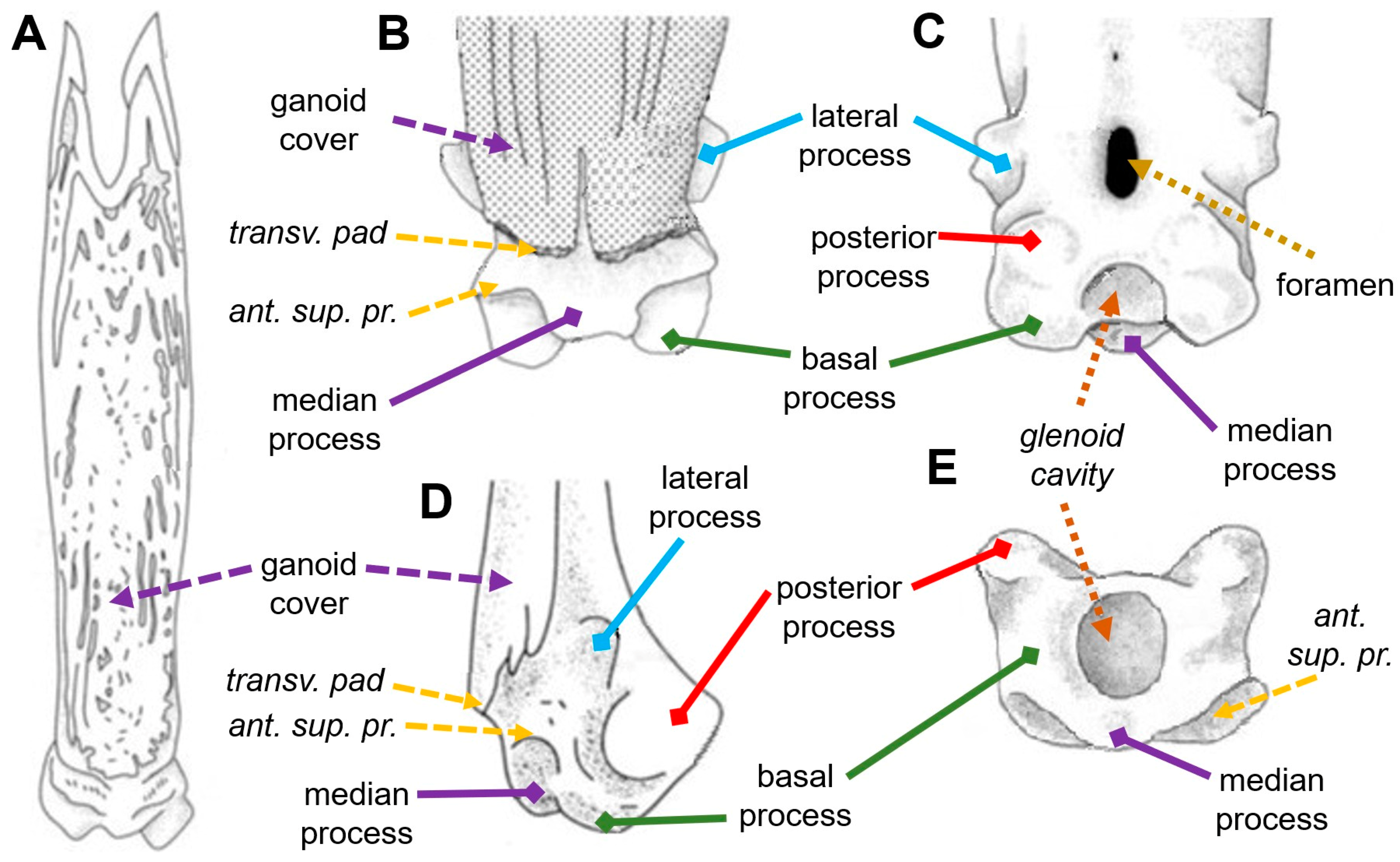
Figure 1. Illustration of the anatomical details on the anterior (A,B), posterior (C), lateral (D), and anterior (E) views of a pinnule (modified from Gayet and Meunier, 2020). The plain lines identify diagnostic structures. trans. pad for transversal pad; ant. sup. pr. for anterior transversal process.
Diversity 15 00517 g001
Figure 2. Microscopic images of the anterior view of different pinnules of different specimens of P. bichir. Each number corresponds to the respective position on the dorsal fin. Scale bars = 1 mm. The white rectangle zooms in on the ganoin ornamentation. The green triangle identifies different levels of concavity below the transversal pad. The yellow lines evidence various relative positions of the median process (dashed oval) compared to the base line of the pinnule (continuous line). Scale = 1 mm.
Diversity 15 00517 g002
Figure 3. Microscopic images of the posterior view of different pinnules of different specimens of P. bichir. Each number corresponds to the respective position on the dorsal fin. Scale bars = 1 mm. The red polygons evidence the various shapes of the head in posterior view. The blue lines underline the various relative orientations of the posterior process (circle), and its position is more or less lateral compared to the basal process (cup). The yellow arrows point at shallow depressions between the basal and posterior processes. The orange oval underlines the foramen with different shapes. Scale = 1 mm.
Diversity 15 00517 g003
Table 1. A summary of the diagnostic features for nominal fossil taxa described based on symmetrical pinnule morphology and their variability are evaluated in this study for the extant species P. bichir. Red represents characters that have been shown to vary intraspecifically. Green represents characters that might be relevant in distinguishing species.
TaxonFeatures with Intraspecific VariationProbably Reliable Features
Shape of the Head of the PinnulaDegree of Individualization of the ProcessesGanoin OrnamentationOpen Glenoid Cavity or Basal ForamenOverall Shape of the Processes
Bartschichthys🗸🗸 🗸
Bartschichthys arnoulti🗸🗸🗸 🗸
Bartschichthys napatensis 🗸🗸
Bartschichthys tubularis🗸 🗸
Polypterus dageti 🗸🗸
Polypterus sudanensis 🗸
Saharichthys africanus 🗸 🗸
Saharichthys nigeriensis 🗸 🗸
Sainthilairia 🗸 🗸
Sainthilairia beccusiformes 🗸
Sainthilairia elongata 🗸
Sainthilairia falcifomis 🗸 🗸
Sainthilairia grandis🗸🗸 🗸
Sainthilairia intermedia 🗸
Sudania🗸🗸
Sudania gracilis 🗸 🗸
Sudania oblonga🗸 🗸
Disclaimer/Publisher’s Note: The statements, opinions and data contained in all publications are solely those of the individual author(s) and contributor(s) and not of MDPI and/or the editor(s). MDPI and/or the editor(s) disclaim responsibility for any injury to people or property resulting from any ideas, methods, instructions or products referred to in the content.