

Description of Cylindrospermum solincola sp. nov. from Jammu and Kashmir, India and Further Insights into the Ecological Distribution and Morphological Attributes of Cylindrospermum badium

Abstract
:1. Introduction
2. Materials and Methods
2.1. Sampling, Isolation and Maintenance of the Isolates
2.2. Morphological Analysis
2.3. Molecular Analysis
2.4. Phylogenetic Analysis
2.5. 16S-23S ITS Secondary Structure Analysis
2.6. Calculations of 16S rRNA Gene Percent Similarity and 16S-23S ITS Percent Dissimilarity
2.7. Preparation of Holotype and Isotype
3. Results
3.1. Site Studied
3.2. Morphological Characterization
3.3. Phylogenetic Analysis Based on 16S rRNA Gene and 16S-23S ITS Region
3.4. Percent Similarity Analysis of 16S rRNA Gene
3.5. Secondary Structure Analysis of 16S-23S ITS Region
3.6. Percent Dissimilarity Analysis of 16S-23S ITS Region
3.7. Taxonomic Description
4. Discussion
Supplementary Materials
Author Contributions
Funding
Institutional Review Board Statement
Data Availability Statement
Acknowledgments
Conflicts of Interest
References
- Kützing, F.T. Phycologia Generalis Oder Anatomie, Physiologie und Systemkunde der Tange: Mit 80 Farbig Gedruckten Tafeln; Brockhaus: Munich, Germany, 1843. [Google Scholar]
- Bornet, E.; Flahault, C. Revision des Nostocacées Hétérocystées Contenues Dans les Principaux Herbiers de France (Quatriéme et Dernier Fragment); Annales des Sciences Naturelles Botanique Septième Série 1886–1888; G. Masson: Paris, France, 1890; pp. 177–262. [Google Scholar]
- Gardner, N.L. The Myxophyceae of Porto Rico and the Virgin Islands; New York Academy of Sciences: New York, NY, USA, 1932. [Google Scholar]
- Geitler, L.S. Klasse schizophyceae. In Natürliche Pflanzenfamilien 1b; Engler, A., Prantl, K., Eds.; Duncker and Humblot: Berlin, Germany, 1942; pp. 1–232. [Google Scholar]
- Desikachary, T.V. Cyanophyta. In ICAR Monograph on Algae; Indian Council of Agricultural Research: New Delhi, India, 1959. [Google Scholar]
- Komárek, J. Süßwasserflora von Mitteleuropa; Bd. 19/3: Cyanoprokaryota. 3. Teil/3rd part: Heterocytous genera. Süßwasserflora von Mitteleuropa; Spektrum Academischer Verlag: Heidelberg, Germany, 2013. [Google Scholar]
- Genuário, D.B.; Sant’Anna, C.L.; Melo, I.S. Elucidating the Cronbergia (Cyanobacteria) dilemma with the description of Cronbergia amazonensis sp. nov. isolated from Solimões river (Amazonia, Brazil). Algal Res. 2018, 29, 233–241. [Google Scholar] [CrossRef]
- Guiry, M.D.; Guiry, G.M. AlgaeBase; World-Wide Electronic Publication, National University of Ireland: Galway, Ireland, 2023; Available online: https://www.algaebase.org (accessed on 10 January 2023).
- Hauer, T.; Komárek, J. CyanoDB 2.0—On-Line Database of Cyanobacterial Genera; World-Wide Electronic Publication, University of South Bohemia and Institute of Botany AS CR: Ceske Budejovice, Czech Republic, 2023; Available online: http://www.cyanodb.cz (accessed on 10 January 2023).
- Pal, S.; Saraf, A.; Kumar, N.; Singh, A.; Talukdar, U.; Kohar, N.; Singh, P. Digging deeper into the taxonomy of Cylindrospermum and description of Johanseniella tripurensis gen. et sp. nov. from India. FEMS Microbiol. Lett. 2022, 369, fnac074. [Google Scholar] [CrossRef] [PubMed]
- Johansen, J.R.; Bohunická, M.; Lukešová, A.; Hrčková, K.; Vaccarino, M.A.; Chesarino, N.M. Morphological and molecular characterization within 26 strains of the genus Cylindrospermum (Nostocaceae, Cyanobacteria), with descriptions of three new species. J. Phycol. 2014, 50, 187–202. [Google Scholar] [CrossRef] [PubMed]
- Komárek, J.; Zapomělová, E.; Hindák, F. Cronbergia gen. nov., a new cyanobacterial genus (Cyanophyta) with a special strategy of heterocyte formation. Cryptogam. Algol. 2010, 31, 321–341. [Google Scholar]
- Komárek, J.; Kaštovský, J.; Mareš, J.; Johansen, J.R. Taxonomic classification of cyanoprokaryotes (cyanobacterial genera) 2014, using a polyphasic approach. Preslia 2014, 86, 295–335. [Google Scholar]
- Tawong, W.; Pongcharoen, P.; Saijuntha, W. Neocylindrospermum variakineticum gen. & sp. nov. (Nostocales, Cyanobacteria), a novel genus separated from Cylindrospermum using a polyphasic method. Phycologia 2022, 61, 653–668. [Google Scholar]
- Rippka, R.; Deruelles, J.; Waterbury, J.B.; Herdman, M.; Stanier, R.Y. Generic assignments, strain histories and properties of pure cultures of cyanobacteria. Microbiology 1979, 111, 1–61. [Google Scholar] [CrossRef]
- Edwards, U.; Rogall, T.; Blöcker, H.; Emde, M.; Böttger, E.C. Isolation and direct complete nucleotide determination of entire genes. Characterization of a gene coding for 16S ribosomal RNA. Nucleic Acids Res. 1989, 17, 7843–7853. [Google Scholar] [CrossRef] [PubMed]
- Gkelis, S.; Rajaniemi, P.; Vardaka, E.; Moustaka-Gouni, M.; Lanaras, T.; Sivonen, K. Limnothrix redekei (Van Goor) Meffert (Cyanobacteria) strains from Lake Kastoria, Greece form a separate phylogenetic group. Microb. Ecol. 2005, 49, 176–182. [Google Scholar] [CrossRef] [PubMed]
- Iteman, I.; Rippka, R.; de Marsac, N.T.; Herdman, M. Comparison of conserved structural and regulatory domains within divergent 16S rRNA–23S rRNA spacer sequences of cyanobacteriaThe GenBank accession numbers for the sequences reported in this paper are AF180968 and AF180969 for ITS-L and ITS-S, respectively. Microbiology 2000, 146, 1275–1286. [Google Scholar] [CrossRef] [PubMed]
- Katoh, K.; Rozewicki, J.; Yamada, K.D. MAFFT online service: Multiple sequence alignment, interactive sequence choice and visualization. Brief. Bioinform. 2019, 20, 1160–1166. [Google Scholar] [CrossRef] [PubMed]
- Ronquist, F.; Teslenko, M.; Van Der Mark, P.; Ayres, D.L.; Darling, A.; Höhna, S.; Larget, B.; Liu, L.; Suchard, M.A.; Huelsenbeck, J.P. MrBayes 3.2: Efficient Bayesian phylogenetic inference and model choice across a large model space. Syst. Biol. 2012, 61, 539–542. [Google Scholar] [CrossRef] [PubMed]
- Trifinopoulos, J.; Nguyen, L.T.; Von Haeseler, A.; Minh, B.Q. W-IQ-TREE: A fast online phylogenetic tool for maximum likelihood analysis. Nucleic Acids Res. 2016, 44, 232–235. [Google Scholar] [CrossRef] [PubMed]
- Minh, B.Q.; Nguyen, M.A.T.; Haeseler, A.V. Ultrafast approximation for phylogenetic bootstrap. Mol. Biol. Evol. 2013, 30, 1188–1195. [Google Scholar] [CrossRef]
- Darriba, D.; Taboada, G.L.; Doallo, R.; Posada, D. jModelTest 2: More models, new heuristics and parallel computing. Nat. Methods 2012, 9, 772. [Google Scholar] [CrossRef]
- Kumar, S.; Stecher, G.; Li, M.; Knyaz, C.; Tamura, K. MEGA X: Molecular evolutionary genetics analysis across computing platforms. Mol. Biol. Evol. 2018, 35, 1547. [Google Scholar] [CrossRef]
- Felsenstein, J. Confidence limits on phylogenies: An approach using the bootstrap. Evolution 1985, 39, 783–791. [Google Scholar] [CrossRef]
- Letunic, I.; Bork, P. Interactive Tree of Life (iTOL): An online tool for phylogenetic tree display and annotation. Bioinformatics 2007, 23, 127–128. [Google Scholar] [CrossRef]
- Zucker, M. Mfold web server for nucleic acid folding and hybridization prediction. Nucleic Acid Res. 2003, 31, 3406–3415. [Google Scholar] [CrossRef]
- Bharadwaja, Y. Contributions to Our Knowledge of the Myxophyceae of India. Ann. Bot. 1933, 47, 117–143. [Google Scholar] [CrossRef]
- Yarza, P.; Yilmaz, P.; Pruesse, E.; Glöckner, F.O.; Ludwig, W.; Schleifer, K.H.; Whitman, W.B.; Euzéby, J.; Amann, R.; Rosselló-Móra, R. Uniting the classification of cultured and uncultured bacteria and archaea using 16S rRNA gene sequences. Nat. Rev. Microbiol. 2014, 12, 635–645. [Google Scholar] [CrossRef] [PubMed]
- Bohunická, M.; Pietrasiak, N.; Johansen, J.R.; Gómez, E.B.; Hauer, T.; Gaysina, L.A.; Lukešová, A. Roholtiella, gen. nov. (Nostocales, Cyanobacteria)—A tapering and branching cyanobacteria of the family Nostocaceae. Phytotaxa 2015, 197, 84–103. [Google Scholar] [CrossRef]
- González-Resendiz, L.; Johansen, J.R.; Alba-Lois, L.; Segal-Kischinevzky, C.; Escobar-Sanchez, V.; Jimenez-Garcia, L.F.; Hauer, T.; León-Tejara, H. Nunduva, a new marine genus of Rivulariaceae (Nostocales, Cyanobacteria) from marine rocky shores. Fottea 2018, 18, 86–105. [Google Scholar] [CrossRef]
- Pal, S.; Saraf, A.; Kumar, N.; Singh, P. Phycological exploration of the global biodiversity hotspots of Northeast India: Discovery of a new species of soil-dwelling cyanobacteria, Desikacharya kailashaharensis sp. nov. FEMS Microbiol. Lett. 2022, 369, fnac099. [Google Scholar] [CrossRef]
- Komárek, J. A polyphasic approach for the taxonomy of cyanobacteria: Principles and applications. Eur. J. Phycol. 2016, 51, 346–353. [Google Scholar] [CrossRef]
- Alvarenga, D.O.; Andreote, A.P.D.; Branco, L.H.Z.; Delbaje, E.; Cruz, R.B.; Varani, A.D.M.; Fiore, M.F. Amazonocrinis nigriterrae gen. nov., sp. nov., Atlanticothrix silvestris gen. nov., sp. nov. and Dendronalium phyllosphericum gen. nov., sp. nov., nostocacean cyanobacteria from Brazilian environments. Int. J. Syst. Evol. Microbiol. 2021, 71, 004811. [Google Scholar] [CrossRef]
- Kumar, N.; Saraf, A.; Pal, S.; Mishra, D.; Singh, P. Insights into the phylogenetic inconsistencies of the genus Amazonocrinis and description of epilithic Amazonocrinis malviyae sp. nov. (Cyanobacteria, Nostocales) from Jammu and Kashmir, India. Int. J. Syst. Evol. Microbiol. 2022, 72, 005658. [Google Scholar] [CrossRef]


| Species | Habitat | Vegetative Cell (Size; LxW) | Heterocyte (Size; LxW) | Akinete (Size; LxW) |
|---|---|---|---|---|
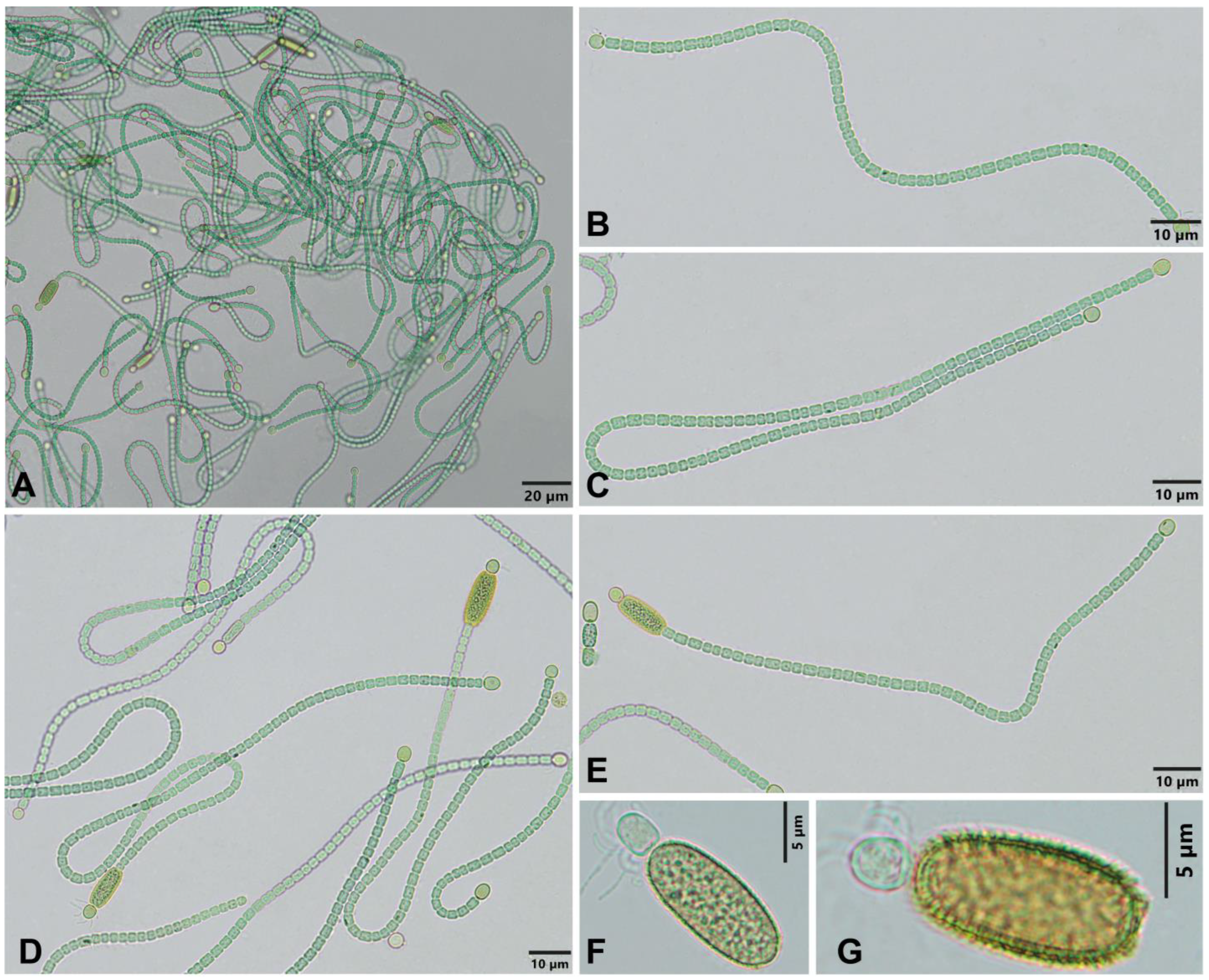
| Cylindrospermum solincola KUT1-PS | Wet soil | Cylindrical, often longer than wide, some isodiametric and shorter than wide, 1.49–3.08 × 1.72–2.11 µm | Terminal, solitary, both terminal ends, oval, rarely spherical, 2.89–4.39 × 2.61–3.33 µm | Large, solitary, at one terminal end, adjoining to heterocyte, young cylindrical, mature oblong, wall serrated with spines, yellowish in colour, 10.21–13.98 × 5.04–6.41 µm |
| Cylindrospermum gorakhpurence | Moist soils in paddy fields | Cylindrical, 7–16.5 × 3.5–5 µm | Terminal, solitary, both terminal ends, ellipsoidal, 13.2–16.5 × 6–7.8 µm | Subterminal, ellipsoidal with rounded ends, wall thick, with delicate, needle-like projections, 26.4–33 × 13.2–20 µm |
| Cylindrospermum dobrudjense | Agricultural soils (Medicago sativa) | Cylindrical, longer than wide, 6.6–9.2 × 4 µm | Terminal, both terminal ends, elongated ellipsoidal, conical or subspherical, 9.4–13.4 × 6.7 µm | Solitary, elongated, ellipsoidal, with rounded ends, wall with very fine and short spines, 22.8–26.6 (28.8) × 9.2–10.5 µm |
| Cylindrospermum ecballiisporum | Edge of wetted, peaty, partly periodically submerged pools and ditches | Cylindrical, ± isodiametric or longer than wide, end cells (without heterocytes) cylindrical and rounded 2.5–7.5 (8) × 2–3.4 µm | Terminal, almost spherical, ellipsoidal, short cylindrical or ovoid, slightly wider than vegetative cells, 5–10 × 3.5–6 µm | Solitary, rarely in pairs, adjoining to heterocytes, cylindrical-oval, rarely ellipsoidal, wall thick, with spines, (14) 20–33 (40) × (9) 11–14.5 (16) µm |
| Cylindrospermum maius CCALA 998 | Recultivated soil after coal mining | Cylindrical, longer than wide, isodiametric to shorter than wide, apical cell rounded to cylindrical, 3.7–6.5 × 3.9–5 µm | Terminal, spherical to elongated, 4–10 × 4.5–6 µm | Solitary, adjoining to heterocyte, cylindrical, oval to ellipsoidal, wall smooth, initially colourless, later brown and layered 1–1.5 µm wide, (18) 21–36 × 10–15 (16) µm |
| Cylindrospermum badium 18C-PS | Vernal pool | Cylindrical, isodiametric to longer than wide, 1.44–2.92 1.47–1.88 µm | Terminal, solitary, one or both terminal ends, oval to ovoid with broad base, 2.59–3.74 × 2.04–2.51 µm | Large, solitary, at one terminal end, adjoining to heterocyte, young oval to oblong, covered with a thin, transparent and diffluent layer of mucilage, mature oblong, wall thick, smooth and chestnut brown, 6.57–10.63 × 4.31–7.19 µm |
| Cylindrospermum badium CCALA 1000 | Recultivated soil after coal mining with sweet gum | Cylindrical, isodiametric to longer than wide, 3.5–7.5 × 3.0–4.8 µm | Terminal, solitary, almost spherical, elongated or slightly conical, 5–10 (13) ×, 3–5 µm | Solitary, paraheterocytic, oval broadly, flattened at ends, wall should be granulated or coarse instead of smooth, wide and chestnut-coloured, 17–30 × 10.0–14.4 µm |
| Cylindrospermum musicola Var. kashmiriensis | Epiphytic on Myriophyllum in shallow pond | Barrel-shaped, longer than broad, 2.6–8.4 × 2.6–3.9 µm | Terminal and intercalary, terminal, solitary, one or both terminal ends, oval or ellipsoidal, intercalary, solitary or in pairs, shorter or longer than vegetative cells, 5.2–10.5 × 3.9–5.9 µm | Adjoining to terminal heterocyte, barrel-ellipsoidal, wall thick and smooth, 9.5–13.6 × 5.2–7.8 µm |
| Cylindrospermum stagnale | Marshes, pools, ponds, lakes | Cylindrical, isodiametric or double longer than wide, (3) 3.8–4.5 (6?) µm wide, apical cylindrical-rounded | Usually oval, ovoid, or cylindrical, with rounded ends, rarely almost spherical, 7–16 × (4.5) 6–7 µm | Solitary, cylindrical or cylindrical-oval, rounded at the ends, smooth-walled, 32–40 × 10–16 µm |
| Cylindrospermum skujae | Wet sandy soils or sand, sometimes among mosses, in drying pools with a sandy bottom | Cylindrical, rarely isodiametric mainly 1.5–2 (4) × longer as wide, 1.6–5 (7) × 2.2–3 µm, terminal conical or cylindrical and rounded | Terminal, at one end only, oval, ovoid to cylindrical with rounded ends, (4.8) 6–8 × 2.8–4.5 µm | Solitary or pairs or up to 3 in a row oval–cylindrical, wall smooth, (12) 14–36 × 7–11.5 µm |
| Cylindrospermum muscicola | Wet soils, among mosses, shallow ditches and pools with water vegetation | More or less cylindrical, ± isodiametric or slightly longer or shorter than wide, terminal conical rounded or cylindrical and rounded, 3.6–5.4 (6) × 3–5.3 µm | Elongated, 5–7 (10.5) × (3.6) 4–5 (7) µm | Solitary, oval or ovoid, rounded at ends, wall smooth, 10–20 (21) × (8.2) 9–12 µm |
| Cylindrospermum alatosporum | Soil or guts of earthworms | Cells isodiametric or longer than wide, rounded at ends, 3–7 (8) × 3.5–5.0 µm | Rounded cylindrical, elongated or almost spherical, 4–9 (11) × 3.5–7.0 µm | Solitary or in pair, oval–rhomboid, wall smooth, 20–32 × (6.5) 10.0–13.0 (17.5) µm |
| Cylindrospermum licheniforme | Prairie remnant soil | Cylindrical, shorter than wide to isodiametric, 3.1–5.1 × 3.6–4.8 µm | Terminal, solitary, oval to elongated to bluntly conical, 5.4–9 × 4.0–5.2 µm | Solitary, paraheterocytic, oval broadly to elongated, wall smooth, 13–23 × 7.0–12.4 µm |
| Cylindrospermum moravicum | Cave sediment | Cylindrical, rarely concave or irregular, isodiametric, longer than wide, 2.7–5 × 3.5–7 µm | Terminal, solitary, spherical to cylindrical elongated, 5.0–9.0 (11) × 2.7–6.0 µm | Solitary, paraheterocytic, granulated, cylindrical, with widened end toward the heterocyte, wall wide with colourless to golden-brown, smooth, internally structured or lamellate exospores, (18) 22–32 × 9–13 µm |
Disclaimer/Publisher’s Note: The statements, opinions and data contained in all publications are solely those of the individual author(s) and contributor(s) and not of MDPI and/or the editor(s). MDPI and/or the editor(s) disclaim responsibility for any injury to people or property resulting from any ideas, methods, instructions or products referred to in the content. |
© 2023 by the authors. Licensee MDPI, Basel, Switzerland. This article is an open access article distributed under the terms and conditions of the Creative Commons Attribution (CC BY) license (https://creativecommons.org/licenses/by/4.0/).
Share and Cite
Kumar, N.; Saraf, A.; Pal, S.; Singh, P. Description of Cylindrospermum solincola sp. nov. from Jammu and Kashmir, India and Further Insights into the Ecological Distribution and Morphological Attributes of Cylindrospermum badium. Diversity 2023, 15, 592. https://doi.org/10.3390/d15050592
Kumar N, Saraf A, Pal S, Singh P. Description of Cylindrospermum solincola sp. nov. from Jammu and Kashmir, India and Further Insights into the Ecological Distribution and Morphological Attributes of Cylindrospermum badium. Diversity. 2023; 15(5):592. https://doi.org/10.3390/d15050592
Chicago/Turabian StyleKumar, Naresh, Aniket Saraf, Sagarika Pal, and Prashant Singh. 2023. "Description of Cylindrospermum solincola sp. nov. from Jammu and Kashmir, India and Further Insights into the Ecological Distribution and Morphological Attributes of Cylindrospermum badium" Diversity 15, no. 5: 592. https://doi.org/10.3390/d15050592
APA StyleKumar, N., Saraf, A., Pal, S., & Singh, P. (2023). Description of Cylindrospermum solincola sp. nov. from Jammu and Kashmir, India and Further Insights into the Ecological Distribution and Morphological Attributes of Cylindrospermum badium. Diversity, 15(5), 592. https://doi.org/10.3390/d15050592