Review

Brief Review of Morphological Characters in the Identification of Muscomorpha (Diptera) of Sanitary and Forensic Importance

by
Paloma Martins Mendonça
1,2,3,*,
Lucas Barbosa Cortinhas
1,2,
Carlos Henrique Garção-Neto
1,2 and
Margareth Maria de Carvalho Queiroz
1,2,3
1
Laboratório Integrado, Simulídeos e Oncocercose & Entomologia Médica e Forense, Instituto Oswaldo Cruz, Fundação Oswaldo Cruz, Rio de Janeiro 21040-360, Brazil
2
Programa de Pós-Graduação em Biodiversidade e Saúde, Instituto Oswaldo Cruz, Fundação Oswaldo Cruz, Rio de Janeiro 21040-360, Brazil
3
Mestrado Profissional em Ciências Ambientais, Universidade de Vassouras, Vassouras 27700-000, Brazil
*
Author to whom correspondence should be addressed.
Diversity 2024, 16(10), 599; https://doi.org/10.3390/d16100599
Submission received: 14 October 2023 / Revised: 15 November 2023 / Accepted: 16 November 2023 / Published: 30 September 2024
(This article belongs to the Special Issue Diversity and the Multiple Importance of the Study of Diptera)

Abstract

:
With more than 125,000 described species, Diptera are considered a megadiverse order. However, immatures display great morphological homogeneity, making few species of Diptera from the Cyclorrhaphan group easy to identify. The main species of medical, sanitary, and forensic importance are found in this group, highlighting the relevance of their description. The uniformity of the group limits sensitive techniques for identifying immatures; therefore, this study reports characters that can be used to identify these insects using scanning electron microscopy. Based on an investigation and analysis of the relevant scientific literature, we suggest combining techniques for accurate morphological identifications of flies.

1. Introduction

Insects are the most diverse and numerous group of organisms on the planet. There are around one million species described, and it is believed that, for each species described, five others are still unknown [1,2]. Within this group, five orders are considered megadiverse, with more than 100,000 species described: Coleoptera (beetles), Lepidoptera (butterflies and moths), Diptera (mosquitos end flies), Hymenoptera (wasps, bees and ants), and Hemipteran (cicadas, stink bugs, aphids and mealybugs), in decreasing order of species richness, corresponding altogether to approximately 920,000 described species [2,3]. It is estimated that around 153–155,000 correspond to representatives of the order Diptera [4,5]. However, the current number of species could be much higher. The number of species of this order could be as high as approximately 400,000 worldwide, with an estimated 60,000 found in Brazil [6]. Currently, the number of species described in Brazil is around 8700 out of a total of 31,000 recognized in the Neotropical region [7].

2. Phylogeny

The monophyly of Diptera is based on a large and valid number of morphological modifications or synapomorphies, such as the development of the mouth apparatus adapted for piercing and sucking and the transformation of the pair of posterior wings (metathoracic) into balance organs for flight (halteres) [8].
The phylogeny of the lower categories is still widely discussed. Classically, Diptera were divided into Nematocera and Brachycera. Nematocera comprise dipterans with a thin antennal flagellum with numerous segments, such as mosquitoes and sand flies.
Brachycerous dipterans have a short and robust antennal flagellum, with the atrophied final portion forming a style or an arista, as found in blow flies and house flies. Only Brachycera are recognized as a monophyletic group. Therefore, the infraorders of Nematoceran dipterans were elevated to suborders [9]. Currently, the most accepted topology comprises two groups, one of them more basal, composed of Tipulomorpha, Psychodomorpha, and Culicomorpha, and another called Neodiptera, formed by Bibionomorpha and Brachycera [5].
Brachycera has four subgroups with poorly resolved internal relationships: Tabanomorpha, Stratiomyomorpha, Xylophagomorpha, and Muscomorpha. The latter is also divided into Asiloidea and Eremoneura (subdivided into Empidoidea and Cyclorrhapha). Cyclorrhapha is considered monophyletic [5], the main characteristics of which are the metamorphosis of the insect within the puparium formed by the chitinization of the skin of the last larval instar and an inflatable sack, the ptilinum, that is expanded by blood (hemolymph) forced into the head from the thorax and abdomen and allows the adult to leave from the puparium (Schyzophora) [10]. Schyzophora is the most diverse group within the order, with approximately 85 of the 157 families and around 50,000 species. In this group, we can also find dipterans without (Acalyptratae) and with calyptra (Calyptratae), which corresponds to two laminar expansions located close to the base of the wings [5,11,12]. Caliptrates are known as muscoid dipterans [11].

3. Medical–Veterinary Importance as Pathogen Transmitters

Flies are widely studied, not only due to the great diversity of the group but also due to the high degree of synanthropy of some species. Synanthropic flies are those species that take advantage of environmental conditions created by man to improve their development [13]. The degree of proximity of each species to humans can be inferred numerically through the synanthropy index [13]. Moreover, other indices were proposed, also considering the size of the insect and the capacity for the contamination and transmission of pathogens [14].
These indices help to determine which species have the greatest potential to transmit pathogens to humans and thus help to establish control measures. Many studies have been carried out, seeking to relate and quantify the pathogens carried by flies.
It is believed that the important role of flies as transmitters of pathogens is related to their ability to fly great distances; some species can fly up to 65 km a day, combined with their high attraction to places where food is processed and stored as well as to decomposing organic matter [15,16,17].
Musca domestica Linnaeus, 1758 (Diptera: Muscidae), is probably the most studied species in the transmission of pathogens as it presents high levels of synanthropy and endophilia; that is, it is closely associated with the domestic environment [18,19].
Some species of Calliphoridae also have these characteristics, however, and are considered urban pests [15]. In the city of Manaus, in the state of Amazonas, Brazil, six genera of bacteria were isolated from the body of Chrysomya megacephala (Fabricius, 1974), Chrysomya putoria (Wiedemann, 1818), Chrysomya albiceps (Wiedemann, 1819), and Lucilia eximia (Wiedemann, 1819) (Calliphoridae) collected in street markets, reinforcing the importance of these fly species as carriers of pathogens to humans [20].
In the city of Rio de Janeiro, Escherichia coli, Citrobacter sp., Proteus mirabilis, Morganella sp., Klebsiella sp., Pseudomonas sp., Enterobacter sp., and Salmonella were isolated from adults of C. megacephala and M. domestica [21]. Multidrug-resistant Escherichia coli was isolated from house flies [22,23]. Escherichia coli serotype 0157:H7 presents a known risk to human health and has been isolated several times from different species of Diptera [19,21,24].

4. Medical–Veterinary Importance as Causes of Myiasis

Larvae of dipterans can develop in the tissue of humans and other animals, where they evolve as ectoparasites. Some species are obligate parasites, and their larvae are called bionthophagous, as they feed on living tissue (myiasis), for example, Dermatobia hominis (Linnaeus Jr., 1781) (Diptera: Oestridae) and Cochliomyia hominivorax (Coquerel, 1858) (Diptera: Calliphoridae). Other species are considered facultative parasites, and their larvae are called necrobionthophagous or scavengers, as they could feed on necrotic tissue that may or may not be in a living organism. As an example of the latter type, we can mention Cochliomyia macellaria (Fabricius, 1775) (Diptera: Calliphoridae) and representatives of the genera Chrysomya, Lucilia, Fannia, and Muscina (Diptera: Calliphoridae), in addition to some members of Sarcophagidae [25,26,27].
Infestation by these larvae causes great damage to animal husbandry on large and small scales. Damage to the animal ranges from increased irritability and decreased appetite to and anemia, leading to weight loss, decreased milk production, and even mastitis and severe infections in the navels of newborns, which can lead to premature weaning and death [28,29,30].

5. Forensic Importance

Flies also stand out among the group of decomposers, as some species can quickly colonize different habitats due to their attraction to the odor released during the decomposition process and the fact that they are agile and good fliers.
According to several authors, flies are the first insects to reach corpses and could oviposit immediately after finding them [31,32]. Furthermore, larvae are responsible for the degradation of body mass, performing an important ecological function as decomposers, and can be found in most corpses investigated [33,34]. Body decomposition is faster when it occurs in the presence of insects [35,36,37].
Although insects are not yet widely used in determining the post-mortem interval, they should be used to complement traditional techniques, which become inaccurate as this interval increases. Histological, chemical, and bacteriological methods, as well as cadaveric phenomena, are also used to determine the post-mortem interval. However, 72–96 h after death, insects are the most accurate tool for estimating the post-mortem interval. Recently, many studies related to entomofauna aimed to define groups of insects as forensic indicators; that is, those species that effectively search the corpse for the development of their immature forms [38,39].

6. Problems in Identification

Correctly identifying species associated with bodies or carcasses is an essential stage in forensic analysis. Some species are phylogenetically close and have similar morphologies. However, they may present different growth rates, responses to diapause, and ecologies.
The differentiation of flies of medical–veterinary and forensic importance is usually based on adults since the identification of immatures is challenging and even, in some cases, impossible [40,41,42]. For most species of adult Diptera, dichotomous identification keys are available [43,44,45,46,47]. However, most of the specimens found in cadavers correspond to the immature stages (eggs, larvae, and pupae) without morphological differences visible even under a light microscope [31,48,49,50]. This is due to the absence of specific diagnostic characters for immatures, in addition to intraspecific variations observed in some geographic regions [50].
To overcome this problem, Diptera in immature stages can be raised until adulthood to allow identification using taxonomic keys. However, in many criminal cases, insects are collected by untrained people who kill or fix the specimens, preventing the complete development and, consequently, the taxonomic identification of the species involved.
Rearing immature flies takes time for the complete development of the species, especially when its biological cycle is still unknown, which is the case for many species. Furthermore, an adequate laboratory is necessary for rearing the insects due to the preferred breeding substrate: feces, beef, pork, fish, and putrefied chicken viscera [49].
In criminal cases, a rapid determination of the post-mortem interval is crucial to the investigation. Often, there is not enough time to rear the immatures until the adults emerge; in addition, the specimens may die before identification at a specific level, thus preventing the analysis of important criminal evidence [49].

7. Identification Using Scanning Electron Microscopy

An alternative to minimize the difficulty of breeding dipterans of forensic and sanitary importance would be scanning electron microscopy (SEM). This accurate technique allows a visualization of diagnostic characters even in the immature stages.
Several authors have used SEM to identify immatures in many countries [50,51,52]. Recently, some descriptions of species collected in Brazil were published [41,42,53,54,55,56,57], but they still do not cover the diversity of species of forensic importance.
Some authors emphasize that certain morphological variations may occur between Diptera populations from different geographic regions and biomes [50], especially in cases where there is an introduction of new species and the isolation of others, as in the case of C. macellaria in Latin America. Therefore, there are no identification keys for immatures that provide diagnostic morphological characters encompassing the diversity of species of forensic importance.

8. Discussion

As mentioned before, immatures of several species have been described using light microscopy and SEM. In the beginning, most authors proposed to describe the specimens, but over time, the need was to compare taxonomic characters to facilitate species identification. In this work, some similarities were found in relation to the shape and other characters observed in the species described in the literature.

8.1. Eggs

Dipteran flies’ eggs are elongated structures covered by chorionic cells with distinct appearances according to their location, whether in the inner portion of the median area or covering the rest of the egg [58]. The median area is differentiated by a dorsal opening from the anterior to the posterior end of the egg, formed by interconnected structures called islands and pillars generating open spaces known as aeropiles [51]. The arrangement of these structures makes the interior of the median area resemble a sponge, retaining air in the aeropiles and allowing gas exchange between the embryo and the environment. In addition to facilitating the embryo’s breathing, the median area offers less resistance to larvae hatching. The larva presses on the median area that breaks right at the junction point of the exochorion’s outermost layer with the islands. The median area is found in several families of terrestrial cyclorrhaphic dipterans [42,57,58,59].
Liu & Greenberg [60] briefly described C. macellaria eggs collected in Chicago, United States of America. These authors report that the median area extends, at most, to half of the micropyle (Y-shape). Furthermore, they also refer to the islands in the median area as structures with a tapered shape, like piles. However, such information is insufficient to identify C. macellaria eggs with, as these can also be observed in other species collected in the Neotropical region.
Some studies reported the same combination of characters in eggs of C. megacephala, L. eximia, and Lucilia cuprina (Wiedemann, 1830) (Diptera: Calliphoridae), representatives of two genera of the same family to which C. macellaria belongs [56,59,61]. Furthermore, these species have very similar ecological habits, can cause secondary myiasis, and are often collected from carcasses [26,38], which could make differentiation between species even more challenging.
The outermost layer of the egg or exochorion is rigid enough to protect the embryo from drying out. The boundaries of the follicular cells from the exochorion become visible through densification, a process causing an increase in the density of the exochorion during the embryonic phase (Figure 1A,B) [10].
The chorionic cells in C. macellaria have a hexagonal pattern and are very similar to those of C. hominivorax and C. megacephala [56,59,60]. The exochorion cells of C. hominivorax decrease in size as they are closer to the posterior region of the egg [62]. In contrast, in C. macellaria, the exochorion has same-size cells throughout the egg. The chorionic sculpture of Lucilia eximia shows a hexagonal pattern with smooth edges [59].
The presence of raised and prominent edges was observed only in C. putoria eggs [59]. In the muscid Hydrotaea aenescens (=Ophyra aenescens) (Widemann, 1830) (Diptera: Muscidae), the authors describe the exochorion as completely ornamented, not only at the edges of the cells but also across the entire surface [57]. In C. macellaria, exochorion cells have a hexagonal shape with smooth edges [56]. Unfortunately, most authors limited themselves to observing structures related to the length and shape of the micropyle and the median area, not highlighting the exochorion cells [50,60].
The micropyle is the hole through which sperm penetrate until they reach the oviduct, where fertilization occurs. Although several studies have reported ornamentation around this hole, this type of ornamentation becomes evident only through SEM. The main ornamentations on the micropylar plate are of the projection type, found in C. megacephala, C. albiceps, Chrysomya nigripes (Aubertin, 1932) (Diptera: Calliphoridae), and C. macellaria, and the depression-type, as reported in C. putoria and H. aenescens (O. aenescens), or even the absence of any type of adornment, as observed in L. cuprina and M. domestica (Figure 1C–E) [42,56,57,59,61].
When combined, the median area’s characters can provide important information to distinguish eggs from members of Calliphoridae (Figure 1F,G) [56,59,63]. The combination of characters makes it possible to differentiate eggs from those of very similar species.
As previously reported, there are few studies describing the ultrastructure of eggs. However, several studies have been published describing Diptera larvae.

8.2. Larvae

Like adult flies, immatures also have very distinct characteristics compared to those of other insect orders. The character that best defines the immatures of Diptera is the absence of articulated thoracic legs combined with active, directed movement [1,3,4,10]. Generally, immature apod insects are immobile or move slowly and erratically. However, immature dipterans move easily through peristaltic body movements aided by protuberances named creeping welt and spines located between the body segments.
Muscomorpha larvae have a body divided into 12 segments, the first considered by some authors to be pseudocephalon due to the almost total reduction in and retraction of the head into the first thoracic segment, followed by three thoracic and eight abdominal segments [10,64,65]. The larvae body resembles a cone, with the mouth opening at the tapered end and the anal segment and the posterior spiracle at the truncated end [10,65]. This seems to be the most ergonomically efficient structure for tissue feeding and burial for pupation [63].

8.2.1. Head

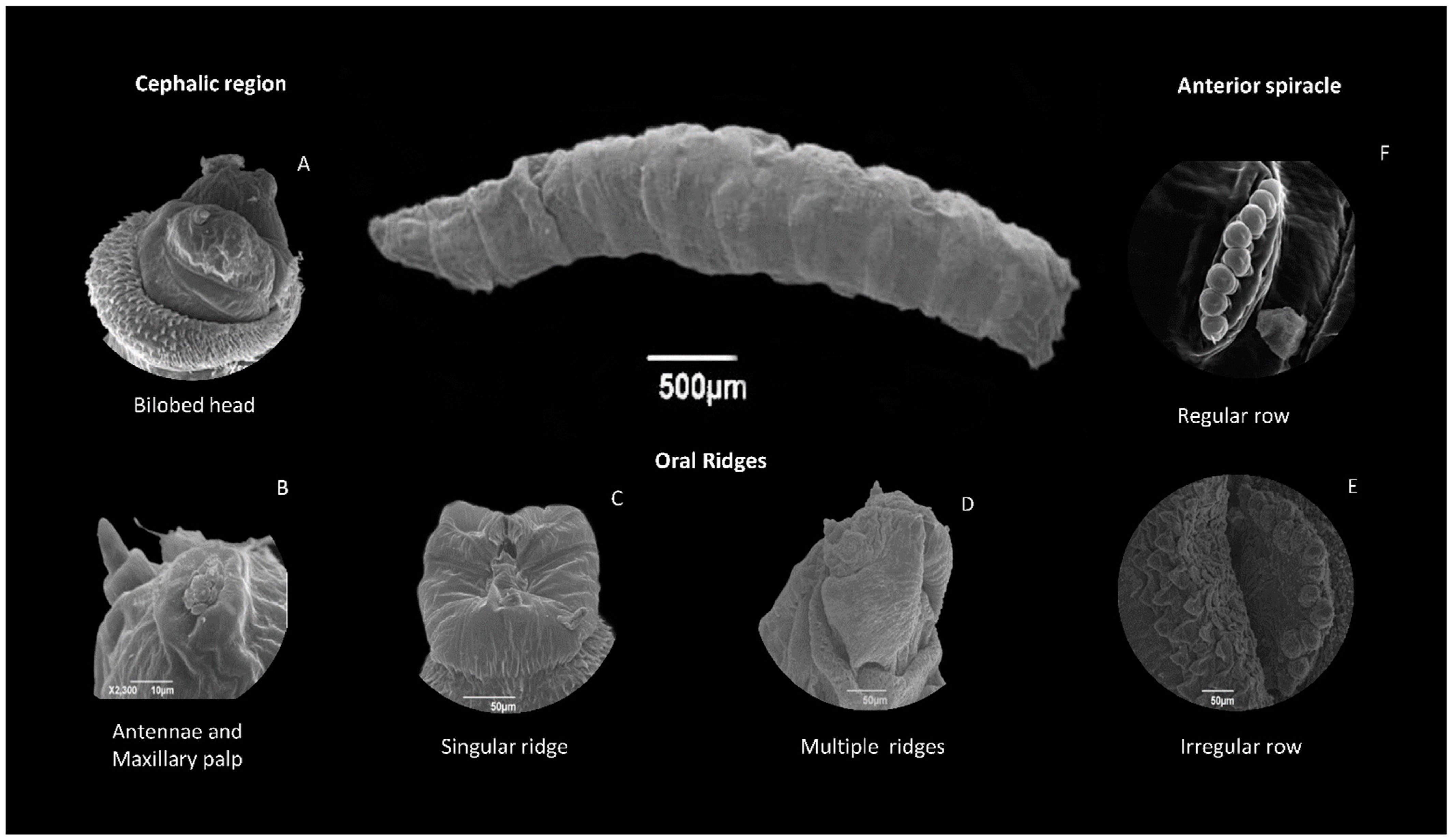
Still, there is no evidence of a sclerotized head in Muscomorpha, with the oral apparatus consisting of only a pair of curved hooks, the mandibles, where teeth can be observed in some species [65]. The main characteristic of Muscomorpha is the presence of the bilobed membranous and sensory pseudocephalon, together with the development of the internal and highly sclerotized cephalopharyngeal skeleton (Figure 2A) [64].
The cephalic or pseudocephalic region is composed of two sensory centers. The first is the antenna–maxilla, which contains the antennae and the maxillary palp complex. The antennae, also known as dorsal organs, are divided into two segments, one basal and the other in a domed shape. The maxillary palp complex or terminal organs are small structures formed by five concentrically arranged papillae. In some species, one or two papillae external to the palp complex could be observed, as in H. aenescens (=O. aenescens) and C. putoria [54,57]. The antennae are believed to have an olfactory function, and the palp complex performs a chemo- and mechano-receptor function, as reported for Musca domestica (Figure 2B) [66].
It is worth highlighting that immature P. (E.) collusor have the papillae of the maxillary palp complex in a cavity, unlike what is observed for L. cuprina and C. macellaria, where these structures are slightly elevated [41,55,56]. This arrangement of the papillae on an elevation was also observed in species from different families, such as M. domestica and C. hominivorax, and species of Chrysomya [42,54,67,68].
The antennae and maxillary palp complex of the sarcophagid Sarcophaga (Liopygia) ruficornis (Fabricius, 1794) were described as a group of papillae surrounded by cuticular ridges resembling a flower [69]. In Peckia (Peckia) chrysostoma (Wiedemann, 1830) (Diptera: Sarcophagidae), another species from the same family, the presence of papillae forming the maxillary palp complex was reported, but not located in a cavity. This arrangement has not been observed in members of other families of Muscomorpha; it is believed that it may be a characteristic restricted to the subgenus Euboettcheria [55], but more studies should be conducted to confirm this.
The second sensory center comprises a very small organ in the middle of the oral ridges, the ventral organ. These structures are innervations originating from the maxillary nerve and are very difficult to find [64]. They are formed by an almost imperceptible sensilla, with a chemo-receptor function and a set of three more visible sensillas that perform a mechano-receptor function [66]. This structure is very similar among all species described [42,54,55,56,57,64,68,69]. In most species, oral ridges have a singular row at the first instar, and it increases at the second and third larval instar (Figure 2C,D).

8.2.2. Body Segments

In the latero-ventral portion of the thoracic segments, it is possible to visualize a set of three sensilla named Keilin’s organs, which have a mechano-receptor function in the larva. However, an analysis of the anatomy and physiology of the neurons present at the base of these sensilla determined that Keilin’s organs give rise to the legs of adult insects [70]. Thus, like the case for the ventral organ, no morphological differences were observed between the species [42,68,69].
The respiratory system of dipterans comprises an internal trachea system and an external spiracle system [64]. Several authors highlight the importance of the number and location of spiracular ramifications in Diptera. The number can vary from 10 pairs (holocaustic larvae) to the absence of spiracles (apneustic), where breathing is carried out through spiracle filaments connected to the larval integument. The larvae of the Cyclorrhapha group, a subdivision within Muscomorpha, are amphipneustic; that is, they have a pair of digitated spiracles located in the dorsolateral area of the first thoracic segment and a pair located in the last abdominal segment.
The anterior spiracle can only be visualized at the second and third larval instar; therefore, the first-instar larvae are metapneustic. This structure is composed of spiracular ramifications arranged in aligned rows or irregularly (Figure 2E,F). The number of ramifications can vary from 1 and 4 to 30, or even more. It is one of the most used characteristics in differentiating species since it is visible under light microscopy [65].
The larvae and puparia of L. cuprina from Brazil have six to seven ramifications [41], as reported in L. eximia [53]. The sheep blowfly L. cuprina from Thailand has four to seven ramifications [69]; however, the puparia were related with five to seven ramifications [52]. The screwworm C. macellaria had 11–12 spiracular ramifications [56]; however, in Peru, 8–12 ramifications were reported, whereas insects collected in Colombia had 8–11 [25,47,71,72]. These observations indicate a variation in the number of openings within the same population and between populations of the same species. These variations reflect the species’ changes to adapt to the new habitat.
In flesh flies, it is possible to observe more than 20 spiracular ramifications [47,73]. This number was also considerably reduced for species collected in Brazil and the Neotropical region. These authors [74] described immatures of four species of sarcophagids (Oxysarcodexia sp.), and the number of ramifications ranged from 8 to 14. In Ravinia belforti (Prado & Fonseca, 1932) (Diptera: Sarcophagidae), it ranged from 16 to 22 [75]. Some species of the same family showed a fewer number of openings: Wohlfahrtia magnifica (Schiner, 1862) (Diptera: Sarcophagidae) (5–6) [76], Sarcophaga (Liopygia) crassipalpis (Macquart, 1839) (Diptera: Sarcophagidae) (11–12) [77], and P. (E.) collusor (11–14) [55]. The organization of spiracular ramifications in a regular row is widely discussed in the Neotropical region. In Oxysarcodexia, larvae and puparia were described as being in an irregular row, as for Sarcophaga dux Thomson, 1869 (Diptera: Sarcophagidae), while in P. (E.) collusor, they were found to be in a regular row [55,73,74].
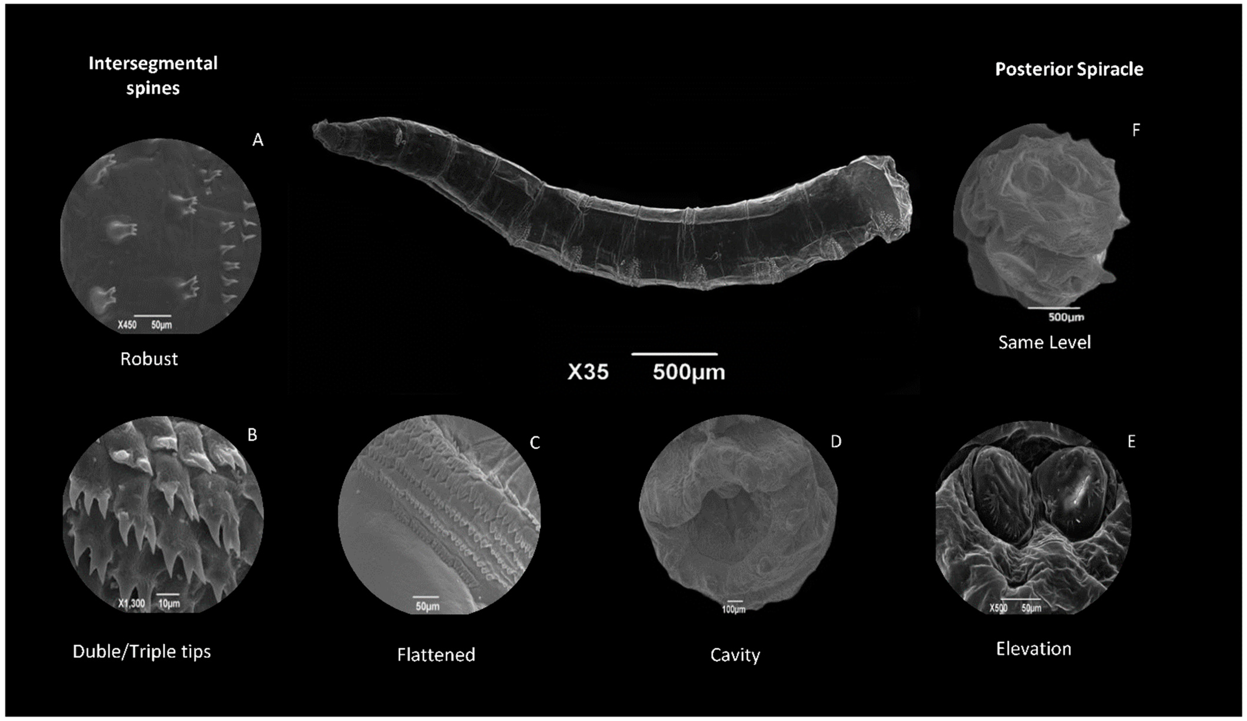
The integument of the larva contains bands of spines located on the anterior and/or posterior part of the segments. These rows can surround the entire body or be restricted to just the ventral, lateral, and dorsal regions of each segment [65]. The number, shape, and arrangement of spines can be a diagnostic characteristic of genera or species of Muscomorpha [72]. The arrangement of these spines can also vary between segments, as well as according to the maturation of the larvae (Figure 3A–C).
The first spine band, or cephalic collar, is located between the cephalic region and the first thoracic segment (TI). In the flesh fly P. (E.) collusor, these spines are sharpened at all stages; however, in third-instar larvae, they become slightly flattened [55]. The spines on the cephalic collar of W. magnifica are wide, conical, elongated, and slightly curved, decreasing in size as they are closer to the posterior region of the body [68]. This similar arrangement was described for L. cuprina, where the spines are flattened with tapered ends in the anterior portion, while in the region closest to the thoracic segment, the spines are filiform. This difference is only observed in the first-instar larva. This kind of arrangement was observed in six species of Lucilia [41]. These authors did not describe the shape of the spines and only used their arrangement to differentiate the immature. According to them, based on the arrangement of the spines of the AVII segment of the abdomen, it is possible to differentiate L. cuprina from L. sericata, species that comprise important taxonomic issues in Europe [41]. It is believed to be a characteristic common to all representatives of this genus. However, it is not exclusive to Lucilia; this same arrangement of the spines was described in C. albiceps. Furthermore, the first-instar larvae of C. macellaria also had more robust spines near the cephalic region and sharper spines near the thoracic region [56].
Some authors considered the cephalic collar part of the intersegmental spines and did not describe this structure specifically [73]. The intersegmental spines are very relevant to the differentiation of immatures from sarcophagids. In P. (E.) collusor, the shape of spines varied during the development of the larva, going from filiform in first-instar larvae to robust and flat in third-instar larvae and puparia. Changes in the shape of the spines were also observed in P. (L.) ruficornis. The spines found in the first-instar larvae were triangular with a sharp tip; in the second instar, the spines were sharper, while in the next instar, they were much smaller [69]. However, this change cannot be considered a pattern within members of this family, as the same change in the spine pattern of P. dux and W. magnifica was not observed [68,73].

8.2.3. Anal Segment

The posterior spiracle is in the last abdominal segment, in the terminal portion of the anal segment. This structure comprises slit-shaped spiracle openings that increase in number as the larva matures; however, in the case of Muscomorpha, it can vary from one to three openings.
The region surrounding these openings is called peritreme, which can be opened, also known as incomplete, or closed, known as complete. Furthermore, a spiracular scar from the previous instar can still be found at the point of convergence of the respiratory openings [64]. The structure and position of the posterior spiracles are very useful in separating genera and species of Cyclorrhapha [65].
The posterior spiracle located in a cavity formed by the tubercles of the anal segment is a typical feature of Sarcophagidae [55,73,75,78]. In Calliphoridae, the spiracular plate is not in a cavity, i.e., it is at the same level as the integument of the anal segment [41,54,56]. In Muscidae, as in species of Hydrotaea (=Ophyra) and Musca, the spiracles are on a projected elevation of the anal segment (Figure 3D–F) [42,57].
Generally, the number of openings in the posterior spiracle corresponds to the instar of the immature. This observation has already been reported for several species [41,57,67,74,78]; however, in some cases, the first-instar larvae may have two spiracle openings, as observed in M. domestica and C. macellaria [42,56]. Surrounding the spiracular plates, some species may have filiform—peristigmatic tufts—or flattened spines, which are better visible via SEM.

8.3. Puparia

Puparia have a general structure similar to that of third-instar larvae since their integument is formed via the chitinization of the larval cuticle. However, inside the puparium, the pupa underwent some adaptations to guarantee their protection and survival, such as the hardening and wrinkling of the cuticle, invagination of the cephalic region, and development of auxiliary respiratory structures. In L. cuprina, for example, pupal respiratory horns were observed in the region close to the first abdominal segment, as in O. aenescens (=H. aenescens) and M. domestica [42,52,57]. A possible explanation is that these horns are present in the pupa enclosed within the puparium and are projected outwards through increased hemolymph pressure. This rupture occurs in a region covered by a membrane and can occur approximately 24 h after pupa formation [63].
However, in some species, the membranes that allow the respiratory horns to project out of the puparia do not exist. Thus, they remain inside the puparium, as is the case of C. macellaria [56]. In sarcophagids, the tubercles are poorly sclerotized and do not have enough strength to break the membranes and exit the puparium [61,65]. According to the literature, the length of pupal respiratory horns in flies varies among species and according to time after puparia formation (Figure 4A–D) [60,63].
A continuous longitudinal line was observed along the second and third thoracic segments, and a slight constriction in the first abdominal segment of the puparia was also observed (Figure 4E,F). These structures were related to the location of the puparium opening for adult emergence.

9. Conclusions and Future Directions

The characterization of immature flies provides support to researchers and criminal professionals in investigations. It is worth highlighting that the number of species described in the literature, mainly in the Neotropical region, is small, as well as the establishment of dichotomous keys that do not correspond to all insects of medical–veterinary and forensic importance in the Neotropical region.
Intersegmental spine bands are relevant characters to describe species of medical-veterinary and forensic importance. We observed that few authors describe them individually. The detailed description of these structures and others presented in this work was possible through SEM, reinforcing the need to popularize this technique. Light microscopy is, undoubtedly, the costless method via which to identify Diptera, but as do all techniques, it has its own limitations. Authors worldwide also recommend the use of light microscopy and SEM for better immature identification.
As observed above, we recommend that taxonomic identification occurs through a combination of characters and not just by choosing a diagnostic character. We also suggest that specialists propose a standardization of morphological characters to be observed so that characteristics that are relevant in identifying fly species can be verified.

Author Contributions

P.M.M., conceptualization; P.M.M., L.B.C. and C.H.G.-N., writing—original draft preparation; P.M.M. and M.M.d.C.Q., financial support. M.M.d.C.Q. writing—review, editing, and supervision. All authors have read and agreed to the published version of the manuscript.

Funding

This research was supported by PAEF-IOC/FIOTEC (Strategic Actions for Development and Strengthening Accredited Laboratories and Research Support Areas) from The Oswaldo Cruz Foundation (FIOCRUZ) (Process Identification: IOC-023-FIO-18-2-30), the National Council of Research and Technological Development—CNPq (Process number: 316254/2021-5), and the Carlos Chagas Filho Research Support Foundation of the State of Rio de Janeiro—FAPERJ (Process Identification: E-26/210.228/2018; E-26/210.982/2021; E-26/201.311/2022; E-26/200471/2023). The last author received a scholarship from the Coordination for the Improvement of Higher Education Personnel—CAPES (Financial Code: 001).

Institutional Review Board Statement

Not applicable.

Data Availability Statement

No new data were created or analyzed in this study. Data sharing is not applicable to this article.

Acknowledgments

We would like to thank Karin Hoch Fehlauer Ale—EscritaLab for English Review and to the Platform Electron Microscopy Rudolf Barth of Instituto Oswaldo Cruz (FIOCRUZ) for the use of the scanning electron microscope.

Conflicts of Interest

The authors declare no conflict of interest. The funders had no role in the design of the study; in the collection, analyses, or interpretation of data; in the writing of the manuscript; or in the decision to publish the results.

References

  1. Grimaldi, D.; Engel, M.S. Evolution of the Insects, 1st ed.; Cambridge University Press: New York, NY, USA, 2005; 755p. [Google Scholar]
  2. Stork, N.E. How Many Species of Insects and Other Terrestrial Arthropods Are There on Earth? Ann. Rev. Entomol. 2018, 63, 31–45. [Google Scholar] [CrossRef]
  3. Rafael, J.A.; Aguiar, A.P.; Amorim, D.S. Knowledge of insect diversity in Brazil: Challenges and Advances. Neotr. Entomol. 2009, 38, 565–570. [Google Scholar] [CrossRef] [PubMed]
  4. Thompson, F.C. The Diptera Site. The biosystematic Database of World Diptera. Nomenclator Status Statistics. Version 10.5. Available online: www.sl.barc.usda.gov/diptera/names/Status/bdwdstat.htm (accessed on 6 October 2023).
  5. Wiegmann, B.M.; Trautwein, M.D.; Winkler, I.S.; Barr, N.B.; Kim, J.W.; Lambkin, C.; Bertone, M.A.; Cassel, B.K.; Bayless, K.M.; Heimberg, A.M.; et al. Episodic radiations in the fly tree of life. Proc. Natl. Acad. Sci. USA 2011, 108, 5690–5695. [Google Scholar] [CrossRef]
  6. Carvalho, C.J.B.; Moura, M.O.; Ribeiro, P.B. Chave para adultos de dípteros (Muscidae, Fanniidae, Anthomyiidae) associados ao ambiente humano no Brasil. Rev. Bras. Entomol. 2002, 46, 107–114. [Google Scholar] [CrossRef]
  7. Amorim, D.S. Neotropical Diptera Diversity: Richness, patterns and perspectives. In Diptera Diversity: Status, Challenges and Tools; Pape, T., Bickel, D., Meier, R., Eds.; Koninklije Brill NV: Leiden, The Netherlands, 2009; pp. 71–97. [Google Scholar]
  8. Yeates, D.K.; Wiegmann, B.M.; Courtney, G.W.; Meier, R.; Lambkin, C.; Pape, T. Phylogeny and systematics of Diptera: Two decades of progress and prospects. Zootaxa 2007, 1668, 565–590. [Google Scholar] [CrossRef]
  9. Amorim, D.S.; Yeates, D.K. Pesky gnats: Ridding dipteran classification of the Nematocera. Stud. Dipterol. 2006, 13, 3–9. [Google Scholar]
  10. Chapman, R.F. The Insects: Structure and Function; Cambridge University Press: Cambridge, UK, 1998. [Google Scholar]
  11. Serra-Freire, N.M.; Mello, R.P. Entomologia & Acarologia na Medicina Veterinária; Ed. LF Livros: Rio de Janeiro, Brazil, 2006. [Google Scholar]
  12. Gullan, P.J.; Cranston, P.S. Insect Systematics: Phylogeny and Classification. In The Insects: An outline of Entomology; Gullan, P.J., Cranston, P.S., Eds.; Wiley Blackwell: London, UK, 2008; pp. 189–222. [Google Scholar]
  13. Nuorteva, P. Synanthropy of blowflies (Dipt. Calliphoridae) in Finland. Ann. Zool. Fenn. 1963, 29, 1–49. [Google Scholar]
  14. Maldonado, M.A.; Centeno, N. Quantifying the potential pathogens transmission of the blowflies (Diptera: Calliphoridae). Mem. Inst. Oswaldo Cruz. 2003, 98, 213–216. [Google Scholar] [CrossRef] [PubMed]
  15. Guimarães, J.H.; Prado, A.P.; Buralli, G.M. Dispersal and distribution of three newly introduced species of Chrysomya R.D. (Diptera: Calliphoridae) in Brazil. Rev. Bras. Entomol. 1979, 23, 245–255. [Google Scholar]
  16. Olsen, A.R. Regulatory action criteria for filth and other extraneous materials. III. Review of flies and foodborne enteric disease. Regul. Toxicol. Pharmacol. 1998, 28, 199–211. [Google Scholar] [CrossRef]
  17. Braack, L.E.; Retief, P.F. Dispersal, density and habitat preference of the blow-flies Chrysomyia albiceps (Wd.) and Chrysomyia marginalis (Wd.) (Diptera: Calliphoridae). Onderstepoort J. Vet. Res. 1986, 53, 13–18. [Google Scholar] [PubMed]
  18. Greenberg, B. Flies and Diseases. Volume I: Ecology, Classification and Biotic Associations; Princeton University: Princeton, NJ, USA, 1971. [Google Scholar]
  19. Butler, J.F.; Garcia-Maruniak, A.; Meek, F.; Maruniak, J.E. Wild Florida House Flies (Musca domestica) as Carriers of Pathogenic Bacteria. Fla. Entomol. 2010, 93, 218–223. [Google Scholar] [CrossRef]
  20. Paraluppi, N.D.; Vasconcelos, J.C.; Aquino, J.S.; Castellón, E.G.; Silva, M.S.B.C. Calliphoridae (Diptera) in Manaus: IV Bacteria isolated from blowflies collected in street markets. Acta Amazon. 1996, 26, 93–96. [Google Scholar] [CrossRef]
  21. Oliveira, V.C.; d’Almeida, J.M.; Abalém-de-Sá, I.V.; Mandarino, J.R.; Solari, C.A. Enterobactérias associadas a adultos de Musca domestica (Linnaeus, 1758) (Diptera: Muscidae) e Chrysomya megacephala (Fabricius, 1754) (Diptera: Calliphoridae) no Jardim Zoológico, Rio de Janeiro. Arq. Bras. Med. Veterinária E Zootec. 2006, 58, 556–561. [Google Scholar] [CrossRef]
  22. Chandrakar, C.; Shakya, S.; Patyal, A.; Jain, A.; Ali, S.L.; Mishra, O.P. ERIC-PCR-based molecular typing of multidrug-resistant Escherichia coli isolated from houseflies (Musca domestica) in the environment of milk and meat shops. Lett. Appl. Microbiol. 2022, 75, 1549–1558. [Google Scholar] [CrossRef] [PubMed]
  23. Bakry, N.; Awad, W.; Ahmed, S.; Kamel, M. The role of Musca domestica and milk in transmitting pathogenic multidrug-resistant Escherichia coli and associated phylogroups to neonatal calves. Environ. Sci. Pollut. Res. Int. 2022, 29, 39593–39609. [Google Scholar] [CrossRef] [PubMed]
  24. Ahmad, A.; Nagaraja, T.G.; Zurek, L. Transmission of Escherichia coli O157:H7 to cattle by house flies. Prev. Vet. Med. 2007, 80, 74–81. [Google Scholar] [CrossRef]
  25. Guimarães, J.H.; Papavaro, N. Myiasis in Man and Animals in the Neotropical Region; Plêiade: São Paulo, Brazil, 1999; 308p. [Google Scholar]
  26. Rodríguez-Ruiz, M.T.; Acosta, A.M.; Cifuentes-Cardozo, E.; Chirveches, M.A.; Rosselli, D. Otomyiasis: Systematic Review. Int. Arch. Otorhinolaryngol. 2019, 23, 104–109. [Google Scholar] [CrossRef]
  27. Rey, L. Bases da Parasitologia Médica, 3rd ed.; Guanabara-Koogan: Rio de Janeiro, Brazil, 2010; 420p. [Google Scholar]
  28. Siqueira, J.A.A.; Ubirajara-Filho, C.R.C.; Silva, T.R.M.; Lima, T.A.R.F.; Costa-Junior, L.M.; Alves, L.C.; Carvalho, G.A.; Ramos, R.A.N. Occurrence and anatomical distribution of myiasis caused by Cochliomyia hominivorax (Diptera: Calliphoridae) in swine. Vet. Parasitol. Reg. Stud. Rep. 2020, 22, 100481. [Google Scholar] [CrossRef]
  29. Barros, A.T.M.; Vazquez, S.A.S. Recomendações para Prevenção e Controle de Bicheiras em Bezerros no Pantanal. Com. Tec. Embrapa 2004, 35, 1–9. [Google Scholar]
  30. Duarte, E.R.; Rocha, F.T.; Teixeira, L.M.; Silva, R.B.; Nogueira, F.A.; Silva, N.O.; Almeida, A.C. Occurrence and treatment of cutaneous myiasis in sheep reared in semi-arid conditions in northern Minas Gerais. Pesq. Vet. Bras. 2012, 32, 490–494. [Google Scholar] [CrossRef]
  31. Smith, K.G.V. A Manual of Forensic Entomology; Cornell University: London, UK, 1986. [Google Scholar]
  32. Carvalho, C.J.B.; Ribeiro, P.B. Chave de identificação das espécies de Calliphoridae (Diptera) do sul do Brasil. Rev. Bras. Parasitol. Vet. 2000, 9, 169–173. [Google Scholar]
  33. Oliveira-Costa, J. Entomologia Forense—Quando os Insetos são Vestígios, 1st ed.; Millenium: Campinas, Brazil, 2003. [Google Scholar]
  34. Garção-Neto, C.H.; Cortinhas, L.B.; Mendonça, P.M.; Duarte, M.L.; Barbosa, T.A.N.; Queiroz, M.M.C. Dípteros Necrófagos do Brasil: Contribuições à Entomologia Forense; Autografia: Rio de Janeiro, Brazil, 2023; 298p. [Google Scholar]
  35. Payne, J.A. A summer carrion of the baby pig Sus scrofa Linnaeus. Ecology 1965, 46, 592–602. [Google Scholar] [CrossRef]
  36. Jirón, L.F.; Cartín, V.M. Insect succession in the decomposition of a mammal in Costa Rica. J. N. Y. Entomol. Soc. 1981, 89, 158–165. [Google Scholar]
  37. Bourel, B.; Callet, B.; Hédouin, V.; Gosset, D. Flies Eggs: A New Method for the Estimation of Short-term Post-mortem Interval? For. Sci. Int. 2003, 135, 27–34. [Google Scholar] [CrossRef]
  38. Barbosa, R.R.; Mello-Patiu, C.A.; Ururahy-Rodrigues, A.; Barbosa, C.G.; Queiroz, M.M.C. Temporal distribution of ten calyptrate dipteran species of medicolegal importance in Rio de Janeiro, Brazil. Mem. Inst. Oswaldo Cruz. 2010, 105, 191–198. [Google Scholar] [CrossRef]
  39. Vasconcelos, S.D.; Cruz, T.M.; Salgado, R.L.; Thyssen, P.J. Dipterans Associated with a Decomposing Animal Carcass in a Rainforest Fragment in Brazil: Notes on the Early Arrival and Colonization by Necrophagous Species. J. Ins. Sci. 2013, 13, 1–11. [Google Scholar] [CrossRef]
  40. Schroeder, H.; Klotzbach, H.; Elias, S.; Augustin, C.; Pueschel, K. Use of PCR-RFLP for differentiation of Calliphoridae larvae (Diptera: Calliphoridae) on human corpses. For. Sci. Int. 2003, 132, 76–78. [Google Scholar] [CrossRef]
  41. Mendonça, P.M.; Barbosa, R.R.; Carriço, C.; Cortinhas, L.B.; Santos-Mallet, J.R.; Queiroz, M.M.C. Ultrastructure of immature stages of Lucilia cuprina (Diptera: Calliphoridae) using scanning electron microscopy. Acta Trop. 2014, 136, 123–128. [Google Scholar] [CrossRef]
  42. Cortinhas, L.B.; Mendonça, P.M.; Braga, M.V.; Queiroz, M.M.C. Ultrastructure of the immature stages of Musca domestica (Diptera: Muscidae: Muscinae). J. Med. Entomol. 2020, 57, 1712–1721. [Google Scholar] [CrossRef]
  43. Albuquerque, D.O.; Pamplona, D.; Carvalho, C.B.J. Contribuição ao conhecimento dos Fannia R. D., 1830 da Região Neotropical (Diptera, Fanniidae). Arch. Mus. Nac. R. J. 1981, 56, 9–34. [Google Scholar]
  44. Carvalho, C.J.B. Muscidae (Diptera) of the Neotropical Region: Taxonomy; Editora da UFPR: Curitiba, Brazil, 2002. [Google Scholar]
  45. Mello, R.P. Chave para identificação das formas adultas das espécies da família Calliphoridae (Diptera, Brachycera, Cyclorrapha) Encontradas no Brasil. Entomol. Vect. 2003, 10, 255–268. [Google Scholar]
  46. Carvalho, C.J.B.; Mello-Patiu, C.A. Key to the adults of the most common forensic species of Diptera in South America. Rev. Bras. Entomol. 2008, 52, 390–406. [Google Scholar] [CrossRef]
  47. Florez, E.; Wolff, M. Descripción y Clave de los Estadios Inmaduros de las Principales Especies de Calliphoridae (Diptera) de Importancia Forense en Colombia. Neotr. Entomol. 2009, 38, 418–429. [Google Scholar] [CrossRef] [PubMed]
  48. Queiroz, M.M.C.; Mello, R.P.; Lima, M.M. Morphological aspects of the larval instars of Chrysomya albiceps (Diptera: Calliphoridae) reared in the laboratory. Mem. Inst. Oswaldo Cruz. 1997, 92, 187–196. [Google Scholar] [CrossRef]
  49. Byrd, J.H.; Castner, J.L. Forensic Entomology: The Utility of Arthropods in Legal Investigations, 2nd ed.; CRC Press: Boca Raton, FL, USA, 2009; 705p. [Google Scholar]
  50. Greenberg, B.; Singh, D. Species identification of Calliphorid (Diptera) eggs. J. Med. Entomol. 1995, 32, 21–26. [Google Scholar] [CrossRef] [PubMed]
  51. Erzinclioglu, Y.Z. The value of chorionic structure and size in the diagnosis of blowfly eggs. Med. Vet. Entomol. 1989, 3, 281–285. [Google Scholar] [CrossRef]
  52. Sukontason, K.L.; Narongchai, P.; Kanchai, C.; Vichairat, K.; Piangjai, S.; Boonsriwong, W.; Sripakdee, D.; Chaiwong, T.; Kuntalue, B.; Siriwattanarungsee, S.; et al. Morphological comparison between Chrysomya rufifacies (Macquart) and Chrysomya villeneuvi Patton (Diptera: Calliphoridae) puparia, forensically important blow flies. Forensic Sci. Int. 2006, 164, 230–234. [Google Scholar] [CrossRef]
  53. Carvalho, L.S. Redescrição das Larvas de Terceiro Instar de Cinco Espécies de Dípteros Califorídeos (Insecta: Diptera) de Importância para a Entomologia Forense; Magister Science; Brasília University: Brasília, Brazil, 2006. [Google Scholar]
  54. Mendonça, P.M.; Santos-Mallet, J.R.; Queiroz, M.M.C. Ultrastructure of immature stages of the blowfly Chrysomya putoria (Wiedemann, 1818) (Diptera: Calliphoridae). Microsc. Res. Tech. 2012, 75, 206–211. [Google Scholar] [CrossRef]
  55. Mendonça, P.M.; Barbosa, R.R.; Cortinhas, L.B.; Santos-Mallet, J.R.; Queiroz, M.M.C. Ultrastructure of immature stages of Peckia (Euboettcheria) collusor (Diptera: Sarcophagidae). Acta Trop. 2013, 128, 522–527. [Google Scholar] [CrossRef]
  56. Mendonça, P.M.; Barbosa, R.R.; Cortinhas, L.B.; Santos-Mallet, J.R.; Queiroz, M.M.C. Ultrastructure of immature stages of Cochliomyia macellaria (Diptera: Calliphoridae), a fly of medical and veterinary importance. Parasitol. Res. 2014, 113, 3675–3683. [Google Scholar] [CrossRef]
  57. Cortinhas, L.B.; Mendonça, P.M.; Barbosa, R.R.; Queiroz, M.M.C. Ultrastructure of immature stages of the black dump fly: Ophyra aenescens (Wiedemann, 1830) (Diptera: Muscidae: Azeliinae). Acta Trop. 2016, 158, 125–129. [Google Scholar] [CrossRef]
  58. Hinton, H.E. A fly larva that tolerates dehydration and temperatures of −270 °C to +102 °C. Nature 1960, 188, 336–337. [Google Scholar] [CrossRef]
  59. Mendonça, P.M.; Santos-Mallet, J.R.; Mello, R.P.; Gomes, L.; Queiroz, M.M.C. Identification of fly eggs using scanning electron microscopy for forensic investigations. Micron 2008, 39, 802–807. [Google Scholar] [CrossRef]
  60. Liu, D.; Greenberg, B. Immature stages of some flies of forensic importance. Ann. Entomol. Soc. Am. 1989, 82, 80–93. [Google Scholar] [CrossRef]
  61. Sukontason, K.L.; Bunchu, N.; Chaiwong, T.; Kuntalue, B.; Sukontason, K. Fine structure of the eggshell of the blow fly, Lucilia cuprina. J. Insect. Sci. 2007, 7, 1–9. [Google Scholar] [CrossRef] [PubMed]
  62. Peterson, R.D.; Newman Junior, S.M. Chorionic structure of the egg of the screwworm Cochliomyia hominivorax (Diptera: Calliphoridae). J. Med. Entomol. 1991, 28, 152–160. [Google Scholar] [CrossRef] [PubMed]
  63. Greenberg, B.; Kunich, J.C. Entomology and the Law: Flies as Forensic Indicators; Cambridge University Press: Cambridge, UK, 2002; 330p. [Google Scholar]
  64. Teskey, H.J. Morphology and Terminology—Larvae. In Manual of Nearctic Diptera, Volume 1; McAlpine, J.F., Peterson, B.V., Shewell, G.E., Teskey, J.H., Vockeroth, J.R., Wood, D.M., Eds.; Research Branch, Supply & Services Canada: Ottawa, ON, Canada, 1981. [Google Scholar]
  65. Guimarães, J.H.; Amorim, D.S. Diptera. In Insetos Imaturos: Metamorfose e Identificação; Costa, C., Ide, S., Simonka, C.E., Eds.; Holos: São Paulo, Brazil, 2006; 249p. [Google Scholar]
  66. Chu, I.W.; Axtell, R.C. Fine structure of the dorsal organ of the house fly larva, Musca domestica. Z. Zellforsch. Mikrosk. Anat. 1971, 117, 17–34. [Google Scholar] [CrossRef] [PubMed]
  67. Leite, A.C.R.; Guevara, J.D.E. Scanning electron microscopy of the larval instars of Cochliomyia hominivorax. Med. Vet. Entomol. 1993, 7, 263–270. [Google Scholar] [CrossRef] [PubMed]
  68. Szpila, K.; Hall, M.J.R.; Wardhana, A.H.; Pape, T. Morphology of the first instar larva of obligatory traumatic myiasis agents (Diptera: Calliphoridae, Sarcophagidae). Parasitol. Res. 2014, 113, 1629–1640. [Google Scholar] [CrossRef]
  69. Singh, D.; Garg, R.; Wadhawan, B. Ultramorphological characteristics of immature stages of a forensically important fly Parasarcophaga ruficornis (Fabricius) (Diptera: Sarcophagidae). Parasitol. Res. 2012, 110, 821–831. [Google Scholar] [CrossRef]
  70. Lakes-Harlan, R.; Bailley, W.J.; Schikorski, T. The auditory system of an atympanate buchcricket Phasmodes ranatriformes (Westwood) (Tettigoniidae, Orthoptera). J. Exp. Biol. 1991, 158, 307–324. [Google Scholar] [CrossRef]
  71. Sukontason, K.; Sribanditmongkol, P.; Ngoen-klan, R.; Klong-klaew, T.; Moophayak, K.; Sukontason, K.L. Differentiation between Lucilia cuprina and Hemipyrellia ligurriens (Diptera: Calliphoridae) larvae for use in forensic entomology applications. Parasitol. Res. 2010, 106, 641–646. [Google Scholar] [CrossRef] [PubMed]
  72. Greenberg, B.; Szyska, M.L. Immature stages and biology of fifteen species of Peruvian Calliphoridae (Diptera). Ann. Entomol. Soc. Am. 1984, 77, 488–517. [Google Scholar] [CrossRef]
  73. Sukontason, K.; Sukontason, K.L.; Piangjai, S.; Chaiwong, T.; Boonchu, N.; Kurahashi, H.; Vogtsberger, R.C. Larval ultrastructure of Parasarcophaga dux (Thomson) (Diptera: Sarcophagidae). Micron. 2003, 34, 359–364. [Google Scholar] [CrossRef] [PubMed]
  74. Lopes, H.S.; Leite, A.C.R. Third contribution to the knowledge of the Raviniini (Diptera: Sarcophagidae), based on observations of the larvae, using scanning electron microscopy. Mem. Inst. Oswaldo Cruz. 1987, 82, 407–413. [Google Scholar] [CrossRef]
  75. Silva-Xavier, A.; Queiroz, M.M.C. Ultrastructure analysis of the immature stages of Ravinia belforti (Diptera: Sarcophagidae), a species of medical-veterinary and forensic importance, by scanning electron microscopy. Acta Trop. 2016, 159, 192–199. [Google Scholar] [CrossRef]
  76. Ruiz-Martinez, I.; Soler-Cruz, M.D.; Benitez-Rodriguez, R.; Perez-Jimenez, J.M.; Diaz-Lopez, M. Postembryonic development of Wohlfahrtia magnifica (Schiner, 1862) (Diptera: Sarcophagidae). J. Parasitol. 1989, 75, 531–539. [Google Scholar] [CrossRef]
  77. Uni, S.; Shinonaga, S.; Nishio, Y.; Fukunaga, A.; Iseki, M.; Okamoto, T.; Ueda, N.; Miki, T. Ophthalmomyiasis caused by Sarcophaga crassipalpis (Diptera: Sarcophagidae) in a hospital patient. J. Med. Entomol. 1999, 36, 906–908. [Google Scholar] [CrossRef]
  78. Vairo, K.P. Sarcophagidae (Diptera) de Potencial Interesse Forense de Curitiba, Paraná: Chave Pictórica para as Espécies e Morfologia dos Estágios Imaturos de Sarcodexia lambens (Wiedemann). Magister Thesis, Federal University from Paraná, Paraná, Brazil, 2011. [Google Scholar]
Figure 1. Some characters used to identify Muscomorpha eggs (100×). Chorionic cells with raised, (A) 500×, or smooth edges; (B) 500×; micropyle with ornamentations as projections, (C) 3700×, depressions, (D) 1200×, and without an adornment, (E) 1200×. The median area’s ending is rounded, (F) 1000×, or bifurcated, (G) 1000×. Pictures: Electron Microscopy Platform Rudolf Barth—IOC/FIOCRUZ.
Diversity 16 00599 g001
Figure 2. Some characters used to identify Muscomorpha larvae (30×). Cephalic region with a bilobed head (A), antennae and maxillary palp, (B) 500×; oral ridges with a singular ridge in L1 (C) and multiple ridges in L2 and L3, (D) 500×; anterior spiracle in L2 and L3 in an irregular row, (E) 330×, and regular row, (F) 800×. Pictures: Electron Microscopy Platform Rudolf Barth—IOC/FIOCRUZ.
Diversity 16 00599 g002
Figure 3. Some characters used to identify Muscomorpha larvae—35×. Robust intersegmental spines, (A) 450×, with double or triple tips, (B) 1300×, and flattened. (C) 1000×; posterior spiracle in a cavity, (D) 100×, at an elevation, (E) 43×, and at the same level as the integument of the anal segment, (F) 500×. Pictures: Electron Microscopy Platform Rudolf Barth—IOC/FIOCRUZ.
Diversity 16 00599 g003
Figure 4. Some characters used to identify Muscomorpha puparia—20×. Respiratory horns 24 h after puparia formation, (A) 300×, 36 h after puparia formation, (B) 500×, and 48 h after puparia formation, (C,D) 400×; an integument with a constriction in the abdominal segment, (E) 20×; wrinkles and spines, (F) 130×. Pictures: Electron Microscopy Platform Rudolf Barth—IOC/FIOCRUZ.
Diversity 16 00599 g004
Disclaimer/Publisher’s Note: The statements, opinions and data contained in all publications are solely those of the individual author(s) and contributor(s) and not of MDPI and/or the editor(s). MDPI and/or the editor(s) disclaim responsibility for any injury to people or property resulting from any ideas, methods, instructions or products referred to in the content.