Article

Diversity and Composition of Posidonia oceanica-Associated Bacterial and Fungal Communities: Effect of Boat-Induced Mechanical Stress in the Villefranche-sur-Mer Bay (France)

by
Sara Frasca
1,2,
Annamaria Alabiso
1,2,
Marco M. D’Andrea
2,
Raffaela Cattaneo
3 and
Luciana Migliore
2,4,*
1
PhD Program in Evolutionary Biology and Ecology, University of Rome Tor Vergata, 00133 Rome, Italy
2
Department of Biology, University of Rome Tor Vergata, 00133 Rome, Italy
3
Institut de la Mer de Villefranche, IMEV, Sorbonne Université, 06230 Villefranche-sur-Mer, France
4
eCampus University, 22060 Novedrate, Italy
*
Author to whom correspondence should be addressed.
Diversity 2024, 16(10), 604; https://doi.org/10.3390/d16100604 (registering DOI)
Submission received: 27 June 2024 / Revised: 9 September 2024 / Accepted: 21 September 2024 / Published: 1 October 2024
(This article belongs to the Topic Marine Ecology, Environmental Stress and Management)

Abstract

:
The anchoring and mooring of boats mechanically damage Posidonia oceanica plants; however, no information is available on the effect of this kind of damage on the plant holobiont, i.e., on the associated bacterial and fungal communities. Indeed, bacterial communities are known to change under different plant stress conditions but the dynamics of seagrass-associated fungi remain largely unexplored. We used DNA metabarcoding to profile the bacterial and fungal colonizers of two nearby P. oceanica patches in the Villefranche-sur-Mer bay (France) differing by the amount of exposure to mechanical stress due to boat transit and anchoring. Bacterial communities showed a significant reduction in diversity with an increase in Vibrio sp. in the rhizome and root samples from the impacted site, where the accumulation of dead organic material favors opportunistic heterotrophs. Conversely, fungal communities showed increased diversity in the leaf samples from the impacted site, where a reduction in the dominant P. oceanica host-specific mutualistic endosymbiont, Posidoniomyces atricolor, was found. This change was probably due to the opening up of new colonizable niches for several fungal species. Although this study represents a preliminary assessment of the effect of mechanical stresses on P. oceanica-associated microbial communities, it further supports their putative use as a seagrass descriptor.

1. Introduction

Posidonia oceanica (L.) Delile is an endemic long-lived seagrass of the Mediterranean Sea, forming extensive meadows along the coastal regions. P. oceanica plays a pivotal role in preserving marine biodiversity by providing habitat and subsistence for a diversified range of organisms due to its extensive distribution and dense leafy canopy [1]. P. oceanica meadows significantly contribute to primary productivity, nutrient cycling, and act as an important carbon sink in the marine environment [2,3,4]. These meadows provide important breeding and feeding grounds for a variety of marine animals and also protect the shoreline from erosion, increasing the preservation of coastal ecosystems [5,6]. Accordingly, P. oceanica meadows have been identified as priority natural habitats [7].
Anthropogenic pressures on coastal areas have a negative impact on the long-term conservation status of P. oceanica meadows. On a large scale, widespread damage to marine ecosystems is mainly attributed to environmental pollution, eutrophication, increased water turbidity, bio-invasions, and climate change [8,9]. On the contrary, at the local scale, the direct mechanical damage derived from human activities, such as boats trawling, mooring, or anchoring is known to exert a significant negative influence on seagrass meadows, contributing to local regressions [9]: indeed, these activities cause mechanical damage, such as shoot-breaking or uprooting and substrate disruption [10,11,12], reducing seagrass biomass and meadow structural complexity [13]. Furthermore, sediment resuspension during anchoring and mooring can reduce water clarity, negatively impacting on the photosynthetic capacity of the seagrass. Globally, these activities have a negative effect on the ecosystem services provided by P. oceanica.
The intimate ecological relationships between plants and their associated microorganisms have long been recognized as a major factor in plant conservation and growth. Seagrasses and their associated microbial communities constitute a functional unit, the so-called holobiont, which responds as a whole to environmental changes [14,15]. Similar to terrestrial plants, the leaves, rhizomes, and roots of P. oceanica provide substrates for a diverse array of epi- and endo-phytic bacteria and fungi [16,17,18,19]. A great majority of the studies about holobionts have taken into consideration the bacterial components.
The association between bacteria and seagrasses involves both generalist and specialist bacteria, with some of them that are a steady component of the association, establishing a core microbiota, that differs from the microbial pool found in the surrounding environment [19,20]. Some microorganisms enhance the nutrient availability through processes such as nitrogen fixation or mineralization of organic compounds, which can be limited resources in the oligotrophic conditions of the Mediterranean Sea. This is the case of the Microtrichaceae and Hyphomonadaceae families or the cyanobacterial taxa found in the bacterial core of P. oceanica, known to be involved in nitrate-supplying to leaves [21,22], while sulfate-reducing bacteria provide nutrients to the roots [23]. Other species, e.g., Marinomonas posidonica, may play a positive effect on P. oceanica by enhancing plant growth and pathogen resistance [24,25], potentially facilitating host adaptation to adverse environmental conditions. Although the bacterial dynamics associated with this plant are still currently under investigation, there is evidence that microbial communities can rapidly change in structure, composition, and diversity according to environmental conditions [20,26].
Conversely, the association between fungi and seagrass is hitherto still little explored; however, fungi comprise a huge diversity of organisms with a wide range of ecological roles, playing a major role in the decomposition of organic matter. Fungi are well-known symbionts of terrestrial plants, critical for their status, adaptation, and evolution [27,28]. Recent studies have highlighted that seagrasses are important reservoirs of fungal diversity in marine environments [29,30,31], albeit still underestimated. Among them, Vohník et al. [32] demonstrated the important role of P. atricolor as a host-specific endosymbiont of P. oceanica [33]; in fact, it has been exclusively found in association with the roots of this seagrass, playing a role in nutrient cycling, interactions with the host, and responsiveness to environmental changes. These activities emerge as critical aspects for the dynamics of P. oceanica meadows. Although the associated fungal communities have been less investigated than bacterial ones, the little information available demonstrates variations in their structure and composition due to interactions with biotic and abiotic variables [34].
Despite the recognized ecological importance of P. oceanica, there is a growing concern about its vulnerability with regard to human-induced stressors, particularly those associated with marine activities. The aim of this study was to assess the diversity and composition of bacterial and fungal communities of two patches of a P. oceanica meadow thriving in the same sea stretch (the bay of Villefranche-sur-Mer, France) but subjected or not to mechanical damage, thereby revealing the potential ecological effects of boat mooring or anchoring on the microbial community associated with P. oceanica. To this end, a DNA metabarcoding approach has been applied, which offers advanced methods for studying the bacterial and fungal taxonomic compositions of environmental DNA samples.

2. Materials and Methods

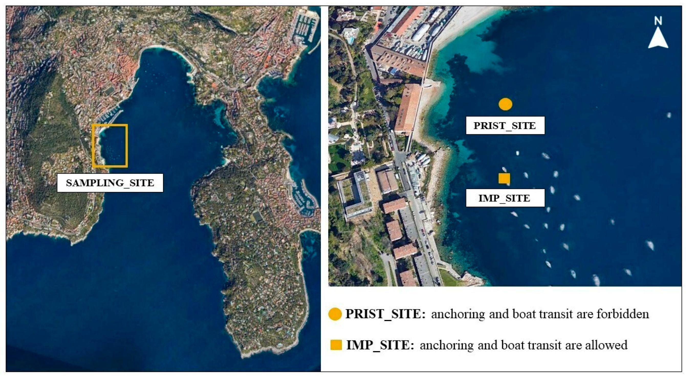
2.1. Study Area and Sampling Activities

Plant and abiotic matrix samples were collected by scuba diving in September 2021 at two sites in the northern Mediterranean Sea, along the coastline of Villefranche-Sur-Mer Bay (Nice, France; Figure 1). Villefranche-sur-Mer is a coastal area characterized by significant maritime activities. The port accommodates approximately 500 berths, including both fixed and floating moorings (Navily, https://www.navily.com/it/port/villefranche-darse/101, accessed on 20 September 2024). The bay provides around 50 to 100 anchoring points, with total capacity varying according to regulatory measures and seasonal conditions (Villefranche-sur-Mer Port Authority, 2024). During summer, Villefranche-sur-Mer experiences heightened maritime activity, regulated to ensure the sustainability of its natural resources and safety of navigation. According to the latest guidelines, anchoring is permitted in designated areas of the bay, focusing on minimizing environmental impact, particularly on seagrass beds of P. oceanica (Navily, https://www.navily.com/it/mouillage/rade-de-villefranche/7032, accessed on 20 September 2024), which characterize its seabed (European Marine Observation and Data Network, EMODnet, https://emodnet.ec.europa.eu/en/seabed-habitats, accessed on 20 September 2024).
Based on this information, the chosen study area comprised two sites of a monospecific P. oceanica meadow exposed to different levels of anthropic pressure and located a few hundred meters apart from each other. The first site, referred to as #impacted, is a boat anchoring and transit area. The second site, referred to as #pristine, is in front of a beach dedicated to seaside tourism, where anchoring and boat transit are forbidden and the main seawater activities involve nearshore swimming. At both sites, the meadow occurred at a depth of 13 m on a muddy sand substrate. The physicochemical parameters of the seawater column at the sampling sites, including temperature, salinity, and pH are reported in Supplementary Table S1A. The nutrient concentrations of NO2, NO3, Si(OH)4, and PO4 were measured in the seawater column and in the interstitial pore water (filtered by a 0.2 μm polycarbonate mesh and analyzed as both sample charged with particles and filtered water) at both the pristine and impacted sites, by the Institut de la Mer de Villefranche (IMEV), following the protocols by [35,36,37]. Results are reported in Supplementary Table S1B.
Seawater, sediment, and plants were haphazardly sampled within each site, at a minimum of 1 m distance from each other, avoiding sampling at the meadow edge. For the microbial analyses, replicates of abiotic matrices from P. oceanica (seawater or sediment) were collected from each site, ensuring the representation of bacterial and fungal communities’ heterogeneity at the sites: three replicates of seawater (1 L) were sampled above the vegetation canopy at each site using a bottle sampler. Simultaneously, three replicates of bulk sediment samples (each measuring 2.5 cm in diameter and 5 cm in depth) were collected using a mini corer. Also, plant parts (the leaves or rhizomes and roots) were collected from each site for microbial analyses: plants were selected underwater, separated into three replicates of the second leaves (in the order they emerged within the shoot) and into three replicates of pooled rhizome and root systems. The plant specimens were individually preserved in sterile ziplock bags to maintain the integrity of microbial communities and transported to the Institut de la Mer de Villefranche laboratory in a cooler at 4 °C.
At the laboratory, each leaf was carefully washed using sterile seawater to remove the presence of non-attached surface microorganisms. Subsequently, each sample was carefully and repeatedly scraped on both sides using a sterile blade while being sprinkled with a pipette containing 2 mL of washing solution (comprising 200 mM Tris–HCl pH 8, 10 mM EDTA, and 0.24% Triton X-100; [38]). Each rhizome/root sample was carefully washed using sterile seawater to remove the presence of non-attached surface microorganisms and, then, the microbes were collected by pipetting (2 mL washing solution) onto the surfaces. This sampling procedure included the collection of both epiphytes and endophytes; therefore, in this entire paper, the indication “in leaves” or “in rhizome/roots” refers to both classes of associated microbes. The microbial suspensions were then centrifuged, and the microbial pellet resuspended in 2 mL of transport solution to preserve the DNA, according to Mejia et al. [39]. The sediment samples (2 g) were mixed and placed immediately in 2 mL transport solution, while the seawater samples were filtered using 0.2 μm filters (Steroglass, Perugia, Italy), each of them stored separately in transport solution (5 mL) until DNA extraction performed using a portion of the filter.

2.2. DNA Metabarcoding

Whole microbial DNA was extracted using the Qiagen DNeasy PowerSoil Pro Kit (QIAGEN, Hilden, Germany), following the manufacturer’s instructions. DNA preparations were submitted for amplicon sequencing of the 16S rDNA and ITS2 rDNA gene region at BMR Genomics (Padova, Italy), where sequencing libraries were prepared and sequenced on the Illumina MiSeq platform (Illumina, Inc., San Diego, CA, USA) generating 300 bp paired-end reads. The bacterial 16S rDNA gene was amplified by PCR with the universal primers targeting the hypervariable 16S gene regions V3-V4 —Pro341F (5′-CCTACGGGNBGCASCAG-3′) and Pro805R (5′-GACTACNVGGGTATCTAATCC-3′; [40])— whereas universal ITS3 (forward, 5′-GCATCGATGAAGAACGCAGC-3′) and ITS4 (reverse, 5′-TCCTCCGCTTATTGATATGC-3′) primers [41], producing amplicons which allow higher-level classifications with incomplete databases, were used to amplify the ITS2 and 5.8S gene region [42].

2.3. Bioinformatics and Statistical Analyses

Raw reads were processed using the Quantitative Insights into Microbial Ecology 2 platform (QIIME 2, v2022.8; [43]). Read trimming was performed using Cutadapt v. 4.2 [44]. Sequences were then filtered, denoised, and checked for quality and chimeras using the DADA2 plugin [45] to generate a table of Amplicon Sequence Variants (ASVs). For DADA2 processing in QIIME 2, quality filtering thresholds were set to 15 for both forward and reverse reads in bacterial sequences, and to 20 for fungal sequences. The resulting 16S rDNA ASVs were taxonomically classified using a Naïve Bayes classifier trained with the SILVA 138 SSU database [46], while fungal taxonomic identification was performed using a combination of Naïve Bayes and VSEARCH classifiers trained on the UNITE database v. 8.3 [47]. Any taxa classified as chloroplast, mitochondria, archaea, or unclassified were filtered out from the feature tables.
The total number of bacterial and fungal raw sequences obtained from sampling was 1,515,660 and 2,507,913, respectively. These were subjected to subsequent bioinformatic analyses, removing low-quality or unmatched sequences to enhance the accuracy and reliability of the final dataset. The details of the filtering procedures are reported in Supplementary Table S2. The final count for bacteria resulted in 534,196 reads, 276,816 from the pristine site and 257,380 from the impacted site; the final count for fungi resulted in 143,613 reads, 65,021 from the pristine site and 78,592 from the impacted site. The heavy reduction in the fungal sequence count has already been observed in terrestrial mycobiome dataset analysis [48] and imputed to artifacts such as index switches and contamination. Rarefaction curves, obtained to assess differences and efficiency in the sampling effort, confirmed that the sequencing depth was good for all bacterial and fungal samples. Clustering analysis of the resulting ASVs revealed the presence of 516 bacterial and 242 fungal Operational Taxonomic Units (OTUs; Supplementary Table S3), obtained using the q2-vsearch algorithm with an identity threshold of 97%. OTU clustering was performed to have a clear picture of the fungal diversity. The bacterial and fungal sequences generated for 16S and ITS2/5.8S rDNA amplicons were deposited in GenBank under accession nos. PRJNA1122770 and PRJNA1076714, respectively.
The diversity indices were computed on bacterial and fungal normalized datasets. We used the q2-srs plugin in QIIME 2 for dataset normalization at 20,000 and 4000 reads for the bacteria and fungi, respectively. Normalization thresholds of 20,000 for 16S and 4000 for ITS were selected to ensure comprehensive representation of microbial diversity, as evidenced by the SRS rarefaction plots (Supplementary Figure S1A,B), thereby including all samples with read counts just above these thresholds. The diversity of the microbial community in P. oceanica was assessed by both α- and β-diversity indices using the QIIME 2 q2-diversity plugin. We used Shannon’s diversity index (H’) to assess within-sample diversity (i.e., α-diversity) in each microbial community and both the Bray–Curtis and weighted-UniFrac distance matrices to determine microbial community dissimilarity (i.e., β-diversity). The weighted UniFrac analysis measures the difference between collections of sequences as the amount of evolutionary history unique to each sample [49,50], evaluated on the OTUs phylogenetic relationships and abundance. Kruskal–Wallis analysis was used to investigate potential significant differences in α-diversity between P. oceanica sample types (seawater or sediment and leaves or rhizomes/roots in impacted or pristine sites), whereas the PERMANOVA test (9999 permutations) was used to determine differences in sample composition and structure. Bar plots were constructed to visually represent the relative abundance of the bacterial and fungal communities’ composition at the lowest observed taxonomic rank.
Sequences classified with QIIME 2 were submitted to the Functional Annotation of Prokaryotic Taxa (FAPROTAX v1.2.7) and FUNGuild v1.0 databases for assigning the putative ecological functional annotations to OTUs [51,52]. FAPROTAX software was used to investigate the potential functions in the pristine and impacted site bacteria, while FUNGuild software was used to focus on the putative trophic or metabolic functions associated with the fungi present in each sample. It must be underlined that this analysis still lacks information due to the scarcity of information on the taxonomy and metabolic activity of several marine fungi.
GraphPad Prism 9 was used to create heatmaps to visualize the relative abundance of putative functional guilds in the different samples. FAPROTAX and FUNGuild analyses allowed for the investigation of possible associations between microbial functional activities and abiotic matrices/plant parts in each sample type. Significant differences in samples’ functional guilds were determined by a two-way PERMANOVA test.

3. Results

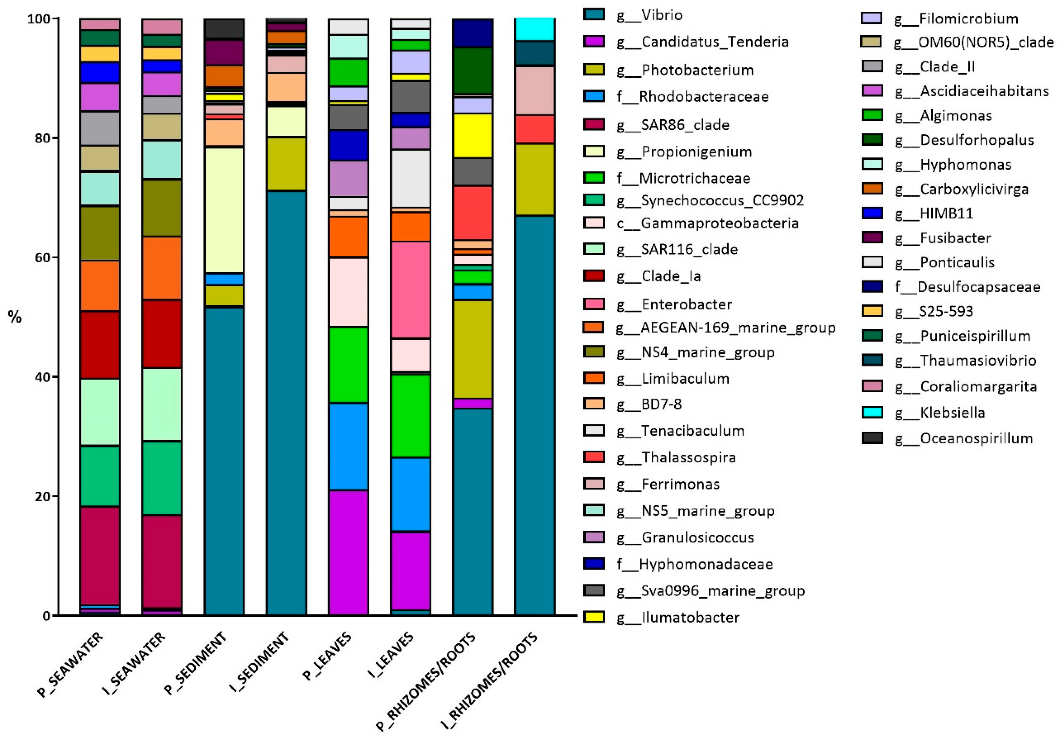
3.1. Bacterial Diversity

The α- and β-diversity of different plant parts and abiotic matrices in the two sites allowed for the evaluation of the effects of mechanical damage on P. oceanica microbial assembly. The α-diversity values, measured by Shannon’s index, showed nearly comparable values for the bacterial community across samples and sites, except for rhizome and root samples at the impacted site, showing a very low value. In these last samples, Shannon’s index decreased in the leaves and rhizome/roots samples, even if a significant difference was only observed for the latter samples (2.86 for the pristine site vs. 1.43 for the impacted one (Table 1).
Considering the β-diversity, seawater, sediment, and P. oceanica plant parts revealed unique bacterial community structures and compositions that differed among the sample types (PERMANOVA test: Bray–Curtis, F = 4.95; p = 0.001; weighted-UniFrac, F = 8.41, p = 0.001). Indeed, PERMANOVA pairwise tests revealed significant differences in seawater vs. sediment samples, (Bray–Curtis, F = 8.231, p = 0.002; weighted-UniFrac, F = 22.92, p = 0.003); while differences between the leaves and rhizome/root samples were significant only with the Bray–Curtis dissimilarity matrix (PERMANOVA test: Bray–Curtis, F = 2.483, p = 0.007; weighted-UniFrac, F = 3.433, p > 0.05). Interestingly, no significant differences were found in the bacterial composition comparing the pristine and impacted site (PERMANOVA test: Bray–Curtis, F = 0.540; p > 0.05; weighted-UniFrac, F = 0.884, p > 0.05).

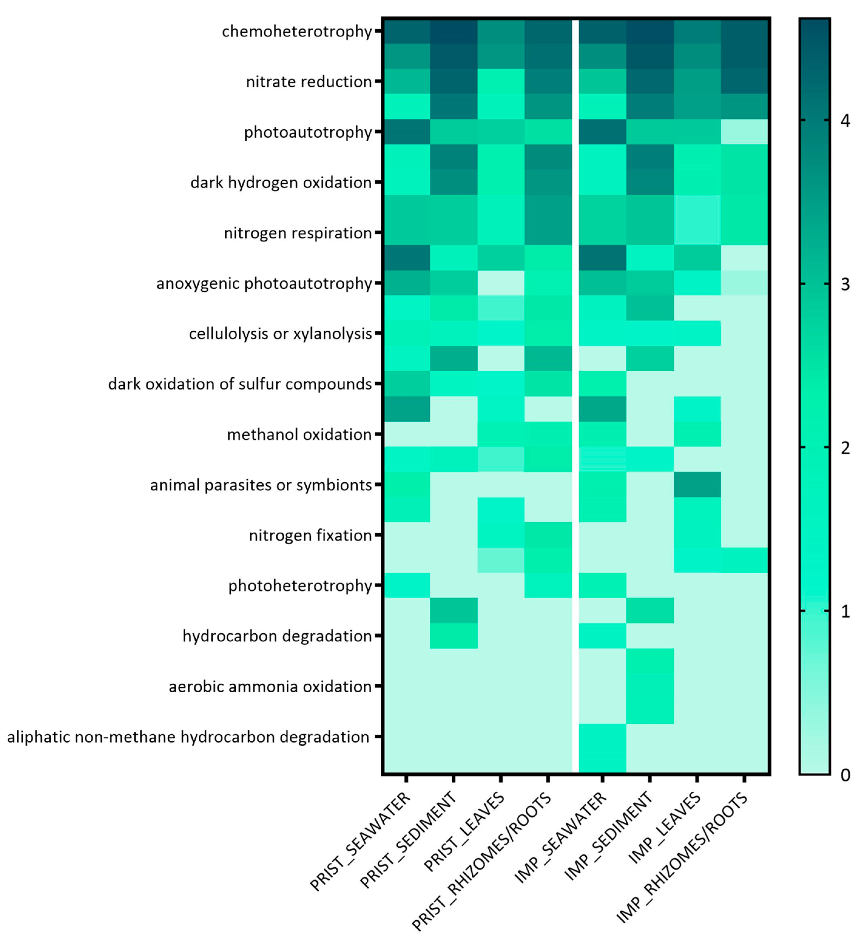
Bacterial Colonizers and FAPROTAX Analysis

The taxonomic identification showed that 90% of the total sequences belonged to five phyla: Proteobacteria (65% of all sequences), Bacteroidota (12% of all sequences), Desulfobacterota, Cyanobacteria (each 5% of all sequences), and Actinobacterota (4% of all sequences). The Proteobacteria phylum was found in all samples at both the pristine and impacted sites but was particularly abundant in the rhizome/root samples from the impacted sites (90% relative abundance); conversely, Bacteroidota were almost absent in rhizome/root samples. Desulfobacterota was mostly abundant in sediment and rhizome/root samples, while Cyanobacteria and Actinobacterota phyla were abundant in seawater and leaf samples, respectively. The remaining ≈10% was composed of a variety of 36 less abundant phyla.
In sediment and rhizome/root samples, the most abundant genera were Vibrio, Photobacterium, and Propionigenium, collectively comprising more than 50% of the relative abundance. The Vibrio genus exhibited the highest relative abundance, steadily increasing from the pristine site to the impacted site: from 52% to 72% in sediment samples and from 36% to 67% in rhizome/root samples. In contrast, the genera Photobacterium and Propionigenium decreased from 17% to 12% in impacted rhizome/root samples and from 21% to 5% in impacted sediment samples, respectively (Figure 2).
Conversely, in seawater samples, bacteria showed similar relative abundance at both the pristine and impacted sites, while in the leaf samples, the dominant bacterial colonizers always showed the highest relative abundance at the pristine sites. In seawater samples, the core bacterial taxa included the genera SAR86 and SAR116 clade (approximately 16% and 12% of relative abundance at both sites, respectively), Synechococcus and Clade Ia (approximately 12% and 11% of relative abundance at both sites, respectively), and the AEGEAN-169 marine group (around 10% of relative abundance at both sites). In contrast, in the leaf samples, the core bacterial taxa belonged to the genus Candidatus Tenderia (20% and 13% of relative abundance at the pristine and impacted sites, respectively) and the families Rhodobacteraceae and Microtrichaceae. The Rhodobacteraceae family was found at 14% and 12% of relative abundance at the pristine and impacted sites, respectively, whereas the Microtrichaceae family was found at 16% and 14% of relative abundance in leaves at the pristine and impacted sites, respectively. A total of 11% and 6% of relative abundance at the pristine and impacted sites, respectively, was covered by an unclassified taxa belonging to the Gammaproteobacteria class. Furthermore, the genus Enterobacter was exclusively found at the impacted site, encompassing 16% of the relative abundance (Figure 2).
Functional annotation of prokaryotic taxa (FAPROTAX) analysis was used to investigate their potential functions at the pristine and the impacted sites. As shown in Figure 3, thirty functional groups are represented. The most abundant functional groups were chemoheterotrophy (109 taxa), including aerobic chemoheterotrophy (76 taxa), and nitrate reduction (7 taxa), found at similar percentages at both the pristine and the impacted sites: chemoheterotrophy was found at 16% and 18% relative abundance, aerobic chemoheterotrophy at 10% and 12%, and nitrate reduction at 6% and 8%, respectively.
As shown in Figure 3, there were no significant differences in functional groups between the pristine and impacted sites, as confirmed by statistical test (two-way PERMANOVA test, F = 1.79, p > 0.05), with the exception of the rhizome/root samples, which displayed a reduced number of functional groups at the impacted site compared to the pristine one (Kruskal–Wallis test, H = 4.29, p = 0.03): the functional groups lacking in the impacted site comprised photosynthetic or photoheterotrophic processes, lysis of cellulose, xylan, chitin, or aromatic compound degradation.

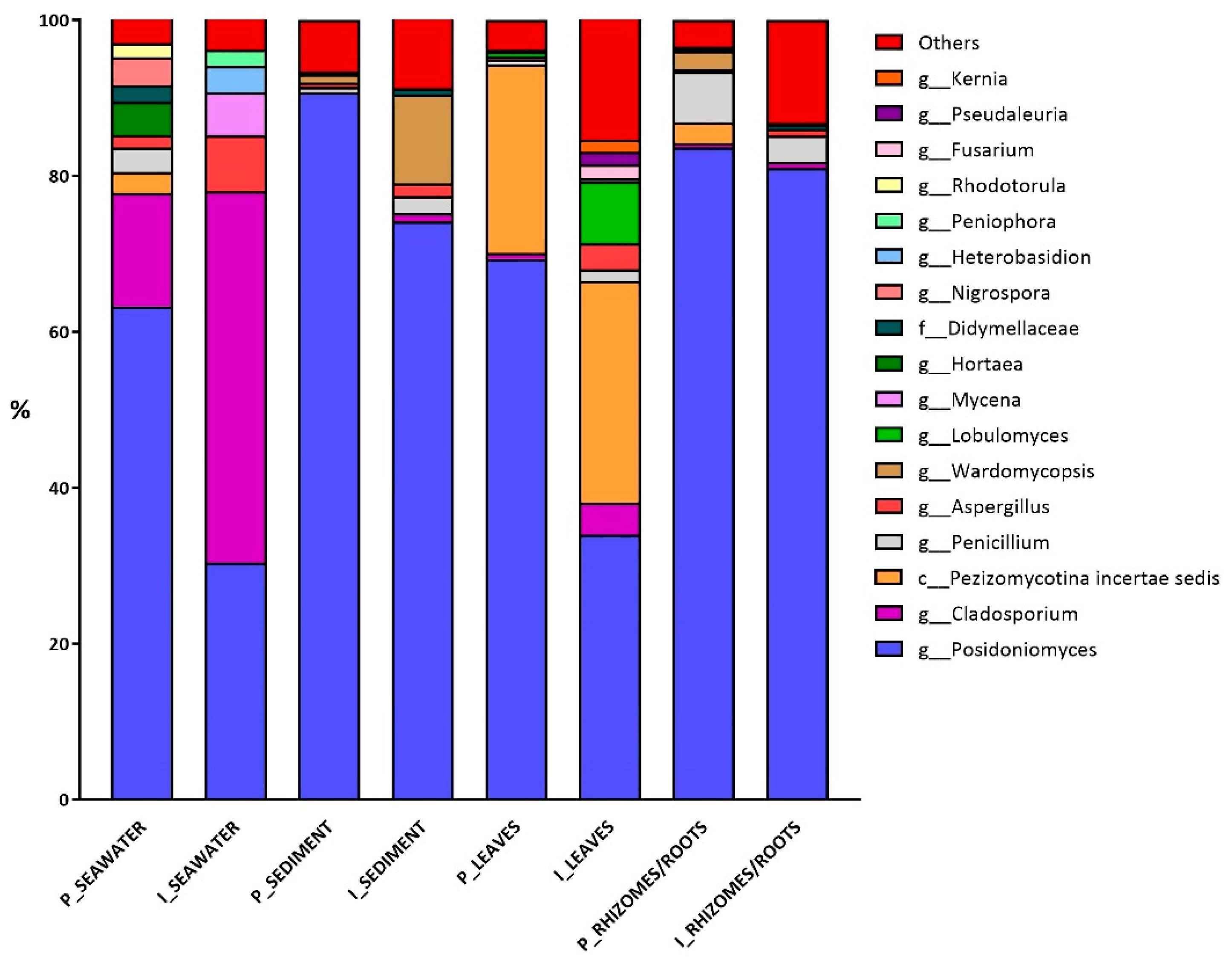
3.2. Fungal Diversity

The P. oceanica mycobiota was analyzed for α- and β-diversity within plant parts and abiotic matrices from the two sites. The α-diversity values, measured by Shannon’s Index, showed variable heterogeneity of fungal communities, ranging from the lowest value observed in the pristine sediment sample to the highest obtained for leaves from the impacted site (Table 2). This trend of increased α-diversity was always maintained in all samples from the impacted site when compared to the pristine one, with the exclusion of seawater samples. The higher diversity at the impacted site is confirmed by taxa richness, which is higher in the plant parts from the impacted site. However, pairwise comparisons using the Kruskal–Wallis test only revealed significant differences in Shannon’s index between the pristine and impacted sites’ leaf samples (Table 2).
As expected, the β-diversity of the fungal communities largely differed between seawater and sediment (PERMANOVA test; F = 2.650 p = 0.001) and between leaves and rhizomes/roots (PERMANOVA test; F = 1.992, p = 0.008) samples based on the Bray–Curtis distance metric, independently from the sampled site. Weighted-UniFrac confirmed these differences (seawater vs. sediment, F = 9.467, p = 0.001; leaves vs. rhizomes/roots, F = 3.637, p = 0.004). Indeed, the comparison of specific pairs, i.e., seawater, sediment, leaves, and rhizome/roots samples from the pristine and impacted sites did not show significant differences in sample composition (PERMANOVA test, F = 1.282, p > 0.05; weighted UniFrac, F = 0.665, p > 0.05).

Fungal Colonizers and FUNGuild Analysis

Taxonomical identification at the phylum level revealed that a highly diversified fungal community, mainly belonging to Ascomycota (95% of all sequences), was present in both abiotic matrices and plant parts. The Basidiomycota phylum (3.5% of all sequences) comprised only a few genera, including Mycena, Heterobasidion, and Peniophora (5%, 4%, and 3% relative abundance, respectively). These last genera were exclusively found in the seawater samples of the impacted site. Conversely, the genus Rhodotorula (2% of relative abundance) was exclusively present in seawater samples from the pristine site and the phylum Chytridiomycota (particularly the genus Lobulomyces) was exclusively found, at a low percentage (1.5% of all sequences), in leaves from the impacted site.
Considering all samples, the most prevalent OTU—identified as Posidoniomyces atricolor (100% confidence)—was represented by 103,708 sequences (72% of total sequences), and was represented by a minimum of 678 sequences in the seawater samples from the impacted site, and a maximum of 29,908 sequences in the rhizome/root sample from the pristine site. Its relative abundance in leaves and seawater from the pristine meadow was 69 and 63%, respectively; these values halved in the corresponding samples from the impacted meadow, falling to ≃30%. At the impacted site, P. atricolor was no longer the dominant species—neither in leaves, where it was superseded by a fungal taxon belonging to Pezizomycotina incertae sedis class (28%), nor in seawater, where the genus Cladosporium (48%) was prevalent. Therefore, Pezizomycotina incertae sedis class and Cladosporium were found to be more abundant at the impacted site than at the pristine site. The relative abundance of Pezizomycotina incertae sedis class slightly increased in the leaf samples from the pristine to the impacted site (from 24 to 28%), while Cladosporium showed a marked increase from the pristine to the impacted site in seawater or leaf samples (from 15 to 48% and from 1 to 4%, respectively). However, both taxa were also detected in all the other samples (except the sediment samples for Pezizomycotina), even if at low percentages. Conversely, P. atricolor remained the dominant species (≃80%) in the rhizome/root and sediment samples from both the pristine and impacted sites. A comparison of the sediment of the pristine vs. impacted site showed that the relative abundance of P. atricolor decreased (from 90 to 74%) while the abundance of the genus Wardomycopsis increased (from 1 to 11%). Additionally, the genus Wardomycopsis was found only in rhizome/root samples from the impacted site, accounting for <3% of the relative abundance.
The fungal community was composed of few frequent OTUs and a large number of rare OTUs. The rare components, represented by taxa with a relative abundance of less than 2% in each sample (reported as ”Others” in Figure 4), were consistently higher at the impacted than at the pristine site; this difference was particularly apparent in leaves and rhizome/root samples (Supplementary Table S4).
To evaluate the fungal community from an ecological perspective, FUNGuild software was employed to identify putative trophic or metabolic functions associated with the taxa present in each sample. Even if the dataset subjected to the FUNGuild analysis included all the identified OTUs (242; see Supplementary Table S3), only 175 successfully matched taxa present in the FUNGuild database (Supplementary Table S5). The 67 OTUs lacking the putative functions analysis included 24 genera, and the majority of them were found at high relative abundance (>7% in at least one sample). Among the fungi lacking the putative functions, there were the recently identified P. atricolor and taxa belonging to Cladosporium, Wardomycopsis, Lobulomyces, and Aspergillus genera as well as the Pezizomycotina incertae sedis class. For this reason, the analysis of the putative trophic or metabolic functions covered only the rare components of the assembly (with a relative abundance of <7% each). The most abundant trophic mode reported for all these taxa was saprotrophy, accounting for 53% of the entire dataset (Figure 5). The figure clearly highlights that more putative trophic or metabolic functions were found at the impacted site than at the pristine site. Two-way PERMANOVA confirmed this observation, as significant differences were found in FUNGuild trophic mode categories both among sample types (seawater, sediment, leaves, and rhizomes/roots) and between sites (F = 1.706, p = 0.03 and F = 2.043, p = 0.04, respectively). In fact, the relative abundance of each trophic mode (Saprotroph, Pathotroph, Symbiotroph, and mixed categories) increased at the impacted site, with the Saprotroph mode showing a 12% increase.

4. Discussion

Anthropogenic activities such as boat transit and anchoring have a notable impact on the dynamics of Posidonia oceanica meadows [8,9]. In Villefranche-Sur-Mer Bay, the maritime activities are strictly regulated to ensure the sustainability of natural resources, particularly to minimize the impact on the seagrass P. oceanica, which characterizes its seabed. Nevertheless, we found that boat transit and anchoring are able to modify the structure and composition of the microbial communities (particularly the fungal ones) associated with P. oceanica, a putative interesting tool for both seagrass monitoring and the regulation of maritime activities.

4.1. Bacterial Community

We identified the changes in the bacterial community due to boat transit and anchoring in P. oceanica meadows by evaluating changes in α- and β-diversity. The α-diversity (Shannon’s index) only significantly decreased in the rhizome/root samples at the impacted site. This decrease in α-diversity may depend on the accumulation of dead/ripped plant debris produced by boat mechanical damage, affecting the belowground section of P. oceanica meadows. Indeed, debris accumulation may lead to a bacterial outburst of a restricted number of bacterial species, including some opportunistic bacteria, which decreases the overall diversity of the site. This is the case in the increase in relative abundance of the genus Vibrio at the impacted site, which increased by 29% in the sediment samples and 31% in the rhizome/root samples; some of these Vibrio spp. are pathogens of marine organisms. The Vibrio genus comprises opportunistic species, extensively found in marine environments and exhibiting significant metabolic adaptability [53], which allows for efficient plant debris degradation. While certain Vibrio species are beneficial to seagrass ecosystems [54,55,56], others can be pathogenic, adversely affecting the health and stability of seagrass beds [57,58]. Earlier research has indicated that Vibrio spp. are uncommon in healthy seagrass; however, our study demonstrated a higher abundance of members of this genus at the impacted site, confirming that the area may be under pressure [59,60]. Another indication of human pressure at the impacted area was the occurrence of the genus Enterobacter, exclusively found in the leaf samples. The Enterobacteriaceae family is considered an indicator of fecal contamination in marine systems and it has already been found in human-impacted areas [26]. Other known human opportunistic pathogens, such as members of the Klebsiella genus [61], were found at low relative abundance in the rhizome/root samples only from the impacted site, offering further evidence of the human impact on the meadow.
In contrast, the β-diversity analyses did not highlight significant differences in the bacterial community structure and composition, which remained stable despite the mechanical impacts from boat transit and anchoring. This stability can be either related to the limited anchoring area/density of boats or to the ability of P. oceanica to preserve the useful bacterial community under undisturbed or limited-disturbed environmental conditions [19,62]. The limited disturbance is confirmed by the maintenance of the proper bacterial communities in the different matrices: significant variations in beta diversity were observed between different seawater and sediment samples or leaves and rhizome/root samples, indicating that each plant part or abiotic compartment preserved its unique microbial community [18,63].
Most of the other bacterial taxa were evenly distributed between the two sampling sites, with variations observed only within sample type. At the phylum level, while the Proteobacteria was dominant across all samples, other phyla showed differences: Cyanobacteria thrived in seawater and on leaf samples, probably favored by light and hydrodynamic conditions [40]; Desulfobacteria was abundant exclusively in the sediment and the rhizome/root samples, where they play a crucial role in sulfate reduction driven by root exudates, especially in seagrass meadows belowground [64]. Furthermore, taxa belonging to Bacteroidota were more abundant in the seawater and leaf samples than in the rhizomes/root and sediment samples, although they are generally found in seagrass meadows belowground as they are well-known decomposers of cellulose and chitin [60].
The bacterial putative functions were analyzed in the P. oceanica-associated community from both the pristine and impacted sites. The key functional groups were chemoheterotroph, nitrate reducer, fermentative, and photoautotroph. According to the decrease in the bacterial community diversity in the rhizome/root samples between the pristine and impacted sites, a reduction in functional diversity at the impacted area was found, homogenizing the bacterial community activities, as already found in both terrestrial and marine environments [65,66].

4.2. Fungal Community

The fungal communities associated with P. oceanica at both the pristine and the impacted sites were found to be quite simple, dominated by a single species, P. atricolor, as already found in a previous study [32]. Although Vohník et al. [32,33] reported that P. atricolor had been exclusively identified in the roots, in this study, we found the species not only in the roots but also in all the other samples (leaves, sediment, and even seawater), demonstrating the association of this fungus to different parts of P. oceanica. P. atricolor is supposed to be responsible for the putative Dark Septate Endophyte (DSE) colonization pattern [32], commonly found in terrestrial systems where the DSE endophyte fungi inhabit plant roots [67]. Their ecological function is still unclear, although evidence suggests a mutualistic relationship that enhances host growth, nutrient acquisition, and abiotic stress tolerance [68,69,70]. Besides P. atricolor, the P. oceanica mycobiome included other fungal taxa accounting for 21% of the entire community: Ascomycota (18%), Basidiomycota (2%), and Chytridiomycota (1%). It is worth noting that the number/diversity of fungal species changes between the pristine and impacted sites, being highest at the impacted one. The increase in α-diversity at the impacted site (significant only in the leaf samples) can be related to the transit of boats, increasing water turbidity, and to a greater extent, to the anchoring and mooring, which results in breaking and/or uprooting the shoots. This increase in plant debris alters the meadow nutrient dynamics and opens up new colonizable niches for the growth of several fungal species, found in low percentages. Hence, the substrate heterogeneity caused by P. oceanica meadow disturbance or decaying plant parts contributes to an increase in fungal diversity, as already found [71,72,73]. There is a downside to this, the increase in fungal diversity due to the stressful environmental conditions reduces the relative abundance of the host-specific P. atricolor in the impacted area. In fact, there was a significant decrease in this species in the seawater and leaves at the impacted site (33% and 35%, respectively; Student’s t-test, p < 0.05 for both comparisons), and a less evident decrease in the sediment and rhizome/root samples (17% and 2%, respectively, Student’s t-test, p > 0.05), where P. atricolor remained the dominant species. This decrease implies a reduction in the symbiotic functionalities of this fungus.
The obvious direct relationship between each plant part and its abiotic matrix explains the different and comparable fungal assemblages in seawater/leaves vs. sediment/rhizomes/roots and determines the overlap of fungal communities in each plant part and the abiotic matrix, as shown by the β-diversity analyses confirming that fungal assemblages have a plant-part-specificity and/or substrate-specificity, as described by Poli et al. [74]; this specific mycobiota may play a crucial role for holobionts by providing specific interactions and competitive advantages [74].
Besides P. atricolor, the other fungal taxa present as rare components of the communities may take advantage of the changed conditions of the impacted area: the genus Cladosporium, present in the seawater column, became dominant at the impacted site; the unidentified taxa belonging to the class Pezizomycotina incertae sedis, found quite abundant in leaves, slightly increased from the pristine to the impacted site, and the genus Wardomycopsis exhibited an increase in the sediment samples from the impacted site. Their increase at the impacted area can be related to their metabolic capability: Cladosporium and Wardomycopsis genera encompass widely distributed species with the capability to degrade and detoxify various organic materials [75]—including recalcitrant substances characterized by a high content of lignin or polyphenolic compounds—which are characteristic of P. oceanica meadows [17], especially under stressful conditions [76,77,78]. It could be hypothesized that in an impacted area, the debris of seagrass accumulates, enhancing the growth of the above-cited efficient degraders. In the same frame, the class Pezizomycotina incertae sedis includes core species of the microfungal community of the terrestrial litter [79], suggesting a role in the litter degradation of the aboveground plant system. The degradative capability of the overmentioned taxa depends on their ability to produce extracellular enzymes, such as laccase or peroxidase, which are active even in extreme conditions and are known to not be produced by terrestrial analogous taxa [17,75,80].
The analysis of the fungal putative functions in the rare component of the mycobiome confirmed that the changed conditions at the impacted area opened up colonizable niches for the growth of different fungal species: disturbance favors the settlement of new trophic categories, particularly in plants, at the expense of the natives, including the symbiont P. atricolor. Among the observed taxa, saprotrophic lifestyles were found, characterizing Aspergillus, Penicillium, Lobulomyces [81,82], and plant pathogens, such as the genus Hortaea [83]. However, the more important effect of boat-transit or anchoring activities on the aboveground plant system is the increase in saprotrophs, which could be involved in the decay of damaged P. oceanica plant parts that represent a nutrient-rich substratum [84]. Among the saprotrophs, the fungi of the genus Penicillium were distributed in all samples, while Aspergillus or Lobulomyces were increasingly abundant in the seawater and leaf samples from the impacted site. The increase in saprotrophs was significant in the leaves (Figure 5; Kruskall–Wallis test, H = 13.4, p = 0.01) and was probably related to the increased organic material (substrate resuspension due to boat transit; plant debris or uprooted shoots due to mooring or anchoring). An increase in saprotrophs was also found in the sediment and rhizomes/roots, although less pronounced; this was a slight but not significant increase in symbiotrophes and saprotrophes to cope with the increased amount of available organic matter as the accumulation of dead organic matter and plant debris is customary in the belowground system.
As a final consideration, the reduced abundance of P. atricolor in plants under mechanical stress offers valuable insights into the plant conservation status and may potentially serve as a tool for its monitoring, which has already been proposed for bacterial communities [19,40,85,86]. In fact, it is well-known that the bacterial communities associated with P. oceanica shift in response to environmental conditions, anthropic stress, or plant traits while supporting plants in adapting to environmental changes [18,19,87]. On this basis, the results of this study suggest also taking into account the structure and composition of the P. oceanica-associated mycobiome as well as the bacterial communities, particularly P. atricolor, as a putative indicator of abiotic or biotic pressures on the seagrass. Nonetheless, future work is still necessary to achieve this goal, as marine fungi have received less attention than terrestrial species [88,89,90] and require more thorough taxonomic and functional investigations.

Supplementary Materials

The following supporting information can be downloaded at: https://www.mdpi.com/article/10.3390/d16100604/s1. Table S1: seawater physicochemical parameters. Table S2: Raw reads, DADA2 output, and final reads obtained after filtering procedures for bacterial and fungal sequences. Figure S1: SRS rarefaction plots for bacterial and fungal sequences. Table S3: OTUs taxonomic identification. Table S4: Rare fungal taxa grouped under the category ‘Others’. Table S5: The 175 OTUs classified based on their putative function through FUNGuild.

Author Contributions

Conceptualization, S.F., A.A., M.M.D. and L.M.; methodology, S.F., A.A. and R.C.; formal analysis, S.F. and A.A.; resources, L.M. and R.C.; writing—original draft preparation, S.F. and L.M.; writing—review and editing, S.F., A.A., M.M.D. and L.M.; funding acquisition, L.M. All authors have read and agreed to the published version of the manuscript.

Funding

This research was funded by Assemble plus TA Fifth call “GASPER-417”, won by L.M. at the Institut de la Mer de Villefranche-sur-Mer IMEV (ex-OOV). S.F. has a three-year PON Ph.D. scholarship funded from the Ph.D. Program in Evolutionary Biology and Ecology, University of Rome Tor Vergata.

Institutional Review Board Statement

Not applicable.

Data Availability Statement

The raw data generated for the 16S and ITS2 amplicon projects have been deposited at the NCBI Sequence Read Archive (SRA) under BioProject IDs PRJNA1122770 and PRJNA1076714, respectively.

Acknowledgments

We acknowledge the CINECA award under the ISCRA initiative for the availability of high-performance computing resources and support. We acknowledge the GT50 s.r.l company for its technical support. This work benefited from access to the Molecular Biology Platform and the Diving Service at the IMEV, an EMBRC-France and EMBRC-ERIC Site.

Conflicts of Interest

The authors declare no conflicts of interest.

References

  1. Arnaud-Haond, S.; Duarte, C.M.; Diaz-Almela, E.; Marba, N.; Sintes, T.; Serrao, E.A. Implications of extreme life span in clonal organisms: Millenary clones in meadows of the threatened seagrass Posidonia oceanica. PLoS ONE 2012, 7, e30454. [Google Scholar] [CrossRef] [PubMed]
  2. Boudouresque, C.F. Marine biodiversity in the Mediterranean: Status of species, populations and communities. Trav. Sci. PNPC 2004, 20, 97–146. [Google Scholar]
  3. Pergent-Martini, C.; Pergent, G.; Monnier, B.; Boudouresque, C.F.; Mori, C.; Valette-Sansevin, A. Contribution of Posidonia oceanica meadows in the context of climate change mitigation in the Mediterranean Sea. Mar. Environ. Res. 2021, 165, 105236. [Google Scholar] [CrossRef] [PubMed]
  4. Apostolaki, E.T.; Caviglia, L.; Santinelli, V.; Cundy, A.B.; Tramati, C.D.; Mazzola, A.; Vizzini, S. The importance of dead seagrass (Posidonia oceanica) matte as a biogeochemical sink. Front. Mar. Sci. 2022, 9, 861998. [Google Scholar] [CrossRef]
  5. Boudouresque, C.F.; Pergent, G.; Pergent-Martini, C.; Ruitton, S.; Thibaut, T.; Verlaque, M. The necromass of the Posidonia oceanica seagrass meadow: Fate, role, ecosystem services and vulnerability. Hydrobiologia 2016, 781, 25–42. [Google Scholar] [CrossRef]
  6. Monnier, B.; Pergent, G.; Valette-Sansevin, A.; Boudouresque, C.F.; Mateo, M.A.; Pergent-Martini, C. The Posidonia oceanica matte: A unique coastal carbon sink for climate change mitigation and implications for management. Vie Milieu/Life Environ. 2020, 70, 17–24. [Google Scholar]
  7. Council Directive 92/43/EEC on the Conservation of Natural Habitats and of Wild Fauna and Flora; Publications Office of the European Union: Luxemburg, 1992; Volume 206, pp. 7–50.
  8. Boudouresque, C.F.; Bernard, G.; Pergent, G.; Shili, A.; Verlaque, M. Regression of Mediterranean seagrasses caused by natural processes and anthropogenic disturbances and stress: A critical review. Bot. Mar. 2009, 52, 395–418. [Google Scholar] [CrossRef]
  9. Montefalcone, M.; Chiantore, M.; Lanzone, A.; Morri, C.; Albertelli, G.; Bianchi, C.N. BACI design reveals the decline of the seagrass Posidonia oceanica induced by anchoring. Mar. Pollut. Bull. 2008, 56, 1637–1645. [Google Scholar] [CrossRef]
  10. Francour, P.; Ganteaume, A.; Poulain, M. Effects of boat anchoring in Posidonia oceanica seagrass beds in the Port-Cros National Park (north-western Mediterranean Sea). Aquat. Conserv. Mar. Freshw. 1999, 9, 391–400. [Google Scholar] [CrossRef]
  11. Ardizzone, G.; Belluscio, A.; Maiorano, L. Long-term change in the structure of a Posidonia oceanica landscape and its reference for a monitoring plan. Mar. Ecol. 2006, 27, 299–309. [Google Scholar] [CrossRef]
  12. Ceccherelli, G.; Campo, D.; Milazzo, M. Short-term response of the slow growing seagrass Posidonia oceanica to simulated anchor impact. Mar. Environ. Res. 2007, 63, 341–349. [Google Scholar] [CrossRef] [PubMed]
  13. Bourque, A.S.; Kenworthy, W.J.; Fourqurean, J.W. Impacts of physical disturbance on ecosystem structure in subtropical seagrass meadows. Mar. Ecol. Prog. Ser. 2015, 540, 27–41. [Google Scholar] [CrossRef]
  14. Rosenberg, E.; Koren, O.; Reshef, L.; Efrony, R.; Zilber-Rosenberg, I. The hologenome theory disregards the coral holobiont: Reply from Rosenberg et al. Nat. Rev. Microbiol. 2007, 5, 826. [Google Scholar] [CrossRef]
  15. Gilbert, S.F.; Rosenberg, E.; Zilber-Rosenberg, I. The holobiont with its hologenome is a level of selection in evolution. In Landscapes of Collectivity in the Life Sciences; MIT Press: Cambridge, MA, USA, 2017; pp. 305–324. [Google Scholar]
  16. Ganley, R.J.; Brunsfeld, S.J.; Newcombe, G. A community of unknown, endophytic fungi in western white pine. Proc. Natl. Acad. Sci. USA 2004, 101, 10107–10112. [Google Scholar] [CrossRef]
  17. Panno, L.; Bruno, M.; Voyron, S.; Anastasi, A.; Gnavi, G.; Miserere, L.; Varese, G.C. Diversity, ecological role and potential biotechnological applications of marine fungi associated to the seagrass Posidonia oceanica. New Biotech. 2013, 30, 685–694. [Google Scholar] [CrossRef] [PubMed]
  18. Conte, C.; Rotini, A.; Manfra, L.; D’Andrea, M.M.; Winters, G.; Migliore, L. The seagrass holobiont: What we know and what we still need to disclose for its possible use as an ecological indicator. Water 2021, 13, 406. [Google Scholar] [CrossRef]
  19. Rotini, A.; Conte, C.; Winters, G.; Vasquez, M.I.; Migliore, L. Undisturbed Posidonia oceanica meadows maintain the epiphytic bacterial community in different environments. Environ. Sci. Pollut. Res. 2023, 30, 95464–95474. [Google Scholar] [CrossRef]
  20. Tarquinio, F.; Hyndes, G.A.; Laverock, B.; Koenders, A.; Säwström, C. The seagrass holobiont: Understanding seagrass-bacteria interactions and their role in seagrass ecosystem functioning. FEMS Microbiol. Lett. 2019, 366, fnz057. [Google Scholar] [CrossRef]
  21. Korlević, M.; Markovski, M.; Zhao, Z.; Herndl, G.J.; Najdek, M. Seasonal dynamics of epiphytic microbial communities on marine macrophyte surfaces. Front. Microbiol. 2021, 12, 671342. [Google Scholar] [CrossRef]
  22. Szitenberg, A.; Beca-Carretero, P.; Azcárate-García, T.; Yergaliyev, T.; Alexander-Shani, R.; Winters, G. Teasing apart the host-related, nutrient-related and temperature-related effects shaping the phenology and microbiome of the tropical seagrass Halophila stipulacea. Environ. Microbiol. 2022, 17, 1–17. [Google Scholar] [CrossRef]
  23. Agawin, N.S.; Ferriol, P.; Cryer, C.; Alcon, E.; Busquets, A.; Sintes, E.; Moyà, G. Significant nitrogen fixation activity associated with the phyllosphere of Mediterranean seagrass Posidonia oceanica: First report. Mar. Ecol. Prog. Ser. 2016, 551, 53–62. [Google Scholar] [CrossRef]
  24. Lucas-Elío, P.; Goodwin, L.; Woyke, T.; Pitluck, S.; Nolan, M.; Kyrpides, N.C.; Detter, J.C.; Copeland, A.; Lu, M.; Bruce, D.; et al. Complete genome sequence of Marinomonas posidonica type strain (IVIA-Po-181(T)). Stand. Genom. Sci. 2012, 7, 31–43. [Google Scholar] [CrossRef] [PubMed]
  25. Espinosa, E.; Marco-Noales, E.; Gómez, D.; Lucas-Elío, P.; Ordax, M.; Garcías-Bonet, N.; Duarte, C.M.; Sanchez-Amat, A. Taxonomic study of Marinomonas strains isolated from the seagrass Posidonia oceanica, with descriptions of Marinomonas balearica sp. nov. and Marinomonas pollencensis sp. nov. Int. J. Syst. Evol. Microbiol. 2010, 60, 93–98. [Google Scholar] [CrossRef] [PubMed]
  26. Rotini, A.; Conte, C.; Seveso, D.; Montano, S.; Galli, P.; Vai, M.; Migliore, L.; Mejia, A. Daily variation of the associated microbial community and the Hsp60 expression in the Maldivian seagrass Thalassia hemprichii. J. Sea Res. 2020, 156, 101835. [Google Scholar] [CrossRef]
  27. Sapp, J. The dynamics of symbiosis: An historical overview. Canad. J. Bot. 2004, 82, 1046–1056. [Google Scholar] [CrossRef]
  28. Brachmann, A.; Parniske, M. The most widespread symbiosis on earth. PLoS Biol. 2006, 4, e239. [Google Scholar] [CrossRef]
  29. Vohník, M.; Borovec, O.; Župan, I.; Vondrášek, D.; Petrtýl, M.; Sudová, R. Anatomically and morphologically unique dark septate endophytic association in the roots of the Mediterranean endemic seagrass Posidonia oceanica. Mycorrhiza 2015, 25, 663–672. [Google Scholar] [CrossRef]
  30. Ettinger, C.L.; Eisen, J.A. Characterization of the mycobiome of the seagrass, Zostera marina, reveals putative associations with marine chytrids. Front. Microbiol. 2019, 10, 491431. [Google Scholar] [CrossRef]
  31. Poli, A.; Varese, G.C.; Garzoli, L.; Prigione, V. Seagrasses, seaweeds and plant debris: An extraordinary reservoir of fungal diversity in the Mediterranean Sea. Fungal Ecol. 2022, 60, 101156. [Google Scholar] [CrossRef]
  32. Vohník, M.; Borovec, O.; Kolaříková, Z.; Sudová, R.; Réblová, M. Extensive sampling and high-throughput sequencing reveal Posidoniomyces atricolor gen. et sp. nov. (Aigialaceae, Pleosporales) as the dominant root mycobiont of the dominant Mediterranean seagrass Posidonia oceanica. MycoKeys 2019, 55, 59. [Google Scholar] [CrossRef]
  33. Vohník, M. Are lulworthioid fungi dark septate endophytes of the dominant Mediterranean seagrass Posidonia oceanica? Plant. Biol. 2022, 24, 127–133. [Google Scholar] [CrossRef] [PubMed]
  34. Lumibao, C.Y.; Harris, G.; Birnbaum, C. Global Diversity and Distribution of Rhizosphere and Root-Associated Fungi in Coastal Wetlands: A Systematic Review. Estuaries Coasts 2024, 47, 905–916. [Google Scholar] [CrossRef]
  35. Aminot, A.; Kérouel, R. Hydrologie des Écosystèmes Marins. Paramètres et Analyses; Ifremer: Plouzané, France, 2004; 336p. [Google Scholar]
  36. Kirkwood, D.S. Stability of solutions of nutrient salts during storage. Mar. Chem. 1992, 38, 151–164. [Google Scholar] [CrossRef]
  37. Treguer, P.; Le Corre, P. Manuel d’Analyse des Sels Nutritifs dans l’Eau de Mer (Utilisation de l’Autoanalyseur II Technicon R); Lab. Océano. Chim. Univ. Bretagne Occidentale: Brest, France, 1974; 110p. [Google Scholar]
  38. Kadivar, H.; Stapleton, A.E. Ultraviolet radiation alters maize phyllosphere bacterial diversity. Microb. Ecol. 2003, 45, 353–361. [Google Scholar] [CrossRef]
  39. Mejia, A.Y.; Rotini, A.; Lacasella, F.; Bookman, R.; Thaller, M.C.; Shem-Tov, R.; Winters, G.; Migliore, L. Assessing the ecological status of seagrasses using morphology, biochemical descriptors and microbial community analyses. A study in Halophila stipulacea (Forsk.) Aschers meadows in the northern Red Sea. Ecol. Indic. 2016, 60, 1150–1163. [Google Scholar] [CrossRef]
  40. Takahashi, S.; Tomita, J.; Nishioka, K.; Hisada, T.; Nishijima, M. Development of a prokaryotic universal primer for simultaneous analysis of Bacteria and Archaea using next-generation sequencing. PLoS ONE 2014, 9, e105592. [Google Scholar] [CrossRef]
  41. White, T.J.; Burns, T.; Lee, S.; Taylor, J. Amplification and Direct Sequencing of Fungal Ribosomal RNA Genes for Phylogenetics; Academic Press: New York, NY, USA, 1990. [Google Scholar]
  42. Heeger, F.; Wurzbacher, C.; Bourne, E.C.; Mazzoni, C.J.; Monaghan, M.T. Combining the 5.8 S and ITS2 to improve classification of fungi. Methods Ecol. Evol. 2019, 10, 1702–1711. [Google Scholar] [CrossRef]
  43. Bolyen, E.; Rideout, J.R.; Dillon, M.R.; Bokulich, N.A.; Abnet, C.C.; Al-Ghalith, G.A.; Caporaso, J.G. Reproducible, interactive, scalable and extensible microbiome data science using QIIME 2. Nat. Biotechnol. 2019, 37, 852–857. [Google Scholar] [CrossRef]
  44. Martin, M. Cutadapt removes adapter sequences from high-throughput sequencing reads. EMBnet. J. 2011, 17, 10–12. [Google Scholar] [CrossRef]
  45. Callahan, B.J.; McMurdie, P.J.; Rosen, M.J.; Han, A.W.; Johnson, A.J.A.; Holmes, S.P. DADA2: High-resolution sample inference from Illumina amplicon data. Nat. Methods 2016, 13, 581–583. [Google Scholar] [CrossRef]
  46. Quast, C.; Pruesse, E.; Yilmaz, P.; Gerken, J.; Schweer, T.; Yarza, P.; Peplies, J.; Glöckner, F.O. The SILVA ribosomal RNA gene database project: Improved data processing and web-based tools. Nucleic Acids Res. 2012, 41, D590–D596. [Google Scholar] [CrossRef] [PubMed]
  47. Kõljalg, U.; Nilsson, R.H.; Abarenkov, K.; Tedersoo, L.; Taylor, A.F.; Bahram, M.; Larsson, K.H. Towards a unified paradigm for sequence-based identification of fungi. Mol. Ecol. 2013, 22, 5271–5277. [Google Scholar] [CrossRef]
  48. Tedersoo, L.; Mikryukov, V.; Anslan, S.; Bahram, M.; Khalid, A.N.; Corrales, A.; Abarenkov, K. The Global Soil Mycobiome consortium dataset for boosting fungal diversity research. Fungal Divers. 2021, 111, 573–588. [Google Scholar] [CrossRef]
  49. Lozupone, C.; Knight, R. UniFrac: A new phylogenetic method for comparing microbial communities. Appl. Environm Microbiol. 2005, 71, 8228–8235. [Google Scholar] [CrossRef] [PubMed]
  50. Lozupone, C.; Lladser, M.E.; Knights, D.; Stombaugh, J.; Knight, R. UniFrac: An effective distance metric for microbial community comparison. ISME J. 2011, 5, 169–172. [Google Scholar] [CrossRef]
  51. Louca, S.; Parfrey, L.W.; Doebeli, M. Decoupling function and taxonomy in the global ocean microbiome. Science 2016, 353, 1272–1277. [Google Scholar] [CrossRef]
  52. Nguyen, N.H.; Song, Z.; Bates, S.T.; Branco, S.; Tedersoo, L.; Menke, J.; Schilling, J.S.; Kennedy, P.G. FUNGuild: An open annotation tool for parsing fungal community datasets by ecological guild. Fungal Ecol. 2016, 20, 241–248. [Google Scholar] [CrossRef]
  53. Liang, J.; Liu, J.; Wang, X.; Sun, H.; Zhang, Y.; Ju, F.; Thompson, F.; Zhang, X.H. Genomic analysis reveals adaptation of Vibrio campbellii to the Hadal Ocean. Appl. Environ. Microbiol. 2022, 88, e00575-22. [Google Scholar] [CrossRef] [PubMed]
  54. Nielsen, J.T.; Liesack, W.; Finster, K. Desulfovibrio zosterae sp. nov., a new sulfate reducer isolated from surface-sterilized roots of the seagrass Zostera marina. Int. J. Syst. Evol. Microbiol. 1999, 49, 859–865. [Google Scholar] [CrossRef]
  55. Ivanova, E.P.; Gorshkova, N.M.; Sawabe, T.; Zhukova, N.V.; Hayashi, K.; Kurilenko, V.V.; Christen, R. Sulfitobacter delicatus sp. nov. and Sulfitobacter dubius sp. nov., respectively from a starfish (Stellaster equestris) and seagrass (Zostera marina). Int. J. Syst. Evol. Microbiol. 2004, 54, 475–480. [Google Scholar] [CrossRef]
  56. Garcias-Bonet, N.; Arrieta, J.M.; Duarte, C.M.; Marbà, N. Nitrogen-fixing bacteria in Mediterranean seagrass (Posidonia oceanica) roots. Aquat. Bot. 2016, 131, 57–60. [Google Scholar] [CrossRef]
  57. Ravikumar, S.; Gnanadesigan, M.; Saravanan, A.; Monisha, N.; Brindha, V.; Muthumari, S. Antagonistic properties of seagrass associated Streptomyces sp. RAUACT-1: A source for anthraquinone rich compound. Asian Pac. J. Trop. Med. 2012, 5, 887–890. [Google Scholar] [CrossRef]
  58. Wu, H.; Chen, W.; Wang, G.H.; Dai, S.K.; Zhou, D.Y.; Zhao, H.Z.; Li, X. Culture-dependent diversity of Actinobacteria associated with seagrass (Thalassia hemprichii). Afr. J. Microbiol. Res. 2012, 6, 87–94. [Google Scholar]
  59. Rabbani, G.; Yan, B.C.; Lee, N.L.Y.; Ooi, J.L.S.; Lee, J.N.; Huang, D.; Wainwright, B.J. Spatial and structural factors shape seagrass-associated bacterial communities in Singapore and peninsular Malaysia. Front. Mar. Sci. 2021, 8, 659180. [Google Scholar] [CrossRef]
  60. Zhang, Y.; Wang, Q.; Yao, Y.; Tan, F.; Jiang, L.; Shi, W.; Liu, J. Bacterial Communities in Zostera marina Seagrass Beds of Northern China. Water 2024, 16, 935. [Google Scholar] [CrossRef]
  61. Brown, C.; Seidler, R.J. Potential pathogens in the environment: Klebsiella pneumoniae, a taxonomic and ecological enigma. Appl. Microbiol. 1973, 25, 900–904. [Google Scholar] [CrossRef] [PubMed]
  62. Shade, A.; Peter, H.; Allison, S.D.; Baho, D.L.; Berga, M.; Bürgmann, H.; Handelsman, J. Fundamentals of microbial community resistance and resilience. Front. Microbiol. 2012, 3, 417. [Google Scholar] [CrossRef] [PubMed]
  63. Tarquinio, F.; Attlan, O.; Vanderklift, M.A.; Berry, O.; Bissett, A. Distinct endophytic bacterial communities inhabiting seagrass seeds. Front. Microbiol. 2021, 12, 703014. [Google Scholar] [CrossRef]
  64. Hansen, J.W.; James, W.U.; Perry, C.J.; Dennison, W.C.; Lomstein, B.A. Effect of the seagrass Zostera capricorni on sediment microbial processes. Mar. Ecol. Prog. Ser. 2000, 199, 83–96. [Google Scholar] [CrossRef]
  65. Singh, B.K.; Quince, C.; Macdonald, C.A.; Khachane, A.; Thomas, N.; Al-Soud, W.A.; Campbell, C.D. Loss of microbial diversity in soils is coincident with reductions in some specialized functions. Environ. Microbiol. 2014, 16, 2408–2420. [Google Scholar] [CrossRef]
  66. Hornick, K.M.; Buschmann, A.H. Insights into the diversity and metabolic function of bacterial communities in sediments from Chilean salmon aquaculture sites. Ann. Microbiol. 2018, 68, 63–77. [Google Scholar] [CrossRef]
  67. Addy, H.D.; Piercey, M.M.; Currah, R.S. Microfungal endophytes in roots. Can. J. Bot. 2005, 83, 1–13. [Google Scholar] [CrossRef]
  68. Hardoim, P.R.; Van Overbeek, L.S.; Berg, G.; Pirttilä, A.M.; Company, S.; Campisano, A.; Döring, M.; Sessitsch, A. The hidden world within plants: Ecological and evolutionary considerations for defining functioning of microbial endophytes. Microbiol. Mol. Biol. Rev. 2015, 79, 293–320. [Google Scholar] [CrossRef] [PubMed]
  69. Bamisile, B.S.; Dash, C.K.; Akutse, K.S.; Keppanan, R.; Wang, L. Fungal endophytes: Beyond herbivore management. Front. Microbiol. 2018, 9, 544–577. [Google Scholar] [CrossRef]
  70. Quesada Moraga, E. Entomopathogenic fungi as endophytes: Their broader contribution to IPM and crop production. Biocontrol. Sci. Technol. 2020, 30, 864–877. [Google Scholar] [CrossRef]
  71. Heilmann-Clausen, J.; Christensen, M. Fungal diversity on decaying beech logs–implications for sustainable forestry. Biodivers. Conserv. 2003, 12, 953–973. [Google Scholar] [CrossRef]
  72. Jönsson, M.T.; Edman, M.; Jonsson, B.G. Colonization and extinction patterns of wood-decaying fungi in a boreal old-growth Picea abies forest. J. Ecol. 2008, 96, 1065–1075. [Google Scholar] [CrossRef]
  73. Meyer, S.; Rusterholz, H.P.; Baur, B. Saproxylic insects and fungi in deciduous forests along a rural–urban gradient. Ecol. Evol. 2021, 11, 1634–1652. [Google Scholar] [CrossRef]
  74. Poli, A.; Bovio, E.; Ranieri, L.; Varese, G.C.; Prigione, V. Fungal Diversity in the Neptune Forest: Comparison of the Mycobiota of Posidonia oceanica, Flabellia petiolata, and Padina pavonica. Front. Microbiol. 2020, 11, 933. [Google Scholar] [CrossRef]
  75. Florio Furno, M.; Ferrero, D.; Poli, A.; Prigione, V.; Tuohy, M.; Oliva, M.; Pretti, C.; Varese, G.C. Chapter Fungi from the Sediments of the Harbour of Livorno as Potential Bioremediation Agents; Firenze University Press: Florence, Italy, 2022; pp. 665–675. [Google Scholar]
  76. Migliore, L.; Rotini, A.; Randazzo, D.; Albanese, N.N.; Giallongo, A. Phenols content and 2-D electrophoresis protein pattern: A promising tool to monitor Posidonia meadows health state. BMC Ecol. 2007, 7, 6. [Google Scholar] [CrossRef]
  77. Rotini, A.; Belmonte, A.; Barrote, I.; Micheli, C.; Peirano, A.; Santos, R.O.; Silva, J.; Migliore, L. Effectiveness and consistency of a suite of descriptors for assessing the ecological status of seagrass meadows (Posidonia oceanica L. Delile). Estuar. Coast. Shelf Sci. 2013, 130, 252–259. [Google Scholar] [CrossRef]
  78. Conte, C.; Apostolaki, E.T.; Vizzini, S.; Migliore, L. A tight interaction between the native seagrass Cymodocea nodosa and the exotic Halophila stipulacea in the Aegean Sea highlights seagrass holobiont variations. Plants 2023, 12, 350. [Google Scholar] [CrossRef] [PubMed]
  79. Lunghini, D.; Granito, V.M.; Di Lonardo, D.P.; Maggi, O.; Persiani, A.M. Fungal diversity of saprotrophic litter fungi in a Mediterranean maquis environment. Mycologia 2013, 105, 1499–1515. [Google Scholar] [CrossRef]
  80. Zalar, P.D.; De Hoog, G.S.; Schroers, H.J.; Crous, P.W.; Groenewald, J.Z.; Gunde-Cimerman, N. Phylogeny and ecology of the ubiquitous saprobe Cladosporium sphaerospermum, with descriptions of seven new species from hypersaline environments. Stud. Mycol. 2007, 58, 157–183. [Google Scholar] [CrossRef] [PubMed]
  81. Vohník, M.; Borovec, O.; Kolařík, M. Communities of cultivable root mycobionts of the seagrass Posidonia oceanica in the northwest Mediterranean Sea are dominated by a hitherto undescribed pleosporalean dark septate endophyte. Microb. Ecol. 2016, 71, 442–451. [Google Scholar] [CrossRef]
  82. Tedersoo, L.; Sánchez-Ramírez, S.; Koljalg, U.; Bahram, M.; Döring, M.; Schigel, D.; May, T.; Ryberg, M.; Abarenkov, K. High-level classification of the Fungi and a tool for evolutionary ecological analyses. Fungal Divers. 2018, 90, 135–159. [Google Scholar] [CrossRef]
  83. Tedersoo, L.; Bahram, M.; Põlme, S.; Kõljalg, U.; Yorou, N.S.; Wijesundera, R.; Abarenkov, K. Global diversity and geography of soil fungi. Science 2014, 346, 1256688. [Google Scholar] [CrossRef] [PubMed]
  84. Borovec, O.; Vohník, M. Ontogenetic transition from specialized root hairs to specific root-fungus symbiosis in the dominant Mediterranean seagrass Posidonia oceanica. Sci. Rep. 2018, 8, 10773. [Google Scholar] [CrossRef]
  85. Rotini, A.; Mejia, A.Y.; Costa, R.; Migliore, L.; Winters, G. Ecophysiological plasticity and bacteriome shift in the seagrass Halophila stipulacea along a depth gradient in the Northern Red Sea. Front. Plant Sci. 2017, 7, 2015. [Google Scholar] [CrossRef]
  86. Conte, C.; Rotini, A.; Winters, G.; Vasquez, M.I.; Piazza, G.; Kletou, D.; Migliore, L. Elective affinities or random choice within the seagrass holobiont? The case of the native Posidonia oceanica (L.) Delile and the exotic Halophila stipulacea (Forssk.) Asch. from the same site (Limassol, Cyprus). Aquat. Bot. 2021, 174, 103420. [Google Scholar] [CrossRef]
  87. Lau, J.A.; Lennon, J.T. Rapid responses of soil microorganisms improve plant fitness in novel environments. Proc. Natl. Acad. Sci. USA 2012, 109, 14058–14062. [Google Scholar] [CrossRef] [PubMed]
  88. Jones, E.B.G. Are there more marine fungi to be described? Bot. Mar. 2011, 54, 343–354. [Google Scholar] [CrossRef]
  89. Gladfelter, A.S.; James, T.Y.; Amend, A.S. Marine fungi. Curr. Biol. 2019, 29, R191–R195. [Google Scholar] [CrossRef] [PubMed]
  90. Jones, E.B.G.; Pang, K.L.; Abdel-Wahab, M.A.; Scholz, B.; Hyde, K.D.; Boekhout, T.; Ebel, R.; Rateb, M.E.; Henderson, L.; Sakayaroj, J.; et al. An online resource for marine fungi. Fungal Divers. 2019, 96, 347–433. [Google Scholar] [CrossRef]
Figure 1. The two sampling stations in the Villefranche-sur-Mer Bay (Nice, France): PRIST_SITE; IMP_SITE (map source: Google Earth Pro).
Diversity 16 00604 g001
Figure 2. Relative abundance (%) of bacterial OTUs at deepest taxonomic level, cumulatively present in P. oceanica samples as more than 2% of the relative abundance; P = pristine site; I = impacted site.
Diversity 16 00604 g002
Figure 3. Heatmap showing the frequency (arbitrary units) of bacterial sequences assigned to FAPROTAX functional groups in abiotic matrix or plant samples collected from the pristine and impacted sites of the Villefranche-sur-Mer bay, France.
Diversity 16 00604 g003
Figure 4. Relative abundance (%) of fungal OTUs at deepest taxonomic level, cumulatively present in P. oceanica samples as more than 2% of the relative abundance; P = pristine site; I = impacted site.
Diversity 16 00604 g004
Figure 5. Heatmap showing the frequency (arbitrary units) of fungal sequences assigned to FUNGuild trophic modes in abiotic matrix or plant samples collected from the pristine and impacted sites of the Villefranche-sur-Mer bay, France.
Diversity 16 00604 g005
Table 1. Shannon’s index (mean H’ ± s.d.), total number of taxa, and bacterial sequences associated with abiotic matrices and P. oceanica plant parts.
Site Status *Shannon’s Index (H’)No. of TaxaTotal No. of SequencesP vs. I
(Kruskall–Wallis)
SeawaterP3.28 ± 0.06310123,400NS
I3.20 ± 0.04234124,233
SedimentP3.12 ± 0.3645073,999NS
I3.05 ± 0.0343273,252
LeavesP3.18 ± 0.1625236,829NS
I2.57 ± 1.1918830,367
Rhizomes–RootsP2.86 ± 0.9026742,588p < 0.05
I1.43 ± 0.386029,528
* P = pristine; I = impacted.
Table 2. Shannon’s index (mean H’± s.d.), total number of taxa, and fungal sequences associated with abiotic matrices and P. oceanica plant parts.
Site Status *Shannon’s Index (H’)No. of TaxaTotal No. of
Sequences
P vs. I
(Kruskall–Wallis)
SeawaterP1.04 ± 0.59205242NS
I0.89 ± 0.42142234
SedimentP0.54 ± 0.074316,475NS
I1.09 ± 0.495816,527
LeavesP0.70 ± 0.19207551p < 0.05
I1.67 ± 0.624623,577
Rhizomes–RootsP0.68 ± 0.184135,753NS
I1.49 ± 1.476636,254
* P = pristine; I = impacted.
Disclaimer/Publisher’s Note: The statements, opinions and data contained in all publications are solely those of the individual author(s) and contributor(s) and not of MDPI and/or the editor(s). MDPI and/or the editor(s) disclaim responsibility for any injury to people or property resulting from any ideas, methods, instructions or products referred to in the content.

