Article

Intertidal Species of Gelidium from the Temperate Coast of Argentina

by
María Emilia Croce
1,2,* and
D. Wilson Freshwater
3
1
Instituto de Investigaciones Bioquímicas de Bahía Blanca (INIBIBB), Universidad Nacional del Sur–CONICET, Camino Carrindanga 7.5 Km, Bahía Blanca B8000FWB, Argentina
2
Departamento de Biología, Bioquímica y Farmacia, Universidad Nacional del Sur, San Juan 670, Bahía Blanca B8000FTN, Argentina
3
Center for Marine Science, University of North Carolina at Wilmington, 5600 Marvin Moss Lane, Wilmington, NC 28409, USA
*
Author to whom correspondence should be addressed.
Diversity 2024, 16(7), 399; https://doi.org/10.3390/d16070399
Submission received: 24 June 2024 / Revised: 8 July 2024 / Accepted: 9 July 2024 / Published: 12 July 2024
(This article belongs to the Section Marine Diversity)

Abstract

:
The Gelidiales comprises economically valuable species of marine red algae that are found globally, in cold, temperate, and warm waters. Although there is much known about the species diversity and distribution of this order, it remains underexplored on the temperate coast of the Southwestern Atlantic Ocean. This study aimed to update current knowledge about the intertidal Gelidiaceae found on the temperate coast of Argentina using a combination of rbcL data and morpho-anatomical studies and to evaluate the morphological variability among species related to habitat characteristics. Three morphotypes were found at the six localities surveyed; two were identified as different morphologies of Gelidium crinale and one was identified as Gelidium carolinianum. Populations of both species were widespread and coexisted extensively from 37° S to 40° S along the Argentinean coast. G. carolinianum is newly reported in the Southern Hemisphere, indicating it has a disjunct distribution that includes the North Atlantic and Mediterranean as well. Molecular data confirmed previous reports of G. crinale in Argentina, a species that exhibited broad morphological variability among sites. The development of both spermatangia and carpogonia on the same fertile gametophyte thalli in G. crinale and G. carolinianum was described for the first time and demonstrated that they are monoecious. These findings shed light on the diversity and biogeography of Gelidiales from temperate South America.

1. Introduction

The order Gelidiales includes a large number of marine macroscopic red algae of economic value for the extraction of agar. They can be found from the intertidal to subtidal zones of cold, temperate, and tropical regions [1,2,3]. Given the widespread nature of these commercially valuable algae, it is important to elucidate the phylogenetic relationships within the order to reveal the distribution patterns and genetic connectivity of cosmopolitan species and/or detect introduced species [4,5,6]. Some authors have also encouraged the examination of Gelidiales species diversity given the current worldwide shortage of agar [7,8].
The Gelidiales includes four families, Pterocladiaceae G.P.Felicini & Perrone, Gelidiellaceae K.-C.Fan, Orthogonacladiaceae G.H.Boo, Le Gall, K.A.Miller & S.M.Boo, and Gelidiaceae Kützing, the latter being the most diverse [9]. Members of the Gelidiaceae are distinguished by the presence of brush-like haptera, endogenous and independent thick-walled cells (known as rhizines, hyphae, or internal rhizoidal filaments) located in the medulla and/or the inner cortex, and bilocular cystocarps on fertile female gametophytes [1,10]. Gelidium J.V.Lamouroux is the most speciose genus within the family, with 144 species listed in the AlgaeBase [11]. Furthermore, it is the most renowned genus for the quality and biotechnological applications of its agar [12].
Gelidium species have uniaxial thalli, apical growth, and a triphasic life history, but vary greatly in shape, color, size, texture, branching pattern, and the degree of development of the prostrate system. Some species are stoloniferous and have modular growth [10]; their heterotrichous thalli are formed by prostrate stolons and upright axes, which can be profusely branched forming turf-like assemblages in the benthic environment. This intricate matrix of entangled prostrate and erect axes is of great ecological importance given its ability to retain particulate matter and provide habitat for benthic animals [13].
The identification of small-sized ‘gelidioid’ taxa through morphology is challenging due to the convergence of characters, morphological plasticity, and/or the absence of fertile material [14]. However, taxonomic studies based on molecular markers, mainly the plastid-encoded rbcL, e.g., [15,16], have helped establish species boundaries in this group of algae, leading to the delimitation of many new genera and species, e.g., [17,18,19].
Although the diversity of Gelidiales of many coasts around the world has been explored by means of both morphological and molecular methods (reviewed in [9]), there are some coastal regions, like temperate South America, where knowledge of this group of algae is still limited. On the Pacific coast of South America, Gelidiales taxa were largely studied on the basis of morphology by Santelices and Montalva [20], Santelices and Stewart [21], and Hoffmann and Santelices [22], while more recent studies have incorporated molecular markers [6,23,24]. The diversity of Gelidiales in the tropical and subtropical Atlantic waters of South America was first reviewed with the use of molecular markers by Iha et al. [14], in which the authors recognized a total of 23 species. Subsequent combined molecular and morphological studies have further clarified their relationships and led to the descriptions of numerous new species [25,26,27,28,29,30]. The situation for the temperate and cold Atlantic coasts of South America is rather different, as the knowledge about Gelidiales diversity comes exclusively from morphological identifications. A recent survey reported seven species in Uruguay and the southern coasts of Brazil, Gelidium crinale (Hare ex Turner) Gaillon, G. floridanum W.R.Taylor, G. microdonticum W.R.Taylor, G. spinosum (S.G.Gmelin) P.C.Silva, Pterocladiella bartlettii (W.R.Taylor) Santelices, P. beachiae Freshwater, and P. capillacea (S.G.Gmelin) Santelices & Hommersand [31]. In Argentina, six species have been recorded, G. pusillum (Stackhouse) Le Jolis [32], G. maggsiae Rico & Guiry and Gelidiella calcicola Maggs & Guiry [33], G. crinale [34,35,36,37], P. capillacea [38], and Gelidiella cf. nigrescens (Feldmann) Feldmann & Hamel [39], although the identity of these taxa is uncertain given that most of these identifications were based on descriptions of benthic floras from other regions of the world. It is therefore crucial to revise their taxonomic identity with the use of molecular data. The Argentinean coastline represents a large proportion of the Southwestern Atlantic Ocean, extending from 34° S to 55° S and encompassing temperate and cold waters. In this study, we aimed to update current knowledge about the diversity and distribution of intertidal Gelidiaceae from the temperate Argentinean coast based on molecular data coupled with morphoanatomical studies, and to evaluate the morphological variability among species related to characteristics of the habitat. Analyses of rbcL demonstrated that the diversity of intertidal Gelidiales in this region of the world is low, represented by only two Gelidium species, G. crinale and G. carolinianum Perrone, Freshwater, Bottalico, G.H.Boo & S.M.Boo, the last one constituting a new record for the intertidal flora of Argentina.

2. Materials and Methods

Surveys of ‘gelidioid’ thalli were conducted in the intertidal zone of six sites from the temperate coast of Argentina (Figure 1). The sites selected for the surveys differ in their oceanographic characteristics like the geomorphology of the coast, the exposure to surf action, the composition of the sediment, the type of hard substrate, water turbidity, and the extent of the intertidal zone (Table 1). At each site, several samples of ‘gelidioid’ thalli were collected in plastic flasks.
Algae were detached from the substrates and were brush-cleaned with seawater to remove sediment and fauna. The thalli found at each location were sorted according to recognizable morphotypes. Two to four specimens of each morphotype recognized from each site were used for DNA extraction. Fragments of these specimens were dehydrated in silica gel, and the remainder of the specimens were used for morpho-anatomical examination. All specimens used for identifications were mounted on herbarium sheets as reference material and housed at the Herbarium BBB of the Universidad Nacional del Sur (acronyms follow Thiers [40]).
Total genomic DNA was extracted from 23 samples using the modified MyTaqExtract-PCR Kit (Bioline, Taunton, MA, USA) following the protocol described in [41]. The resulting extractions were further cleaned with a OneStep PCR Inhibitor Removal Kit (Zymo Research, Irvine, CA, USA) following the manufacturer’s protocol. PCR amplification reactions of rbcL contained 1–10 ng of template DNA and 0.2 µM of each primer and MyTaq HS Red Mix (Bioline, Taunton, MA, USA), following the manufacturer’s protocol, and were thermocycled using the program described in [41]. Amplification products were cleaned using ExoSap (ThermoFisher Scientific, Waltham, MA, USA) and sequenced using Brilliant Dye sequencing chemistry (NimaGen, Nijmegen, The Netherlands) and BDX64 enhancing buffer (Molecular Cloning Lab [MCLAB], San Francisco, CA, USA) following the MCLAB BDX64 protocol. The oligonucleotide primers used for amplification and sequencing reactions were F57, F753, R1144, and RrbcSstart [15,42]. Sequence reactions were analyzed on an ABI 3500 Genetic Analyzer (Life Technologies Corp., Grand Island, NY, USA), and the sequences were edited and assembled using Geneious Prime (Biomatters, Auckland, New Zealand). Unique rbcL sequences were deposited in the GenBank public database (accession numbers are listed in Table S1).
An alignment of 53 rbcL sequences from Gelidium specimens and five species from other Gelidiaceae genera as outgroups was compiled for analyses. Two additional alignments of 17 and 36 rbcL sequences that included specimens closely related to the Argentina Gelidium species were also compiled and analyzed. The sequences for all three were aligned with Muscle [43], and phylogenetic analyses were conducted using RAxML [44] and Mr. Bayes [45], as implemented in Geneious Prime (Biomatters, Auckland, NZ). RAxML analyses of the three data sets used the Rapid hill-climbing algorithm and GTR CAT I model, with data partitioned by codon position, run 10 times with different starting trees. Support for nodes was assessed by 500 (53-sequence data set) or 1000 (17- and 36-sequence data sets) replications of Rapid bootstrapping using the same model and data partitioning. Bayesian analyses were performed using the GTR + gamma + invariable sites model and two simultaneous runs, with four Monte-Carlo Markov Chains (three heated and one cold) for 1,000,000 generations, sampling every 900 generations, and with a burnin value of 100,000 generations (53-sequence data set), or for 1,000,000 generations, sampling every 950 generations, and with a burnin value of 50,000 generations (17- and 36-sequence data sets).
The examination of external and anatomical characters was performed on five to ten specimens of each morphotype found in each site. When necessary, longitudinal or cross sections were handmade on vegetative and reproductive parts of the thalli and stained with 1% aniline blue. To observe specific anatomical details, especially on minute thalli, thin microtome sections were obtained following the protocol of Armiñana and Breijo [46] with some modifications. Samples were dehydrated through a series of tert-butyl alcohol, embedded in Paraplast, mounted on slides with Canada balm, and stained with 1% methylene blue in 50% ethanol (3:1) or Fast Green-saturated in 96% ethanol.
Observations were made with an ARCANO ZTX-T stereomicroscope (ARCANO, Beijing, China) and a NIKON ECLIPSE E100 (NIKON, Tokyo, Japan) and a CARL ZEISS AxioLab A1 (ZEISS, Jena, Germany) compound microscopes. Photographs were taken with a ZEISS AXIOCAM ERc5s (ZEISS, Jena, Germany). Morphometric measurements were obtained under the microscope using an ocular micrometer and/or calculated from photographs using ImageJ v.1.52.
Additional observations were made of the ultrastructure of vegetative cells. Selected fragments were preserved at 5 °C for 2 h in 2.5% glutaraldehyde with cacodylate buffer, treated with 1% osmium tetroxide, and dehydrated in a graded acetone series [47]. Samples were embedded in Spurr’s low-viscosity resin and thin transversal sections of the thalli were cut with a Reicher Ultracut OM U2 ultramicrotome (Vienna, Austria). The sections were observed with a transmission electron microscope, Jeol 100CX-II (Jeol Ltd., Tokyo, Japan).

3. Results

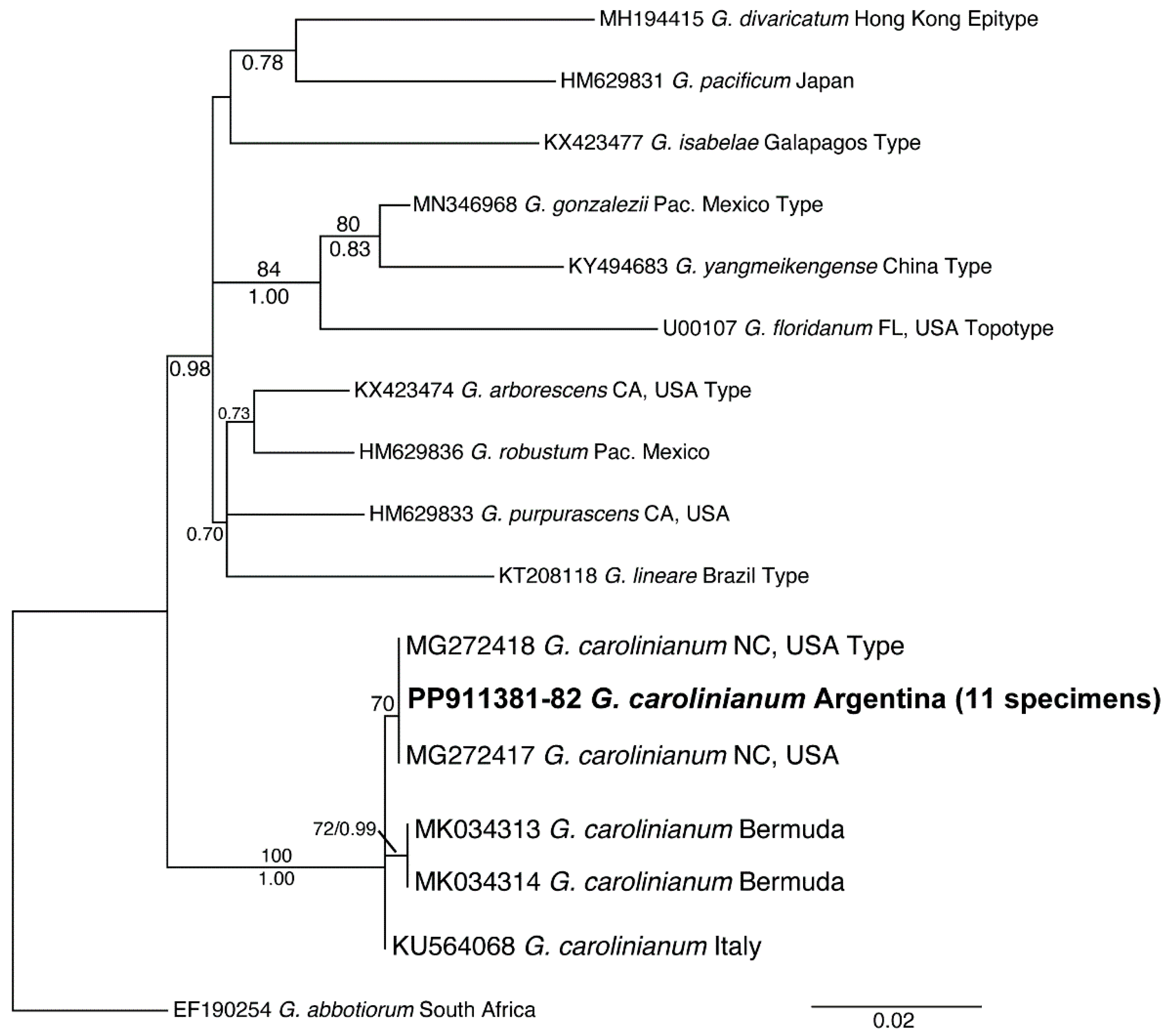
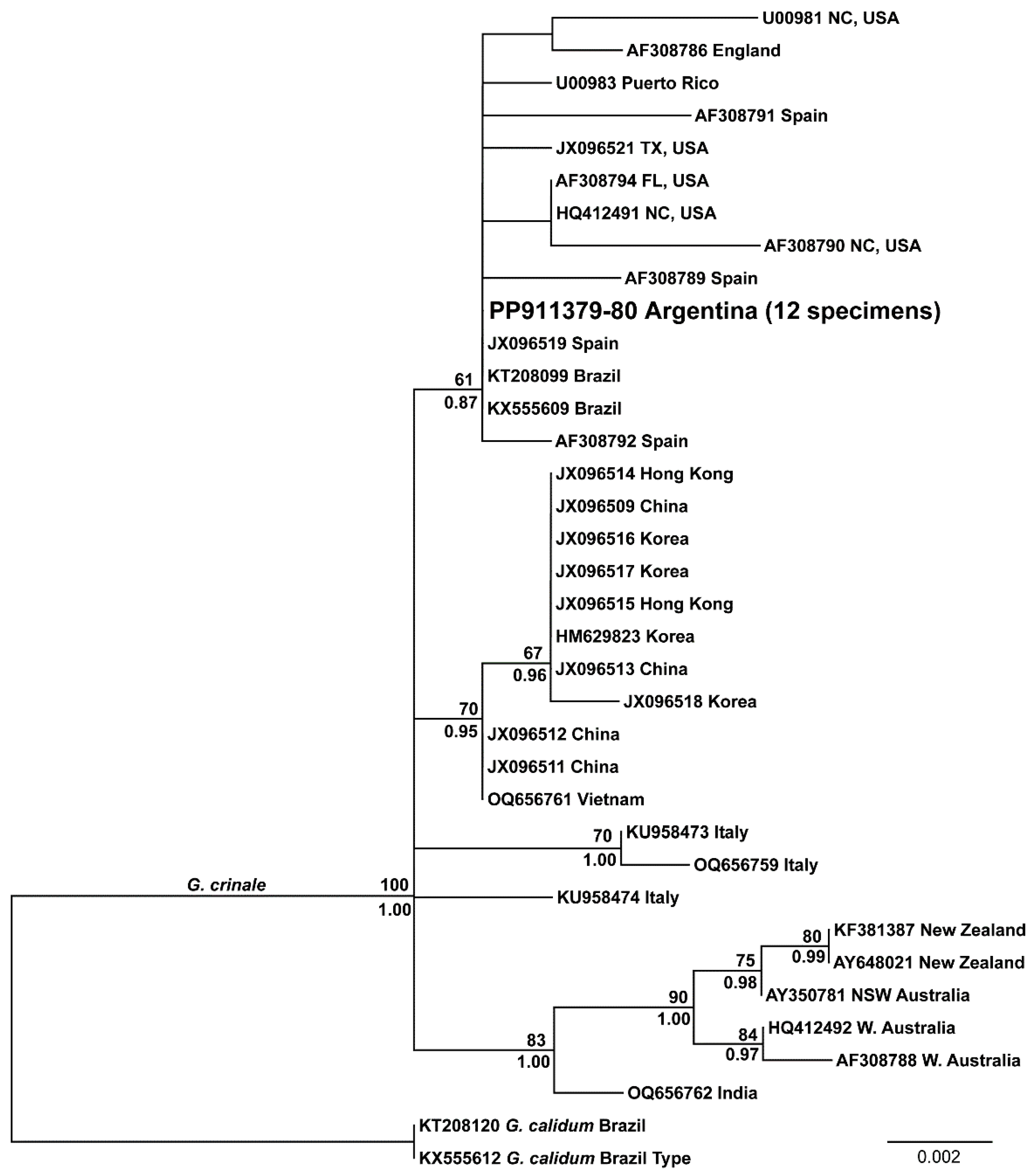
3.1. Molecular Identification of the Morphotypes

Three morphotypes were recognized from the samples which had clear differences in the architecture of the thalli. Morphotype A had a short and fibrous thallus, dark red in color, highly tufted in the upper part, and with an arborescent aspect (Figure 2a). The erect axes of this morphotype were terete to compressed and profusely branched. Morphotype B had more elongated and less branched thalli, almost blackish in color, with filiform and fibrous erect axes that were terete to compressed (Figure 2b). Morphotype C had short and tufted thalli, crimson in color, and profusely branched, and the erect axes were flattened and membranous (Figure 2c).
Molecular analyses of the rbcL sequences obtained from these three morphotypes showed that two morphotypes corresponded to Gelidium crinale and one morphotype corresponded to Gelidium carolinianum. Three specimens with morphotype A and nine specimens with morphotype B shared identical rbcL or rbcL-3P sequences and were resolved within the clade of G. crinale with high support, whereas eleven specimens with morphotype C shared identical rbcL or rbcL-3P sequences and were resolved within the clade of G. carolinianum with full support (Figure 3).

3.2. Distribution and Phylogeny of G. crinale and G. carolinianum

Two species, G. crinale and G. carolinianum, coexisted in the intertidal region of most of the sites surveyed. G. crinale was found in five of the sites studied, whereas G. carolinianum appeared in four of the sites studied (Table S1). Morphotype A of G. crinale was only found at the site with the highest exposure to surf action (site S1), in the high intertidal zone, and the thalli were exposed to air during low tides on emerged substrates. The other morphotypes were found submerged, inside tidal pools. In the tidal channels of site S5, where pebbles and cobbles are the main hard substrate available, only G. carolinianum was found.
The rbcL analyses showed that within the G. carolinianum clade, specimens collected from the four populations surveyed in Argentina had identical sequences to specimens from North Carolina (USA) and were only 1 bp different from sequences of specimens collected in Italy and Bermuda (Figure 4, Table 2). All rbcL and rbcL-3P sequences from G. crinale specimens collected from the five populations surveyed in Argentina were identical and they were also identical to those from Brazil specimens and a single specimen from Spain (Figure 5, Table 3). Pairwise distances of 34 publicly available rbcL sequences revealed an intraspecific divergence range of 0–15 bp (0.00–1.21%, Table 3). Maximum-likelihood analysis of these sequences with G. calidum as an outgroup resolved a well-supported Australia–New Zealand clade that was moderately supported as being most closely related to a specimen from India (Figure 5). An East Asia clade of specimens from Vietnam, China, and Korea was poorly supported and there was no support for geographic groups among the Atlantic Ocean and Mediterranean specimens.

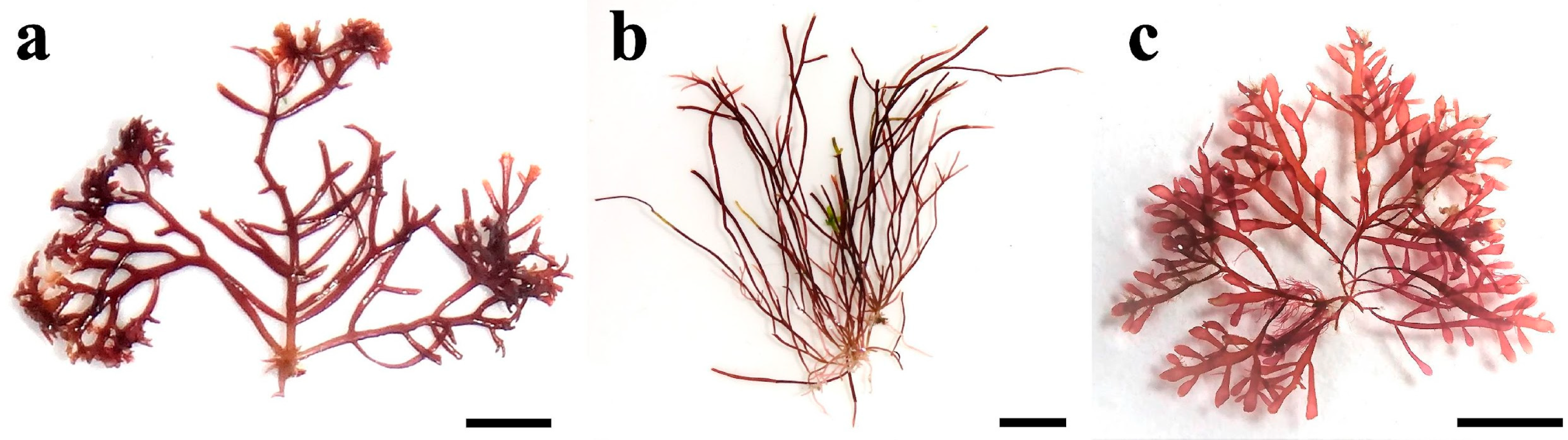
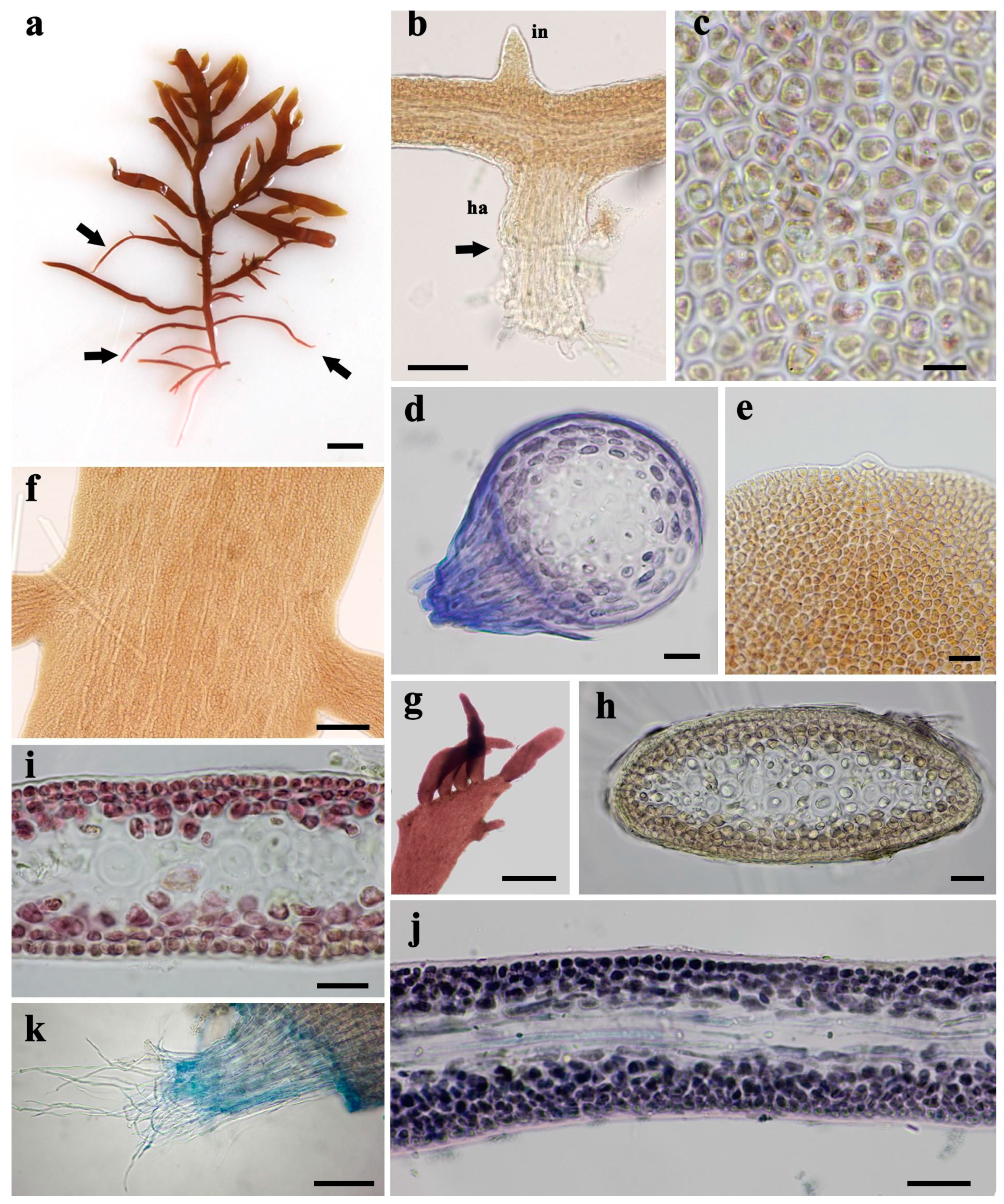
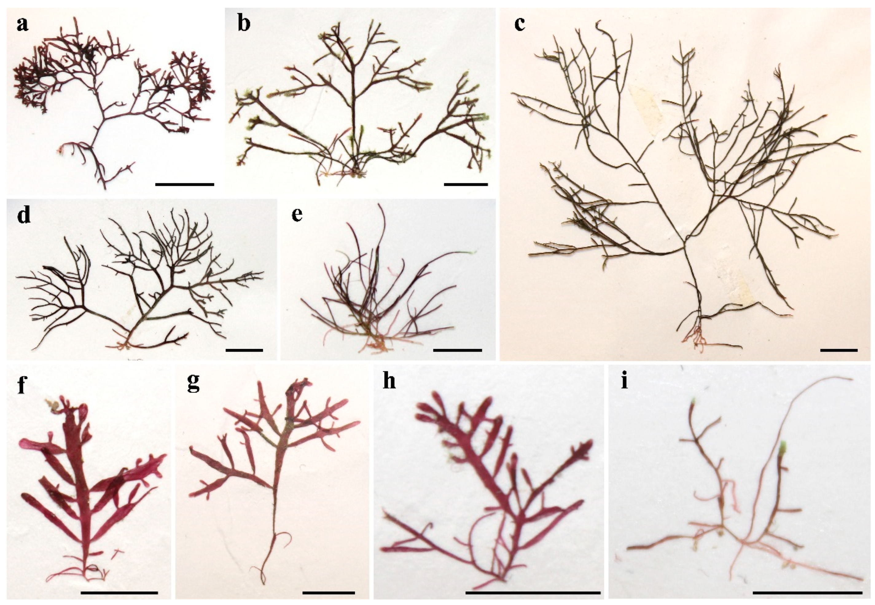
3.3. Morphological Characteristics of Gelidium carolinianum

Specimens identified as G. carolinianum are crimson, membranous, and short (Figure 2c). The thalli are composed of stolons that give rise to erect axes and form turfs up to 2 cm long (Figure 6a) that attach to rocks or shells of bivalves and gastropods, frequently epizoic on the gastropod Bostrycapulus aculeatus (Gmelin, 1791). Stolons are paler than erect axes and have many brush-like haptera, which generally develop opposite the erect axes or their initials (Figure 6b). Haptera are produced by the protrusion of internal rhizoidal filaments out of the stolon and are partially covered with elongated cortical cells. The internal rhizoidal filaments that form the haptera have swollen tips for adhesion to substrates. Cortical cells are polygonal in the surface view, with parietal chloroplasts (Figure 6c). In cross sections, stolons are terete and have a thick cuticle (Figure 6d). They are formed by three to four layers of highly pigmented cortical cells surrounding a medulla of colorless, thick-walled cells (Figure 6d). The medulla has one central medullary cell surrounded by six, seven or more medullary cells depending on the thickness of the stolon (Figure 6d). Cortical cells are rounded in cross sections, with the surface cortical cells wider than they are tall (Figure 6d). Internal rhizoidal filaments are sparsely located in the medulla, and solitary or in small groups (Figure 6d).
Erect axes are cylindrical at the base where they arise from the stolons and become compressed within 1–3 mm before gradually becoming flattened and up to 1 mm wide. The branching is opposite or subopposite, sometimes bipinnate and fastigiate, up to three orders (Figure 6a). First-order branches appear every 1–2 mm along the main axis, forming acute angles with it. The more basal branches are terete throughout and reflexed downwards, often developing into stolons (Figure 6a). The branches are slightly constricted at the base and have acute or rounded apices with a conspicuous dome-shaped apical cell that protrudes from the axis tip (Figure 6e). Striations are noticeable in the surface view of flattened axes (Figure 6f) due to the files of elongated medullary cells showing through the cortex. Several new axes usually develop from truncated damaged axes, which may obscure the branching pattern (Figure 6g).
In the cross sections of compressed (Figure 6h) and flattened axes (Figure 6i), there are three to four layers of cortical cells, with the outermost layer formed by smaller, tightly packed quadrangular cells in sections, whereas the innermost layers are formed by larger, rounded, and loosely arranged cells. The medullary cells are cylindrical and longitudinally elongated (Figure 6j). In compressed and flattened parts of the axes, large medullary cells are arranged along a median row and surrounded by two or three layers of smaller medullary cells. Internal rhizoidal filaments are abundant and solitary or in tightly packed groups within the medulla but are absent in the cortex (Figure 6h–j). They have a fibrous aspect that is clearly visible in torn axes (Figure 6k).
Tetrasporangial sori are irregularly distributed on the main axes and spatulate branchlets of tetrasporophytes and are surrounded by a narrow sterile margin (Figure 7a,b). The tetrasporangia are cruciate or decussately cruciate (Figure 7c). Gametophytes are monoecious. Spermatangia develop in colorless or yellowish sori of irregular shape and extent on the surface of cystocarpic thalli, near the cystocarps (Figure 7d,e). Cystocarps are bilocular, strongly protruding on both surfaces of branches or axes, and lack peristomes around the ostioles (Figure 7f). They are solitary or in pairs, with one or two ostioles on each locule. In cross sections, carpospores are located on both sides of a central placenta and elongated third-order filament cells extend from the central placental tissue to the pericarp (Figure 7g).
G. carolinianum displayed significant variability in some morphological characters among the specimens collected in different locations (Table 4). The height of thalli and the length of branches were greater in specimens collected in site S3. The maximum width of erect axes was lower in specimens collected in site S6. The diameter of stolons was higher in specimens collected in site S2. The diameter of cortical and medullary cells in cross section was lower in specimens collected from site S6. Branches were frequently shorter and spatulated in specimens from exposed shores like site S2, compared with specimens collected from protected coasts like site S3, where branches are more frequently ligulate and longer. The specimens from site S6 had more irregular branching and abundant internal rhizoidal filaments.

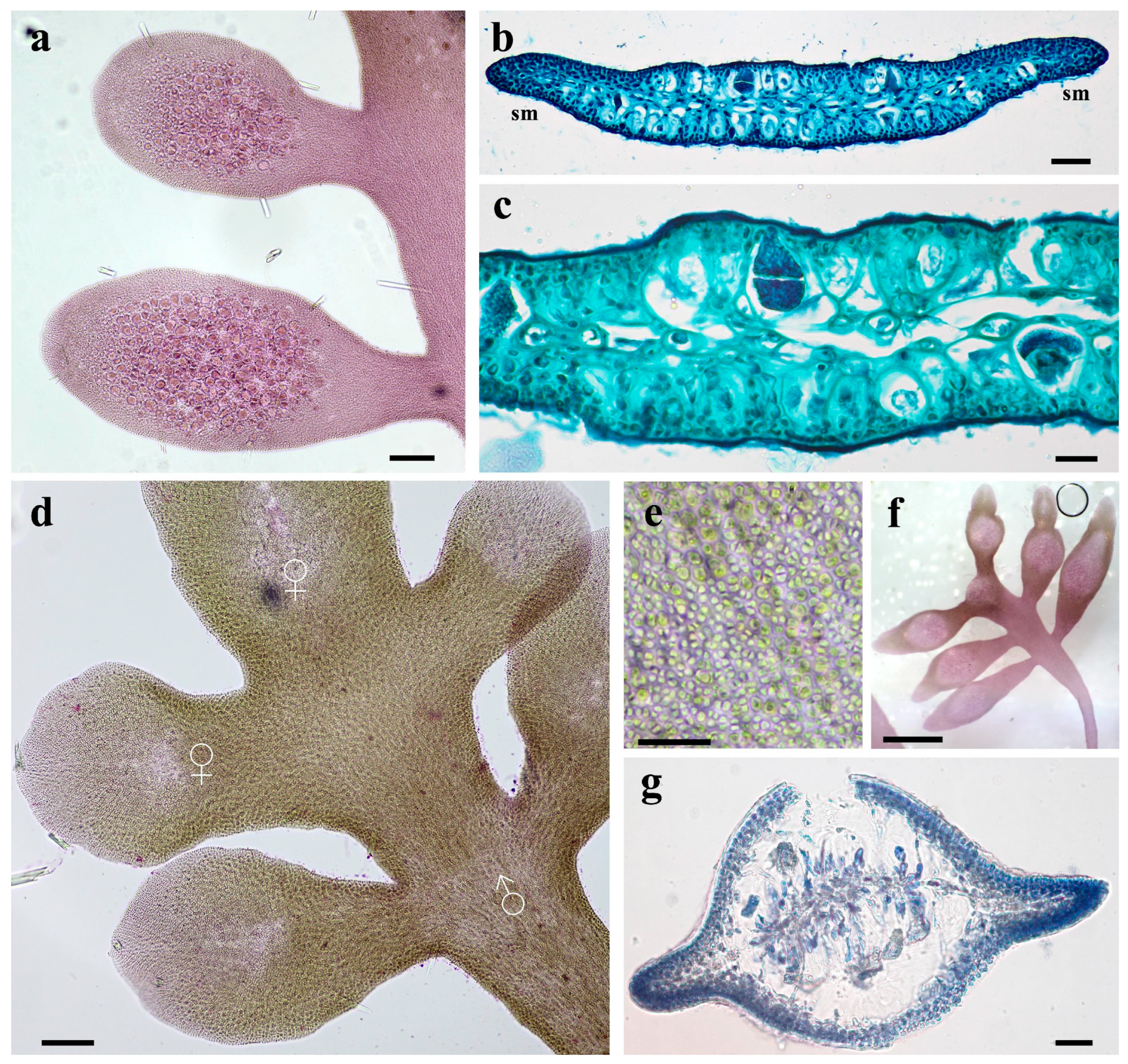
3.4. Morphological Characteristics of Gelidium crinale

Specimens identified as G. crinale were attached to rocks or shells, mainly of the oyster Magallana gigas (Thunberg, 1793) and the mussel Brachidontes rodriguezii (A. d’Orbigny, 1842). The thalli are dark red or blackish and fibrous, and consist of stolons giving rise to erect axes that form turfs up to 5.2 cm tall (Figure 2b). The erect axes are basally cylindrical where they arise from the stolon and become compressed and up to 0.6 mm in width. The branching is opposite, subopposite, or irregular and up to three orders (Figure 8a). First-order branches appear every 1–2 mm along the axis. The more basal branches are reflexed downwards and often develop into stolons (Figure 8b). Erect axes are frequently damaged and new axes develop from the truncated axis (Figure 8c). The stolons are pinkish and have brush-like haptera that are frequently opposite to erect axes or their initials (Figure 8d). Haptera are formed by the protrusion of internal rhizoidal filaments out of the stolon. Rhizoidal filaments have swollen tips for adhesion to substrates. Stolons are terete in cross section (Figure 8e). They are formed by three rows of highly pigmented cortical cells that are rounded in cross section and surround a medulla of colorless thick-walled cells (Figure 8e). The medulla is formed by one central medullary cell surrounded by two or three layers of medullary cells (Figure 8e). Internal rhizoidal filaments are abundant in the medulla and distributed separately or in small groups (Figure 8e). Dome-shaped apical cells protrude from the tips of the axes (Figure 8f). Cortical cells are polygonal in the surface view with parietal chloroplasts (Figure 8g).
In cross sections, erect axes have three to four layers of cortical cells; the outermost layer is formed by tightly packed and smaller rectangular cells, with the longest axis perpendicular to the surface, whereas the inner layers are formed by larger, rounded, and more loosely arranged cells (Figure 8h). The medullary cells are cylindrical, thick-walled, and longitudinally elongated. A median row of large medullary cells surrounded by two to three layers of smaller medullary cells is often visible in transverse sections through compressed parts of the axes. The medullary cells are cylindrical and elongated, clearly visible in longitudinal sections (Figure 8i). Internal rhizoidal filaments are solitary or in groups, located predominately in the outer medulla and absent in the cortex. They have a fibrous aspect that is clearly visible in torn axes (Figure 8j).
Tetrasporangial sori seem scattered, with no clear arrangement on spatulate branchlets; they lack sterile margins and can become elongated with the continued growth of the axes (Figure 9a–c). Tetrasporangia are cruciate or decussately cruciate (Figure 9c,d). Gametophytic thalli are monoecious. Young fertile gametophytes have colorless areas on the apex of axes and branches where carpogonia develop, and spermatangial sori below that appear as colorless or yellowish patches (Figure 9e–h). Spermatangial sori are variable in size and irregularly shaped (Figure 9g). Spermatia are cut off by transverse divisions of the spermatangial initials (Figure 9h). Mature cystocarpic thalli are recognized by protruding cystocarps on lanceolate branches (Figure 9i). Cystocarps are bilocular, strongly protruding on both thallus surfaces, and have one or two ostioles lacking peristomes on each locule. In cross sections, carpospores are located on both sides of a central placenta (Figure 9j).
G. crinale displayed significant variability in some morphological characters among the specimens collected at different sites (Table 5). The height of thalli and the length of branches were greater in specimens collected at site S3. The average distance among branches was lower in specimens collected at site S1, although this was not significantly different. The distance between haptera along stolons was similar among all specimens studied. The maximum width of erect axes was lower in specimens collected at site S6 and greater in specimens collected at site S2. The stolon diameter and basal width of erect axes showed no significant differences among sites. The cortical cell width was lower in specimens collected from site S1 and the branches were shorter in specimens collected from site S2.

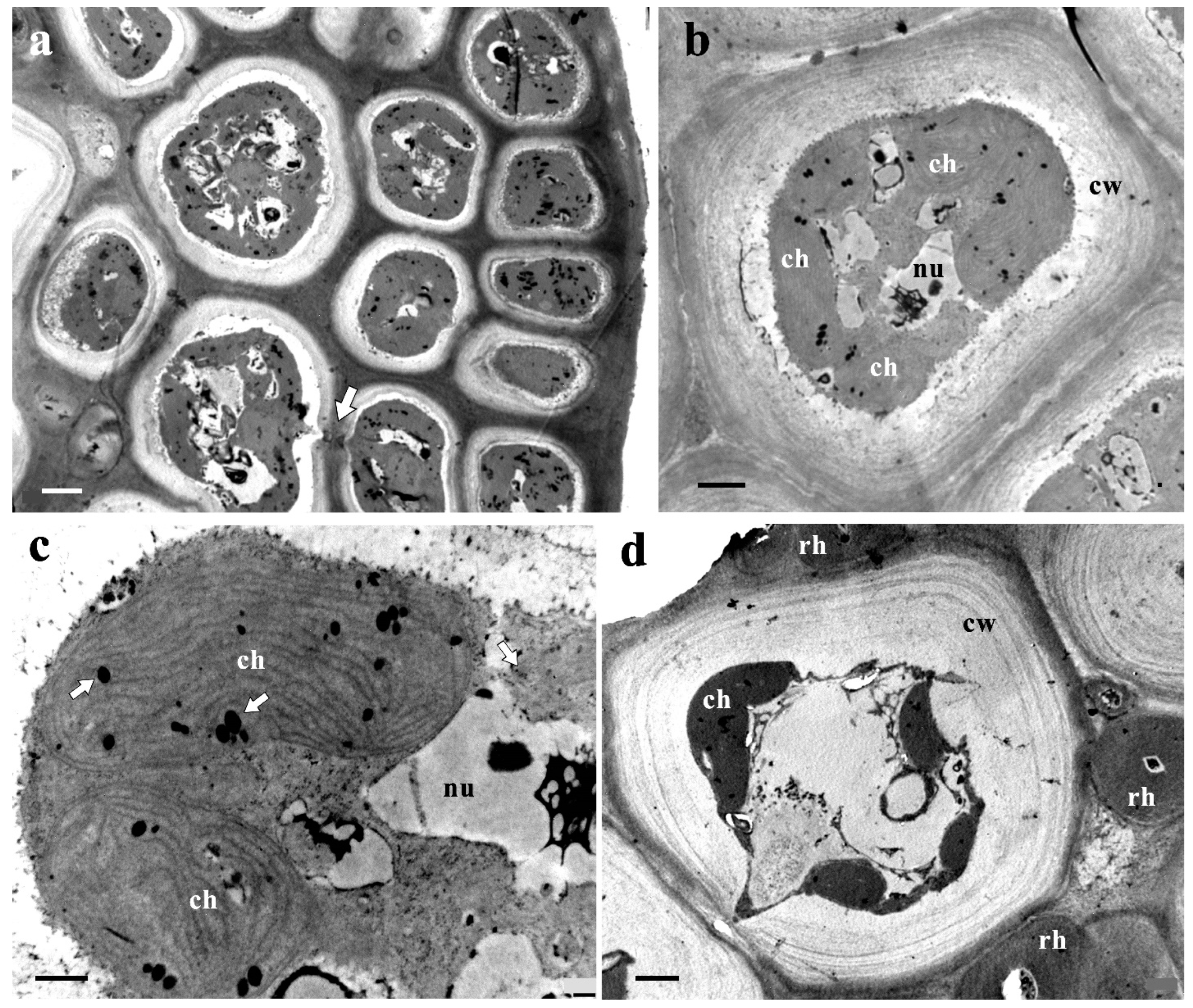
3.5. Ultrastructure of Vegetative Cells

Cortical cells of both species have a thick cell wall composed of fibrillar and amorphous parts and several chloroplasts occupying most of the cell (Figure 10a). Chloroplasts are largely developed in cortical cells (Figure 10b), with many thylakoids (Figure 10c). Among the thylakoids, there are several dark inclusions of variable size (plastoglobuli), which appear solitary or in groups (Figure 10c). Medullary cells have a thick cell wall formed by fibrillar and amorphous parts, and less developed chloroplasts (Figure 10d). Internal rhizoidal filaments are 3.08 (±0.77) µm in diameter and have a thick and homogeneous cell wall (Figure 10d).

4. Discussion

The order Gelidiales has historically received little attention in Argentinean phycology, as demonstrated by the scarcity of research studies available in the literature (reviewed in [48]). This is the first taxonomic study of Argentina Gelidiales species to include evidence from DNA markers. RbcL data revealed the occurrence of two species of Gelidiaceae, Gelidium crinale and Gelidium carolinianum, coexisting in the intertidal zone of the temperate coast of Argentina. These species represent the only two Argentina Gelidiaceae taxa whose identities have been confirmed by DNA sequence data. The results presented here provide new knowledge about Gelidiaceae diversity in South America and contribute to our understanding of their phylogenetic relationships and geographical distribution.
The two species of Gelidium found in this study were widely spread along the temperate coast of Argentina and their distribution ranges mostly overlapped. At locations where both species coexisted, their populations occupied the same habitat, and sometimes, their thalli were entangled. This coalescence of thalli was also noticed by Perrone et al. [49] in turfs of G. crinale. The presence of entangled “red algal turfs” that were a mixture of Gelidium maggsiae and Gelidiella calcicola was reported in a previous ecological survey carried out at site S4 [33]. Subsequently, a detailed study of the vegetative and reproductive morphology of new specimens from the same population revealed that one of the species forming the turf had morphological characteristics of G. crinale [37]. Our analysis of rbcL sequences of additional samples collected from the same site confirmed the identity of G. crinale, but also revealed the presence of G. carolinianum and no other Gelidiales species. Considering that the names previously assigned to these records were based on morphological observations, the reports of G. maggsiae and G. calcicola in Croce and Parodi [33] should be interpreted as misidentifications of G. carolinianum and G. crinale. Boraso [38] mentions that Pterocladiella capillacea is present on the temperate coast of Argentina as a flattened, bipinnately branched small turf; however, the author notes the uncertain identity of this record due to the lack of female reproductive structures in the specimens studied. We believe this report could also be a misidentification of G. carolinianum due to some external morphological resemblance with P. capillacea [50]. These findings highlight once more the value of conducting molecular taxonomic studies on challenging algal groups such as Gelidiales.
The rbcL data confirmed the occurrence of several intertidal populations of G. crinale on the temperate coast of Argentina, some of which had been documented in previous surveys, and also demonstrated that the distribution range of this species in the Western Atlantic Ocean extends beyond its currently known southern limit on the coast of Brazil [51]. Populations of G. crinale were found at five locations separated by at least 30 km along the Argentinean coast. According to existing records, the distribution of G. crinale in the Southwestern Atlantic ranges from 20° S to 54° S [14,31,34,35,37,52,53,54]. In these records, G. crinale is frequently mentioned as a common intertidal alga; however, it has rarely been recorded in cold waters. Therefore, the identities of populations located above 40°S on the Argentinean coast need to be confirmed by molecular markers. Given that G. crinale was found at the highest latitude surveyed (S6, 40°53′ S; 65°07′ W), we infer that the lack of samples from site S5 (40°43′ S; 64°57′ W) may be explained by the absence of appropriate substrates. G. crinale was mainly attached to sedimentary rocks or shells of Magallana gigas, and these types of substrates were infrequent at site S5; instead, the main hard substrates available were small pebbles.
G. crinale is a cosmopolitan species, probably the most widespread species of Gelidium along the warm and temperate coasts of the world. Past molecular studies based on rbcL and cox1 data have demonstrated that this species is represented by three clades corresponding to different geographical populations: Australia, East Asia, and Eastern North America and Europe [2,16]. More recently, Boo et al. [51] investigated the causes of the cosmopolitan distribution of G. crinale and inferred a single geographical origin from which the species dispersed prior to the Last Glacial Maximum (LGM). According to these authors, the relict populations gave rise to the current distribution of G. crinale around the world in five geographical clades, Atlantic–Mediterranean, Ionian, Asian, Adriatic–Ionian, and Australasia–India–Tanzania–Easter Island [51]. No evidence of introductions in new habitats was found and this suggests that the species outlives the changing environmental conditions by a strong tolerance to stress. Our findings showed that the populations of G. crinale from Argentina fall within the Atlantic–Mediterranean clade. The 12 specimens studied were collected from five locations on the Argentinean coast and were closely related to Brazilian specimens. According to these results, the distribution of G. crinale along the Southwestern Atlantic coast appears to be continuous from the equator down to 40° S.
The species G. carolinianum was recently described on the basis of field and herbarium material from the North Atlantic and Mediterranean; it had been previously identified as G. americanum or P. americana [18] and was later sequenced by Perrone et al. [49]. Sequences of rbcL and cox1 indicated that G. carolinianum has a narrow distribution that includes the coasts of North Carolina, Bermuda, and Italy [49]. However, our finding of G. carolinianum on the Argentinean coast indicates a much wider distribution range for this species. Molecular surveys of Gelidiales on other coasts of South America did not find G. carolinianum [14,28]; therefore, this study reports for the first time the occurrence of this species in the Southern Hemisphere.
The results obtained in this study indicate that G. carolinianum has a disjunct distribution encompassing the Atlantic Ocean and the Mediterranean Sea. The populations occur on warm–temperate coasts, but are absent in tropical waters, which may be indicative of a stenothermal species. The rbcL analyses of G. carolinianum specimens found three different haplotypes that were geographically structured. One haplotype was found in specimens from continental regions of the southern West Atlantic (Argentina) and northern West Atlantic (North Carolina), the second haplotype was present in insular specimens from northern West Atlantic (Bermuda), and the third haplotype was found in the specimen from the Adriatic Sea (Italy). The reason for this scattered distribution is uncertain, but may have originated from relic populations, as explained by the theory of driven-off relicts [55]. This theory postulates that marine organisms originally occupied continuous ranges and were driven away toward the poles by climate changes, giving rise to their actual discontinuous bipolar distribution [56]. According to this theory, G. carolinianum may have been widely distributed along the Western Atlantic during the Pleistocene, when the current tropics were temperate [57], and moved to higher latitudes when the waters became warmer after the glaciers retreated, disappearing from the tropics and prevailing on temperate coasts. Nevertheless, relationships among populations of this species are puzzling. The populations from both hemispheres are closely related despite the wide biogeographical barrier (the tropical warm belt) that separates them. A natural introduction by long-distance dispersion among countries is unlikely due to the mentioned biogeographical barrier; therefore, a possibility is that thalli or fragments of G. carolinianum had been accidentally transported perhaps via ballast water or ballast stones, either southwards or northwards in the Atlantic Ocean. Given that G. carolinianum is widely dispersed and well established along the Argentinean coast, and that the species has a well-known interspecific relationship (unpubl. data) with the native snail Bostrycapulus odites Collin, 2005 [58], it is reasonable to expect that the species may have been introduced in North Carolina. The type locality of G. carolinianum is Topsail Sound (Pender Co.) in North Carolina, USA [49]. The coast at this locality is formed by salt marshes with fine sediment, meadows of Sporobolus alterniflorus (Loisel.) P.M. Peterson & Saarela, and intertidal oyster reefs [59] and is geomorphologically and oceanographically similar to the sites were G. carolinianum was found in Argentina. If an introduction may have occurred in North Carolina, the species may have found an appropriate habitat to establish its populations. It is important to notice, however, that the species is distributed at much higher latitudes in the South Atlantic (38° S to 40° S) than where it has been reported in the North Atlantic (33° N to 36° N). If the introduction of G. carolinianum may have been southwards (that is, from USA to Argentina) by vessel transport of thalli or fragments, the entrance site could have been site S3, an estuary with high transit of international vessels from which the species ultimately dispersed to the other localities. The sites where G. carolinianum was found in Argentina are relatively close to each other (<300 km); therefore, a dispersion driven by the warm Brazil current may have occurred. The high capacity of this species for replication through fragments [60] could explain its success at colonizing new habitats. Additional studies of molecular markers with faster evolution rates like COI-5P are needed to shed light on the connectivity of these American populations and their relationship to the European ones.
Specimens of G. carolinianum from Argentina share most of the morphological characteristics described for those of North Carolina and Italy [49]. They had a characteristic single median row of medullary cells, compressed to flattened branches constricted at the base, pinnate branching up to three orders, abundant internal rhizoidal filaments surrounding the medullary cells, and tetrasporangial sori with sterile margins. The Argentinean specimens of G. carolinianum differed from North Carolina and Italy specimens in the size and consistency of the thallus, being smaller and membranous instead of cartilaginous, and having longitudinal striations on the surface of the main axes. The variation in the size and robustness of the thalli among specimens from different coasts was noted by Perrone et al. [49]. These authors related the variation in the texture of the thalli to differences in the abundance of internal rhizoidal filaments in the medullary zone. Although the specimens studied here had many internal rhizoidal filaments among medullary cells, the texture of the thalli was membranous instead of cartilaginous.
G. carolinianum showed significant variability in anatomical characters among the sites surveyed; however, the external appearance of the thalli was similar and morphotypes could not be distinguished (Figure 11). On the contrary, the populations of G. crinale showed notable variability in the external morphology among specimens that was attributable to the influence of oceanographic factors. This phenotypic diversity was recognized as two morphotypes: a short form (morphotype A), found in site S1 (Figure 11a), where waters have low turbidity and there is a strong influence of the surf on the intertidal, and a long form (morphotype B), found in sites S2, S3, S4, and S6 that are more protected from wave exposure and have high water turbidity (Figure 11b–e). Morphotype A of G. crinale (Figure 2a and Figure 11a) resembles the specimen from Korea which has an arborescent aspect, shown in Figure 4A from Kim and Boo [2]. The phenotypic plasticity of G. crinale and related species has been documented in the natural environment, and variability in length, pattern, and the abundance of branching has been related to geographical factors such as wave exposure or seasonality [61,62]. For example, Echegaray Taborga and Seaone Camba [61] have reported changes in length and the degree of branching according to the season, with Gelidium species being larger and more branched in the warmer seasons, which coincides with reproduction. Also, plants on wave-exposed shores are usually smaller than those at wave-protected sites, which was evident in morphotype A of G. crinale found at site S1.
Perrone et al. [49] noted that G. carolinianum could be confused with fertile G. crinale when the apices of the latter become enlarged. In our surveys, we also noticed that G. carolinianum may be confused with G. crinale when the young thalli of G. carolinianum have many basal stoloniferous branches or when the thalli have many truncated axes. In both situations, the axes appear terete, similar to those of young G. crinale thalli. Externally, the prostrate system of both species is indistinguishable, as they both have pale and terete stolons attached by several haptera. However, when plants are more developed, they can be easily distinguished in the field due to their differences in color (G. carolinianum is crimson-red and G. crinale is blackish-red) and texture (G. crinale is cartilaginous and G. carolinianum is membranous).
It is important to notice that the thalli of both species were largest at site S3 (Figure 11), a site characterized by high turbidity, where the thalli were frequently heavily covered in fine sediment. The sediment may benefit the development of the thalli in two aspects. Sediment deposition on the thalli may increase the availability of nutrients on the surface of branches, favoring growth, and it may reduce the exposure to high irradiances during the long periods of low tide, avoiding photoinhibition and heat stress. Evidence from current culture studies indicates that reduced exposure to light intensity benefits the growth of G. carolinianum in terms of an increased growth rate and reduced epiphytism (unpub. data).
Both G. crinale and G. carolinianum had monoecious thalli, a characteristic that has been reported for few other Gelidiaceae [63,64,65,66]. To our knowledge, this is the first study to describe the spermatangia of either species. As reviewed by Santelices [67], male gametophytes of Gelidiaceae have been less frequently reported than female gametophytes. The spermatangia of red seaweeds occur during a short period of time, which must be perfectly synchronized with the emergence of female fertile tissue. Mature cystocarpic thalli of Gelidiaceae are easily recognizable by the conspicuous dome-shaped protrusions on the surface of apical branches. At this stage, the female gametophyte has been fertilized; therefore, it is unlikely to find remaining spermatangia unless the female gametophyte is still under development. This may explain the difficulty of finding male reproductive structures on collected thalli, and male sori might have passed undetected in most previous studies [64]. The spermatangia found in our study were observed in the early stages of cystocarpic development, as also reported by Santelices and Flores [64] for Gelidium macnabbianum (E.Y.Dawson) Santelices (as Pterocladia macnabbiana E.Y.Dawson). Spermatangia are recognized as whitish or yellowish patches of irregular shape that are located on the axes in proximity to the fertile female tissue. The spermatangia in the surface view are seen as patches of small ovoid and colorless cells, whereas in longitudinal sections, they appear as piled coins and can sometimes be covered with a mucilaginous cuticle.
The populations of G. crinale and G. carolinianum from the Argentinean coast are widespread and persistent. The occurrence of all reproductive structures representing the complete life cycle of Rhodophyta (spermatangia, cystocarps, and tetrasporangia) suggests that the two species are well established in the region. Like other turf-forming species, these assemblages have a strong influence on the dynamics of particulate matter, and they offer habitats for a number of benthic invertebrates [32,68,69]. The genus Gelidium is highly diverse, and species are adapted to a great variety of habitats. Given that the genus may have originated in the Southern Hemisphere [3,70] we believe that other species may be found if efforts are focused on surveying both intertidal and subtidal habitats at latitudes higher than 40° S.

5. Conclusions

This study revealed that only two species of Gelidiaceae inhabit the intertidal region of the temperate coast of Argentina, showing that this economically valuable group of red algae is underrepresented in this region of the world, as compared to other warm–temperate coasts. The rbcL data confirmed the taxonomic identity of G. crinale as part of the intertidal flora of the Argentinean coasts, for which records had been previously based only on morphological identifications, and the presence of an additional Gelidium species, G. carolinianum, which has a disjunct distribution on both hemispheres. The wide distribution of these species on the Southwestern Atlantic coast suggests that their turf-like assemblages are a major component of the coastal intertidal communities. Further studies on population dynamics and interactions with other elements of the ecosystem are therefore encouraged.

Supplementary Materials

The following supporting information can be downloaded at: https://www.mdpi.com/article/10.3390/d16070399/s1, Supplementary Table S1. List of samples used and accession numbers of rbcL and rbcL-3P sequences. Only one representative of identical rbcL and rbcL-3P sequences for each species was submitted to GenBank.

Author Contributions

Conceptualization, M.E.C.; formal analysis, M.E.C. and D.W.F.; investigation, M.E.C. and D.W.F.; resources, M.E.C. and D.W.F.; writing—original draft preparation, M.E.C. and D.W.F.; writing—review and editing, M.E.C. and D.W.F.; visualization, M.E.C. and D.W.F.; funding acquisition, M.E.C. and D.W.F. All authors have read and agreed to the published version of the manuscript.

Funding

This study was funded by the National Agency of Scientific and Technological Promotion FONCYT AGENCIA, grant number PICT-2019-04106, the National Council of Scientific and Technological Research CONICET, grant number PIP 11220130100070CO, the US National Science Foundation Biotic Surveys and Inventories, grant number 0743334, and the Center for Marine Science DNA-Algal Trust.

Institutional Review Board Statement

Not applicable.

Data Availability Statement

DNA sequence data used in this study are available from GenBank (www.ncbi.nlm.nih) and accession numbers are provided in the Supplementary Materials of this paper.

Conflicts of Interest

The authors declare no conflicts of interest. The funders had no role in the design of the study; in the collection, analyses, or interpretation of the data; in the writing of the manuscript; or in the decision to publish the results.

References

  1. Womersley, H.B.S.; Guiry, M.D. The Marine Benthic Flora of Southern Australia. Part IIIA; Australian Biological Resources Study and State Herbarium of South Australia: Canberra, Australia, 1994; pp. 118–142. [Google Scholar]
  2. Kim, K.M.; Boo, S.M. Phylogenetic relationships and distribution of Gelidium crinale and G. pusillum (Gelidiales, Rhodophyta) using cox1 and rbcL sequences. Algae 2012, 27, 83–94. [Google Scholar] [CrossRef]
  3. Boo, G.H.; Kim, K.M.; Nelson, W.A.; Riosmena-Rodríguez, R.; Yoon, K.J.; Boo, S.M. Taxonomy and distribution of selected species of the agarophyte genus Gelidium (Gelidiales, Rhodophyta). J. Appl. Phycol. 2014, 26, 1243–1251. [Google Scholar] [CrossRef]
  4. Fujita, D.; Ishikawa, T.; Kodama, S.; Kato, Y.; Notoya, M. Distribution and recent reduction of Gelidium beds in Toyama Bay, Japan. J. Appl. Phycol. 2006, 18, 591–598. [Google Scholar] [CrossRef]
  5. Boo, G.H.; Mansilla, A.; Nelson, W.A.; Bellgrove, A.; Boo, S.M. Genetic connectivity between trans-oceanic populations of Capreolia implexa (Gelidiales, Rhodophyta) in cool temperate waters of Australasia and Chile. Aquat. Bot. 2014, 119, 73–79. [Google Scholar] [CrossRef]
  6. López, B.A.; Tellier, F.; Retamal-Alarcón, J.C.; Pérez-Araneda, K.; Fierro, A.O.; Macaya, E.C.; Tala, F.; Thiel, M. Phylogeography of two intertidal seaweeds, Gelidium lingulatum and G. rex (Rhodophyta: Gelidiales), along the South East Pacific: Patterns explained by rafting dispersal? Mar. Biol. 2017, 164, 1–19. [Google Scholar] [CrossRef]
  7. Boo, G.H.; Hughey, J.R. Phylogenomics and multigene phylogenies decipher two new cryptic marine algae from California, Gelidium gabrielsonii and G. kathyanniae (Gelidiales, Rhodophyta). J. Phycol. 2019, 55, 160–172. [Google Scholar] [CrossRef]
  8. Callaway, E. Lab staple agar runs low: Dwindling seaweed harvest imperils reagent essential for culturing microbes. Nature 2015, 528, 171–172. [Google Scholar] [CrossRef]
  9. Boo, G.H.; Le Gall, L.; Miller, K.A.; Freshwater, D.W.; Wernberg, T.; Terada, R.; Yoon, K.J.; Boo, S.M. A novel phylogeny of the Gelidiales (Rhodophyta) based on five genes including the nuclear CesA, with descriptions of Orthogonacladia gen. nov. and Orthogonacladiaceae fam. nov. Mol. Phylogenetics Evol. 2016, 101, 359–372. [Google Scholar] [CrossRef]
  10. Perrone, C.; Felicini, G.P.; Bottalico, A. The prostrate system of the Gelidiales: Diagnostic and taxonomic importance. Bot. Mar. 2006, 49, 23–33. [Google Scholar] [CrossRef]
  11. Guiry, M.D.; Guiry, G.M. AlgaeBase. World-Wide Electronic Publication, National University of Ireland, Galway. Available online: https://www.algaebase.org (accessed on 14 June 2024).
  12. Meinita, M.D.N.; Harwanto, D.; Amron, M.T.; Hannan, M.A.; Jeong, G.T.; Moon, I.S.; Choi, J.S. A concise review of the potential utilization based on bioactivity and pharmacological properties of the genus Gelidium (Gelidiales, Rhodophyta). J. Appl. Phycol. 2023, 35, 1499–1523. [Google Scholar] [CrossRef]
  13. Airoldi, L.; Fabiano, M.; Cinelli, F. Sediment deposition and movement over a turf assemblage in a shallow rocky coastal area of the Ligurian Sea. Mar. Ecol. Prog. Ser. 1996, 133, 241–251. [Google Scholar] [CrossRef]
  14. Iha, C.; Milstein, D.; Guimarães, S.M.P.B.; Freshwater, D.W.; Oliveira, M.C. DNA barcoding reveals high diversity in the Gelidiales of the Brazilian southeast coast. Bot. Mar. 2015, 58, 295–305. [Google Scholar] [CrossRef]
  15. Freshwater, D.W.; Rueness, J. Phylogenetic relationships of some European Gelidium (Gelidiales, Rhodophyta) species, based on rbcL nucleotide sequence analysis. Phycologia 1994, 33, 187–194. [Google Scholar] [CrossRef]
  16. Freshwater, D.W.; Tudor, K.; O’Shaughnessy, K.; Wysor, B. DNA barcoding in the red algal order Gelidiales: Comparison of COI with rbcL and verification of the ‘barcoding gap’. Cryptogam. Algol. 2010, 31, 435–449. [Google Scholar]
  17. Kim, M.K.; Hwang, L.K.; Yoon, H.S.; Boo, S.M. Four novel Gelidium species (Gelidiales, Rhodophyta) discovered in Korea: G. coreanum, G. jejuensis, G. minimum and G. prostratum. Phycologia 2012, 51, 461–474. [Google Scholar] [CrossRef]
  18. Bottalico, A.; Boo, G.H.; Russo, C.; Boo, S.M.; Perrone, C. Parviphycus albertanoae sp. nov. (Gelidiales, Rhodophyta) from the Mediterranean Sea. Phycologia 2014, 53, 243–251. [Google Scholar] [CrossRef]
  19. Boo, G.H.; Park, J.K.; Han, K.S.; Yoon, H.S. Gelidium rosulatum (Gelidiales, Rhodophyta), a new species of subtidal marine algae from Korea. Phycologia 2022, 61, 332–340. [Google Scholar] [CrossRef]
  20. Santelices, B.; Montalva, S. Taxonomic studies on Gelidiaceae (Rhodophyta) from central Chile. Phycologia 1983, 22, 185–196. [Google Scholar] [CrossRef]
  21. Santelices, B.; Stewart, J.G. Pacific species of Gelidium Lamouroux and other Gelidiales (Rhodophyta), with keys and descriptions to the common or economically important species. In Taxonomy of Economic Seaweeds with Reference to Some Pacific and Caribbean Species; Abbott, I.A., Norris, J.N., Eds.; California Sea Grant College Program: San Diego, CA, USA, 1985; pp. 17–31. [Google Scholar]
  22. Hoffmann, A.; Santelices, B. Flora Marina de Chile Central; Ediciones Universidad Católica de Chile: Santiago, Chile, 1997; p. 434. [Google Scholar]
  23. Boo, G.H.; Hughey, J.R.; Miller, K.A.; Boo, S.M. Mitogenomes from type specimens, a genotyping tool for morphologically simple species: Ten genomes of agar-producing red algae. Sci. Rep. 2016, 6, 35337. [Google Scholar] [CrossRef]
  24. Boo, G.H.; Calderon, M.S.; Boo, S.M. A new marine alga, Pterocladiella andresii sp. nov.(Gelidiales, Rhodophyta) and its relationship to P. caloglossoides from Pacific South America. Phytotaxa 2017, 319, 139–148. [Google Scholar] [CrossRef]
  25. Iha, C.; O’Shaughnessy, K.A.; Guimarães, S.M.P.B.; Oliveira, M.C.; Freshwater, D.W. Taxonomic reappraisal of Gelidium coarctacum (Gelidiales, Rhodophyta) and Gelidium lineare sp. nov. from the tropical western Atlantic. Phycologia 2016, 55, 555–563. [Google Scholar] [CrossRef]
  26. Iha, C.; Jamas, M.; Guimarães, S.M.P.B.; Fujii, M.T.; Freshwater, D.W.; Oliveira, M.C. Pterocladiella (Gelidiales, Rhodophyta) species of Brazil including morphological studies of Pterocladiella media and a reassessment of Pterocladiella taylorii. Phycologia 2017, 56, 624–637. [Google Scholar] [CrossRef]
  27. Iha, C.; Freshwater, D.W.; Guimarães, S.M.P.B.; Oliveira, M.C. Gelidiorariphycus gen. nov. (Gelidiales, Rhodophyta): A rare genus found in the Americas. Phycologia 2022, 61, 473–483. [Google Scholar] [CrossRef]
  28. Jamas, M.; Iha, C.; Oliveira, M.C.; Guimarães, S.M.P.B.; Fujii, M.T. Morphological and molecular studies on Gelidiaceae and Gelidiellaceae (Gelidiales, Rhodophyta) from Brazil with description of the new species Gelidium calidum. Phytotaxa 2017, 314, 195–218. [Google Scholar] [CrossRef]
  29. Brunelli, B.; Milstein, D.; Boo, S.M.; Fujii, M.T. Gelidium guimaraesiae sp. nov. (Gelidiaceae, Rhodophyta) from the Western Atlantic segregated from G. floridanum by morphological and molecular evidence. Phytotaxa 2019, 388, 275–286. [Google Scholar] [CrossRef]
  30. Brunelli, B.; Jamas, M.; Milstein, D.; Boo, S.M.; Fujii, M.T. Gelidium brasiliense sp. nov. (Gelidiales, Rhodophyta): A diminutive agarophyte from Brazil. J. Appl. Phycol. 2019, 31, 951–958. [Google Scholar] [CrossRef]
  31. Steigleder, K.M.; Copertino, M.S.; Lanari, M.; Camargo, M.; Fujii, M.T. Latitudinal gradient in intertidal seaweed composition off the coast of southern Brazil and Uruguay. Aquat. Bot. 2019, 156, 47–56. [Google Scholar] [CrossRef]
  32. Croce, M.E.; Gauna, M.C.; Fernández, C.; Parodi, E.R. Intertidal seaweeds from North Atlantic Patagonian coasts, Argentina. Checklist 2015, 11, 1–8. [Google Scholar] [CrossRef]
  33. Croce, M.E.; Parodi, E.R. Seasonal dynamic of macroalgae in intertidal pools formed by beds of Crassostrea gigas (Mollusca, Bivalvia) on the north Patagonian Atlantic coast. Bot. Mar. 2012, 55, 49–58. [Google Scholar] [CrossRef]
  34. Pujals, C. Catálogo de Rhodophyta citadas para la Argentina; Museo Argentino de Ciencias Naturales e Instituto Nacional de Investigación de las Ciencias Naturales: Buenos Aires, Argentina, 1963; p. 139. [Google Scholar]
  35. Sar, E.; Pascual, M.; Parma, A. Consideraciones ecológicas sobre las algas del litoral rocoso bonaerense. Rev. Mus. Plata 1984, 13, 143–147. [Google Scholar]
  36. Parma, A.; Pascual, M.; Sar, E. Clave Para el Reconocimiento de los Géneros de Algas Macrofitas del Intermareal Rocoso Bonaerense; Universidad Nacional de La Plata, Serie Técnica y Didáctica: La Plata, Argentina, 1987; p. 29. [Google Scholar]
  37. Croce, M.E.; Parodi, E.R. The turf-forming alga Gelidium crinale (Florideophyceae, Rhodophyta) on Atlantic Patagonian coasts. Bot. Mar. 2013, 56, 131–141. [Google Scholar] [CrossRef]
  38. Boraso, A.L. Elementos Para el Estudio de las Macroalgas de Argentina; Editorial Universitaria de la Patagonia: Comodoro Rivadavia, Argentina, 2013; p. 204. [Google Scholar]
  39. López Gappa, J.; Tablado, A.; Magaldi, N.H. Seasonal changes in an intertidal community affected by sewage pollution. Environ. Pollut. 1993, 82, 157–165. [Google Scholar] [CrossRef]
  40. Thiers, B. Index Herbariorum: A Global Directory of Public Herbaria and Associated Staff. New York Botanical Garden’s Virtual Herbarium. Available online: http://sweetgum.nybg.org/science/ih/ (accessed on 20 June 2024).
  41. Taylor, R.L.; Bailey, J.C.; Freshwater, D.W. Systematics of Cladophora spp. (Chlorophyta) from North Carolina, USA, based upon morphology and DNA sequence data with a description of Cladophora subtilissima sp. nov. J. Phycol. 2017, 53, 541–556. [Google Scholar] [CrossRef]
  42. Freshwater, D.W.; Scott, S.; Tronchin, E.M.; Saunders, G.W. Reassessment of Tristan da Cunha Gelidium (Gelidiales, Rhodophyta) species. Bot. Mar. 2020, 63, 455–462. [Google Scholar] [CrossRef]
  43. Edgar, R.C. MUSCLE: Multiple sequence alignment with high accuracy and high throughput. Nucleic Acids Res. 2004, 32, 1792–1797. [Google Scholar] [CrossRef]
  44. Stamatakis, A. RAxML Version 8: A tool for phylogenetic analysis and post-analysis of large phylogenies. Bioinformatics 2014, 30, 1312–1313. [Google Scholar] [CrossRef]
  45. Huelsenbeck, J.P.; Ronquist, F. MRBAYES: Bayesian inference of phylogenetic trees. Bioinformatics 2001, 30, 754–775. [Google Scholar] [CrossRef]
  46. Armiñana, J.R.; Breijo, F.G. Técnicas de Histología Vegetal; Jardín Botánico de Valencia: Valencia, Spain, 2006; p. 40. [Google Scholar]
  47. Cáceres, E.J. Métodos de preparación de algas para su observación con microscopía electrónica de transmisión convencional (METC). In Manual de Métodos Ficológicos; Alveal, K., Ferrario, M.E., Oliveira, E.C., Sar, E., Eds.; Universidad de Concepción: Concepción, Chile, 1995; pp. 147–168. [Google Scholar]
  48. Raffo, M.P.; Dellatorre, F.; Ciancia, M. Seaweed resources of Argentina (S W Atlantic): Production, bio-ecological, applied research and challenges for sustainable development. Appl. Phycol. 2022, 3, 383–421. [Google Scholar] [CrossRef]
  49. Perrone, C.; Bottalico, A.; Boo, G.H.; Boo, S.M.; Miller, K.A.; Freshwater, D.W. Gelidium adriaticum sp. nov. and Gelidium carolinianum sp. nov. (Gelidiales, Rhodophyta) from the Mediterranean Sea. Phycologia 2019, 58, 359–373. [Google Scholar] [CrossRef]
  50. Patarra, R.F.; Iha, C.; Pereira, L.; Neto, A.I. Concise review of the species Pterocladiella capillacea (S.G. Gmelin) Santelices & Hommersand. J. Appl. Phycol. 2020, 32, 787–808. [Google Scholar] [CrossRef]
  51. Boo, G.H.; Bottalico, A.; Le Gall, L.; Yoon, H.S. Genetic diversity and phylogeography of a turf-forming cosmopolitan marine alga, Gelidium crinale (Gelidiales, Rhodophyta). Int. J. Mol. Sci. 2023, 24, 5263. [Google Scholar] [CrossRef]
  52. Hariot, P. Algues. Algues: Mission Scientifique du Cap Horn (1882–83); Botanique: Paris, France, 1888; pp. 1–109. [Google Scholar]
  53. Cotton, A.D. Cryptogams from the Falkland Islands collected by Mrs. Vallentin. J. Linn. Soc. Bot. 1915, 43, 137–231. [Google Scholar] [CrossRef]
  54. Skottsberg, C. Marine Algae. 2. Rhodophyceae. In Botan. Ergebn. der Schwed. Expedition Nach Patagonien und dem Feuerlande 1907–1909, IX; Svenska, K., Ed.; Vetenkapasakad Handlingar: Stockholm, Sweden, 1923; 70p. [Google Scholar]
  55. Murray, J. On the deep and shallow-water marine fauna of the Kerguelen Region of the Great Southern Ocean. Trans. R. Soc. Edinb. 1897, 38, 343–500. [Google Scholar] [CrossRef]
  56. Okolodkov, Y.B. Biogeografía Marina; Universidad Autónoma de Campeche: Campeche, México, 2010; p. 217. [Google Scholar]
  57. Provan, J.; Bennett, K.D. Phylogeographic insights into cryptic glacial refugia. Trends Ecol. Evol. 2008, 23, 564–571. [Google Scholar] [CrossRef]
  58. Collin, R.; Ramos-Esplá, A.A.; Izquierdo, A. Identification of the South Atlantic spiny slipper limpet Bostrycapulus odites Collin, 2005 (Caenogastropoda: Calyptraeidae) on the Spanish Mediterranean coast. Aquat. Invasions 2010, 5, 197–200. [Google Scholar] [CrossRef]
  59. Gammill, S.P.; Hosier, P.E. Coastal saltmarsh development at Southern Topsail sound, North Carolina. Estuaries 1992, 15, 122–129. [Google Scholar] [CrossRef]
  60. Gauna, M.C.; Croce, M.E. Evaluación del crecimiento de Gelidium en cultivos con bioestimulantes a base de algas. In Boletín de la Sociedad Argentina de Botánica, Proceedings of the XXXIX Jornadas Argentinas de Botánica, Catamarca, Argentina, 21 September 2023; Sociedad Argentina de Botánica: La Plata, Argentina, 2023. [Google Scholar]
  61. Echegaray Taborga, J.; Seaone Camba, J.A. Estudio comparativo sobre la variación morfológica y fisiológica de Gelidium crinale y Gelidium spathulatum colectados en el mediterráneo catalán. Collect. Botánica 1982, 13, 803–816. [Google Scholar]
  62. Prathep, A.; Lewmanomontb, K.; Buapeta, P. Effects of wave exposure on population and reproductive phenology of an algal turf, Gelidium pusillum (Gelidales, Rhodophyta), Songkhla, Thailand. Aquat. Bot. 2009, 90, 179–183. [Google Scholar] [CrossRef]
  63. Renfrew, D.E.; Gabrielson, P.W.; Scagel, R.F. The marine algae of British Columbia, northern Washington, and southeast Alaska: Division Rhodophyta (red algae), class Rhodophyceae, order Gelidiales. Can. J. Bot. 1989, 67, 3297–3314. [Google Scholar] [CrossRef]
  64. Santelices, B.; Flores, V. Spermatangial sori on cystocarpic branchlets of species of Gelidium and Pterocladia (Gelidiales, Rhodophyta). Phycologia 1995, 34, 337–341. [Google Scholar] [CrossRef]
  65. Thomas, D.T.; Freshwater, D.W. Studies of Costa Rican Gelidiales (Rhodophyta): Four Caribbean taxa including Pterocladiella beachii sp. nov. Phycologia 2001, 40, 340–350. [Google Scholar] [CrossRef]
  66. Tronchin, E.M.; Freshwater, D.W. Four Gelidiales (Rhodophyta) new to southern Africa, Aphanta pachyrrhiza gen. et sp. nov., Gelidium profundum sp. nov., Pterocladiella caerulescens and P. psammophila sp. nov. Phycologia 2007, 46, 325–348. [Google Scholar] [CrossRef]
  67. Santelices, B. Synopsis of Biological Data on the Seaweed Genera Gelidium and Pterocladia (Rhodophyta); Food & Agriculture Organization of the United Nations: Rome, Italy, 1988; Volume 145, pp. 1–55. [Google Scholar]
  68. Whorff, J.S.; Whorff, L.L.; Sweet, M.H. Spatial variation in an algal turf community with respect to substratum slope and wave height. J. Mar. Biol. Assoc. UK 1995, 75, 429–444. [Google Scholar] [CrossRef]
  69. Pell, M.V.; Carcedo, M.C.; Fernández, C.; Croce, M.E. Evaluación de algas Gelidiales cespitosas como formadoras de hábitat del estuario de Bahía Blanca. In Boletín de la Sociedad Argentina de Botánica, Proceedings of the XXXIX Jornadas Argentinas de Botánica, Catamarca, Argentina, 21 September 2023; Sociedad Argentina de Botánica: La Plata, Argentina, 2023. [Google Scholar]
  70. Hommersand, M.H. Global biogeography and relationships of the Australasian marine macroalgae. In Algae of Australia: Introduction; McCarthy, P.M., Ed.; Australian Biological Resources Study and CSIRO Publishing: Canberra, Australia, 2007; pp. 511–542. [Google Scholar]
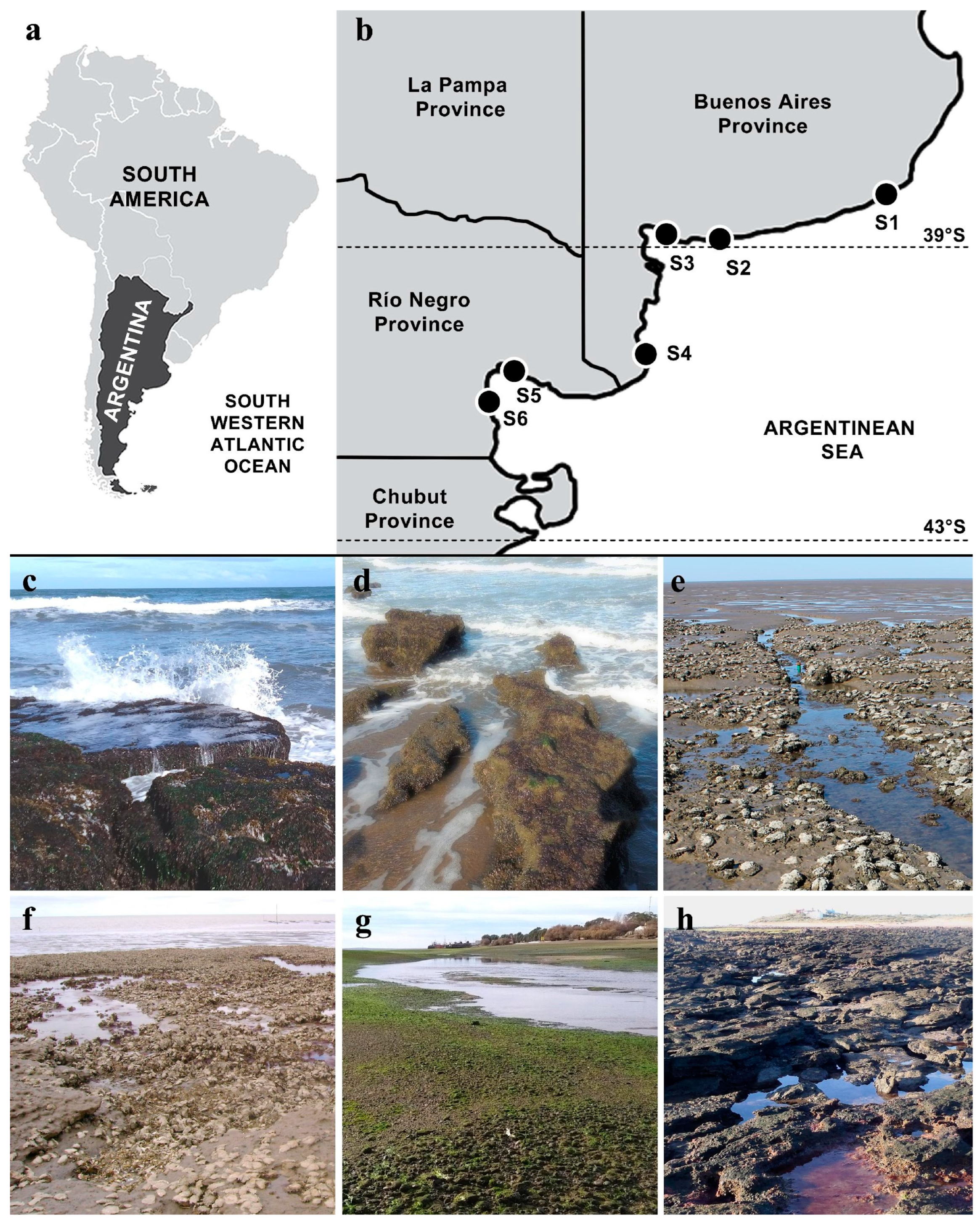
Figure 1. The geographic location and general view of the intertidal regions surveyed at the temperate coast of Argentina. (a) A map of South America showing the location of Argentina in the Southwestern Atlantic Ocean; (b) a section of the Argentinean coast showing the location of sites S1 to S6; (c) intertidal orthoquartzite rocks exposed to surf action in site S1; (d) intertidal sandstone outcrops exposed to surf action in site S2; (e) tidal flats (back) and intertidal sandstone outcrops colonized by Magallana gigas (front) in site S3; (f) oyster reefs of M. gigas covering intertidal sandstone outcrops in site S4; (g) an intertidal zone with pebbles in a bay channel in site S5; (h) intertidal sandstone outcrops in site S6.
Diversity 16 00399 g001
Figure 2. External aspects of the species of Gelidium present on the temperate coast of Argentina. (a) Morphotype A of G. crinale, scale = 2.5 mm; (b) morphotype B of G. crinale, scale = 5 mm; (c) G. carolinianum (morphotype C), scale = 5 mm.
Diversity 16 00399 g002
Figure 3. A maximum-likelihood tree of Gelidiaceae rbcL sequences from 53 Gelidium specimens and outgroup species from five Gelidiaceae genera. Maximum-likelihood bootstrap values and Bayesian posterior probabilities are shown when >70 and >0.80, respectively. ‘*’ indicates full support. C. = Capreolia; G. = Gelidium; Gphyc. = Gelidiophycus; Grari. = Gelidiorariphycus; P. = Ptilophora.
Diversity 16 00399 g003
Figure 4. A maximum-likelihood tree of Gelidium rbcL sequences from 16 specimens representing Gelidium carolinianum and 11 other closely related species. Maximum-likelihood bootstrap values and Bayesian posterior probabilities are shown when >70 and >0.70, respectively.
Diversity 16 00399 g004
Figure 5. A maximum-likelihood tree of rbcL sequences from 45 Gelidium crinale specimens and using G. calidum as an outgroup. The values above the branches are maximum-likelihood bootstrap values, only shown when >60.
Diversity 16 00399 g005
Figure 6. Vegetative morphology of Gelidium carolinianum from the temperate coast of Argentina. (a) External aspect of thallus showing basal branches pointing downwards (arrows), scale = 1 mm; (b) stolon showing haptera (ha) and erect axis initial (in); arrow indicates end of part of haptera covered by cortical cells, scale=100 µm; (c) surface view of cortical cells, scale = 10 µm; (d) cross section of stolon, scale = 20 µm; (e) detail of apical cell, scale = 20 µm; (f) striated surface of flattened erect axes, scale = 100 µm; (g) truncated erect axis with several proliferations, scale = 500 µm; (h) cross section through compressed axes, scale = 30 µm; (i) cross section through flattened axis, scale = 20 µm; (j) longitudinal section of erect axis, scale = 30 µm; (k) internal rhizoidal filaments protruding from torn axis, scale = 50 µm.
Diversity 16 00399 g006
Figure 7. Reproductive morphology of Gelidium carolinianum from the temperate coast of Argentina. (a) Fertile tetrasporangial branches with sori having sterile margins, scale = 100 µm; (b) cross section through tetrasporangial branch, showing sterile margins (sm), scale = 50 µm; (c) detail of tetrasporangia in a section through a tetrasporangial sorus, scale = 20 µm; (d) monoecious gametophyte with branchlets bearing carpogonia (♀) and also spermatangial sori (♂) on branch axis, scale = 100 µm; (e) surface view of spermatangia, scale = 20 µm; (f) terminal branches bearing mature cystocarps, scale = 1 mm; (g) cross section through mature cystocarp, showing two locules and central placenta, scale = 150 µm.
Diversity 16 00399 g007
Figure 8. Vegetative morphology of Gelidium crinale from the temperate coast of Argentina. (a) External aspect of thallus, scale = 1 cm; (b) detail of basal branches reflexed downwards, scale = 150 µm; (c) truncated apex of erect axis with several proliferations (arrows), scale = 250 µm; (d) stolon with haptera (ha) opposite erect axis initials (in), scale = 50 µm; (e) cross section through stolon, scale = 50 µm; (f) detail of apical cell, scale = 10 µm; (g) surface view of cortical cells, scale = 25 µm; (h) cross section through compressed axis, scale = 100 µm; (i) longitudinal section through erect axis, scale = 50 µm; (j) torn axis showing medullary cells (me) and internal rhizoidal filaments (rh), scale = 20 µm.
Diversity 16 00399 g008
Figure 9. Reproductive morphology of Gelidium crinale from the temperate coast of Argentina. (a) General aspect of a tetrasporophyte, scale = 5 mm; (b) detail of fertile branches, scale = 100 µm; (c) surface view of tetrasporangia, scale = 50 µm; (d) cross section through tetrasporangial branch, showing mature cruciate tetrasporangia, scale = 50 µm; (e) monoecious gamethophyte, with carpogonia developing near apices (♀) and spermatangial sori more proximal on branches (♂), scale = 100 µm; (f) surface view of spermatangial sorus, scale = 100 µm; (g) group of spermatangia on margin of thallus, scale = 20 µm; (h) detail of spermatium cut off by transverse division of spermatangial initial, scale = 20 µm; (i) general aspect of fertilized gametophyte; arrows point to axes and branches with mature cystocarps, scale = 5 mm; (j) cross section through mature cystocarp, showing two locules and central placenta, scale = 150 µm.
Diversity 16 00399 g009
Figure 10. Ultrastructure of vegetative cells of Gelidium crinale and Gelidium carolinianum. (a) General view of cortex of G. carolinianum showing 3–4 layers of cortical cells that become larger inwards; arrow points to pit connection, scale = 2 µm; (b) cortical cell of G. crinale showing large chloroplasts (ch), scale = 1 µm; (c) detail of cortical cell of G. crinale showing chloroplasts (ch) with several thylakoids and plastoglobuli (arrows); nu = nucleus, scale = 0.5 µm; (d) section through medulla of G. carolinianum showing internal rhizoidal filaments (rh) with thick cell walls and medullary cell with thick cell wall (cw) and less developed chloroplasts (ch), scale = 1 µm.
Diversity 16 00399 g010
Figure 11. Morphological variability in Gelidium crinale and Gelidium carolinianum from temperate coast of Argentina. (a) G. crinale from site S1 (morphotype A); (b) G. crinale from site S2 (morphotype B); (c) G. crinale from site S3 (morphotype B); (d) G. crinale from site S4 (morphotype B); (e) G. crinale from site S6 (morphotype B); (f) G. carolinianum from site S2; (g) G. carolinianum from site S3; (h) G. carolinianum from site S5; (i) G. carolinianum from site S6. Scales = 5 mm.
Diversity 16 00399 g011
Table 1. The geographic location and the main oceanographic characteristics of the six sites surveyed for ‘gelidioid’ algae on the temperate coast of Argentina.
SiteLocality, ProvinceCoordinatesCollection DatesType of CoastTidal AmplitudeType of Hard SubstrateDominant SedimentSurf ExposureWater Turbidity
S1Mar del Plata, Buenos Aires37°59′48.5″ S 57°32′27.7″ WApril 2015; March 2023Exposed sandy beachMicro-tidalOrthoquartzite rocksSandHighLow/Medium
S2Pehuen Co, Buenos Aires39°00′13.0″ S 61°36′55.5″ WJune 2015Exposed sandy beachMeso-tidalSandstone outcropsSandHigh/MediumMedium
S3Villa del Mar, Buenos Aires38°51′24.6″ S 62°07′04.5″ WMay 2015; Jan–Dec 2022Protected estuarial bay with tidal flats and salt marshes Meso-tidalSandstone outcropsLime–clayLowHigh
S4Los Pocitos, Buenos Aires40°26′30.0″ S 62°25′15.1″ WJan–Dec 2009Protected bay with salt marshes Meso-tidalSandstone outcropsLime–clayLowHigh
S5San Antonio Oeste, Río Negro40°43′37.0″ S 64°57′05.3″ WApril 2017Protected tidal channelsMacro-tidalPebbles/cobblesSand-gravelLowMedium
S6Las Grutas, Río Negro40°53′02.3″ S 65°07′29.1″ WMarch 2016Sandy beach with meadowsMacro-tidalSandstone outcropsSandMediumLow
Table 2. Number of differences and percent sequence divergence values for rbcL sequences among specimens of Gelidium carolinianum.
Argentina
(n = 11)
North Carolina
(n = 2)
Italy
(n = 1)
Bermuda
(n = 2)
Argentina (n = 11)0 bp
0.00%
North Carolina (n = 2)0 bp
0.00%
0 bp
0.00%
Italy (n = 1)1 bp
0.08%
1 bp
0.08%
0 bp
0.00%
Bermuda (n = 2)1 bp
0.13%
1 bp
0.13%
1 bp
0.13%
0 bp
0.00%
Table 3. Number of differences and percent sequence divergence values for rbcL sequences among specimens of Gelidium crinale.
Australia, New Zealand,
and India (n = 6)
Eastern Asia
(n = 11)
Atlantic Ocean and Mediterranean
(n = 17)
Australia, New Zealand, and India (n = 6)0–6 bp
0.00–0.48%
Eastern Asia (n = 11)4–10 bp
0.32–0.81%
0–2 bp
0.00–0.16%
Atlantic Ocean and Mediterranean (n = 17)4–15 bp
0.32–1.21%
2–11 bp
0.16–0.89%
0–14 bp
0.00–1.13%
Table 4. Measures of morphological and anatomical characters of specimens of Gelidium carolinianum from where they were collected (see Table 1 for site references) and statistical results of ANOVA (F) and Tuckey comparisons (letters). Values are mean (±SD). All tests were highly significant (p << 0.01; d.f. = 3).
Pehuen CoVilla
del Mar
San Antonio OesteLas
Grutas
CharacterS2S3S5S6F
Height (cm)0.74
(±0.33)
a
1.22
(±0.36)
b
0.67
(±0.25)
a
0.63
(±0.11)
a
24.01
Basal width of erect axes (mm)0.16 (±0.03)
c
0.15
(±0.03)
bc
0.13
(±0.04)
ab
0.12
(±0.02)
a
6.54
Maximum width of erect axes (mm)0.78
(±0.28)
b
0.65
(±0.20)
b
0.39
(±0.12)
c
0.22
(±0.04)
a
43.03
Diameter of stolon (mm)0.15
(±0.02)
b
0.10
(±0.02)
a
0.11
(±0.03)
a
0.12
(±0.02)
a
13.56
Length of 1st-order branches (mm)1.78
(±0.90)
a
3.31
(±1.61)
b
1.97
(±1.17)
a
2.02
(±1.08)
a
10.46
Width of apical cell (µm)8.19
(±1.40)
a
8.93
(±1.69)
a
10.21
(±1.72)
b
9.25
(±1.88)
ab
9.68
Width of cortical cell (µm)8.47
(±2.77)
a
7.53
(±1.46)
ab
6.86
(±1.79)
b
8.17
(±1.63)
a
5.58
Distance between haptera (mm)0.62
(±0.35)
b
0.78
(±0.45)
ab
0.64
(±0.28)
b
1.02
(±0.41)
a
4.68
Distance between 1st-order branches (mm)0.98
(±0.45)
bc
1.21
(±0.70)
c
0.55
(±0.26)
a
0.70
(±0.47)
ab
14.90
Width of sterile margin (µm)187.5
(±72.17)
a
89.10
(±36.43)
c
32.88
(±20.36)
b
-45.01
Diameter of cortical cells (µm)9.73
(±2.08)
b
9.43
(±2.11)
b
8.63
(±2.49)
b
5.92
(±1.45)
a
12.33
Diameter of medullary cells (µm)17.43
(±3.02)
b
15.55
(±3.27)
b
16.43
(±3.50)
b
11.93
(±1.71)
a
7.70
Table 5. Measures of morphological and anatomical characters of specimens of Gelidium crinale from sites where they were collected (see Table 1 for site references) and statistical results of ANOVA (F) and Tuckey comparisons (letters). Values are mean (±SD). Highly significant differences (p << 0.01; d.f. = 3) are indicated as **.
Mar del PlataPehuen CoVilla del MarLos
Pocitos
Las
Grutas
CharacterS1S2S3S4S6F
Height (cm)1.12
(±0.33)
a
1.64
(±0.66)
ab
3.16
(±1.27)
c
2.07
(±0.76)
b
1.38
(±0.31)
a
26.93 **
Basal width of erect axes (mm)0.17
(±0.04)
a
0.16
(±0.03)
a
0.16
(±0.03)
a
0.17
(±0.02)
a
0.15
(±0.02)
a
1.57
Maximum width of erect axes (mm)0.29
(±0.07)
a
0.38
(±0.09)
b
0.29
(±0.05)
a
0.28
(±0.05)
a
0.19
(±0.04)
c
39.9 **
Diameter of stolon (mm)0.18
(±0.04)
a
0.17
(±0.04)
a
0.15
(±0.03)
a
0.18
(±0.03)
a
0.18
(±0.03)
a
2.5
Length of 1st-order branches (mm)1.72
(±1.24)
a
6.22
(±3.44)
a
19.96
(±10.04)
b
5.91
(±3.06)
a
3.27
(±2.64)
a
45.7 **
Width of apical cell (µm)9.06
(±1.53)
a
10.97
(±1.49)
b
10.26
(±2.52)
ab
9.26
(±1.66)
a
9.42
(±1.25)
a
5.39 **
Width of cortical cell (µm)6.76
(±1.26)
a
8.48
(±1.51)
b
8.20
(±1.57)
b
9.03
(±2.58)
b
9.17
(±1.97)
b
8.16 **
Distance between haptera (mm)0.87
(±0.39)
a
0.65
(±0.29)
a
0.66
(±0.35)
a
0.81
(±0.54)
a
0.70
(±0.38)
a
1.49
Distance between 1st-order branches (mm)0.66
(±0.34)
a
0.68
(±0.75)
a
2.01
(±1.61)
b
1.01
(±0.59)
a
1.18
(±0.33)
ab
5.34 **
Diameter of cortical cells (µm)10.18
(±2.86)
a
14.80
(±2.52)
c
11.70
(±2.98)
ab
9.74
(±1.77)
a
13.00
(±3.56)
bc
15.54 **
Diameter of medullary cells (µm)17.12
(±3.60)
bc
14.29
(±1.96)
a
19.22
(±2.85)
c
19.61
(±3.94)
c
14.65
(±3.47)
ab
11.33 **
Disclaimer/Publisher’s Note: The statements, opinions and data contained in all publications are solely those of the individual author(s) and contributor(s) and not of MDPI and/or the editor(s). MDPI and/or the editor(s) disclaim responsibility for any injury to people or property resulting from any ideas, methods, instructions or products referred to in the content.

