Article

Inocybaceae (Basidiomycota) in Ectomycorrhizal Symbiosis with Halimium (Cistaceae), and the Description of Two New Species of Inocybe from Sardinia (Italy)

1
Associazione Micologica Bresadola, Gruppo ‘Sette Fratelli’, Via Cavaro 45, 09131 Cagliari, CA, Italy
2
Department of Biomedical Sciences, University of Cagliari, 09042 Monserrato, CA, Italy
*
Authors to whom correspondence should be addressed.
Diversity 2024, 16(8), 505; https://doi.org/10.3390/d16080505
Submission received: 1 July 2024 / Revised: 10 August 2024 / Accepted: 12 August 2024 / Published: 19 August 2024
(This article belongs to the Special Issue Biogeography and Macroecology Hotspots in 2024)

Abstract

:
Halimium, Cistaceae, is a genus of shrubs restricted to the western part of the Mediterranean basin, where it thrives in diverse habitats. Despite this ecological adaptability, little attention has been devoted to understanding the mycorrhizal biology of Halimium. Through the examination of both sporocarps and ectomycorrhizal root tip collections, together with a thorough study of data previously reported in the relevant literature, we revealed a rich diversity of ectomycorrhizal mycobiota associated with Halimium spp. In this study, we describe the noteworthy diversity of the basidiomycetous family Inocybaceae in Halimium stands. Some 17 species of Inocybe and Pseudosperma are currently reported as linked to Halimium spp., mostly on the basis of sporocarp occurrence in the proximity of the potential host plant. Of these species, over 40% (7 of 17) come from our own study based on observations in pure Halimium stands in southwestern Sardinia, Italy. We also describe two new species of Inocybe, I. halimiphila sp. nov. and Inocybe rupribes sp. nov., that were collected under Halimium and demonstrated to establish ectomycorrhizal symbiosis with the shrub. We discuss the main morphological and ecological characteristics that distinguish the new species, unveiling their evolutionary relationships by inferring a phylogeny based on multiple loci.

1. Introduction

The territories surrounding the Mediterranean basin are home to one of the broadest biological diversities in the world. Among the various vegetation types that characterize this vast area, shrublands (maquis) make recurrent occurrence, covering a significant overall extension. Shaped by thousands of years of anthropogenic disturbance and land use, including massive deforestation, aggressive agricultural development, and pastoralism, Mediterranean shrublands nevertheless provide key ecosystem services like carbon sequestration, the protection of soils from desertification, and habitat for threatened species [1,2]. In order to conserve and manage these communities in a sustainable manner, their ecological complexity and dynamics must be fully disclosed, also taking into account the role played by fungal symbionts [3].
The genus Halimium (Dunal) Spach belongs to the Cistaceae, with 12 recognized species, subspecies, and hybrids of evergreen or semi-deciduous small-to-large shrubs with yellow or white flowers with three locules in each ovary [4]. Halimium is closely related to Cistus, and some sources consider Halimium a synonym of Cistus, e.g., [5,6]. However, the most recent molecular phylogenetic analyses have clearly shown the two genera as distinct [7,8,9]. Although the distribution of the two genera largely overlaps, Halimium is restricted to the western part of the Mediterranean basin [9]. Halimium species thrive in diverse habitats ranging from matorral shrublands to coastal sandy soils, but also occur in degraded forest patches, at the verges of woods, and in abandoned fields [10,11].
Despite this ecological adaptability, little attention has been devoted to understanding the mycorrhizal biology of Halimium. Existing research indicates the potential for both ectomycorrhizal and vesicular arbuscular mycorrhizal associations [12,13,14]. To address this knowledge gap, we initiated a long-term research program focusing on the mycorrhizal interactions of Halimium species. Our investigations, conducted in Sardinia, Italy, shed light on the diversity of fungal symbionts associated with Halimium, including the first morphoanatomical description of an ectomycorrhiza, formed by Scleroderma meridionale Demoulin & Malençon, on Halimium halimifolium (L.) Willk. [15]. Through the examination of both sporocarps and ectomycorrhizal root tip collections, together with a thorough study of data previously reported in the relevant literature, we revealed a rich diversity of ectomycorrhizal mycobiota associated with Halimium spp. [16].
Inocybaceae [17] is a large family of Agaricales, hosting ectomycorrhizal species growing in association with a vast range of host plants, and distributed both in temperate and tropical habitats. The family has recently undergone significant taxonomic changes based on multi-locus phylogenetic analyses. As a consequence, seven major clades have been identified, each one erected to the genus level, as follows: Auritella, Inocybe sensu stricto, Inosperma, Mallocybe, Nothocybe, Pseudosperma, and Tubariomyces [18,19,20]. The diversity within Inocybaceae is remarkable. Only in Inocybe, more than 1000 species are known, but a continuous stream of new species are described, e.g., [21,22,23,24], an ongoing process facilitated by the application of molecular tools to an otherwise morphologically rather homogenous group of macrofungi.
In this article, we describe the noteworthy diversity of the basidiomycetous family Inocybaceae in Halimium stands. We also describe two new species of Inocybe that were collected under Halimium and demonstrated to establish ectomycorrhizal symbiosis with the shrub. We will discuss the main morphological and ecological characters that distinguish the new species, unveiling their evolutionary relationships as inferred by a phylogeny based on multiple loci.

2. Materials and Methods

2.1. Collecting Site and Fungal Sampling

Sporocarps were harvested in a costal (about 100 m asl) sandy area close to Gonnesa, about 70 km west of Cagliari, Sardinia, Italy; sporocarps were photographed in situ and tentatively identified on the basis of published descriptions of macroscopic and microscopic characteristics before confirmation through molecular analysis (see below). The collection site is characterized by extended stands of Halimium halimifolium (L.) Willk. (Figure 1) that here occurs practically in pure form (with the exception of a few scattered Cistus salviifolius L. plants). For ectomycorrhizae, 40 soil cores (about 20 × 20 × 20 cm) were excavated randomly in proximity to Halimium shrubs (not underneath the sporocarps) at least 5 m apart from each other. Soil cores were immersed overnight in water, and ectomycorrhizal roots were carefully separated under a dissecting microscope; several tips for each type were immediately transferred into 90% EtOH and stored at −20 °C for subsequent DNA analysis or fixed in 2.5% (v/v) glutaraldehyde in a 10 mM Na-phosphate buffer (pH of 7.2). Reference materials for the sporocarps and ectomycorrhizae were deposited in the collection of the Department of Biomedical Sciences, University of Cagliari, Monserrato, Italy, and in the private herbaria of Alberto Mua (M, Quartu St. Elena, Italy) and Massimo Sanna (MS, Cagliari, Italy).

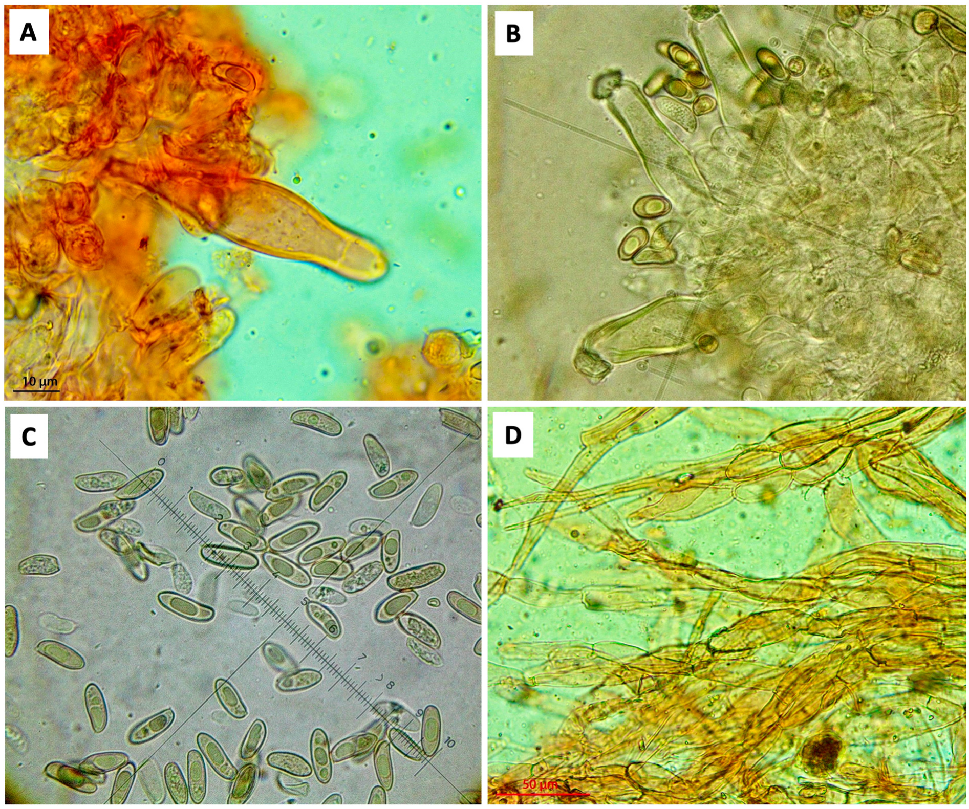
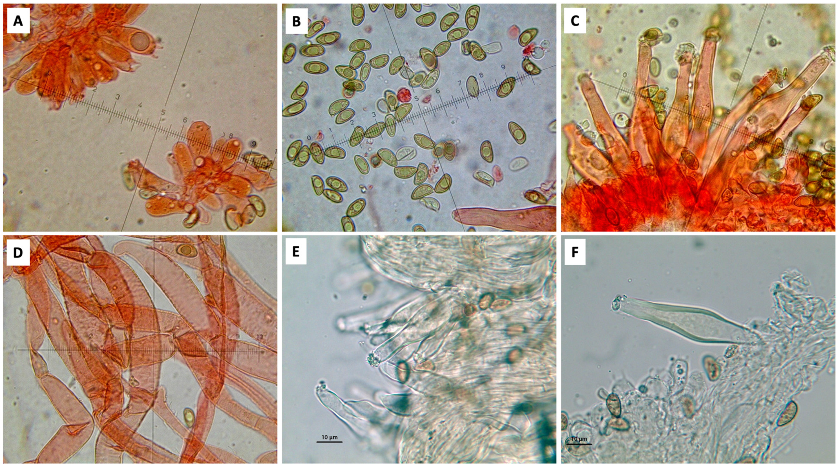
2.2. Morphological Observations

The description of the macromorphological characteristics was carried out on fresh basidiomata, which were photographed in the field using Canon 450D and Canon 1100D digital cameras (Canon Inc., London, UK). The study of the micromorphological characteristics was carried out on exsiccatae using Optika B-383 PLi (Optika Italy, Ponteranica, BG, Italy) and Motic BA 400 (Motic Europe, Barcelona, Spain) trinocular microscopes equipped with integrated digital cameras and with an Optech B4 (Optech Technology, Taipei City, Taiwan) binocular microscope equipped with a Nikon Coolpix SQ Optec digital camera (Nikon Europe B.V., Campi Bisenzio, FI, Italy). The following dyes and reagents were also used for the slide preparation: 2% anionic Congo Red, Phloxin, and 6% NH4OH. The exsiccatae were rehydrated with 5% KOH or with distilled H2O. The pileipellis was observed in both water and 2% anionic Congo Red. The sporal measurements were taken from at least 32 measurements obtained from the spore deposits of each of three distinct collections (186 spores in total), as indicated by Parmasto and Parmasto [25]. The dimensions of the basidiospores are given as (minimum–) average minus standard deviation–average–average plus standard deviation (–maximum) of length × (minimum–) average minus standard deviation–average–average plus standard deviation (–maximum) of width. The spore quotient (Q; length/width ratio) minimum–maximum is also reported. The Qm (=average quotient) is given by the arithmetic mean of single spore quotients. The hilar appendix was excluded from the sporal measurements; sterigmata were excluded from the basidia measurements. The dimensions of the basidia, cystidia, and their Q are expressed as the minimum–maximum value, followed by the average values. The color code used was that of Ridgway [26]. For the definitions in the morphological descriptions, we referred to Kuyper [27]. Collections of the new species were deposited at CAG (Cagliari Herbarium, Italy) with the numbers (CAG) n° B/5.6.4 (Inocybe halimiphila sp. nov., holotype) and B/5.6.5 (Inocybe rubripes sp. nov., holotype). Duplicates of the collections are found in the collection of the Department of Biomedical Sciences, University of Cagliari, Monserrato, Italy, and in the private herbaria of Alberto Mua (M, Quartu St. Elena, Italy) and Massimo Sanna (MS, Cagliari, Italy).

2.3. DNA Extraction, Amplification, and Sequencing

Total DNA was extracted in the Molecular Solutions laboratory (Portland, OR, USA). DNA was extracted from fungal tissue by heating with a Chelex extraction buffer (100 mM Tris with a pH of 8.5, 4% Chelex 100, and 1% Triton X-100) at 99 °C for 20 min, then freezing. After thawing, the resulting supernatant was used in the PCR. The PCR was performed in 25 mL reactions with 1 µL of DNA extract, 0.4 mM of each primer, 0.2 mM of the dNTP mixture, 5 µg of bovine serum albumin, and 0.5 U of OneTaq Hot Start DNA polymerase (New England Biolabs, Ipswich, MA, USA) in 1X OneTaq standard buffer. The PCR conditions were as follows: 94 °C for 30 s, followed by 36 cycles of 94 °C for 15 s, 57 °C for 30 s, and 68 °C for 60 s, followed by a final extension at 68 °C for 5 min. The primers ITS1F [28] and ITS4 [29] were used to amplify the ITS1-5.8S-ITS2 region. Sequencing was performed at Eurofins Genomics (Louisville, KY, USA) using the same primers used for the PCR. Forward and reverse reads were aligned using Genious 10.2.4 and resolved manually, as needed.

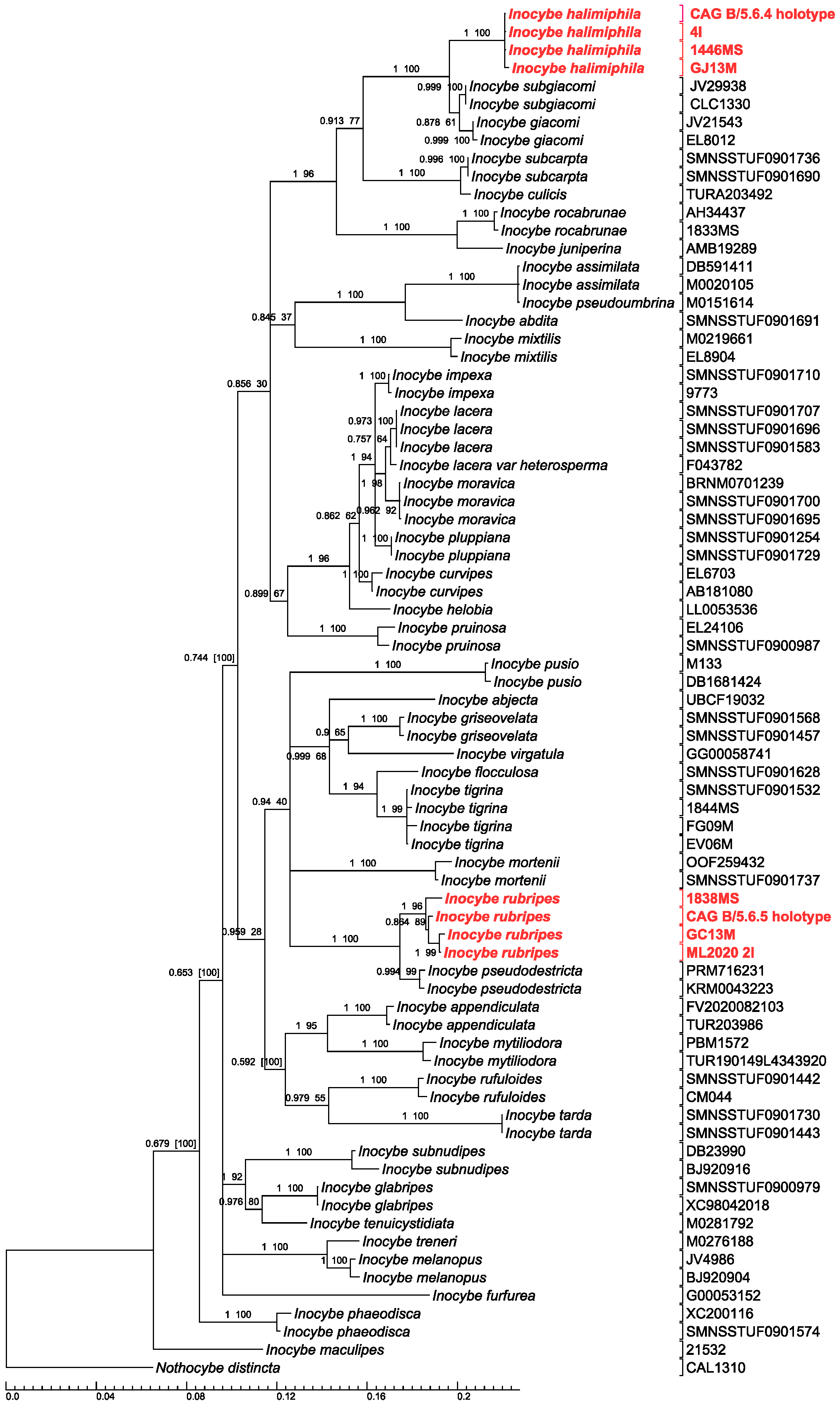
2.4. Phylogenetic Analysis

The sequences used in the phylogenetic analysis were retrieved from GenBank based on the BLASTn results [30] and from phylogenetic studies that focused on Inocybe. The final nrITS and nrLSU datasets containing 76 and 45 total sequences, respectively, were aligned with the MUSCLE application of the MEGA X software, version 10.2.2 [31], manually refining the alignment and concatenating with Mesquite v. 3.61 [32]. Maximum Likelihood analysis (ML) was performed with RAxMLGUI 2.0 version 2.0.10 [33] using 1000 bootstrap replicates and the GTRGAMMA substitution model for the three delimited partitions (nrITS1+2, 5.8S, nrLSU) support. Bayesian Inference analysis (BI) was carried out with MrBayes v. 3.2.7 on the XSEDE application in the CIPRES (Cyber Infrastructure for Phylogenetic RESearch) portal [34,35], setting as parameters 1 × 10⁷ generations, the burnin fraction to 0.25, the sample frequency to 1000, and the number of chains to 4 and 3673 characters. The phylogenetic trees thus obtained were processed with the TREEGRAPH2 program. The Nothocybe distincta (K.P.D. Latha & Manim) Matheny & K.P.D. Latha sequence KX171343 (GenBank) was chosen as the outgroup. The significance threshold was set to ≥0.95 for the Bayesian posterior probability (BPP) and ≥70% for the ML bootstrap (MLB) values.

3. Results and Discussion
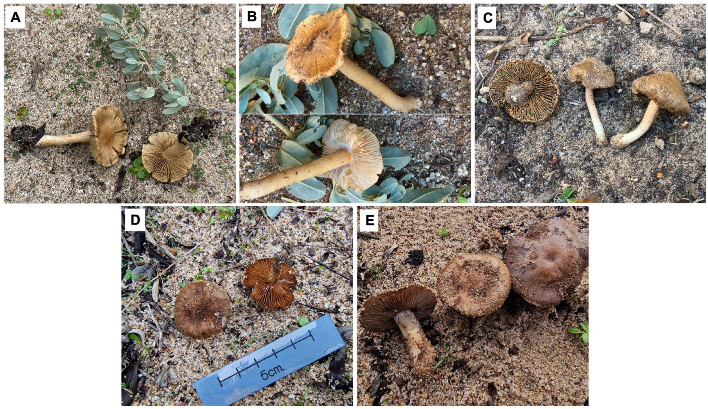
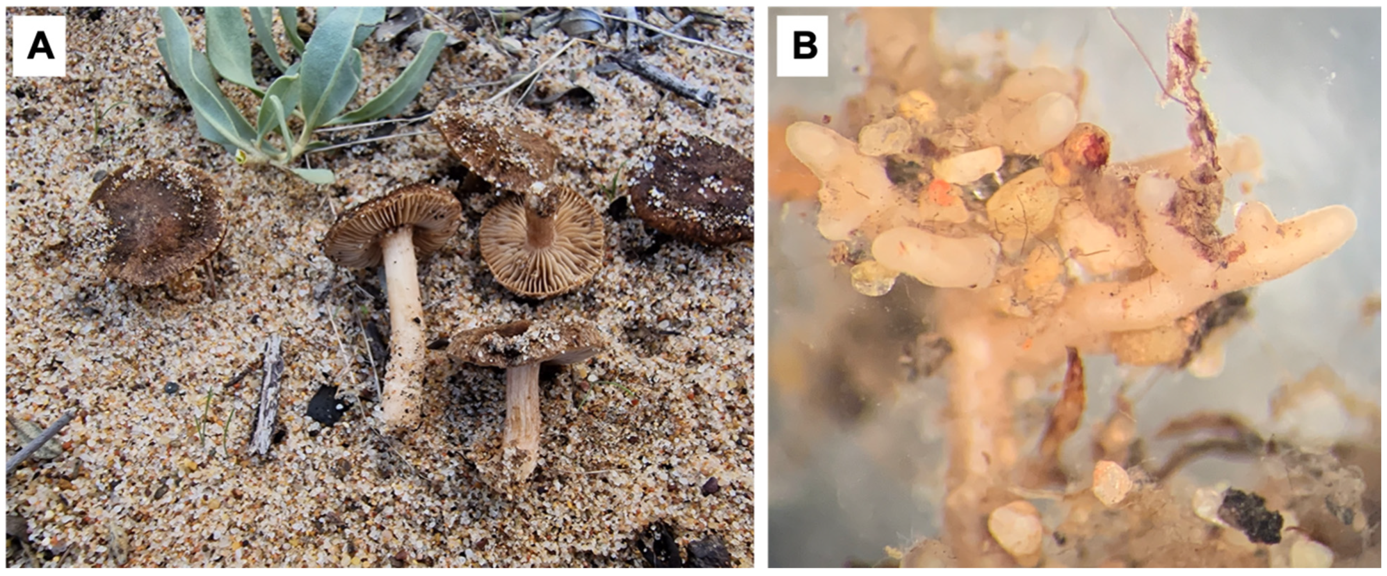
Table 1 lists the 17 species of Inocybe and Pseudosperma that are currently reported as linked to Halimium spp., mostly on the basis of sporocarp occurrence in the proximity of the potential host plant. Over 40% (7 of 17) of the recorded species come from our own study based on observations in pure Halimium stands in southwestern Sardinia, Italy. These include several new Inocybe species, two of which (Inocybe halimiphila sp. nov. and Inocybe rupribes sp. nov.) are described in the present article, while the others will be the object of future contributions. Considering the close systematic and ecological proximity of Halimium and Cistus, and the fact that they usually share many ectomycorrhizal mycobionts, see [16], it is somehow surprising that, in our Halimium surveys, we found a single Inocybaceae species previously linked to Cistus. It is Inocybe rocabrunae Esteve-Rav. & Vila (Figure 2), a species described from Cistus shrublands in Spain and characterized by spores with an angular to subnodulose contour [36]. Besides I. rocabrunae, just one other species previously reported as associated with Cistus has been somehow linked to Halimium, Inocybe pruinosa R. Heim, while two others, Inocybe iberilepora Fachada & Bandini and Inocybe phaeosquamosa Fachada & Bandini, have been described from Cistaceae ecosystems, with the co-occurrence of Cistus and Halimium [37] (Table 1).
Inocybe tigrina R. Heim (Figure 2) was certainly the most commonly encountered species during our Halimium surveys. This smooth-spored taxon has been recently discussed in detail by Bandini and colleagues, who also designated an epitype for it on the basis of material collected in Germany [42]. The species has a large European distribution (it has also been reported elsewhere, like China) and a flexible mycorrhizal biology, apparently entering into ectomycorrhizal relationship with a range of symbionts, both broadleaved and coniferous (e.g., Quercus, Tilia, Populus, Fagus, Pinus, Picea, and so on). Our findings in thermophilic Mediterranean shrublands confirm the ecological plasticity of I. tigrina. Another interesting finding of our surveys in Halimium shrublands is Pseudosperma sp. (Figure 2). The initial BLAST analysis of the ITS sequences of our collections with either GenBank and Unite databases showed Pseudosperma mimicum (Massee) Matheny & Esteve-Rav. (=Inocybe mimica Massee) as the closest match, but with a 97% identity or below, while the LSU sequences matched with the same species, but with a greater similarity (over 99%). Pseudosperma mimicum is a rare species, reported mostly in northern Europe, where it occurs associated with Betula, Picea, and Pinus, but also from Pinus wallichiana forests in Pakistan [43,44]. As we are waiting for more data, the classification of our collections is thus provisional, possibly pointing to a new, so far undescribed Pseudoderma species linked to Halimium, the first for this genus.
A total of 12 nrITS and 9 nrLSU sequences were generated de novo from the Sardinian collections of Inocybe and Pseudosperma under H. halimifolium (Table 1), while the dataset used to infer the phylogenetical tree of Inocybe spp. comprised a total of 76 nrITS sequences (10 newly generated and 66 from GenBank) and 45 nrLSU sequences (8 newly generated and 37 from GenBank), respectively (see Table 2). All of the newly generated sequences were obtained from Sardinian collections (Table 1). The tree produced following the Bayesian Inference and Maximum Likelihood analyses is shown in Figure 3, with the bootstrap (MLB) and posterior probability (BPP) values corresponding to the individual nodes. The final concatenated multi-locus (nrITS1+2, 5.8S and nrLSU) dataset used for inferring the ML and Bayesian phylogenies, which consisted of 76 specimens, had a total number of 3673 bp. The multi-locus phylogeny produced with RAxML had a Final ML Optimization Likelihood value of −16,924.678604, whereas the MrBayes analysis reached an average standard deviation of split frequencies of 0.005 after 6.77 × 10⁶ generations.
The new species, Inocybe halimiphila, formed a single, strongly supported clade (MLB = 100%; BPP = 1) (Figure 3). In the phylogenetic analysis, the closest species are Inocybe giacomi Favre and Inocybe subgiacomi C.L. Cripps, Vauras & E. Larss, from which I. halimiphila nevertheless differs due to its smooth and non-nodular spores (Figure 4 and Figure 5). The distance of the nrITS type sequence of the new I. halimiphila compared with the sequences of the closest species in the BLASTN suite ranged from 2.16% (Inocybe sp., accession number JQ711960) to 3.97% (I. giacomi, accession number MK153653). As for the shape and size of the spores, I. halimiphila is similar to Inocybe lacera (Fr.) P. Kumm., a species that has also been observed to be in association with H. halimifolium (see Table 1). However, the two species are phylogenetically distinct (Figure 3) and differ in several macro- and micromorphological features (Figure 4 and Figure 5; see the Taxonomy section for further details).
In our analysis, the sequences of Inocybe rubripes equally formed a strongly supported clade (MLB = 96%; BPP = 1) that is placed near Inocybe pseudodestricta Stangl & J. Veselský (Figure 1). The genetic distance of the nrITS type sequence of I. rubripes from the closest GenBank entries ranged from 2.40% (Inocybe sp., environmental sample, accession number JQ898553) to 5.46% (Inocybe glabrescens Velen., accession number HQ604513). Inocybe rubripes has a brown cap, with radial fibrils, fairly dense clay–brown gills with a yellowish tinge, and a stem tinged reddish pink, widened at the base, with whitish fibrils (Figure 6 and Figure 7). The main characteristics distinguishing I. rubripes from the morphologically and/or phylogenetically closest Inocybe species, such as Inocybe roseipes Malençon and I. pseudodestricta, are discussed in the Taxonomy section.
Through the random sampling of soil in the proximity of H. halimifolium shrubs during our study, we were able to detect and confirm molecularly the ectomycorrhizae formed by the two new Inocybe species herewith described (Table 1; Figure 4 and Figure 6). This finding is significant because it confirms the ectomycorrhizal relationship between both I. halimiphila and I. rubripes and Halimium. In addition, since the new species are so far known only from the type locality and a single host, they could be the first Halimium-specific ectomycorrhizal fungal species ever reported and known both as sporocarp and mycorrhiza. The ectomycorrhizae of both species display the classic features of the Inocybe ectomycorrhizae described so far [45,46], such as pale color tones (whitish to yellowish brown) and a mantle surface with a cottony appearance (Figure 4 and Figure 6). In both cases, the mycorrhizal system varies from 0.5 to 2 mm in length, from monopodial pinnate to coralloid, with the presence of hyphal strands.
A final comment on conservation-related issues is necessary. Indeed, after being traditionally largely neglected in biodiversity conservation plans, fungi are eventually in the spotlight. Thanks to the increasingly appreciated ecological role of these organisms, their close relationships with both plants and animals, and their so far ungauged but bewildering diversity, fungi are more and more frequently the object of both the assessment of conservation threats and of specific conservation actions [47,48]. However, given the specific features of fungi biology and life cycles (e.g., fructification of some species is rarely observed, or can occur with lapses that can last decades), to decide which kind of measures could be put in place to conserve an endangered species it is not always straightforward, although habitat and/or host protection seem in most cases a viable possibility. Under this point of view, putting into action protective measures aimed at conserving the Halimium shrublands of southwestern Sardinia is much needed. This ecosystem is not only the locus tipicus of I. halimiphila, I. rubripes, and of other new Inocybe and Pseudosperma species under study, it is also a thriving ectomycorrhizal community from which new species belonging to other fungal families and genera have already been described, like Cyanoboletus mediterraneensis Biketova, A. Rinaldi & Simonini [49], and Coltricia insularis P.-A. Moreau, Bellanger, Loizides & A. Rinaldi [50].

4. Taxonomy

Inocybe halimiphila Mua, Sanna, A. Rinaldi & Casula, sp. nov. (Figure 4 and Figure 5).
Type: Italy, Sardinia, Gonnesa (CA), Guroneddu locality, about 100 m asl, on sandy dune soil near the sea, under Halimium halimifolium, 22 December 2022, voucher GA15M (holotype in CAG B/5.6.4), legit A. Mua, M. Sanna, and A.C. Rinaldi, ITS sequence: GenBank PP941799, LSU sequence: GenBank PP941805. Isotypus in Mua herbarium (GA15M) and in Sanna herbarium (1496MS).
MycoBank # 855453
Description: Pileus: 20–30 mm in diameter, hemispherical then convex, finally flat, often with a wide and low umbo; margin first facing downwards then extended; dry surface, more or less smooth on the disc, without veil residues, fibrillose towards the periphery, dark brown in color. Lamellae: unmarginated or hooked, medium-spaced, quite thick, a little pot-bellied when ripe, pale ocher–gray then ocher–brown in color; more or less regular thread, slightly fimbriated, lighter than the faces; few lamellulae: L/l = 1/1. Stipe: 30–70 × 6–8 mm, cylindrical, even slightly curved, not bulbous, whitish–ocher throughout its length, crossed by longitudinal fibrils; fistulous at maturity; no partial film residues observed. Context: firm, white. Smell: spermatic odor, flavor not significant. Basidiospores: (9.62) 10.94–12.75–14.57 (15.48) × (3.74) 4.01–4.44–14.57 (5.5) µm, Qm = 2.87 (70 measurements), smooth, cylindrical, yellowish, often a little irregular, subreniform (tattered type), largely monoguttulate, less frequently with more than one guttula or with granular contents, little visible apicle, sublateral. Basidia: 20–30 × 7–11 µm, clavate tetrasporic, buckled at the base, sterigmata 2.5–3 µm long. Cheilocystidia: (46.3) 47.3–53.6 (65) × (14.4) 14.5–17 (18) µm, Qm = (2.8) 3.2–3.5 (3.7), n = 7, wall thickness of (1.08) 1.2–1.85 (3.15) µm, abundant, metuloid, fusiform, subcapitulate, with an apex partly adorned with crystals, more often naked, with walls clearly yellowing with KOH and Na4OH. Pleurocystidia: abundant, very similar to cheilocystidia in shape, size, and the presence of crystals. Paracystidia: 10–20 × 5–12 µm, clubbed, numerous, with thin parts, mixed with cheilocystidia. Caulocystidia: very rare or absent; if present, localized only at the apex of the stem, without crystals, with a thin wall. Pileipellis: cutis type consisting of a suprapellis with a thin layer of thin hyphae, 1–3 µm thick, with a long distance between the septa, with terminal elements with an obtuse apex and with the presence of fine encrusting membrane pigment; the mediopellis is made up of sausage-shaped hyphae, 12–25 µm long and 7–16 µm wide, with fine encrusting parietal and yellow–brown cytoplasmic pigment; subpellis little differentiated from the mediopellis, with even thicker hyphae, up to 20 µm. Buckle joints present and abundant.
Etymology: The epithet refers to the symbiotic link of the new species with Halimium.
Additional materials examined: Paratypus, voucher 1446MS, collected on 28 November 2018, legit M. Sanna, A. Mua, and A.C. Rinaldi, in Guroneddu locality, municipality of Gonnesa, Sardinia (Italy), on sandy dune soil near the sea, in pure Halimium halimifolium stands, deposited in the Sanna herbarium (1146MS). GenBank PP941800. Paratypus, voucher GJ13M, collected on 20 December 2023, legit A. Mua and A.C. Rinaldi, in Guroneddu locality, municipality of Gonnesa, Sardinia (Italy), on sandy dune soil near the sea, in pure Halimium halimifolium stands, deposited in the Mua herbarium (GJ13M). GenBank PP941802 (ITS), PP941806 (LSU). Collection of mycorrhiza H. halimifolium + I. halimiphila on 29 May 2018, legit Andrea Rinaldi, in Guroneddu locality, municipality of Gonnesa, Sardinia (Italy), on sandy dune soil near the sea, in pure Halimium halimifolium stands. GenBank MT594518 (ITS).
Ecology and distribution: Terrestrial, gregarious, in Mediterranean shrubland, with Halimium halimifolium, in autumn and early winter. At present, basidiocarps and ectomycorrhizae collected only from the type locality in Sardinia (Italy).
Notes: Inocybe halimiphila, despite having smooth spores, is similar from a phylogenetic point of view, positioning itself in the same clade, to Inocybe giacomi Favre and Inocybe subgiacomi C.L. Cripps, Vauras & E. Larss., both with nodular spores (Figure 3). Inocybe giacomi differs in the larger size of the basidiomes, the adnate lamellae, the shorter and more nodular spores, the cheilocystidia and pleurocystidia, almost completely adorned with crystals at the apex, and the nordic or alpine habitat, near Betula sp., Pinus sylvestris, Dryas octopetala, and Salix reticulata [51]. The species has been reported in both Europe and North America [52]. Inocybe subgiacomi differs in its slightly nodular, shorter spores, the basidia, which are on average longer, the longer cheilocystidia, with the apex always adorned with crystals, and the nordic or alpine habitat near D. octopetala, S. reticulata, and Salix herbacea on calcareous soil. The species has been reported in the USA and northern Europe [52]. The spores of I. halimiphila are similar to those of Inocybe lacera (Fr.) P. Kumm., but differs from the latter in the smaller average size of the basidiomes, in the darker colored cap, for the absence of traces of veil on the stem, for the regular, non-polymorphic cheilocystidia, for the rare presence or even absence of caulocystidia, and for the spores, which are on average smaller. Moreover, the two species have a fair phylogenetic distance, positioning themselves in different clades (Figure 3). Inocybe lacera and related species also have genetic affinity with species with nodular spores, such as, for example, with Inocybe curvipes P. Karst. and, to a lesser extent, with Inocybe goniopusio Stangl. In particular, Inocybe pluppiana Bandini, B. Oertel & U. Eberh., a species very close to I. lacera, has almost smooth spores [53], such as to represent a transition point between leiospores and nodulospores.
 
Inocybe rubripes Sanna, Mua, A. Rinaldi & Casula, sp. nov. (Figure 6 and Figure 7).
Type: Italy, Sardinia, Gonnesa (CA), Guroneddu locality, about 100 m asl, on sandy dune soil near the sea, under Halimium halimifolium, 27 January 2024, voucher 1926MS (holotype in CAG B/5.6.5), legit A. Mua, M. Sanna, and A.C. Rinaldi, ITS sequence: GenBank PP941801, LSU sequence: GenBank PP941847. Isotypus in Mua herbarium (GA17M) and in Sanna herbarium (1926MS).
MycoBank # 855454
Description: Pileus: 8–42 mm in diameter, conical to convex, the presence of obtuse umbo even in adult specimens; dry cuticle, brown in color, joined to the disc, with evident radial fibrils elsewhere; margin initially involuted then rounded, entire, sometimes broken; presence of scanty white veiled residues only in very young specimens. Lamellae: quite dense, adnate-marginated, ventricose, sinuous, first clay then clay–brown, tinged with a yellowish, lighter thread, whitish, entire. Stipe: 20–45 × 4–0.9 mm, cylindrical, slightly enlarged at the bottom, whitish, then tinged with reddish–pinkish tones at the base, pruinose up to the median area, crossed by fine whitish fibrils in adult specimens. Context: white in the cap, whitish in the stem, and yellowish in its base. Smell: light, herbaceous–sour. Basidiospores: (8) 9.35–10.21–11.09 (12) × (4) 5.01–5.59–6.16 (6.33) µm, Qm = 1.84 (84 measurements), smooth, amygdaliform, subreniform, slightly pronounced apicle, sub-rounded apex, sub-ogival or even ogival, with a thick wall, brown in mass. Cheilocystidia: measuring (52.85) 56.77 (66.28) × (11.02) 13.53 (13.97) µm, metuloid, pin-shaped, sub-lageniform, sub-cylindrical, not capitulated, even with one septum, a 1–2.5 µm thick wall reacting −/+ with NH4OH, partly free of crystals or slightly muricated with small–medium-sized crystals. Paracystidia: numerous, pyriform. Pleurocystidia: metuloid, fusiform, pin-shaped, non-capitulated, muricate, walls 2–2.5 µm thick, −/+ with NH4OH., 55.35–73.90 × 14.13–16.30 µm. Basidia: claviform, 28–30 × 8–10 µm, tetrasporic, with guttula. Caulocystidia: in the upper third, there are caulocystidia, arranged in tufts, with a 0.5–1.5 µm thick wall, sub-lageniform, sub-fusiform, also muricate, with small- and medium-sized crystals, also with one septum near the base, (44.86) 45.915 (61.47) × (6.27) 11.035 (11.48) µm, mixed with club-shaped and cylindrical non-metuloid caulocystidioid elements, arranged in tufts, 28.63–8.38 × 7.80–11.10 µm; in the lower third, there are piliform, cylindrical structures, with a 0.5 µm thick wall, non-metuloid and non-muric, also septate, in tufts, 46.69–86.42 × 7.58–8.68 µm. Pileipellis: cutis type, suprapellis formed by cylindrical hyphae 4–9 µm wide, the presence of brown cytoplasmic and encrusting pigment, hyphae of the mediopellis up to 13 µm wide, and in the subpellis, the presence of hyphae up to 15 µm wide. Buckle joints present.
Etymology: The epithet refers to the reddish–pinkish tones at the base of the stipe.
Additional materials examined: Paratypus, voucher 1838MS, collected on 14 January 2023, legit M. Sanna, A. Mua, and A.C. Rinaldi, Guroneddu locality, municipality of Gonnesa, Sardinia (Italy), on sandy dune soil near the sea, in pure Halimium halimifolium stands, deposited in the Sanna herbarium (1838MS). GenBank PP941803 (ITS), PP941846 (LSU). Paratypus, voucher GC13M, collected on 14 January 2023, legit A. Mua and A.C. Rinaldi, Guroneddu locality, municipality of Gonnesa, Sardinia (Italy), on sandy dune soil near the sea, in pure Halimium halimifolium stands, deposited in the Mua herbarium (GC13M). GenBank PP941804 (ITS). Collection of mycorrhiza H. halimifolium + I. rubripes on 29 May 2018, legit Andrea Rinaldi, Guroneddu locality, municipality of Gonnesa, Sardinia (Italy), on sandy dune soil near the sea, in pure Halimium halimifolium stands. GenBank MT594517 (ITS).
Ecology and distribution: Terrestrial, gregarious, in Mediterranean shrubland, with Halimium halimifolium, in early winter. At present, basidiocarps and ectomycorrhizae collected only from the type locality in Sardinia (Italy).
Notes: Inocybe rubripes is characterized by a fibrillose cap, a stem with pink–reddish shades, and the presence of caulocystides in the upper third of the stem. Several species of Inocybe have these characteristics, including those that Bon places in the subsection Tardinae [54]. Inocybe roseipes Malençon differs in the cap, defined as woolly fibrillose with the disc generally lighter in color than the remaining part of the cap, the flesh with a weak spermatic odor, the habitat linked to Cedrus, the cheilocystids with thicker walls compared to I. rubripes, and the presence of basidia in the lamellar thread and the pleurocystidia, which have a mucous bubble at the apex, where immersed crystals can be observed [55]. Inocybe pseudodestricta Stangl & J. Veselský, the closest from the point of view of phylogeny, differs in the greater robustness of the basidiomes, the reddish–brown center of the cap, the hymenial cystidia, which are on average larger, and the habitat under oak, beech, lime, and birch [56].

5. Conclusions

The current study confirms the high biodiversity of the ectomycorrhizal community inhabiting Halimium shrublands, which host a number of so far undescribed species of macromycetes. Our results also reassert the knowledge of Inocybaceae as one of the most diverse families of ectomycorrhizal basidiomycetes, and it is quite safe to speculate that future research conducted in cistaceous shrublands will lead to a further, significant increase in newly described species.

Author Contributions

Conceptualization, M.S., M.C., A.M. and A.C.R.; methodology, M.S., A.M., M.C. and A.C.R.; software, M.S.; validation, M.S., M.C., A.M. and A.C.R.; formal analysis, M.S., A.M. and A.C.R.; investigation, M.S., A.M. and A.C.R.; resources, A.C.R.; data curation, M.S. and A.C.R.; writing—original draft preparation, M.S., A.M. and A.C.R.; writing—review and editing, M.S., A.M. and A.C.R.; visualization, M.S., A.M., M.C. and A.C.R.; supervision A.C.R.; project administration, M.S. and A.C.R.; funding acquisition, A.C.R. All authors have read and agreed to the published version of the manuscript.

Funding

This research was funded by a PRIN PNRR research grant from the Italian Ministry of Research and University, awarded to A.C.R., in the frame of the project entitled “Tapping into the biological potential of wild mushrooms from a range of ecosystems in Sardinia and Abruzzi: FUNSarAbr” (F53D23012210001).

Institutional Review Board Statement

Not applicable.

Data Availability Statement

Publicly available datasets were analyzed in this study. These data can be found at http://www.ncbi.nlm.nih.gov/ (accessed on 5 January 2024) and https://unite.ut.ee/ (accessed on 10 January 2024).

Conflicts of Interest

The authors declare no conflicts of interest.

References

  1. Henne, P.D.; Elkin, C.; Franke, C.; Colombaroli, D.; Calò, C.; La Mantia, T.; Pasta, S.; Conedera, M.; Dermody, O.; Tinner, W. Reviving extinct Mediterranean forests communities may improve ecosystem potential in a warmer future. Front. Ecol. Environ. 2015, 13, 356–362. [Google Scholar] [CrossRef] [PubMed]
  2. Guarino, R.; Vrahnakis, M.; Rodriguez Rojo, M.P.; Giuga, L.; Salvatore, P. Grasslands and Shrublands of the Mediterranean Region. In Encyclopedia of the World’s Biomes; Goldstein, M.I., DellaSala, D.A., Eds.; Elsevier: Amsterdam, The Netherlands, 2020; pp. 638–655. [Google Scholar]
  3. Comandini, O.; Contu, M.; Rinaldi, A.C. An overview of Cistus ectomycorrhizal fungi. Mycorrhiza 2006, 16, 381–395. [Google Scholar] [CrossRef] [PubMed]
  4. WorldPlants. Available online: https://www.worldplants.de (accessed on 24 May 2024).
  5. Govaerts, R.; Nic Lughadha, E.; Black, N.; Turner, R.; Paton, A. The World Checklist of Vascular Plants, a continuously updated resource for exploring global plant diversity. Sci. Data 2021, 8, 215. [Google Scholar] [CrossRef] [PubMed]
  6. The World Flora Online Plant List. Available online: https://wfoplantlist.org/ (accessed on 24 May 2024).
  7. Guzmán, B.; Vargas, P. Systematics, character evolution, and biogeography of Cistus L. (Cistaceae) based on ITS, trnL-trnF, and matK sequences. Mol. Phylogenet. Evol. 2005, 37, 644–660. [Google Scholar] [CrossRef] [PubMed]
  8. Guzmán, B.; Vargas, P. Historical biogeography and character evolution of Cistaceae (Malvales) based on analysis of plastid rbcL and trnL-trnF sequences. Org. Divers. Evol. 2009, 9, 83–99. [Google Scholar] [CrossRef]
  9. Civeyrel, L.; Leclercq, J.; Demoly, J.-P.; Agnan, Y.; Quèbre, N.; Pélissier, C.; Otto, T. Molecular systematics, character evolution, and pollen morphology of Cistus and Halimium (Cistaceae). Plant Syst. Evol. 2011, 295, 23–54. [Google Scholar] [CrossRef]
  10. Zunzunegui, M.; Díaz Barradas, M.C.; Aguilar, F.; Ain Lhout, F.; Clavijo, A.; García Novo, F. Growth response of Halimium halimifolium at four sites with different soil water availability regimes in two contrasted hydrological cycles. Plant Soil 2002, 247, 271–281. [Google Scholar] [CrossRef]
  11. Zunzunegui, M.; Ain-Lhout, F.; Díaz Barradas, M.C.; Álvarez-Cansino, L.A.; Esquivias, M.P.; García Novo, F. Physiological, morphological and allocation plasticity of a semi-deciduous shrub. Acta Oecol. 2009, 35, 370–379. [Google Scholar] [CrossRef]
  12. Camprubi, A.; Estaun, V.; Calvet, C. Greenhouse inoculation of psammophilic plant species with arbuscular mycorrhizal fungi to improve survival and early growth. Eur. J. Soil Biol. 2011, 47, 194–197. [Google Scholar] [CrossRef]
  13. Buscardo, E.; Rodríguez-Echeverría, S.; Barrico, L.; García, M.Á.; Freitas, H.; Martín, M.P.; De Angelis, P.; Muller, L.A.H. Is the potential for the formation of common mycorrhizal networks influenced by fire frequency? Soil Biol. Biochem. 2012, 46, 136–144. [Google Scholar] [CrossRef]
  14. Beddiar, A.; Adouane, M.; Boudiaf, I.; Fraga, A. Mycorrhizal status of main spontaneous or introduced forest trees in El Tarf province (Algerian north-east). Online J. Sci. Technol. 2015, 5, 40–45. [Google Scholar]
  15. Leonardi, M.; Neves, M.A.; Comandini, O.; Rinaldi, A.C. Scleroderma meridionale ectomycorrhizae on Halimium halimifolium: Expanding the Mediterranean symbiotic repertoire. Symbiosis 2018, 76, 199–208. [Google Scholar] [CrossRef]
  16. Leonardi, M.; Furtado, A.N.M.; Comandini, O.; Geml, J.; Rinaldi, A.C. Halimium as an ectomycorrhizal symbiont. New records and an appreciation of known fungal diversity. Mycol. Prog. 2020, 19, 1495–1509. [Google Scholar] [CrossRef]
  17. Jülich, W. Higher Taxa of Basidiomycetes; J. Cramer: Vaduz, Liechtenstein, 1981; Volume 85, pp. 1–485. [Google Scholar]
  18. Matheny, P.B. Improving phylogenetic inference of mushrooms using RPB1 and RPB2 sequences (Inocybe; Agaricales). Mol. Phylogenet. Evol. 2005, 35, 1–20. [Google Scholar] [CrossRef] [PubMed]
  19. Ryberg, M.; Larsson, E.; Jacobsson, S. An evolutionary perspective on morphological and ecological characters in the mushroom family Inocybaceae (Agaricomycotina, Fungi). Mol. Phylogenet. Evol. 2010, 55, 431–442. [Google Scholar] [CrossRef] [PubMed]
  20. Matheny, P.B.; Hobbs, A.M.; Esteve-Raventós, F. Genera of Inocybaceae: New skin for the old ceremony. Mycologia 2020, 112, 83–120. [Google Scholar] [CrossRef] [PubMed]
  21. Cervini, M. Inocybe messapica (Inocybaceae, Agaricales, Basidiomycota), a new species in section Splendentes, from Mediterranean oak woods. Phytotaxa 2021, 480, 174–184. [Google Scholar] [CrossRef]
  22. Dovana, F.; Bizio, E.; Garbelotto, M.; Ferisin, G. Inocybe cervenianensis (Agaricales, Inocybaceae), a new species in the I. flavoalbida clade from Italy. Phytotaxa 2021, 484, 227–236. [Google Scholar] [CrossRef]
  23. Sanna, M.; Mua, A.; Porcu, G.; Casula, M.; Rinaldi, A.C.; Mifsud, S.; Garrido-Benavent, I. Pseudosperma calciphilum (Inocybaceae), a new Mediterranean species Sardinia (Italy), Malta, and Valencia (Spain). Phytotaxa 2024, 633, 253–264. [Google Scholar] [CrossRef]
  24. Sanna, M.; Mua, A.; Porcu, G.; Casula, M.; Rinaldi, A.C. Pseudosperma subvolvatum (Inocybaceae), sp. nov. Fungal Divers. Notes 2024, in press. [Google Scholar]
  25. Parmasto, E.; Parmasto, I. Variation in Basidiospores in the Hymenomycetes and Its Significance to Their Taxonomy; Cramer in d. Borntraeger-Verl.-Buchh: Berlin, Germany, 1987; Volume 115, pp. 1–168. [Google Scholar]
  26. Ridgway, R. Color Standards and Color Nomenclature; The Author: Washington, DC, USA, 1912; pp. 1–172. [Google Scholar]
  27. Kuyper, T.W. A revision of the genus Inocybe in Europe I. Subgenus Inosperma and the smooth-spored species of subgenus Inocybe. Persoonia 1986, 3, 1–247. [Google Scholar]
  28. Gardes, M.; Bruns, T.D. ITS primers with enhanced specificity for Basidiomycetes—Application to the identification of mycorrhizae and rusts. Mol. Ecol. 1993, 2, 113–118. [Google Scholar] [CrossRef]
  29. White, T.J.; Bruns, T.; Lee, S.; Taylor, J. Amplification and direct sequencing of fungal ribosomal RNA genes for phylogenetics. In PCR Protocols: A Guide to Methods and Applications; Innis, M.A., Gelfand, D.H., Sninsky, J.J., White, T.J., Eds.; Academic Press: New York, NY, USA, 1990; pp. 315–322. [Google Scholar]
  30. Altschul, S.F.; Gish, W.; Miller, W.; Myers, E.W.; Lipman, D.J. Basic local alignment search tool. J. Mol. Biol. 1990, 215, 403–410. [Google Scholar] [CrossRef] [PubMed]
  31. Kumar, S.; Stecher, G.; Li, M.; Knyaz, C.; Tamura, K. MEGA X: Molecular evolutionary genetics analysis across computing platforms. Mol. Biol. Evol. 2018, 35, 1547–1549. [Google Scholar] [CrossRef]
  32. Maddison, W.P.; Maddison, D.R. Mesquite: A Modular System for Evolutionary Analysis, Version 3.61. 2019. Available online: http://mesquiteproject.org (accessed on 18 February 2024).
  33. Edler, D.; Klein, J.; Antonelli, A.; Silvestro, D. raxmlGUI 2.0: A graphical interface and toolkit for phylogenetic analyses using RAxML. Methods Ecol. Evol. 2021, 12, 373–377. [Google Scholar] [CrossRef]
  34. Rambaut, A. FigTree, v1. 4.0; University of Oxford: Oxford, UK, 2012. Available online: http://tree.bio.ed.ac.uk/software/figtree (accessed on 15 February 2024).
  35. Miller, M.A.; Pfeiffer, W.; Schwartz, T. Creating the CIPRES Science Gateway for Inference of Large Phylogenetic Trees Conference: Gateway Computing Environments Workshop (GCE). In Proceedings of the 2010 Gateway Computing Environments Workshop (GCE), New Orleans, LA, USA, 14 November 2010; pp. 1–8. [Google Scholar]
  36. Esteve-Raventós, F.; Vila, J.; Llimona, X. Estudios sobre el género Inocybe (Cortinariales) en los jarales de Cataluña. I. Rev. Catalana Micol. 2002, 24, 135–146. [Google Scholar]
  37. Fachada, V.; Bandini, D.; Beja-Pereira, A. Two new species of Inocybe from Mediterranean Cistaceae heathlands. Mycologia 2024, 116, 1–16. [Google Scholar] [CrossRef]
  38. Moreno-Arroyo, B. (Ed.) Inventario Micológico Básico de Andalucía; Consejería de Medio Ambiente, Junta de Andalucía: Córdoba, Spain, 2004; pp. 1–680. [Google Scholar]
  39. Moreau, P.A.; Corriol, G.; Borgarino, D.; Auber, P.; Lavoise, C.; Richard, F.; Selosse, M.-A. Contribution à la connaissance des champignons de l’étage thermoméditerranéen Corse II. Bull. Semest. Féd. Assoc. Mycol. Méditerr. 2007, 31, 9–31. [Google Scholar]
  40. Moreau, P.A.; Corriol, G.; Borgarino, D.; Auber, P.; Lavoise, C. Contribution à la connaissance des champignons de l’étage thermoméditerranéen Corse III. Bull. Semest. Féd. Assoc. Mycol. Méditerr. 2007, 31, 33–84. [Google Scholar]
  41. Alvarado, P.; Manjón, J.L.; Matheny, P.B.; Esteve-Raventós, F. Tubariomyces, a new genus of Inocybaceae from the Mediterranean region. Mycologia 2010, 102, 1389–1397. [Google Scholar] [CrossRef]
  42. Bandini, D.; Oertel, B.; Eberhardt, U. A fresh outlook on the smooth-spored species of Inocybe: Type studies and 18 new species. Mycol. Prog. 2021, 20, 1019–1114. [Google Scholar] [CrossRef]
  43. Larsson, E.; Ryberg, M.; Moreau, P.-A.; Delcuse Mathiesen, Å.; Jacobsson, S. Taxonomy and evolutionary relationships within species of section Rimosae (Inocybe) based on ITS, LSU and mtSSU sequence data. Persoonia 2009, 23, 86–98. [Google Scholar] [CrossRef]
  44. Saba, M.; Ahmad, I.; Khalid, A.N. New reports of Inocybe from pine forests in Pakistan. Mycotaxon 2015, 130, 671–681. [Google Scholar] [CrossRef]
  45. Agerer, R. Fungal relationships and structural identity of their ectomycorrhizae. Mycol. Prog. 2006, 5, 67–107. [Google Scholar] [CrossRef]
  46. Daskalopoulos, V.; Polemis, E.; Fryssouli, V.; Kottis, L.; Bandini, D.; Dima, B.; Zervakis, G.I. Mallocybe heimii ectomycorrhizae with Cistus creticus and Pinus halepensis in Mediterranean littoral sand dunes-assessment of phylogenetic relationships to M. arenaria and M. agardhii. Mycorrhiza 2021, 31, 497–510. [Google Scholar] [CrossRef] [PubMed]
  47. Leonardi, M.; Comandini, O.; Sanjust, E.; Rinaldi, A.C. Conservation status of milkcaps (Basidiomycota, Russulales, Russulaceae), with notes on poorly known species. Sustainability 2021, 13, 10365. [Google Scholar] [CrossRef]
  48. Niskanen, T.; Lücking, R.; Dahlberg, A.; Gaya, E.; Suz, L.M.; Mikryukov, V.; Liimatainen, K.; Druzhinina, I.; Westrip, J.R.S.; Mueller, G.M.; et al. Pushing the frontiers of biodiversity research: Unveiling the global diversity, distribution, and conservation of fungi. Ann. Rev. Environ. Resour. 2023, 48, 149–176. [Google Scholar] [CrossRef]
  49. Biketova, A.Y.; Simonini, G.; Rinaldi, A.C. Nomenclatural novelties: Cyanoboletus mediterraneensis Biketova, A. Rinaldi & Simonini, sp. nov. Index Fungorum 2022, 516, 1. [Google Scholar]
  50. Jayawardena, R.S.; Hyde, K.D.; Wang, S.; Sun, Y.R.; Suwannarach, N.; Sysouphanthong, P.; Abdel-Wahab, M.A.; Abdel-Aziz, F.A.; Abeywickrama, P.D.; Abreu, V.P.; et al. Fungal diversity notes 1512–1610: Taxonomic and phylogenetic contributions on genera and species of fungal taxa. Fungal Divers. 2022, 117, 1–272. [Google Scholar] [CrossRef] [PubMed]
  51. Kokkonen, K.; Vauras, J. Eleven new boreal species of Inocybe with nodulose spores. Mycol. Prog. 2012, 11, 299–341. [Google Scholar] [CrossRef]
  52. Cripps, C.L.; Larsson, E.; Vauras, J. Nodulose-spored Inocybe from the Rocky Mountain alpine zone molecularly linked to European and type specimens. Mycologia 2019, 112, 133–153. [Google Scholar] [CrossRef] [PubMed]
  53. Bandini, D.; Oertel, B.; Schüssler, C.; Eberhardt, U. Noch mehr Risspilze: Fünfzehn neue und zwei wenig bekannte Arten der Gattung Inocybe. Mycol. Bavar. 2020, 20, 13–101. [Google Scholar]
  54. Bon, M. Clé monographique dù genre Inocybe (Fr.) Fr. Doc. Mycol. 1997, 27, 1–77. [Google Scholar]
  55. Malençon, G.; Bertault, R. Flore des Champignons Superieurs du Maroc; Faculté des Sciences: Rabat, Morocco, 1970; Volume 1, pp. 1–604. [Google Scholar]
  56. Stangl, J.; Veselský, J. Zweiter Beitrag zur Kenntnis der selteneren Inocybe-Arten. Česká Mykol. 1973, 27, 11–25. [Google Scholar]
Figure 1. Halimiun halimifolium, Cistaceae. (A) Plant in blossom; (B) Flower close-up; (C,D) View of collection site.
Diversity 16 00505 g001
Figure 2. Diversity of Halimium-linked Inocybaceae in Sardinia. (A) Inocybe tigrina; (B) Inocybe sp. 1 (Hal-BP-68); (C) Inocybe sp. 4 (Hal-BP-32); (D) Inocybe rocabrunae; (E) Pseudosperma sp.
Diversity 16 00505 g002
Figure 3. Phylogenetic tree. The Maximum Likelihood (ML) and Bayesian posterior probabilities (BPPs) phylogram obtained from the ITS and LSU region sequences alignment of Inocybe spp. Nothocybe distincta was used as the outgroup taxon. All ML values and PP values are shown on the branches. The sequences of newly described species are in red and their types are indicated.
Diversity 16 00505 g003
Figure 4. Inocybe halimiphila sp. nov. (A) Basidiomata; (B) Ectomycorrhizal morphotype, collected under Halimium halimifolium. See Table 2 for further details.
Diversity 16 00505 g004
Figure 5. Inocybe halimiphila sp. nov. (all holotype). Micromorphological features. (A) Cheilocystidia 1000x; (B) Cheilocystidia 1000x; (C) Spores 400x; (D) Pileipellis 400x. (A,D) in Congo Red, (B,C) in NH4OH. Photos: A. Mua and M. Casula.
Diversity 16 00505 g005
Figure 6. Inocybe rubripes sp. nov. (A) Basidiomata; (B) Ectomycorrhizal morphotype, collected under Halimium halimifolium. See Table 2 for further details.
Diversity 16 00505 g006
Figure 7. Inocybe rubripes sp. nov. (all holotype). Micromorphological features. (A) Basidia 1000x; (B) Spores 400x; (C) Cheilocystidia 1000x; (D) Pileipellis 1000x. (E) Caulocystidia 1000x; (F) Pleurocystida 1000x. (A,C,D) in Congo Red, (B,E,F) in NH4OH. Photos: M. Casula and M. Sanna.
Diversity 16 00505 g007
Table 1. Inocybaceae reported to be associated with Halimium spp. §.
SpeciesHostReferenceGenBank nrITS Accession NumberGenBank nrLSU Accession Number
Inocybe asterospora Quél.Halimium sp.,
Halimium halimifolium
[38]
Inocybe calida Velen.Halimium halimifolium[38] (a)
Inocybe corydalina Quél.Halimium sp.[38]
Inocybe halimiphila sp. nov.Halimium halimifoliumthis studyPP941799PP941805
Inocybe halimiphila sp. nov.Halimium halimifoliumthis studyPP941800
Inocybe halimiphila sp. nov.Halimium halimifoliumthis studyPP941802PP941806
Inocybe halimiphila sp. nov. (b)Halimium halimifolium[16] (ECM)MT594518
Inocybe halophila R. Heim. (c)Halimium halimifolium[39,40]
Inocybe iberilepora
Fachada & Bandini
Halimium sp. (?)[37]OQ690007
Inocybe lacera (Fr.) P. Kumm.Halimium halimifolium[38]
Inocybe phaeosquamosa
Fachada & Bandini
Halimium cfr. umbellatum (?)[37]OQ690006
Inocybe pruinosa R. Heim (d)Halimium sp.[38]
Inocybe rocabrunae
Esteve-Rav. & Vila (d)
Halimium halimifoliumthis studyPP935492PP935502
Inocybe rubripes sp. nov.Halimium halimifoliumthis studyPP941804
Inocybe rubripes sp. nov.Halimium halimifoliumthis studyPP941801PP941847
Inocybe rubripes sp. nov.Halimium halimifoliumthis studyPP941803PP941846
Inocybe rubripes sp. nov. (e)Halimium halimifolium[16] (ECM)MT594517
Inocybe tigrina R. HeimHalimium halimifolium[16]MT594512
Inocybe tigrina R. HeimHalimium halimifolium[16]MT594513
Inocybe tigrina R. HeimHalimium halimifolium[16]MT594514
Inocybe tigrina R. HeimHalimium halimifolium[16]MT594516
Inocybe tigrina R. HeimHalimium halimifoliumthis studyPP935492PP935539
Inocybe tigrina R. HeimHalimium halimifoliumthis studyPP946889PP946893
Inocybe tigrina R. HeimHalimium halimifoliumthis studyPP946890PP946896
Inocybe sp. 1 (Hal-BP-68)Halimium halimifolium[16]MT594515
Inocybe sp. 4 (Hal-BP-32)Halimium halimifoliumthis studyPP859462
Pseudosperma sp.Halimium halimifoliumthis studyPP958644PP958508
Tubariomyces hygrophoroides
Esteve-Rav., P.-A. Moreau & C.E. Hermos
Halimium halimifolium[41]
Tubariomyces inexpectatus (M. Villarreal, Esteve-Raventós, Heykoop & E. Horak) Esteve-Raventós & MathenyHalimium halimifolium[39] (f)
§ For the names of taxa and synonymy, we followed Index Fungorum (http://www.indexfungorum.org/) and MycoBank (http://www.mycobank.org); ?, uncertain ectomycorrhizal association; ECM, ectomycorrhiza; (a) reported as Inocybe brunneorufa Stangl & J. Veselsky in [38]; (b) erroneously reported as Inocybe sp. 3 in [16] and in the relevant GenBank entry; (c) sometimes reported as a synonym of I. pruinosa R. Heim (see [MB#252517]); (d) previously reported as associated with Cistus, see [3,36]; (e) reported as Inocybe sp. 3 in [16] and in the relevant GenBank entry; (f) reported as Inocybe inexpectata Villarreal, Esteve-Rav., Heykoop & E. Horak in [39].
Table 2. Specimens included in the phylogenetic analyses. Data about collections (country, type specimens, and/or herbarium no.) as well as GenBank accession numbers for nrITS and nrLSU loci are provided. Here, “n/a” indicates the lack of data or information in public sequence databases. Sequences in bold have been obtained during this study.
SpeciesVoucher/StrainCountryGenBank
nrITS
Accession Number
GenBank
nrLSU
Accession Number
Inocybe abditaSTU:SMNS-STU-F-0901691,
holotype
GERMANYOP164062OP164062
Inocybe abjectaUBC:F19032 18SCANADAHQ604518HQ604518
Inocybe appendiculataFV2020082103FRANCEON622929n/a
Inocybe appendiculataTUR203986ITALYOR387048n/a
Inocybe assimilataDB5-9-14-11GERMANYMG136880 MG137006
Inocybe assimilataM 0020105, epitypeGERMANYKM873366n/a
Inocybe culicisTUR-A 203492FINLANDOP164108OP164108
Inocybe curvipesEL6703SWEDENAM882813AM882813
Inocybe curvipesAB 18-10-80FRANCEON322974n/a
Inocybe flocculosaSTU:SMNS-STU-F-0901628NETHERLANDSOK057165OK057165
Inocybe furfureaG:G00053152, lectotypeFRANCEMG012472n/a
Inocybe giacomiJV21543FINLANDMK153656MK153656
Inocybe giacomiEL80-12NORWAYMK153657MK153657
Inocybe glabripesSTU:SMNS-STU-F-0900979,
neotype
GERMANYMW845881MW845881
Inocybe glabripesXC98-04-20-18FRANCEON129669n/a
Inocybe griseovelataSTU:SMNS-STU-F-0901568, epitypeGERMANYMW845942MW845942
Inocybe griseovelataSTU:SMNS-STU-F-0901457NETHERLANDSMW845883MW845883
Inocybe halimiphilaCAG B/5.6.4 (GA15M), holotypeITALYPP941799P941805
Inocybe halimiphila1446MSITALYPP941800n/a
Inocybe halimiphilaGJ13MITALYPP941802PP941806
Inocybe halimiphila4I, ectomycorrhizaITALYMT594518n/a
Inocybe helobiaL:L-0053536, holotypeNETHERLANDSMN319699 n/a
Inocybe impexa9773ITALYJF908134n/a
Inocybe impexaSMNS-STU-F-0901710FINLANDOP164066OP164066
Inocybe juniperinaAMB 19289, paratypeITALYOR656747OR656748
Inocybe laceraSTU:SMNS-STU-F-0901707NETHERLANDSOP164102OP164102
Inocybe laceraSTU: SMNS-STU-F-0901696GERMANYOP164052OP164052
Inocybe laceraSTU:SMNS-STU-F-0901583,
neotype
NETHERLANDSOK057130OK057130
Inocybe lacera var. heterospermaWTU:F 043782, isotypeCANADAKY923044n/a
Inocybe maculipes21532ITALYJF908217n/a
Inocybe melanopusJV4986FINLANDAM882727AM882727
Inocybe melanopusBJ920904SWEDENAM882725 AM882725
Inocybe mixtilisM 0219661, epitypeGERMANYKM873369n/a
Inocybe mixtilisEL8904SWEDENAM882836AM882836
Inocybe moravicaBRNM:07012/39, holotypeCZECH REPUBLICOP712321n/a
Inocybe moravicaSTU:SMNS-STU-F-0901700NETHERLANDSOP164097n/a
Inocybe moravicaSTU:SMNS-STU-F-0901695NETHERLANDSOP164099OP164099
Inocybe morteniiO:O-F-259432NORWAYOP164040n/a
Inocybe morteniiSTU:SMNS-STU-F-0901737AUSTRIAOP164072OP164072
Inocybe mytiliodoraPBM1572USAOR387055JN974947
Inocybe mytiliodoraTUR190149_L4343920FINLANDOR387054n/a
Inocybe phaeodiscaXC2001-16FRANCEON129688 n/a
Inocybe phaeodiscaSTU:SMNS-STU-F-0901574GERMANYMW845950MW845950
Inocybe pluppianaSTU:SMNS-STU-F-0901254,
holotype
NETHERLANDSMN512327MN512327
Inocybe pluppianaSTU:SMNS-STU-F-0901729NETHERLANDSOP164088OP164088
Inocybe pruinosaEL24106FRANCEFN550904FN550904
Inocybe pruinosaSTU:SMNS-STU-F-0900987GERMANYMT101877MT101877
Inocybe pseudodestrictaPRM:PRM716231, holotypeGERMANYMG012468n/a
Inocybe pseudodestrictaKR:KR-M-0043223NETHERLANDSMT101892n/a
Inocybe pseudoumbrinaM:M-0151614, holotypeGERMANYMF782552n/a
Inocybe pusioM133HUNGARYOM228863n/a
Inocybe pusioDB16-8-14-24GERMANYMH366588 n/a
Inocybe rocabrunaeAH 34437SPAINOR364880OR364880
Inocybe rocabrunae1833MSITALYPP935492PP935502
Inocybe rubripesGC13MITALYPP941804n/a
Inocybe rubripesCAG B/5.6.5 (1926MS), holotypeITALYPP941801PP941847
Inocybe rubripes1838MSITALYPP941803PP941846
Inocybe rubripesML2020 2I, ectomycorrhizaITALYMT594517n/a
Inocybe rufuloidesSTU:SMNS-STU-F-0901442GERMANYMT101878n/a
Inocybe rufuloidesCM044ALGERIAKP826750n/a
Inocybe subcarptaSTU:SMNS-STU-F-0901736GERMANYOP164086OP164086
Inocybe subcarptaSTU:SMNS-STU-F-0901690AUSTRIAOP164077OP164077
Inocybe subgiacomiJV29938, holotypeSWEDENMK153665MK153665
Inocybe subgiacomiCLC1330USAMK153664MK153664
Inocybe subnudipesDB23-9-90-VaurasFINLANDOP164034n/a
Inocybe subnudipesBJ920916SWEDENAM882809n/a
Inocybe tardaSTU:SMNS-STU-F-0901730, epitypeGERMANYOP164094OP164094
Inocybe tardaSTU:SMNS-STU-F-0901443GERMANYMW845922MW845922
Inocybe tenuicystidiataM:M-0281792, holotypeGERMANYMW856453MW856453
Inocybe tigrinaSTU:SMNS-STU-F-0901532, epitypeGERMANYMW845933MW845933
Inocybe tigrina1844MSITALYPP935492PP935539
Inocybe tigrinaEV06MITALYPP946889PP946893
Inocybe tigrinaFG09MITALYPP946890PP946896
Inocybe treneriM-0276188(M)GERMANYMF083695n/a
Inocybe virgatulaG:G00058741, lectotypeFRANCEMW845923n/a
Nothocybe distinctaCAL 1310, holotypeINDIAKX171343KX171344
Disclaimer/Publisher’s Note: The statements, opinions and data contained in all publications are solely those of the individual author(s) and contributor(s) and not of MDPI and/or the editor(s). MDPI and/or the editor(s) disclaim responsibility for any injury to people or property resulting from any ideas, methods, instructions or products referred to in the content.

