Article

A Molecular-Informed Species Inventory of the Order Ceramiales (Rhodophyta) in the Narragansett Bay Area (Rhode Island and Massachusetts), USA

1
School of Life Sciences, University of Hawai‘i at Mānoa, 3190 Maile Way Room 101, Honolulu, HI 96822, USA
2
Department of Biology, Marine Biology and Environmental Science, Roger Williams University, Bristol, RI 02809, USA
*
Authors to whom correspondence should be addressed.
Diversity 2024, 16(9), 554; https://doi.org/10.3390/d16090554
Submission received: 29 June 2024 / Revised: 5 August 2024 / Accepted: 26 August 2024 / Published: 5 September 2024
(This article belongs to the Special Issue DNA Barcodes for Evolution and Biodiversity—2nd Edition)

Abstract

:
Narragansett Bay is an estuarine system in the western North Atlantic Ocean that harbors a diverse marine flora, providing structure, habitat, and food for native biodiversity. This area has been the center of numerous environmental, biological, ecological, and oceanographic studies; however, marine macroalgae have not been extensively examined using modern molecular methods. Here, we document the biodiversity of the red algal order Ceramiales based on DNA sequence comparisons of the 3′ end of the RuBisCo large subunit (rbcL-3P) and the universal plastid amplicon (UPA). Thirty-seven distinct species of this order were identified and validated with molecular data, including five new species reports and at least one new report of an introduced species, Antithamnionella spirographidis, in the vicinity of Narraganset Bay. Novel sequence data were generated for numerous species, and it was discovered that the UPA marker, which has been less frequently used in red algal floristics, revealed an identical inventory of ceramialean algae as the rbcL-3P marker. Thus, the shorter length of the UPA marker holds promise for DNA metabarcoding studies that seek to elucidate biodiversity across algal phyla.

1. Introduction

Introduced species are organisms that successfully establish a new population outside of their natural range, frequently as a result of human activity. These organisms are of particular importance because of their potential to wreak havoc on the ecosystems into which they are introduced [1,2]. Algae introduced to marine and estuarine ecosystems can cause declines in biodiversity, potentially creating more uniform environments that are more vulnerable to environmental disturbances and climate change [3,4]. Furthermore, preserving biodiversity in marine ecosystems is integral to maintaining ecosystem services, such as productive fisheries, ecotourism, and shoreline protection [5]. Introduced algae can also reshape ecosystems by outcompeting sessile invertebrates for space to grow [6] and by replacing primary producers [7], both of which cause declines in animals that feed exclusively on these native producers [8,9].
Marine algae can be introduced to new habitats through a variety of vectors, including ballast water from ships [10], growing on ship hulls [11], intentional or accidental release into a non-native ecosystem [12], or through poleward range expansions [13]. Poleward range expansions of marine algal species are also becoming increasingly common due to climate change, which increases sea surface temperatures, driving algal species into cooler habitats that were previously uninhabitable for tropical and subtropical species. This can be problematic as more equatorial populations begin to shift poleward and replace colder-water species, causing breakdowns in local ecosystems that rely on the presence of the native colder-water species [14].
One particularly notable group of algae that has a greater propensity for invading or being introduced to new habitats than any other group of algae due to its ability to foul shipping hulls and its general resilience when introduced to new habitats is the order Ceramiales [15]. The order Ceramiales (phylum: Rhodophyta) is a highly diverse group of red algae encompassing over 2700 species. This group is the most species-rich group of rhodophytes, making up almost 40% of all described red algal species [16]. Algae within this order are morphologically diverse, exhibiting filamentous, foliose, and fleshy morphotypes with a wide variety of branching patterns, cell morphologies, and reproduction strategies [17]. Furthermore, recent studies examining the Ceramiales have found many new and cryptogenic species that were previously overlooked or mistakenly lumped under names with other algae species [18,19,20]. High intraspecies morphological plasticity, along with morphological convergence, has made algal classification challenging, especially within the order Ceramiales [21,22].
Due to the challenges of classifying Ceramiales, molecular techniques have become the standard for reconciling morphological identifications and classifying red algae. DNA barcoding provides an objective method by which algal diversity can be scrutinized despite well-known problems of morphological convergence and morphological plasticity that make it challenging to identify species by morphology alone [23]. DNA barcoding is a method used for identifying species based on the divergence of sequences of a particular gene between individuals. This method has been used extensively across the world to assess algal diversity to resolve taxonomic uncertainties of easily confused red algal species, revealing cryptic diversity and objectively determining what species are present in a certain ecosystem [21,22,24]. A few examples of large-scale DNA barcoding studies include work on Rhodophyta of the Hawaiian archipelago [25], turf algae in the Pacific Panama [26], and invasive/introduced species in the Azores [27], among many others. While specimen-by-specimen studies, such as the ones mentioned above, effectively assess algal biodiversity, they are limited by incomplete libraries of publicly available sequence data, which can lead to erroneous identifications [28]. Incomplete public databases also restrict the capabilities of metabarcoding studies that attempt to identify species assemblages and the biodiversity of entire environments [29]. Hence, there is an exigence for more specimen-by-specimen studies to generate reliable sequence data for this group of organisms, so that specimen-independent studies have a reliable foundation that they can draw upon.
In this study, sequences from two different chloroplast-encoded genes were used: Domain V of the plastid-encoded large ribosomal subunit gene, also known as the universal plastid amplicon (UPA) [30], and the 3′ end of the RuBisCO large subunit gene (rbcL-3P) [31]. The rbcL-3P marker is a widely used marker for phylogeny and identification of marine algae, as it is conserved between members of the same species but diverges between genera. Public sequence repositories are replete with rbcL sequence data, which makes this marker particularly useful for contextualizing newly generated sequence data. A potential disadvantage, though, is that the rbcL-3P marker can diverge at priming sites, such that different lineages may require different primer sets to amplify effectively [32]. The UPA marker amplifies across nearly the full range of autotrophic life with only a single set of primers, and its short length (~370 bp) requires fewer sequence reads than the full rbcL gene, which can reduce overall costs. Furthermore, the short length of UPA suits it well to next-generation sequencing strategies because its full length can be recovered. Despite these advantages, the UPA marker has not been widely used, and thus public sequence repositories remain incomplete. Where it has been used for floristic treatments, estimates of species richness are largely concordant with those determined from the rbcL-3P and COI-5P sequences [25,26]. Since we have enjoyed greater success amplifying the rbcL-3P marker over the COI-5P marker, we used it as the comparative foundation to establish a preliminary UPA context for future, specimen-independent metabarcoding endeavors on a taxonomically challenging group in a well-studied estuary, subject to the pressures of human disturbance, including global climate change. Here, we compare contemporary, molecularly-validated marine flora with historical reports from the Narragansett Bay Area (NBA). Using DNA barcoding, morphological analysis, and multiple algal reference compendia, 37 species of the order Ceramiales were identified in the NBA, including an introduced species not previously recorded in the western Atlantic. Sequence data generated in this study establish a molecular foundation tied directly to contemporary specimen vouchers and morphological species determinations. This foundation provides a baseline for future study of introduced species in the NBA, and considering that the order Ceramiales includes numerous introduced species, and at least one new introduced species is reported here, there is a need for such a baseline study [33]. Furthermore, this foundation is necessary for future monitoring projects that would rely on specimen-independent molecular techniques, including studies that seek to explore changes in biodiversity or ecosystem niche shifts as a result of climate change or other disturbances.

2. Materials and Methods

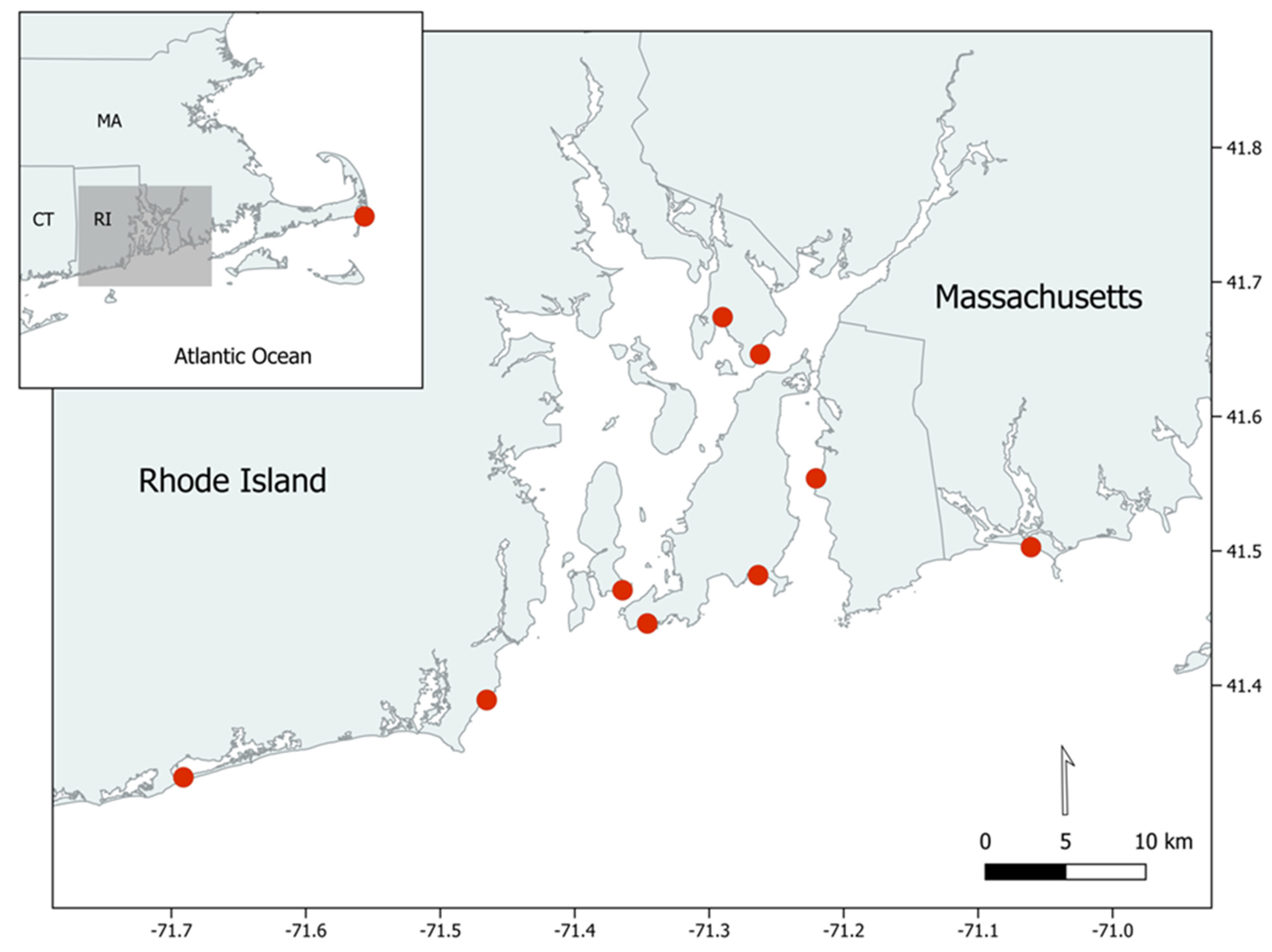
2.1. Specimen Collection

The majority of sequenced specimens were collected over the summer and fall of 2023. Ten sites representing a variety of intertidal and shallow subtidal habitats from around the NBA were visited over the course of the summer and fall from the end of May to early November 2023 (Figure 1). Algal specimens were collected while snorkeling or wading. In situ images as well as lab macroscopic and microscopic images were recorded for newly collected specimens. New specimens were also entered into the Marine Algae of Rhode Island (MARI) database maintained in the Seaweed Biodiversity Lab at Roger Williams University, and both herbarium presses and silica gel vouchers were created and saved in RWU holdings. Many specimens collected in the summer of 2022, as well as older herbarium specimens and silica gel vouchers from the NBA, were also sequenced to include species that were not collected over the summer of 2023.

2.2. Morphological Examination

All specimens were examined morphologically and compared to historical NBA reports from four main compendia, including Sears [34], Villalard−Bohnsack [35], Mathieson & Dawes [17], and Saunders [36]. For species where there were discrepancies with historical reports, further literature review was conducted. Commonly referenced macroscopic characters for separating algae of the order Ceramiales include general thallus arrangement and branching patterns, while microscopic characters include seriation and cortical development on main axes.

2.3. DNA Extraction, Amplification and Sequencing

Extraction of total genomic DNA was completed using the BioLine MyTaq Extract-PCR Kit (Meridian Bioscience; Cincinatti, OH, USA) for almost all samples collected after the spring of 2023, including live, silica-gel-dried, and herbarium vouchers. Extractions from silica-gel-dried vouchers completed before spring 2023 were performed with the DNEasy Plant Mini-Kit (QIAGEN Sciences Inc.; Germantown, MD, USA). For the MyTaq Extract-PCR Kit extraction method on live, fresh specimens, about 5 mm of algal tissue from a new branch tip was added to the extraction solution and incubated overnight at 75 °C. The samples were then centrifuged at 14 krpm for two minutes to pellet excess undissolved tissue. About 85 µL of the supernatant, the isolated total genomic DNA, was retained. The optimal incubation time appears to be around 22 h, although clean sequences were obtained from extracts incubated from as short as 13 h to as long as 24 h. Following extraction, 1:10 dilutions of the DNA extracts were prepared to be used in PCR, as preliminary experiments showed that diluted extracts were far more likely to amplify than undiluted extracts, and these samples often amplified much stronger than the undiluted extracts if both samples amplified. The primers used for both the rbcL-3P and UPA loci are given in Table 1.
Table 1. Primer names and sequences used in this study.
MarkerNameSequence (5′-3′)Source
UPAp23SrV_f1GGA CAG AAA GAC CCT ATG AA[25]
p23SrV_r1TCA GCC TGT TAT CCC TAG AG[25]
rbcL-3PF753GGA AGA TAT GTA TGA AAG AGC[37]
rbcLrevNEWACA TTT GCT GTT GGA GTY TC[32]
R1442AAA CAT TAG CTG TTG GAG TTT CTA C[38]
Standard PCR reagents (Table 2) and thermocycling profiles (Table 3) were used for amplifying the target gene regions.
Following amplification, PCR products were electrophoresed on 1.2% UltraPure LMP agarose (Invitrogen; Carlsbad, CA, USA) gels prepared with a final concentration of 0.5x GelGreen Nucleic Acid Stain (Biotium; Fremont, CA, USA). Amplicons were excised from the gel with a clean razor blade, and DNA was isolated using the Bioline Isolate II PCR and Gel Kit (Meridian Bioscience; Cincinatti, OH, USA) following standard protocol, but substituting PCR water for the provided elution reagent. Two microliters of each cleaned PCR product was then immediately used to determine DNA concentrations (ngDNA/µL) using the NanoDrop One C spectrophotometer (Thermo Scientific; Waltham, MA, USA). All cleaned PCR amplicons were prepared for Sanger sequencing at the University of Rhode Island Molecular Informatics Core Facility, using approximately 5 and 8.75 ng templates for UPA and rbcL-3P, respectively, and 5 pmol/reaction of forward or reverse primer. The Applied Biosystems BigDye® Terminator v3.1 chemistry was used, and cycle sequencing products were analyzed on the ABI 3500xl genetic analyzer following removal of unincorporated reagents using magnetic bead clean-up.

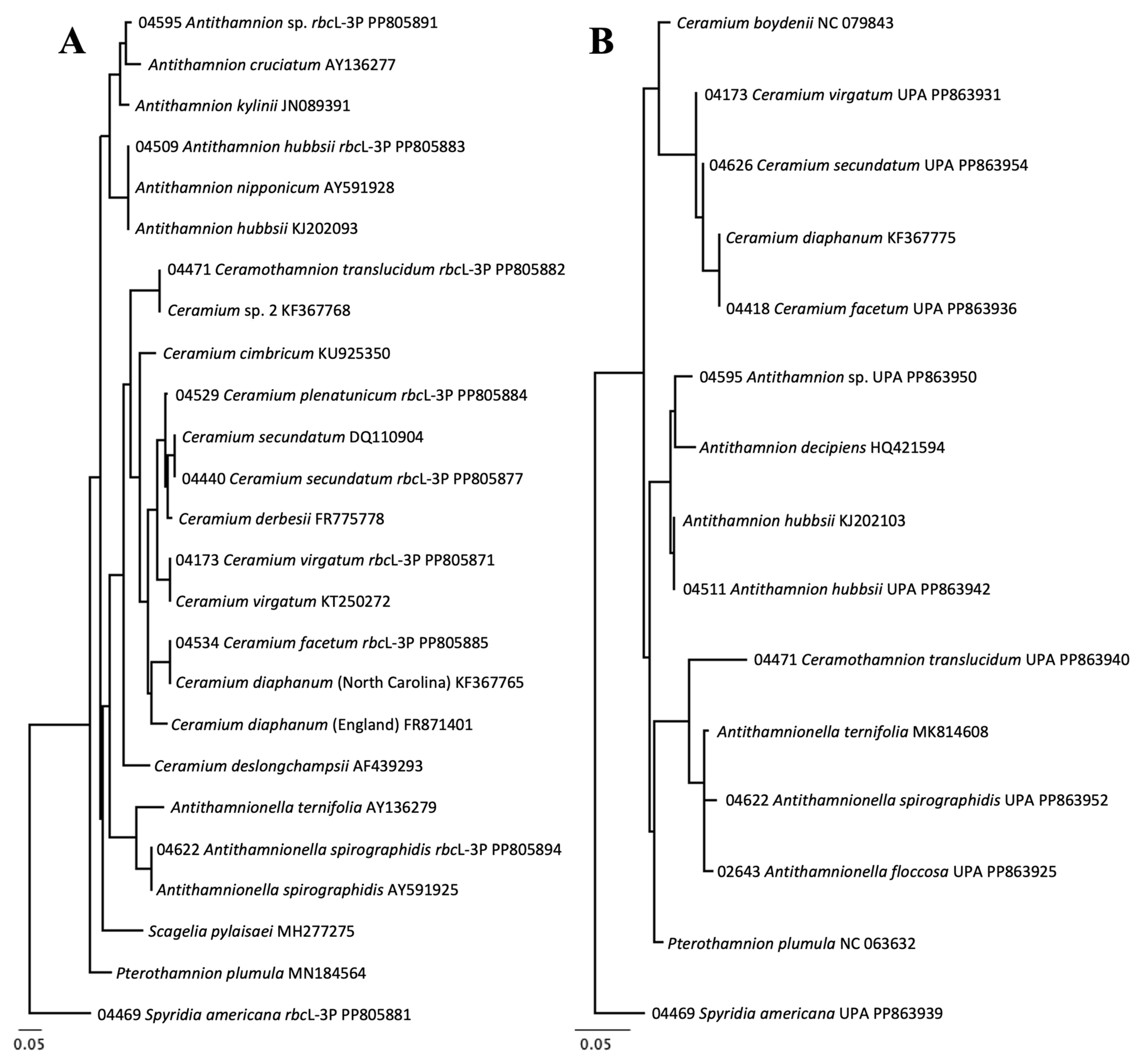
2.4. Molecular Analysis

DNA sequences were analyzed using Geneious Prime software (Version 2022.1.1, Dotmatics, Auckland, New Zealand). Completed sequences were trimmed to remove the primer sequences and standardized sequence lengths for analysis. The rbcL-3P sequences were 667–670 base pairs in length, and the UPA sequences ranged from 368 to 370 base pairs. Two types of phylogenetic trees were generated using different algorithms. Prior to generating trees for both algorithms, the sequences were aligned using MUSCLE [39] through Geneious Prime. From the MUSCLE alignment, UPGMA cluster diagrams (trees) were generated using Jukes–Cantor distances in Geneious Prime using only sequences newly generated in this study. The trees revealed clusters of specimens with low levels of molecular variation, which were used as indicators of sequence-based species. The UPGMA trees were used to estimate how many species were sampled in the NBA, as determined by each genetic marker (rbcL-3P and UPA). To compare phylogenetic relationships between our generated sequence data and published data in GenBank, RAxML trees [40] were generated from MUSCLE alignments in Geneious Prime. Bootstrap values were calculated by setting the algorithm iteration count to 1000. The RAxML trees were generated using our unique sequences and GenBank sequences of the following categories: closest BLAST search result [41], ceramialean species reported from the NBA, ceramialean species that are found south of the NBA and may expand their ranges into the NBA as a result of climate change or other vectors, and ceramialean species that are known to be introduced outside their native ranges.

2.5. Species Determinations

Species determinations were reached by combining BLAST search results of molecular data with morphological examination based on the reference compendia mentioned above. Molecular results were determined simply by using the BLAST search engine to compare newly generated sequences. The closest match by percent identity was generally used to confirm genus assignments, and where the percent identity was greater than 99% or 99.5% for rbcL-3P and UPA, respectively, the same species was presumed (see Section 3.3 for assessment of molecular variation). In many instances, we generated the first sequences for a certain species for either the UPA or rbcL-3P marker, so the closest BLAST search results were not in fact the same species as the species we were searching. Historical reports that included descriptions of the morphology and ecology of algae were used to reconcile molecular species determinations.

3. Results

3.1. Sequencing

A total of 415 sequences were generated from 268 specimens of red algae of the order Ceramiales in the Narragansett Bay Area (NBA), including 203 rbcL-3P sequences and 212 UPA sequences. The UPA marker, in general, had much higher amplification and sequence success than that of the rbcL-3P primers used (Table 4). In fact, over 40 more UPA sequences were generated than rbcL-3P sequences, even though over 1.5 times as many rbcL-3P PCR experiments were carried out than UPA PCR experiments. Generally, PCR experiments using the UPA primers yielded amplicons of up to fivefold greater concentrations than PCR experiments using the rbcL-3P primers, even when amplifying the same DNA extract.

3.2. Molecular Biodiversity of Ceramiales

A total of 37 species were molecularly validated from the NBA, including 2 potentially undescribed species and at least one new report of an introduced species in the NBA (Figure 2). Of the 37 molecularly validated species, 32 were validated with the rbcL-3P marker, 36 were validated with the UPA marker, and 31 were validated with both markers (Figure 3A,B, Table 5). The availability of rbcL-3P sequence data in GenBank is robust; however, this study generated the first rbcL-3P sequences for four species (Table 5). Conversely, there were far less published ceramialean UPA sequence data to which our sequences could be compared, and this study generated the first UPA sequences for 21 species (Table 5).
In most cases, morphological species identities based on historical reports were consistent with molecular species identities (Figure 4). However, there were six taxa where molecular species determinations were inconsistent with historical morphological reports (Figure 4, gray triangles). Along with the taxonomic inconsistencies, another five species were collected in this study, which have not been previously reported in the NBA at all (Figure 2 and Figure 4, black circles). The species treatment section below includes descriptions and detailed discussion of these 11 taxa.
While molecular data was generated for 37 species, about 47 species were reported from the NBA based on historical records and reconciliation of these reports with molecular data (Figure 2). The number of historically reported names from the NBA is an approximate estimate, since there have been a variety of complicated taxonomic changes in the ceramialean flora over the past several decades. To give a few examples of these complications, many of these names were erroneously applied to taxa, in some cases calling a single highly morphologically plastic species multiple different names [42], in some cases not recognizing the cryptic diversity of a single species complex and compiling multiple species under one name [43], or applying an extralimital name to a morphologically similar taxon [44,45,46]. Of the 47 species reported from the NBA, about 15 species were not collected during the summer of 2023 or molecularly validated through attempts to DNA barcode RWU herbarium presses (Figure 2). Most of the 15 species that were unaccounted for were either winter annuals or deeper-water algae that would not have been encountered during the summertime shallow intertidal snorkeling collections.

3.3. Molecular-Assisted Identification of Ceramialean Species in the NBA

The barcoding gap for separating species using the rbcL-3P marker is around 1.00% (7 base pairs), which separated the close pair of Dasya elegans and Dasya pedicellata. However, all other similar species pairs in this study were over 2% (14 base pairs) divergent from one another for the rbcL-3P marker. The barcoding gap for the UPA marker is about 0.50% (2 base pairs), which was the interspecies divergence between Ceramium secundatum and Ceramium virgatum, but again all other species pairs were over 1% (4 base pairs) divergent from one another. It is to be noted, though, that in most cases, if the interspecies divergence for one marker was particularly low, this was not the case for the other marker. For example, the minimum interspecies divergence between D. elegans and D. pedicellata was 1.00% (7 base pairs) for rbcL-3P, but it was 1.63% (6 base pairs) for UPA. Similarly, where the minimum interspecies divergence was 0.54% (2 base pairs) between C. secundatum and C. virgatum for the UPA marker, it was no less than 4.24% (28 base pairs) for the rbcL-3P marker. Using both markers together provided a helpful check to determine if two genetic groups were the same species or not, which was useful for informing species identifications.
Molecular species identifications between the two markers (UPA and rbcL-3P) were 100% in accordance with one another. This appeared to be the case even when the rbcL-3P and UPA sequences for a given specimen did not always have the same closest BLAST search results (Table 6). This seemed to be a result of one of two things depending on the species: no sequence data for that species were available for one or both markers, so the closest BLAST search results were inherently inconsistent between the markers (e.g., the rbcL-3P sequence for MARI-04622 matched Antithamnionella spirographidis, but the UPA sequence was closest to Antithamnionella ternifolia since there is no Antithamnionella spirographidis UPA sequence data available), or there is some taxonomic ambiguity or potentially misnamed sequences in GenBank (this appeared to be the case only for Dasya spp. and Melanothamnus spp.). The rbcL-3P marker was found to be similarly conserved between individuals of a species with respect to the UPA marker, as the intraspecies divergence of rbcL-3P was at most 0.30% (2 base pair divergence; this occurred in Callithamnion corymbosum and Vertebrata fucoides; Table 6), and the UPA marker had a maximum intraspecies divergence of 0.31% (1 base pair divergence; this occurred in multiple taxa; Table 6). However, as mentioned above, the lowest interspecies divergences for both markers were above these thresholds, with the lowest rbcL-3P interspecies divergence being between Dasya elegans and Dasya pedicellata at 1.00% (7 base pairs), and for UPA it was 0.54% (2 base pairs) between Ceramium secundatum and Ceramium virgatum. The Melanothamnus species group was excluded from these analyses, since this group includes a few hybridized species in the Northwest Atlantic and displays high genetic variance [43].
For species that had very close neighbors, such as species of the genera Ceramium, Dasya, or Vertebrata, intraspecies divergences for both markers were well below those genetic distances (percent identity) to the nearest neighbor. For example, a close pair such as C. secundatum and C. virgatum, which were at minimum 0.54% (2 base pairs) divergent, had a 0% intraspecies divergence (Table 6).
Since the UPA marker had an established barcoding gap that was reliable and had species clusters and identifications consistent with the rbcL-3P marker, the UPA marker was found to be just as effective as the rbcL-3P marker in identifying species of the order Ceramiales. This is consistent with other studies that have used the UPA marker for molecular surveys of red algae [47,48].
There were two taxa for which sequence data were challenging to obtain. These species that posed challenges during the barcoding process included Callithamnion tetragonum and Spermothamnion repens. The former posed no challenges when working with the UPA primers; in fact, complete sequences were generated for five C. tetragonum specimens. However, no rbcL-3P sequences were generated for C. tetragonum. Considering the success with the UPA marker, the lack of success with the rbcL-3P primers was likely a result of the primers not being a good base pair match to their complements in the C. tetragonum genome. Spermothamnion repens, on the other hand, seemed to be much more challenging to obtain usable DNA extracts using the BioLine extraction method. The only sequence data generated for this species were from older DNA extracts that had been prepared by grinding liquid nitrogen frozen material with a mortar and pestle.

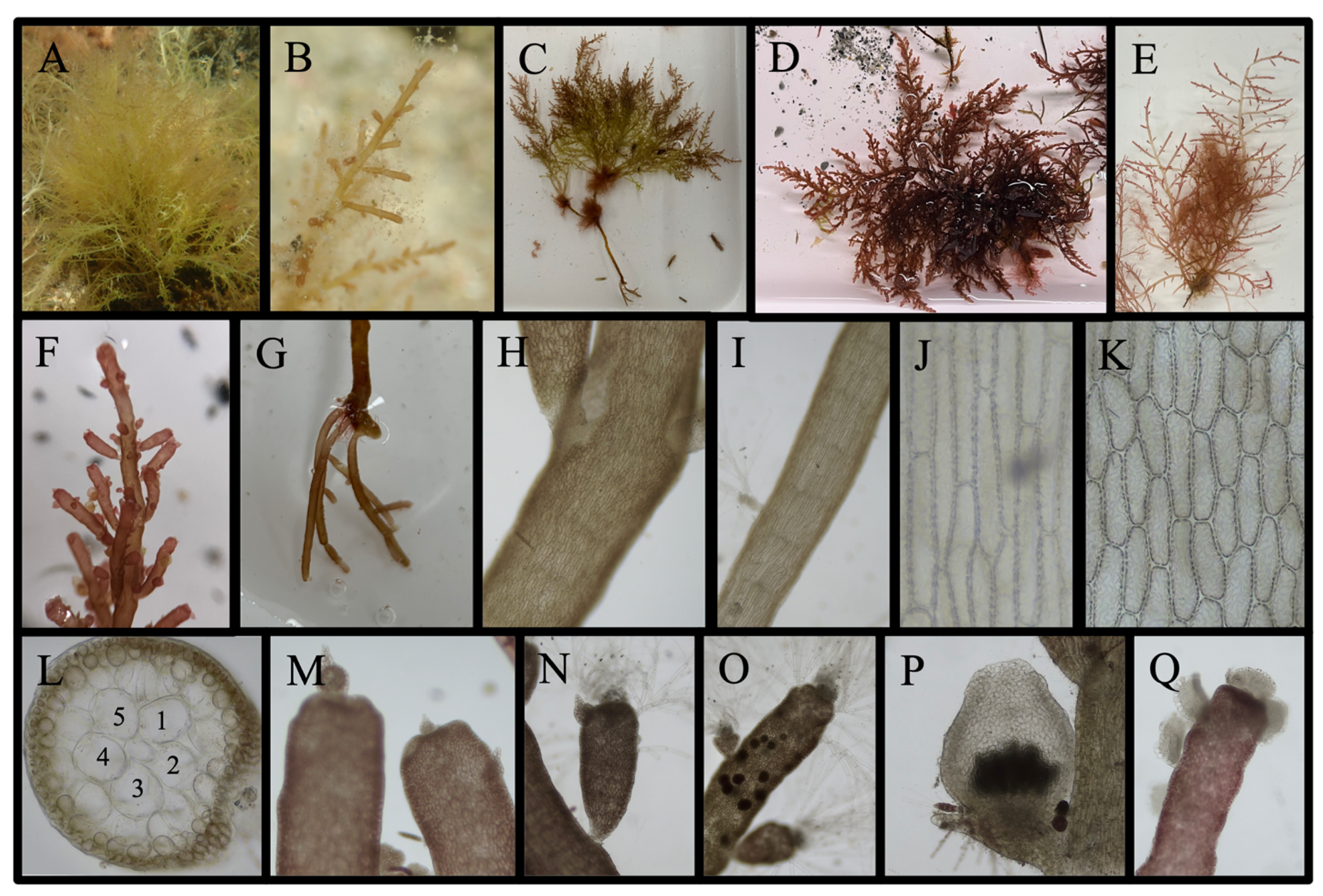
3.4. Species Treatments

Most taxa sampled in this study have been reported from the NBA historically, and both the morphological and molecular species determinations agree with the reports of these species’ identities and presence in the NBA (Figure 2). However, 11 species of interest are discussed below, namely new reports for the area and species for which there are discrepancies between the molecular results, morphological results, and historical documentation.

3.4.1. New Reports for the Narragansett Bay Area (5 Species)

Acanthosiphonia echinata (Harvey) Savoie & G. W. Saunders

Molecular Results: Both rbcL-3P and UPA sequences were generated for a single specimen, MARI-04540 (Table 5). The rbcL-3P and UPA sequences were a 100% match to Acanthosiphonia echinata MF120866 (Figure 5A) from New Brunswick and a 97.57% match to Polysiphonia binneyi KY573931 from Pacific Panama (Figure 5B), respectively. Both markers supported Carradoriella elongata as being the nearest genetic neighbor that we collected, and MARI-04540 differed from C. elongata by 7.81–8.04% and 3.51–3.68% for rbcL-3P and UPA, respectively.
Locality and Morphology: One specimen, MARI-04540, was collected from drift material on 29 July 2023 along the open coast at Horseneck Beach, Westport, MA, USA (Table 7). The thalli were small, matting epiphytes no more than 30 mm tall that covered a strand of the Chorda filum (Figure 6A,B). Branching was alternate to irregular, with adventitious branching being common and giving the thallus an echinate, “spine-like” appearance (Figure 6C,D). Filaments were erect, lacking any prostrate systems, and arose from discoid, rhizoidal holdfasts (Figure 6E). Axes were ecorticate throughout, and branches were borne from cell lineage nodes (Figure 6F,G). The axes were typically shaped such that cells would be slightly concave and the axes would be wider at internodes between segments in older axes (Figure 6H), but sometimes, cells were inflated near branchlet tips, giving the appearance of corn on the cob (Figure 6I,J). The axes had four pericentral cells throughout, as seen in the cross-section (Figure 6J,K). Trichoblasts were abundant, colorless, highly branched, and present at the branchlet tips (Figure 6M). At least some of the material collected was cystocarpic, but there may have been multiple reproductive stages present in the mix (Figure 6N). The morphology described above, particularly the echinate branching pattern, ecorticate character, and four pericentral cells, is consistent with the description of A. echinata [49].
Remarks: Based on the molecular results and morphology, this species was identified as A. echinata. This is the first report of A. echinata from the NBA and the surrounding regions (Figure 2). The type locality of the species is Florida, and it has been reported widely throughout the Caribbean and Gulf of Mexico, but it has recently been introduced to the Mediterranean and Indonesia [16,50,51]. It has also recently been reported in North Carolina and New Brunswick, but not anywhere in between [49,52]. However, just as with Ceramothamnion translucidum, considering that A. echinata has been reported in Florida, North Carolina, and New Brunswick, it would follow that A. echinata would also be present in the NBA since the NBA is within the established range of the species. However, A. echinata has only been reported outside its original range in the Caribbean and Gulf of Mexico within the last two decades. There are a few possibilities for this trend. A. echinata may have recently been introduced outside its range up the east coast of North America and elsewhere in the world, which would explain why it has only recently been reported in so many other locations outside of the region of the type locality. The introduction of the species to the Mediterranean and Indonesia would likely have been enabled by anthropogenic means, but the spread of A. echinata up the east coast of North America could be a natural consequence of the species extending its range as a result of global warming, making northerly waters more habitable to subtropical species in the summer [13]. There is also another possible reason that this species has only recently been recognized in new habitats, and that could be because it is, at first glance, very similar to a variety of other ceramialean species (e.g., Melanothamnus spp. or Neosiphonia spp.). The increased popularity of molecular surveying methods, such as those employed here, may have been the reason A. echinata was identified in these new regions only within the last two decades and not earlier, since this species is morphologically similar to many other species. Further examination of older herbarium material may elucidate whether A. echinata has been present in places such as the NBA for a while and has been overlooked due to its easily mistakable identity, or if it truly was not present in these waters until recently.

Antithamnion sp.

Molecular Results: For two specimens, MARI-04535 and MARI-04595, both rbcL-3P and UPA sequences were obtained, and another UPA sequence was generated from MARI-04594. The rbcL-3P sequences were 0% divergent from each other, and their BLAST search results were a 96.40% match to a cultured specimen of uncertain origin, Antithamnion kylinii JN089393 (Figure 7A). The UPA sequences were also identical to one another, and their BLAST search results were a 98.37% match to Antithamnion hubbsii from North Carolina (Figure 7A). Both of these BLAST search results were more than 1% divergent from this species, indicating that MARI-04535 and MARI-04595 are species not currently represented molecularly in GenBank. The nearest molecular neighbor that we collected according to both markers was Antithamnion hubbsii, which differed from this species by 6.15–6.30% and 1.63% for rbcL-3P and UPA, respectively, meaning that this species is distinct from A. hubbsii, as the sequence divergence for both markers is above the maximum intraspecies divergences of ~1% and ~0.5% for each marker, respectively.
Locality and Morphology: MARI-04535 was collected from Fogland Beach South on 25 July 2023, and MARI-04594 and MARI-04595 were collected at King’s Beach on 19 September 2023 (Table 7). A few very young vegetative thalli, including MARI-04535, were found growing on shells and rocks at a depth of about 1.5–2 m in late July; these were inconspicuous and did not appear to be common (Figure 8A). However, multiple examples of this species were collected in the drift as epiphytes on coarse algae in September at King’s Beach, generally not growing to be more than a few centimeters tall (Figure 8B). Thalli were generally bushy, with axes appearing fuzzy, and unilaterally branched ultimate branchlets (Figure 8C,E). The ultimate branchlet tips were composed of cylindrical or swollen, slightly ovoid cells (Figure 8F,G). Every axial cell bore two opposing, distally arranged branchlets (Figure 8H). Pairs of branchlets were oppositely arranged on the same axial cell; however, opposite pairs were whorled around the axes, branching in multiple planes and giving the axes a bushy appearance. Furthermore, basal cells attaching branchlets to axial cells were globose to spherical and much smaller than the rest of the branchlet cells (Figure 8I). Rhizoids in the upper thallus developed unilaterally and most often in triplets from the spherical ultimate branchlet basal cells, similar to Antithamnion hubbsii (Figure 8J,K). In the lower thallus, basal rhizoids occasionally develop singly from axial cells or inconspicuous basal branchlet cells (Figure 8L). One specimen collected in September was tetrasporophytic and bore cruciately divided tetrasporangia borne on short, multicellular stalks (Figure 8M,N).
Remarks: Based on both morphological and molecular results, this species is within the genus Antithamnion. This species did not match published sequence data of either species of Antithamnion reported from the NBA (Table 6); however, it groups with various other Antithamnion in both the rbcL-3P and UPA RAxML trees (Figure 7A,B). Also, this species shares the following morphological characteristics that place it in this group: basal branchlet cells smaller than other branchlet cells, at least one order of pinnate branching, rhizoidal development from basal branchlet cells, and cruciate tetrasporangia borne on short stalks [17,53]. Currently, only a few specimens of this species have been collected, and no older specimens that may be the same species as MARI-04595 have been molecularly validated from the area; whether this species is native or introduced cannot be assessed. What is known, though, is that MARI-04595 is a representative of a species distinct from other Ceramiaceae historically reported in the NBA and is considered a new report for the area (Figure 2).

Antithamnionella spirographidis (Schiffner) E. M. Wollaston, 1968

Molecular Results: A single specimen, MARI-04622, was collected, and both an rbcL-3P and UPA sequence were generated for the specimen (Table 5). The rbcL-3P sequence was a 100% match to A. spirographidis DQ022810 from the Netherlands (Figure 5A), and the UPA sequence was a 99.19% match to A. ternifolia MK814608 from Spain (Figure 7B). Note that the UPA sequences of both these species, A. spirographidis MARI-04622, and another species not discussed in detail in the species treatments, A. floccosa MARI-02643, share top BLAST search results and match A. ternifolia MK814608 (Table 1). Both A. floccosa and A. spirographidis are 0.81% (3bp) divergent from MK814608; however, two of the three base pair differences are at different locations in the UPA gene between MARI-04622 and MARI-02643. Even though MARI-04622 and MARI-02643 are the closest neighbors to each other out of the species we collected, they are 1.35% divergent from one another; they are further apart from each other than from MK814608 (Figure 7B). The nearest neighbor based on the rbcL-3P sequences was the other Antithamnion sp. (Species Treatment II), which differed from MARI-04622 by 9.74–9.81%. However, the large interspecies gap in the rbcL-3P data that is not seen in the UPA data is likely because we do not have rbcL-3P sequence data for MARI-02643 or other potentially closer-related species.
Locality and Morphology: The single specimen MARI-04622 was found floating in shallow waters off Lighthouse Beach, Chatham, MA, on 29 October 2023 (Table 7; Figure 9A). The thallus was light pink, uniseriate throughout, and had adventitious branchlets on every axial cell at the base of the thallus (Figure 9B). Branching was somewhat planar and in two orders: alternate then pinnate; however, sometimes branchlets would not be paired near branchlet tips and would be unilaterally arranged or paired branchlets were of unequal lengths (Figure 9C–F). In the lower axes, 2–3 ultimate branchlets are whorled and situated distally on axial cells. These branchlets often have one to a few gland cells growing adaxially on cells near the bases of the branchlets (Figure 9G,H). Tetrahedrally divided, sessile tetrasporangia are present in the upper axes, growing singularly and adaxially on basal cells of ultimate branchlets (Figure 9I–L).
Remarks: The specimen collected from Chatham, MA, is a 100% match (rbcL-3P) and is a strong morphological match to Antithamnionella spirographidis [42,54]. However, MARI-04622 also matches the description of Scagelia americana, as reported from the NBA [17]. The description for A. spirographidis, as given by [42,54], is nearly identical to that of S. americana in Mathieson & Dawes [17]. All three described a species with irregular to alternate branching, with every axial cell bearing 2–4 whorled branchlets, branchlet pairs having uneven lengths, apical cells with pointed tips, abundant gland cells growing adaxially on basal branchlet cells, and tetrasporangia being sessile, oblong, cruciately divided, and near the bases of branchlets. However, while S. americana is still a valid name [16], it was listed as a heterotypic synonym of Scagelia pylaisaei by Bruce [42]. S. pylaisaei was reported to be a highly morphologically variable species, and there was also high molecular diversity within the group that was inconsistent with morphologies, which was presumed to account for the confusion in species determinations of Scagelia in Canadian waters [42]. Therefore, since the identity of S. americana is ambiguous and the description of S. americana in Mathieson & Dawes [17] is nearly identical to the less ambiguously described A. spirographidis, the description of S. americana may refer to a misidentified A. spirographidis. If this is true, then A. spirographidis may have been present in the NBA for some time; however, this cannot be confirmed without assessing older material. Regardless of what species Mathieson & Dawes [17] were referring to, based on the morphological and molecular data, MARI-04622 is determined to be A. spirographidis, and this taxon is reported from the western Atlantic for the first time (Figure 1). A single collection of this species, especially one that was not found to be actively growing, is not enough to determine whether this species has established a population in the area, but this species should be monitored. The origin of A. spirographidis is somewhat disputed; however, it is likely native to the Pacific [55]. It is found throughout the Pacific, including the west coast of the United States and Canada, Japan, Russia, China, and Australia, as well as much of the Mediterranean [16]. Due to its widespread distribution in the Pacific, it is believed that this species was introduced to the Mediterranean some time before the 1970s, likely through hull fouling or contaminated ballast water [56,57].

Dasya elegans (G. Martens) C. Agardh

Molecular Results: Both rbcL-3P and UPA sequences were obtained from two specimens, MARI-04392 and MARI-04407. The rbcL-3P sequences were a 100% match to Dasya sp. 1 baillouviana MW698713 from Nova Scotia as well as D. “baillouviana” FM993088 from The Netherlands (Figure 5A). D. “baillouviana” FM993088 separates from MARI-04392 in the rbcL-3P RAxML tree because the sequences do not completely overlap (each is a sequence from slightly different segments of the rbcL gene, Figure 5A). Both UPA sequence BLAST search results were a 98.64% match to Dasya sp. HQ421299 from Hawaii, and a 98.37% match to D. baillouviana HQ421392, also from Hawaii (Figure 5B). Intraspecies divergences were 0% for both markers (Table 6). The nearest neighbor we collected was D. pedicellata according to both markers, and this species was 1.00–1.16% and 1.63% divergent from D. pedicellata for rbcL-3P and UPA, respectively.
Locality and Morphology: Two specimens were collected from the estuarine RWU waterfront, one on 24 May 2023, and the other on 4 June 2023. Both specimens were lithophytes in the shallow subtidal, and they were abundant during late spring/early summer at this site (Table 7). Thalli were light, peach pink to deep red, with main axes that were covered in adventitious, pigmented, uniseriate branchlets that gave the thallus a fuzzy appearance (Figure 10A–E). Often, branching was rather sparse, and there was a discernible central axis from which laterals extended almost perpendicularly (Figure 10A). In cross-section, the main axes had 5–6 pericentral cells (Figure 10F,G). The main axes were heavily corticated (Figure 10H). Adventitious branchlets were composed of often long, cylindrical cells, and branchlets divided dichotomously, bearing tetrasporangial stichidia on short multicellular stalks (Figure 10I–K). This species is almost inseparable from D. pedicellata based on morphology alone in the NBA; however, D. elegans had much sparser lateral branches from the main axes, and adventitious branchlets were not as abundant and did not obscure the main axes as much as in D. pedicellata. However, aside from the fact that these characters are relatively subjective, since very few specimens of D. elegans were examined in this study, these characters may also not be universal.
Remarks: Only one species of Dasya has previously been reported from the NBA, and that was D. baillouviana, which is now considered an invalid name due to the lack of any surviving type material and the ambiguity of the identity of the type specimen [58]. Following the rejection of the name D. baillouviana, D. pedicellata was promoted back to full species status and has been applied as the name for Dasya collected in the NBA since the type specimen of D. pedicellata was from New York [58]. However, two genotypes of Dasya were recovered in this study and these were named D. elegans and D. pedicellata (Section Dasya pedicellata (C. Agardh) C. Agardh, 1824 Dasya pedicellata). Although both recovered species were very similar morphologically, molecular data suggest that these are different species, since the minimum sequence divergence between D. elegans and D. pedicellata (1.00% and 1.63% for rbcL-3P and UPA, respectively) were both greater than their intraspecies divergences of 0% for both markers. Therefore, we report two species of Dasya from the NBA. The historical reports of only a single species of Dasya from the NBA were likely a result of the nearly identical morphologies of the two molecularly validated species, and/or potentially because D. elegans was introduced recently to the Atlantic Northwest from the Eastern Atlantic, since our sequence data matches some from the Netherlands (D. “baillouviana” FM993088). The species represented by MARI-04392 and MARI-04407, which is molecularly distinct from the multiple specimens of D. pedicellata that were also collected in this study, is provisionally named D. elegans. D. elegans is a name still considered synonymous to D. pedicellata, but it is currently being considered to be resurrected for this second species [16] (G.W. Saunders, pers. comm.). The type specimen of D. elegans is from Italy, which suggests that if MARI-04392 and MARI-04407, collected in the NBA, are the same species as the Italian holotype, then this would support the idea that this species was introduced from the Mediterranean, as mentioned above. More taxonomic work is needed in this genus to fully understand the origins and distributions of the constituents of this genus, as many species are morphologically challenging to separate; however, this is a new report from the NBA considering that only one species of Dasya has been reported historically from the area (Figure 2).

Streblocladieae sp.

Molecular Results: Both rbcL-3P and UPA sequences were generated for a single specimen, MARI-04466 (Table 5). Neither marker produced a very close BLAST search result with any published sequences, indicating that there are no published sequence data for the species of MARI-04466 (Table 6, Figure 5A and Figure 6B). The rbcL-3P sequence was closest to Kapraunia pentamera HM573564 from Panama, which was only a 93.30% match (Figure 5A), and the UPA sequence was a 97.83% match to Polysiphonia sp. HQ421052 from Hawaii. The nearest molecular neighbor we collected according to the rbcL-3P markers was Kapraunia schneideri, which was 7.26–7.94% divergent from MARI-04466. Acanthosiphonia echinata was the nearest molecular neighbor according to the UPA marker, which was 3.78% divergent from MARI-04466.
Locality and Morphology: MARI-04466 was collected from the southwestern end of Ninigret Pond by the East Beach boat launch in shallows less than one meter deep on 21 June 2023 (Table 7). During a snorkel that lasted over 30 min, only the one specimen was collected, growing as a large (~11 cm tall) but inconspicuous lithophyte that looked and felt very similar to a lot of brown and green algal growth in the same area. The thallus was dull orangish brown and had very thin branches that were not easy to make out with the naked eye (Figure 11A). Branching was alternate to irregular throughout, with central, uncurved axes that bore adventitious branchlets. Branching was abundant and followed the echinate branching patterns of some members of the tribe Streblocladieae (Figure 11B,C).
Axes were ecorticate throughout and had four pericentral cells (Figure 11D–F). Trichoblasts were present around branchlet apices, and a single apical cell was often clearly visible at branch termini (Figure 11G). Rhizoids were connected to axial cells via an open connection and were abundant on the basal growth of the thallus (Figure 11H,I). MARI-04466 was a tetrasporophyte, whose tetrasporangia were arranged in spiral series near branch apices, causing branchlets to swell and warp (Figure 11J). Sometimes, the spiral character is not very pronounced in some branchlets (Figure 11K). Tetrasporangia were tetrahedrally divided (Figure 11L). The morphology of MARI-04466 loosely matches the description of Bryocladia subtilissima (previously Polysiphonia subtilissima), which has been reported from the NBA by multiple sources [17,34,35]. B. subtilissima is an euryhaline species that can grow in both freshwater and marine environments and is often found in estuarine/brackish waters, is ecorticate, has four pericentral cells throughout, and has unicellular rhizoids with an open connection to the parent axial cells [17,59]. However, MARI-04466 differed in at least one major way from B. subtilissima, which is known to be a characteristic used for separating species in this group. Tetrasporangia were arranged in spiral series in MARI-04466, whereas tetrasporangia were arranged in straight series in B. subtilissima [17].
Remarks: Considering the molecular results indicating that this species does not match any published sequence data, and that this species does not match any species historically reported from the area morphologically, this species appears to be either an undescribed species or potentially an introduced species of obscure origin for which no sequence data is currently available. There is another possibility that seems to be the more likely situation. As mentioned above, this species would key out to B. subtilissima, which is a species reported from the NBA and which we did not otherwise collect. However, MARI-04466 is almost certainly not B. subtilissima, since our specimen differs morphologically from B. subtilissima since the tetrasporangia are arranged differently, as discussed above. Furthermore, the molecular results agree that these are not the same species, since there are published sequence data for B. subtilissima, which MARI-04466 does not match well with at all. The closest published B. subtilissima sequence was B. subtilissima (as Polysiphonia subtilissima) JX294918 from Spain, which was only an 86.85% match to the rbcL-3P sequence for MARI-04466 (Figure 5A). However, there is the possibility that specimens of the species as MARI-04466 were collected in the NBA and were mistakenly identified as B. subtilissima, just as the case was with Chondria atropurpurea being called Chondria capillaris and Dasya pedicellata being called Dasya baillouviana. Further collections of the species of MARI-04466 and comparison to historical collections from the NBA may help clarify the status of this species. What is clear, though, is that this species is not Bryocladia subtilissima and is considered a new report for the region (Figure 2).

3.4.2. Species with Taxonomic Ambiguities That Are Not New Reports: (6 Species)

Antithamnion hubbsii E. Y. Dawson

Molecular Results: Sequences were obtained from seven specimens, four of which both rbcL-3P and UPA sequences were generated for. The rbcL-3P sequences were a 100% match to both A. nipponicum AY591928 and A. hubbsii KJ202093, both specimens from North Carolina, as well as a 99.85% match to three more A. nipponicum and one A. hubbsii from various other localities (Figure 7A). UPA sequences were a 100% match to A. hubbsii KJ202103 from North Carolina (Figure 7B). Intraspecies divergence was 0% for both markers (Table 6). The closest neighbor to A. hubbsii for both markers was Antithamnion sp. (Species Treatment II), from which A. hubbsii sequences differed by 6.15–6.30% and 1.63% for rbcL-3P and UPA, respectively.
Locality and Morphology: Sequenced specimens were collected from Black Point, King’s Beach, and Fort Wetherill from June, July, and September (Table 7). Specimens were collected frequently on various coarse algae in high wave action zones along the open coast from July through November. In most cases, thalli were gregarious and created spreading, noticeable, epiphytic turfs on the distal, wave-exposed growth of subtidal algae, most commonly Chondrus (Figure 12A–C). Tetrasporophytes were observed in late July; however, none of this material was imaged or sequenced. Thalli were uniseriate throughout, and while specimens had decussate branching patterns (Figure 12D), most specimens had strictly planar pinnate branching in two orders (Figure 12E–H). Branchlets were connected to main axes by small spherical basal cells smaller than the rest of the branchlet cells, gland cells were common (Figure 12I–K), occasionally more than 5 per branchlet, and rhizoids generally grew in triplets from the spherical branchlet basal cells (Figure 12K,L). The uniseriate thallus structure, planar pinnate branching patterns, and small and spherical basal branchlet cells are distinctive characteristics that are consistent with the morphology described for A. hubbsii [17,53].
Remarks: The name Antithamnion hubbsii has a complicated taxonomic history. A. hubbsii was first described from Baja California, Mexico, in 1962 and has since been reported widely across the globe [53]. However, this species has long been confused and used almost interchangeably with A. pectinatum and A. nipponicum [53]. A. pectinatum has been identified as a species that was introduced to the Mediterranean [60,61]; however, it was determined that these specimens thought to be A. pectinatum should be A. hubbsii [62]. Furthermore, A. nipponicum, which has been recorded from the Western Pacific and has also been reported to have been introduced in the Mediterranean [63] and North Carolina [53], has since been synonymized with A. pectinatum, whose type locality is in New Zealand [62]. In short, all specimens that were called A. hubbsii, A. nipponicum, or A. pectinatum outside of New Zealand have been renamed A. hubbsii. A. nipponicum is no longer a valid name, and A. pectinatum is only present in New Zealand. However, no A. hubbsii from the type locality of Baja California have been sequenced, so there is a chance that molecular A. hubbsii recorded from the Mediterranean, western Pacific, and western Atlantic are not the same as true A. hubbsii. For the purpose of this study, the name A. hubbsii was maintained for specimens collected and sequenced in the NBA. Five of the seven specimens collected in this study that were a molecular match to North Carolinian Antithamnion hubbsii were also a morphological match to this species, as described above. Furthermore, both UPA and rbcL-3P sequences were generated for three of these five specimens, and molecular species determinations for both markers for each of the three specimens agreed with A. hubbsii. Furthermore, A. hubbsii has been reported from the NBA [17], and it was first reported to have been introduced to Connecticut in 1991 under the name Antithamnion cf. nipponicum [64]. Therefore, since the morphology and molecular data agree for these specimens as well as with historical reports, the current presence and identity of A. hubbsii is reaffirmed in the NBA. However, two samples from Fort Wetherill and one from King’s Beach did not fit the above morphological descriptions for A. hubbsii, but were molecular matches to A. hubbsii (for those that were barcoded). These included MARI-04431, for which a UPA sequence was generated; MARI-04476, for which we did not have any sequence data; and MARI-04560, for which an rbcL-3P sequence was generated. Each of these specimens had decussate branching patterns (i.e., branching in two more or less perpendicular planes, Figure 12D). While MARI-04560 was found as a turf on Chondrus-like typical A. hubbsii specimens, MARI-04431 and MARI-04476 were found to be growing in sparse patches along more sheltered, lower axes of coarse algae. There are two species of Antithamnion reported from the NBA—A. hubbsii and A. cruciatum—and MARI-04431, MARI-04476, and MARI-04560 agree morphologically with A. cruciatum. A. hubbsii has been described as having a planar branching pattern, while A. cruciatum has a decussate branching pattern [17]. However, the UPA sequence for MARI-04431 and the rbcL-3P sequence for MARI-04560 are matches to other A. hubbsii sequences we generated, and the A. hubbsii rbcL-3P sequences were distinct from A. cruciatum AY136277 (Figure 7A). This, along with the fact that sequences were 0% divergent from one another for both markers, suggests that there may be only one species of Antithamnion present in the NBA, or if A. cruciatum is also present and it was not encountered by this study, then the two species cannot be separated by their branching pattern, as multiple specimens that barcoded to A. hubbsii had the decussate branching pattern that is supposed to be unique to A. cruciatum [17].

Ceramium facetum G.W. Saunders & C. W. Schneider, 2024

Molecular Results: A total of 13 specimens were sequenced, and for 8 of these, both rbcL-3P and UPA sequences were generated. The rbcL-3P sequences were a 100% match to C. facetum (Ceramium sp.1GoSL) in the Barcode of Life Data System (BOLDS; [65]) and a 100% match to C. diaphanum KF367765 from North Carolina in GenBank (Figure 7A). The UPA sequences were also a 100% match to the same GenBank specimen as rbcL-3P, C. diaphanum KF367775 (Figure 7B). The intraspecies divergence for rbcL-3P and UPA was 0–0.07% and 0–0.28%, respectively (Table 6). The nearest neighbor we collected to this species was C. virgatum for rbcL-3P, which differed from C. facetum by 6.78–7.62%, and the nearest neighbor according to our UPA sequence data was C. secundatum, which differed from our sequences by 1.08–1.41%.
Locality and Morphology: Specimens were collected throughout the summer from late May to mid-August in a variety of habitats, from protected estuaries to salt marsh headwaters to tidepools and drift material on the open coast (Table 7). This was most commonly collected as a lithophyte forming dense, bushy mats on rocks in the low intertidal zone in more protected areas but also occasionally collected as an epiphyte on coarser algae. Thalli were incompletely corticated, profusely branched, and often strictly dichotomously branched (Figure 13A–D). Branchlet tips were recurved and pincer-shaped, a characteristic typical of the genus Ceramium (Figure 13E). Branching dichotomies occurred at the nodes (Figure 13F). Branchlet tips were recurved and pincer-shaped, a characteristic typical of the genus Ceramium (Figure 13E). In cross-section, nodes had 6–7 large, round periaxial cells that were distinct from cortical cells, which separates this species from the only other incompletely corticated ceramiacean alga reported from the area, Ceramothamnion translucidum, whose periaxial cells are fewer (~5) and are similar in size to cortical cells (Figure 13G,H). Sun-bleached thalli from much calmer, protected waters appear much paler than thalli from high wave action or shaded areas (Figure 13H). In some thalli, adventitious branchlets were present, but were still mostly dichotomously branched with pincer-shaped tips (Figure 13I,J). Tetrasporophytes were collected in early June and had tetrasporangia organized in whorls around the nodes (Figure 13L,M). Cystocarpic thalli were collected in late July, and pairs of gonimolobes developed at branching dichotomies (Figure 13N).
Remarks: This species has long been reported from the NBA as C. diaphanum, whose holotype is from Scotland. However, routine DNA barcoding of specimens from the Northwest Atlantic has revealed that the incompletely corticated Ceramium that has long been identified as C. diaphanum in the northwest Atlantic is distinct from the true C. diaphanum from the eastern Atlantic [36]. The northwestern Atlantic species has just recently been described as a new species, C. facetum [66]. The sequences generated in this study are consistent with this finding and match “C. diaphanum” from the northwestern Atlantic, not Europe. A specimen from Cornwall, England, published as C. diaphanum FR871401, is only a 94.66% match to the rbcL-3P sequences we generated and is the closest molecular match of any published European C. diaphanum sequences (Figure 7A). Considering both the morphology discussed above and the molecular results, C. facetum is regarded as established in the NBA and has been mistaken for an extralimital but morphologically similar European species.

Ceramium plenatunicum G. W. Saunders & C. W. Schneider, 2024

Molecular Results: For three different specimens, rbcL-3P sequences were obtained; however, no complete UPA sequences were generated. The rbcL-3P sequences were a 100% match to C. plenatunicum (Ceramium sp. 2virgatum) GWS006236 from New Brunswick in BOLDS, and a 98.80% match to C. derbesii FR775779 from Italy in GenBank (Figure 7A). The intraspecies divergence of the three sequences was 0% (Table 6). The nearest collected neighbor was C. secundatum, which differed from this species by 2.54–3.63%.
Locality and Morphology: Two specimens were collected from the protected, estuarine north shore of Fogland Beach on 25 July 2023 (Table 7). An rbcL-3P sequence from another herbarium specimen was also obtained, but the collection data for that specimen are not included here. The summer 2023 specimens were abundant epiphytes in shallow, <20 cm deep water at low tide, often growing on gracilariacean algae and mixed with Chondria baileyana (Figure 14A–C). Branching was strictly dichotomous with occasional adventitious branches, and branching angles were wide, spreading, and often >60° except at the branchlet tips (Figure 14D). Thalli were completely corticated throughout but may still appear banded, as the nodes were sometimes darker than internodes (Figure 14E–G). In the cross-section, nodes had 6–7 circular periaxial cells (Figure 14H). Branchlet tips were typical for the genus Ceramium, with pincer-shaped termini that were slightly inrolled (Figure 14I). No reproductive material was collected.
Remarks: Historically, there have been two completely corticated species of Ceramium reported from the NBA. These would be C. secundatum and C. virgatum, both of which were recovered in this study, and we have multiple rbcL-3P and UPA sequences that agree with the morphological identifications confirming these identities (Table 1). However, a third species of completely corticated Ceramium was recovered as well, and that species both matches the morphology and barcode of C. plenatunicum, which was only recently described based on specimens collected in New Brunswick [36]. This species has also been genetically verified in Rhode Island from an epiphyte on Grateloupia [36]. This species mainly differs from the other two completely corticated Ceramium because it has much wider branching dichotomies, but there may be other differences that would be better elucidated by collection/study of more specimens. One such potential difference is how the periaxial cells of C. plenatunicum seem to be much more circular than those of C. secundatum and virgatum, whose periaxial cells are often slightly oblong or sector-shaped. Since this species is morphologically very similar to the other completely corticated Ceramium reported from the NBA, it is likely that this species has long been overlooked and is probably not introduced to the area; but the origin and history of this species is uncertain in the NBA. However, considering that C. plenatunicum has been molecularly verified from the NBA by another lab [66], it is not included as a new report for the area (Figure 2).

Ceramothamnion translucidum G. W. Saunders & C. W. Schneider, 2024

Molecular Results: Sequences for five specimens were obtained, and for three of these specimens, both rbcL-3P and UPA sequences were generated. The rbcL-3P sequences were a 100% match to “Ceramium sp. 2 DWF-2013” KF367768 from North Carolina (Figure 7A). UPA sequences were also a 100% match to the same species from the same locality, “Ceramium sp. 2 DWF-2013,” from North Carolina (Figure 7B). These species, “Ceramium sp. 2 DWF-2013,” are matches to holotype sequences of Ceramothamnion translucidum [36], so the species validated in this study is also therefore C. translucidum. Intraspecies divergence for the rbcL-3P and UPA sequences generated were 0–0.15% and 0%, respectively (Table 6). The nearest neighbor we collected, as indicated by both rbcL-3P and UPA sequence data, was C. virgatum, which this species differed from by 8.57–9.54% and 5.41% for rbcL-3P and UPA, respectively.
Locality and Morphology: Four specimens from three different sites at Ninigret Pond and one specimen from Horseneck Beach were collected in June and July of 2023 (Table 7). A fifth specimen, an older herbarium specimen, MARI-03342, collected from Sakonnet Point in September 1972, was also barcoded to this species. This specimen was described as an epiphyte on Fucus and had an annotation and drawing that indicated that it was tetrasporophytic, with tetrahedrally divided tetraspores growing emergent from axial nodes. All specimens collected in the summer of 2023 were nonreproductive. Thalli were generally in small, epiphytic tangled clusters no more than a few centimeters across, collected in shallow subtidal (<1 m depth) at Ninigret Pond (Figure 15A,B). The specimen from Horseneck Beach was a small tuft growing on Ascophyllum, collected in the drift. The older specimen was much larger and spreading, a dense mat over 10 centimeters across. Branching was strictly dichotomous, and branches had acute angles of around 60° or less, and axes were incompletely corticated throughout (Figure 15C–E). The darker, central axial filament could often be seen from the surface view of newer axes near branchlet tips (Figure 15F). In cross-section, nodes had about 5 periaxial cells that were slightly larger than the abundant, surrounding cortical cells (Figure 15G). Periaxial cells were generally small enough so that it was possible to take a cross-section at a node that did not include the periaxial cells and only revealed cortical cells (Figure 15H). The branchlet tips were often slightly inrolled and pincer-shaped (Figure 15I–K); however, sometimes, the branchlet tips were straight and not recurved at all (Figure 15L). These characteristics are consistent with the original description of Ceramothamnion translucidum [36]. Furthermore, the relatively smaller size of periaxial cells in Ceramothamnion translucidum separates it from species of the genus Ceramium, whose periaxial cells are often more numerous and are generally much larger and distinct from cortical cells at nodes [67].
Remarks: Based on molecular and morphological results, this species is a strong match to Ceramothamnion translucidum, formally described in April 2024, from material collected in New Brunswick [66]. Sequences generated in this study match those of specimens collected in North Carolina (Figure 7A,B), as well as specimens from the Gulf of St. Lawrence [66]. Considering the range of localities from which this species has been collected and molecularly validated, both from our study and from others, as well as MARI-03342, which was collected in 1972, this taxon is likely established in the NBA and has been present for multiple decades. This species may have been historically overlooked or confused with the similar C. deslongchampsii, which is reported to have 4 large periaxial cells, incomplete cortication, and forms mats on rocks and algae in the mid to low intertidal [17]. No specimens that matched the morphology or matched existing published DNA barcodes of C. deslongchampsii were collected in this study. However, it is still reported in the area by numerous sources [17,34,35,36]. MARI-03342 had originally been identified as Ceramium fastigiatum, which is now a synonym of C. cimbricum, a species whose type locality is Denmark and to which MARI-03342 and the rest of the Ceramothamnion specimens are a poor molecular match [16]. C. cimbricum has been reported from Connecticut and the NBA [17,68], but this study did not uncover any specimens that were a molecular match to any European sequence data of this species.

Chondria littoralis Harvey 1853/Chondria sedifolia Harvey 1853

Molecular Results: Sequences were obtained from 13 specimens, and for 10 of these specimens, both rbcL-3P and UPA sequences were generated. The rbcL-3P BLAST search results were a 100% match to C. littoralis KF672853 from North Carolina (Figure 5A), and the UPA BLAST search results were a 97.02% match to Chondria sp. MF101429 (Figure 5B). The intraspecies divergence for the rbcL-3P and UPA markers was 0–0.15% and 0–0.27%, respectively (Table 6). The rbcL-3P and UPA markers disagreed on what the nearest neighbor we collected was, the former being closest to C. baileyana and the latter being closest to C. atropurpurea, with divergences from these species being 6.34–8.56% and 3.52–3.87%, respectively.
Locality and Morphology: This species was collected throughout the summer and into the fall from as early as 23 June to as late as 12 October. Thalli were abundant on open coast sites as lithophytes in subtidal waters, but occasionally young thalli were found as epiphytes on Zostera (Table 7). Also often collected in the drift. Tetrasporophytes were collected on 23 June 2022 and 1 August 2023; a single male gametophyte was collected on 29 July 2023, and female gametophytes were collected from mid-July to October. Thalli were bushy with thick axes and visibly blunt branchlet tips, and they varied greatly in color from straw yellow to a deep purplish maroon (Figure 16A–G). Axes were thick, heavily corticated throughout and had 5 pericentral cells in cross-section (Figure 16H–L). This species was easily distinguished from other Chondria in the NBA, as the apices of branchlet tips were concave with the apical cells usually hidden in a sunken pit (Figure 16M). Trichoblasts were absent in some thalli (Figure 16M), while in others, they were abundant, highly dichotomously branched, and often had small, dark, pigmented cells interspersed throughout the trichoblasts at branching dichotomies (Figure 16N). Tetrasporangia were interspersed throughout the cortex of branchlet tips without much pattern (Figure 16O). Cystocarps were usually found on distal growth on ultimate branchlets, but occasionally on distal main axes. They were urn-shaped and were borne on pedestal-like lateral stalks, upon which cystocarps developed adaxially (Figure 16P). Spermatangial sori were small, hyaline plates that were organized in whorls around ultimate branchlet tips (Figure 16Q).
Remarks: There are many taxonomic issues with the genus Chondria in the NBA. Four species have long been reported from the NBA, including C. baileyana, C. capillaris, C. dasyphylla, and C. sedifolia. Three species were recovered in this study: C. baileyana, C. atropurpurea, and C. littoralis/sedifolia. There were no issues with the identity and presence of C. baileyana in the NBA, as the recovered specimens matched the morphological descriptions of C. baileyana and were barcoded to C. baileyana with no disagreements (Figure 5A). Also, in a recent paper, old herbarium specimens that had been initially identified and used as the basis of the presence of C. capillaris in the NBA were determined to actually be C. atropurpurea, a species previously thought to be confined to the Gulf of Mexico and the Caribbean [45]. Similarly, this study found that all C. capillaris-like specimens from the NBA had DNA barcodes that matched C. atropurpurea (Figure 5A). These three species—C. baileyana, C. capillaris, and C. atropurpurea—comprise the exposed-apical-cell Chondria group, and based on this study, only C. baileyana and C. atropurpurea are present in the NBA (Figure 2). That leaves the sunken-apical-cell Chondria group, C. dasyphylla, C. sedifolia, and C. littoralis/sedifolia. According to Mathieson & Dawes (2017), C. dasyphylla and C. sedifolia can be differentiated by the thallus being “soft” or “tough,” by looking at the size of the thallus, axis diameter, branchlet length, or the presence/absence of trichoblasts. However, the numerous specimens we collected represented a gradient across all the size and physical characteristics described by Mathieson & Dawes (2017), and they all barcoded to one species with very low sequence divergence. Considering this, the molecular and morphological data suggest that there is only one species of morphologically plastic sunken-apical-cell, Chondria, present in the NBA. Molecularly, this species matches C. littoralis (rbcL-3P, Figure 5A) and Chondria sp. (UPA, Figure 5B). C. littoralis is a southern species reported throughout the Caribbean and Gulf of Mexico [16], a situation reminiscent of C. atropurpurea, as discussed above. On another note, there are public rbcL-3P sequence data for C. dasyphylla available, and our sequences are at most a 94.94% and 97.99% match to that species for rbcL-3P (MH388513, Figure 5A) and UPA (MT898439, Figure 5B), respectively. Therefore, the sunken-apical-cell Chondria recovered in this study are not C. dasyphylla. However, there are no published sequence data for C. sedifolia, so there is a chance that the C. littoralis sequence to which our data matches could be a misidentified C. sedifolia. This is supported by the morphological characterization of C. littoralis. C. littoralis is apparently an exposed-apical-cell Chondria [69], which is inconsistent with the morphology of the sunken-apical-cell species recovered in this study. In conclusion, the identity of this species is still unclear; however, there is at least (likely at most) one species of sunken-apical-cell Chondria that has been molecularly validated and has likely been present in the NBA for a long time based on historical reports (Figure 2).

Dasya pedicellata (C. Agardh) C. Agardh, 1824

Molecular Results: Sequences were generated for six specimens, and for three of these, both rbcL-3P and UPA sequences were generated. The rbcL-3P sequences were a 100% match to D. pedicellata ON002436 from Rhode Island (Figure 5A); UPA sequences were a 100% match to D. baillouviana HQ421392 from Rhode Island, with Dasya sp. HQ421299 being the second closest, which was a 98.10% match (Figure 5B). Considering that D. baillouviana is an invalid name, the UPA GenBank voucher D. baillouviana HQ421392 may well be D. pedicellata. Furthermore, since our rbcL-3P data matched D. pedicellata ON002436, and the locality of that specimen was Rhode Island, the six specimens recovered in this study are named D. pedicellata. The intraspecies divergence for both DNA markers was 0%, and the species closest to D. pedicellata that we collected was D. elegans, which differed from this species by 1.00–1.16% and 1.63% for the rbcL-3P and UPA markers, respectively.
Locality and Morphology: All specimens were collected from four open coast sites. Thalli were abundant from late July to late October and were most frequently encountered as a subtidal lithophyte. Some younger, developing thalli were found as epiphytes on Zostera or coarse algae, but the largest individuals were always found growing on hard substrata (Table 7). Many thalli were frequently found to be free-floating in the drift as well. Tetrasporophytes and female gametophytes were collected throughout the late summer from late July to October; however, male gametophytes were only collected in late October. The thalli were most often a deep rose red and grew to be large, flowy bushes that were easily recognizable due to their fuzzy appearance and bright coloration (Figure 17A,B). Lateral branches were abundant, giving the thallus a much denser, fuller structure than that of D. elegans (Figure 17C,D). The axes were heavily corticated and covered in uniseriate, pigmented, dichotomously branched adventitious branchlets (Figure 17E–H). In cross-section, the axes had 5–6 pericentral cells (Figure 17I). Developing cystocarps were often narrow and cone-shaped, but fully developed cystocarps were large, urn-shaped with clearly tapered, somewhat umbonate ostioles, clearly visible to the naked eye even when not fully developed, and borne off the main corticated axes (Figure 17J,K). Both spermatangial stichidia and tetrasporangial stichidia were abundant and borne from the adventitious branchlets on male gametophytes and tetrasporophytes, respectively (Figure 17L–N). While the morphologies of D. pedicellata and D. cf. elegans were very similar and may differ in the ways discussed above under the D. cf. elegans treatment, the localities and seasonality of these species seem to be different. D. cf. elegans was only collected from calm, estuarine waters in the early summer, whereas D. pedicellata was only collected in the late summer at open coast sites. However, this could simply be an artifact of having only collected Dasya from warm, protected, estuarine waters in the early summer and only collecting Dasya from open coast areas in the late summer. Both species may be present throughout the entire summer and spring, and considering the sparsity of Dasya collected from estuarine waters, D. pedicellata may also be present in these habitats. Further collections and work are needed to fully flesh out the ecology of these two species.
Remarks: The rbcL-3P sequence data support the determination of D. pedicellata, whose type locality is in New York and was first described two hundred years ago [70]. While D. cf. elegans may be an introduced species, D. pedicellata is likely not and has been reported from the area for many years under the invalid name D. baillouviana. For further discussion on NBA Dasya spp., see remarks under D. cf. elegans (Section Dasya elegans (G. Martens) C. Agardh Dasya elegans).

4. Discussion

DNA barcoding has proved to be a useful tool for assessing the biodiversity of the order Ceramiales in the NBA. Molecular results not only helped identify species independent of overlapping morphologies, but they also provided insight into numerous taxonomic discrepancies. Most discrepancies were a result of algae from the NBA being mistakenly identified as morphologically similar but distinct species from abroad, mostly Europe. A few examples of other local NBA Ceramiales not discussed in detail in this study that were mistaken for extralimital species whose identities were resolved within the last few years include Spyridia americana, which had long been misidentified in the NBA as the eastern Atlantic S. filamentosa, and Pleonosporium novae-angliae, which had been mistaken for P. borreri [24,44]. This study also agreed with these works and provided new sequence data that can be used to resolve outstanding taxonomic issues in the genera Ceramium and Dasya. Furthermore, five species were reported from the NBA for the first time, including at least two potentially undescribed species and at least two new reports of the introduced species.
The first potentially undescribed species, Streblocladieae sp., MARI-04466, loosely matches the historical morphological records of Bryocladia subtilissima, but MARI-04466 was molecularly distinct from this species (as reported in Genbank) and was not a close match to any other published sequence data. The second unknown species, Antithamnion sp., for which a couple of specimens were collected and sequenced, matched no historical morphological records or any published sequence data. The two new records of introduced species in the NBA include Acanthosiphonia echinata and Antithamnionella spirographidis. A single specimen, MARI-04540, was collected from Horseneck Beach, Westport, MA, and was both a strong morphological and molecular match to A. echinata. This species was described from Florida, but has only recently been reported in the Northwestern Atlantic (New Brunswick) and has been reportedly introduced to the Mediterranean and Indonesia [49,50,52]. This species may have been present in the NBA for a long time, but there is also a chance that this species only recently migrated northward from the type locality as a result of global warming or through other introduction vectors. Another singular specimen, MARI-04622, was collected at Lighthouse Beach, Chatham, MA, and was a strong morphological match and molecular match to A. spirographidis [54]. Unlike A. echinata, this species is likely native to the Pacific and was introduced throughout Europe [55,57]. This is the first report of A. spirographidis from the western Atlantic, indicating that this may be a newly introduced species to the region.
While most of the sampling was conducted over the summer months, with some sparse collecting in the fall of 2023, the majority of historically reported species from the area were collected and molecularly validated (Figure 2). Further collections in the winter and spring months, as well as more attempts at sequencing older herbarium specimens, should be able to fill the sampling gaps and help determine if some of the historically reported species not encountered in this study are no longer present.
Although there are still some gaps left to fill with respect to understanding ceramialean biodiversity and ecology in the NBA, many new sequences as well as specimen-tied sequences were generated, which will serve as useful comparisons for future work involving species of Ceramiales in the NBA and more distant waters. Furthermore, the UPA gene has again been shown to be a reliable marker for separating species. The UPA marker determined the same species identities as the traditionally used rbcL-3P marker, and there was a universal barcoding gap where maximum intraspecies divergences were never above the threshold of about 0.3% sequence divergence, which was below the minimum recorded interspecies divergence of about 0.5% (but which was over 1.0% in every case, except for Ceramium secundatum/virgatum). We conclude that there is a discrete limit for separating ceramialean species with the UPA marker that is comparable to other more widely-used markers like the rbcL-3P marker. This, along with the ease of amplification and sequencing (Table 4), and its short length, makes the UPA a useful marker for specimen-independent methods of biodiversity assessment and discovery.
Finally, the library of ceramialean UPA sequences generated here, which represent common fouling organisms with a propensity for survival beyond their native ranges [15,33], establishes an important foundation for detecting species in nature from nondescript dispersal stages (i.e., spores, gametes, zygotes, fragments). As this foundation continues to grow, DNA metabarcoding using UPA sequences will become a strategic tool for rapid biodiversity assessment to help understand shifts in biodiversity and the discovery of non-native introduction pathways. In particular, the detection of marine species beyond their native ranges is often ascribed to ballast water [3,71,72,73,74,75,76,77,78,79,80,81], and though this vector is confirmed for many organismal groups, relatively few studies [3,79,80,81] have verifiably established this for marine macroalgae. We think that DNA metabarcoding with the UPA marker holds great promise both for ballast water monitoring and for comprehensive assessments of natural waters, but it necessarily relies on a library of DNA barcodes grounded in vouchered, specimen-by-specimen assessment.

Author Contributions

B.W. conceived and designed the study, secured funding, generated data, oversaw field collections, data management, and analysis, and undertook technical and editorial review of the manuscript. T.I. drafted the original manuscript. T.I. and A.B. conducted field collections, curated specimens, and generated and analyzed morphological and DNA sequence data. All authors have read and agreed to the published version of the manuscript.

Funding

This project is a component of the Rhode Island Seaweed Biodiversity Project, funding for which was provided by the Rhode Island Science and Technology Advisory Council and RI EPSCoR (NSF Award #1004057). The latter awarded a Summer Undergraduate Research Fellowship to A.B. Additional funding was provided by the Roger Williams University (RWU) Foundation to Promote Scholarship & Teaching and an RWU Sabbatical award to B.W. Additional funding from RWU was provided to T.I. in the form of a Senior Research Thesis supplies grant, and the Mark Gould Memorial Summer Student Fellowship.

Institutional Review Board Statement

Not applicable.

Data Availability Statement

DNA sequence data are available in GenBank under the accession numbers listed Table 5. Herbarium specimens are housed at Roger Williams University.

Acknowledgments

Students in the Fall 2015 Marine Phycology (BIO 355) and Genetics (BIO 200) courses at RWU, as well as fellow undergraduate researchers, including Benjamin Carolan, Abigail St. Jean, and Matthew Koch, are gratefully acknowledged for contributing specimens and/or data to this project. We gratefully acknowledge the scholarly and editorial review of earlier versions of this manuscript by Larry Liddle, Marcie Marston, and Koty Sharp.

Conflicts of Interest

The authors declare no conflicts of interest. The funders had no role in the design of the study; in the collection, analyses, or interpretation of data; in the writing of the manuscript; or in the decision to publish the results.

References

  1. Streftaris, N.; Zenetos, A. Alien marine species in the Mediterranean-the 100 ‘Worst Invasives’ and their impact. Med. Mar. Sci. 2006, 7, 87–118. [Google Scholar] [CrossRef]
  2. Lorenti, M.; Gambi, M.C.; Guglielmo, R.; Patti, F.P.; Scipione, M.B.; Zupo, V.; Buia, M.C. Soft-bottom macrofaunal assemblages in the Gulf of Salerno, Tyrrhenian Sea, Italy, an area affected by the invasion of the seaweed Caulerpa racemosa var. cylindracea. Mar. Ecol. 2011, 32, 320–334. [Google Scholar] [CrossRef]
  3. Cohen, A.N.; Carlton, J.T. Accelerating invasion rate in a highly invaded estuary. Science 1998, 279, 555–558. [Google Scholar] [CrossRef] [PubMed]
  4. Piazzi, L.; Cinelli, F. Evaluation of benthic macroalgal invasion in a harbour area of the western Mediterranean Sea. Eur. J. Phycol. 2003, 38, 223–231. [Google Scholar] [CrossRef]
  5. Palumbi, S.R.; Sandifer, P.A.; Allan, J.D.; Beck, M.W.; Fautin, D.G.; Fogarty, M.J.; Halpern, B.S.; Incze, L.S.; Leong, J.-A.; Norse, E.; et al. Managing for ocean biodiversity to sustain marine ecosystem services. Front. Ecol. Environ. 2009, 7, 204–211. [Google Scholar] [CrossRef]
  6. Deudero, S.; Blanco, A.; Box, A.; Mateu-Vicens, G.; Cabanellas-Reboredo, M.; Sureda, A. Interaction between the invasive macroalga Lophocladia lallemandii and the bryozoan Reteporella grimaldii at seagrass meadows: Density and physiological responses. Biol. Invasions 2010, 12, 41–52. [Google Scholar] [CrossRef]
  7. Gravez, V.; Ruitton, S.; Boudouresque, C.F.; Meinesz, A.; Scabbia, G.; Verlaque, M. (Eds.) Fourth International Workshop on Caulerpa taxifolia; GIS Posidonie Publ.: Marseille, France, 2001; p. 406. [Google Scholar]
  8. Salvaterra, T.; Green, D.S.; Crowe, T.P.; O’Gorman, E.J. Impacts of the invasive alga Sargassum muticum on ecosystem functioning and food web structure. Biol. Invasions 2013, 15, 2563–2576. [Google Scholar] [CrossRef]
  9. Guy-Haim, T.; Lyons, D.A.; Kotta, J.; Ojaveer, H.; Queirós, A.M.; Chatzinikolaou, E.; Arvanitidis, C.; Como, S.; Magni, P.; Blight, A.J.; et al. Diverse effects of invasive ecosystem engineers on marine biodiversity and ecosystem functions: A global review and meta-analysis. Glob. Chang. Biol. 2018, 24, 906–924. [Google Scholar] [CrossRef]
  10. Choi, K.H.; Kimmerer, W.; Smith, G.; Ruiz, G.M.; Lion, K. Post-exchange zooplankton in ballast water of ships entering the San Francisco Estuary. J. Plankton Res. 2005, 27, 707–714. [Google Scholar] [CrossRef]
  11. Godwin, L.S. Hull fouling of maritime vessels as a pathway for marine species invasions to the Hawaiian Islands. Biofouling 2003, 19, 123–131. [Google Scholar] [CrossRef] [PubMed]
  12. Bax, N.; Williamson, A.; Aguero, M.; Gonzalez, E.; Geeves, W. Marine invasive alien species: A threat to global biodiversity. Mar. Policy 2003, 27, 313–323. [Google Scholar] [CrossRef]
  13. Jueterbock, A.; Tyberghein, L.; Verbruggen, H.; Coyer, J.A.; Olsen, J.L.; Hoarau, G. Climate change impact on seaweed meadow distribution in the North Atlantic rocky intertidal. Ecol. Evol. 2013, 3, 1356–1373. [Google Scholar] [CrossRef] [PubMed]
  14. Smale, D.A.; Teagle, H.; Hawkins, S.J.; Jenkins, H.L.; Frontier, N.; Wilding, C.; King, N.; Jackson-Bué, M.; Moore, P.J. Climate-driven substitution of foundation species causes breakdown of a facilitation cascade with potential implications for higher trophic levels. J. Ecol. 2022, 110, 2132–2144. [Google Scholar] [CrossRef]
  15. Williams, S.; Smith, J. A global review of the distribution, taxonomy, and impacts of introduced seaweeds. Annu. Rev. Ecol. Evol. Syst. 2007, 38, 327–359. [Google Scholar] [CrossRef]
  16. Guiry, M.D.; Guiry, G.M. AlgaeBase; World-Wide Electronic Publication, National University of Ireland: Galway, Ireland, 2024; Available online: https://www.algaebase.org (accessed on 24 March 2023).
  17. Mathieson, A.C.; Dawes, C.J. Seaweeds of the Northwest Atlantic; University of Massachusetts: Amherst, MA, USA, 2017; p. 798. [Google Scholar]
  18. Piñeiro-Corbeira, C.; Verbruggen, H.; Díaz-Tapia, P. Molecular survey of the red algal family Rhodomelaceae (Ceramiales, Rhodophyta) in Australia reveals new introduced species. J. Appl. Phycol. 2020, 32, 2535–2547. [Google Scholar] [CrossRef]
  19. Serio, D.; Furnari, G.; Moro, I.; Sciuto, K. Molecular and morphological characterisation of Melanothamnus testudinis sp. nov. (Rhodophyta, Rhodomelaceae) and its distinction from Polysiphonia carettia. Phycologia 2020, 59, 281–291. [Google Scholar]
  20. Cassano, V.; Santos, G.D.N.; Pestana, E.M.D.S.; Nunes, J.M.D.C.; Oliveira, M.C.; Fujii, M.T. Laurencia longiramea sp. nov. for Brazil and an emendation of the generic delineation of Corynecladia (Ceramiales, Rhodophyta). Phycologia 2019, 58, 115–127. [Google Scholar]
  21. Robba, L.; Russell, S.J.; Barker, G.L.; Brodie, J. Assessing the use of the mitochondrial cox1 marker for use in DNA barcoding of red algae (Rhodophyta). Am. J. Bot. 2006, 93, 1101–1108. [Google Scholar] [CrossRef]
  22. Saunders, G.W. Applying DNA barcoding to red macroalgae: A preliminary appraisal holds promise for future applications. Philos. Trans. R. Soc. Lond. B Biol. Sci. 2005, 360, 1879–1888. [Google Scholar]
  23. Cianciola, E.N.; Popolizio, T.R.; Schneider, C.W.; Lane, C.E. Using molecular-assisted alpha taxonomy to better understand red algal biodiversity in Bermuda. Diversity 2010, 2, 946–958. [Google Scholar] [CrossRef]
  24. Wolf, M.A.; Sciuto, K.; Betto, V.M.; Moro, I.; Maggs, C.A.; Sfriso, A. Updating Ceramium (Rhodophyta, Ceramiales) biodiversity in the North Adriatic Sea (Mediterranean): Ceramium rothianum sp. nov. and rediscovery of three forgotten species. Eur. J. Phycol. 2019, 54, 571–584. [Google Scholar] [CrossRef]
  25. Sherwood, A.R.; Kurihara, A.; Conklin, K.Y.; Sauvage, T.; Presting, G.G. The Hawaiian Rhodophyta Biodiversity Survey (2006–2010): A summary of principal findings. BMC Plant Biol. 2010, 10, 258. [Google Scholar] [CrossRef]
  26. Freshwater, D.W.; Idol, J.N.; Parham, S.L.; Fernández-García, C.; León, N.; Gabrielson, P.W.; Wysor, B. Molecular assisted identification reveals hidden red algae diversity from the Burica Peninsula, Pacific Panama. Diversity 2017, 9, 19. [Google Scholar] [CrossRef]
  27. Gabriel, D.; Ferreira, A.I.; Micael, J.; Fredericq, S. Non-Native Marine Macroalgae of the Azores: An Updated Inventory. Diversity 2023, 15, 1089. [Google Scholar] [CrossRef]
  28. Bast, F.; Bhushan, S.; Ahmad John, A. Brown barcoded as red but reality is green! How epiphytic green algae confuse phycologists? Webbia 2015, 70, 59–63. [Google Scholar] [CrossRef]
  29. Zangaro, F.; Saccomanno, B.; Tzafesta, E.; Bozzeda, F.; Specchia, V.; Pinna, M. Current limitations and future prospects of detection and biomonitoring of NIS in the Mediterranean Sea through environmental DNA. NeoBiota 2021, 70, 151–165. [Google Scholar] [CrossRef]
  30. Sherwood, A.R.; Presting, G.G. Universal primers amplify a 23S rDNA plastid marker in eukaryotic algae and cyanobacteria. J. Phycol. 2007, 43, 605–608. [Google Scholar] [CrossRef]
  31. Freshwater, D.W.; Tudor, K.; O’shaughnessy, K.; Wysor, B. DNA barcoding in the red algal order Gelidiales: Comparison of COI with rbcL and verification of the “barcoding gap”. Cryptogam. Algol. 2010, 3, 435–449. [Google Scholar]
  32. Saunders, G.W.; Moore, T.E. Refinements for the amplification and sequencing of red algal DNA barcode and RedToL phylogenetic markers: A summary of current primers, profiles and strategies. Algae 2013, 28, 31–43. [Google Scholar] [CrossRef]
  33. Davidson, A.D.; Campbell, M.L.; Hewitt, C.L.; Schaffelke, B. Assessing the impacts of nonindigenous marine macroalgae: An update of current knowledge. Bot. Mar. 2015, 58, 55–79. [Google Scholar] [CrossRef]
  34. Sears, J.R. NEAS Keys to Benthic Marine Algae of the Northeastern Coast of North America from Long Island Sound to the Strait of Belle Isle; Northeast Algal Society Contr. No. 2; Univ. Mass. Dartmouth Campus Book Store: Dartmouth, MA, USA, 2002; pp. 1–161. [Google Scholar]
  35. Villalard-Bohnsack, M. Illustrated Key to the Seaweeds of New England; Rhode Island Natural History Survey: South Kingstown, RI, USA, 2003; pp. 1–149. [Google Scholar]
  36. Saunders, G.W. NEAS Keys to the Benthic Marine Algae of the Northwest Atlantic and Canadian Arctic from Long Island Sound to Cambridge Bay, 3rd ed.; With the Description of Eight New Species; Northeast Algal Society Contr. No. 3; Northeast Algal Society: Bronx, NY, USA, 2024; pp. 1–87. [Google Scholar]
  37. Hommersand, M.H.; Fredericq, S.; Freshwater, D.W. Phylogenetic systematics and biogeography of the Gigartinaceae (Gigartinales, Rhodophyta) based on sequence analysis of rbcL. Bot. Mar. 1994, 37, 193–203. [Google Scholar] [CrossRef]
  38. Kim, M.S.; Kim, S.Y.; Nelson, W. Symphyocladia lithophila sp. nov. (Rhodomelaceae, Ceramiales), a new Korean red algal species based on morphology and rbcL sequences. Bot. Mar. 2010, 53, 233–241. [Google Scholar]
  39. Edgar, R.C. MUSCLE: Multiple sequence alignment with high accuracy and high throughput. Nucleic Acids Res. 2004, 32, 1792–1797. [Google Scholar] [CrossRef]
  40. Stamatakis, A. RAxML-VI-HPC: Maximum likelihood-based phylogenetic analyses with thousands of taxa and mixed models. Bioinformatics 2006, 22, 2688–2690. [Google Scholar] [CrossRef]
  41. Altschul, S.F.; Gish, W.; Miller, W.; Myers, E.W.; Lipman, D.J. Basic local alignment search tool. J. Molec. Biol. 1990, 215, 403–410. [Google Scholar] [CrossRef] [PubMed]
  42. Bruce, M.R.; Saunders, G.W. A molecular-assisted investigation of diversity, biogeography and phylogenetic relationships for species of Neoptilota and Ptilota (Wrangeliaceae, Rhodophyta) reported along Canadian coasts. Phycologia 2017, 56, 36–53. [Google Scholar] [CrossRef]
  43. Savoie, A.M.; Saunders, G.W. Evidence for the introduction of the Asian red alga Neosiphonia japonica and its introgression with Neosiphonia harveyi (Ceramiales, Rhodophyta) in the Northwest Atlantic. Mol. Ecol. 2015, 24, 5927–5937. [Google Scholar] [CrossRef]
  44. Schneider, C.W.; Wynne, M.J.; Saunders, G.W. On the nomenclatural reinstatement and lectotypification of Spyridia americana Durant (1850). Bot. Mar. 2021, 64, 221–225. [Google Scholar] [CrossRef]
  45. Schneider, C.W.; Saunders, G.W. Correcting an historical oversight: Chondria atropurpurea Harvey (Rhodomelaceae, Rhodophyta) is present in the northeastern North American flora. Not. Algarum 2023, 279, 1–5. [Google Scholar]
  46. Schneider, C.W.; Saunders, G.W. Australasian Lophothamnion J. Agardh aligns genetically with Pleonosporium Nägeli (Wrangeliaceae, Spongoclonieae): New species from the western Atlantic. Cryptogam. Algol. 2024, 45, 1–10. [Google Scholar]
  47. Vis, M.L.; Necchi Jr, O.; Chiasson, W.B.; Entwisle, T.J. Molecular phylogeny of the genus Kumanoa (Batrachospermales, Rhodophyta). J. Phycol. 2012, 48, 750–758. [Google Scholar] [CrossRef]
  48. Lyra Gde, M.; Costa Eda, S.; de Jesus, P.B.; de Matos, J.C.; Caires, T.A.; Oliveira, M.C.; Oliveira, E.C.; Xi, Z.; Nunes, J.M.; Davis, C.C. Phylogeny of Gracilariaceae (Rhodophyta): Evidence from plastid and mitochondrial nucleotide sequences. J Phycol. 2015, 51, 356–366. [Google Scholar] [CrossRef]
  49. Savoie, A.M.; Saunders, G.W. A molecular assessment of species diversity and generic boundaries in the red algal tribes Polysiphonieae and Streblocladieae (Rhodomelaceae, Rhodophyta) in Canada. Eur. J. Phycol. 2019, 54, 1–25. [Google Scholar]
  50. Wolf, M.A.; Buosi, A.; Sfriso, A. First record of Acanthosiphonia echinata (Rhodomelaceae, Rhodophyta) in the Mediterranean Sea, molecular and morphological characterization. Bot. Mar. 2020, 63, 241–245. [Google Scholar] [CrossRef]
  51. Bustamante, D.E.; Won, B.Y.; Cho, T.O. First record of Neosiphonia echinata (Rhodomelaceae, Rhodophyta) in the South Pacific: An introduced species in Southeast Asia. Bot. Mar. 2015, 58, 345–354. [Google Scholar] [CrossRef]
  52. Mamoozadeh, N.R.; Freshwater, D.W. Taxonomic notes on Caribbean Neosiphonia and Polysiphonia (Ceramiales, Florideophyceae): Five species from Florida, USA and Mexico. Bot. Mar. 2011, 54, 269–292. [Google Scholar] [CrossRef]
  53. Cho, T.O.; Yeon Won, B.; Fredericq, S. Antithamnion nipponicum (Ceramiaceae, Rhodophyta), incorrectly known as A. pectinatum in western Europe, is a recent introduction along the North Carolina and Pacific coasts of North America. Eur. J. Phycol. 2005, 40, 323–335. [Google Scholar]
  54. Cormaci, M.; Furnari, G.; Alongi, G.; Serio, D. Flora marina bentonica del Mediterraneo: Rhodophyta—Rhodymeniophycidae III: Ceramiales I (Rhodomelaceae escluse). Bull. Gioenia Acad. Nat. Sci. Catania 2023, 56, FP81–FP615. [Google Scholar] [CrossRef]
  55. Maggs, C.A.; Stegenga, H. Red algal exotics on North Sea coasts. Helgoländer Meeresunters. 1998, 52, 243–258. [Google Scholar]
  56. Stegenga, H.; Prud’homme Van Reine, W.F. Changes in the seaweed flora of the Netherlands. Changes Mar. Flora North Sea 1998, 7, 7–8. [Google Scholar]
  57. Verlaque, M.; Ruitton, S.; Mineur, F.; Boudouresque, C.F. CIESM atlas of exotic species in the Mediterranean: 4. Macrophytes 2009. Available online: https://www.ciesm.org/atlas/appendix4.html (accessed on 25 August 2024).
  58. Pena-Martín, C.; Gómez-Garreta, A.; Crespo, M.B. Proposals to conserve or reject names. Taxon 2016, 65, 882–883. [Google Scholar] [CrossRef]
  59. Lam, D.W.; García-Fernández, M.E.; Aboal, M.; Vis, M.L. Polysiphonia subtilissima (Ceramiales, Rhodophyta) from freshwater habitats in North America and Europe is confirmed as conspecific with marine collections. Phycologia 2013, 52, 156–160. [Google Scholar] [CrossRef]
  60. Curiel, D.; Marzocchi, M.; Bellemo, G. First report of fertile Antithamnion pectinatum (Ceramiales, Rhodophyceae) in the North Adriatic Sea (Lagoon of Venice, Italy). Bot. Mar. 1996, 39, 19–22. [Google Scholar] [CrossRef]
  61. Verlaque, M. Checklist of the macroalgae of Thau Lagoon (Hérault, France), a hot spot of marine species introduction in Europe. Oceanol. Acta 2001, 24, 29–49. [Google Scholar] [CrossRef]
  62. Athanasiadis, A. Typification of Antithamnion nipponicum Yamada et Inagaki (Antithamnieae, Ceramioideae, Ceramiaceae, Ceramiales, Rhodophyta). Bot. Mar. 2009, 52, 256–261. [Google Scholar] [CrossRef]
  63. Verlaque, M.; Riouall, R. Introduction de Polysiphonia nigrescens et d’Antithamnion nipponicum Rhodophyta, Ceramiales sur le littoral méditerranéen français. Cryptogamie. Algol. 1989, 10, 313–323. [Google Scholar]
  64. Foertch, J.R.; Swenarton, J.; Keser, M. Introduction of a new Antithamnion (cf. nipponicum) to Long Island Sound. In Proceedings of the 48th Northeast Algal Symposium, Amherst, MA, USA, 17–19 April 2009; University of Massachusetts: Amherst, MA, USA, 1991; p. 21. [Google Scholar]
  65. Ratnasingham, S.; Hebert, P.D. BOLD: The Barcode of Life Data System. Mol. Ecol. Notes 2007, 7, 355–364. [Google Scholar] [CrossRef]
  66. Saunders, G.W.; Schneider, C.W. Taxonomic addendum. In NEAS Keys to the Benthic Marine Algae of the Northwest Atlantic and Canadian Arctic from Long Island Sound to Cambridge Bay, 3rd ed.; With the Description of Eight New Species; Northeast Algal Society Contr. No. 3; Saunders, G.W., Ed.; Northeast Algal Society: Bronx, NY, USA, 2024; pp. 63–69. [Google Scholar]
  67. Barros-Barreto, M.B.; Jaramillo, M.A.; Hommersand, M.H.; Ferreira, P.C.G.; Maggs, C.A. Phylogenetic analysis of the red algal tribe Ceramieae reveals multiple morphological homoplasies but defines new genera. Cryptogam. Algol. 2023, 44, 13–58. [Google Scholar] [CrossRef]
  68. Schneider, C.W.; Suyemoto, M.M.; Yarish, C. An annotated checklist of Connecticut seaweeds. Bull. Connecticut State Geol. Nat. Hist. Survey 1979, 108, 1–20. [Google Scholar]
  69. Dawes, C.J.; Mathieson, A.C. The Seaweeds of Florida; University Press of Florida: Gainesville, FL, USA, 2008; 592p. [Google Scholar]
  70. Agardh, C.A. Systema Algarum; Lundae: Lund, Sweden; Literis Berlingianis: Berlin, Germany, 1824; pp. 1–312. [Google Scholar]
  71. Carlton, J.T. Transoceanic and interoceanic dispersal of coastal marine organisms: The biology of ballast water. Oceanogr. Mar. Biol. Ann. Rev. 1985, 23, 313–371. [Google Scholar]
  72. Carlton, J.T. Man’s role in changing the face of the ocean: Biological invasions and implications for conservation of near-shore environments. Cons. Biol. 1989, 3, 265–273. [Google Scholar] [CrossRef]
  73. Ellegaard, M.; Ribeiro, S. The long-term persistence of phytoplankton resting stages in aquatic ‘seed banks’. Biol. Rev. Camb Philos. Soc. 2018, 93, 166–183. [Google Scholar] [CrossRef] [PubMed]
  74. Gollasch, S.; MacDonald, E.; Belson, S.; Botnen, H.; Christensen, J.T.; Hamer, J.P.; Houvvenaghel, G.; Jelmert, A.; Luca, I.; Masson, D.; et al. Life in Ballast Tanks. In Invasive Aquatic Species of Europe, Distribution, Impact and Management; Leppakoski, E., Gollasch, S., Olenin, S., Eds.; Springer: Dordrecht, The Netherlands, 2002; pp. 217–231. [Google Scholar]
  75. Hallegraeff, G.M.; Bolch, C.J. Transport of toxic dinoflagellate cysts in ship’s ballast water. Mar. Pollut. Bull. 1991, 22, 27–30. [Google Scholar] [CrossRef]
  76. McCarthy, H.; Crowder, L. An overlooked scale of global transport: Phytoplankton species richness in ship’s ballast water. Biol. Invas. 2000, 2, 321–322. [Google Scholar] [CrossRef]
  77. Smith, L.D.; Wonham, M.J.; McCann, L.D.; Ruize, G.M.; Hines, A.H.; Carlton, J.T. Invasion pressure to a ballast-flooded estuary and an assessment of inoculant survival. Biol. Invas. 1999, 1, 67–87. [Google Scholar] [CrossRef]
  78. Ware, C.; Berge, J.; Sundet, J.H.; Kirkpatrick, J.B.; Coutts, A.D.M.; Jelmert, A.; Olsen, S.M.; Floerl, O.; Wisz, M.S.; Alsos, I.G.; et al. Climate change, non-indigenous species and shipping: Assessing the risk of species introduction to a high-Arctic archipelago. Divers. Distrib. 2014, 20, 10–19. [Google Scholar] [CrossRef]
  79. Flagella, M.M.; Verlaque, M.; Soria, A.; Buia, M.C. Macroalgal survival in ballast water tanks. Mar. Pollut. Bull. 2007, 54, 1395–1401. [Google Scholar] [CrossRef]
  80. Flagella, M.M.; Andreakis, N.; Hiraoka, M.; Verlaque, M.; Buia, M.C. Identification of cryptic Ulva species (Chlorophyta, Ulvales) transported by ballast water. J. Biol. Res. Thessalon. 2010, 13, 47–57. [Google Scholar]
  81. Zaiko, A.; Martinez, J.L.; Schmidt-Petersen, J.; Ribicic, D.; Samuiloviene, A.; Garcia-Vazquez, E. Metabarcoding approach for the ballast water surveillance—An advantageous solution or an awkward challenge? Mar. Pollut. Bull. 2015, 92, 25–34. [Google Scholar] [CrossRef] [PubMed]
Figure 1. Map of collection site locations (red dots) in the Narragansett Bay Area for fresh specimen collection in 2023. The dark gray box in the inset shows the main map with respect to a slightly more removed collection site, Lighthouse Beach in Chatham, MA.
Diversity 16 00554 g001
Figure 2. Venn diagram summarizing all species historically reported from the NBA and those that we encountered. Unnumbered bullets indicate species reported from the NBA that are synonyms of the above name (syn.), previously misidentified, but are now recognized as the above name (prev.), or are included in the species group of the above name (inc.).
Diversity 16 00554 g002
Figure 3. UPGMA phylogenetic trees showing species of the order Ceramiales based on sequence data generated in this study. (A) rbcL-3P. (B) UPA. A total of 32 species were validated with the rbcL-3P marker, 36 were validated with the UPA marker, and 31 were validated with both. Species names are colored by family: yellow—Callithamniaceae; red—Ceramiaceae; light blue—Dasyaceae; dark blue—Delesseriaceae; pink—Rhodomelaceae; cyan—Spyridiaceae; green—Wrangeliaceae. The n-values indicate how many sequences were generated for that particular species. Black stars indicate species for which we have sequence data for only one DNA marker (e.g., no UPA sequences were generated for Ceramium plenatunicum).
Diversity 16 00554 g003
Figure 4. RAxML phylogenetic trees showing all species barcoded in this study, closest BLAST search results in GenBank, species historically reported from the NBA, and any other contextually relevant species. (A) rbcL-3P. (B) UPA. Sequences generated in this study start with the five-digit MARI number and end with their GenBank accession numbers; outsourced sequences do not start with a MARI number. Gray triangles indicate species with discrepancies between morphological and molecular species identities, and black circles indicate species that are newly reported in this study. Note that there are 6 gray triangles in the rbcL-3P tree (A) and only five in the UPA tree (B) because no UPA sequences were generated for Ceramium plenatunicum. Species marked by colored shapes are discussed in the species treatments.
Diversity 16 00554 g004
Figure 5. RAxML phylogenetic trees of Rhodomelaceae and Dasyaceae, focusing on species discussed in the species treatments. (A) rbcL-3P. (B) UPA. Closely related taxa and other published sequences were included as context. Sequences generated in this study start with the 5-digit MARI number and end with their GenBank accession numbers. Outsourced sequences do not start with a MARI number.
Diversity 16 00554 g005
Figure 6. Acanthosiphonia echinata epiphytic on Chorda filum collected in the drift at Horseneck Beach, Westport, MA, USA. (A) The light golden epiphytes covered a strand of Chorda filum. (BD) Detail of echinate branching patterns. (E) Basal rhizoidal disk of an erect filament. (FH) Axes are ecorticate throughout, and segments are usually 1–1.5 diameters long. (I) Detail of the distal portion of a branchlet with “spine-like” adventitious branchlets (arrowheads). (J) Some distal axes had very swollen cells that give the appearance of corn on the cob. (K,L) Axes have 4 pericentral cells (numbered) throughout. (M) Branchlet terminus with sparse but present trichoblasts. (N) A young cystocarp developing on a short stalk.
Diversity 16 00554 g006
Figure 7. RAxML phylogenetic trees of Ceramiaceae with Spyridia included as an outgroup. (A) rbcL-3P. (B) UPA. All unique Ceramiaceae sequences generated in this study are included in these trees as well as published sequences in GenBank used as context. Sequences generated in this study start with the five-digit MARI number and end with their GenBank accession numbers; outsourced sequences do not start with a MARI number.
Diversity 16 00554 g007
Figure 8. Antithamnion sp. collected from Fogland Beach South and King’s Beach. (A) Habit of thallus in situ, growing on a shell. (B) Habit of thallus in lab. (C,D) Detail of bushy, prolific branching. (EG) Detail of branchlet tips, showing cylindrical cells of a vegetative thallus in (E,F), and more globose cells from a tetrasporophyte in (G). (H) Detail of branchlet near branch apex, showing larger axial cells with branchlets spreading in multiple planes. (I) Basal cells (arrowheads) of the ultimate branchlets are globose and smaller than the rest of the branchlet cells; branchlets are whorled around axial cells. (J) Numerous unpigmented rhizoids extending from a prostrate branchlet. (K) Rhizoids grow in triplets (arrowhead) from globose basal branchlet cells in the upper thallus. (L) Rhizoids (arrowheads) grow singly from axial cells in the basal main axes. (M) Tetrasporophyte with many dark tetraspores interspersed throughout the axes. (N) Cruciately divided tetraspores are borne on small stalks a few cells tall (arrowhead) on ultimate branchlets.
Diversity 16 00554 g008
Figure 9. Antithamnionella spirographidis collected from Lighthouse Beach, Chatham, MA. (A) Full specimen. (B) Lower axes. (CE) Detail of axes in two orders of branching, alternate and opposite/pinnate. (F) Axis termini end in a slightly sinusoidal arrangement (black line). (G,H) Detail of lower axes with whorled branchlets. Gland cells (arrowheads) present and adaxial on whorled branchlets near the bases of branchlets. (IK) Numerous developing tetrasporangia are present, growing singularly, sessile, and adaxial on basal cells of ultimate branchlets. (L) Detail of tetrahedrally divided tetrasporangium.
Diversity 16 00554 g009
Figure 10. Dasya cf. elegans collected from the RWU waterfront, Bristol, RI. (A,B) General habit of thallus in lab. (CE) Detail of axes with relatively sparse adventitious branchlets that give axes a fuzzy appearance, but by no means obscure the main axes. (F,G) Cross-sections of axes near branch termini, showing heavy cortication and six and five pericentral cells (numbered) in (F,G), respectively. (H) Surface view of the corticated main axis, devoid of adventitious branchlets. (I,J) Detail of uniseriate adventitious branchlets, which in (J) can be seen dividing dichotomously. (K) Tetrasporangial stichidia are borne on short stalks of a few cells off the adventitious branchlets.
Diversity 16 00554 g010
Figure 11. Unidentified species of Streblocladieae collected from Ninigret Pond. (A) Habit of thallus in lab. (B) Detail of branching pattern of herbarium press. (CE) Detail of axes in surface view. Branchlets arise from nodes between cells and stem from the central axis. (F) Cross-section showing four pericentral cells (numbered). (G) The branchlet tip has a few trichoblasts and an exposed, singular apical cell. (H) Rhizoid has an open connection to the axial cell (arrowhead). (I) Three large rhizoids (arrowheads) found at the base of the thallus. (J,K) Branchlets are swollen with tetrasporangia in a slightly spiral series (arrowheads). (L) A tetrahedrally divided tetrasporangium. Note: Images of (H,I,L) were taken after rehydrating material from the herbarium specimen.
Diversity 16 00554 g011
Figure 12. Antithamnion hubbsii collected from Black Point, King’s Beach, and Fort Wetherill. (A) General habit in situ. (B,C) Habit of thallus. (D) Detail of non-planar, slightly decussate branching that is atypical of the species MARI-04431. (EG) Detail of typical planar, pinnate branching pattern. (H) Lateral view showing the planar structure of two fronds. (I) Detail of branchlet with four adaxial gland cells (arrowheads) on the basal ultimate branchlet cells. (J,K) Detail of branchlets along axial cells. Note that the basal branchlet cells (arrowheads) are globose and smaller than the rest of the branchlet cells. In (K), there are two clear rhizoids (arrows) growing from the central, lower-right basal branchlet cell. (L) Multiple bundles of unpigmented rhizoids extending up and to the left from the prostrate axis.
Diversity 16 00554 g012
Figure 13. Ceramium facetum collected from a variety of estuarine habitats throughout Rhode Island. (A) Darker thallus collected in murky rapids at the head of Bristol Harbor, RI. (B,C) Detail of dichotomous branching pattern. (DF) Axes are incompletely corticated throughout and end in recurved, pincer-shaped tips. (G) Cross-section of node revealing six periaxial cells (numbered). (H) Lighter thallus collected in calm clear waters where there is high light exposure on the north side of Fogland Beach, Tiverton, RI. (I) Detail of branching pattern, which is mostly dichotomous but has numerous adventitious branchlets (arrowheads) arising from nodes. (J) Branchlet tips are recurved. (K) Cross-section of node revealing seven periaxial cells (numbered). (L,M) Emergent tetrasporangia (arrowheads) organized around nodes. (N) Dark, paired gonimolobes growing at the branch node.
Diversity 16 00554 g013
Figure 14. Ceramium plenatunicum collected from Fogland Beach, Tiverton, RI. (A,B) General habit of thallus in situ. (C) Thallus in the lab. (D) Detail of wide-angled, spreading dichotomous branching pattern. (E,F) Axes are corticated throughout but may still appear banded, as cortical cells at nodes appear as dark bands in surface view. (G) Branching is dichotomous throughout. (H) Cross-section at node revealing seven circular periaxial cells (numbered). (I) Branchlet tips are not strongly recurved in these thalli, but they are still pincer-shaped.
Diversity 16 00554 g014
Figure 15. Ceramothamnion translucidum collected from Ninigret Pond and Horseneck Beach. (A,B) General habit of thallus. (C) Detail of the branching pattern. (D,E) Branching is dichotomous and mostly acute, often around 60° angles. (F) Detail of cortication at nodes, along with the clearly visible, dark central axial filament (arrowheads). (G) Cross-section at a node showing five periaxial cells (numbered) that are only slightly larger than the surrounding cortical cells. (H) Cross-section at a node showing a single layer of cortical cells and no periaxial cells. (IL) Variation in branchlet termini. Note the dark central axial filament visible in each branch.
Diversity 16 00554 g015
Figure 16. Chondria littoralis/sedifolia collected from numerous open coast sites in the NBA. (A) Habit of thallus in situ. (B) Detail of branchlet in situ. (CE) Thalli in lab from Second Beach, Newport, RI, Sheep Point Cove, Newport, RI, and Horseneck Beach, Westport, MA. (F) Detail of branchlets in lab. (G) Prostrate branchlets. (H,I) Surface view of lower axes, which are heavily corticated. (J,K) Surface view of cortical cells that are longitudinally elongated. (L) Cross-section showing five pericentral cells (numbered) and a few layers of cortical cells. (M) Ultimate branchlets are blunt and have apical pits that often obscure the apical cell. These branchlets had no trichoblasts. (N) Ultimate branchlets with many trichoblasts, some of which have dark pigmentations in a few cells. (O) Tetrasporangia are interspersed throughout the axes of ultimate branchlets or distal regions of main axes near the branch termini. (P) Cystocarps are subspherical and are borne on small lateral pedestals. (Q) Spermatangial sori are oriented around the apices of ultimate branchlets.
Diversity 16 00554 g016
Figure 17. Dasya pedicellata collected from various open coast or protected open coast NBA sites. (A,B) General habit of thallus in situ at King’s Beach, Newport, RI, and Fort Wetherill, Jamestown, RI. (C,D) Thalli in lab collected from Lighthouse Beach, Chatham, MA, and Sheep Point Cove, Newport, RI. (EG) Detail of axes covered in uniseriate adventitious branchlets. Dark cystocarps are growing on the axes in (E,F,H) Detail of uniseriate adventitious branchlets. (I) Cross-section of the main axis near the branch terminus revealing heavy cortication and six pericentral cells (numbered). (J) Detail of axes with numerous immature cystocarps. (K) Mature cystocarp-releasing carpospores. (L) Spermatangial stichidia are sessile and borne on uniseriate adventitious branchlets. (M) Detail of axes with myriad tetrasporangial stichidia. (N) Tetrasporangial stichidia are borne on short stalks on adventitious branchlets.
Diversity 16 00554 g017
Table 2. Individual reaction ingredients for the 20 µL PCR experiments. The same reagents and volumes were used to create PCR cocktails for both target genes using the primers listed in Table 1.
ReagentReaction Volume (mL)Final PCR Concentration
BioLine 2× MyTaq Red HS Mix10
10 mM Forward Primer10.5 mM
10 mM Reverse Primer10.5 mM
5M Betaine41 M
PCR Water3--
DNA Template1--
Table 3. Thermocycling profile for PCR. The same parameters were used for both markers and are shown below.
PCR StageTemperature (°C)Time (min)# Cycles
Initial Denature9551
Cycle StartDenature950.535
Annealing500.535
Cycle EndExtension721.535
Final Extension7251
Table 4. Summary of PCR and sequencing success. Fractions show successes over attempts and the percent success.
Primer PairPCRSequencing#Sequences
UPA-F/UPA-R185/198 (93.4%)136/172 (79.1%)133
F753/R1442204/294 (69.4%)101/169 (59.8%)91
F753/rbcLrevNEW48/61 (78.7%)17/31 (54.8%)17
Table 5. Overview of sequence data generated in this study, including the number of rbcL-3P and UPA sequences generated, overlap (the number of specimens for which both rbcL-3P and UPA sequences were generated), and accession numbers for published data generated in this study. Asterisks (*) indicate species for which this study generated the first sequences for a particular marker. Gray-highlighted rows indicate species discussed in greater detail in the species treatments. Hyperlinks to iNaturalist observations of representative specimens with images of sequenced algae are embedded in the species names. All linked images show specimens that were sequenced in this study, with the exception of the linked observation of Spermothamnion repens, showing a specimen that was not sequenced but is representative of the species.
FamilySpecies# of rbcL-3P # of UPA OverlaprbcL-3P Acc. #rbcL-3P MARI #UPA Acc. #UPA MARI #
Callithamniaceae—4 spp.132213
Aglaothamnion halliae77 *7PP805876MARI-04397PP862934MARI-04399
Callithamnion corymbosum696PP805878MARI-04464PP862957MARI-04642
Callithamnion tetragonum05 *0------PP862946MARI-04554
Seirospora interrupta01 *0------PP862927MARI-03581
Ceramiaceae—9 spp.443929
Antithamnion hubbsii564PP805883MARI-04509PP862942MARI-04511
Antithamnion sp.2 *3 *2PP805891MARI-04595PP862950MARI-04595
Antithamnionella floccosa01 *0------PP862925MARI-02643
Antithamnionella spirographidis1 *1 *1PP805894MARI-04622PP862952MARI-04622
Ceramium facetum1298PP805885MARI-04534PP862936MARI-04418
Ceramium plenatunicum300PP805884MARI-04529------
Ceramium secundatum1411 *9PP805877MARI-04440PP862954, PP862958MARI-04626, MARI-04513
Ceramium virgatum32 *2PP805871MARI-04173PP862931MARI-04173
Ceramothamnion translucidum453PP805882MARI-04471PP862940MARI-04471
Dasyaceae—3 spp.8148
Dasya cf. elegans222PP805874MARI-04392PP862935MARI-04407
Dasya pedicellata363PP805893MARI-04604PP862955, PP862959MARI-04628, MARI-04604
Dasysiphonia japonica343PP805875MARI-04393PP862933MARI-04393
Delesseriaceae—2 spp.151
Grinnellia americana13 *1PP805892MARI-04597PP862951MARI-04597
Phycodrys sp.02 *0------PP862926MARI-02955
Rhodomelaceae – 14 spp.12311581
Acanthosiphonia echinata11 *1PP805886MARI-04540PP862943MARI-04540
Bostrychia radicans25 *2PP805870MARI-04163PP862930MARI-04169
Carradoriella elongata332PP805872MARI-04185PP862945MARI-04547
Chondria atropurpurea79 *5PP805879MARI-04465PP862937MARI-04465
Chondria baileyana1415 *10PP805887MARI-04545PP862944MARI-04545
Chondria littoralis/sedifolia1012 *10PP805888MARI-04557PP862947MARI-04556
Kapraunia schneideri675PP805865MARI-01246PP862929MARI-04168
Melanothamnus spp.443929------------
Polysiphonia stricta741PP805869MARI-02324PP862928MARI-04157
Rhodomela sp.010------PP862924MARI-02262
Streblocladieae sp.1 *1 *1PP805880MARI-04466PP862938MARI-04466
Vertebrata fucoides189 *7PP805867, PP805873MARI-01882, MARI04391PP862941MARI-04482
Vertebrata lanosa665PP805868MARI-02045PP862932MARI-04181
Vertebrata nigra43 *3PP805895MARI-04624PP862953MARI-04624
Spyridiaceae—1 sp.633
Spyridia americana63 *3PP805881MARI-04469PP862939MARI-04469
Wrangeliaceae—4 spp.8147
Griffithsia globulifera4 *8 *4PP805889MARI-4581PP862948MARI-04582
Pleonosporium novae-angliae13 *1PP805890MARI-04590PP862949MARI-04591
Plumaria plumosa121PP805896MARI-04636PP862956MARI-04636
Spermothamnion repens211PP805866MARI-01818PP862923MARI-02047
Table 6. Overview of molecular species determinations of all species validated in this study, including nearest GenBank BLAST search results (percent identity to closest match, species, and GenBank accession number) and intraspecies divergences for each marker. Gray-highlighted rows indicate species discussed in greater detail in the species treatments. BLAST search results are not included for Melanothamnus spp., since this group has a large sequence divergence and matches numerous different species within the genus.
Family SpeciesrbcL-3P
(% ID)
Nearest BLASTUPA (% ID)Nearest BLASTrbcL-3P Intra. Div.UPA Intra. Div
Callithamniaceae—4 spp.
Aglaothamnion halliae100%Aglaothamnion halliae
AF439305
98.10%Aglaothamnion sp.
KY573954
0%0%
Callithamnion corymbosum99.40%Callithamnion corymbosum DQ110896100%Callithamnion corymbosum KC7958920–0.30%0–0.28%
Callithamnion tetragonum --- --- 97.83%Callithamnion tetragonum MK814616---0%
Seirospora interrupta --- --- 94.86%Cryptopleura ramosa
MK814633
------
Ceramiaceae—9 spp.
Antithamnion hubbsii100%Antithamnion hubbsii
KJ202093
100%Antithamnion hubbsii
KJ202103
0%0%
Antithamnion sp.96.40%Antithamnion kylinii
JN089393
98.37%Antithamnion hubbsii
KJ202103
0%0%
Antithamnionella floccosa --- --- 99.19%Antithamnionella ternifolia MK814608------
Antithamnionella spirographidis100%Antithamnionella spirographidis DQ02281099.19%Antithamnionella ternifolia MK814608------
Ceramium facetum99.85%Ceramium diaphanum
KF367765
100%Ceramium diaphanum
KF367765
0–0.07%0–0.28%
Ceramium plenatunicum98.80%Ceramium derbesii
FR775779
--- --- 0%---
Ceramium secundatum100%Ceramium secundatum
DQ110904
98.92%Ceramium diaphanum
KF367775
0%0%
Ceramium virgatum100%Ceramium virgatum
KT250272
98.38%Ceramium diaphanum
KF367775
0.07–0.22%0%
Ceramothamnion translucidum100%“Ceramium sp. 2
KF367768
100%“Ceramium sp. 2
KF367781
0–0.15%0%
Dasyaceae—3 spp.
Dasya cf. elegans100%“Dasya sp. 1 baillouviana”
MW698713
98.64%Dasya sp.
HQ421299
0%0%
Dasya pedicellata100%Dasya pedicellata
ON002436
100%Dasya baillouviana
HQ421392
0%0%
Dasysiphonia japonica100%Dasysiphonia japonica
MH287465
100%Dasysiphonia japonica
MK814640
0%0%
Delesseriaceae—2 spp.
Grinnellia americana100%Grinnellia americana
AF254184
97.83%Membranoptera tenuis
NC_032399
---0%
Phycodrys sp. --- --- 100%Phycodrys radicosa
KC795887
---0%
Rhodomelaceae—14 spp.
Acanthosiphonia echinata100%Acanthosiphonia echinate
MF120866
97.57%Polysiphonia binneyi
KY573931
------
Bostrychia radicans100%Bostrychia radicans
AY920882
96.48%Bostrychia moritziana
NC_035266
0.30%0%
Carradoriella elongata100%Carradoriella elongata
MF120875
99.73%Carradoriella elongata
NC_035274
0%0%
Chondria atropurpurea100%Chondria atropurpurea
MH388516
98.10%Chondria sp.
MF101431
0–0.08%0%
Chondria baileyana100%Chondria baileyana
KU564500
97.83%Chondria sp.
OM468962
0–0.22%0–0.28%
Chondria littoralis/sedifolia100%Chondria littoralis
KF672853
97.02%Chondria sp.
MF101429
0–0.15%0–0.27%
Kapraunia schneideri100%Kapraunia schneideri
MT597079
99.19%Kapraunia schneideri
NC_035296
0–0.29%0%
Melanothamnus spp.------------0–1.94%0–0.73%
Polysiphonia stricta99.85%Polysiphonia stricta
EU492916
100%Polysiphonia stricta
MF101428
0%0–0.31%
Rhodomela sp.------100%Rhodomela confervoides
NC_035271
------
Streblocladieae sp.93.30%Kapraunia pentamera
HM573564
97.83%Polysiphonia sp.
HQ421052
------
Vertebrata fucoides100%Vertebrata fucoides
EU492913
98.37%Vertebrata isogona
NC_035278
0–0.30%0%
Vertebrata lanosa100%Vertebrata lanosa
KU564487
100%Vertebrata lanosa
KP208097
0%0%
Vertebrata nigra100%Vertebrata nigra
MF120893
98.37%Vertebrata isogona
NC_035278
0%0%
Spyridiaceae—1 sp.
Spyridia americana100%Spyridia americana
MW770750
99.19%Spyridia filamentosa
HQ421086
0–0.15%0%
Wrangeliaceae—4 spp.
Griffithsia globulifera89.66%Griffithsia okiensis
EU195056
95.93%Plumaria plumosa
MK814703
0–0.16%0%
Pleonosporium novae-angliae96.20%Lophothamnion comatum
KU381977
99.18%Aglaothamnion boergesenii
HQ421367
---0%
Plumaria plumosa100%Plumaria plumosa
KU381993
99.19%Plumaria plumosa
MK814703
---0.27%
Spermothamnion repens100%Spermothamnion repens
MK814735
100%Spermothamnion repens
MK814735
0.15%---
Table 7. Ecological data of molecularly validated Ceramiales from the NBA. Most of these data are from specimens collected in the summer and fall of 2023, with some data of older specimens from numerous other sites that were also sequenced in this study. Asterisks (*) indicate data that are not molecularly validated but are strong morphological matches to sequenced specimens. (Key: BH: Bristol Harbor, Bristol, RI; BM: Belmont Beach, Newport RI; BP: Black Point, Narragansett, RI; BSP: Brenton State Park, Newport, RI; BTP: Beavertail Point, Jamestown, RI; CRN: Cormorant Rock, Newport, RI; CSP: Colt State Park, Bristol, RI; CWB: Cherry & Webb Beach, Westport, MA; EAS: Easton’s Beach, Newport, RI; FBN: Fogland Beach North, Tiverton, RI; FBS: Fogland Beach South, Tiverton, RI; FW: Fort Wetherill, Jamestown, RI; GB: Garbage Beach, Falmouth, MA; GOO: Gooseberry Beach, Newport, RI; HHP: High Hill Point, Tiverton, RI; HNB: Horseneck Beach, Westport, MA; KB: King’s Beach, Newport, RI; KR: Kickemuit River, Bristol, RI; LHB: Lighthouse Beach, Chatham, MA; MAC: Mackerel Cove, Jamestown, RI; NMS: Northeastern Marine Science Center, Nahant, MA; NP: Ninigret Pond, Charlestown, RI; NTB: Narragansett Town Beach, Narragansett, RI; PB: Pebble Beach, Rockport, MA; PLC: Police Cove, Barrington, RI; RWU: Roger Williams University Waterfront, Bristol, RI; SCB: Second Beach, Middletown, RI; SP: Sakonnet Point, Little Compton, RI; SPG: Sandy Point, Greenwich, RI).
Families and SpeciesDates CollectedSubstratumHabitatLocalities
Callithamniaceae—4 spp.
Aglaothamnion halliaeJUNEpiphytic on Gracilaria, Spartina, and shellsLow intertidal to shallow subtidal, estuarine, common in JuneRWU
Callithamnion corymbosumMAY–OCT, NOV *, DEC *Most often lithophytic, occasionally epiphytic on coarse algaeLow intertidal to subtidal, estuarine, commonBH, CSP *, HHP, NP, PLC *, SPG
Callithamnion tetragonumMAR, JUN–OCTEpiphytic on Codium, Chondrus and other coarse algaeLower intertidal, subtidal, tidepools, open coast, more abundant JUL—OCTBSP, BTP, FW, KB, SCB *
Seirospora interruptaAUG(Drift)EstuarineGB
Ceramiaceae—9 spp.
Antithamnion hubbsiiJUN–OCTEpiphytic on Phyllophora, Chondrus, and other coarse algaeSubtidal, open coast with high wave action, common annuals with new growth appearing in June and larger growth in fallBP, CRN, FW, KB, SCB *
Antithamnion sp.AUG–SEPEpiphytic on coarse algae and shellsSubtidal, open coast to unprotected estuarine waters, uncommonFBS, KB
Antithamnionella floccosaAPREpiphytic on jetty“Common, exposed area 2 feet above datum”PB
Antithamnionella spirographidisOCT(Drift)Open coastLHB
Ceramium facetumMAY–SEPMostly lithophytic, occasionally epiphytic on GracilariaUpper intertidal, shallow subtidal, estuarine and open coast, commonBH, BP, FBN, FW, PLC, RWU
Ceramium plenatunicumAUGEpiphytic on GracilariaLow intertidal to shallow subtidal, protected estuarine, abundant at this siteFBN
Ceramium secundatumMAY–NOVEpiphytic on coarse algae or lithophyticSubtidal, mostly open coast but occasionally in estuarine drift, commonBP, BSP, FW, HNB, KB, LHB, RWU
Ceramium virgatumMAY–AUGEpiphytic on coarse algae or lithophyticSubtidal, open coast, uncommonFW, SCB
Ceramothamnion translucidumJUN–SEPEpiphytic on Fucus and other coarse and mixed with soft algaeOn algae growing on floating docks or attached to drift algae, estuarine and open coast, inconspicuous but commonHNB, NP, SP
Dasyaceae—3 spp.
Dasya cf. elegansMAY–JUNLithophyticLow intertidal to subtidal, estuarineRWU
Dasya pedicellataJUL, SEP–NOVMostly lithophytic, but younger thalli epiphytic on coarse algae and ZosteraSubtidal, open coast, commonBM, HNB, KB, LHB
Dasysiphonia japonicaJUN, JUL, AUG *–MAY *Lithophytic or epiphytic on coarse algaeLow intertidal to subtidal, estuarine and open coast, common and widespreadBH *, BM *, CSP *, FW *, HNB *, KB, NP, RWU, SCB *
Delesseriaceae—2 spp.
Grinnellia americanaJUN, JUL *, AUG *, OCT, NOV *Lithophytic, occasionally on shells, once collected on raphyrus mass of Cliona celataSubtidal, estuarine and open coast, common and widespreadCSP *, FBS, FW, PLC *, RWU
Phycodrys sp.OCT, NOV(Drift)Subtidal, open coastBSP, NMS
Rhodomelaceae—14 spp.
Acanthosiphonia echinataJULEpiphytic on Chorda in driftProbably subtidal, open coastHNB
Bostrychia radicansJUN, JUL, NOV *Mostly epiphytic on live Geukensia, occasionally on Spartina or spreading over rocksHigh intertidal, estuarine, inconspicuous but common at these sitesPLC, RWU
Carradoriella elongataAPR *, JUL, AUG *–OCT *, NOV(Drift)Probably subtidal, open coast, new growth in April and older growth growth JUL–OCT, commonBM *, CWB *, HNB, KB *, SCB
Chondria atropurpureaJUN–OCTStrictly lithophyticShallow subtidal, open coast and estuarine, widespread and commonBM *, FBN, FBS, HNB, KB, NP, RWU, SCB
Chondria baileyanaJUN–OCTLithophytic or epiphytic on coarse algae and ZosteraLow intertidal to shallow subtidal and in tidepools, estuarine and open coast, widespread and common, new growth appearing in June, older growth in the fallBH, BP, EAS, FW, HNB, KB, PLC, RWU
Chondria littoralis/sedifoliaJUN–OCTMostly lithophytic, but younger thalli epiphytic on coarse algae and ZosteraSubtidal, open coast, new growth appearing in June, older growth in the fall, commonBM *, FW, HNB, KB, SCB
Kapraunia schneideriJUN–AUGLithophytic or on pilings or floating docksSubtidal, estuarine or open coast, widespread and commonBH, BP *, PLC, RWU
Melanothamnus spp.APR–OCT, DEC *–MAR *Lithophytic or epiphytic on coarse algae or floating docksMid intertidal to shallow subtidal or in tidepools, estuarine and open coast, widespread and commonBH, BSP, BTP, CRN, CSP, EAS, GOO, FW *, KB, RWU, SCB, SPG
Polysiphonia strictaDEC *, FEB *, MAR–JUNLithophyticMid intertidal to shallow subtidal, new growth appearing in December, older growth present in June.BSP, CSP, FW, KB, RWU
Rhodomela sp.APRLithophyteIn tidepool, open coastBSP
Streblocladieae sp.JUNLithophyticShallow subtidal, estuarineNP
Vertebrata fucoidesFEB *, MAY, JUN, AUG *, OCTLithophyticSubtidal, estuarine and open coast, widespread, common, very old growth present in AugustBSP, COR, CSP, GOO, FW, KB, RWU, SCB, SPG
Vertebrata lanosaJUN, JUL *, AUG *, SEP–NOVGrowing on Ascophyllum nodosumMid intertidal to low intertidal, open coast, commonBP, GOO, FW, KB, SCB
Vertebrata nigraAPR *, JUN, OCTLithophyticSubtidal, open coast, uncommonCWB *, GOO, FW *, LHB
Spyridiaceae—1 sp.
Spyridia americanaJUN, JUL, AUG *–OCT *Mostly lithophytic, occasionally in tangled masses with other algaeSubtidal, open coast and estuarine, widespread, common in certain localities, older growth present in OctoberKB *, NP, RWU
Wrangeliaceae—4 spp.
Griffithsia globuliferaJUN, JUL, SEPMostly lithophytic, occasionally epiphytic on shellsShallow subtidal, estuarine, common in certain localitiesFBS, KR, PLC, RWU *
Pleonosporium novae-angliaeAUG, SEPEpiphytic on coarse algaeSubtidal, open coast, uncommon, new growth appearing in AugustKB
Plumaria plumosaOCT(Drift)Open coastNTB
Spermothamnion repensJUN *–SEP *Lithophytic or spreading onto basal axes of coarse algaeSubtidal, open coast, commonBP *, FW *, KB *, SCB *
Disclaimer/Publisher’s Note: The statements, opinions and data contained in all publications are solely those of the individual author(s) and contributor(s) and not of MDPI and/or the editor(s). MDPI and/or the editor(s) disclaim responsibility for any injury to people or property resulting from any ideas, methods, instructions or products referred to in the content.

