
Exploring Aesthetic Perception in Impaired Aging: A Multimodal Brain—Computer Interface Study

 ,
,  , , ,
, , ,  , ,
, ,  and
and 
Abstract
1. Introduction
2. Materials and Methods
2.1. Study Participants
2.2. Recording Techniques
2.2.1. Paradigm
2.2.2. fNIRS Data Processing
2.3. EEG Data Processing
2.4. Statistical Methods
2.4.1. fNIRS
2.4.2. EEG
3. Results
3.1. fNIRS Results
3.1.1. Between Groups Comparison
3.1.2. ANOVA
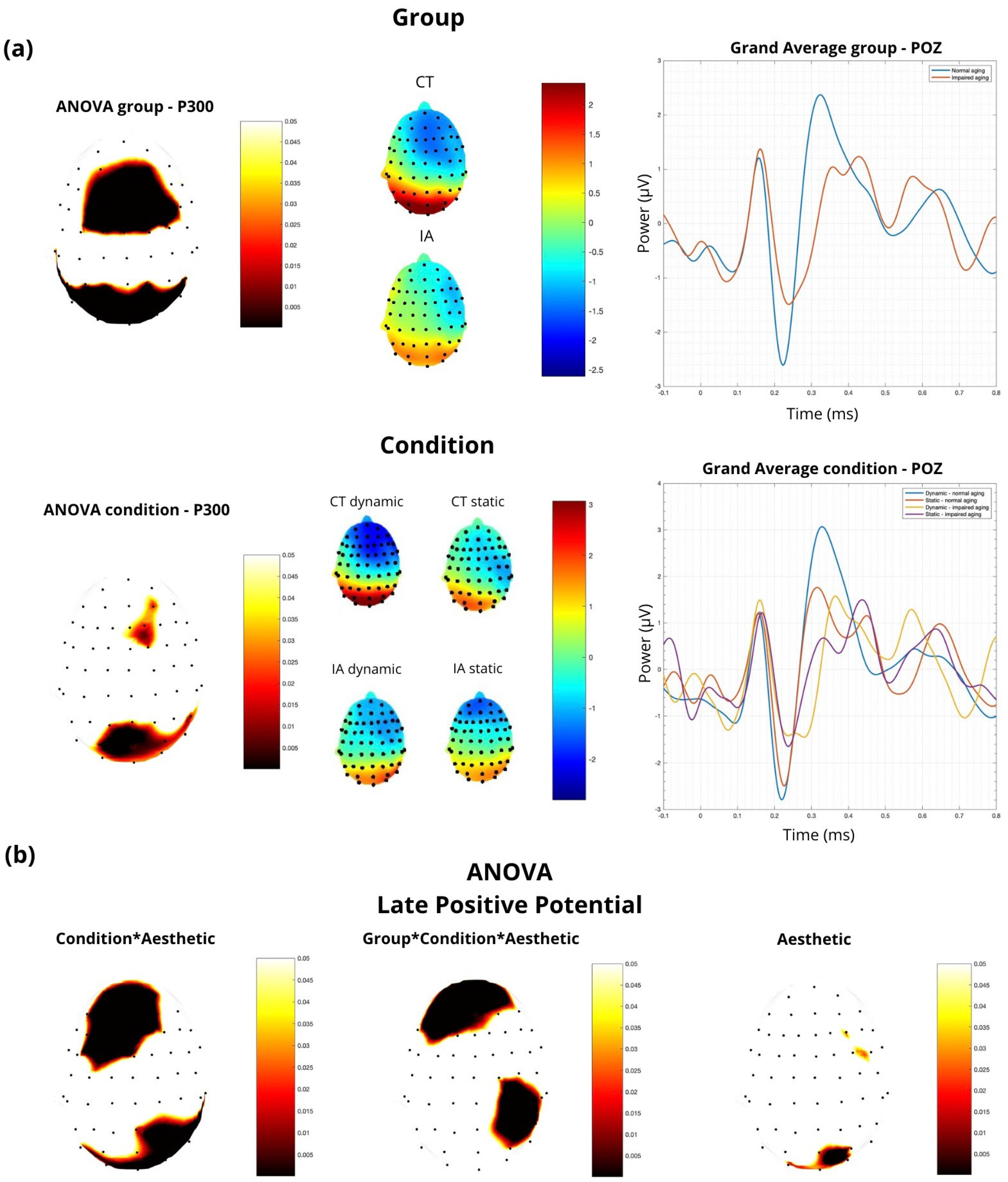
3.2. EEG Results
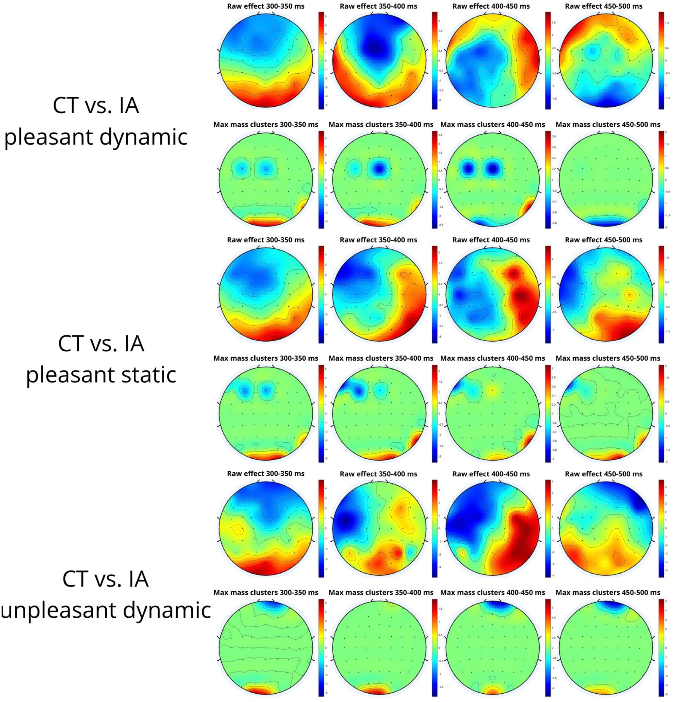
Non-Parametric Cluster-Based Permutation Analysis
4. Discussion
4.1. fNIRS
4.2. EEG
4.3. Limitations
5. Conclusions
Author Contributions
Funding
Institutional Review Board Statement
Informed Consent Statement
Data Availability Statement
Acknowledgments
Conflicts of Interest
References
- Mudgal, S.K.; Sharma, S.K.; Chaturvedi, J.; Sharma, A. Brain computer interface advancement in neurosciences: Applications and issues. Interdiscip. Neurosurg. 2020, 20, 100694. [Google Scholar] [CrossRef]
- Rosenfeld, J.V.; Wong, Y.T. Neurobionics and the brain–computer interface: Current applications and future horizons. Med. J. Aust. 2017, 206, 363–368. [Google Scholar] [CrossRef] [PubMed]
- Nicolas-Alonso, L.F.; Gomez-Gil, J. Brain Computer Interfaces, a Review. Sensors 2012, 12, 1211–1279. [Google Scholar] [CrossRef] [PubMed]
- Buccino, A.P.; Keles, H.O.; Omurtag, A. Hybrid EEG-fNIRS Asynchronous Brain-Computer Interface for Multiple Motor Tasks. PLoS ONE 2016, 11, e0146610. [Google Scholar] [CrossRef] [PubMed]
- Mane, R.; Chouhan, T.; Guan, C. BCI for stroke rehabilitation: Motor and beyond. J. Neural Eng. 2020, 17, 041001. [Google Scholar] [CrossRef] [PubMed]
- Mansour, S.; Ang, K.K.; Nair, K.P.S.; Phua, K.S.; Arvaneh, M. Efficacy of Brain–Computer Interface and the Impact of Its Design Characteristics on Poststroke Upper-limb Rehabilitation: A Systematic Review and Meta-analysis of Randomized Controlled Trials. Clin. EEG Neurosci. 2022, 53, 79–90. [Google Scholar] [CrossRef] [PubMed]
- Ma, P.; Dong, C.; Lin, R.; Ma, S.; Liu, H.; Lei, D.; Chen, X. Effect of Local Network Characteristics on the Performance of the SSVEP Brain-Computer Interface. IRBM 2023, 44, 100781. [Google Scholar] [CrossRef]
- Saeidi, M.; Karwowski, W.; Farahani, F.V.; Fiok, K.; Taiar, R.; Hancock, P.A.; Al-Juaid, A. Neural Decoding of EEG Signals with Machine Learning: A Systematic Review. Brain Sci. 2021, 11, 1525. [Google Scholar] [CrossRef]
- La Rocca, M.; Laporta, A.; Clemente, L.; Ammendola, E.; Delussi, M.D.; Ricci, K.; Tancredi, G.; Stramaglia, S.; de Tommaso, M. Galcanezumab treatment changes visual related EEG connectivity patterns in migraine patients. Cephalalgia 2023, 43, 03331024231189751. [Google Scholar] [CrossRef]
- Saha, S.; Mamun, K.A.; Ahmed, K.; Mostafa, R.; Naik, G.R.; Darvishi, S.; Khandoker, A.H.; Baumert, M. Progress in Brain Computer Interface: Challenges and Opportunities. Front. Syst. Neurosci. 2021, 15, 578875. [Google Scholar] [CrossRef]
- Pandarinath, C.; Nuyujukian, P.; Blabe, C.H.; Sorice, B.L.; Saab, J.; Willett, F.R.; Hochberg, L.R.; Shenoy, K.V.; Henderson, J.M. High performance communication by people with paralysis using an intracortical brain-computer interface. eLife 2017, 6, e18554. [Google Scholar] [CrossRef]
- Kaiju, T.; Doi, K.; Yokota, M.; Watanabe, K.; Inoue, M.; Ando, H.; Takahashi, K.; Yoshida, F.; Hirata, M.; Suzuki, T. High Spatiotemporal Resolution ECoG Recording of Somatosensory Evoked Potentials with Flexible Micro-Electrode Arrays. Front. Neural Circuits 2017, 11, 20. [Google Scholar] [CrossRef] [PubMed]
- Wolpaw, J.R. Brain–computer interfaces. In Handbook of Clinical Neurology; Elsevier: Amsterdam, The Netherlands, 2013; Volume 110, pp. 67–74. [Google Scholar] [CrossRef]
- Zhao, Z.-P.; Nie, C.; Jiang, C.-T.; Cao, S.-H.; Tian, K.-X.; Yu, S.; Gu, J.-W. Modulating Brain Activity with Invasive Brain–Computer Interface: A Narrative Review. Brain Sci. 2023, 13, 134. [Google Scholar] [CrossRef] [PubMed]
- Wolpaw, J.R.; Birbaumer, N.; McFarland, D.J.; Pfurtscheller, G.; Vaughan, T.M. Brain–computer interfaces for communication and control. Clin. Neurophysiol. 2002, 113, 767–791. [Google Scholar] [CrossRef] [PubMed]
- Sciaraffa, N.; Di Flumeri, G.; Germano, D.; Giorgi, A.; Di Florio, A.; Borghini, G.; Vozzi, A.; Ronca, V.; Babiloni, F.; Aricò, P. Evaluation of a New Lightweight EEG Technology for Translational Applications of Passive Brain-Computer Interfaces. Front. Hum. Neurosci. 2022, 16, 901387. [Google Scholar] [CrossRef]
- Roman-Gonzalez, A. EEG Signal Processing for BCI Applications. In Human—Computer Systems Interaction: Backgrounds and Applications 2; Hippe, Z.S., Kulikowski, J.L., Mroczek, T., Eds.; Advances in Intelligent and Soft Computing; Springer: Berlin/Heidelberg, Germany, 2012; Volume 98, pp. 571–591. [Google Scholar] [CrossRef]
- Sörnmo, L.; Laguna, P. Bioelectrical Signal Processing in Cardiac and Neurological Applications; Elsevier Academic Press: Amsterdam, The Netherlands, 2005. [Google Scholar]
- De Tommaso, M.; Pecoraro, C.; Sardaro, M.; Serpino, C.; Lancioni, G.; Livrea, P. Influence of aesthetic perception on visual event-related potentials. Conscious. Cogn. 2008, 17, 933–945. [Google Scholar] [CrossRef] [PubMed]
- De Tommaso, M.; La Rocca, M.; Quitadamo, S.G.; Ricci, K.; Tancredi, G.; Clemente, L.; Gentile, E.; Ammendola, E.; Delussi, M. Central effects of galcanezumab in migraine: A pilot study on Steady State Visual Evoked Potentials and occipital hemodynamic response in migraine patients. J. Headache Pain 2022, 23, 52. [Google Scholar] [CrossRef]
- Ayaz, H.; Izzetoglu, M.; Bunce, S.; Heiman-Patterson, T.; Onaral, B. Detecting cognitive activity related hemodynamic signal for brain computer interface using functional near infrared spectroscopy. In Proceedings of the 2007 3rd International IEEE/EMBS Conference on Neural Engineering, Kohala Coast, HI, USA, 2–5 May 2007; IEEE: Piscataway, NJ, USA, 2007; pp. 342–345. [Google Scholar] [CrossRef]
- Wilcox, T.; Biondi, M. fNIRS in the developmental sciences. WIRES Cogn. Sci. 2015, 6, 263–283. [Google Scholar] [CrossRef]
- La Rocca, M.; Clemente, L.; Gentile, E.; Ricci, K.; Delussi, M.; De Tommaso, M. Effect of Single Session of Anodal M1 Transcranial Direct Current Stimulation—TDCS—On Cortical Hemodynamic Activity: A Pilot Study in Fibromyalgia. Brain Sci. 2022, 12, 1569. [Google Scholar] [CrossRef]
- Coyle, S.; Ward, T.; Markham, C.; McDarby, G. On the suitability of near-infrared (NIR) systems for next-generation brain–computer interfaces. Physiol. Meas. 2004, 25, 815–822. [Google Scholar] [CrossRef]
- Di Dio, C.; Ardizzi, M.; Massaro, D.; Di Cesare, G.; Gilli, G.; Marchetti, A.; Gallese, V. Human, Nature, Dynamism: The Effects of Content and Movement Perception on Brain Activations during the Aesthetic Judgment of Representational Paintings. Front. Hum. Neurosci. 2016, 9, 705. [Google Scholar] [CrossRef] [PubMed]
- Thakral, P.P.; Moo, L.R.; Slotnick, S.D. A neural mechanism for aesthetic experience. NeuroReport 2012, 23, 310–313. [Google Scholar] [CrossRef] [PubMed]
- Battaglia, F.; Lisanby, S.H.; Freedberg, D. Corticomotor Excitability during Observation and Imagination of a Work of Art. Front. Hum. Neurosci. 2011, 5, 79. [Google Scholar] [CrossRef] [PubMed]
- Proverbio, A.M.; Riva, F.; Zani, A. Observation of Static Pictures of Dynamic Actions Enhances the Activity of Movement-Related Brain Areas. PLoS ONE 2009, 4, e5389. [Google Scholar] [CrossRef] [PubMed]
- Uchitel, J.; Vidal-Rosas, E.E.; Cooper, R.J.; Zhao, H. Wearable, Integrated EEG–fNIRS Technologies: A Review. Sensors 2021, 21, 6106. [Google Scholar] [CrossRef] [PubMed]
- Li, R.; Li, S.; Roh, J.; Wang, C.; Zhang, Y. Multimodal Neuroimaging Using Concurrent EEG/fNIRS for Poststroke Recovery Assessment: An Exploratory Study. Neurorehabilit. Neural Repair 2020, 34, 1099–1110. [Google Scholar] [CrossRef] [PubMed]
- Chopra, A.; Cavalieri, T.A.; Libon, D.J. Dementia Screening Tools for the Primary Care Physician. Clin. Geriatr. 2007, 15, 38–45. [Google Scholar]
- Machado, A.; Cai, Z.; Pellegrino, G.; Marcotte, O.; Vincent, T.; Lina, J.-M.; Kobayashi, E.; Grova, C. Optimal positioning of optodes on the scalp for personalized functional near-infrared spectroscopy investigations. J. Neurosci. Methods 2018, 309, 91–108. [Google Scholar] [CrossRef]
- Delpy, D.T.; Cope, M.; Zee, P.V.D.; Arridge, S.; Wray, S.; Wyatt, J. Estimation of optical pathlength through tissue from direct time of flight measurement. Phys. Med. Biol. 1988, 33, 1433–1442. [Google Scholar] [CrossRef]
- Maris, E.; Oostenveld, R. Nonparametric statistical testing of EEG- and MEG-data. J. Neurosci. Methods 2007, 164, 177–190. [Google Scholar] [CrossRef]
- Liu, Z.; Shore, J.; Wang, M.; Yuan, F.; Buss, A.; Zhao, X. A systematic review on hybrid EEG/fNIRS in brain-computer interface. Biomed. Signal Process. Control 2021, 68, 102595. [Google Scholar] [CrossRef]
- Sadjadi, S.M.; Ebrahimzadeh, E.; Shams, M.; Seraji, M.; Soltanian-Zadeh, H. Localization of Epileptic Foci Based on Simultaneous EEG–fMRI Data. Front. Neurol. 2021, 12, 645594. [Google Scholar] [CrossRef] [PubMed]
- Formaggio, E.; Tonellato, M.; Antonini, A.; Castiglia, L.; Gallo, L.; Manganotti, P.; Masiero, S.; Del Felice, A. Oscillatory EEG-TMS Reactivity in Parkinson Disease. J. Clin. Neurophysiol. 2023, 40, 263–268. [Google Scholar] [CrossRef] [PubMed]
- Holtzer, R.; Rakitin, B.C.; Steffener, J.; Flynn, J.; Kumar, A.; Stern, Y. Age effects on load-dependent brain activations in working memory for novel material. Brain Res. 2009, 1249, 148–161. [Google Scholar] [CrossRef] [PubMed][Green Version]
- Doi, T.; Makizako, H.; Shimada, H.; Park, H.; Tsutsumimoto, K.; Uemura, K.; Suzuki, T. Brain activation during dual-task walking and executive function among older adults with mild cognitive impairment: A fNIRS study. Aging Clin. Exp. Res. 2013, 25, 539–544. [Google Scholar] [CrossRef] [PubMed]
- Chaparro, G.; Balto, J.M.; Sandroff, B.M.; Holtzer, R.; Izzetoglu, M.; Motl, R.W.; Hernandez, M.E. Frontal brain activation changes due to dual-tasking under partial body weight support conditions in older adults with multiple sclerosis. J. Neuroeng. Rehabil. 2017, 14, 65. [Google Scholar] [CrossRef] [PubMed]
- Mahoney, J.R.; Holtzer, R.; Izzetoglu, M.; Zemon, V.; Verghese, J.; Allali, G. The role of prefrontal cortex during postural control in Parkinsonian syndromes a functional near-infrared spectroscopy study. Brain Res. 2016, 1633, 126–138. [Google Scholar] [CrossRef] [PubMed]
- Chatterjee, A.; Vartanian, O. Neuroaesthetics. Trends Cogn. Sci. 2014, 18, 370–375. [Google Scholar] [CrossRef]
- Niu, H.; Li, X.; Chen, Y.; Ma, C.; Zhang, J.; Zhang, Z. Reduced Frontal Activation during a Working Memory Task in Mild Cognitive Impairment: A Non-Invasive Near-Infrared Spectroscopy Study. CNS Neurosci. Ther. 2013, 19, 125–131. [Google Scholar] [CrossRef]
- Yeung, M.K.; Sze, S.L.; Woo, J.; Kwok, T.; Shum, D.H.; Yu, R.; Chan, A.S. Reduced Frontal Activations at High Working Memory Load in Mild Cognitive Impairment: Near-Infrared Spectroscopy. Dement. Geriatr. Cogn. Disord. 2016, 42, 278–296. [Google Scholar] [CrossRef]
- Papaliagkas, V.T.; Kimiskidis, V.K.; Tsolaki, M.N.; Anogianakis, G. Cognitive event-related potentials: Longitudinal changes in mild cognitive impairment. Clin. Neurophysiol. 2011, 122, 1322–1326. [Google Scholar] [CrossRef] [PubMed]
- Babiloni, C.; Lizio, R.; Del Percio, C.; Marzano, N.; Soricelli, A.; Salvatore, E.; Ferri, R.; Cosentino, F.I.; Tedeschi, G.; Montella, P.; et al. Cortical Sources of Resting State EEG Rhythms are Sensitive to the Progression of Early Stage Alzheimer’s Disease. J. Alzheimer’s Dis. 2013, 34, 1015–1035. [Google Scholar] [CrossRef] [PubMed]
- Polich, J. Updating P300: An integrative theory of P3a and P3b. Clin. Neurophysiol. 2007, 118, 2128–2148. [Google Scholar] [CrossRef] [PubMed]
- Medvidovic, S.; Titlic, M.; MarasSimunic, M. P300 Evoked Potential in Patients with Mild Cognitive Impairment. Acta Inform. Medica 2013, 21, 89–92. [Google Scholar] [CrossRef] [PubMed]
- Papadaniil, C.D.; Kosmidou, V.E.; Tsolaki, A.; Tsolaki, M.; Kompatsiaris, I.; Hadjileontiadis, L.J. Cognitive MMN and P300 in mild cognitive impairment and Alzheimer’s disease: A high density EEG-3D vector field tomography approach. Brain Res. 2016, 1648, 425–433. [Google Scholar] [CrossRef] [PubMed]
- Howe, A.S.; Bani-Fatemi, A.; De Luca, V. The clinical utility of the auditory P300 latency subcomponent event-related potential in preclinical diagnosis of patients with mild cognitive impairment and Alzheimer’s disease. Brain Cogn. 2014, 86, 64–74. [Google Scholar] [CrossRef]
- Fix, S.T.; Arruda, J.E.; Andrasik, F.; Beach, J.; Groom, K. Using visual evoked potentials for the early detection of amnestic mild cognitive impairment: A pilot investigation. Int. J. Geriatr. Psychiatry 2015, 30, 72–79. [Google Scholar] [CrossRef] [PubMed]
- Bennys, K.; Rondouin, G.; Benattar, E.; Gabelle, A.; Touchon, J. Can Event-Related Potential Predict the Progression of Mild Cognitive Impairment? J. Clin. Neurophysiol. 2011, 28, 625–632. [Google Scholar] [CrossRef]
- Gozke, E.; Tomrukcu, S.; Erdal, N. Visual Event-Related Potentials in Patients with Mild Cognitive Impairment. Int. J. Gerontol. 2016, 10, 190–192. [Google Scholar] [CrossRef][Green Version]
- Leder, H.; Belke, B.; Oeberst, A.; Augustin, D. A model of aesthetic appreciation and aesthetic judgments. Br. J. Psychol. 2004, 95, 489–508. [Google Scholar] [CrossRef]
- Clemente, L.; Gasparre, D.; Alfeo, F.; Battista, F.; Abbatantuono, C.; Curci, A.; Lanciano, T.; Taurisano, P. Theory of Mind and Executive Functions in Individuals with Mild Cognitive Impairment or Healthy Aging. Brain Sci. 2023, 13, 1356. [Google Scholar] [CrossRef] [PubMed]
- Höfel, L.; Jacobsen, T. Electrophysiological indices of processing aesthetics: Spontaneous or intentional processes? Int. J. Psychophysiol. 2007, 65, 20–31. [Google Scholar] [CrossRef] [PubMed]
- Righi, S.; Orlando, V.; Marzi, T. Attractiveness and affordance shape tools neural coding: Insight from ERPs. Int. J. Psychophysiol. 2014, 91, 240–253. [Google Scholar] [CrossRef] [PubMed]


| ch 10 | Sum of Squares | df | Mean Square | F | p |
|---|---|---|---|---|---|
| group | 4.37 × 10⁻⁷ | 1 | 4.37 × 10⁻⁷ | 1.63 | 0.205 |
| condition | 4.76 × 10⁻⁷ | 1 | 4.76 × 10⁻⁷ | 1.77 | 0.186 |
| aesthetic | 1.02 × 10⁻⁶ | 2 | 5.08 × 10⁻⁷ | 1.89 | 0.156 |
| group x condition | 6.08 × 10⁻⁷ | 1 | 6.08 × 10⁻⁷ | 2.27 | 0.135 |
| group x aesthetic | 1.76 × 10⁻⁶ | 2 | 8.80 × 10⁻⁷ | 3.28 | 0.042 * |
| condition x aesthetic | 1.07 × 10⁻⁶ | 2 | 5.36 × 10⁻⁷ | 2.00 | 0.141 |
| group x condition x aesthetic | 1.20 × 10⁻⁶ | 2 | 5.98 × 10⁻⁷ | 2.23 | 0.113 |
| Residuals | 2.63 × 10⁻⁵ | 98 | 2.68 × 10⁻⁷ |
| ch 20 | Sum of Squares | df | Mean Square | F | p |
|---|---|---|---|---|---|
| group | 2.30 × 10⁻⁸ | 1 | 2.30 × 10⁻⁸ | 0.06 | 0.809 |
| condition | 9.68 × 10⁻⁷ | 1 | 9.68 × 10⁻⁷ | 2.47 | 0.119 |
| aesthetic | 3.99 × 10⁻⁸ | 2 | 2.00 × 10⁻⁸ | 0.05 | 0.950 |
| group x condition | 1.48 × 10⁻⁶ | 1 | 1.48 × 10⁻⁶ | 3.78 | 0.055 |
| group x aesthetic | 9.25 × 10⁻⁸ | 2 | 4.62 × 10⁻⁸ | 0.12 | 0.889 |
| condition x aesthetic | 2.22 × 10⁻⁶ | 2 | 1.11 × 10⁻⁶ | 2.84 | 0.063 |
| group x condition x aesthetic | 2.70 × 10⁻⁶ | 2 | 1.35 × 10⁻⁶ | 3.45 | 0.036 * |
| Residuals | 3.83 × 10⁻⁵ | 98 | 3.91 × 10⁻⁷ |
| Max Positive Cluster | Max Negative Cluster | ||||||
|---|---|---|---|---|---|---|---|
| Cohen’s D | Mass | ERP (Mean) | ERP (sd) | Mass | ERP (Mean) | ERP (sd) | |
| Pleasant dynamic CT vs. IA | 2.10 | 118.10 | 3.37 | 3.24 | 102.92 | 0.20 | 1.55 |
| Pleasant static CT vs. IA | 2.27 | 128.42 | 2.80 | 4.83 | 88.80 | −0.69 | 2.76 |
| Unpleasant dynamic CT vs. IA | 1.96 | 38.21 | 0.57 | 5.06 | 16.06 | 1.30 | 8.12 |
| Unpleasant static CT vs. IA | 2.33 | 34.67 | −1.31 | 1.57 | 73.25 | 1.52 | 4.18 |
| Neutral dynamic CT vs. IA | 2.05 | 53.02 | 4.44 | 4.45 | 14.24 | 1.37 | 6.17 |
| Neutral static CT vs. IA | 1.76 | 21.31 | −0.65 | 2.26 | 27.98 | 0.95 | 3.15 |
| Control dynamic pleasant vs. unpleasant | 2.18 | 11.40 | −0.48 | 2.09 | 34.99 | 0.06 | 2.36 |
| Control dynamic pleasant vs. neutral | - | - | - | - | - | - | - |
| Control dynamic unpleasant vs. neutral | 1.95 | 16.85 | 0.06 | 2.36 | 7.13 | −3.48 | 1.07 |
| Patient dynamic pleasant vs. unpleasant | 2.12 | 130.55 | 1.37 | 1.31 | 75.97 | 0.28 | 6.43 |
| Patient dynamic pleasant vs. neutral | 1.81 | 41.33 | 0.46 | 1.64 | 19.79 | 4.17 | 7.08 |
| Patient dynamic unpleasant vs. neutral | 2.04 | 11.98 | −0.28 | 4.73 | 30.83 | 1.98 | 6.07 |
| Control static pleasant vs. unpleasant | 2.15 | 20.29 | 0.21 | 1.94 | 7.66 | −0.76 | 2.41 |
| Control static pleasant vs. neutral | 2.10 | 15.41 | 2.25 | 3.14 | 9.44 | 0.56 | 1.03 |
| Control static unpleasant vs. neutral | 2.82 | 20.15 | 0.69 | 3.99 | 17.51 | −1.70 | 0.94 |
| Patient static pleasant vs. unpleasant | 2.32 | 24.60 | −1.47 | 2.62 | 46.32 | 0.40 | 3.31 |
| Patient static pleasant vs. neutral | 1.75 | 26.75 | −1.17 | 2.65 | 12.06 | 0.42 | 3.66 |
| Patient static unpleasant vs. neutral | 2.16 | 27.86 | 0.10 | 5.33 | 22.55 | 0.42 | 3.66 |
| Control pleasant dynamic vs. static | 1.62 | 15.38 | −3.57 | 2.98 | 14.80 | −0.85 | 2.60 |
| Control unpleasant dynamic vs. static | 2.45 | 61.84 | 1.62 | 4.66 | 21.37 | −1.60 | 2.18 |
| Control neutral dynamic vs. static | 2.23 | 16.94 | 0.37 | 1.60 | 24.52 | −1.85 | 1.75 |
| Patient pleasant dynamic vs. static | 1.57 | 31.34 | 0.71 | 1.04 | 14.01 | −1.86 | 2.42 |
| Patient unpleasant dynamic vs. static | 2.51 | 60.60 | 0.22 | 5.44 | 57.08 | 0.93 | 4.19 |
| Patient neutral dynamic vs. static | 1.49 | 27.86 | 4.36 | 5.26 | 33.90 | 0.32 | 3.33 |
Disclaimer/Publisher’s Note: The statements, opinions and data contained in all publications are solely those of the individual author(s) and contributor(s) and not of MDPI and/or the editor(s). MDPI and/or the editor(s) disclaim responsibility for any injury to people or property resulting from any ideas, methods, instructions or products referred to in the content. |
© 2024 by the authors. Licensee MDPI, Basel, Switzerland. This article is an open access article distributed under the terms and conditions of the Creative Commons Attribution (CC BY) license (https://creativecommons.org/licenses/by/4.0/).
Share and Cite
Clemente, L.; La Rocca, M.; Paparella, G.; Delussi, M.; Tancredi, G.; Ricci, K.; Procida, G.; Introna, A.; Brunetti, A.; Taurisano, P.; et al. Exploring Aesthetic Perception in Impaired Aging: A Multimodal Brain—Computer Interface Study. Sensors 2024, 24, 2329. https://doi.org/10.3390/s24072329
Clemente L, La Rocca M, Paparella G, Delussi M, Tancredi G, Ricci K, Procida G, Introna A, Brunetti A, Taurisano P, et al. Exploring Aesthetic Perception in Impaired Aging: A Multimodal Brain—Computer Interface Study. Sensors. 2024; 24(7):2329. https://doi.org/10.3390/s24072329
Chicago/Turabian StyleClemente, Livio, Marianna La Rocca, Giulia Paparella, Marianna Delussi, Giusy Tancredi, Katia Ricci, Giuseppe Procida, Alessandro Introna, Antonio Brunetti, Paolo Taurisano, and et al. 2024. "Exploring Aesthetic Perception in Impaired Aging: A Multimodal Brain—Computer Interface Study" Sensors 24, no. 7: 2329. https://doi.org/10.3390/s24072329
APA StyleClemente, L., La Rocca, M., Paparella, G., Delussi, M., Tancredi, G., Ricci, K., Procida, G., Introna, A., Brunetti, A., Taurisano, P., Bevilacqua, V., & de Tommaso, M. (2024). Exploring Aesthetic Perception in Impaired Aging: A Multimodal Brain—Computer Interface Study. Sensors, 24(7), 2329. https://doi.org/10.3390/s24072329