
Pyrazolo[4,3-c]pyridine Sulfonamides as Carbonic Anhydrase Inhibitors: Synthesis, Biological and In Silico Studies

 , , , , , and
Abstract
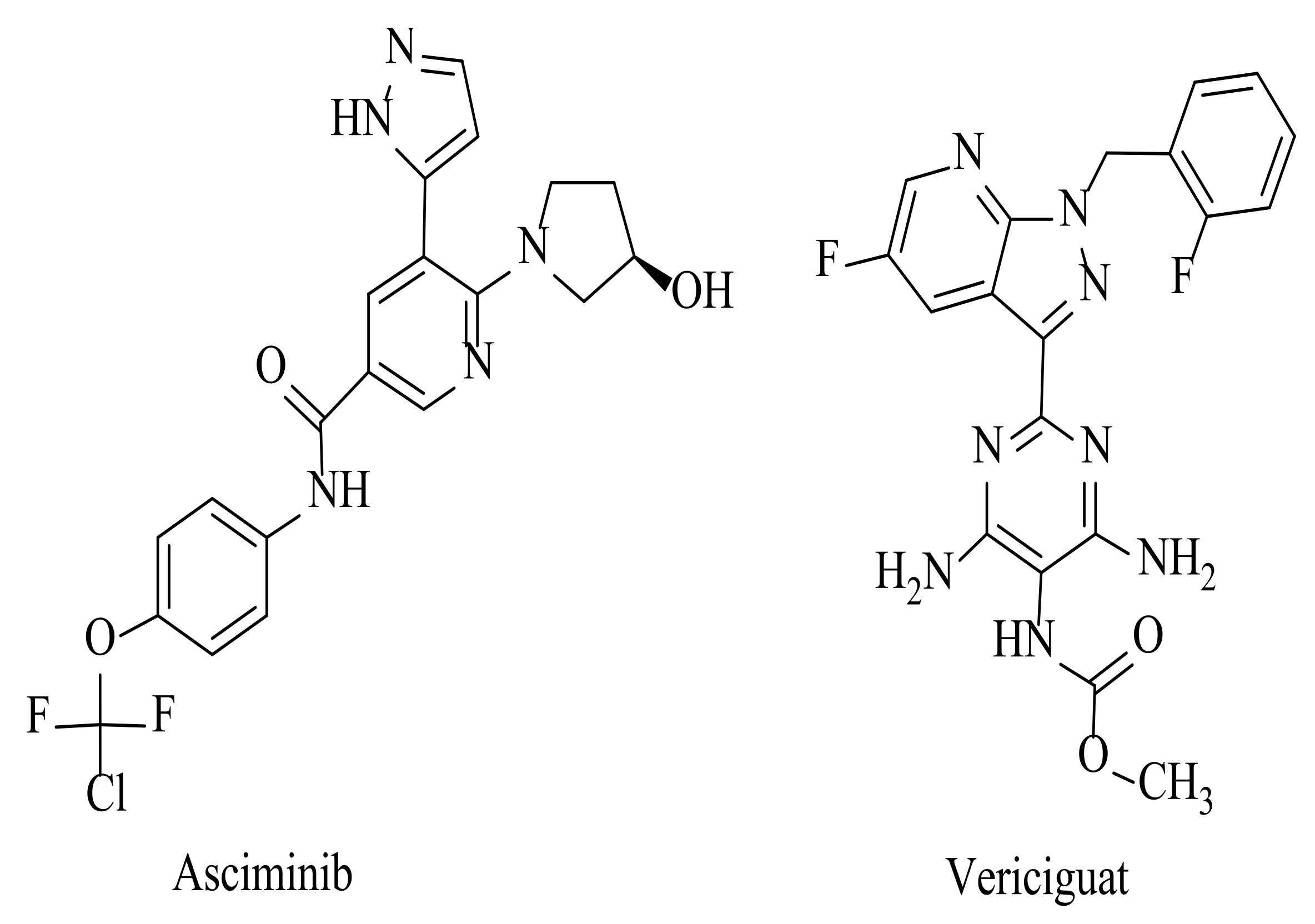
:1. Introduction
2. Results and Discussion
2.1. Chemistry
2.2. Carbonic Anhydrase Inhibition
2.3. Molecular Docking Studies
2.3.1. Molecular Docking Studies in Human CA Isoforms
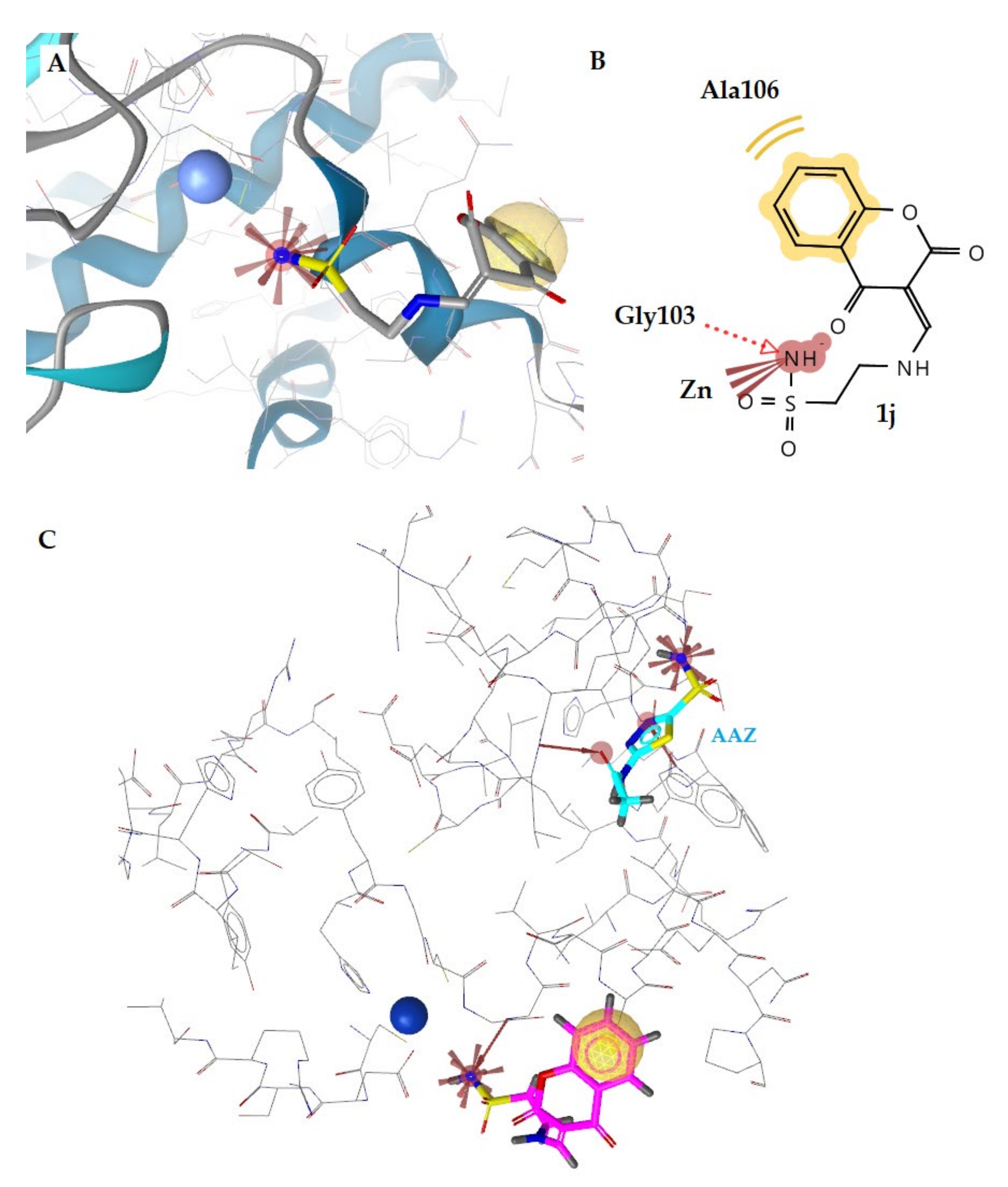
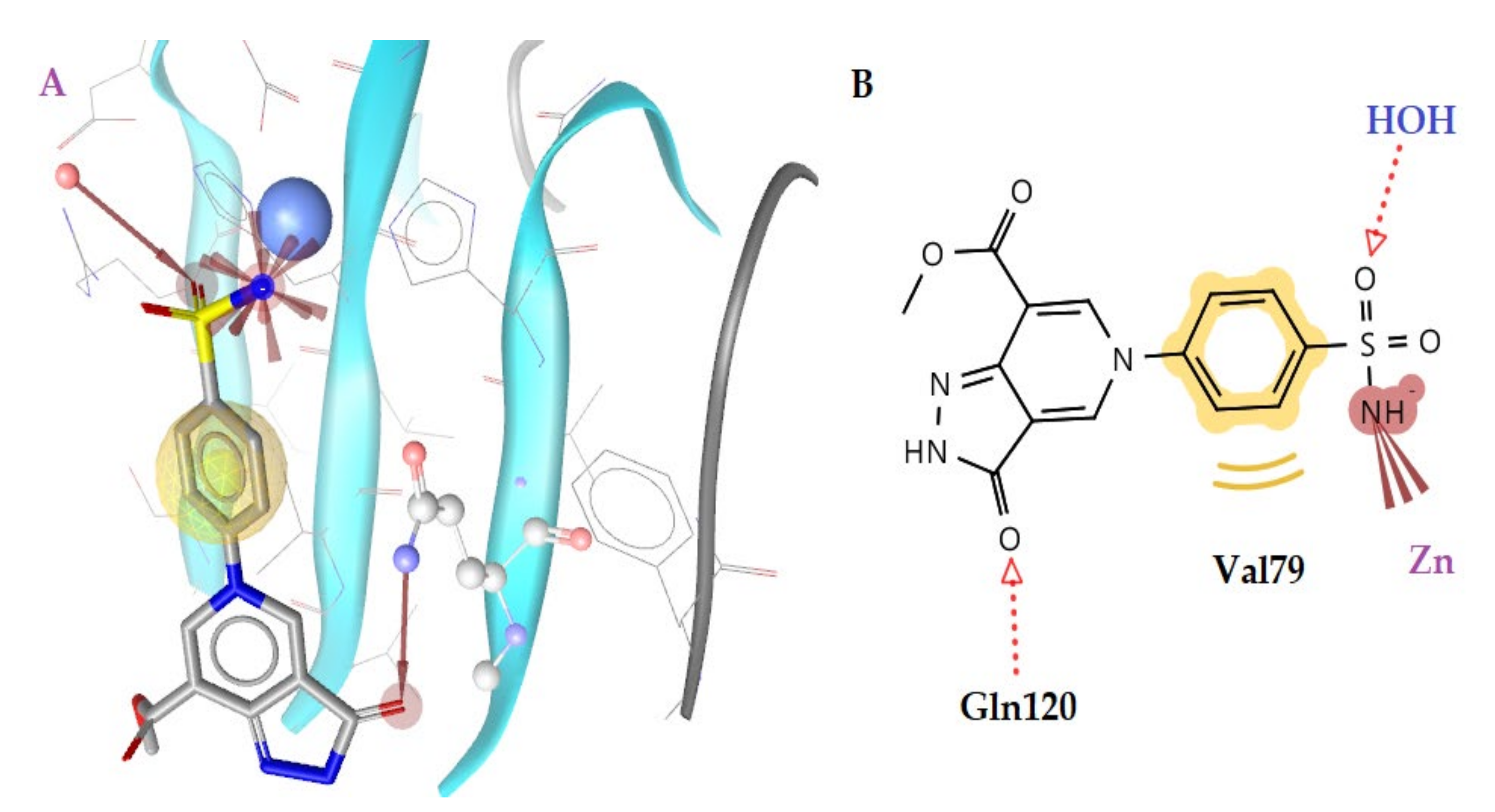
2.3.2. Molecular Docking Studies in β- and γ-CA Classes
2.4. In Silico Prediction Studies
Drug-Likeness
3. Materials and Methods
3.1. Chemistry
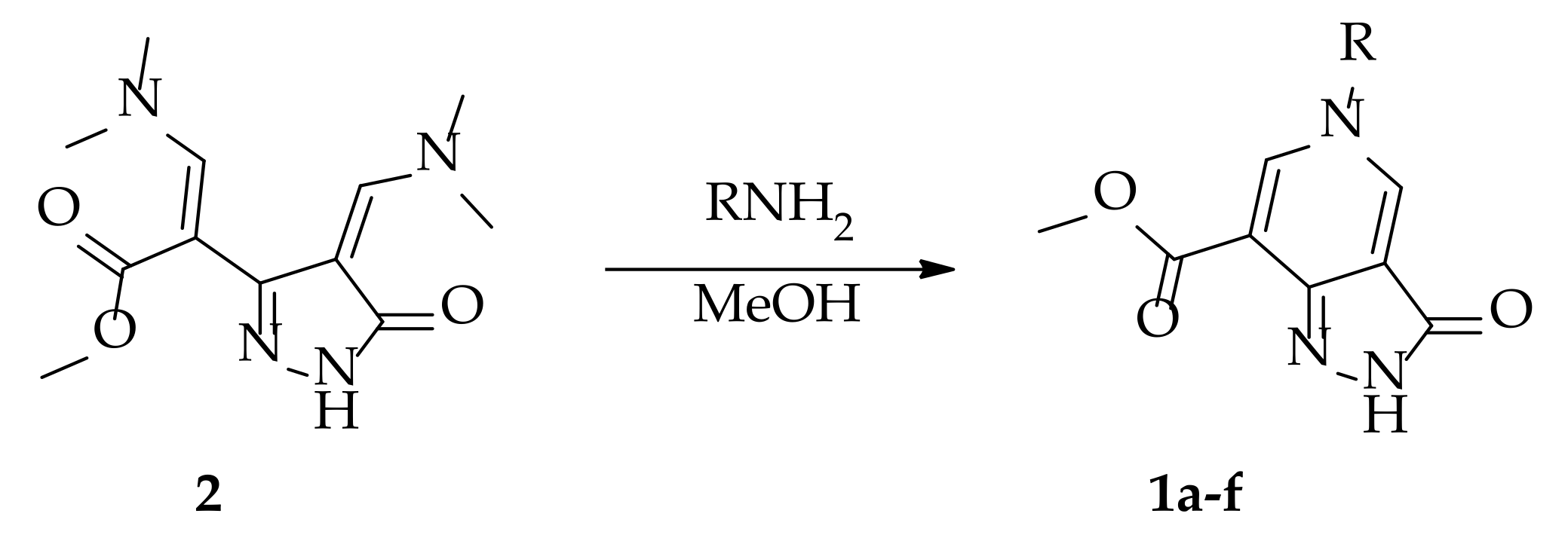
3.1.1. Synthesis of 5-Substituted Methyl 3-Oxo-3,5-dihydro-2H-pyrazolo[4,3-c]pyridine-7-carboxylates 1a–f (General Procedure)
3.1.2. Synthesis of 1-Acetyl-4-(arylamino)-1,5-dihydro-2H-pyrrol-2-ones 1g,h (General Procedure)
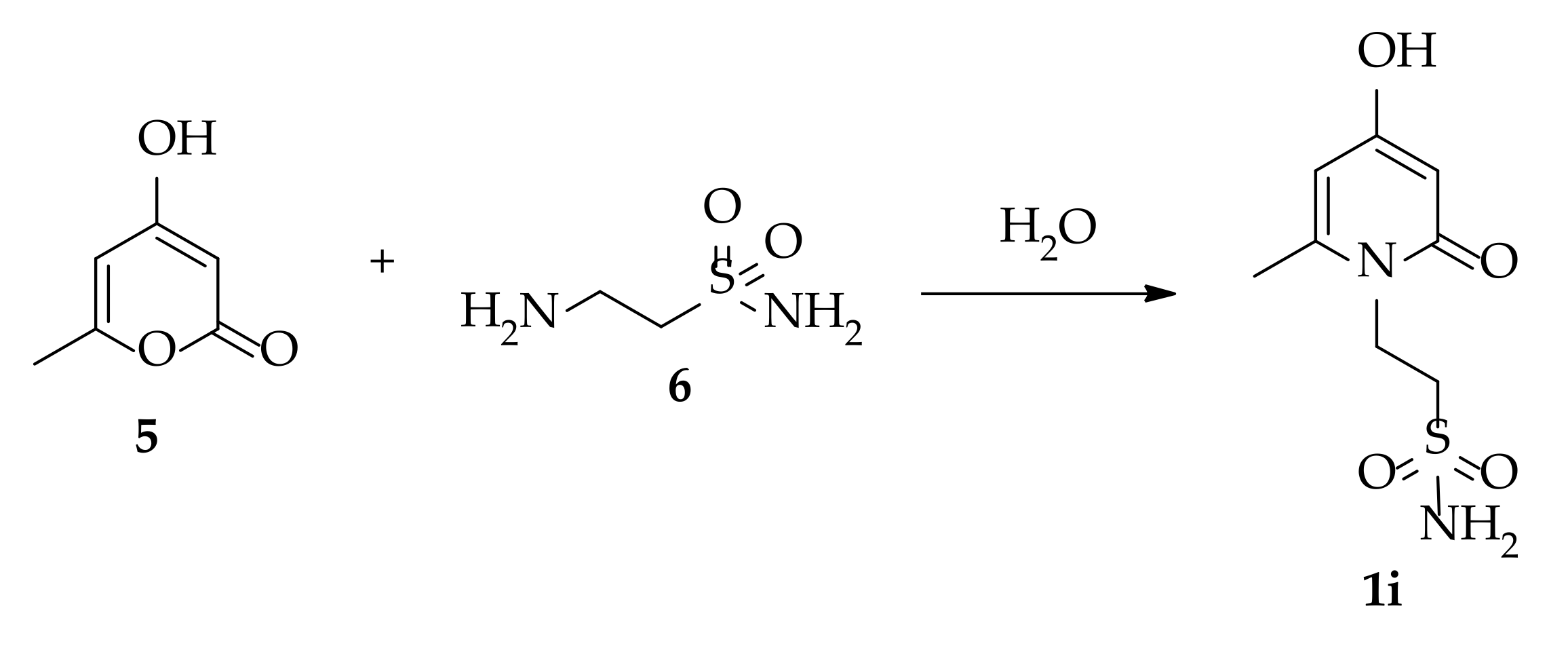
3.1.3. Synthesis of 2-(4-Hydroxy-6-methyl-2-oxopyridin-1(2H)-yl)ethane-1-sulfonamide 1i
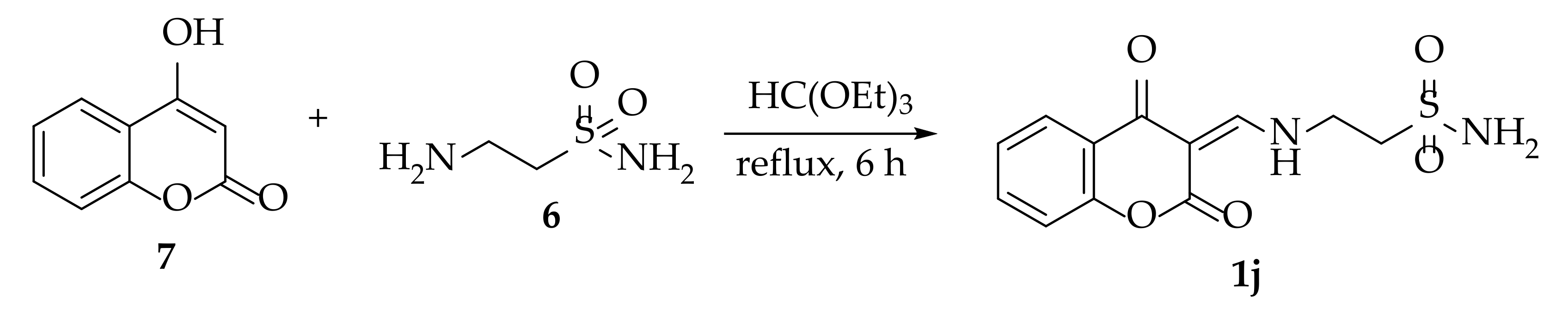
3.1.4. Synthesis of 2-(((2,4-Dioxochroman-3-ylidene)methyl)amino)ethane-1-sulfonamide 1j
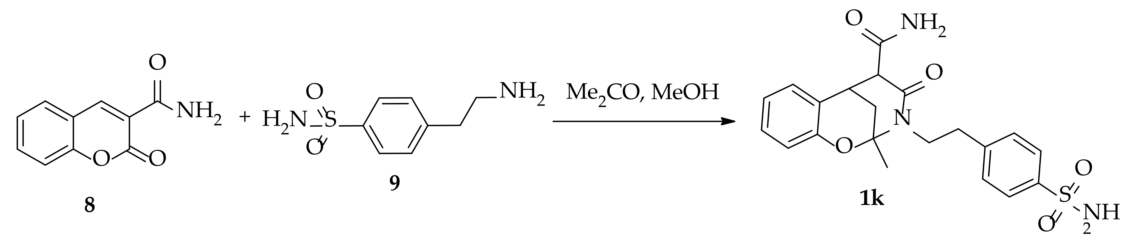
3.1.5. Synthesis of 2-Methyl-4-oxo-3-(4-sulfamoylphenethyl)-3,4,5,6-tetrahydro-2H-2,6-methanobenzo[g] [1,3]oxazocine-5-carboxamide 1k
3.2. Molecular Docking Studies
3.3. CA Inhibition Assay
3.4. Drug-Likness
4. Conclusions
Supplementary Materials
Author Contributions
Funding
Institutional Review Board Statement
Informed Consent Statement
Data Availability Statement
Conflicts of Interest
References
- Supuran, C.T.; Capasso, C. An Overview of the Bacterial Carbonic Anhydrases. Metabolites 2017, 7, 56. [Google Scholar] [CrossRef] [PubMed] [Green Version]
- Supuran, C.T.; Capasso, C. Antibacterial carbonic anhydrase inhibitors: An update on the recent literature. Expert Opin. Ther. Pat. 2020, 30, 963–982. [Google Scholar] [CrossRef] [PubMed]
- Supuran, C.T. Carbonic anhydrases: Novel therapeutic applications for inhibitors and activators. Nat. Rev. Drug Discov. 2008, 7, 168–181. [Google Scholar] [CrossRef] [PubMed]
- Supuran, C.T. Emerging role of carbonic anhydrase inhibitors. Clin. Sci. 2021, 135, 1233–1249. [Google Scholar] [CrossRef]
- Alterio, V.; Di Fiore, A.; D’Ambrosio, K.; Supuran, C.T.; De Simone, G. Multiple binding modes of inhibitors to carbonic anhydrases: How to design specific drugs targeting 15 different isoforms? Chem. Rev. 2012, 112, 4421–4468. [Google Scholar] [CrossRef] [Green Version]
- Penning, T.D.; Talley, J.J.; Bertenshaw, S.R.; Carter, J.S.; Collins, P.W.; Docter, S.; Graneto, M.J.; Lee, L.F.; Malecha, J.W.; Miyashiro, J.M.; et al. Synthesis and biological evaluation of the 1,5-diarylpyrazole class of cyclooxygenase-2 inhibitors: Identification of 4-[5-(4-methylphenyl)-3-(trifluoromethyl)-1H-pyrazol-1-yl]benze nesulfonamide (SC-58635, celecoxib). J. Med. Chem. 1997, 40, 1347–1365. [Google Scholar] [CrossRef]
- Fioravanti, R.; Bolasco, A.; Manna, F.; Rossi, F.; Orallo, F.; Ortuso, F.; Alcaro, S.; Cirilli, R. Synthesis and biological evaluation of N-substituted-3,5-diphenyl-2-pyrazoline derivatives as cyclooxygenase (COX-2) inhibitors. Eur. J. Med. Chem. 2010, 45, 6135–6138. [Google Scholar] [CrossRef]
- Devi, N.; Shankar, R.; Singh, V. 4-Formyl-Pyrazole-3-Carboxylate: A Useful Aldo-X Bifunctional Precursor for the Syntheses of Pyrazole-fused/Substituted Frameworks. J. Heterocycl. Chem. 2018, 55, 373–390. [Google Scholar] [CrossRef]
- Katz, R. Effects of zometapine, A structurally novel antidepressant, in an animal model of depression. Pharmacol. Biochem. Behav. 1984, 21, 487–490. [Google Scholar] [CrossRef]
- d’Aniello, F.; Santos, B.; Guglietta, A. Lorediplon: A New GABAA Modulator Drug for Treatment of Insomnia. Drug Treat. Sleep Disord. 2015, 49, 121–145. [Google Scholar] [CrossRef]
- Ervinna, N.; Mita, T.; Yasunari, E.; Azuma, K.; Tanaka, R.; Fujimura, S.; Sukmawati, D.; Nomiyama, T.; Kanazawa, A.; Kawamori, R.; et al. Anagliptin, a DPP-4 Inhibitor, Suppresses Proliferation of Vascular Smooth Muscles and Monocyte Inflammatory Reaction and Attenuates Atherosclerosis in Male apo E-Deficient Mice. Endocrinology 2013, 154, 1260–1270. [Google Scholar] [CrossRef] [Green Version]
- Marinescu, M. Synthesis of Antimicrobial Benzimidazole–Pyrazole Compounds and Their Biological Activities. Antibiotics 2021, 10, 1002. [Google Scholar] [CrossRef]
- Cetin, A.; Bildirici, I. A study on synthesis and antimicrobial activity of 4-acyl-pyrazoles. J. Saudi Chem. Soc. 2018, 22, 279–296. [Google Scholar] [CrossRef] [Green Version]
- Muhammad, Z.A.; Alshehrei, F.; Zayed, M.E.M.; Farghaly, T.A.; Abdallah, M.A. Synthesis of Novel Bis-pyrazole Derivatives as Antimicrobial Agents. Mini Rev. Med. Chem. 2019, 19, 1276–1290. [Google Scholar] [CrossRef]
- Da Costa, L.; Scheers, E.; Coluccia, A.; Casulli, A.; Roche, M.; Di Giorgio, C.; Neyts, J.; Terme, T.; Cirilli, R.; La Regina, G.; et al. Structure-Based Drug Design of Potent Pyrazole Derivatives against Rhinovirus Replication. J. Med. Chem. 2018, 61, 8402–8416. [Google Scholar] [CrossRef] [Green Version]
- Corona, A.; Onnis, V.; Deplano, A.; Bianco, G.; Demurtas, M.; Distinto, S.; Cheng, Y.-C.; Alcaro, S.; Esposito, F.; Tramontano, E. Design, synthesis and antiviral evaluation of novel heteroarylcarbothioamide derivatives as dual inhibitors of HIV-1 reverse transcriptase-associated RNase H and RDDP functions. Pathog. Dis. 2017, 75, ftx078. [Google Scholar] [CrossRef] [Green Version]
- Al-Zharani, M.; Al-Eissa, M.S.; Rudayni, H.A.; Ali, D.; Alkahtani, S.; Surendrakumar, R.; Idhayadhulla, A. Pyrazolo[3,4-b]pyridin-3(2H)-one derivatives: Synthesis and their investigation of mosquito larvicidal activity. J. King Saud Univ. Sci. 2022, 34, 101767. [Google Scholar] [CrossRef]
- Naim, M.J.; Alam, O.; Alam, M.J.; Hassan, M.Q.; Siddiqui, N.; Naidu, V.G.M.; Alam, M.I. Design, synthesis and molecular docking of thiazolidinedione based benzene sulphonamide derivatives containing pyrazole core as potential anti-diabetic agents. Bioorg. Chem. 2018, 76, 98–112. [Google Scholar] [CrossRef]
- Faidallah, H.M.; Al-Mohammadi, M.M.; Alamry, K.A.; Khan, K.A. Synthesis and biological evaluation of fluoropyrazolesulfonylurea and thiourea derivatives as possible antidiabetic agents. J. Enzyme Inhib. Med. Chem. 2016, 31, 157–163. [Google Scholar] [CrossRef] [Green Version]
- Li, X.; Yu, Y.; Tu, Z. Pyrazole Scaffold Synthesis, Functionalization, and Applications in Alzheimer’s Disease and Parkinson’s Disease Treatment (2011–2020). Molecules 2021, 26, 1202. [Google Scholar] [CrossRef]
- Lather, V.; Grewal, A.S.; Sharma, S.K.; Pandita, D. Synthesis, Docking and Evaluation of Novel Pyrazole Carboxamide Derivatives as Multifunctional Anti-Alzheimer’s Agents. J. Med. Chem. Toxicol. 2017, 2, 47–54. [Google Scholar] [CrossRef]
- Meta, E.; Brullo, C.; Tonelli, M.; Franzblau, S.G.; Wang, Y.; Ma, R.; Baojie, W.; Orena, B.S.; Pasca, M.R.; Bruno, O. Pyrazole and imidazo[1,2-b]pyrazole Derivatives as New Potential Antituberculosis Agents. Med. Chem. 2019, 15, 17–27. [Google Scholar] [CrossRef]
- Xu, Z.; Gao, C.; Ren, Q.-C.; Song, X.-F.; Feng, L.-S.; Lv, Z.-S. Recent advances of pyrazole-containing derivatives as anti-tubercular agents. Eur. J. Med. Chem. 2017, 139, 429–440. [Google Scholar] [CrossRef]
- Bekhit, A.A.; Saudi, M.N.; Hassan, A.M.; Fahmy, S.M.; Ibrahim, T.M.; Ghareeb, D.; El-Seidy, A.M.; Al-Qallaf, S.M.; Habib, H.J.; Bekhit, A.E.A.-D. Synthesis, molecular modeling and biological screening of some pyrazole derivatives as antileishmanial agents. Future Med. Chem. 2018, 10, 2325–2344. [Google Scholar] [CrossRef] [PubMed]
- Chaudhry, F.; Naureen, S.; Choudhry, S.; Huma, R.; Ashraf, M.; Al-Rashida, M.; Jahan, B.; Khan, M.H.; Iqbal, F.; Munawar, M.A.; et al. Evaluation of α-glucosidase inhibiting potentials with docking calculations of synthesized arylidene-pyrazolones. Bioorg. Chem. 2018, 77, 507–514. [Google Scholar] [CrossRef] [PubMed]
- El-Gohary, N.; Shaaban, M. New pyrazolopyridine analogs: Synthesis, antimicrobial, antiquorum-sensing and antitumor screening. Eur. J. Med. Chem. 2018, 152, 126–136. [Google Scholar] [CrossRef] [PubMed]
- Salem, M.S.; Ali, M.A.M. Novel Pyrazolo[3,4-b]pyridine Derivatives: Synthesis, Characterization, Antimicrobial and Antiproliferative Profile. Biol. Pharm. Bull. 2016, 39, 473–483. [Google Scholar] [CrossRef] [PubMed] [Green Version]
- El-Borai, M.A.; Rizk, H.F.; Beltagy, D.M.; El-Deeb, I.Y. Microwave-assisted synthesis of some new pyrazolopyridines and their antioxidant, antitumor and antimicrobial activities. Eur. J. Med. Chem. 2013, 66, 415–422. [Google Scholar] [CrossRef]
- Bare, T.M.; McLaren, C.D.; Campbell, J.B.; Firor, J.W.; Resch, J.F.; Walters, C.P.; Salama, A.I.; Meiners, B.A.; Patel, J.B. Synthesis and structure-activity relationships of a series of anxioselective pyrazolopyridine ester and amide anxiolytic agents. J. Med. Chem. 1989, 32, 2561–2573. [Google Scholar] [CrossRef]
- Gu, X.; Ma, S. Recent Advances in the Development of Pyrazolopyridines as Anticancer Agents. Anti Cancer Agents Med. Chem. 2021. [Google Scholar] [CrossRef]
- Mor, S.; Khatri, M.; Punia, R.; Sindhu, S. Recent Progress in Anticancer Agents Incorporating Pyrazole Scaffold. Mini Rev. Med. Chem. 2022, 22, 115–163. [Google Scholar] [CrossRef]
- Gavriil, E.-S.; Lougiakis, N.; Pouli, N.; Marakos, P.; Skaltsounis, A.-L.; Nam, S.; Jove, R.; Horne, D.; Gioti, K.; Pratsinis, H.; et al. Synthesis and antiproliferative activity of new pyrazolo[3,4-c]pyridines. Med. Chem. 2017, 13, 365–374. [Google Scholar] [CrossRef]
- Giannouli, V.; Lougiakis, N.; Kostakis, I.K.; Pouli, N.; Marakos, P.; Skaltsounis, A.-L.; Nam, S.; Jove, R.; Horne, D.; Tenta, R.; et al. The discovery of new cytotoxic pyrazolopyridine derivatives. Bioorganic Med. Chem. Lett. 2016, 26, 5229–5233. [Google Scholar] [CrossRef]
- Anand, D.; Yadav, P.K.; Patel, O.P.S.; Parmar, N.; Maurya, R.K.; Vishwakarma, P.; Raju, K.S.R.; Taneja, I.; Wahajuddin, M.; Kar, S.; et al. Antileishmanial Activity of Pyrazolopyridine Derivatives and Their Potential as an Adjunct Therapy with Miltefosine. J. Med. Chem. 2017, 60, 1041–1059. [Google Scholar] [CrossRef]
- de Mello, H.; Echevarria, A.; Bernardino, A.M.R.; Canto-Cavalheiro, A.M.; Leon, L. Antileishmanial Pyrazolopyridine Derivatives: Synthesis and Structure−Activity Relationship Analysis. J. Med. Chem. 2004, 47, 5427–5432. [Google Scholar] [CrossRef]
- Pinheiro, L.C.S.; Feitosa, L.M.; Gandi, M.O.; Silveira, F.F.; Boechat, N. The Development of Novel Compounds Against Malaria: Quinolines, Triazolpyridines, Pyrazolopyridines and Pyrazolopyrimidines. Molecules 2019, 24, 4095. [Google Scholar] [CrossRef] [Green Version]
- Hamblin, J.N.; Angell, T.D.; Ballantine, S.P.; Cook, C.M.; Cooper, A.W.; Dawson, J.; Delves, C.J.; Jones, P.S.; Lindvall, M.; Lucas, F.S.; et al. Pyrazolopyridines as a novel structural class of potent and selective PDE4 inhibitors. Bioorganic Med. Chem. Lett. 2008, 18, 4237–4241. [Google Scholar] [CrossRef]
- Sklepari, M.; Lougiakis, N.; Papastathopoulos, A.; Pouli, N.; Marakos, P.; Myrianthopoulos, V.; Robert, T.; Bach, S.; Mikros, E.; Ruchaud, S. Synthesis, Docking Study and Kinase Inhibitory Activity of a Number of New Substituted Pyrazolo[3,4-c]pyridines. Chem. Pharm. Bull. 2017, 65, 66–81. [Google Scholar] [CrossRef] [Green Version]
- Michailidou, M.; Giannouli, V.; Kotsikoris, V.; Papadodima, O.; Kontogianni, G.; Kostakis, I.K.; Lougiakis, N.; Chatziioannou, A.; Kolisis, F.N.; Marakos, P.; et al. Novel pyrazolopyridine derivatives as potential angiogenesis inhibitors: Synthesis, biological evaluation and transcriptome-based mechanistic analysis. Eur. J. Med. Chem. 2016, 121, 143–157. [Google Scholar] [CrossRef]
- Abbas, H.-A.; El-Karim, S.A.; Abdelwahed, N.A.M. Synthesis and biological evaluation of sulfonamide derivatives as antimicrobial agents. Acta Pol. Pharm. Drug Res. 2017, 74, 849–860. [Google Scholar]
- Shahzad, S.; Qadir, M.A.; Ahmed, M.; Ahmad, S.; Khan, M.J.; Gulzar, A.; Muddassar, M. Folic acid-sulfonamide conjugates as antibacterial agents: Design, synthesis and molecular docking studies. RSC Adv. 2020, 10, 42983–42992. [Google Scholar] [CrossRef]
- Akili, A.; Hadda, D.; Bitar, Y.; Balash, A.; Chehna, A. Design, Synthesis and Characterization of Novel Sulfonamides Derivatives as Anticancer Agent Targeting EGFR TK, and Development of New Methods of Synthesis by Microwave Irradiation. Int. J. Org. Chem. 2021, 11, 199–223. [Google Scholar] [CrossRef]
- Pingaew, R.; Mandi, P.; Prachayasittikul, V.; Thongnum, A.; Prachayasittikul, S.; Ruchirawat, S.; Prachayasittikul, V. Investigations on Anticancer and Antimalarial Activities of Indole-Sulfonamide Derivatives and In Silico Studies. ACS Omega 2021, 6, 31854–31868. [Google Scholar] [CrossRef]
- Alshibl, H.M.; Al-Abdullah, E.S.; Haiba, M.E.; Alkahtani, H.M.; Awad, G.E.; Mahmoud, A.H.; Ibrahim, B.M.; Bari, A.; Villinger, A. Synthesis and Evaluation of New Coumarin Derivatives as Antioxidant, Antimicrobial, and Anti-Inflammatory Agents. Molecules 2020, 25, 3251. [Google Scholar] [CrossRef]
- Akgul, O.; Di Cesare Mannelli, L.; Vullo, D.; Angeli, A.; Ghelardini, C.; Bartolucci, G.; Altamimi, A.S.A.; Scozzafava, A.; Supuran, C.T.; Carta, F. Discovery of Novel Nonsteroidal Anti-Inflammatory Drugs and Carbonic Anhydrase Inhibitors Hybrids (NSAIDs–CAIs) for the Management of Rheumatoid Arthritis. J. Med. Chem. 2018, 61, 4961–4977. [Google Scholar] [CrossRef]
- Salve, M.; Jadhav, S., Sr. Synthesis, Characterization and Antidiabetic Evaluation of Sulfonamide in Corporated with 1,3,4-Oxadiazole Derivatives. IJPER 2021, 55, 1145–1150. [Google Scholar] [CrossRef]
- Azzam, R.A.; Elsayed, R.E.; Elgemeie, G.H. Design, Synthesis, and Antimicrobial Evaluation of a New Series of N-Sulfonamide 2-Pyridones as Dual Inhibitors of DHPS and DHFR Enzymes. ACS Omega 2020, 5, 10401–10414. [Google Scholar] [CrossRef] [PubMed]
- Gokcen, T.; Gulcin, I.; Ozturk, T.; Goren, A.C. A class of sulfonamides as carbonic anhydrase I and II inhibitors. J. Enzyme Inhib. Med. Chem. 2016, 31, 180–188. [Google Scholar] [CrossRef] [PubMed] [Green Version]
- Giovannuzzi, S.; D’Ambrosio, M.; Luceri, S.; Osman, S.M.; Pallecchi, M.; Bartolucci, G.; Nocentini, A.; Supuran, C.T. Aromatic Sulfonamides including a Sulfonic Acid Tail: New Membrane Impermeant Carbonic Anhydrase Inhibitors for Targeting Selectively the Cancer-Associated Isoforms. Int. J. Mol. Sci. 2022, 23, 461. [Google Scholar] [CrossRef] [PubMed]
- Bonardi, A.; Nocentini, A.; Bua, S.; Combs, J.; Lomelino, C.; Andring, J.; Lucarini, L.; Sgambellone, S.; Masini, E.; McKenna, R.; et al. Sulfonamide Inhibitors of Human Carbonic Anhydrases Designed through a Three-Tails Approach: Improving Ligand/Isoform Matching and Selectivity of Action. J. Med. Chem. 2020, 63, 7422–7444. [Google Scholar] [CrossRef] [PubMed]
- Angeli, A.; Kartsev, V.; Petrou, A.; Pinteala, M.; Brovarets, V.; Vydzhak, R.; Panchishin, S.; Geronikaki, A.; Supuran, C.T. Carbonic Anhydrase Inhibition with Sulfonamides Incorporating Pyrazole- and Pyridazinecarboxamide Moieties Provides Examples of Isoform-Selective Inhibitors. Molecules 2021, 26, 7023. [Google Scholar] [CrossRef]
- Angeli, A.; Kartsev, V.; Petrou, A.; Pinteala, M.; Vydzhak, R.M.; Panchishin, S.Y.; Brovarets, V.; De Luca, V.; Capasso, C.; Geronikaki, A.; et al. New Sulfanilamide Derivatives Incorporating Heterocyclic Carboxamide Moieties as Carbonic Anhydrase Inhibitors. Pharmaceuticals 2021, 14, 828. [Google Scholar] [CrossRef]
- Angeli, A.; Kartsev, V.; Petrou, A.; Pinteala, M.; Brovarets, V.; Slyvchuk, S.; Pilyo, S.; Geronikaki, A.; Supuran, C. Chromene-Containing Aromatic Sulfonamides with Carbonic Anhydrase Inhibitory Properties. Int. J. Mol. Sci. 2021, 22, 5082. [Google Scholar] [CrossRef]
- Bevk, D.; Svete, J.; Stanovnik, B. Synthesis of 2-unsubstituted 2,3,5,6,7,8-Hexahydropyrazolo[4,3-d][1,2]diazepinone-8-carboxylates. Heterocycles 2007, 71, 657–668. [Google Scholar] [CrossRef]
- Hamilakis, S.; Kontonassios, D.; Sandris, C. Acylaminoacetyl derivatives of active methylene compounds. 3.C-Acylation Reactionsviathe Hippuric Acid Azlactone. J. Heterocycl. Chem. 1994, 31, 1145–1150. [Google Scholar] [CrossRef]
- Hiersemann, M.; Tymann, D.; Tymann, D.C.; Benedix, L.; Iovkova, L.; Pallach, R.; Henke, S. Photochemical Approach to the Cyclohepta[b]indole Scaffold by Annulative Two-Carbon Ring-Expansion. Chem. A Eur. J. 2020, 26, 11974–11978. [Google Scholar] [CrossRef]
- Kraus, G.A.; Wanninayake, U.K.; Bottoms, J. Triacetic acid lactone as a common intermediate for the synthesis of 4-hydroxy-2-pyridones and 4-amino-2-pyrones. Tetrahedron Lett. 2016, 57, 1293–1295. [Google Scholar] [CrossRef] [Green Version]
- Hamdi, M.; Granier, P.; Sakellariou, R.; Spéziale, V. Reaction of amines on 3-ureidomethylenecoumarins. A new route to N-(methylene-4-oxocoumarinyl)amines. J. Heterocycl. Chem. 1993, 30, 1155–1157. [Google Scholar] [CrossRef]
- O’Callaghan, C.N.; Mcmurry, T.B.H.; O’Brien, J.E. The Formation of Polyheterocyclic Systems by the Reaction of 2-Oxo-2H- 1-benzopyran-3-carboxamide and Related Compounds with Active Methylene Compounds. J. Chem. Res. 1995, 12, 3001–3017. [Google Scholar] [CrossRef]
- Alterio, V.; Hilvo, M.; Di Fiore, A.; Supuran, C.T.; Pan, P.; Parkkila, S.; Scaloni, A.; Pastorek, J.; Pastorekova, S.; Scozzafava, A.; et al. Crystal structure of the catalytic domain of the tumor-associated human carbonic anhydrase IX. Proc. Natl. Acad. Sci. USA 2009, 106, 16233–16238. [Google Scholar] [CrossRef] [Green Version]
- Di Fiore, A.; Truppo, E.; Supuran, C.T.; Alterio, V.; Dathan, N.; Bootorabi, F.; Parkkila, S.; Monti, S.M.; De Simone, G. Crystal structure of the C183S/C217S mutant of human CA VII in complex with acetazolamide. Bioorgan. Med. Chem. Lett. 2010, 20, 5023–5026. [Google Scholar] [CrossRef]
- Whittington, D.A.; Waheed, A.; Ulmasov, B.; Shah, G.N.; Grubb, J.H.; Sly, W.S.; Christianson, D.W. Crystal structure of the dimeric extracellular domain of human carbonic anhydrase XII, a bitopic membrane protein overexpressed in certain cancer tumor cells. Proc. Natl. Acad. Sci. USA 2001, 98, 9545–9550. [Google Scholar] [CrossRef] [Green Version]
- Supuran, C.T. Structure-based drug discovery of carbonic anhydrase inhibitors. J. Enzyme Inhib. Med. Chem. 2012, 27, 759–772. [Google Scholar] [CrossRef] [PubMed]
- Capasso, C.; Supuran, C.T. An Overview of the Carbonic Anhydrases from Two Pathogens of the Oral Cavity: Streptococcus mutans and Porphyromonas gingivalis. Curr. Top. Med. Chem. 2016, 16, 2359–2368. [Google Scholar] [CrossRef] [PubMed]
- Capasso, C.; Supuran, C.T. Sulfa and trimethoprim-like drugs-antimetabolites acting as carbonic anhydrase, dihydropteroate synthase and dihydrofolate reductase inhibitors. J. Enzyme Inhib. Med. Chem. 2014, 29, 379–387. [Google Scholar] [CrossRef] [PubMed]
- Pinard, M.A.; Lotlikar, S.R.; Boone, C.D.; Vullo, D.; Supuran, C.T.; Patrauchan, M.A.; McKenna, R. Structure and inhibition studies of a type II beta-carbonic anhydrase psCA3 from Pseudomonas aeruginosa. Bioorgan. Med. Chem. 2015, 23, 4831–4838. [Google Scholar] [CrossRef] [PubMed]
- Vullo, D.; Isik, S.; Del Prete, S.; De Luca, V.; Carginale, V.; Scozzafava, A.; Supuran, C.T.; Capasso, C. Anion inhibition studies of the α-carbonic anhydrase from the pathogenic bacterium Vibrio cholerae. Bioorgan. Med. Chem. Lett. 2013, 23, 1636–1638. [Google Scholar] [CrossRef] [PubMed]
- Iverson, T.M.; Alber, B.E.; Kisker, C.; Ferry, J.G.; Rees, D.C. A closer look at the active site of gamma-class carbonic anhydrases: High-resolution crystallographic studies of the carbonic anhydrase from Methanosarcina thermophila. Biochemistry 2000, 39, 9222–9231. [Google Scholar] [CrossRef]
- Lipinski, C.A. Lead- and drug-like compounds: The rule-of-five revolution. Drug Discov. Today Technol. 2004, 1, 337–341. [Google Scholar] [CrossRef]
- Morris, G.M.; Huey, R.; Lindstrom, W.; Sanner, M.F.; Belew, R.K.; Goodsell, D.S.; Olson, A.J. AutoDock4 and AutoDockTools4: Automated docking with selective receptor flexibility. J. Comput. Chem. 2009, 30, 2785–2791. [Google Scholar] [CrossRef] [Green Version]
- A Structural View of Biology. Available online: http://www.rcsb.org/ (accessed on 30 May 2020).
- Stefanucci, A.; Angeli, A.; Dimmito, M.P.; Luisi, G.; Del Prete, S.; Capasso, C.; Donald, W.A.; Mollica, A.; Supuran, C.T. Activation of β- and γ-carbonic anhydrases from pathogenic bacteria with tripeptides. J. Enzyme Inhib. Med. Chem. 2018, 33, 945–950. [Google Scholar] [CrossRef]
- Angeli, A.; Mannelli, L.d.C.; Lucarini, E.; Peat, T.S.; Ghelardini, C.; Supuran, C.T. Design, synthesis and X-ray crystallography of selenides bearing benzenesulfonamide moiety with neuropathic pain modulating effects. Eur. J. Med. Chem. 2018, 154, 210–219. [Google Scholar] [CrossRef]
- Angeli, A.; Pinteala, M.; Maier, S.S.; Simionescu, B.C.; Milaneschi, A.; Abbas, G.; Del Prete, S.; Capasso, C.; Capperucci, A.; Tanini, D.; et al. Evaluation of Thio- and Seleno-Acetamides Bearing Benzenesulfonamide as Inhibitor of Carbonic Anhydrases from Different Pathogenic Bacteria. Int. J. Mol. Sci. 2020, 21, 598. [Google Scholar] [CrossRef] [Green Version]


| Compound | Structure | Compound | Structure |
|---|---|---|---|
| 1a | 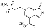 | 1g | 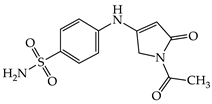 |
| 1b |  | 1h | 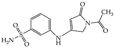 |
| 1c |  | 1i |  |
| 1d | 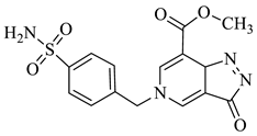 | 1j |  |
| 1e | 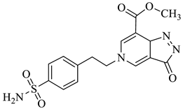 | 1k |  |
| 1f | 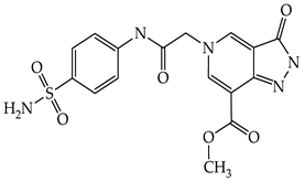 |
| KI (nM) * | ||||
|---|---|---|---|---|
| Cmp | hCA I | hCAII | hCA IX | hCA XII |
| 1a | 8010 | 7329 | 97.9 | 282.3 |
| 1b | 156.8 | 51.4 | 319.1 | 358.2 |
| 1c | 1443 | 247.4 | 589.5 | 143.2 |
| 1d | 847.7 | 779.3 | 644.7 | 262.4 |
| 1e | 864.2 | 658.3 | 848.8 | 397.4 |
| 1f | 58.8 | 6.6 | 907.5 | 474.8 |
| 1g | 66.8 | 41.7 | 294.2 | 508.5 |
| 1h | 135.8 | 61.7 | 94.3 | 713.6 |
| 1i | 5439 | 6791 | 79.6 | 104.8 |
| 1j | 3865 | 5712 | 97.8 | 285.1 |
| 1k | 88.3 | 5.6 | 421.4 | 34.5 |
| AAZ | 250.0 | 12.1 | 25.8 | 5.7 |
| KI (nM) * | ||||||
|---|---|---|---|---|---|---|
| Cmp | E.coli β | E.coli γ | BpsCAβ | BpsCAγ | PgiCAγ | VhCAβ |
| 1a | 861.9 | 61.8 | 654.3 | 912.8 | 783.0 | 2334 |
| 1b | 3457 | 57.8 | 229.1 | 513.2 | 91.0 | 844.2 |
| 1c | 3836 | 79.1 | 785.4 | 613.5 | 637.1 | 913.3 |
| 1d | 5027 | 189.7 | 644.4 | 805.1 | 848.9 | 670.7 |
| 1e | 3136 | 58.1 | 682.9 | 1341 | 96.1 | 840.0 |
| 1f | 3650 | 66.8 | 236.3 | 2179 | 667.8 | 466.6 |
| 1g | 453.8 | 204.7 | 664.3 | 97.1 | 83.1 | 1449 |
| 1h | 711.9 | 524.3 | 2961 | 833.2 | 90.0 | 2617 |
| 1i | 3048 | 92.7 | 96.4 | 191.5 | 95.6 | 2241 |
| 1j | 94.9 | 67.1 | 788.8 | 625.4 | 84.3 | 642.3 |
| 1k | 3864 | 63.5 | 212.5 | 952.3 | 201.6 | 355.8 |
| AAZ | 227 | 248 | 745 | 149 | 324 | 451 |
| No | hCA Isoform | Estimated Free Binding Energy (Kcal/mol) | Chelating the Zn (II) Ion | Residues Involved in H-Bond Interactions | Residues Involved in Hydrophobic Interactions |
|---|---|---|---|---|---|
| 1c | hCA I | −4.70 | No | - | - |
| hCA II | −5.03 | No | - | Ile91, Phe131 | |
| hCA IX | −6.06 | Yes | Thr199 | Val121, Leu198 | |
| hCA XII | −5.92 | Yes | - | Leu198 | |
| 1g | hCA I | −10.42 | Yes | Trp5, Thr199, His200 | Leu198, His200 |
| hCA II | −6.89 | Yes | Thr199 | Val121, Leu198 | |
| hCA IX | −7.65 | Yes | Thr199, Thr200 | Leu198 | |
| hCA XII | 6.11 | Yes | Thr200 | Trp5, Leu198 | |
| 1f | hCA I | −11.37 | Yes | Trp5, Ser136, Thr199 | Ala121, Leu198 |
| hCA II | −10.12 | Yes | Gln92, Thr199 | Val121, Leu198, Thr200 | |
| hCA IX | −4.29 | Yes | - | Val121, Leu198 | |
| hCA XII | −5.50 | Yes | Gln92 | Val121, Leu198 | |
| 1k | hCA I | −9.25 | Yes | Thr199, His200 | Leu198, His200 |
| hCA II | −10.53 | Yes | Gln92, Thr199 (2) | Val121, Phe131, Val135, Leu198 | |
| hCA IX | −6.17 | Yes | - | Val121, Leu198 | |
| hCA XII | −6.79 | Yes | Thr199 | Val121, Leu198, Trp209 | |
| AAZ | hCA I | −8.28 | Yes | Gln92 | Leu198, Thr199, His200, Pro201, Trp209 |
| hCA II | −8.87 | Yes | Thr199, Thr200 | Val121, Phe131, Leu198, Trp209 | |
| hCA IX | −9.02 | Yes | Thr199, Thr200 | Val121, Val143, Val131, Leu198, Trp209 | |
| hCA XII | −9.14 | Yes | Thr199, Thr200 | Val121, Val143, Leu198, Trp209 |
| No | hCA Isoform | Estimated Free Binding Energy (Kcal/mol) | Chelating The Zn (II) Ion | Residues Involved in H-Bond Interactions | Residues Involved in Hydrophobic Interactions |
|---|---|---|---|---|---|
| 1a | E. coli β | −3.15 | No | - | - |
| γ | −5.18 | No | - | Leu80, Ala82 | |
| 1b | E. coli β | −1.07 | No | - | - |
| γ | −10.86 | Yes | Gln120, H2O | Val79 | |
| 1c | E. coli β | −2.40 | No | - | - |
| γ | −7.52 | Yes | Ser57 | Val78 | |
| 1d | E. coli β | −1.66 | No | - | - |
| γ | - | No | - | - | |
| 1e | E. coli β | −2.71 | No | - | - |
| γ | −10.57 | Yes | H2O | Val78, Val79 | |
| 1f | E. coli β | - | No | - | - |
| γ | −9.16 | Yes | Ser57, Arg59 | Val79, Leu83 | |
| 1g | E. coli β | −3.16 | No | - | Ala106 |
| γ | −2.55 | No | - | Val78 | |
| 1h | E. coli β | −3.02 | No | - | Ile126 |
| γ | −2.61 | No | - | Val78 | |
| 1i | E. coli β | −1.28 | No | - | - |
| γ | −7.43 | Yes | Glu62 | Val79 | |
| 1j | E. coli β | −8.61 | Yes | Gly103 | Ala106 |
| γ | −10.35 | Yes | Arg59, H2O | Val79 | |
| 1k | E. coli β | −2.58 | No | - | - |
| γ | −10.59 | Yes | Arg59, H2O | Val78 | |
| AAZ | E. coli β | −3.46 | No | - | Ala106, Val198 |
| γ | −4.27 | No | Glu140 | - |
| Cmp | MW | Number of HBA a | Number of HBD b | Log Po/w (iLOGP) c | Log S d | TPSA e | Lipinski Violations | Bioavailability Score | Drug-Likeness Model Score |
|---|---|---|---|---|---|---|---|---|---|
| 1a | 300.29 | 7 | 2 | −0.01 | Very soluble | 145.52 | 0 | 0.55 | −0.43 |
| 1b | 348.33 | 7 | 2 | 1.36 | Soluble | 145.52 | 0 | 0.55 | −0.47 |
| 1c | 348.33 | 7 | 2 | 0.92 | Soluble | 145.52 | 0 | 0.55 | −0.94 |
| 1d | 362.36 | 7 | 2 | 1.45 | Moderately soluble | 145.52 | 0 | 0.55 | −0.08 |
| 1e | 376.39 | 7 | 2 | 1.74 | Moderately soluble | 145.52 | 0 | 0.55 | −0.04 |
| 1f | 405.39 | 8 | 3 | 0.57 | Moderately soluble | 174.62 | 1 * | 0.55 | 0.83 |
| 1g | 295.31 | 5 | 2 | 1.29 | Very soluble | 117.95 | 0 | 0.55 | 0.90 |
| 1h | 294.31 | 6 | 1 | −2.99 | Very Soluble | 105.92 | 0 | 0.55 | −0.13 |
| 1i | 232.26 | 5 | 2 | 0.16 | Very soluble | 105.92 | 0 | 0.55 | −0.08 |
| 1j | 296.30 | 6 | 2 | 0.57 | Soluble | 123.94 | 0 | 0.55 | 0.01 |
| 1k | 429.49 | 6 | 2 | 1.09 | Moderately soluble | 141.17 | 0 | 0.55 | 0.44 |
Publisher’s Note: MDPI stays neutral with regard to jurisdictional claims in published maps and institutional affiliations. |
© 2022 by the authors. Licensee MDPI, Basel, Switzerland. This article is an open access article distributed under the terms and conditions of the Creative Commons Attribution (CC BY) license (https://creativecommons.org/licenses/by/4.0/).
Share and Cite
Angeli, A.; Kartsev, V.; Petrou, A.; Lichitsky, B.; Komogortsev, A.; Pinteala, M.; Geronikaki, A.; Supuran, C.T. Pyrazolo[4,3-c]pyridine Sulfonamides as Carbonic Anhydrase Inhibitors: Synthesis, Biological and In Silico Studies. Pharmaceuticals 2022, 15, 316. https://doi.org/10.3390/ph15030316
Angeli A, Kartsev V, Petrou A, Lichitsky B, Komogortsev A, Pinteala M, Geronikaki A, Supuran CT. Pyrazolo[4,3-c]pyridine Sulfonamides as Carbonic Anhydrase Inhibitors: Synthesis, Biological and In Silico Studies. Pharmaceuticals. 2022; 15(3):316. https://doi.org/10.3390/ph15030316
Chicago/Turabian StyleAngeli, Andrea, Victor Kartsev, Anthi Petrou, Boris Lichitsky, Andrey Komogortsev, Mariana Pinteala, Athina Geronikaki, and Claudiu T. Supuran. 2022. "Pyrazolo[4,3-c]pyridine Sulfonamides as Carbonic Anhydrase Inhibitors: Synthesis, Biological and In Silico Studies" Pharmaceuticals 15, no. 3: 316. https://doi.org/10.3390/ph15030316