
Examining the Antioxidant and Superoxide Radical Scavenging Activity of Anise, (Pimpinella anisum L. Seeds), Esculetin, and 4-Methyl-Esculetin Using X-ray Diffraction, Hydrodynamic Voltammetry and DFT Methods

 , , and
Abstract
:1. Introduction
2. Materials and Methods
2.1. Plant Material and Extraction
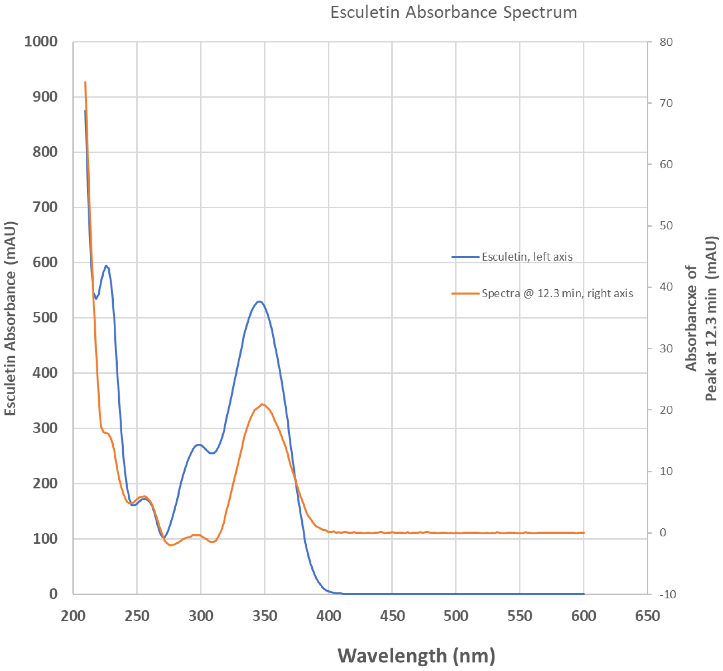
2.2. HPLC Study
2.3. Single Crystal X-ray Diffraction Analysis
2.3.1. Esculetin
2.3.2. 4-Methyl-Esculetin
2.4. Hydrodynamic Voltammetry (RRDE)
2.5. Computational Study
3. Results and Discussion
3.1. HPLC
3.2. X-ray Diffraction Analysis
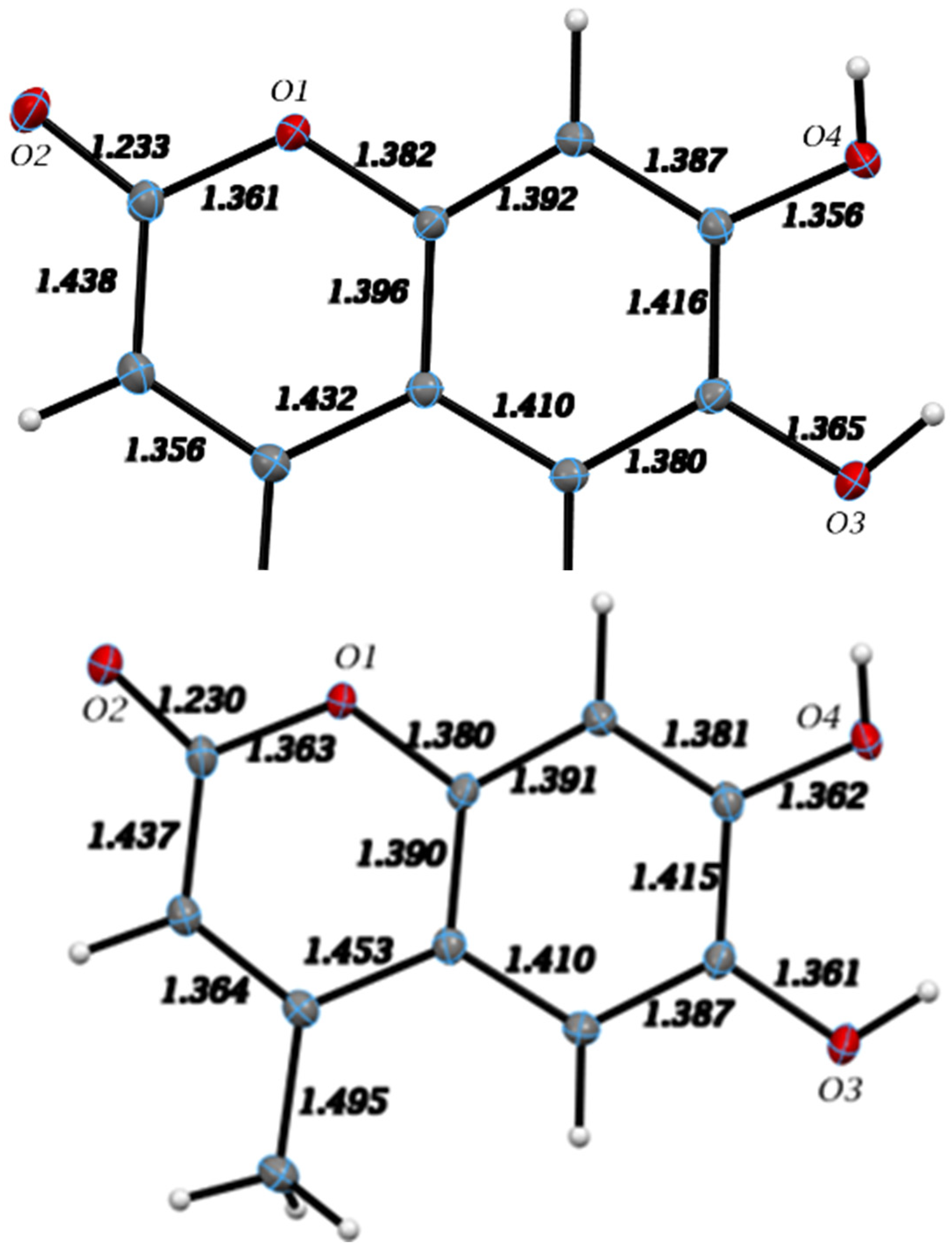
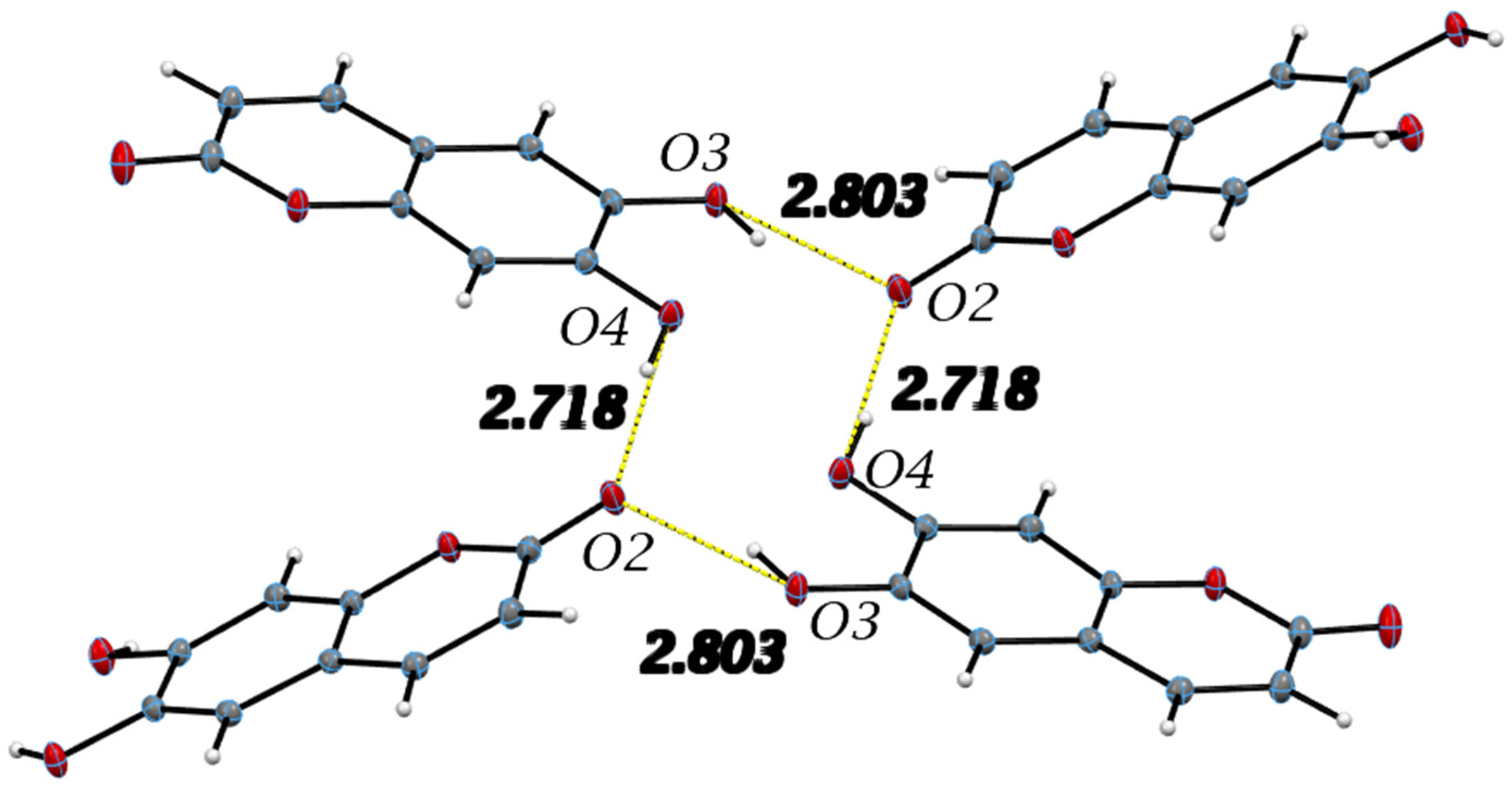
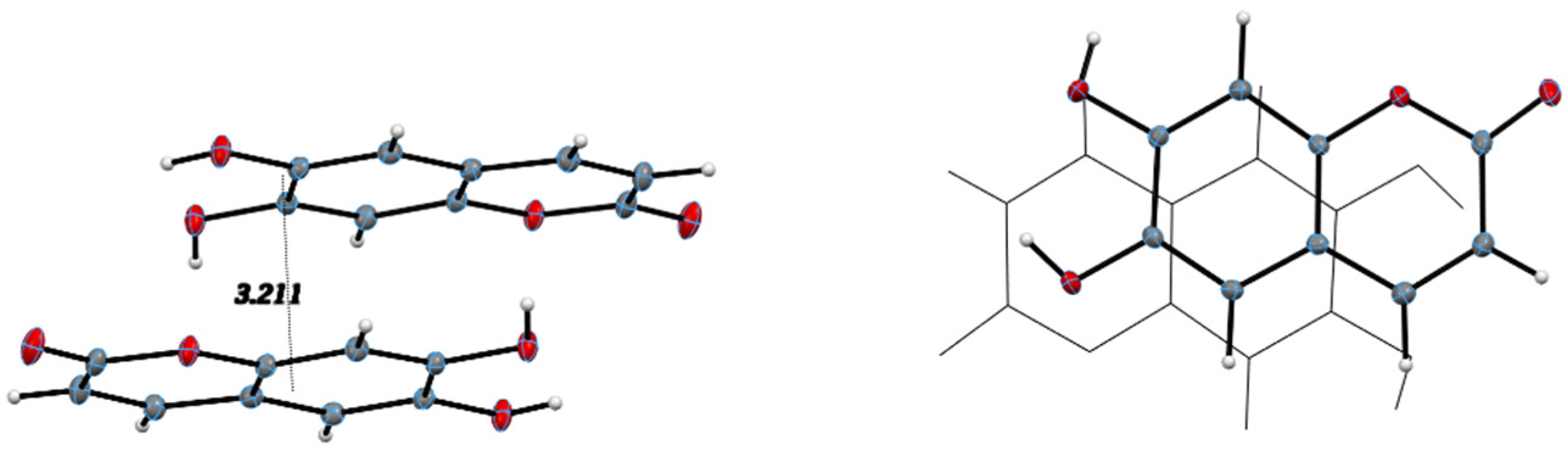
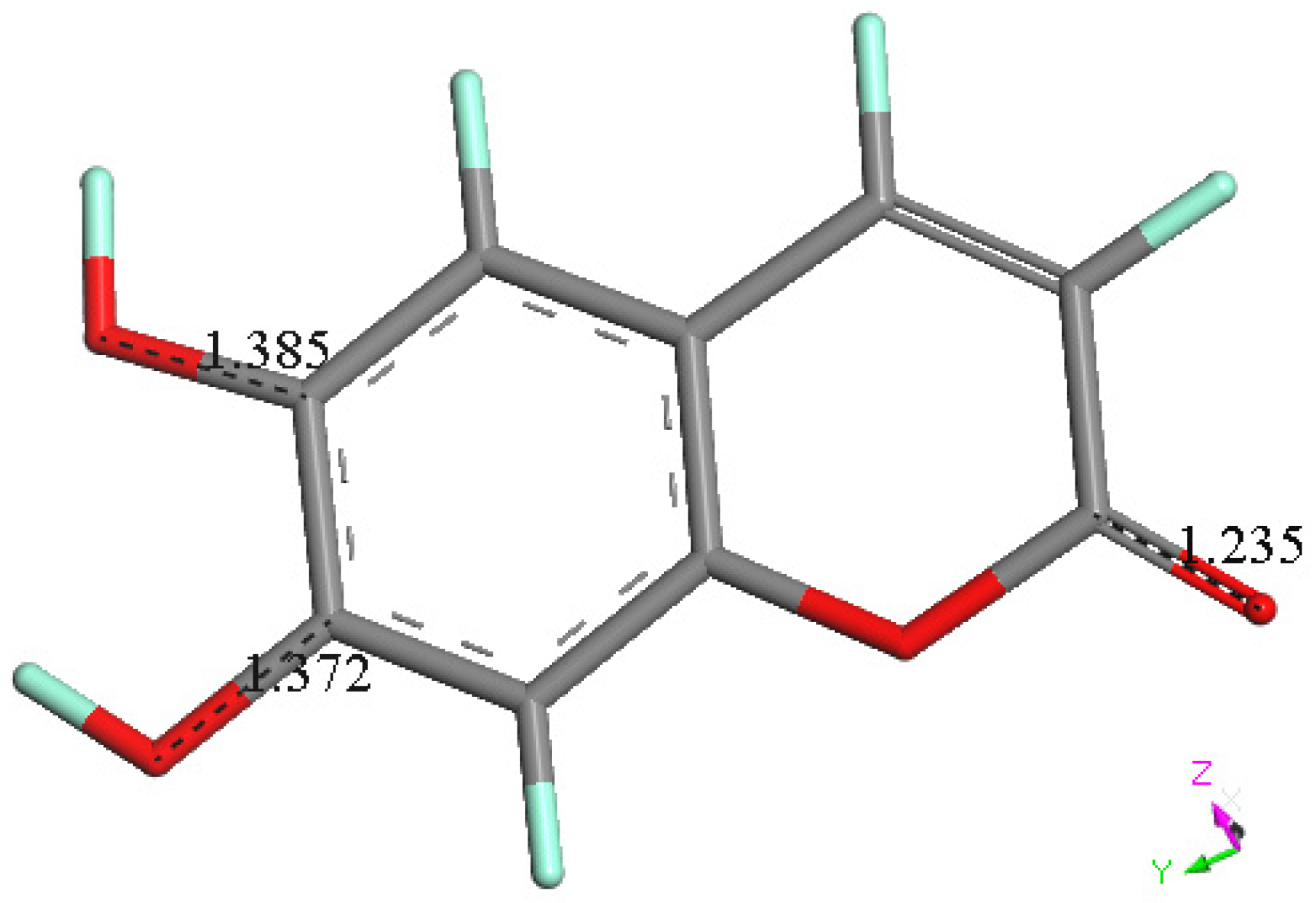
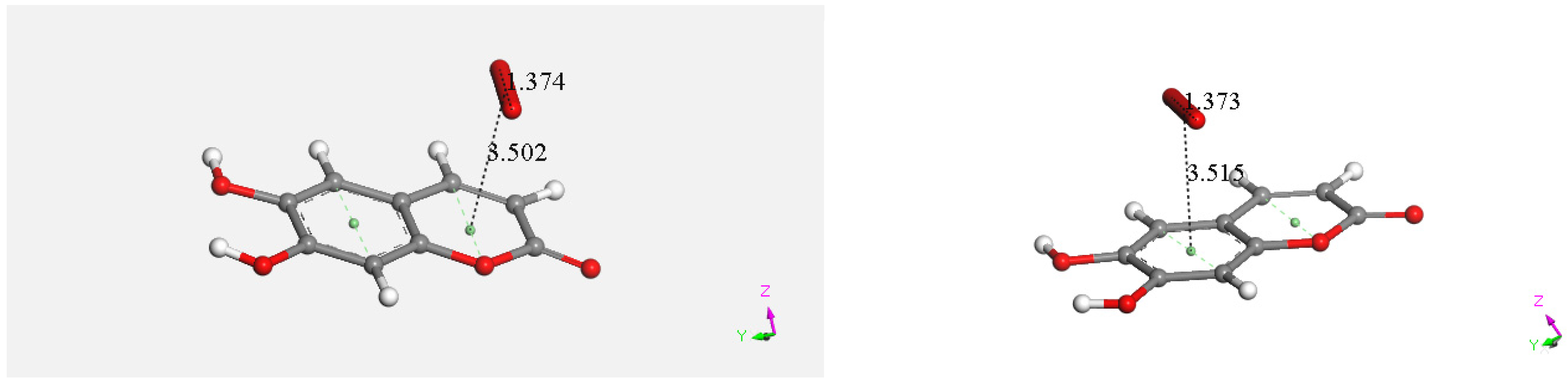
3.2.1. Esculetin
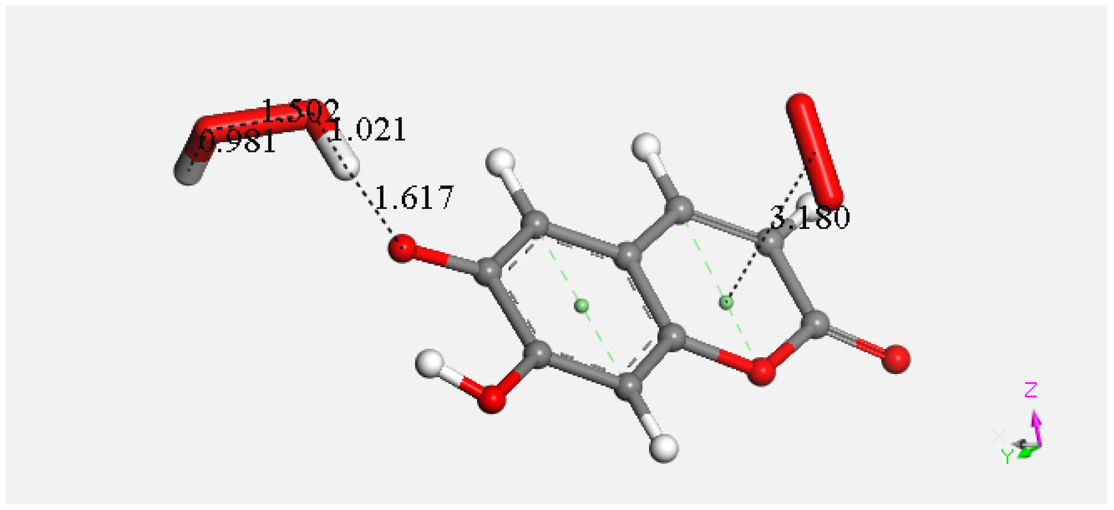
3.2.2. 4-Methyl-Esculetin


| Donor-H | Acceptor-H | Donor–Acceptor | Angle | |
|---|---|---|---|---|
| O3-H6⋯O4 | 0.90(2) | 2.35(2) | 2.746(10) | 106.8(17) |
| C8-H8⋯O1 #2 | 1.043(15) | 2.198(15) | 3.1544(13) | 151.5(11) |
| O4-H7⋯O2 #2 | 0.827(17) | 2.088(17) | 2.9094(11) | 172.0(14) |
| O3-H6⋯O2 #1 | 0.90(2) | 1.87(2) | 2.7491(10) | 154.9(15) |
| Symmetry transformations used to generate equivalent atoms: | ||||
| #1 | x, y, z − 1 | |||
| #2 | −x, −y + 2, −z + 1 | |||
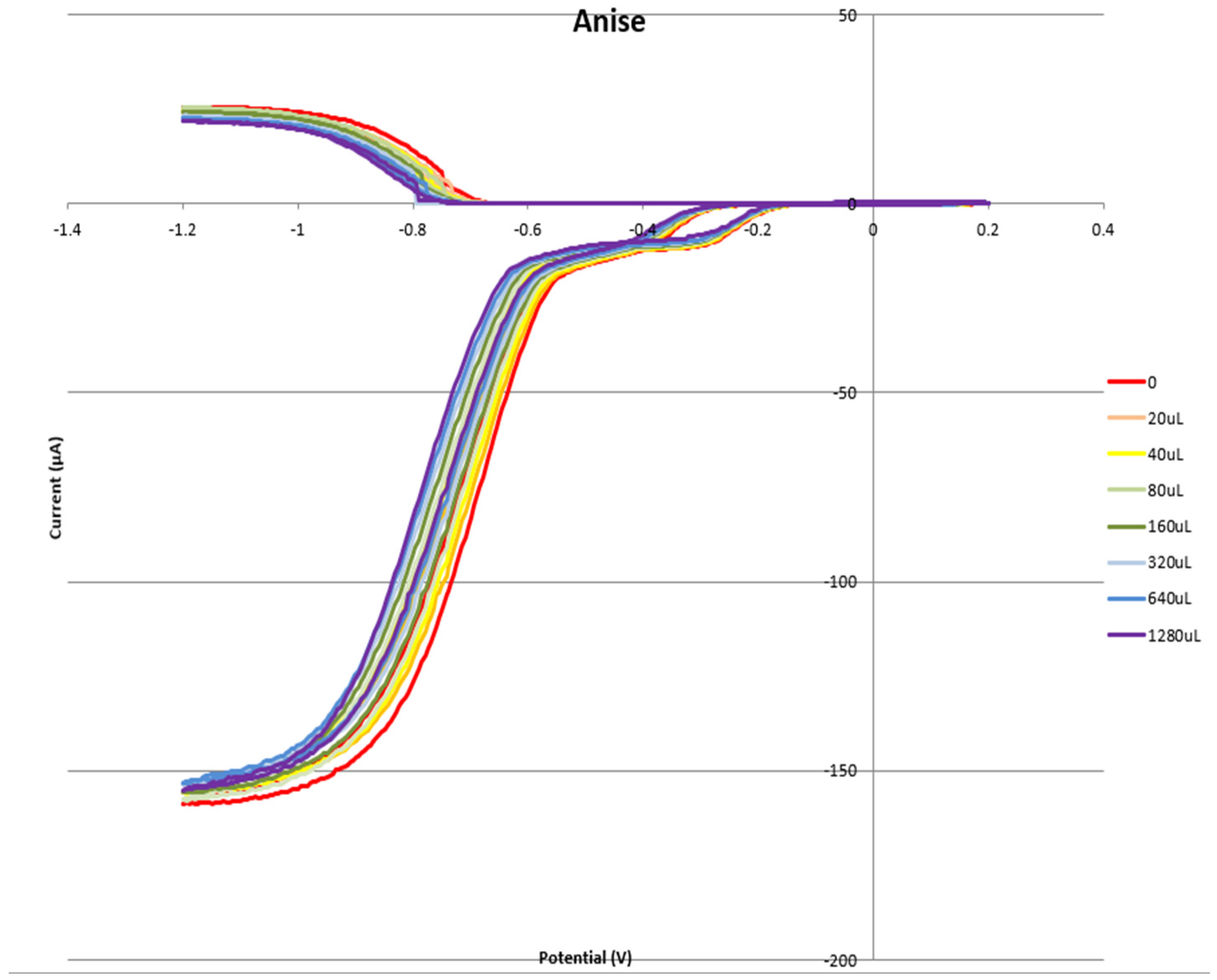
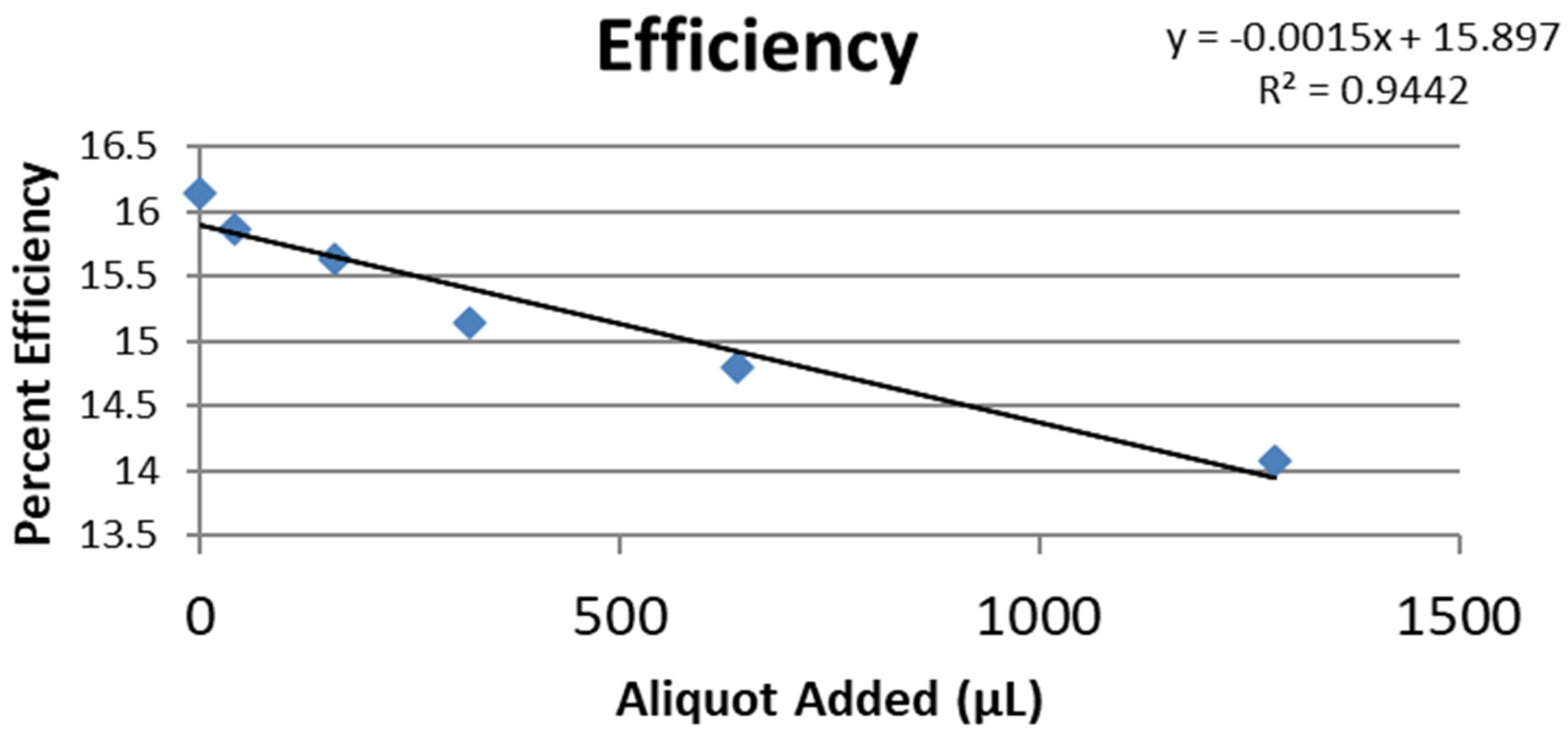
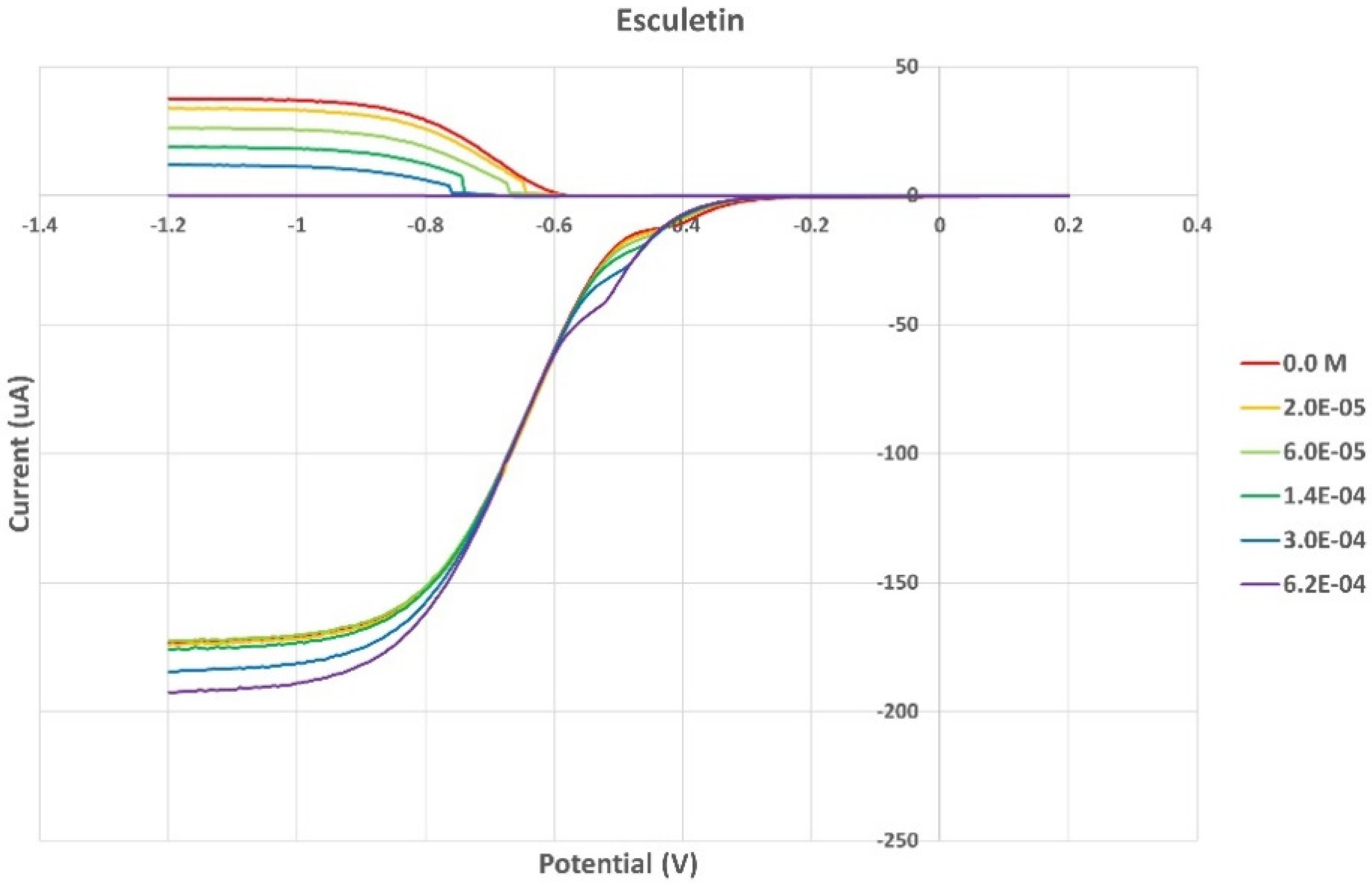
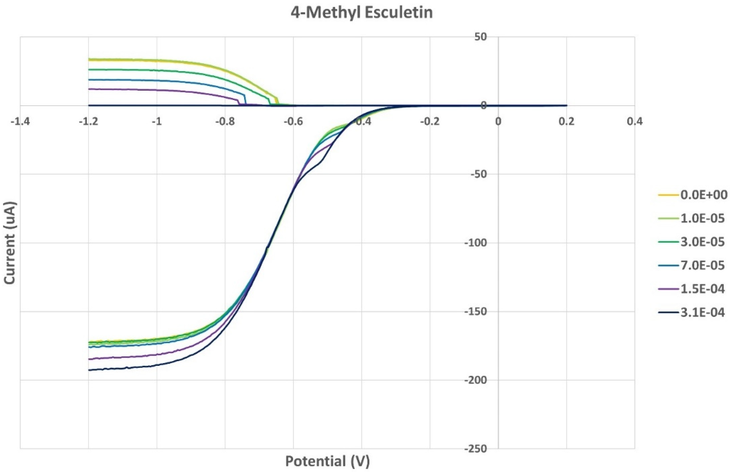
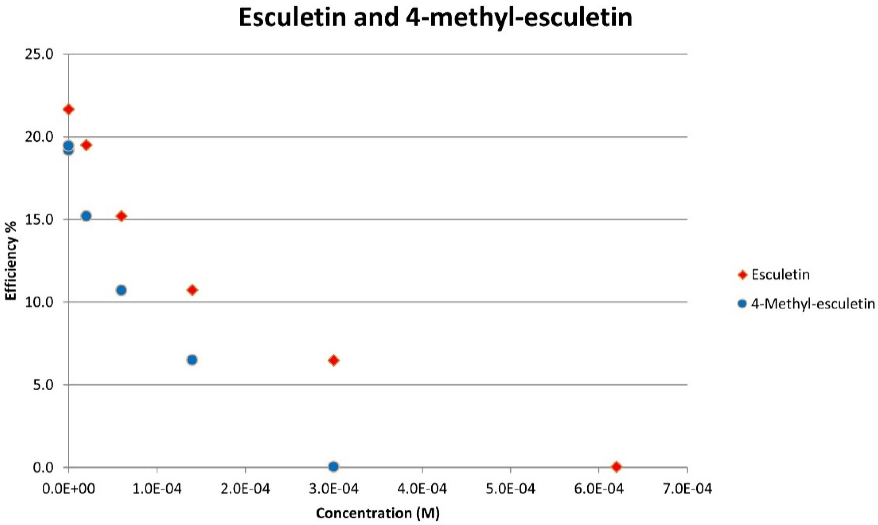
3.3. RRDE
3.4. DFT
4. Conclusions
Supplementary Materials
Author Contributions
Funding
Data Availability Statement
Acknowledgments
Conflicts of Interest
References
- Metwaly, A.M.; Ghoneim, M.M.; Eissa, I.H.; Elsehemy, I.A.; Mostafa, A.E.; Hegazy, M.M.; Afifi, W.M.; Dou, D. Traditional ancient Egyptian medicine: A review. Saudi J. Biol. Sci. 2021, 28, 5823–5832. [Google Scholar] [CrossRef] [PubMed]
- Sun, W.; Shahrajabian, M.H.; Cheng, Q. Anise (Pimpinella anisum L.), a dominant spice and traditional medicinal herb for both food and medicinal purposes. Cogent Biol. 2019, 5, 1. [Google Scholar] [CrossRef]
- Singletary, K.W. Anise: Potential Health Benefits. Nutr. Today 2022, 57, 96–109. [Google Scholar] [CrossRef]
- Wagner, J.; Granvogl, M.; Schieberle, P. Characterization of the Key Aroma Compounds in Raw Licorice (Glycyrrhiza glabra L.) by Means of Molecular Sensory Science. J. Agric. Food Chem. 2016, 64, 8388–8396. [Google Scholar] [CrossRef] [PubMed]
- Soto-Blanco, B. Chapter 12—Herbal glycosides in healthcare. In Herbal Biomolecules in Healthcare Applications; Mandal, S.C., Nayak, A.K., Dhara, A.K., Eds.; Academic Press: Cambridge, MA, USA, 2022; pp. 239–282. ISBN 9780323858526. [Google Scholar] [CrossRef]
- Matos, M.J.; Santana, L.; Uriarte, E.; Abreu, O.A.; Molina, E.; Yordi, E.G. Coumarins—An Important Class of Phytochemicals; InTech: London, UK, 2015. [Google Scholar] [CrossRef]
- Pang, Z.; Chen, J.; Wang, T.; Gao, C.; Li, Z.; Guo, L.; Xu, J.; Cheng, Y. Linking Plant Secondary Metabolites and Plant Microbiomes: A Review. Front. Plant. Sci. 2021, 12, 621276. [Google Scholar] [CrossRef] [PubMed]
- Annunziata, F.; Pinna, C.; Dallavalle, S.; Tamborini, L.; Pinto, A. An Overview of Coumarin as a Versatile and Readily Accessible Scaffold with Broad-Ranging Biological Activities. Int. J. Mol. Sci. 2020, 21, 4618. [Google Scholar] [CrossRef]
- Sharifi-Rad, J.; Cruz-Martins, N.; López-Jornet, P.; Lopez, E.P.; Harun, N.; Yeskaliyeva, B.; Beyatli, A.; Sytar, O.; Shaheen, S.; Sharopov, F.; et al. Natural Coumarins: Exploring the Pharmacological Complexity and Underlying Molecular Mechanisms. Oxid. Med. Cell Longev. 2021, 2021, 6492346. [Google Scholar] [CrossRef]
- Rossi, M.; Aktar, S.; Davis, M.; Hefter Feuss, E.; Roman-Holba, S.; Wen, K.; Gahn, C.; Caruso, F. The Grapefruit Effect: Interaction between Cytochrome P450 and Coumarin Food Components, Bergamottin, Fraxidin and Osthole. X-ray Crystal Structure and DFT Studies. Molecules 2020, 25, 3158. [Google Scholar] [CrossRef]
- Caruso, F.; Incerpi, S.; Pedersen, J.; Belli, S.; Kaur, S.; Rossi, M. Aromatic Polyphenol π-π Interactions with Superoxide Radicals Contribute to Radical Scavenging and Can Make Polyphenols Mimic Superoxide Dismutase Activity. Curr. Issues Mol. Biol. 2022, 44, 5209–5220. [Google Scholar] [CrossRef]
- Liang, C.; Ju, W.; Pei, S.; Tang, Y.; Xiao, Y. Pharmacological Activities and Synthesis of Esculetin and Its Derivatives: A Mini-Review. Molecules 2017, 22, 387. [Google Scholar] [CrossRef]
- Kang, Y.S.; Chung, Y.C.; Lee, J.N.; Kim, B.S.; Hyun, C.-G. Anti-Inflammatory Effects of 6,7-Dihydroxy-4-Methylcoumarin on LPS-Stimulated Macrophage Phosphorylation in MAPK Signaling Pathways. Nat. Prod. Comm. 2021, 16. [Google Scholar] [CrossRef]
- Pedersen, J.Z.; Oliveira, C.; Incerpi, S.; Kumar, V.; Fiore, A.M.; De Vito, P.; Prasad, A.K.; Malhotra, S.V.; Parmar, V.S.; Saso, L. Antioxidant activity of 4-methylcoumarins. J. Pharm. Pharmacol. 2007, 59, 1721–1728. [Google Scholar] [CrossRef] [PubMed]
- Barzegar, A.; Davari, M.D.; Chaparzadeh, N.; Zarghami, N.; Pedersen, J.Z.; Incerpi, S.; Saso, L.; Moosavi-Movahedi, A.A. Theoretical and experimental studies on the structure-antioxidant activity relationship of synthetic 4-methylcoumarins. J. Iran. Chem. Soc. 2011, 8, 973–982. [Google Scholar] [CrossRef]
- Hoult, J.R.S.; Payá, M. Pharmacological and biochemical actions of simple coumarins: Natural products with therapeutic potential. Gen. Pharmacol. Vasc. Syst. 1996, 27, 713–722. [Google Scholar] [CrossRef] [PubMed]
- Sheng, Y.; Abreu, I.A.; Cabelli, D.E.; Maroney, M.; Miller, A.F.; Teixeira, M.; Valentine, J.S. Superoxide dismutases and superoxide reductases. Chem. Rev. 2014, 114, 3854–3918. [Google Scholar] [CrossRef] [PubMed]
- Hayyan, M.; Hashim, M.A.; AlNashef, I.M. Superoxide Ion: Generation and Chemical Implications. Chem. Rev. 2016, 116, 3029–3085. [Google Scholar] [CrossRef] [PubMed]
- Gulcn, I.; Oktay, M.; Krecc, E.; Kufrevoglu, O.I. Screening of antioxidant and antimicrobial activities of anise (Pimpinella anisum L.) seed extracts. Food Chem. 2003, 83, 371–382. [Google Scholar] [CrossRef]
- Bruker Software Package; Bruker AXS Inc.: Madison, WI, USA, 2017.
- Sheldrick, G.M. Crystal structure refinement with SHELXL. Acta Crystallogr. 2015, C71, 3–8. [Google Scholar] [CrossRef]
- Dolomanov, O.V.; Bourhis, L.J.; Gildea, R.J.; Howard, J.A.K.; Puschmann, H. OLEX2: A complete structure solution, refinement and analysis program. J. Appl. Crystallogr. 2009, 42, 339–341. [Google Scholar] [CrossRef]
- Macrae, C.F.; Sovago, I.; Cottrell, S.J.; Galek, P.T.A.; McCabe, P.; Pidcock, E.; Platings, M.; Shields, G.P.; Stevens, J.S.; Towler, M.; et al. Mercury 4.0: From visualization to analysis, design and prediction. J. Appl. Crystallogr. 2020, 53 Pt 1, 226–235. [Google Scholar] [CrossRef]
- Belli, S.; Rossi, M.; Molasky, N.; Middleton, L.; Caldwell, C.; Bartow-McKenney, C.; Duong, M.; Chiu, J.; Gibbs, E.; Caldwell, A.; et al. Effective and novel application of superoxide radical scavenging by natural phenolic antioxidants. Antioxidants 2019, 8, 14. [Google Scholar] [CrossRef] [PubMed]
- BIOVIA Discovery Studio Software Package; Dassault Systèmes: San Diego, CA, USA, 2023.
- Delley, B.J. From molecules to solids with the DMol3 approach. J. Chem. Phys. 2000, 113, 7756–7764. [Google Scholar] [CrossRef]
- Perdew, J.P.; Chevary, J.A.; Vosko, S.H.; Jackson, K.A.; Pederson, M.R.; Singh, D.J.; Fiolhais, C. Atoms, molecules, solids, and surfaces: Applications of the generalized gradient approximation for exchange and correlation. Phys. Rev. 1992, 46, 6671–6687. [Google Scholar] [CrossRef] [PubMed]
- Becke, A.D. Density-functional exchange-energy approximation with correct asymptotic behavior. Phys. Rev. A 1988, 38, 3098–3100. [Google Scholar] [CrossRef] [PubMed]
- Yu, S.; Caruso, F.; Belli, S.; Rossi, M. Scavenging of Superoxide in Aprotic Solvents of Four Isoflavones That Mimic Superoxide Dismutase. Int. J. Mol. Sci. 2023, 24, 3815. [Google Scholar] [CrossRef] [PubMed]
- Caruso, F.; Berinato, M.; Hernandez, M.; Belli, S.; Smart, C.; Rossi, M. Antioxidant properties of bee propolis and an important component, galangin, described by X-ray crystal structure, DFT-D and hydrodynamic voltammetry. PLoS ONE 2022, 17, e0267624. [Google Scholar] [CrossRef] [PubMed]
- Okoye, I.; Yu, S.; Caruso, F.; Rossi, M. X-ray Structure Determination, Antioxidant Voltammetry Studies of Butein and 2′,4′-Dihydroxy-3,4-dimethoxychalcone. Computational Studies of 4 Structurally Related 2′,4′-diOH Chalcones to Examine Their Antimalarial Activity by Binding to Falcipain-2. Molecules 2021, 26, 6511. [Google Scholar] [CrossRef] [PubMed]
- Rossi, M.; Wen, K.; Caruso, F.; Belli, S. Emodin Scavenging of Superoxide Radical Includes π–π Interaction. X-ray Crystal Structure, Hydrodynamic Voltammetry and Theoretical Studies. Antioxidants 2020, 9, 194. [Google Scholar] [CrossRef]
- Sakib, R.; Caruso, F.; Aktar, S.; Belli, S.; Kaur, S.; Hernandez, M.; Rossi, M. Antioxidant Properties of Thymoquinone, Thymohydroquinone and Black Cumin (Nigella sativa L.) Seed Oil: Scavenging of Superoxide Radical Studied Using Cyclic Voltammetry, DFT and Single Crystal X-ray Diffraction. Antioxidants 2023, 12, 607. [Google Scholar] [CrossRef]
- Caruso, F.; Rossi, M.; Kaur, S.; Garcia-Villar, E.; Molasky, N.; Belli, S.; Sitek, J.D.; Gionfra, F.; Pedersen, J.Z.; Incerpi, S. Antioxidant Properties of Embelin in Cell Culture. Electrochemistry and Theoretical Mechanism of Scavenging. Potential Scavenging of Superoxide Radical through the Cell Membrane. Antioxidants 2020, 9, 382. [Google Scholar] [CrossRef]
- Rossi, M.; Caruso, F.; Kwok, L.; Lee, G.; Caruso, A.; Gionfra, F.; Candelotti, E.; Belli, S.; Molasky, N.; Raley-Susman, K.M.; et al. Protection by extra virgin olive oil against oxidative stress in vitro and in vivo. Chemical and biological studies on the health benefits due to a major component of the Mediterranean diet. PLoS ONE 2017, 12, e0189341. [Google Scholar] [CrossRef] [PubMed]
- Ueno, K.; Saito, N. Esculetin; 6,7-dihydroxycoumarin. Acta Crystallogr. 1977, B33, 283–285. [Google Scholar] [CrossRef]
- Yang, S.-P.; Han, L.-J.; Wang, D.-Q.; Xia, H.-T. 6,7-Dihydroxy-4-methylcoumarin. Acta Crystallogr. 2007, E63, o4785. [Google Scholar] [CrossRef]
- Ali, I.; Sharma, S.; Bezbaruah, B. Quantum Mechanical Study on the π-π Stacking Interaction and Change in Conformation of Phenolic Systems with Different Intermolecular Rotations. Comput. Chem. 2018, 6, 71–86. [Google Scholar] [CrossRef]
- Plais, R.; Clavier, G.; Salpin, J.-Y.; Gaucher, A.; Prim, D. Anion-π Interaction for Molecular Recognition of Anions: Focus on Cooperativity with Hydrogen Bonding. Eur. J. Org. Chem. 2023, 26, e202201281. [Google Scholar] [CrossRef]
- Wang, D.X.; Wang, M.X. Exploring Anion-π Interactions and Their Applications in Supramolecular Chemistry. Acc. Chem. Res. 2020, 53, 1364–1380. [Google Scholar] [CrossRef]
- Rosokha, S.V. Anion-π Interactions: What’s in the Name? Chempluschem 2023, 88, e202300350. [Google Scholar] [CrossRef]
- Giese, M.; Albrecht, M.; Rissanen, K. Experimental investigation of anion-π interactions--applications and biochemical relevance. Chem. Commun. 2016, 52, 1778–1795. [Google Scholar] [CrossRef]
- Borges, F.; Roleira, F.; Milhazes, N.; Santana, L.; Uriarte, E. Simple coumarins and analogues in medicinal chemistry: Occurrence, synthesis and biological activity. Curr. Med. Chem. 2005, 12, 887–916. [Google Scholar] [CrossRef]
- Kostova, I.; Bhatia, S.; Grigorov, P.; Balkansky, S.; Parmar, V.S.; Prasad, A.K.; Saso, L. Coumarins as antioxidants. Curr. Med. Chem. 2011, 18, 3929–3951. [Google Scholar] [CrossRef]
- Peng, X.M.; Damu, G.L.; Zhou, C. Current developments of coumarin compounds in medicinal chemistry. Curr. Pharm. Des. 2013, 19, 3884–3930. [Google Scholar] [CrossRef] [PubMed]
- Sandhu, S.; Bansal, Y.; Silakari, O.; Bansal, G. Coumarin hybrids as novel therapeutic agents. Bioorg. Med. Chem. 2014, 22, 3806–3814. [Google Scholar] [CrossRef] [PubMed]
- Kostova, I. Synthetic and natural coumarins as antioxidants. Mini Rev. Med. Chem. 2006, 6, 365–374. [Google Scholar] [CrossRef] [PubMed]
- Sinha, S.; Singh, K.; Ved, A.; Hasan, S.M.; Mujeeb, S. Therapeutic journey and recent advances in the synthesis of coumarin derivatives. Mini Rev. Med. Chem. 2022, 22, 1314–1330. [Google Scholar] [CrossRef] [PubMed]
- Acosta-Quiroga, K.; Rojas-Pena, C.; Nerio, L.S.; Gutierrez, M.; Polo-Cuadrado, E. Spirocyclic derivatives as antioxidants: A review. RSC Adv. 2021, 11, 21926. [Google Scholar] [CrossRef]
- Todorov, L.; Saso, L.; Kostova, I. Antioxidant Activity of Coumarins and Their Metal Complexes. Pharmaceuticals 2023, 16, 651. [Google Scholar] [CrossRef] [PubMed]
- Malankar, G.S.; Shelar, D.S.; Butcher, R.J.; Manjare, S.T. Synthesis and single crystal X-ray study of phenylselenyl embedded coumarin-based sensors for selective detection of superoxide. Dalton Trans. 2022, 51, 10518–10526. [Google Scholar] [CrossRef]
- Sulaiman Al-Ayed, A. 4-Hydroxy-3-carboxycoumarin as an efficient building block for new ruthenium(II) complexes: Synthesis, characterization, antibacterial, antioxidant and anti-inflammatory activities. Appl. Organometal. Chem. 2017, 31. [Google Scholar] [CrossRef]
- Nongpiur, C.G.L.; Soh, C.; Diengdoh, D.F.; Verma, A.K.; Gogoi, R.; Banothu, V.; Kaminsky, W.; Kollipara, M.R. 3-acetyl-coumarin-substituted thiosemicarbazones and their ruthenium, rhodium and iridium metal complexes: An investigation of the antibacterial, antioxidant and cytotoxicity activities. J. Organometal. Chem. 2023, 998, 122788. [Google Scholar] [CrossRef]


| Crystal Data | Esculetin | 4-Methyl-esculetin |
|---|---|---|
| Chemical formula | C9H6O4 | C10H8O4 |
| Mr, g/mol | 178.14 | 192.16 |
| Crystal system, space group | Monoclinic, P21/c | Triclinic, P − 1 |
| Cell parameters, a, b, c (Å) | 8.2707 (5), 6.7867 (4), 13.2033 (7) | 6.7550 (6), 7.1429 (6), 9.5517 (8) |
| α (°) | 90 | 68.166 (1) |
| β (°) | 103.558 (1) | 85.275 (1) |
| γ (°) | 90 | 69.616 (1) |
| V (Å3) | 720.46 (7) | 400.43 (6) |
| Z | 4 | 2 |
| Density (calculated) | 1.642 g/cm3 | 1.594 g/cm3 |
| Absorption coefficient, μ (mm−1) | 0.132 | 0.125 |
| F(000) | 368 | 200 |
| Crystal size (mm) | 0.150 × 0.190 × 0.340 | 0.100 × 0.170 × 0.270 |
| Tmin, Tmax | 0.981, 0.957 | 0.988, 0.967 |
| No. of measured, independent and observed [I > 2σ(I)] reflections | 17,336, 2201, 2027 | 9707, 2339, 1861 |
| R int | 0.0238 | 0.0205 |
| R[F2 > 2σ(F2)], wR(F2), S | 0.036, 0.109, 1.032 | 0.0397, 0.1107,1.110 |
| No. of reflections | 2201 | 2339 |
| No. of parameters | 142 | 160 |
| Δρmax, Δρmin (e Å−3) | 0.538, −0.277 | 0.472, −0.280 |
| Donor-H | Acceptor-H | Donor–Acceptor | Angle | |
|---|---|---|---|---|
| C7-H7⋯O3 #2 | 0.995(14) | 2.573(14) | 3.5362(10) | 162.8(11) |
| O4-H6⋯O2 #1 | 0.845(16) | 1.877(16) | 2.7178(9) | 172.4(15) |
| O3-H5⋯O4 | 0.872(18) | 2.216(16) | 2.6946(9) | 114.3(13) |
| O3-H5⋯O2 #4 | 0.872(18) | 1.989(18) | 2.8030(9) | 154.9(15) |
| C4-H4⋯O1 #3 | 1.003(13) | 2.508(13) | 3.4952(10) | 167.8(11) |
| Symmetry transformations used to generate equivalent atoms: | ||||
| #1 | −x + 1, y + 1/2, −z + 1/2 | |||
| #2 | x, −y + 3/2, z − 1/2 | |||
| #3 | x, −y + 3/2, z + 1/2 | |||
| #4 | X + 1, −y + 3/2, z + 1/2 | |||
Disclaimer/Publisher’s Note: The statements, opinions and data contained in all publications are solely those of the individual author(s) and contributor(s) and not of MDPI and/or the editor(s). MDPI and/or the editor(s) disclaim responsibility for any injury to people or property resulting from any ideas, methods, instructions or products referred to in the content. |
© 2023 by the authors. Licensee MDPI, Basel, Switzerland. This article is an open access article distributed under the terms and conditions of the Creative Commons Attribution (CC BY) license (https://creativecommons.org/licenses/by/4.0/).
Share and Cite
Rossi, M.; Caruso, F.; Thieke, N.; Belli, S.; Kim, A.; Damiani, E.; Morresi, C.; Bacchetti, T. Examining the Antioxidant and Superoxide Radical Scavenging Activity of Anise, (Pimpinella anisum L. Seeds), Esculetin, and 4-Methyl-Esculetin Using X-ray Diffraction, Hydrodynamic Voltammetry and DFT Methods. Pharmaceuticals 2024, 17, 67. https://doi.org/10.3390/ph17010067
Rossi M, Caruso F, Thieke N, Belli S, Kim A, Damiani E, Morresi C, Bacchetti T. Examining the Antioxidant and Superoxide Radical Scavenging Activity of Anise, (Pimpinella anisum L. Seeds), Esculetin, and 4-Methyl-Esculetin Using X-ray Diffraction, Hydrodynamic Voltammetry and DFT Methods. Pharmaceuticals. 2024; 17(1):67. https://doi.org/10.3390/ph17010067
Chicago/Turabian StyleRossi, Miriam, Francesco Caruso, Natalie Thieke, Stuart Belli, Alana Kim, Elisabetta Damiani, Camilla Morresi, and Tiziana Bacchetti. 2024. "Examining the Antioxidant and Superoxide Radical Scavenging Activity of Anise, (Pimpinella anisum L. Seeds), Esculetin, and 4-Methyl-Esculetin Using X-ray Diffraction, Hydrodynamic Voltammetry and DFT Methods" Pharmaceuticals 17, no. 1: 67. https://doi.org/10.3390/ph17010067