Review

Singlet Oxygen in Photodynamic Therapy

1
MOE Key Laboratory of Medical Optoelectronics Science and Technology, Key Laboratory of Photonics Technology of Fujian Province, School of Optoelectronics and Information Engineering, Fujian Normal University, Fuzhou 350117, China
2
Department of Radiation Oncology, University of Pennsylvania, Philadelphia, PA 19104, USA
*
Author to whom correspondence should be addressed.
Pharmaceuticals 2024, 17(10), 1274; https://doi.org/10.3390/ph17101274
Submission received: 30 July 2024 / Revised: 10 September 2024 / Accepted: 10 September 2024 / Published: 26 September 2024
(This article belongs to the Special Issue Photodynamic Therapy 2023)

Abstract

:
Photodynamic therapy (PDT) is a therapeutic modality that depends on the interaction of light, photosensitizers, and oxygen. The photon absorption and energy transfer process can lead to the Type II photochemical reaction of the photosensitizer and the production of singlet oxygen (1O2), which strongly oxidizes and reacts with biomolecules, ultimately causing oxidative damage to the target cells. Therefore, 1O2 is regarded as the key photocytotoxic species accountable for the initial photodynamic reactions for Type II photosensitizers. This article will provide a comprehensive review of 1O2 properties, 1O2 production, and 1O2 detection in the PDT process. The available 1O2 data of regulatory-approved photosensitizing drugs will also be discussed.

1. Introduction

Photodynamic therapy (PDT) is a modern therapeutic technology that depends on the interdisciplinary integration of pharmaceutics, optics, and medicine. PDT has been used to treat various benign and malignant diseases [1]. The therapeutic responses of PDT depend on the interaction of light, photosensitizers (PSs), and molecular oxygen within the target cells and tissues [2]. In general, PDT involves two main steps: the topical or systemic administration of photosensitizing drugs and light irradiation of the treatment site accumulated with the PS. In the presence of molecular oxygen, this process leads to the generation of reactive oxygen species (ROS) and, particularly, the photosensitized formation of singlet oxygen (1O2), the key photophysical step in Type II photosensitization. The initial biological responses and effectiveness of PDT treatment are ultimately determined by the temporal and spatial distribution of 1O2.
Photosensitizers commonly employed in clinical practice can be classified into groups such as porphyrins, chlorins, or other dyes from the chemist’s point of view. Porphyrins are a widely used PS, which include Porfimer Sodium, PhotoGem, and Hiporfin [3], also known as hematoporphyrin derivative (HpD), which is the complex mixture of water-soluble porphyrin monomers and oligomers that are purified from animal blood [4]. Aminolevulinic acid (ALA) and its ester derivatives are prodrugs that are metabolized in cells and enzymatically converted to protoporphyrin IX (PpIX), a potent endogenous porphyrin type PS [5]. Chlorin-type PSs used in clinical practice includes Temoporfin (m-THPC) and Laserphyrin (LS11) [6,7]. Phthalocyanines and organic dyes, for instance, aluminum phthalocyanine tetrasulfonate (ALPcS4), usually have long absorption wavelengths and high extinction coefficients [8].
Most PSs exhibit PDT effects primarily characterized by the Type II photochemical reaction and the efficient production of 1O2 [9]. 1O2 strongly oxidizes and reacts with biomolecules. The mechanism of 1O2-mediated PDT highly depends on the subcellular localization of the PS and the oxidative destruction of organelles, such as mitochondria, lysosomes, endoplasmic reticulum, Golgi apparatus, and plasma membranes. When exceeding the cell’s antioxidation capacity, the acute oxidative stress to these organelles can induce changes in calcium and lipid metabolism, the production of cytokines and stress proteins, and the activation of protein kinases and transcription factors. These cellular responses can ultimately lead to apoptosis and necrosis of targeted eukaryotic cells and prokaryotic cells. Subsequently, 1O2-generated cytotoxicity exerts a distinctive therapeutic mechanism of the direct killing of target cells, damaging blood vessels, and inducing an immune effect [10]. Therefore, 1O2 is regarded as the key photocytotoxic species accountable for the initial tissue damage during PDT. The triplet sensitization in the PDT process is a very effective way to generate 1O2, which makes 1O2-mediated PDT a powerful treatment modality with unique mechanisms and broad clinical applications [11]. This article provides a comprehensive review focusing on the 1O2 production from a clinically used PS based on the recent literature.

2. Properties of Singlet Oxygen

2.1. Electronic Structure and Leaps in Singlet Oxygen

According to the molecular orbital theory, the two highest energy electrons of triplet-state oxygen should be filled in two π-antibonding orbitals and spin-parallel, respectively. It has two unpaired electrons; thus it is paramagnetic. This oxygen molecule with unpaired electrons is triplet-state oxygen, symbolized as 3O2 (3 g ) [12]. Figure 1 illustrates the three lowest electronic state energy level transitions and molecular orbital schematics of oxygen molecules. When the 3O2 becomes excited, the two electrons with opposite spins and the highest energy can be arranged in two ways: (1) The two electrons with opposite spins occupy a single π orbital simultaneously, and the other orbital is the empty orbital, which is the first excited singlet state of the oxygen molecule, i.e., singlet oxygen, symbolized as 1O2 (1Δg). (2) The two electrons with opposite spins each occupy two π orbitals, which is the second excited singlet state oxygen molecule, symbolized as 1O2 (1 g + ) [13]. The three forms of the oxygen molecule are called spin isomers: 3O2 (3 g ), 1O2 (1Δg), and 1O2 (1 g + ). The energy difference between 1O2 (1 g + ) and 1O2 (1Δg) is 0.56 eV, while the energy difference between 1O2 (1Δg) and 3O2 (3 g ) is 0.98 eV [14]. The decay of 1O2 (1 g + ) to 1O2 (1Δg) by the internal conversion process is fast, while 1O2 (1Δg) to 3O2 (3 g ) is a slower process. So, in general, singlet oxygen refers to the 1O2 (1Δg) state [13].
Salokhiddinov et al. report that the radioluminescence intensity of transition (5) (λ = 1270 nm) is 60 times higher than that of transition (4) (λ = 1590 nm), based on the detection of the 1O2 luminescence signal of mesoporphyrin IX dimethyl ester dissolved in carbon tetrachloride (CCl4) using a highly sensitive germanium diode detector [15]. Macpherson et al. report that the radioluminescence intensity of transition (5) is 100 times higher than that of transition (4), based on the study of the 1O2 luminescence signals of 5,10,15,20-tetra(4-sulfonato) phenylporphine (TTPS), 5,10,15,20-tetrakis (pentafluorophenyl) porphine (TPPF), and 5,10,15,20-tetraphenylporphine (TTP) in different solutions (e.g., methanol, toluene, CCl4, etc.) [16].
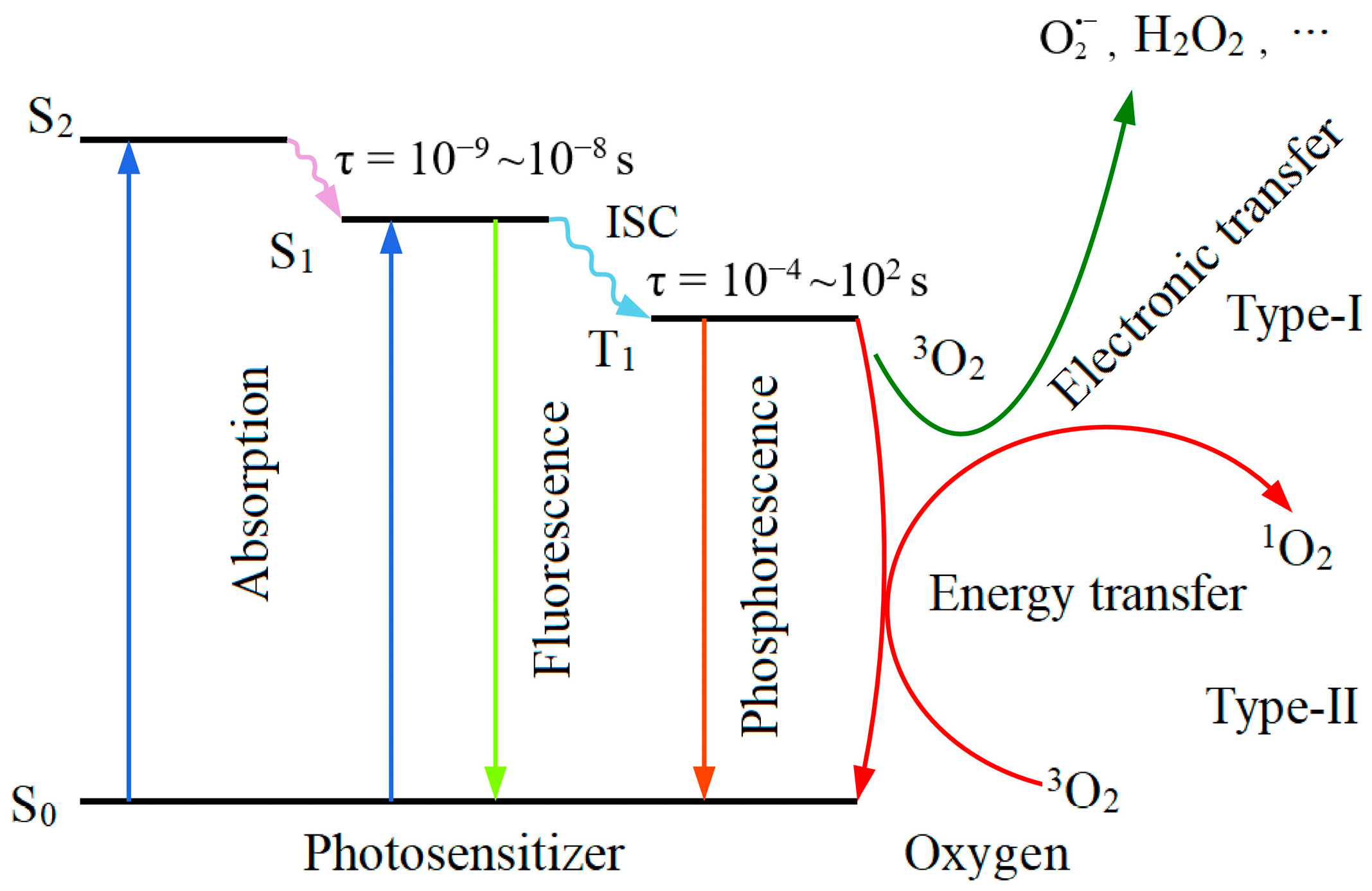
Since the energy (ΔE) of 1O2 (1Δg) is 0.98 eV higher than that of 3O2 (3 g ), the difference in excitation energy from the S0 to the S1 and the difference in energy transfer from the T1 to the S0 of the PS should be higher than 0.98 eV, which is a necessary but not sufficient condition for the production of 1O2 by the PS. Spiller et al. suggest that the excitation energy difference of S0 to S1 (see Figure 2) is 1.85 eV for phthalocyanine zinc(II) (ZnPc) and 1.63 eV for 1,4,8,11,15,18,22,25-octa (hexyloxy) phthalo cyanine zinc(II) (ZnOHP), respectively [17]. Darwent et al. suggest that the T1 to S0 energy transfer differences for 2,9,16,23-tetrasulfophthalocyanine zinc(II) (ZnPTC) and 5,10,15,20-tetraphenyl porphyrin zinc(II) (ZnTPP) are 1.12 eV and 1.59 eV, respectively [18].

2.2. Physical Chemistry Properties of Singlet Oxygen

Without reacting with the surrounding molecules, 1O2 can return to the ground state and emit photons through spontaneous radiation transition, known as 1O2 luminescence (or phosphorescence), corresponding the wavelength (λ) of an emission peak near 1270 nm [19]. But not all 1O2 produced emits light through spontaneous radiation. The luminescence rate constant (ke) for 1O2 is the rate at which 1O2 is consumed by the luminescence process upon return to the ground state. The ke of 1O2 varies in different solvents, e.g., the ke of 1O2 in water is 0.25 s−1 [20]. The intrinsic lifetime of 1O2 refers to its lifetime in an ideal state without undergoing any chemical reactions, and it is described by τ = 1/ke. τ is dependent on the properties of molecular structure and energy level. In experimental conditions, the lifetimes of 1O2 can vary in different solutions, cellular environments, or biological environments. Table 1 lists the lifetimes of 1O2 in different solvents.
The lifetime of 1O2 is mainly determined by the quenching rate constant (kq), which falls under two categories: physical and chemical. Physical quenching involves energy transfer without a chemical reaction. In this process, 1O2 interacts with a quenching agent and transfers its energy to heat or other forms of energy before returning to 3O2. For instance, excited singlet-state β-carotene can transfer energy to 1O2, producing triplet-state β-carotene and 3O2, therefore, effectively quenching 250~1000 molecules of 1O2 [21]. The chemical quenching process involves a reaction in which a chemical reacts with the quencher to create a new compound, and ultimately leads to a change in the chemical structure of the quencher or even results in quencher depletion. For example, 1O2 reacts with amines to produce free radicals and ascorbate to form hydrogen peroxide (H2O2) [21,22]. The quenching effect reduces the concentration of 1O2. The rate of quenching depends on the stereoelectronic structure of the quencher molecule. For example, C-H, N-H, and O-H groups can quickly transfer vibrational energy, a non-radiative transition process, and can distribute the excited molecular energy through the vibrational modes within the molecule, thus speeding up the quenching process [21]. The quenching rate directly impacts the lifetime of 1O2. A fast quenching process shortens the lifetime, whereas a slow process or low quencher concentration prolongs it. The kq, ke, and τ of porphyrins are listed in Table 1.
Table 1. The quenching rate constant, luminescence rate constant, and lifetime of 1O2 in different solvents.
Solventkq (M−1s−1)ke (M−1s−1)τ (μs)
H2O2.9 × 109 a0.25 b3.1 a,b,c
CH3OH1.2 × 109 c0.81 b9.5 a
C6H145.5 × 108 aN/A23.4 a
C6H63.9 × 108 a4.5 b30 a
C5H124.5 × 108 aN/A34.7 a
D2O3.7 × 108 a0.22 b68 a,b
CD3ODN/A0.79 b270 b
a: data from [23]; b: data from [20]; and c: data from [24]. N/A: not available.
Quenching can be achieved by adding a quenching agent, which is now mostly used to verify the existence of 1O2. The use of sodium azide (NaN3) in an aqueous solution at a concentration greater than 9 mM can quench 1O2, and adding deuterium oxide (D2O) possibly also extends the lifetime of 1O2 to make detection easier. Table 2 summarizes the kq and the quenching mode of several commonly used 1O2 quenchers.
Cells contain various biomolecules, including lipids, proteins, and nucleic acids. These biomolecules can react rapidly with 1O2 and cause quenching. Lipids play essential roles as a fundamental part of the cell membrane, a source of energy storage, and as an intermediary in various signaling processes. The reaction of 1O2 with the free and esterified forms of unsaturated fatty acids in lipid molecules mainly involves para-reactions, resulting in isomeric allylic hydroperoxides. 1O2 also reacts with cholesterol to produce cholesterol aldehydes [30]. 1O2 reacts with several amino acid residues in proteins (e.g., histidine, tryptophan, and tyrosine) in a reaction that is essentially a chemical quenching of 1O2. Therefore, 1O2 has a short lifetime and limited diffusion distance within the cell. Moan et al. demonstrate that the average diffusion distance of 1O2 in the cellular environment is 100~150 nm, and it is 20 nm intracellularly with the 1O2 lifetime of 40 ns in the skin and liver of living rats [31]. Ethirajan et al. suggest that the diffusion distance of 1O2 in the cellular environment is only 45 nm, as the lifetimes in the lipid and cytoplasmic regions of the cell membrane are 100 ns and 250 ns, respectively [32]. These data suggest that 1O2 cannot penetrate through the cellular membrane.

2.3. Oxidizing Activity of Singlet Oxygen

The oxidizing capacity can be measured by its standard redox potential. The standard redox potential of 1O2 is 2.23 V, which is 1 V higher than that of 3O2 [33]. H2O2, a Type I ROS, is also a strong oxidizer with a standard redox potential of 1.77 V [34]. Although H2O2 is a two-electron strong oxidizer, it exhibits minimal or no interaction with the majority of biomolecules, including low molecular weight antioxidants (e.g., vitamin E and vitamin C) [35]. The presence of a pair of electrons with opposite spins in the highest occupied molecular orbital confers dienophile features to 1O2. 1O2 readily reacts with unsaturated organic compounds by electrophilic addition and electron extraction [13]. It can also react with free radicals through energy transfer or electron transfer. In certain chemical reactions, 1O2 can transfer energy to other molecules, resulting in the creation of free radicals. For instance, 1O2 reacts with unsaturated fatty acids in cell membranes, leading to the formation of hydroperoxides through Alder-ene reactions with linoleic and linolenic acids, and ultimately causing lipid peroxidation [36]. The oxidation of amino acid residues in proteins by 1O2 (e.g., the oxidation of methionine residues to sulfoxides, oxidation of histidine to hydroxyimidazolone, and oxidation of tryptophan to N-formylkynurenine) leads to protein structural changes and function loss [37,38]. 1O2 can also react with lipids or thiols via electron transfer, leading to free radical formation [39,40]. 1O2 also induces the oxidation of guanine and causes oxidative damage of DNA [41]. These reactions and the high redox potential suggest that the oxidizing power of 1O2 is stronger than H2O2. However, the decomposition of H2O2 creates super-oxidizing hydroxyl radicals (OH) with an oxidation potential of 2.8 V, making them potent oxidizing ROS produced in the PDT process [42]. This can also be reflected by the difference between the wide ranges of the half-life of different ROS. For instance, the half-life of 1O2 ranges between 10−9 and 10−6 s, and that of OH between 10−9 and 10−7 s [43].
Antioxidants are substances that, when present in low concentrations compared to those of an oxidizable substrate, significantly inhibit or prevent the oxidation of that substrate. They act as reducing agents, donating electrons to neutralize free radicals, thereby preventing the formation of oxidative chain reactions that can damage cells and lead to various diseases and conditions [44]. An imbalance between oxidants and antioxidants in a biological system favors oxidants, which can lead to damage known as oxidative stress [45]. Both light-dependent and light-independent processes in biological systems may produce 1O2. The light-independent reactions include those catalyzed by peroxidases or oxygenases, and reactions of H2O2 with hypochlorite (ClO) or nitrite. Additionally, the recombination of peroxyl radicals (ROO) derived from biomolecules can lead to the release of 1O2 [30]. Additionally, light-dependent processes undergo photosensitization to generate 1O2. Experimental evidence has directly or indirectly suggested that the following naturally occurring ROS are major ones causing oxidative damage in the human body: superoxide radical anion ( O 2 ), H2O2, ROO, OH, 1O2, and peroxynitrite (ONOO) [46]. To counteract the assault of these ROS, living cells have a biological defense system composed of enzymatic antioxidants that convert ROS to harmless species. For example, O 2 can be converted to 3O2 and H2O2 by superoxide dismutase, and H2O2 can be converted to water and 3O2 [46]. In contrast, some ROS are dependent on quenching by various non-enzymatic antioxidants. Non-enzymatic endogenous antioxidants include lipid-based antioxidants (e.g., carotenoids and vitamin E) and water-soluble antioxidants (e.g., vitamin C, glutathione, and hemoglobin) [47].
Excess 1O2 in biological systems can destroy cells or tissues, potentially causing irreversible oxidative damage, carcinogenesis, and promoting tumor survival. When the antioxidant system is dysregulated, such as through the overexpression of glutamate–cysteine ligase and glutamate–cysteine catalytic subunit, it can lead to an imbalance in the glutathione/glutathione disulfide ratio [48], generating an excess of 1O2. This imbalance can disrupt the integrity of biomolecules, cause cellular damage, generate oxidative stress, and damage mitochondria and DNA, ultimately leading to apoptosis, necrosis, senescence, and a proliferation blockade [49].
Physiological levels of 1O2 have signaling roles within the cell and may act as modulators of cell signaling [49]. It exerts a significant influence on cellular activities, usually with reversibly oxidized hydrogen sulfide groups as signaling molecules to maintain a dynamic equilibrium of generated and quenched 1O2. The amount of 1O2 in an organism is regulated by various factors, including the physiological state and environmental conditions. Nevertheless, it is difficult to define a high or low level of 1O2 as a specific value.

3. Singlet Oxygen Production in PDT Process

PDT is a two-step procedure: (1) the patient is first topically or systemically given a PS (“photosensitizing drug”) that preferentially accumulates in the target tissue, and (2) the light of a specific wavelength generated by a laser light source, LED light source, or daylight is then applied to the treatment site in order to active the PS. The process of PDT to produce 1O2 involves the interaction of PS molecules with light irradiation in the presence of molecular oxygen. Therefore, the PS, light, and oxygen are considered as the three crucial elements of PDT. The process of 1O2 production includes photon absorption, emission, and energy transfer as the PS undergoes different electronic states to eventually generate an excited triplet-state PS and convert the absorbed light energy to highly cytotoxic 1O2. PSs used in PDT are usually conjugated unsaturated organic molecules that possess high light absorption coefficients, high intersystem crossing efficiencies, and high quantum yields for the production of 1O2 or other ROS [50].
When the PS is exposed to the light of a specific wavelength and absorbs photon energy, PS molecules in the ground state (S0) are transitioned to an energy level above the first excited singlet state (S1). Subsequently, the PS molecules in the S1 will undergo vibrational relaxation and either decay to the lowest vibrational energy level of that electronic state or decay through internal conversion and vibrational relaxation to the lowest energy level of S1.
The lifetime of S1 is short (10−8~10−9 s) due to the rapid rate of internal transitions [51]. Therefore, the observed luminescence of an S1 radiation, accompanied by the S1 to S0 radiative transitions, is called fluorescence. PS molecules in the S1 undergo intersystem crossing (ISC) through non-radiative transitions from the S1 to the slightly lower energy excited triplet state (T1). The emission of light resulting from the transition from the T1 to S0 is known as phosphorescence. This progress is spin-forbidden, leading to a low rate constant, with a lifetime ranging from 10−4 to 102 s for the T1 [51].
Because the S1 of the PS molecule has a much shorter lifetime than the T1, the likelihood of energy transfer with 3O2 is reduced. The T1-state PS, with its longer lifetime, can effectively transfer energy to 3O2, leading to the formation of 1O2. A simplified Jablonski energy level diagram for the Type I and Type II photochemical reactions in the PDT process is shown in Figure 2.
The concentration of 1O2 generated during PDT is a crucial factor in determining the therapeutic effect, and its concentration is related to many factors and can be expressed as the following:
O 2 1 t = N δ S 0 Φ Δ τ Δ τ T τ Δ exp t τ T exp t τ Δ
where [1O2] represents the concentration of 1O2 at time t, N is the number of photons during the excitation pulse, δ the absorption cross-section of the PS, [S0] is the concentration of the ground-state PS, Φ is the quantum yield of 1O2, τT is the lifetime of the PS in its triplet state, and τΔ is the lifetime of 1O2.

4. Singlet Oxygen Detection

4.1. Direct Detection of Singlet Oxygen

Detection methods for 1O2 are broadly categorized into direct and indirect methods. The direct detection of 1O2 luminescence is considered the “gold standard” for PDT dosimetry. 1O2 is directly detected by measuring its luminescence at 1270 nm [52]. The long wavelength and low energy of 1O2, along with its low emission probability in biological media (approximately 10−8), contribute to its inactivation through collision with water molecules under physiological conditions. This results in a short lifetime of 1O2 on solvents or biological environments, rendering its accurate detection exceedingly arduous using direct detection methods. Therefore, near-infrared photodetectors based on high sensitivity, when integrated with time-resolved counting techniques (e.g., gated photon counting, multichannel counting, and time-correlated single-photon counting), have emerged as a pivotal method for the direct detection of 1O2 lifetimes. Currently, high-sensitivity NIR detectors for the direct detection of 1O2 luminescence include the photomultiplier tube (PMT) [53], InGaAs/InP single photon avalanche diode (SPAD) [54], and superconducting nanowire single photon detector (SNSPD) [55,56].

4.2. Indirect Detection of Singlet Oxygen

4.2.1. Electron Paramagnetic Resonance

The electron paramagnetic resonance (EPR) method detects 1O2 by utilizing the change in the EPR signal after the EPR spin-trapping agent interacts with the 1O2. The method can determine the yield of 1O2 by detecting the change in the EPR signal. EPR has a high degree of sensitivity and selectivity in the detection of 1O2, but it is susceptible to the interference of solvents and coexisting ions. In 1976, McIntosh and Bolton combined the phenomenon of chemically induced dynamic electron polarization with changes in the rate of free radical generation to specifically detect the production of 1O2 as low as 100 nM [57]. Many researchers have tried to improve the EPR method for the study of 1O2. For instance, to study the 1O2 quenching effect of hydroxyl radicals in the presence of thioredoxin by using 2,2,6,6,-tetramethyl-4-piperidine (TMP) as a trapping agent [58]. However, EPR spectroscopy is not ideal for detecting intracellular 1O2, given that its temporal resolution typically spans from microseconds to milliseconds, whereas the lifetime of 1O2 within cells is usually in the nanosecond scale [12].

4.2.2. Fluorescence Photometry

Fluorescent probes are characterized by high sensitivity, simple data acquisition, high imaging resolution, etc., and are excellent sensors for detecting 1O2 [59]. By detecting changes in fluorescence properties (fluorescence at specific wavelengths, changes in fluorescence signal intensity, fluorescence quantum yield, etc.), it is possible to determine whether or not 1O2 is produced. Commonly used organic fluorescent probes mainly include probes with fluorescent signals of their own, which have a significant change in fluorescent signal intensity after 1O2 is captured, and probes that do not have fluorescent signals of their own, but will generate strong fluorescent signal after 1O2 is captured. The fluorescence probe includes 1,3 Diphenylisobenzofuran (DPBF) [60], 9-[2-(3-Carboxy-9,10-diphenyl)anyhryl]-6-hydroxy-3H-xanthen-3-ones (DPAXs) [61], and 9-[2-(3-carboxy-9,10-dimethyl)anyhryl]-6-hydroxy-3H-xanthen-3-one (DMAX) [62]. DMAX reacts faster with 1O2 and is 53 times more sensitive than DPAXs. While DPBF (9.6 × 10⁸ M−1s−1) exhibits a higher rate constant for its reaction with 1O2 in water environments compared to DMAX (9.1 × 10⁸ M−1s−1), its reactivity with ClO and hydroxyl radicals diminishes its selectivity as a probe for 1O2 detection [63]. These probes react with 1O2 to form stable endoperoxides. They have been used to detect 1O2 in neutral or alkaline aqueous solutions.
The singlet oxygen green sensor (SOSG) is a fluorescent probe for in vitro detection that is highly selective for 1O2 [64]. To apply the fluorescent probe to biological samples, the fluorescent probe needs to penetrate the cell membrane and localize inside the cell to directly react with the 1O2 molecules. Recently, 9,10-dimethylanthracene (DMA) and silicon-containing rhodamine (Si-rhodamine) moieties, a far-red fluorescent probe namely Si-DMA, have been developed to detect 1O2 in the mitochondria. Murotomi et al. demonstrated that Si-DMA can quantitatively measure intracellular 1O2 in living cells [65].

4.2.3. Spectrophotometry

Spectrophotometry utilizes an absorption probe to detect 1O2, where the absorbance value changes upon capture of the 1O2 and the difference in absorbance is used to express the amount of 1O2 produced. Spectrophotometry is a relatively simple method for the detection of 1O2, and the commonly used absorption probes include 9,10-anthracenedipro-pionicacid (ADPA) [66] and 9,10-diphenyl anthracene (DPA) [67]. Some probes can dissolve in water and undergo irreversible reactions with 1O2, leading to the creation of stable endoperoxides. For instance, ADPA reacts with 1O2 to produce endoperoxides, with a peak photobleaching absorption occurring at around 378 nm [68]. Absorption probes enable real-time visualization and monitoring of 1O2, providing a useful tool for exploring the generation, distribution, and dynamics of 1O2 in biological environments.

4.2.4. Chemiluminescence

Fluorescence photometry and spectrophotometry are based on the reaction of probe molecules with 1O2, which results in a significant increase in fluorescence or a decrease in absorbance. On the other hand, chemiluminescence does not require an excitation light source and can directly eliminate the interference of background light, making it a suitable method for detecting 1O2. When the chemical probe reacts with 1O2, it produces highly energetic compounds, which are usually unstable and release energy during photolysis. The presence or absence of 1O2 is determined by detecting light at a specific wavelength. Some common chemical probes include 2-methyl-6-phenyl-3,7-dihydroimidazo[1,2-α]-pyrazine-3-one (CLA) [69] and its derivatives, such as 4,4′(5′)-bis[2-(9-anthryloxy)ethyl-thio]tetra-fulvalene (TTF). In the H2O2/ClO system, the sensitivity of the detection of 1O2 is as low as 76 nM [70].

5. Available Singlet Oxygen Data of Regulatory-Approved PS

Quantum yield is an important parameter that measures the ability of a PS to produce 1O2. It is crucial to accurately determine the 1O2 quantum yield of a particular PS in various media. When an excited PS is quenched by 3O2, it produces 1O2. The efficiency of this process is referred to as the 1O2 quantum yield (Φ), which is the ratio between the number of 1O2 produced and the number of photons absorbed by the PS.
Φ Δ = number   of   sin glet   oxygen   generated number   of   photons   absorbed
The Φ of a PS is an inherent characteristic that varies depending on the molecular structure and photophysical properties of the PS. Accurately quantifying the Φ of a Type II PS is important for evaluating PDT effectiveness [71].
Oxygen depletion is an indirect technique to accurately measure the Φ by dissolving the PS in a solvent and passing oxygen through the solution utilizing a gas pump to ensure a steady supply of dissolved oxygen during the reaction. Oxygen consumption is accurately measured under steady-state irradiation conditions using gas microtubes; it is a process that requires sufficient substrate to quantitatively capture and intercept all 1O2 produced during the photoreaction. The Φ can be determined from the ratio between the number of moles of oxygen consumed and the number of einsteins absorbed by the PS [72]. Lysozyme inactivation can be used as an indirect method for measuring the Φ. When 1O2 reacts with lysozyme, it leads to the inactivation of lysozyme, and this inactivation is proportional to the concentration of 1O2. The production of 1O2 can be determined by measuring the activity of the remaining lysozyme. The Φ can be calculated by comparing the difference between lysozyme and lysozyme-free activities [73]. In addition, the excited-state PS is a common precursor of O 2 and 1O2. The O 2 reacts with cytochrome C, leading to a cytochrome C reduction. This process can be measured by checking the production of O 2 and the number of photons absorbed by the PS, which helps calculate the quantum yield of O 2 . The production of 1O2 also depends on the number of photons absorbed by the PS. Thus, the Φ can be indirectly determined by comparing the relative yields of O 2 and 1O2 [74].
The time-resolved infrared luminescence (TRIL) method can also be used to directly detect the Φ generated by a PS. First, the absorption spectrum of the PS is recorded using a spectrophotometer to determine the absorption coefficient. Then, the luminescence intensity of 1O2 is directly detected using a PMT. By measuring the number of photons absorbed by the PS and the luminescence intensity of 1O2 the Φ can be calculate [75]. These methods are often calibrated using the quantum yield of a reference PS. The most commonly used calibration PSs are Rose Bengal (RB: Φ = 0.75) and Methylene Blue (MB: Φ = 0.52) [75]; Pheophrbide-a is also used as a calibration reference by some researchers [17]. Table 3 provides a summary of the methods used for the determination of quantum yield and specific values for several regulatory-approved PSs for clinical applications. It should be noted that many direct and indirect measurements are often carried out in deuterated solvents, which can improve 1O2 detection but might increase experiment costs and complications depending upon the required solvent [75,76].
Based on Table 3, it is evident that various PSs exhibit unique optical properties. The maximum absorption wavelengths (λmax) and molar extinction coefficients (Ɛmax) of these PSs can impact their light absorption efficiency and tissue penetration depth, thereby influencing the effectiveness of PDT. The development and design of new PSs of different chemical, biological, and optical properties continue to be the focus of PDT research.
The extinction coefficient (Ɛ) quantifies how efficiently a PS absorbs light at a specific wavelength (λ). The subscript “max” denotes the peak extinction coefficient (Ɛmax) and peak absorption wavelength (λmax), respectively. The penetration depth of light in a target tissue depends on the scattering and absorption properties of the tissue at the specific wavelength of the light [81]. The actual “phototherapeutic window” of PDT ranges between 600 nm and 800 nm [82]. The λmax and Ɛmax of porphyrin PSs are relatively low compared with those of other types of PSs. For example, Photofrin®, the drug form of Porfimer Sodium, has an Ɛmax of 3000 M−1cm−1 at 630 nm, while ALPcS4 has an Ɛmax of 200,000 M−1cm−1 at 676 nm [73]. The low Ɛmax value of the PS at λmax indicates that the PS absorbs light weakly at the λmax. Generally speaking, the larger the Ɛmax value, the higher potential PDT response. However, in contrast, the lower Ɛmax value of a certain PS does not necessarily correlate to a reduced Φ. For instance, Photofrin® and ALScP4 exhibit Φ of 0.89 and 0.38 in phosphate buffer saline (PBS) (pH 7.4) in the presence of 1 vol% Triton X-100 (PBS/TX100), demonstrating a complex interplay between PS’s absorption optical properties, energy transfer, and efficacy in generating 1O2 [73]. Numerous studies demonstrate that non-halogenated and halogenated BODIPYs (4,4-difluoro-4-bora-3a,4a-diaza-s-indacene) have very similar λmax values, but very different Φ values, suggesting no absolute correlation between Φ and λmax [83].
The Φ of the PS is influenced not only by their inherent structure but also by the measurement technique, ambient environmental conditions, and excitation wavelength. The Φ for the same PS, measured under consistent conditions, may differ by measurement methods (e.g., the Φ values for PpIX in PBS/TX100 solution are 0.8 and 0.56 when measured using TRIL and Lysozyme inactivation methods, respectively) [73,75]. The Φ for a PS measured by the same method can also vary under different environmental conditions. For example, using the oxygen depletion method, the Φ values for HpD in methanol and methanol-D solutions are 0.64 and 0.76, respectively [72,77]. Using the TRIL method, the Φ values for Talaporfin sodium in Dulbecco’s PBS (D-PBS) and D-PBS/TX100 solutions are 0.53 and 0.59, respectively [73,75]. Even when the same method is used to measure the Φ for the same PS under the same solution conditions, there may be some differences. This could be related to the polarity of the solvent, ionic strength, or the interactions between the PS and solvent molecules.
In clinical applications, the λmax, Ɛmax, and Φ of the PS can be used as the main criteria for selecting a particular PS. Factors such as the metabolism time of the PS in the body, the duration of the skin photosensitivity response, and the selectivity to the diseased tissue also affect the PDT effect. Porphyrin-type PSs are the most extensively studied, but they have longer skin photosensitivity durations and a lower selectivity for diseased tissues. For instance, Porfimer Sodium, which is purified from HpD, requires a higher drug/light dose during treatment and necessitates 4 to 6 weeks of light avoidance after treatment [84]. Chlorin PSs are widely used clinically due to their strong photosensitivity, high target specificity, and minimal side effects. For example, NPe6 hardly participates in metabolic processes in the body, offering better safety [85]. Phthalocyanine PSs have strong absorption in the “phototherapeutic window”, but they exhibit very low or no absorption in the spectrum of sunlight (400~600 nm), thereby reducing the degree of skin photosensitivity [86]. Nevertheless, the selection and use of PSs also require a comprehensive consideration of these properties, pharmacological and therapeutic strategies, and their impact on therapeutic efficacy and patient safety to achieve optimal PDT outcomes [87,88].

6. Conclusive Remarks

Singlet oxygen is molecular oxygen in the excited state and widely exists in nature. The fundamental properties of 1O2 have been well documented. It is well-known that 1O2 can react with simple organic molecules, complex biological molecules, and cellular components with high-rate constants. 1O2 could be generated via various processes. However, triplet sensitization is a very effective way to generate the lowest excited singlet state of oxygen. The term “1O2” usually refers to the lowest energy excited species (1Δg) of molecular oxygen. Triplet states of PS molecules under illumination can undergo energy transfer with oxygen molecules in the 3 g + state, facilitating the ISC process from the triplet to singlet states of O2, producing 1O2. By understanding and utilizing 1O2 reactivity, it has been possible to develop unique therapeutic tools that can be precisely employed for destructive purposes via 1O2-mediated oxidative damages, for example, the medical applications of the photoinduced production of 1O2 in PDT.
Ever since Photofrin® was officially approved as the first PDT PS drug for the treatment of superficial and solid cancers, the development of new non-toxic PS dyes has been focused on improving PDT efficacy, shortening cutaneous photosensitivity, and expanding indications worldwide. Several PSs have received regulatory approval over the past three decades globally. Currently, PDT mediated by these traditional PSs has been used topically and systemically for the treatment of various cancer, pre-cancer, vascular, and infectious diseases. It should be recognized that each PS has its advantages and disadvantages. Nonetheless, the Type II mechanism and the production of 1O2 are regarded as the fundamental process in current PDT protocols. PSs should ideally possess a high triplet quantum yield leading to the substantial production of 1O2 upon irradiation. The research on PS should also comprehensively consider their photophysical properties, biocompatibility, targeting, and metabolic pathways to ensure the safety and efficacy of clinical PDT application. It should also be noted that PDT is less effective in hypoxia conditions, since 1O2 generation in the PDT process requires oxygen and the process also consumes oxygen; therefore, PDT effectiveness can be directly affected by the tissue oxygen partial pressure and oxygenation status. Since the severity of PDT-induced photocytotoxicity is governed by the temporal and spatial distribution of 1O2, the overproduction of 1O2 in normal tissue during PDT treatment could cause unwanted collateral damage and side effects.
However, due to the technique difficulty in the direct detection of 1O2 and the implementation of 1O2 dosimetry, quantifying the in vivo production of 1O2 is often an overlooked factor in PS evaluation and PDT dosimetry. In clinical practice, the λmax, Ɛmax, and Φ of PS can be used as the main criteria for selecting a particular PS. The Φ is often used for the evaluation of the ability of a PS to produce 1O2. It should be noted that, as described above, the reported Φ values of regulatory-approved PS are often determined in organic solvents. Even measured under the same condition, the Φ for the same PS may differ by measurement methods. Although the direct detection of 1O2 and Φ is more technically challenging within biological environments and particularly in clinical setup, the quantitative techniques for 1O2 measurement during photosensitization are of immense importance of values for both preclinical and clinical evaluation of potential PSs for future clinical use, for example, the direct singlet oxygen luminescence dosimetry (SOLD). In the scenarios where direct 1O2 luminescence detection is difficult, the macroscopic singlet oxygen explicit dosimetry (SOED), which involves the measurement of the key components in the PDT photosensitizer concentration, ground-state oxygen concentration ([3O2]), and light fluence to calculate the amount of reacted 1O2, might offer a viable alternative for quantifying 1O2 in PDT.

Author Contributions

Conceptualization, S.C., X.G., Z.H. and T.C.Z.; draft preparation, S.C., X.G., S.W., Z.W. and D.H.; proofreading, X.Z., T.C.Z. and Z.H.; and typesetting, S.C., X.G., Z.W. and D.H. All authors have read and agreed to the published version of the manuscript.

Funding

This research and APC are funded in part by the National Natural Science Foundation of China (No. 81471703).

Conflicts of Interest

The authors declare no conflicts of interest.

References

  1. Patrizia, A.; Kristian, B.; Keith, A.C.; Thomas, H.F.; Albert, W.G.; Sandra, O.G.; Stephen, M.H.; Michael, R.H.; Asta, J.; David, K.; et al. Photodynamic Therapy of Cancer: An Update. CA Cancer J. Clin. 2011, 61, 250–281. [Google Scholar]
  2. Kamanli, A.F.; Çetinel, G.; Yıldız, M.Z. A New Handheld Singlet Oxygen Detection System (SODS) and NIR Light Source Based Phantom Environment for Photodynamic Therapy Applications. Photodiagnosis Photodyn. Ther. 2020, 29, 101577. [Google Scholar] [CrossRef] [PubMed]
  3. Allison, R.R.; Huang, Z.; Dallimore, I.; Moghissi, K. Tools of Clinical Photodynamic Therapy (PDT): A Mini Compendium. Photodiagnosis Photodyn. Ther. 2024, 46, 104058. [Google Scholar] [CrossRef]
  4. Kwiatkowski, S.; Knap, B.; Przystupski, D.; Saczko, J.; Kędzierska, E.; Knap-Czop, K.; Kotlińska, J.; Michel, O.; Kotowski, K.; Kulbacka, J. Photodynamic Therapy—Mechanisms, Photosensitizers and Combinations. Biomed. Pharmacother. 2018, 106, 1098–1107. [Google Scholar] [CrossRef]
  5. Pustimbara, A.; Li, C.; Ogura, S.I. Hemin Enhance s the 5-aminolevulinic Acid-Photodynamic Therapy Effect Through the Changes of Cellular Iron Homeostasis. Photodiagnosis Photodyn. Ther. 2024, 48, 104253. [Google Scholar] [CrossRef]
  6. Pierre, G.; Jean-François, S.; Georges, W.; Mizeret, J.; Woodtli, A.; Jean-François, T.; Fontolliet, C.; Hubert, V.; Ph, M. Tetra(m-hydroxyphenyl)chlorin Clinical Photodynamic Therapy of Early Bronchial and Oesophageal Cancers. Laser Med. Sci. 1996, 11, 227–235. [Google Scholar]
  7. David, K. Pharmacokinetics of N-aspartyl Chlorin e6 in Cancer Patients. J. Photochem. Photobiol. B 1997, 39, 81–83. [Google Scholar]
  8. Ming, L.; Changhua, L. Recent Advances in Activatable Organic Photosensitizers for Specific Photodynamic Therapy. ChemPlusChem 2020, 85, 948–957. [Google Scholar]
  9. Ochsner, M. Photophysical and Photobiological Processes in the Photodynamic Therapy of Tumours. J. Photochem. Photobiol. B 1997, 39, 1–18. [Google Scholar] [CrossRef]
  10. Castano, A.P.; Demidova, T.N.; Hamblin, M.R. Mechanisms in Photodynamic Therapy: Part Two-Cellular Signaling, Cell Metabolism and Modes of Cell Death. Photodiagnosis Photodyn. Ther. 2005, 2, 1–23. [Google Scholar] [CrossRef]
  11. Weishaupt, K.R.; Gomer, C.J.; Dougherty, T.J. Identification of Singlet Oxygen as the Cytotoxic Agent in Photoinactivation of a Murine Tumor. Cancer Res. 1976, 36, 2326–2329. [Google Scholar] [PubMed]
  12. Murotomi, K.; Umeno, A.; Shichiri, M.; Tanito, M.; Yoshida, Y. Significance of Singlet Oxygen Molecule in Pathologies. Int. J. Mol. Sci. 2023, 24, 2739. [Google Scholar] [CrossRef]
  13. DeRosa, M.C.; Crutchley, R.J. Photosensitized Singlet Oxygen and its Applications. Coord. Chem. Rev. 2002, 233–234, 351–371. [Google Scholar] [CrossRef]
  14. Foote, C.S. Mechanisms of Photosensitized Oxidation. There are Several Different Types of Photosensitized Oxidation Which May be Important in Biological Systems. Science 1968, 162, 963–970. [Google Scholar] [CrossRef] [PubMed]
  15. Salokhiddinov, K.I.; Dzhagarov, B.M.; Byteva, I.M.; Gurinovich, G.P. Photosensitized Luminescence of Singlet Oxygen in Solutions at 1588 nm. Chem. Phys. Lett. 1980, 76, 85–87. [Google Scholar] [CrossRef]
  16. Macpherson, A.N.; Truscott, T.G.; Turner, P.H. Fourier-Transform Luminescence Spectroscopy of Solvated Singlet Oxygen. J. Chem. Soc. Faraday Trans. 1994, 90, 1065–1072. [Google Scholar] [CrossRef]
  17. Spiller, W.; Kliesch, H.; Wöhrle, D.; Hackbarth, S.; Röder, B.; Schnurpfeil, G. Singlet Oxygen Quantum Yields of Different Photosensitizers in Polar Solvents and Micellar Solutions. J. Porphyr. Phthalocya 1998, 2, 145–158. [Google Scholar] [CrossRef]
  18. Darwent, J.R.; Douglas, P.; Harriman, A.; Porter, G.; Richoux, M.C. Metal Phthalocyanines and Porphyrins as Photosensitizers for Reduction of Water to Hydrogen. Coord. Chem. Rev. 1982, 44, 83–126. [Google Scholar] [CrossRef]
  19. Gunduz, H.; Kolemen, S.; Akkaya, E.U. Singlet Oxygen Probes: Diversity in Signal Generation Mechanisms Yields a Larger Color Palette. Coord. Chem. Rev. 2021, 429, 213641. [Google Scholar] [CrossRef]
  20. Losev, A.P.; Nichiporovich, I.N.; Byteva, I.M.; Drozdov, N.N.; Jghgami, I.F.A. The Perturbing Effect of Solvents on the Luminescence Rate Constant of Singlet Molecular Oxygen. Chem. Phys. Lett. 1991, 181, 45–50. [Google Scholar] [CrossRef]
  21. Kearns, D.R. Physical and Chemical Properties of Singlet Molecular Oxygen. Chem. Rev. 1971, 71, 395–427. [Google Scholar] [CrossRef]
  22. Kramarenko, G.G.; Hummel, S.G.; Martin, S.M.; Buettner, G.R. Ascorbate Reacts with Singlet Oxygen to Produce Hydrogen Peroxide. Photochem. Photobiol. 2006, 82, 1634–1637. [Google Scholar] [CrossRef] [PubMed]
  23. Schweitzer, C.; Schmidt, R. Physical Mechanisms of Generation and Deactivation of Singlet Oxygen. Chem. Rev. 2003, 103, 1685–1757. [Google Scholar] [CrossRef] [PubMed]
  24. Gollnick, K.; Schenck, G.O. Mechanism and Stereoselectivity of Photosensitized Oxygen Transfer Reactions. Pure Appl. Chem. 1964, 9, 507–526. [Google Scholar] [CrossRef]
  25. Foote, C.S.; Denny, R.W. Chemistry of Singlet Oxygen. VII. Quenching by Beta.-Carotene. J. Am. Chem. Soc. 1968, 90, 6233–6235. [Google Scholar] [CrossRef]
  26. Hall, R.D.; Chignell, C.F. Steady-State Near-Infrared Detection of Singlet Molecular Oxygen: A Stern-Volmer Quenching Experiment with Sodium Azide. Photochem. Photobiol. 1987, 45, 459–464. [Google Scholar] [CrossRef]
  27. Gorman, A.A.; Ian, R.G.; Hamblett, I.; Standen, M.C. Reversible Exciplex Formation Between Singlet Oxygen, 1.DELTA.g, and Vitamin E. Solvent and Temperature Effects. J. Am. Chem. Soc. 1984, 106, 6956–6959. [Google Scholar] [CrossRef]
  28. Michaeli, A.; Feitelson, J. Reactivity of Singlet Oxygen Toward Amino Acids and Peptides. Photochem. Photobiol. 1994, 59, 284–289. [Google Scholar] [CrossRef] [PubMed]
  29. Matheson, I.B.; Etheridge, R.D.; Kratowich, N.R.; Lee, J. The Quenching of Singlet Oxygen by Amino Acids and Proteins. Photochem. Photobiol. 1975, 21, 165–171. [Google Scholar] [CrossRef]
  30. Di Mascio, P.; Martinez, G.R.; Miyamoto, S.; Ronsein, G.E.; Medeiros, M.H.G.; Cadet, J. Singlet Molecular Oxygen Reactions with Nucleic Acids, Lipids, and Proteins. Chem. Rev. 2019, 119, 2043–2086. [Google Scholar] [CrossRef]
  31. Moan, J. On the Diffusion Length of Singlet Oxygen in Cells and Tissues. J. Photochem. 1990, 6, 343–344. [Google Scholar] [CrossRef]
  32. Ethirajan, M.; Chen, Y.; Joshi, P.; Pandey, R.K. The Role of Porphyrin Chemistry in Tumor Imaging and Photodynamic Therapy. Chem. Soc. Rev. 2011, 40, 340–362. [Google Scholar] [CrossRef] [PubMed]
  33. Klaper, M.; Linker, T. New Singlet Oxygen Donors Based on Naphthalenes: Synthesis, Physical Chemical Data, and Improved Stability. Chemistry 2015, 21, 8569–8577. [Google Scholar] [CrossRef]
  34. Winterbourn, C.C. Reconciling the Chemistry and Biology of Reactive Oxygen Species. Nat. Chem. Biol. 2008, 4, 278–286. [Google Scholar] [CrossRef] [PubMed]
  35. Grune, T.; Schröder, P.; Biesalski, H.K. Low Molecular Weight Antioxidants. In Reactions, Processes: Oxidants and Antioxidant Defense Systems; Grune, T., Ed.; Springer Berlin Heidelberg: Berlin/Heidelberg, Germany, 2005; pp. 77–90. [Google Scholar]
  36. Kruk, J.; Szymańska, R. Singlet Oxygen Oxidation Products of Carotenoids, Fatty Acids and Phenolic Prenyllipids. J. Photochem. Photobiol. B 2021, 216, 112148. [Google Scholar] [CrossRef]
  37. Kim, J.; Rodriguez, M.E.; Guo, M.; Kenney, M.E.; Oleinick, N.L.; Anderson, V.E. Oxidative Modification of Cytochrome c by Singlet Oxygen. Free Radic. Biol. Med. 2008, 44, 1700–1711. [Google Scholar] [CrossRef]
  38. Marques, E.F.; Medeiros, M.H.G.; Di Mascio, P. Lysozyme Oxidation by Singlet Molecular Oxygen: Peptide Characterization Using [(18) O]-Labeling Oxygen and nLC-MS/MS. J. Mass. Spectrom. 2017, 52, 739–751. [Google Scholar] [CrossRef]
  39. Yin, H.; Xu, L.; Porter, N.A. Free Radical Lipid Peroxidation: Mechanisms and Analysis. Chem. Rev. 2011, 111, 5944–5972. [Google Scholar] [CrossRef]
  40. Rougee, M.; Bensasson, R.V.; Land, E.J.; Pariente, R. Deactivation of Singlet Molecular Oxygen by Thiols and Related Compounds, Possible Protectors Against Skin Photosensitivity. Photochem. Photobiol. 1988, 47, 485–489. [Google Scholar] [CrossRef]
  41. Cadet, J.; Davies, K.J.A.; Medeiros, M.H.; Di Mascio, P.; Wagner, J.R. Formation and Repair of Oxidatively Generated Damage in Cellular DNA. Free Radic. Biol. Med. 2017, 107, 13–34. [Google Scholar] [CrossRef]
  42. Liu, Y.; Yu, X.; Kamali, M.; Zhang, X.; Feijoo, S.; Al-Salem, S.M.; Dewil, R.; Appels, L. Biochar in Hydroxyl Radical-Based Electrochemical Advanced Oxidation Processes (eAOPs)—Mechanisms and Prospects. Chem. Eng. J. 2023, 467, 143291. [Google Scholar] [CrossRef]
  43. Gulcin, İ. Antioxidants and Antioxidant Methods: An Updated Overview. Arch. Toxicol. 2020, 94, 651–715. [Google Scholar] [CrossRef]
  44. Halliwell, B.; Gutteridge, J.M.C. Free Radicals in Biology and Medicine; Oxford University Press: Oxford, UK, 2015. [Google Scholar]
  45. Sies, H. Oxidative Stress: Oxidants and Antioxidants. Exp. Physiol. 1997, 82, 291–295. [Google Scholar] [CrossRef] [PubMed]
  46. Huang, D.; Ou, B.; Prior, R.L. The Chemistry Behind Antioxidant Capacity Assays. J. Agric. Food Chem. 2005, 53, 1841–1856. [Google Scholar] [CrossRef] [PubMed]
  47. Vani, R.; Masannagari, P.; Aastha, C.; Chaitra, B.; Prerana, B.; Ranjithvishal; Shruthi, L.; Sudharshan, N. Reactive Oxygen Species and Antioxidant Interactions in Erythrocytes. In The Erythrocyte; Vani, R., Ed.; IntechOpen: Rijeka, Croatia, 2022; Chapter 1. [Google Scholar]
  48. Zhang, H.; Limphong, P.; Pieper, J.; Liu, Q.; Rodesch, C.K.; Christians, E.; Benjamin, I.J. Glutathione-Dependent Reductive Stress Triggers Mitochondrial Oxidation and Cytotoxicity. FASEB J. 2012, 26, 1442–1451. [Google Scholar] [CrossRef]
  49. Fujii, J.; Soma, Y.; Matsuda, Y. Biological Action of Singlet Molecular Oxygen from the Standpoint of Cell Signaling, Injury and Death. Molecules 2023, 28, 4085. [Google Scholar] [CrossRef]
  50. Huang, Z.; Xu, H.; Meyers, A.D.; Musani, A.I.; Wang, L.; Tagg, R.; Barqawi, A.B.; Chen, Y.K. Photodynamic Therapy for Treatment of Solid Tumors--Potential and Technical Challenges. Technol. Cancer Res. Treat. 2008, 7, 309–320. [Google Scholar] [CrossRef]
  51. Uzdensky, A.B.; Berezhnaya, E.; Kovaleva, V.; Neginskaya, M.; Rudkovskii, M.; Sharifulina, S. Photodynamic Therapy: A Review of Applications in Neurooncology and Neuropathology. J. Biomed. Opt. 2015, 20, 61108. [Google Scholar] [CrossRef]
  52. Yang, W.; Rastogi, V.; Sun, H.; Sharma, D.; Wilson, B.C.; Hadfield, R.H.; Zhu, T.C. Multispectral Singlet Oxygen Luminescent Dosimetry (MSOLD) for Photofrin-mediated Photodynamic Therapy. Proc. SPIE Int. Soc. Opt. Eng. 2023, 12359, 1235908. [Google Scholar]
  53. Li, B.; Shen, Y.; Lin, H.; Wilson, B.C. Correlation of in Vitro Cell Viability and Cumulative Singlet Oxygen Luminescence from Protoporphyrin IX in Mitochondria and Plasma Membrane. Photodiagnosis Photodyn. Ther. 2024, 46, 104080. [Google Scholar] [CrossRef]
  54. Gemmell, N.R.; McCarthy, A.; Kim, M.M.; Veilleux, I.; Zhu, T.C.; Buller, G.S.; Wilson, B.C.; Hadfield, R.H. A Compact Fiber-Optic Probe-Based Singlet Oxygen Luminescence Detection System. J. Biophotonics 2017, 10, 320–326. [Google Scholar] [CrossRef] [PubMed]
  55. Gemmell, N.R.; McCarthy, A.; Liu, B.; Tanner, M.G.; Dorenbos, S.D.; Zwiller, V.; Patterson, M.S.; Buller, G.S.; Wilson, B.C.; Hadfield, R.H. Singlet Oxygen Luminescence Detection with a Fiber-Coupled Superconducting Nanowire Single-Photon Detector. Opt. Express 2013, 21, 5005–5013. [Google Scholar] [CrossRef]
  56. Cui, S.; Ke, C.; Peng, W.; You, L.; Zhang, X.; Huang, Z. Optimizing Single Photon Detection Based on Superconducting Strip Photon Detector (SSPD) for Singlet Oxygen Luminescence Detection. Proc. SPIE 2023, 12770, 127702S1–127702S8. [Google Scholar]
  57. McIntosh, A.R.; Bolton, J.R. Triplet State Involvement in Primary Photochemistry of Photosynthetic Photosystem II. Nature 1976, 263, 443–445. [Google Scholar] [CrossRef]
  58. Nishide, N.; Miyoshi, N. Singlet Oxygen Trapping by DRD156 in Micellar Solutions. Life Sci. 2002, 72, 321–328. [Google Scholar] [CrossRef]
  59. Gomes, A.; Fernandes, E.; Lima, J.L. Fluorescence Probes Used for Detection of Reactive Oxygen Species. J. Biochem. Biophys. Methods 2005, 65, 45–80. [Google Scholar] [CrossRef] [PubMed]
  60. Song, D.; Cho, S.; Han, Y.; You, Y.; Nam, W. Ratiometric Fluorescent Probes for Detection of Intracellular Singlet Oxygen. Org. Lett. 2013, 15, 3582–3585. [Google Scholar] [CrossRef]
  61. Umezawa, N.; Tanaka, K.; Urano, Y.; Kikuchi, K.; Higuchi, T.; Nagano, T. Novel Fluorescent Probes for Singlet Oxygen. Angew. Chem. Int. Ed. Engl. 1999, 38, 2899–2901. [Google Scholar] [CrossRef]
  62. Tanaka, K.; Miura, T.; Umezawa, N.; Urano, Y.; Kikuchi, K.; Higuchi, T.; Nagano, T. Rational Design of Fluorescein-Based Fluorescence Probes. Mechanism-Based Design of a Maximum Fluorescence Probe for Singlet Oxygen. J. Am. Chem. Soc. 2001, 123, 2530–2536. [Google Scholar] [CrossRef]
  63. Arnab, M. Photophysical Detection of Singlet Oxygen. In Reactive Oxygen Species; Ahmad, R., Surguchov, A., Eds.; IntechOpen: London, UK, 2022; Chapter 2; pp. 1–20. 2022. [Google Scholar]
  64. Ragàs, X.; Jiménez-Banzo, A.; Sánchez-García, D.; Batllori, X.; Nonell, S. Singlet Oxygen Photosensitisation by the Fluorescent Probe Singlet Oxygen Sensor Green. Chem Commun (Camb) 2009, 20, 2920–2922. [Google Scholar] [CrossRef]
  65. Murotomi, K.; Umeno, A.; Sugino, S.; Yoshida, Y. Quantitative Kinetics of Intracellular Singlet Oxygen Generation Using a Fluorescence Probe. Sci. Rep. 2020, 10, 10616. [Google Scholar] [CrossRef] [PubMed]
  66. Lindig, B.A.; Rodgers, M.A.J.; Schaap, A.P. Determination of the Lifetime of Singlet Oxygen in Water-d2 Using 9,10-anthracenedipropionic Acid, a Water-Soluble Probe. J. Am. Chem. Soc. 1980, 102, 5590–5593. [Google Scholar] [CrossRef]
  67. Song, B.; Wang, G.; Yuan, J. Measurement and Characterization of Singlet Oxygen Production in Copper ion-Catalyzed Aerobic Oxidation of Ascorbic Acid. Talanta 2007, 72, 231–236. [Google Scholar] [CrossRef] [PubMed]
  68. Moreno, M.J.; Monson, E.; Reddy, R.G.; Rehemtulla, A.; Ross, B.D.; Philbert, M.; Schneider, R.J.; Kopelman, R. Production of Singlet Oxygen by Ru(dpp(SO3)2)3 Incorporated in Polyacrylamide PEBBLES. Sens. Actuat B-Chem. 2003, 90, 82–89. [Google Scholar] [CrossRef]
  69. Yasuta, N.; Takenaka, N.; Takemura, T. Mechanism of Photosensitized Chemiluminescence of 2-Methyl-6-phenylimidazo[1,2-a]pyrazin-3(7H)-one (CLA) in Aqueous Solution. Chem. Lett. 2003, 28, 451–452. [Google Scholar] [CrossRef]
  70. Li, X.; Zhang, G.; Ma, H.; Zhang, D.; Li, J.; Zhu, D. 4,5-Dimethylthio-4′-[2-(9-anthryloxy)ethylthio]tetrathiafulvalene, a Highly Selective and Sensitive Chemiluminescence Probe for Singlet Oxygen. J. Am. Chem. Soc. 2004, 126, 11543–11548. [Google Scholar] [CrossRef]
  71. Bresolí-Obach, R.; Torra, J.; Zanocco, R.P.; Zanocco, A.L.; Nonell, S. Singlet Oxygen Quantum Yield Determination Using Chemical Acceptors. Methods Mol. Biol. 2021, 2202, 165–188. [Google Scholar]
  72. Tanielian, C.; Wolff, C.; Esch, M. Singlet Oxygen Production in Water:  Aggregation and Charge-Transfer Effects. J. Phys. Chem. 1996, 100, 6555–6560. [Google Scholar] [CrossRef]
  73. Fernandez, J.M.; Bilgin, M.D.; Grossweiner, L.I. Singlet Oxygen Generation by Photodynamic Agents. J. Photochem. Photobiol. B 1997, 37, 131–140. [Google Scholar] [CrossRef]
  74. Hadjur, C.; Wagnieres, G.; Monnier, P.; van den Bergh, H. EPR and Spectrophotometric Studies of Free Radicals (O2, OH, BPD-MA) and Singlet Oxygen (1O2) Generated by Irradiation of Benzoporphyrin Derivative Monoacid Ring A. Photochem. Photobiol. 1997, 65, 818–827. [Google Scholar] [CrossRef]
  75. Nishimura, T.; Hara, K.; Honda, N.; Okazaki, S.; Hazama, H.; Awazu, K. Determination and Analysis of Singlet Oxygen Quantum Yields of Talaporfin Sodium, Protoporphyrin IX, and Lipidated Protoporphyrin IX Using Near-Infrared Luminescence Spectroscopy. Lasers Med. Sci. 2020, 35, 1289–1297. [Google Scholar] [CrossRef] [PubMed]
  76. Bregnhøj, M.; Thorning, F.; Ogilby, P.R. Singlet Oxygen Photophysics: From Liquid Solvents to Mammalian Cells. Chem. Rev. 2024, in press. [Google Scholar] [CrossRef]
  77. Tanielian, C.; Schweitzer, C.; Mechin, R.; Wolff, C. Quantum Yield of Singlet Oxygen Production by Monomeric and Aggregated Forms of Hematoporphyrin Derivative. Free Radical Biol. Med. 2001, 30, 208–212. [Google Scholar] [CrossRef] [PubMed]
  78. Bautista-Sanchez, A.; Kasselouri, A.; Desroches, M.C.; Blais, J.; Maillard, P.; de Oliveira, D.M.; Tedesco, A.C.; Prognon, P.; Delaire, J. Photophysical Properties of Glucoconjugated Chlorins and Porphyrins and their Associations with Cyclodextrins. J. Photochem. Photobiol. B 2005, 81, 154–162. [Google Scholar] [CrossRef]
  79. Spikes, J.D.; Bommer, J.C. Photosensitizing Properties of Mono-L-Aspartyl Chlorin e6 (NPe6): A Candidate Sensitizer for the Photodynamic Therapy of Tumors. J. Photochem. Photobiol. B 1993, 17, 135–143. [Google Scholar] [CrossRef] [PubMed]
  80. Aveline, B.; Hasan, T.; Redmond, R.W. Photophysical and Photosensitizing Properties of Benzoporphyrin Derivative Monoacid Ring A (BPD-MA)*. Photochem. Photobiol. 1994, 59, 328–335. [Google Scholar] [CrossRef] [PubMed]
  81. Yoon, I.; Li, J.Z.; Shim, Y.K. Advance in Photosensitizers and Light Delivery for Photodynamic Therapy. Clin. Endosc. 2013, 46, 7–23. [Google Scholar] [CrossRef]
  82. Przygoda, M.; Bartusik-Aebisher, D.; Dynarowicz, K.; Cieślar, G.; Kawczyk-Krupka, A.; Aebisher, D. Cellular Mechanisms of Singlet Oxygen in Photodynamic Therapy. Int. J. Mol. Sci. 2023, 24, 16890. [Google Scholar] [CrossRef]
  83. Hu, W.; Zhang, R.; Zhang, X.F.; Liu, J.; Luo, L. Halogenated BODIPY Photosensitizers: Photophysical Processes for Generation of Excited Triplet State, Excited Singlet State and Singlet Oxygen. Spectrochim. Acta A Mol. Biomol. Spectrosc. 2022, 272, 120965. [Google Scholar] [CrossRef]
  84. Li, N.; Cui, S.; Yang, A.; Xiao, B.; Cao, Y.; Yang, X.; Lin, C. Sequence-Dependent Effects of Hematoporphyrin Derivatives (HPD) Photodynamic Therapy and Cisplatin on Lung Adenocarcinoma Cells. Photodiagnosis Photodyn. Ther. 2024, 47, 104102. [Google Scholar] [CrossRef]
  85. Wang, S.; Bromley, E.; Xu, L.; Chen, J.C.; Keltner, L. Talaporfin sodium. Expert. Opin. Pharmacother. 2010, 11, 133–140. [Google Scholar] [CrossRef] [PubMed]
  86. Zheng, B.D.; He, Q.X.; Li, X.; Yoon, J.; Huang, J.D. Phthalocyanines as Contrast Agents for Photothermal Therapy. Coord. Chem. Rev. 2021, 426, 213548. [Google Scholar] [CrossRef]
  87. Hurley, D.J.; Gallagher, D.; Petronzi, V.; O’Rourke, M.; Kinsella, F.; Townley, D. Examining the Efficacy of Verteporfin Photo-Dynamic Therapy (PDT) at Different Dose & Fluence Levels. Photodiagnosis Photodyn. Ther. 2023, 44, 103848. [Google Scholar] [PubMed]
  88. Pihl, C.; Lerche, C.M.; Andersen, F.; Bjerring, P.; Haedersdal, M. Improving the Efficacy of Photodynamic Therapy for Actinic Keratosis: A Comprehensive Review of Pharmacological Pretreatment Strategies. Photodiagnosis Photodyn. Ther. 2023, 43, 103703. [Google Scholar] [CrossRef]
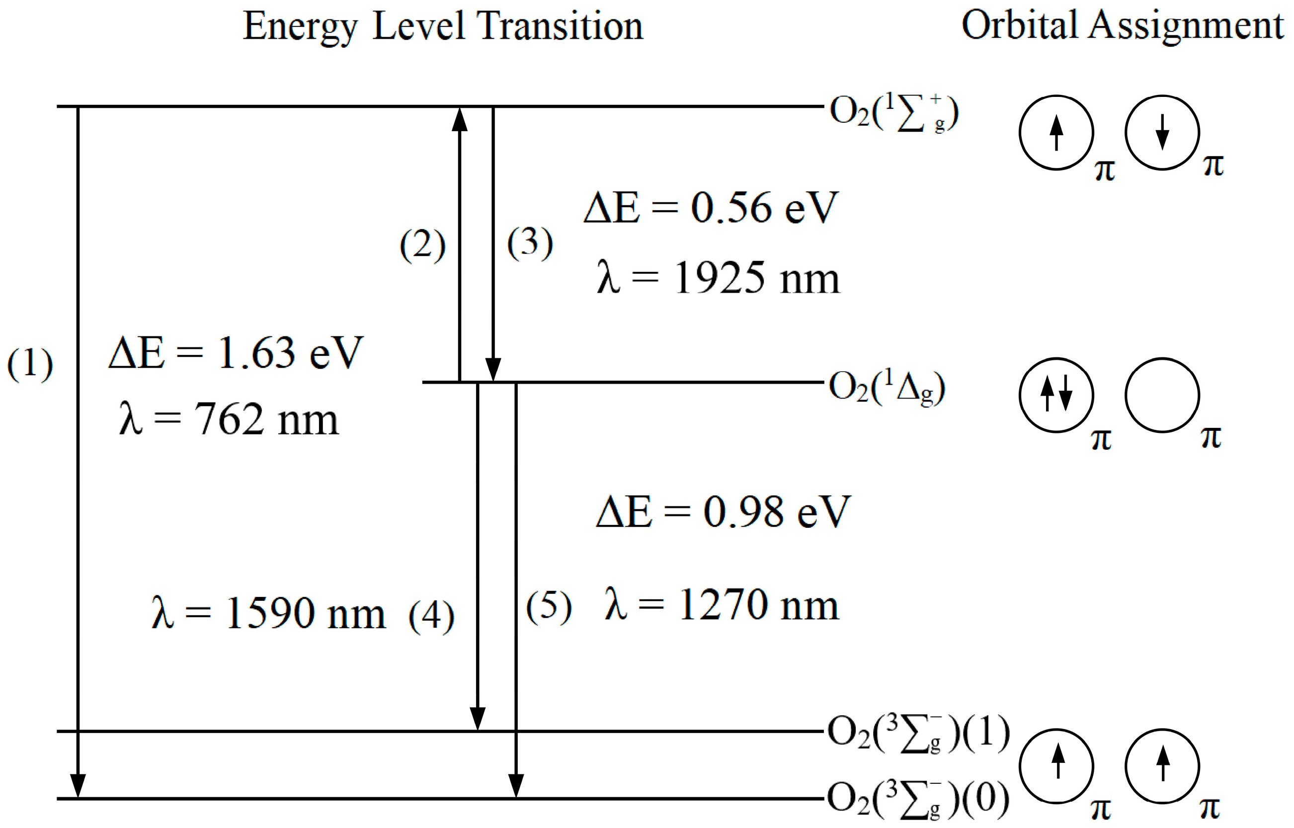
Figure 1. The lowest electronic state energy level transitions and molecular orbital schematics of the oxygen molecule. The configuration of the molecular orbitals of the 1Δg can be described as follows: O2KK(2σg)2(2σu)2(3σg)2(1πu)4( 1 π g + )( 1 π g + ).
Pharmaceuticals 17 01274 g001
Figure 2. Simplified Jablonski energy level diagram of Type I and Type II photochemical reaction.
Pharmaceuticals 17 01274 g002
Table 2. Quenching rate constant and quenching mode of commonly used 1O2 quenchers.
PhotosensitizerQuencherSolventkq (M−1s−1)Type of QuenchingReference
Chlorophyllβ-Carotene
(C40H56)
Benzene1.3 × 1010Physical[25]
Methanol9.3 × 109Physical
Rose Bengal
and Eosin Y
Sodium azide
(NaN3)
Water6 × 108Physical[26]
2-AcetonaphthoneVitamin E
(α-Tocopherol)
Methanol3 × 108Physical[27]
Toluene2.2 × 108Physical
Rose Bengal and PorphyrinHistidine
(C6H9N3O2)
Water4.6 × 107Physical and Chemical[28]
Rose Bengal and PorphyrinTryptophanWater3.2 × 107Physical and Chemical[28]
Methylene BlueTyrosinewater7 × 106Chemical[29]
Table 3. Characterization of photosensitizer approved for PDT.
Photosensitizerλmax (nm)/
Ɛmax (M−1cm−1)
Solvent/(Standard)ΦMethodReference
Porfimer Sodium632/3000PBS/TX100 (RB:0.75)0.89Lysozyme inactivation[73]
Hematoporphyrin derivative (HpD)630Methanol0.64Oxygen depletion[77]
Methanol-D0.76Oxygen depletion[72]
Protoporphyrin IX (PpIX)630/3480D-PBS/TX100 (RB:0.75)0.78TRIL[75]
PBS/TX100 (RB:0.75)0.8TRIL[75]
PBS/TX100 (MB:0.52)0.56Lysozyme inactivation[73]
Meta-tetra(hydroxyl
pheny) chlorin
(m-THPC)
652/35,000Ethanol
(Pheophorbide-a:0.52)
0.42TRIL[78]
N-aspartyl chlorin e6 (NPe6)664/40,000D2O (RB:0.75)0.66Oxygen depletion[79]
PBS (MB:0.52)0.64Lysozyme inactivation[80]
Benzoporphyrin
derivative monoacid ring A
(BPD-MA)
689/34,000Ethanol0.81Cytochrome C
reduction
[74]
Methanol0.78Cytochrome C
reduction
[74]
Benzene0.77TRIL[80]
Talaporfin sodium
(mono-L-aspartyl chlorine e6)
654/40,000D-PBS (RB:0.75)0.53TRIL[75]
D-PBS/TX100 (RB:0.75)0.59TRIL[73]
Aluminum phthalocyanine tetrasulfonate (ALPcS4)676/200,000PBS/TX1000.38Lysozyme inactivation[73]
Disclaimer/Publisher’s Note: The statements, opinions and data contained in all publications are solely those of the individual author(s) and contributor(s) and not of MDPI and/or the editor(s). MDPI and/or the editor(s) disclaim responsibility for any injury to people or property resulting from any ideas, methods, instructions or products referred to in the content.