Article

Metabolomic Profiling of Leptadenia reticulata: Unveiling Therapeutic Potential for Inflammatory Diseases through Network Pharmacology and Docking Studies

by
Yashaswini Mallepura Adinarayanaswamy
1,
Deepthi Padmanabhan
1,
Purushothaman Natarajan
2 and
Senthilkumar Palanisamy
1,*
1
Department of Genetic Engineering, School of Bioengineering, SRM Institute of Science and Technology, Kattankulathur 603203, Tamil Nadu, India
2
Department of Biology, West Virginia State University, Institute, WV 25112-1000, USA
*
Author to whom correspondence should be addressed.
Pharmaceuticals 2024, 17(4), 423; https://doi.org/10.3390/ph17040423
Submission received: 19 February 2024 / Revised: 18 March 2024 / Accepted: 22 March 2024 / Published: 26 March 2024
(This article belongs to the Section Natural Products)

Abstract

:
Medicinal plants have been utilized since ancient times for their therapeutic properties, offering potential solutions for various ailments, including epidemics. Among these, Leptadenia reticulata, a member of the Asclepiadaceae family, has been traditionally employed to address numerous conditions such as diarrhea, cancer, and fever. In this study, employing HR-LCMS/MS(Q-TOF) analysis, we identified 113 compounds from the methanolic extract of L. reticulata. Utilizing Lipinski’s rule of five, we evaluated the drug-likeness of these compounds using SwissADME and ProTox II. SwissTarget Prediction facilitated the identification of potential inflammatory targets, and these targets were discerned through the Genecard, TTD, and CTD databases. A network pharmacology analysis unveiled hub proteins including CCR2, ICAM1, KIT, MPO, NOS2, and STAT3. Molecular docking studies identified various constituents of L. reticulata, exhibiting high binding affinity scores. Further investigations involving in vivo testing and genomic analyses of metabolite-encoding genes will be pivotal in developing efficacious natural-source drugs. Additionally, the potential of molecular dynamics simulations warrants exploration, offering insights into the dynamic behavior of protein–compound interactions and guiding the design of novel therapeutics.

Graphical Abstract

1. Introduction

Inflammation is a painful redness or swelling of a body part brought on by an injury, disease, or infection. Inflammation may not always have a beneficial impact on the body. Autoimmune diseases arise whenever the immune system’s response inappropriately attacks the human body’s native cells, leading to harmful inflammation [1]. The production of cytokines, cell trafficking, mediator synthesis, fibrolysis, coagulation, extravasation changes in hemodynamic properties, and ultimately microvascular death-causing permeability are all part of a cascade of events which leads to inflammation [2]. Normally, the process results in healing and recovery from infection. Nevertheless, if targeted destruction and assistance with repair are not properly designed, inflammation may result in lasting damage to tissue by affecting collagen, leukocytes, or lymphocytes [3]. Inflammation is categorized as acute when marked by swift and intense onset due to factors like toxins or trauma, with symptoms lasting a short time, whereas chronic inflammation, lasting for months to years, depends on the body’s healing capacity and the nature of the initial injury [4]. The WHO says chronic illnesses are the largest health concern. Researchers expect long-term inflammation-related disorders to grow in the US during the next 30 years. In 2000, 125 million people had chronic diseases, and 61 million (21%) had several disorders [5]. Chronic inflammatory diseases such as stroke, lung, heart, cancer, obesity, and diabetes affect three in five people worldwide [6]. The activity of inflammatory-related cytokines, chemokines, adhesion proteins, and pro-inflammatory enzymes has been associated with chronic inflammation [7]. Nonsteroidal anti-inflammatory medicines (NSAIDs) are widely used pharmaceuticals for treating inflammation and associated illnesses, with a global consumption estimated to exceed 30 million per day [8,9,10]. Regrettably, the therapeutic use of NSAIDs is restricted due to the severe adverse effects they may cause, including gastrointestinal (GI) ulcers, perforation, obstruction, and bleeding, despite their strong anti-inflammatory efficacy [10]. NSAIDs may also elevate the likelihood of experiencing falls, heighten the occurrence of geriatric mental episodes, and amplify the danger of stroke [11].
Over 80,000 plants have medicinal properties, most of which have been utilized for centuries [12]. Traditional medicinal plants are receiving more attention from medical research and healthcare. India possesses a total of 45,000 medicinal plants in the Andaman and Nicobar Islands, Western Ghats, and Eastern Himalayas [13]. There are approximately 3000 therapeutic herbs that have received regulatory approval, although traditional practitioners utilize roughly 6000 herbs [14]. Approximately 75–80% of the global population residing in underdeveloped nations depend on natural products for their fundamental healthcare needs, owing to their greater cultural acceptance, compatibility with the human body, and lack of adverse side effects. Traditional medicinal plants produce compounds with anti-inflammatory, antioxidant, and antimicrobial effects [15]. Medicinal plants include a significant abundance of secondary metabolites, which play a crucial role in the identification of novel drugs. Medicinal plants yield a diverse array of secondary metabolites, including flavonoids, terpenoids, tannins, steroids, quinones, coumarins, and alkaloids. These compounds exhibit a broad spectrum of pharmacological properties [16]. Leptadenia reticulata is one of the traditional medicinal plants; it belongs to the family Apocynaceae [17]. It is commonly known as Jivanti (Sanskrit) and Palaikkodi (Tamil). It is a branched shrub which has greenish yellow flowers, the leaves’ length and width are between 2 and 5 cm, and it has an ovate form. It is widely grown in warm subtropical and tropical areas [18]. The whole plant is used in many Ayurvedic remedies and possesses several pharmaceutical properties such as antimicrobial, anti-inflammatory, antipyretic, hepatoprotective, wound healing, diuretic activity, antioxidative, analgesic activity, cytotoxic activity, and an immunomodulatory effect [19]. Our study pioneers an integrated approach combining metabolomics, network pharmacology, molecular docking, and molecular dynamics simulations to elucidate complex biochemical interactions and therapeutic targets. Metabolomics provides insights into physiological states and perturbations [20]. Network pharmacology maps interactions within biological networks, highlighting potential drug targets [21]. Molecular docking identifies promising ligand–receptor interactions, which are then refined through molecular dynamics simulations to assess the stability and dynamics of these complexes in a realistic environment [22]. This comprehensive framework enhances our understanding of molecular mechanisms and drug action, offering a robust strategy for drug discovery and development, with potential applications in personalized medicine and disease treatment. The objective of this study was to characterize the bioactive compounds present in the methanolic extract of L. reticulata using high-resolution liquid chromatography–quadrupole time-of-flight mass spectrometry (HR-LCMS/MS(Q-TOF)). The identified phytochemicals went through ADMET (absorption, distribution, metabolism, excretion, and toxicity) testing, utilizing online tools. Additionally, a network pharmacology analysis was conducted to elucidate the component–target–pathway interactions, thereby uncovering potential molecular mechanisms of action for the identified phytocompounds. Subsequent assessment of the therapeutic potential of these compounds involved molecular docking and molecular dynamics simulation experiments, facilitating the development of novel therapies for inflammatory conditions.

2. Results

2.1. Identification of Phytochemicals Using HR-LCMS/MS(Q-TOF)

Phytocompound separation and analysis were conducted using Q-TOF in both negative and positive modes, in conjunction with HR-LCMS/MS. Table 1 presents a comprehensive list of 113 identified phytocompounds detected in the methanolic extract. Notable among these compounds are kaempferol, known for its diverse medicinal properties such as anticarcinogenic, antibacterial, antifungal, antiprotozoal, anti-inflammatory, and antioxidant activities [23,24]; luteolin, which boasts numerous health benefits including cancer prevention, mitigation of oxidative stress, management of behavioral issues, neuroinflammation, inflammation, cardiovascular diseases, and its role in preventing metabolic disorders like diabetes, hepatic steatosis, and obesity [25]; ferulic acid, recognized for its versatility as a bioactive molecule with anti-inflammatory and antioxidant properties, offering some degree of protection against cardiovascular and renal diseases [26]; quercitrin, which exhibits a wide range of bioactivities including antioxidant effects, anti-inflammatory properties, antimicrobial activity, immune system regulation, pain reduction, wound healing promotion, and vasodilation [27]; catechin, known for its antibacterial, antitumor, antihypertensive, anticoagulant, and antiulcer properties [28]; and ellagic acid, possessing significant anti-inflammatory, anti-mutagenic, anti-proliferative, and antioxidant properties, showing promise in the treatment of various human ailments. Additionally, several other phytocompounds were identified, consistent with findings from previous studies [29].

2.2. ADMET Profiling

SwissADME, an online tool, was used to assess the phytocompounds’ pharmacokinetic and drug-likeness characteristics as well as their distribution, metabolism, excretion, and absorption capabilities. We predicted ADME profiling based on Lipinski’s rule of five, where compounds with a molecular weight less than 500, topological surface area (TSA) < 150, number of hydrogen bond donors < 150, quantity of donors for hydrogen bonds < 5, quantity of donors for hydrogen acceptors < 10, and two breaches of the rules are permitted. Except for 2,3,5,7,9-Pentathiadecane 2,2-dioxide, beta-D-3[5-Deoxy-5-(dimethylarsinyl)ribofuranosyloxy]-2-hydroxy-1-propanesulfonic acid, and Methyl(3×,10R)-dihydroxy-11-dodecene-6,8-diynoate 10-glucoside, all phytocompounds which cleared both ADME and toxicity exhibited excellent gastrointestinal absorption. It was predicted that certain compounds cannot pass through the blood–brain barrier (BBB), including Neotussilagine, 1-Pyrenylsulfate, 2,3,5,7,9-Pentathiadecane 2,2-dioxide, Kaempferol, Malic acid, (Z)-3-(1-Formyl-1-propenyl)pentanedioic acid, (1S,4R)-10-Hydroxyfenchone glucoside, beta-D-3[5-Deoxy-5-(dimethylarsinyl)ribofuranosyloxy]-2-hydroxy-1-propanesulfonic acid, and Albuterol, Methyl(3×,10R)-dihydroxy-11-dodecene-6,8-diynoate 10-glucoside. Pro Tox II was used to determine the toxicity, and apart from Neotussilagine, the majority of the phytocompounds would not be mutagenic, cytotoxic, carcinogenic, immunotoxic, or hepatotoxic. The substances with LD50 values greater than 2000 mg/kg indicated potential safety for usage as future medicines in in vivo research. Table 2 shows the list of compounds with the best ADME properties. Table 3 shows the list of compounds with the best toxicity profiling.

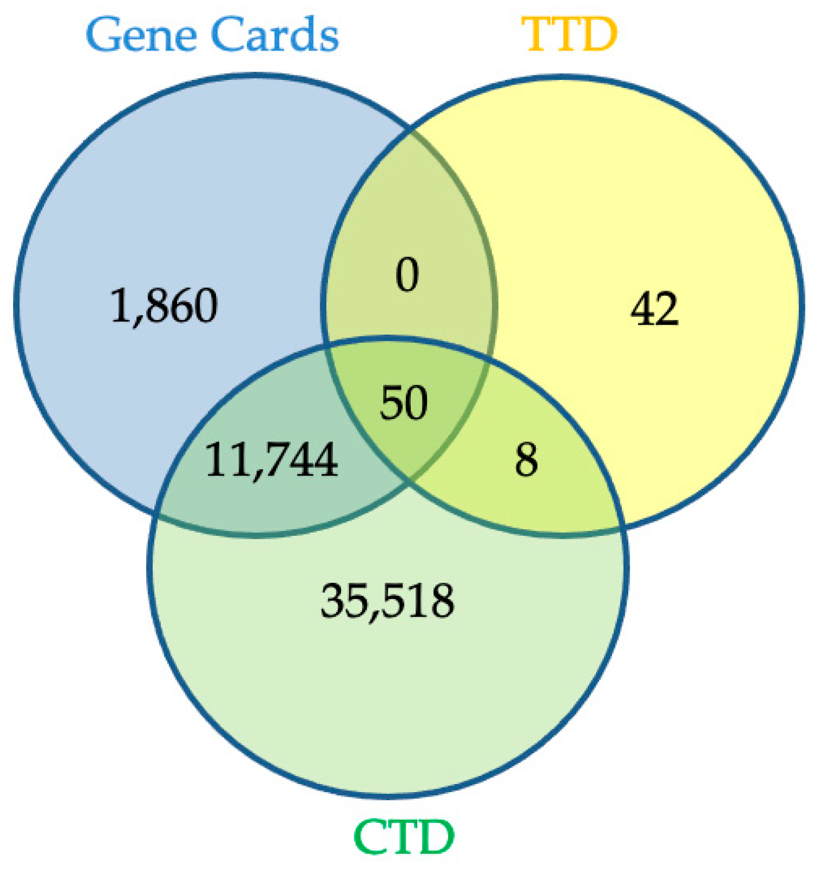
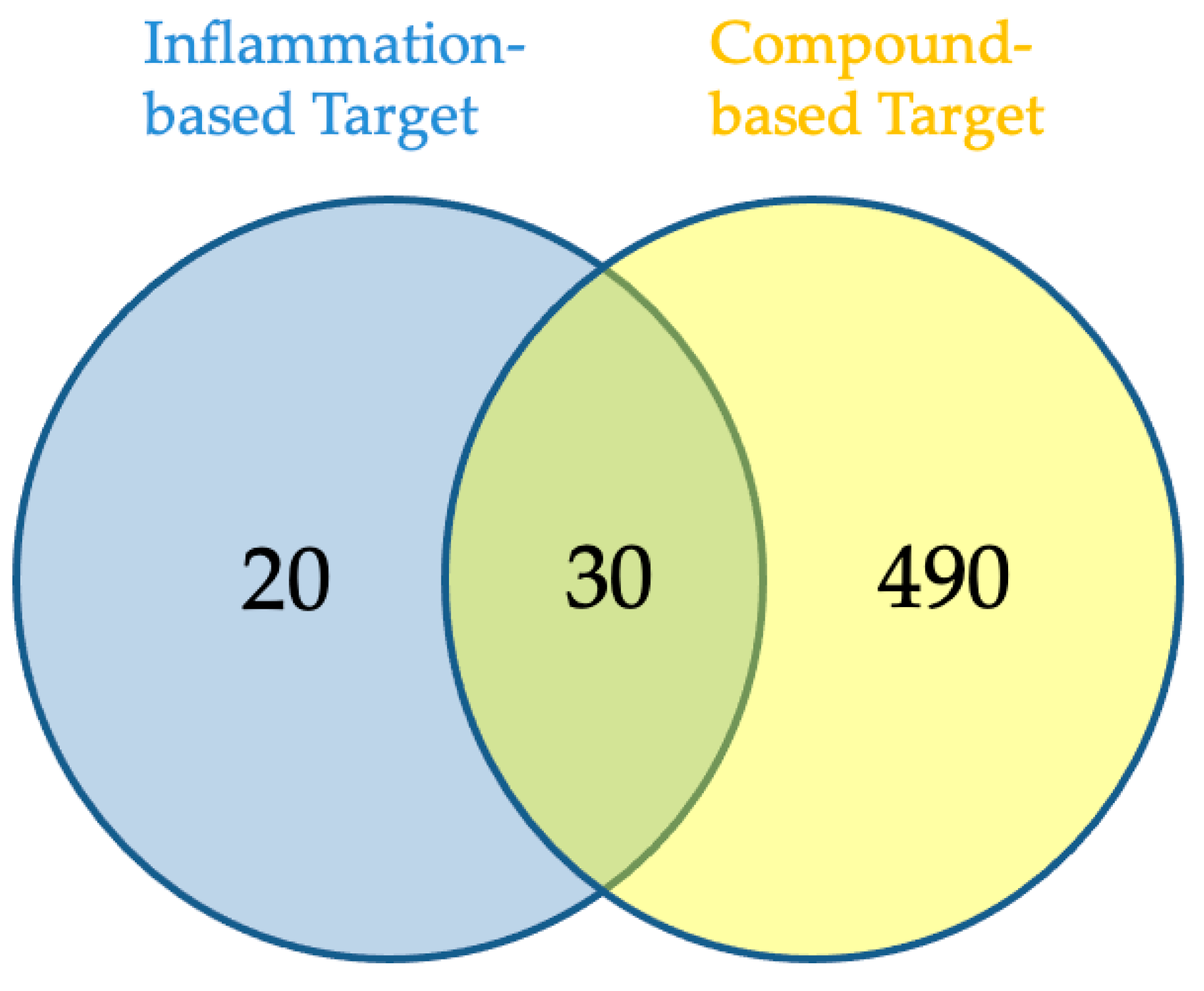
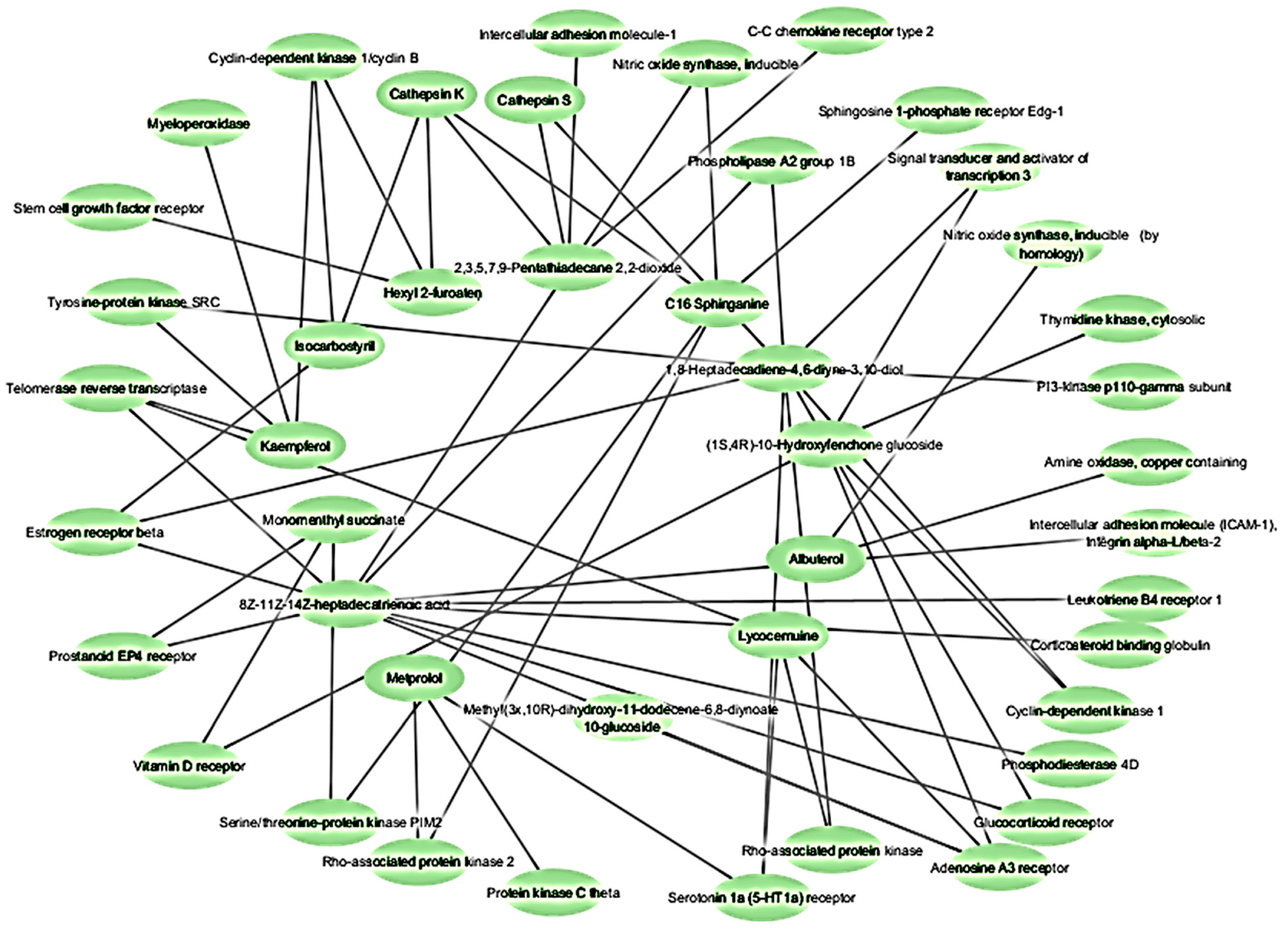
2.3. Network Pharmacology Analysis for Potential Active Compound Targets and Anti-Inflammatory Targets

Eighteen of the eighty compounds in the plant L. reticulata were chosen using Lipinski’s rule of five and ADME. A total of 520 potential targets could be found for 18 of the components combined using SwissTarget Prediction of having a likelihood greater than zero. Using the term “inflammation”, the associated genes were selected from the disease gene databases (GeneCards, CTD, TTD). After pooling the findings and removing duplicates, 50 records remained for screening, as shown in Figure 1. The Venn diagram (Figure 2) revealed 30 crossovers between the active compounds and inflammation-related targets. To create a compound–target network diagram, the targets and corresponding phytocompounds were loaded into Cytoscape 3.9.1. This network (Figure 3) shows the synergistic multi-component and multitargeted effects of the L. reticulata contributing to their anti-inflammatory activities. Table 4 shows the list of target proteins which play vital roles in inflammation.

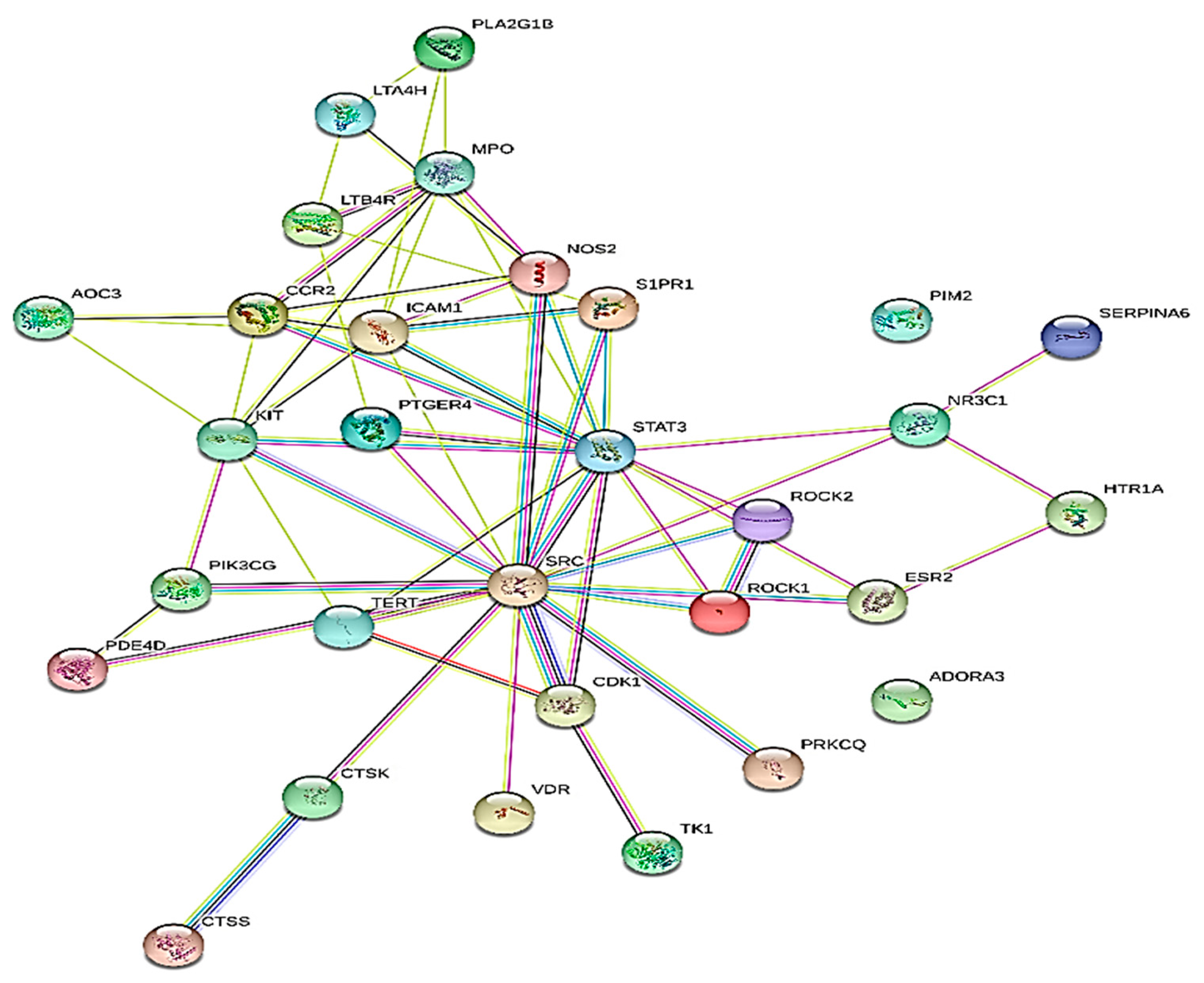
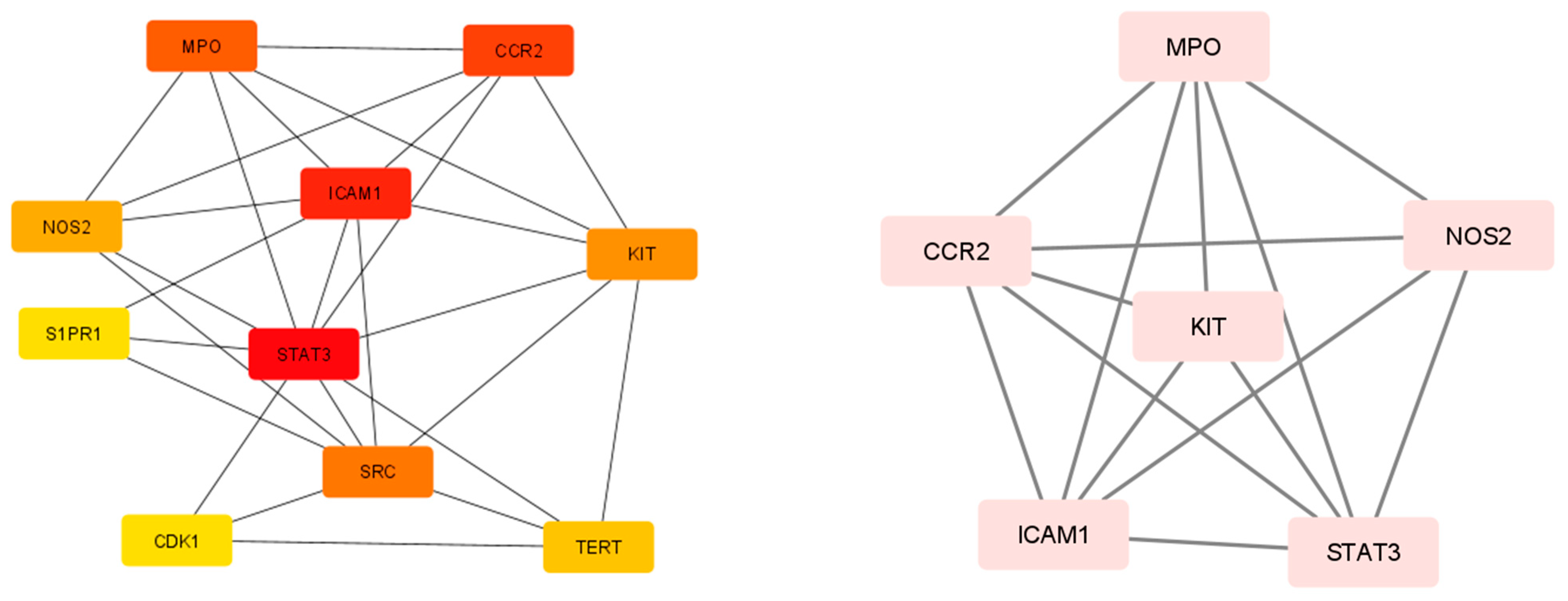
2.4. Protein–Protein Interaction

Using the STRING database, a PPI network analysis was carried out to recognize the hub genes in critical modules. The selected results required an overall score of at least 0.4. Figure 4 shows the protein–protein interaction (PPI) network, which serves as a therapeutic target for the reduction of inflammation. Within the PPI network, the top 10 hub genes were selected using the MCC algorithm and the CytoHubba plugin. The top functional clusters of the module were chosen, as shown in Figure 5. By examining the MCC and M-CODE junction targets, six gene hubs were found (CCR2, ICAM1, KIT, MPO, NOS2, and STAT3). The eighteen metabolites showed anti-inflammatory effects, namely, receptors (C-C chemokine receptor type 2, stem cell growth factor receptor, and others), enzymes (myeloperoxidase, nitric oxide synthase, and others), and proteins (intercellular adhesion molecule-1, signal transducer and activator of transcription 3, and others).

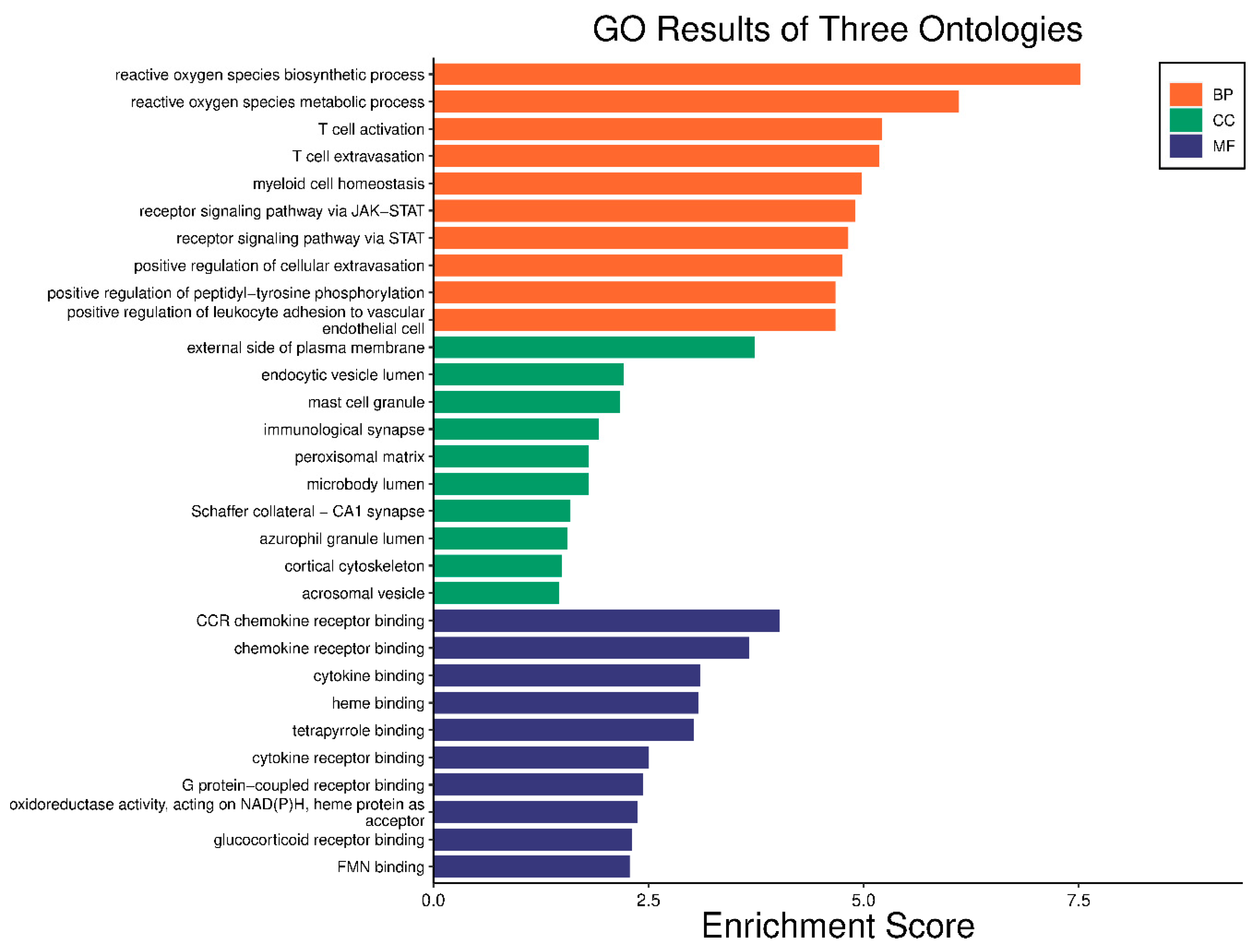
2.5. GO Enrichment and KEGG Analysis

KEGG pathway enrichment and GO analysis were carried out on the six major targets using the DAVID 6.8 database. With the use of GO analysis, 24 GO items with p < 0.05 were found; they included entries for 17 biological processes, five cell component entries, and two molecular functions; Figure 6 shows the biological processes, cellular components, and gene molecular functions. The biological processes included a reactive oxygen species biosynthesis process, a reactive oxygen species metabolic process, T cell activation, and T cell extravasation. The cellular component enrichment included the external side of the plasma membrane, mast cell granules, immunological synapses, endocytic vesicle lumen, and microbody lumen. The molecular functions included CCR chemokine receptor binding, chemokine receptor binding, cytokine binding, and heme binding. Afterwards, an enrichment analysis of the KEGG pathway was carried out (Figure 7). Every route that had a p value less than 0.05 was screened and then sorted based on the p value. Acute myeloid leukemia, the AGE–RAGE signaling route in diabetic compilations, and the HIF-1 signaling pathway are the top three mechanisms.

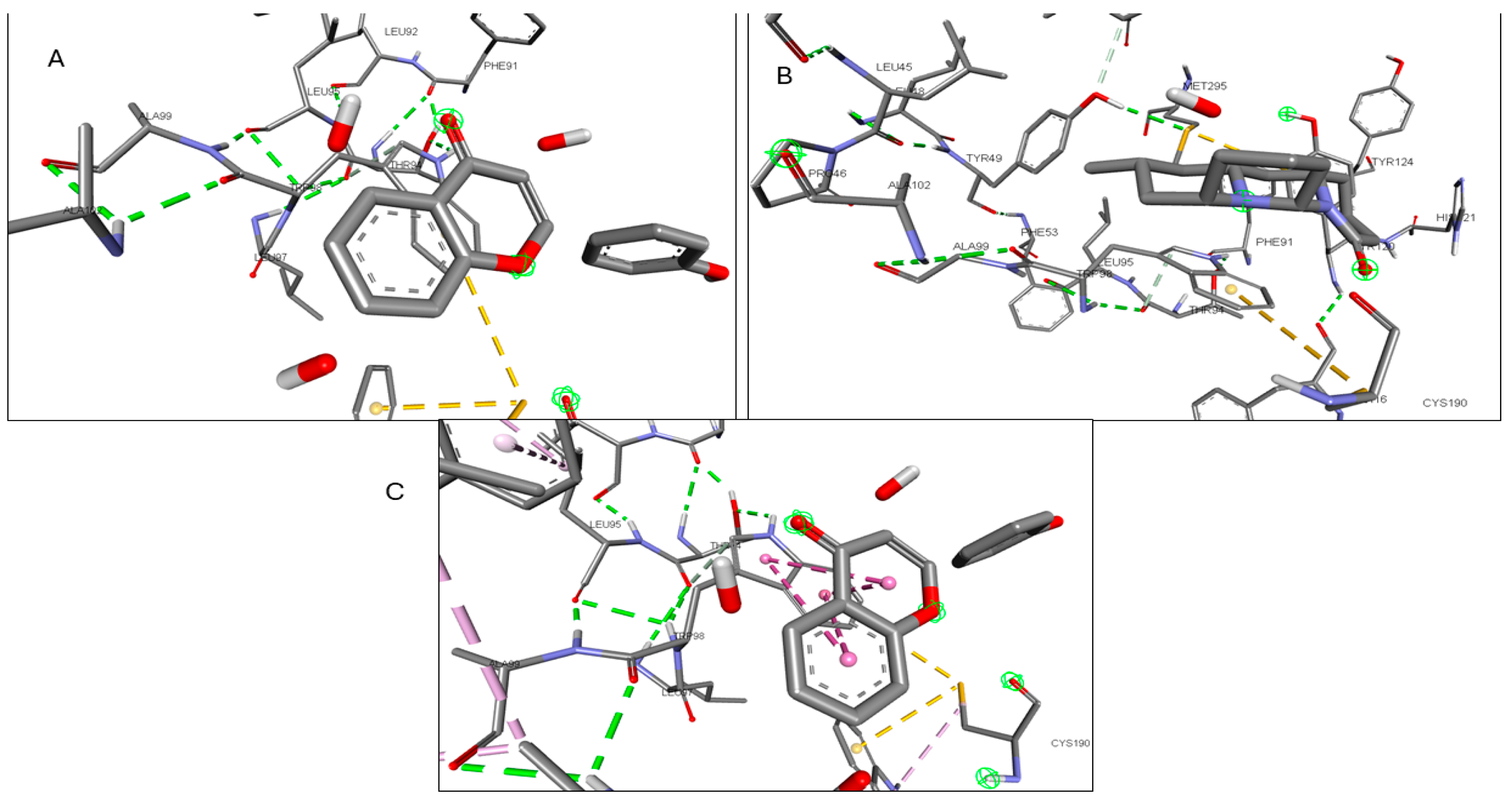
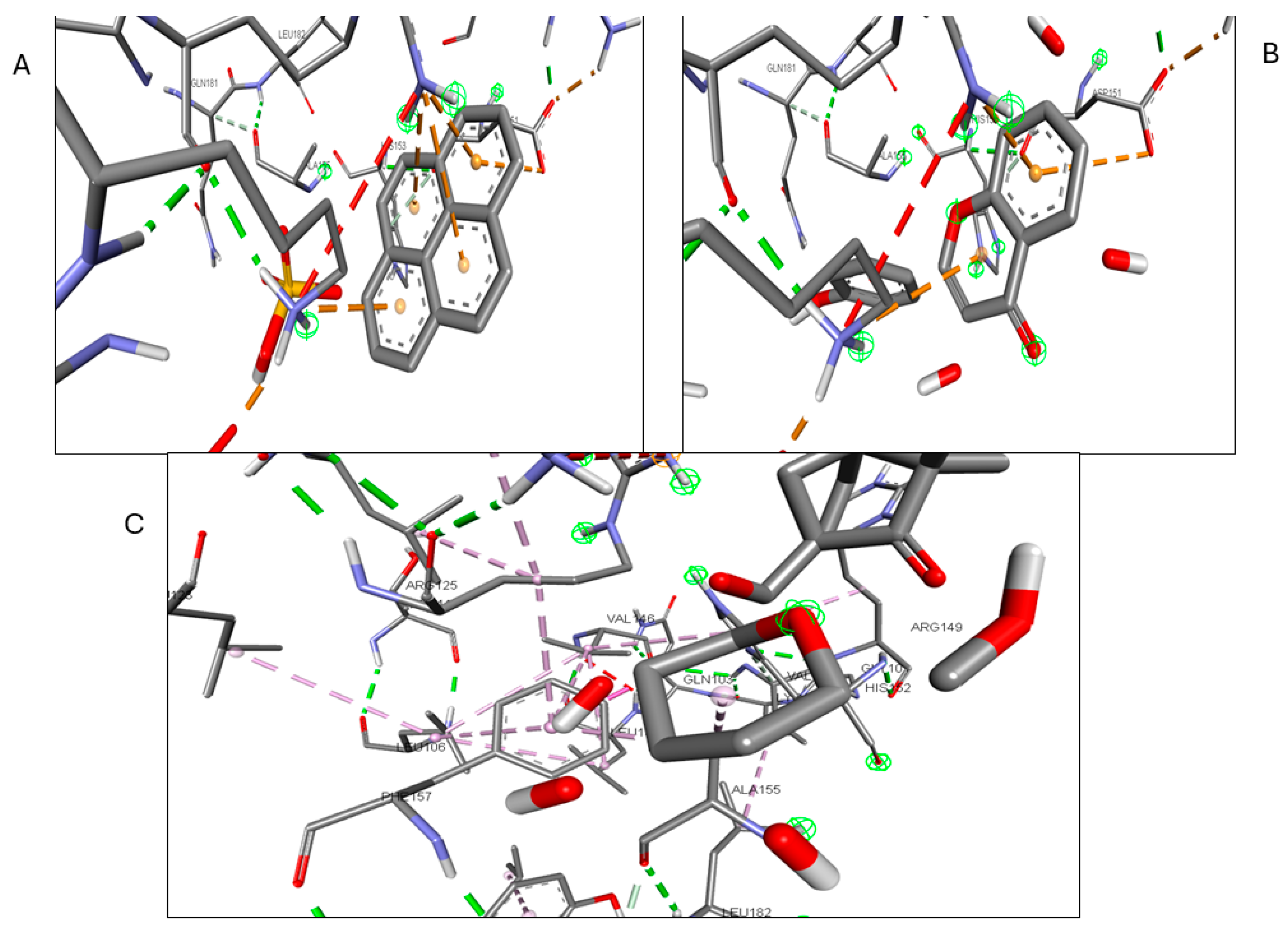
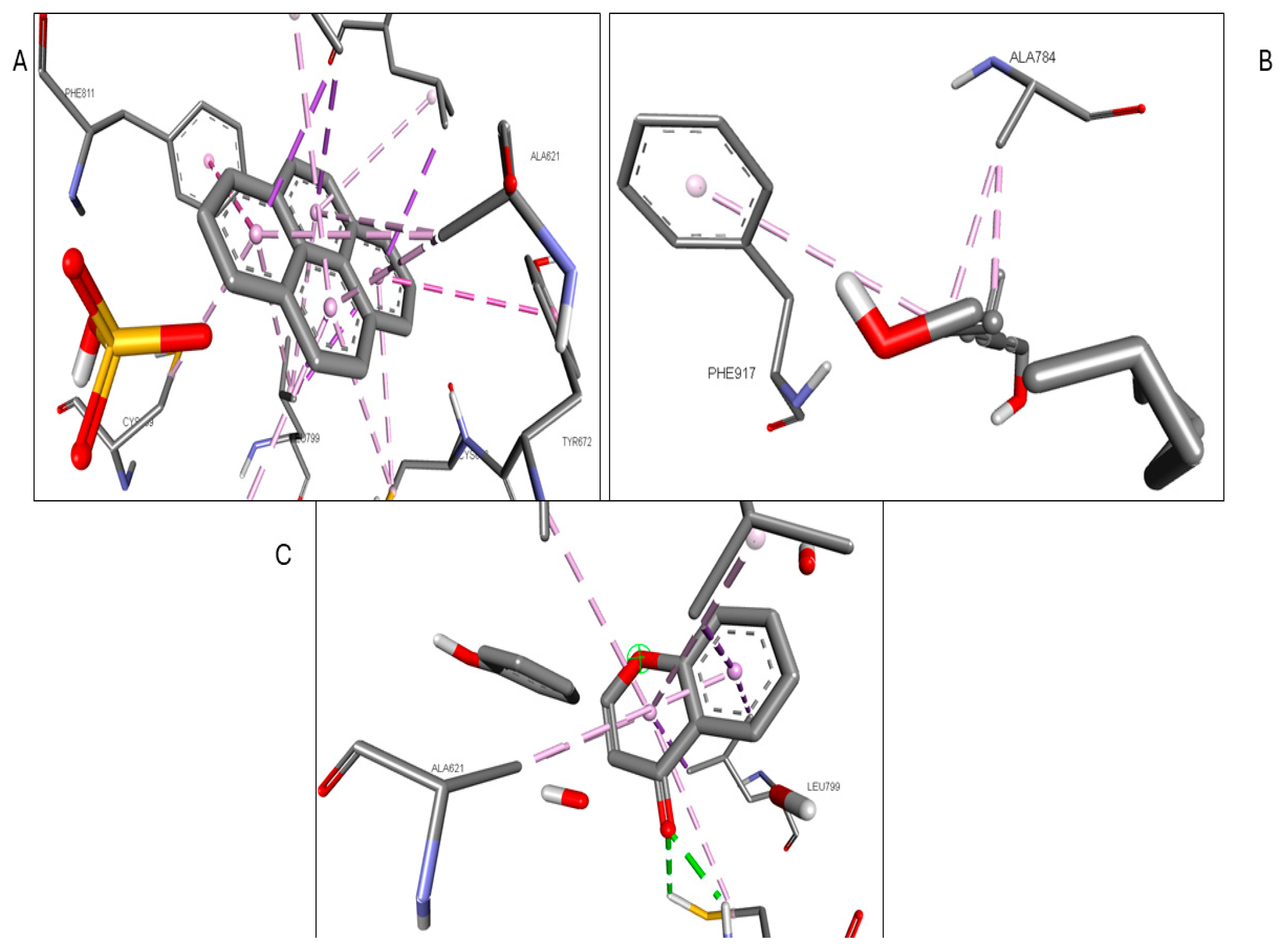
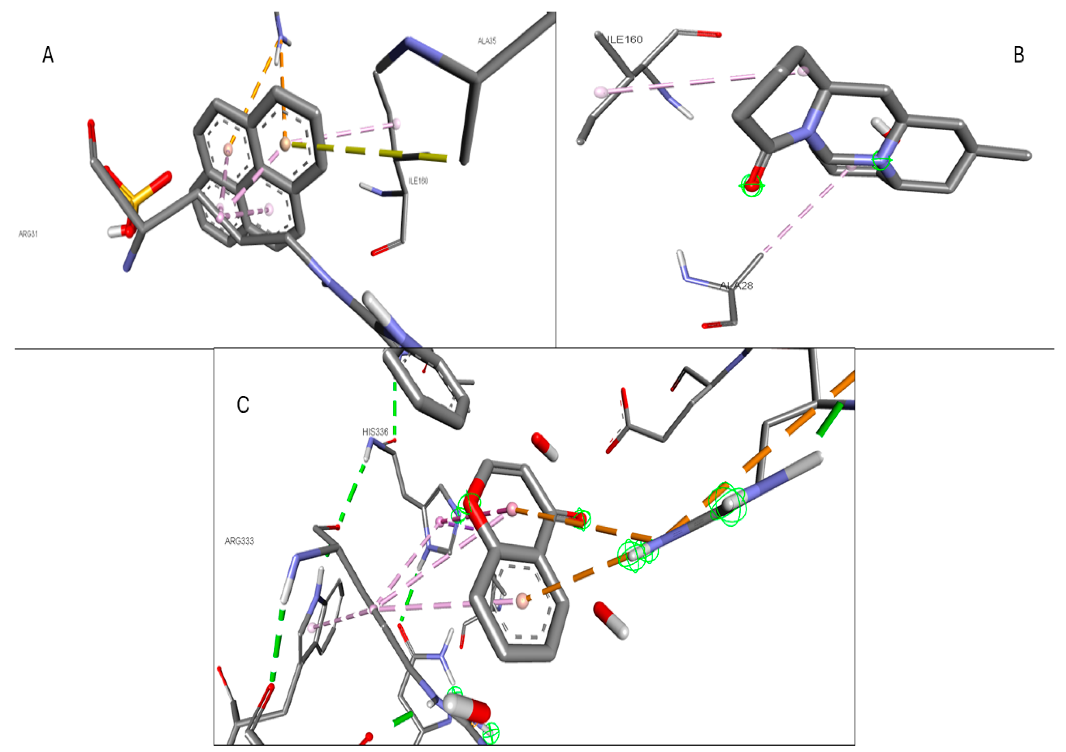
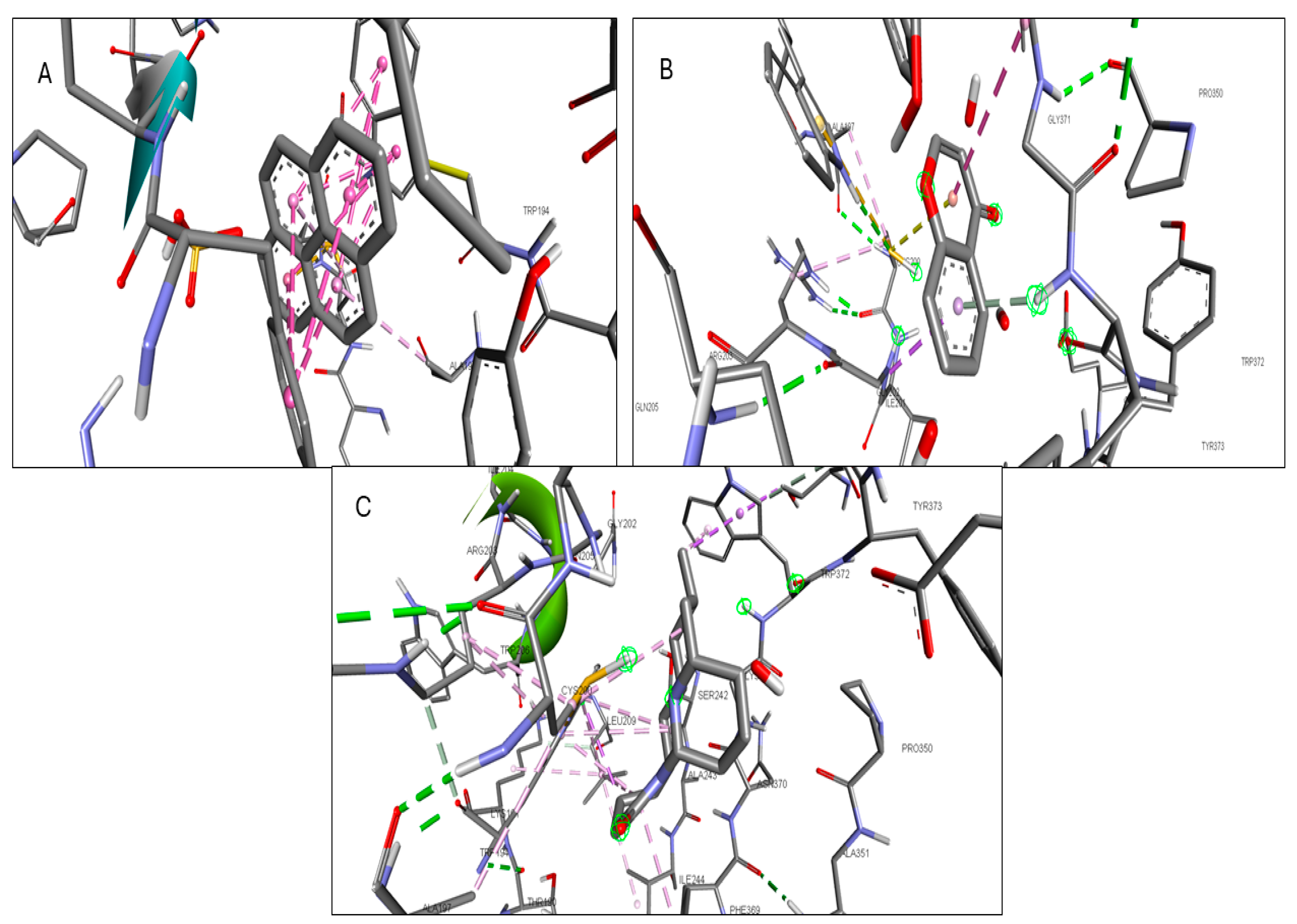
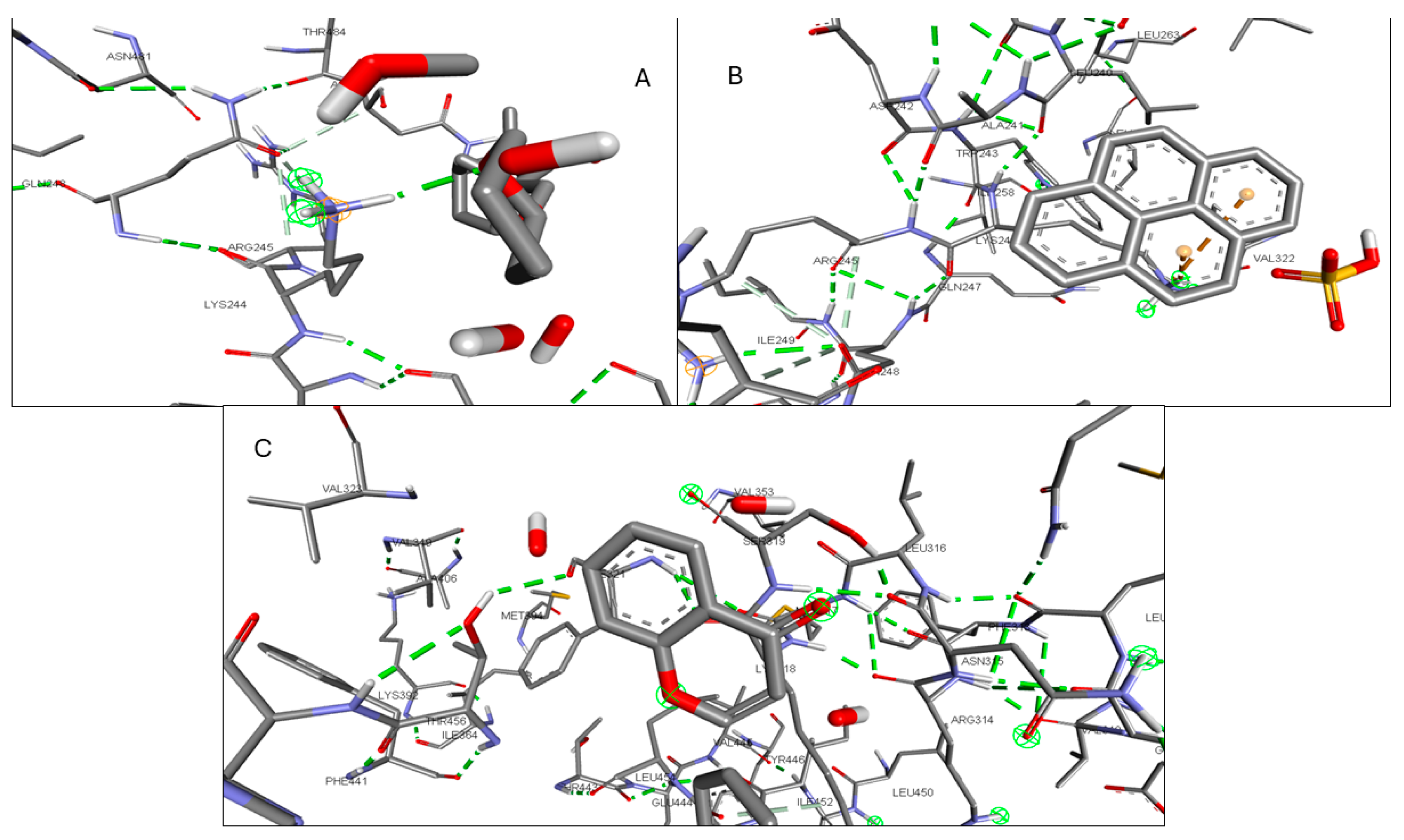
2.6. Molecular Docking of Active Compounds and Key Targets

Validation of the docking software was conducted by removing the crystallized ligand from the protein and then rebinding it to the same pocket. Supplementary Figure S1 illustrates the three-dimensional interaction between the ligand and the target proteins. The top six targets, ranked by degree, along with eighteen active components, were selected for molecular docking. According to convention, a binding energy score higher than 4.25 indicates a binding capability between the compounds and proteins. Scores exceeding 5.0 denote a relatively high binding affinity, while scores exceeding 7.0 indicate a strong ligand–receptor interaction. Table 5 presents the optimal binding affinities of the phytocompounds and proteins. Using the BIOVIA Discovery Studio 2024 Client Visualizer, interactions between amino acid residues and the ligand, as well as the types of forces involved, were investigated in both three-dimensional (Figure 8, Figure 9, Figure 10, Figure 11, Figure 12 and Figure 13) and two-dimensional formats (Supplementary Figures S2–S7).

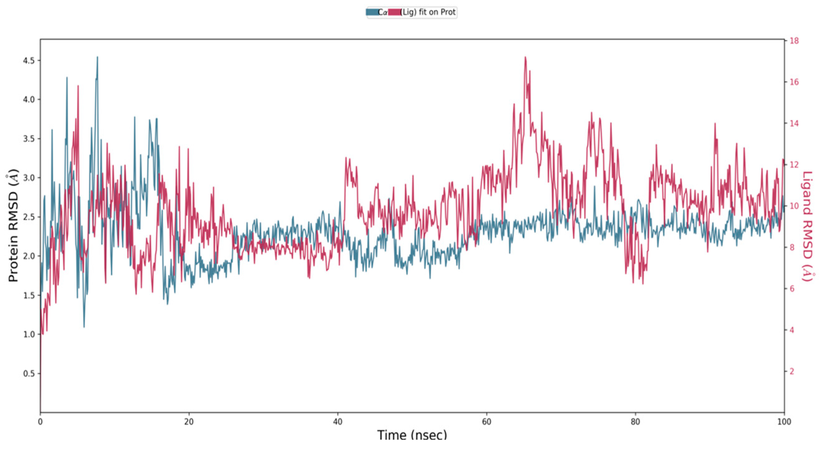
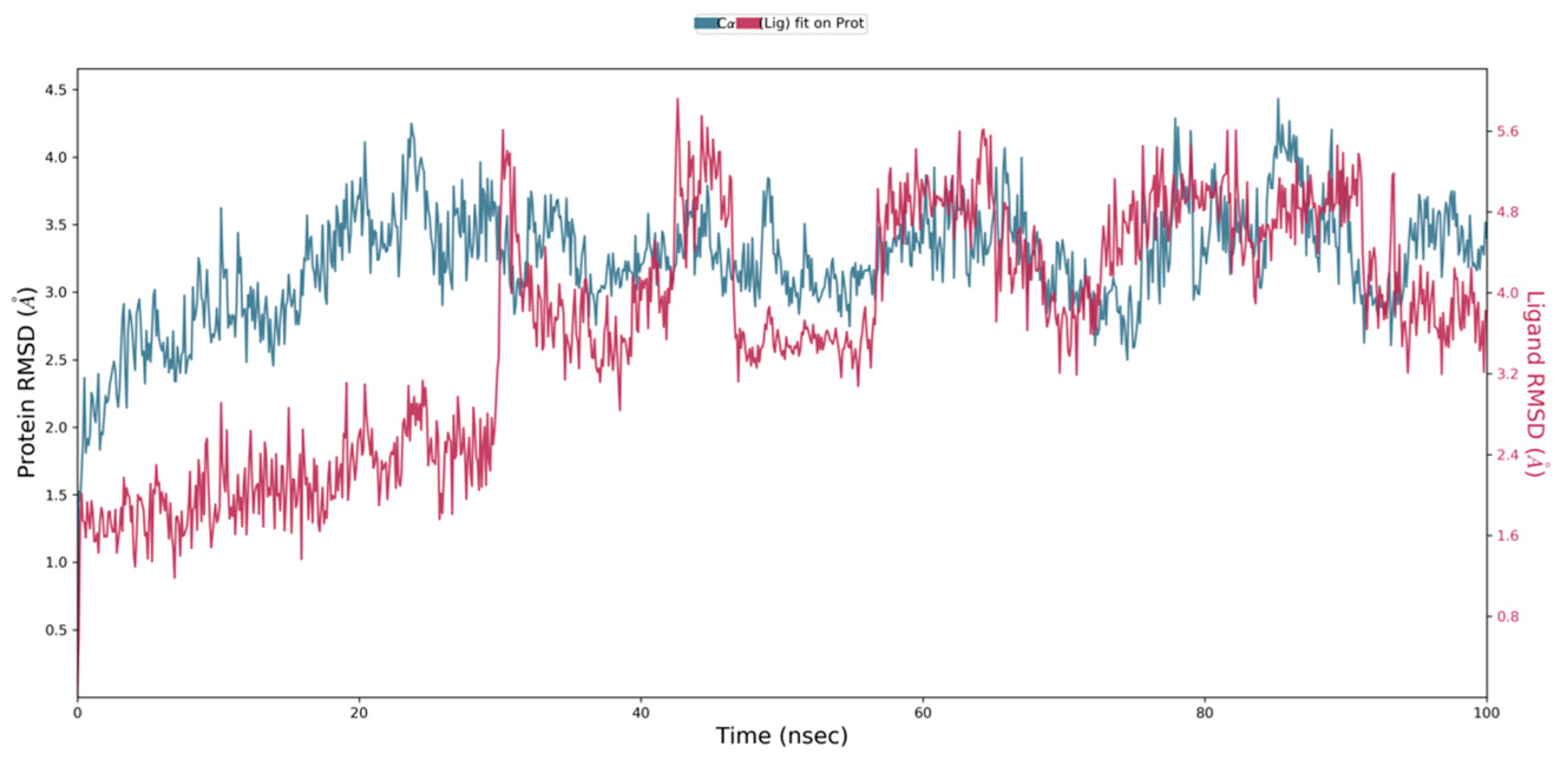
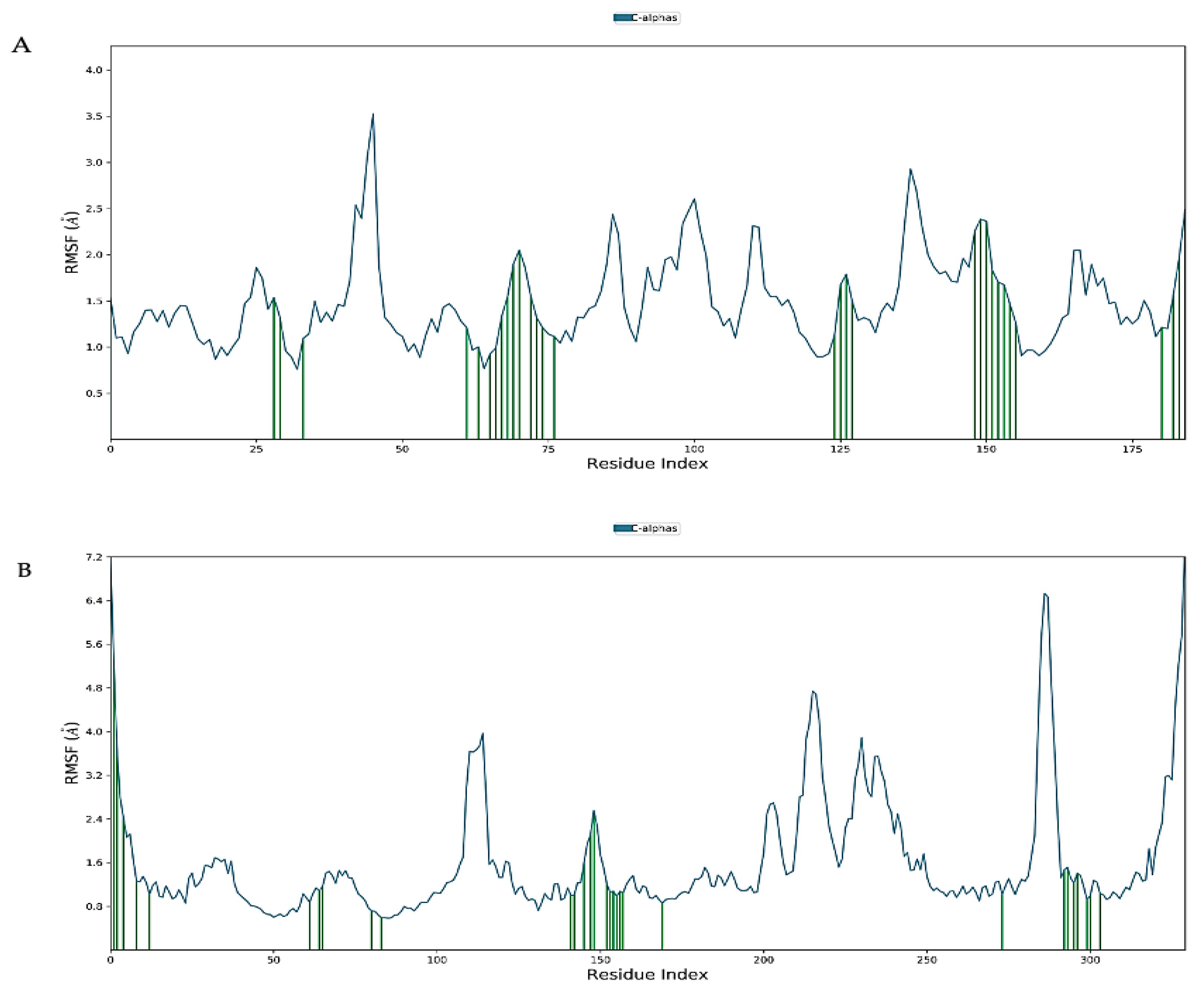
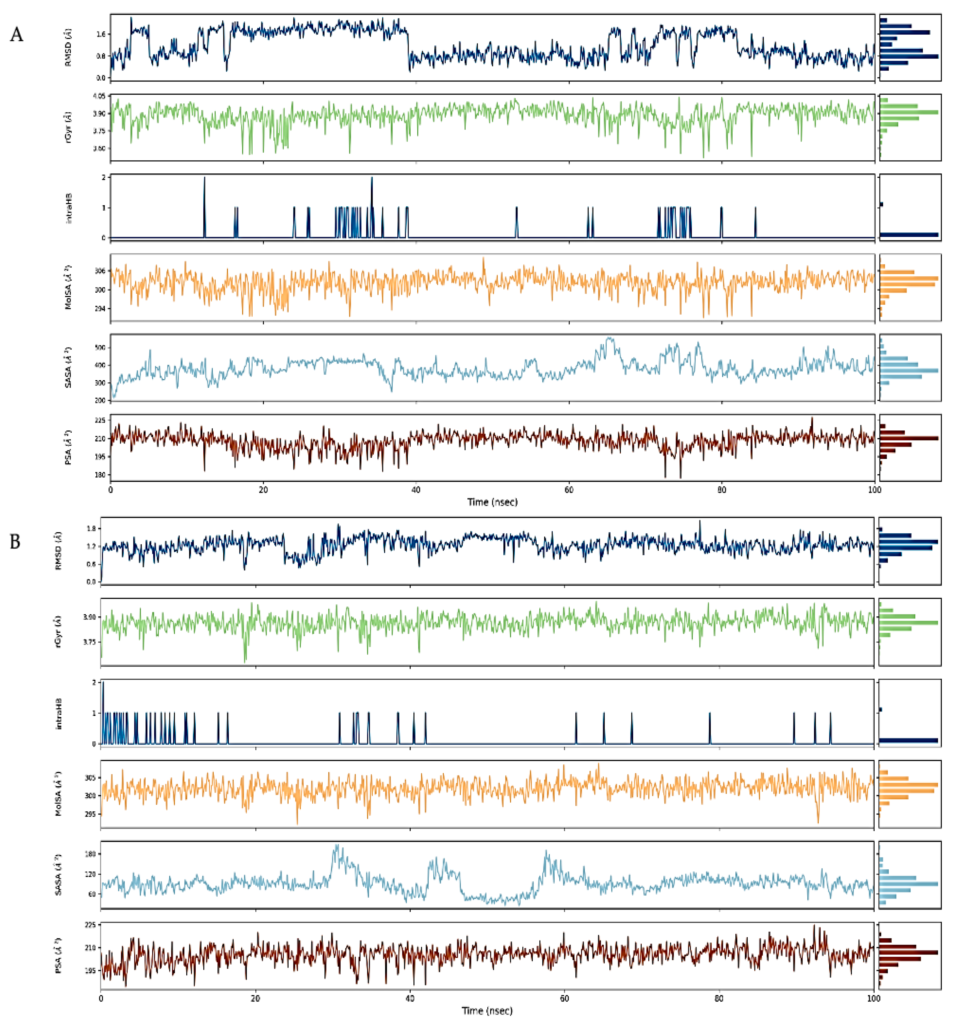
2.7. Molecular Dynamics Simulation

Virtual screening of phytocompounds from L. reticulata identified fifteen potent hub protein antagonists. Among these, (1S,4R)-10-Hydroxyfenchone glucoside, 1-Pyrenylsulfate, Kaempferol, and Lycocernuine exhibited the highest binding affinities. Consequently, the conformational stability of (1S,4R)-10-Hydroxyfenchone glucoside–ICAM1 and (1S,4R)-10-Hydroxyfenchone glucoside–CCR2 complexes were assessed through 100-nanosecond dynamics simulations. The structural integrity of the ligand–protein complex was evaluated using the root mean square deviation (RMSD) obtained from the MD simulation trajectory. For the (1S,4R)-10-Hydroxyfenchone glucoside–ICAM1 complex (depicted in Figure 14), there was initial stability observed between 0 and 40 nanoseconds, followed by fluctuation from 40 to nearly 80 nanoseconds. The ligand exhibited fluctuations ranging from 2.5 Å to 4.5 Å, while the protein showed lesser fluctuations, between 2.0 Å and 2.5 Å. The stability of the ligand within the binding site of the target persisted throughout the simulation, indicating prolonged interaction, which correlates with the observed high binding affinity from docking analysis. The simulation of the (1S,4R)-10-Hydroxyfenchone glucoside–CCR2 complex (illustrated in Figure 15) also showed minimal fluctuation, with stability observed from 30 nanoseconds onwards, exhibiting a deviation of 0.5 Å throughout the 100-nanosecond simulation period. A root-mean-square fluctuation (RMSF) analysis was employed to assess variations within the CCR2 and ICAM1 proteins. Peaks in the RMSF plot represent the regions of the protein with the highest degree of fluctuation during the simulations. Figure 16 illustrates the backbone atoms of the protein moiety (shown in blue) and the side chain atoms (in green), with RMSF values quantifying protein flexibility and fluctuation. Further evaluation of the protein–ligand complex during the 100-nanosecond simulation included analysis of the root mean square deviation (RMSD), polar surface area (PSA), molecular surface area (MolSA), radius of gyration (rGyr), and solvent-accessible surface area (SASA), as depicted in Figure 17. Supplementary Figure S8 illustrates the hydrogen bond formation process, while Figure 18 visualizes the interaction between protein–ligand complexes during molecular dynamics simulations. Supplementary Figure S9 represents the RMSF of protein ICAM 1 and CCR2. Supplementary Figure S10 provides a timeline depiction of the protein’s residues interactions with the ligand molecule.

3. Discussion

Inflammation serves as the body’s innate defense mechanism against various harmful substances and unpleasant stimuli. However, conventional anti-inflammatory medications often come with a host of adverse effects. Natural remedies have long been employed to alleviate inflammation, reflecting ancient practices. Throughout history, the use of medicinal herbs has been widely accepted as safe, cost effective, and prevalent. In Serbia, traditional medicine reigns as the predominant form of therapy, rooted in a steadfast belief in the healing properties of herbs. Numerous studies have highlighted the potent anti-inflammatory properties of several components found in L. reticulata [29].
The methanolic extract derived from L. reticulata underwent analysis through high-resolution liquid chromatography–mass spectrometry (HR-LCMS/MS) coupled with Q-TOF analysis, revealing a total of 260 compounds. Upon comparison of the high-resolution liquid chromatography and mass spectrum data with the MassHunter library, 113 compounds were successfully characterized and identified. These identifications relied on factors such as retention time, molecular formula, and mass. The chromatograms (Supplementary Figures S3–S8) provide significant insights into the relative amounts of various bioactive chemicals. Prominent phytocompounds confirmed through the HR-LCMS/MS(Q-TOF) analysis included Methyl N-methylanthranilate, Brassilexin, Kaempferol, Ferulic acid, Ellagic acid, Neuraminic acid, Hydroquinidine, Catechin, Madasiatic acid, Luteolin, Caulerpin, 2-Hexaprenyl-3-methyl-6-methoxy-1,4 benzoquinone, Malic acid, Quercitrin, Albuterol, Colnelenic acid, Lamprolobine, 1-Pyrenylsulfate, 14,19-Dihydroaspidospermatine, Muricatalin, Hexazinone, Tetradecyl sulfate, 9-HOTE, Isocarbostyril, 6-Methylquinoline, L-Tryptophan, and 2,4,6-Triethyl-1,3,5-trioxane. In a similar study on Alangium salviifolium bark, LC/Q-TOF analysis led to the identification of 449 compounds using the METLIN library.
ADMET studies play a crucial role in pharmaceutical research, providing a comprehensive assessment of a medication’s pharmacokinetics, including absorption, distribution, metabolism, excretion, and toxicity. Predicting a medication’s fate and its physiological impacts, such as oral and gastrointestinal absorption, is essential for drug development. Inadequate absorption can adversely affect distribution and metabolism, potentially leading to neurotoxicity and nephrotoxicity. Understanding a medication molecule’s distribution within an organism is a primary objective of research, making ADMET studies vital in computational drug design. In the current study, 18 phytocompounds from L. reticulata successfully underwent ADMET profiling. The observations indicated satisfactory oral absorption, along with appropriate solubility and absorption qualities, aligning with drug-like principles.
Network pharmacology was utilized to pinpoint the compounds primarily responsible for the anti-inflammatory effects of the phytocompounds. After undergoing the ADME filter procedure and adhering to Lipinski’s rule of five, eighteen compounds were chosen for target prediction. Analysis of the protein–protein interaction (PPI) network using MCODE revealed hub genes, including CCR2, ICAM1, KIT, MPO, NOS2, and STAT3, which are pivotal in inflammation, guiding immune cell migration, adhesion, and activation. Their roles range from facilitating leukocyte endothelial transmigration (ICAM1) to regulating inflammatory responses and signaling (STAT3). Targeting these proteins could significantly impact treating inflammatory and autoimmune diseases by modulating immune responses [30,31,32,33,34,35].
The investigation culminated in molecular docking and molecular dynamic simulations to elucidate the precise medicinal mechanism of phytocompounds found in the plant. The docking studies identified key interactions between proteins and compounds, suggesting paths for further investigation into their therapeutic effects. We found that the 1S,4R)-10-Hydroxyfenchone glucoside phytocompound had a great interaction with CCR2 and ICAM1. Due to this interaction, we took this complex forward for a molecular dynamics simulation study. Despite their complexity, the molecular dynamics simulations offered insights into the receptor–ligand interactions, emphasizing the role of water molecules in drug development. Changes in the root mean square deviation (RMSD) highlighted deviations in protein conformation, while the root mean square fluctuation (RMSF) values pointed to stability and strong hydrogen bonding, crucial for understanding compound efficacy.
Other phytocompounds with notable binding affinities with different targets include (1S,4R)-10-Hydroxyfenchone glucoside and Lycocernuin. Kaempferol, known for its anti-inflammatory properties, has been validated in various studies as a safe and effective natural dietary anti-inflammatory agent. Conversely, (1S,4R)-10-Hydroxyfenchone glucoside boasts antioxidant, antimicrobial, and other bioactive effects, warranting further investigation to confirm its connection and underlying mechanisms.
Overall, this research underscores the anti-inflammatory properties of Leptadenia reticulata methanolic extract, mediated through the synergy of multiple components, targets, and pathways. Network pharmacology played a pivotal role in elucidating this mechanism of action, offering valuable insights for future therapeutic development.

4. Materials and Methods

4.1. Plant Material

Plant samples were collected from Hosur, Tamil Nadu and were duly authenticated by Dr. Senthilkumar Umapathy (https://mcc.edu.in/; accessed on 9 September 2023; [email protected]), Assistant Professor, Department of Botany at Madras Christian College campus.

4.2. Extraction of Phytochemicals from L. reticulata

The leaves, stem, and root of Leptadenia reticulata were shade dried for two weeks at room temperature. The dried samples were ground finely using liquid nitrogen and 1 mL of 99.9% methanol was added to 100 mg of sample and stored at −20 °C overnight. The sample was subjected to water bath sonication for 15 min at 35 °C, and then stored at −4 °C overnight [36,37]. After 24 h, the sample was centrifuged at 10,000 rpm for 10 min. The centrifuged sample was then filtered using a syringe filter (PVDF/L 0.22 µm) and stored at −20 °C for further analysis.

4.3. Identification of Phytochemicals Using HR-LCMS/MS(Q-TOF)

Identification of secondary metabolites from Leptadenia reticulata was achieved using an Agilent 6550 iFunnel Q-TOF (Agilent Technologies, Santa Clara, CA, USA), with both positive and negative modes [38]. Hypersil Gold C-18 (3 µm particle size, 2.1 mm internal diameter, and 100 mm length) was used for the separation of secondary metabolites. A flow rate of 300 µL/min was used. An aliquot of 3 µL was injected independently. 100% acetonitrile with 100% methanol made up mobile phase B, whereas 0.1% formic acid in water made up mobile phase A [39]. With the following parameters, a complete scan mode was attained in the 100–1000 amu range: capillary voltage (3500 V); nozzle voltage (1000 V); 13 L/min gas flow rate at 300 °C; and nebulization set at 35 psi. Mass Hunter Workstation was used for identification of secondary metabolites based on the m/z (mass/charge) values and spectrum graph.

4.4. Active Ingredient Screening

The SwissADME web server (http://www.swissadme.ch/; accessed on 29 September 2023) was used to conduct the in silico ADME toxicity and drug-likeness assessments on the discovered phytocompounds. To evaluate the drug-likeness of the compound various parameters were computed, including number of hydrogen bond donors and acceptors, molecular weight, molecule polar surface area, Veber’s rule, the logarithm of the n-octanol/water partition coefficient(logP), and Lipinski’s rule of five [40]. Additionally, ADMET prediction was carried out while considering elements including cytochrome P450(CYP) 2D6 inhibition, plasma membrane binding, blood–brain barrier penetration, aqueous solubility, and hepatotoxicity [16]. The ProTox II (https://tox-new.charite.de/; accessed on 2 October 2023) online server was used to calculate the LD50 value of the phytocompounds and to predict organ toxicity.

4.5. Inflammation-Related Target and Associated Drug Target Screening

The SwissTargetPrediction tool (http://www.swisstargetprediction.ch; accessed on 7 October 2023) was used to screen the target corresponding to the component and screen the targets based on the possible criteria before (probability > 0) merging the targets and removing repeated values [41]. In order to find relevant targets, we used the search term “inflammation” to gather and combine targets from the Human Gene Database (GeneCards; https://www.genecards.org/; accessed on 9 October 2023) [42], Therapeutic Target Database (TTD; https://db.idrblab.net/ttd/; accessed on 9 October 2023) [43], and Comparative Toxicogenomics Database (CTD; http://ctdbase.org/; accessed on 9 October 2023) [44]. Then, the target dataset was imported into the tool called Venny2.1.0 (https://bioinfogp.cnb.csic.es/tools/venny/index.html; accessed on 12 October 2023) to create a Venn diagram that showed the intersection of the identified drug targets and disease, i.e., the potential targets of phytocompounds against inflammation-related diseases.

4.6. Protein–Protein Interaction

To build a protein–protein interaction (PPI) network, the common targets were imported into the STRING 12.0 database (https://www.string-db.org; accessed on 13 October 2023) [45]. Subsequently, the acquired data were transferred into Cytoscape 3.9.1. (http://cytoscape.org; accessed on 13 October 2023) for topological analysis to screen out the primary targets of diseases associated with inflammation [46].

4.7. GO Enrichment and KEGG Analysis

GO function and KEGG (https://www.kegg.jp/kegg/kegg1.html; accessed on 14 October 2023) pathway enrichment analysis were carried out using DAVID (https://david.ncifcrf.gov/; accessed on 14 October 2023) [47] using Homo sapiens as the selected species. SRplots (https://www.bioinformatics.com.cn/en; accessed on 14 October 2023) was used to visualize the GO function and KEGG pathway enrichment. The threshold point was set at p < 0.05 for all GO enrichment and pathway studies [48]. The final pathway map was created by integrating and plotting the top-ranked paths.

4.8. Molecular Docking

The interactions between the target protein and the bioactive phytocompounds identified in the HR-LCMS/MS(Q-TOF) investigation of L. reticulata were determined using the PyRx-0.8 software. The RCBS Protein Data Bank (https://www.rcsb.org/; accessed on 17 October 2023) was accessed to obtain the target protein’s 3D structures (PDB ID: 1IAM, 1T46, 4NOS, 4C1M, 6NJS, 6GPS) [49]. Using BIOVIA Discovery Studio, the protein structures were altered by removing water and hydrogen atoms; later the Chimera 1.16 tool was used to add polar hydrogen bonds. Subsequently, PyRx was utilized to convert the structures into PDBQT format [50,51,52]. The SDF format of the found compounds’ structures were downloaded from PubChem (https://pubchem.ncbi.nlm.nih.gov/; accessed on 17 October 2023) [53] and PyRx was used to minimize the energy and convert it into the PDBQT format. Table 6 gives the list of phytocompounds downloaded to perform molecular docking. PyRx was used to carry out the molecular docking. The validation of the docking software was achieved by removing the crystallized ligand from the pocket and rebinding it to the same pocket. For docking studies, the size of the grid box was selected to encompass the active site of the protein. The best docked posture for the ligand and protein was chosen based on the binding affinity, and it was then displayed in 2D analysis using Discovery Studio to identify the residues interacting with different bonds.

4.9. Molecular Docking Simulations

Molecular dynamics (MD) simulation is a computer technique used in drug design to generate a trajectory that monitors the movements of molecules over time [54]. In this study, we studied the interactions of the highest-scoring protein–ligand complexes using Maestro Schrodinger 2017v1 during a period of 100 ns.
The simulation model utilized an explicit solvent system, employing the four-point TIP4P rigid water model. Additionally, a crystalline system with a unit cell in the form of a right prism with a rectangular base, known as an orthorhombic box, was used for model preparation. Sodium and chloride ions (Na+ and Cl) were introduced at a pressure of 1.01325 bar and a temperature of 310 K to neutralize the model. The simulation was conducted using the OPLS-2005 force field [52]. The simulation was run for 100 nanoseconds. The trajectory created was evaluated to determine the root mean square deviation (RMSD), root mean square fluctuation (RMSF), hydrogen mapping, and radius of gyration [51].

5. Conclusions

Traditional medicine often relies on plants like L. reticulata, which contain a myriad of bioactive compounds yet we lack a comprehensive understanding of their therapeutic mechanisms. To address this gap, our study employed HR-LCMS/MS(Q-TOF), unveiling 113 constituents within the methanolic extract of L. reticulata’s leaves, stems, and roots. Among these compounds, flavonoids, organic acids, and polyphenols emerged as predominant components, hinting at their potential therapeutic significance. Through network pharmacology, we identified 18 compounds and six targets implicated in the plant’s bioactivity, notably targeting inflammation. Among these, neotussilagine, kaempferol, and (1S,4R)-10-Hydroxyfenchone glucoside stood out for their potential anti-inflammatory properties. These findings align with the traditional use of L. reticulata in managing inflammatory conditions, shedding light on its molecular underpinnings. Of particular interest was the strong binding affinity demonstrated by (1S,4R)-10-Hydroxyfenchone glucoside towards CCR2 and ICAM-1, key players in inflammatory pathways. This suggests a promising avenue for targeted medication design against inflammatory disorders, potentially paving the way for novel therapeutics. Moreover, our investigation highlighted several signaling pathways implicated in the anti-inflammatory effects of L. reticulata compounds. Notably, the HIF-1 and AGE–RAGE signaling pathways, along with acute myeloid leukemia pathways, emerged as potential targets for modulating inflammation. These insights provide a roadmap for future research into the mechanisms underlying L. reticulata’s anti-inflammatory properties, offering new avenues for drug development and therapeutic intervention. However, while molecular docking studies revealed promising interactions between L. reticulata compounds and their protein targets, further validation through in vitro and in vivo studies is essential. These experiments will provide a deeper understanding of the efficacy and safety profiles of these compounds, ultimately translating into tangible clinical benefits for individuals suffering from inflammatory conditions. In summary, our study provides valuable insights into the bioactive potential of L. reticulata and lays the groundwork for future research aimed at harnessing its anti-inflammatory properties for therapeutic purposes.

Supplementary Materials

The following supporting information can be downloaded at: https://www.mdpi.com/article/10.3390/ph17040423/s1, Figure S1: (A) Interaction of CCR2 and ligand; (B) Interaction of ICAM-1 and ligand; (C) Interaction of KIT and ligand; (D) Interaction of MPO and ligand; (E) Interaction of NOS2 and ligand; (F) Interaction of STAT3 and ligand. Figure S2: (A) Interaction residue of (1S,4R)-10-Hydroxyfenchone glucoside with CCR2; (B) Interaction residue of Kaempferol with CCR2; (C) Interaction residue of Lycocernuine with CCR2. Figure S3: (A) Interaction residue of (1S,4R)-10-Hydroxyfenchone glucoside with ICAM-1; (B) Interaction residue of 1-Pyrenylsulfate with ICAM-1; (C) Interaction residue of Kaempferol with ICAM-1. Figure S4: (A) Interaction residue of 1,8-Heptadecadiene-4,6-diyne-3,10-diol with KIT; (B) Interaction residue of 1-Pyrenylsulfate with KIT; (C) Interaction residue of Kaempferol with KIT. Figure S5: (A) Interaction residue of 1-Pyrenylsulfate with MPO; (B) Interaction residue of Kaempferol with MPO; (C) Interaction residue of Lycocernuine with MPO. Figure S6: (A) Interaction residue of 1-Pyrenylsulfate with NOS2; (B) Interaction residue of Kaempferol with NOS2; (C) Interaction residue of Lycocernuine with NOS2. Figure S7: (A) Interaction residue of (1S,4R)-10-Hydroxyfenchone with STAT3; (B) Interaction residue of glucoside1-Pyrenylsulfate with STAT3; (C) Interaction residue of Kaempferol with STAT3. Figure S8: (A) Interactions of (1S,4R)-10-Hydroxyfenchone glucoside with ICAM1; (B) Interactions of (1S,4R)-10-Hydroxyfenchone glucoside with CCR2. Figure S9: (A) ICAM1 Protein RMSF; (B) CCR2 Protein RMSF. Figure S10: (A) The number of ligand contacts with ICAM1 protein and the amino acid sites; (B) The number of ligand contacts with CCR2 protein and the amino acid sites.

Author Contributions

Conceptualization, investigation, supervision, project administration, S.P.; methodology, formal analysis, resources, data curation, visualization, Y.M.A. and D.P.; writing—original draft preparation, Y.M.A.; writing—review and editing, D.P.; software, validation—P.N. All authors have read and agreed to the published version of the manuscript.

Funding

This research received no external funding.

Institutional Review Board Statement

Not applicable.

Informed Consent Statement

Not applicable.

Data Availability Statement

Data is contained within the article and Supplementary Materials.

Acknowledgments

We would like to give our special thanks to SOPHISTICATED ANALYTICAL INSTRUMENT FACILITY (SAIF) Indian Institute of Technology Bombay, for the excellent technical support in the HR-LCMS/MS(Q-TOF) analysis. All authors acknowledge the SAIF, IIT Bombay for their support in HR-LCMS/MS(Q-TOF) analysis.

Conflicts of Interest

The authors declare no conflicts of interest.

References

  1. Ashley, N.T.; Weil, Z.M.; Nelson, R.J. Inflammation: Mechanisms, Costs, and Natural Variation. Annu. Rev. Ecol. Evol. Syst. 2012, 43, 385–406. [Google Scholar] [CrossRef]
  2. Pfitzner, E.; Kliem, S.; Baus, D.; Litterst, C.M. The Role of STATs in Inflammation and Inflammatory Diseases. Curr. Pharm. Des. 2004, 10, 2839–2850. [Google Scholar] [CrossRef] [PubMed]
  3. Nathan, C. Points of Control in Inflammation. Nature 2002, 420, 846–852. [Google Scholar] [CrossRef]
  4. Furman, D.; Campisi, J.; Verdin, E.; Carrera-Bastos, P.; Targ, S.; Franceschi, C.; Ferrucci, L.; Gilroy, D.W.; Fasano, A.; Miller, G.W.; et al. Chronic Inflammation in the Etiology of Disease across the Life Span. Nat. Med. 2019, 25, 1822–1832. [Google Scholar] [CrossRef]
  5. Pahwa, R.; Goyal, A.; Jialal, I. Chronic Inflammation. In StatPearls; StatPearls Publishing: Treasure Island, FL, USA, 2024. [Google Scholar]
  6. Germolec, D.R.; Shipkowski, K.A.; Frawley, R.P.; Evans, E. Markers of Inflammation. Methods Mol. Biol. 2018, 1803, 57–79. [Google Scholar] [CrossRef] [PubMed]
  7. Singh, N.; Baby, D.; Rajguru, J.P.; Patil, P.B.; Thakkannavar, S.S.; Pujari, V.B. Inflammation and Cancer. Ann. Afr. Med. 2019, 18, 121. [Google Scholar] [CrossRef]
  8. Korniluk, A.; Koper, O.; Kemona, H.; Dymicka-Piekarska, V. From Inflammation to Cancer. Ir. J. Med. Sci. 2017, 186, 57–62. [Google Scholar] [CrossRef]
  9. AAbdulla, A.; Adams, N.; Bone, M.; Elliott, A.M.; Gaffin, J.; Jones, D.; Knaggs, R.; Martin, D.; Sampson, L.; Schofield, P. Guidance on the Management of Pain in Older People. Age Ageing 2013, 42 (Suppl. S1), i1–i57. [Google Scholar] [CrossRef]
  10. Onder, G.; Pellicciotti, F.; Gambassi, G.; Bernabei, R. NSAID-Related Psychiatric Adverse Events: Who Is at Risk? Drugs 2004, 64, 2619–2627. [Google Scholar] [CrossRef]
  11. Wongrakpanich, S.; Wongrakpanich, A.; Melhado, K.; Rangaswami, J. A Comprehensive Review of Non-Steroidal Anti-Inflammatory Drug Use in The Elderly. Aging Dis. 2018, 9, 143–150. [Google Scholar] [CrossRef]
  12. Rajčević, N.; Bukvički, D.; Dodoš, T.; Marin, P.D. Interactions between Natural Products—A Review. Metabolites 2022, 12, 1256. [Google Scholar] [CrossRef]
  13. Harish, K.; Sivasankaran, S.M.; Manoharan, S. Indian Medicinal Plants with Multiple Pharmacological Efficacies: A Comprehensive Review. J. Drug Deliv. Ther. 2024, 14, 143–154. [Google Scholar] [CrossRef]
  14. Bamola, N.; Verma, P.; Negi, C. A Review on Some Traditional Medicinal Plants. Int. J. Life-Sci. Sci. Res. 2018, 4, 1550–1556. [Google Scholar] [CrossRef]
  15. Sharma, G.; Agarwal, S.; Verma, K.; Bhardwaj, R.; Mathur, V. Therapeutic Compounds from Medicinal Plant Endophytes: Molecular and Metabolic Adaptations. J. Appl. Microbiol. 2023, 134, lxad074. [Google Scholar] [CrossRef]
  16. Padmanabhan, D.; Siddiqui, M.H.; Natarajan, P.; Palanisamy, S. Hecogenin a Plant Derived Small Molecule as an Antagonist to BACE-1: A Potential Target for Neurodegenerative Disorders. Metabolites 2023, 13, 758. [Google Scholar] [CrossRef]
  17. Bawra, B.; Dixit, M.; Chauhan, N.; Dixit, V.; Saraf, D.K. Leptadenia Reticulata a Rasayana Herbs: A Review. Asian J. Plant Sci. 2010, 9, 314–319. [Google Scholar] [CrossRef]
  18. Mohanty, S.K.; Swamy, M.K.; Sinniah, U.R.; Anuradha, M. Leptadenia reticulata (Retz.) Wight & Arn. (Jivanti): Botanical, Agronomical, Phytochemical, Pharmacological, and Biotechnological Aspects. Molecules 2017, 22, 1019. [Google Scholar] [CrossRef]
  19. Pullaiah, T. (Ed.) Phytochemical Composition and Pharmacy of Medicinal Plants: 2-Volume Set; Apple Academic Press: New York, NY, USA, 2023; ISBN 978-1-00-339049-7. [Google Scholar]
  20. Božunović, J.; Ivanov, M.; Petrović, J.; Gašić, U.; Nakarada, Đ.; Milutinović, M.; Aničić, N.; Giba, Z.; Mišić, D.; Stojković, D. Solvent System-Guided Extraction of Centaurium spicatum (L.) Fritch Provides Optimized Conditions for the Biological and Chemical Characteristics of the Herbal Extracts. Pharmaceuticals 2023, 16, 245. [Google Scholar] [CrossRef] [PubMed]
  21. Huang, Z.; Nie, S.; Wang, S.; Wang, H.; Gong, J.; Yan, W.; Tian, D.; Liu, M. Therapeutic Effect of Costunolide in Autoimmune Hepatitis: Network Pharmacology and Experimental Validation. Pharmaceuticals 2023, 16, 316. [Google Scholar] [CrossRef]
  22. Ramírez, G.L.S.; López Rincón, A.; García, J.M.; Correa, J.B.; Martínez, M.A. In Silico Screening of Drugs that Target Different Forms of E Protein for Potential Treatment of COVID-19. Pharmaceuticals 2023, 16, 296. [Google Scholar] [CrossRef]
  23. Periferakis, A.; Periferakis, K.; Badarau, I.A.; Petran, E.M.; Popa, D.C.; Caruntu, A.; Costache, R.S.; Scheau, C.; Caruntu, C.; Costache, D.O. Kaempferol: Antimicrobial Properties, Sources, Clinical, and Traditional Applications. Int. J. Mol. Sci. 2022, 23, 15054. [Google Scholar] [CrossRef] [PubMed]
  24. Devi, K.P.; Malar, D.S.; Nabavi, S.F.; Sureda, A.; Xiao, J.; Nabavi, S.M.; Daglia, M. Kaempferol and Inflammation: From Chemistry to Medicine. Pharmacol. Res. 2015, 99, 1–10. [Google Scholar] [CrossRef] [PubMed]
  25. Taheri, Y.; Sharifi-Rad, J.; Antika, G.; Yılmaz, Y.B.; Tumer, T.B.; Abuhamdah, S.; Chandra, S.; Saklani, S.; Kılıç, C.S.; Sestito, S.; et al. Paving Luteolin Therapeutic Potentialities and Agro-Food-Pharma Applications: Emphasis on In Vivo Pharmacological Effects and Bioavailability Traits. Oxidative Med. Cell. Longev. 2021, 2021, 1987588. [Google Scholar] [CrossRef]
  26. Li, D.; Rui, Y.-X.; Guo, S.-D.; Luan, F.; Liu, R.; Zeng, N. Ferulic Acid: A Review of Its Pharmacology, Pharmacokinetics and Derivatives. Life Sci. 2021, 284, 119921. [Google Scholar] [CrossRef] [PubMed]
  27. Chen, J.; Li, G.; Sun, C.; Peng, F.; Yu, L.; Chen, Y.; Tan, Y.; Cao, X.; Tang, Y.; Xie, X.; et al. Chemistry, Pharmacokinetics, Pharmacological Activities, and Toxicity of Quercitrin. Phytother. Res. 2022, 36, 1545–1575. [Google Scholar] [CrossRef] [PubMed]
  28. Ganeshpurkar, A.; Saluja, A. The Pharmacological Potential of Catechin. Indian J. Biochem. Biophys. 2020, 57, 285–299. [Google Scholar]
  29. Kumari, S.; Saini, R.; Bhatnagar, A.; Mishra, A. A Comprehensive Review on Ayurvedic Herb Leptadenia reticulata (Jeevanti): A Phytochemistry and Pharmacological Perspective. Nat. Prod. Res. 2023, 1–27. [Google Scholar] [CrossRef] [PubMed]
  30. Chen, W.; Fang, Y.; Wang, H.; Tan, X.; Zhu, X.; Xu, Z.; Jiang, H.; Wu, X.; Hong, W.; Wang, X.; et al. Role of Chemokine Receptor 2 in Rheumatoid Arthritis: A Research Update. Int. Immunopharmacol. 2023, 116, 109755. [Google Scholar] [CrossRef]
  31. Hao, Q.; Vadgama, J.V.; Wang, P. CCL2/CCR2 Signaling in Cancer Pathogenesis. Cell Commun. Signal. 2020, 18, 82. [Google Scholar] [CrossRef]
  32. Bui, T.M.; Wiesolek, H.L.; Sumagin, R. ICAM-1: A Master Regulator of Cellular Responses in Inflammation, Injury Resolution, and Tumorigenesis. J. Leukoc. Biol. 2020, 108, 787–799. [Google Scholar] [CrossRef]
  33. Kargapolova, Y.; Geißen, S.; Zheng, R.; Baldus, S.; Winkels, H.; Adam, M. The Enzymatic and Non-Enzymatic Function of Myeloperoxidase (MPO) in Inflammatory Communication. Antioxidants 2021, 10, 562. [Google Scholar] [CrossRef] [PubMed]
  34. Miranda, K.M.; Ridnour, L.A.; Cheng, R.Y.S.; Wink, D.A.; Thomas, D.D. The Chemical Biology of NO That Regulates Oncogenic Signaling and Metabolism: NOS2 and Its Role in Inflammatory Disease. Crit. Rev. Oncog. 2023, 28, 27–45. [Google Scholar] [CrossRef] [PubMed]
  35. Yu, H.; Pardoll, D.; Jove, R. STATs in Cancer Inflammation and Immunity: A Leading Role for STAT3. Nat. Rev. Cancer 2009, 9, 798–809. [Google Scholar] [CrossRef] [PubMed]
  36. Sasidharan, S.; Chen, Y.; Saravanan, D.; Sundram, K.M.; Yoga Latha, L. Extraction, Isolation and Characterization of Bioactive Compounds from Plants’ Extracts. Afr. J. Tradit. Complement. Altern. Med. 2010, 8, 1–10. [Google Scholar] [CrossRef] [PubMed]
  37. Taamalli, A.; Arráez-Román, D.; Abaza, L.; Iswaldi, I.; Fernández-Gutiérrez, A.; Zarrouk, M.; Segura-Carretero, A. LC-MS-Based Metabolite Profiling of Methanolic Extracts from the Medicinal and Aromatic Species Mentha Pulegium and Origanum Majorana. Phytochem. Anal. 2015, 26, 320–330. [Google Scholar] [CrossRef] [PubMed]
  38. Yang, M.; Li, J.; Zhao, C.; Xiao, H.; Fang, X.; Zheng, J. LC-Q-TOF-MS/MS Detection of Food Flavonoids: Principle, Methodology, and Applications. Crit. Rev. Food Sci. Nutr. 2023, 63, 3750–3770. [Google Scholar] [CrossRef] [PubMed]
  39. Patil, S.P. Proton NMR and HR-LC/MS Based Phytochemical Analysis of Methanolic Fraction of Alectra Parasitica A. Rich. Rhizomes. Heliyon 2020, 6, e03171. [Google Scholar] [CrossRef]
  40. Benet, L.Z.; Hosey, C.M.; Ursu, O.; Oprea, T.I. BDDCS, the Rule of 5 and Drugability. Adv. Drug Deliv. Rev. 2016, 101, 89–98. [Google Scholar] [CrossRef] [PubMed]
  41. Daina, A.; Michielin, O.; Zoete, V. SwissTargetPrediction: Updated Data and New Features for Efficient Prediction of Protein Targets of Small Molecules. Nucleic Acids Res. 2019, 47, W357–W364. [Google Scholar] [CrossRef]
  42. Stelzer, G.; Rosen, N.; Plaschkes, I.; Zimmerman, S.; Twik, M.; Fishilevich, S.; Stein, T.I.; Nudel, R.; Lieder, I.; Mazor, Y.; et al. The GeneCards Suite: From Gene Data Mining to Disease Genome Sequence Analyses. Curr. Protoc. Bioinform. 2016, 54, 1.30.1–1.30.33. [Google Scholar] [CrossRef]
  43. Zhou, Y.; Zhang, Y.; Zhao, D.; Yu, X.; Shen, X.; Zhou, Y.; Wang, S.; Qiu, Y.; Chen, Y.; Zhu, F. TTD: Therapeutic Target Database Describing Target Druggability Information. Nucleic Acids Res. 2024, 52, D1465–D1477. [Google Scholar] [CrossRef] [PubMed]
  44. Davis, A.P.; Wiegers, T.C.; Wiegers, J.; Wyatt, B.; Johnson, R.J.; Sciaky, D.; Barkalow, F.; Strong, M.; Planchart, A.; Mattingly, C.J. CTD Tetramers: A New Online Tool that Computationally Links Curated Chemicals, Genes, Phenotypes, and Diseases to Inform Molecular Mechanisms for Environmental Health. Toxicol. Sci. 2023, 195, 155–168. [Google Scholar] [CrossRef] [PubMed]
  45. Mehryary, F.; Nastou, K.; Ohta, T.; Jensen, L.J.; Pyysalo, S. STRING-Ing Together Protein Complexes: Extracting Physical Protein Interactions from the Literature. bioRxiv 2023. bioRxiv:2023.12.10.570999. [Google Scholar]
  46. Liao, Y.; Ding, Y. Exploring the Mechanism of Alisma Orientale for the Treatment of Pregnancy Induced Hypertension and Potential Hepato-Nephrotoxicity by Using Network Pharmacology, Network Toxicology, Molecular Docking and Molecular Dynamics Simulation. Front. Pharmacol. 2022, 13, 1027112. [Google Scholar] [CrossRef] [PubMed]
  47. Sherman, B.T.; Hao, M.; Qiu, J.; Jiao, X.; Baseler, M.W.; Lane, H.C.; Imamichi, T.; Chang, W. DAVID: A Web Server for Functional Enrichment Analysis and Functional Annotation of Gene Lists (2021 Update). Nucleic Acids Res. 2022, 50, W216–W221. [Google Scholar] [CrossRef] [PubMed]
  48. Gene Ontology Consortium, T. Expansion of the Gene Ontology Knowledgebase and Resources. Nucleic Acids Res. 2017, 45, D331. [Google Scholar] [CrossRef]
  49. Burley, S.K.; Bhikadiya, C.; Bi, C.; Bittrich, S.; Chao, H.; Chen, L.; Craig, P.A.; Crichlow, G.V.; Dalenberg, K.; Duarte, J.M.; et al. RCSB Protein Data Bank (RCSB.Org): Delivery of Experimentally-Determined PDB Structures alongside One Million Computed Structure Models of Proteins from Artificial Intelligence/Machine Learning. Nucleic Acids Res. 2023, 51, D488–D508. [Google Scholar] [CrossRef]
  50. Gökşen Tosun, N. Enhancing Therapeutic Efficacy in Breast Cancer: A Study on the Combined Cytotoxic Effects of Doxorubicin and MPC-3100. Naunyn Schmiedebergs Arch. Pharmacol. 2023. [Google Scholar] [CrossRef]
  51. Houshmand, F.; Houshmand, S. Potentially Highly Effective Drugs for COVID-19: Virtual Screening and Molecular Docking Study through PyRx-Vina Approach. Front. Health Inform. 2023, 12, 150. [Google Scholar] [CrossRef]
  52. Umi Baroroh, S.S.; Muscifa, Z.S.; Destiarani, W.; Rohmatullah, F.G.; Yusuf, M. Molecular Interaction Analysis and Visualization of Protein-Ligand Docking Using Biovia Discovery Studio Visualizer. Indones. J. Comput. Biol. 2023, 2, 22–30. [Google Scholar] [CrossRef]
  53. Kim, S.; Chen, J.; Cheng, T.; Gindulyte, A.; He, J.; He, S.; Li, Q.; Shoemaker, B.A.; Thiessen, P.A.; Yu, B.; et al. PubChem 2023 Update. Nucleic Acids Res. 2023, 51, D1373–D1380. [Google Scholar] [CrossRef] [PubMed]
  54. Hollingsworth, S.A.; Dror, R.O. Molecular Dynamics Simulation for All. Neuron 2018, 99, 1129–1143. [Google Scholar] [CrossRef] [PubMed]
Figure 1. Target proteins for inflammation from different databases.
Pharmaceuticals 17 00423 g001
Figure 2. The common target proteins from compound-based and disease-based databases.
Pharmaceuticals 17 00423 g002
Figure 3. Interaction network between active components and intersection targets in L. reticulata.
Pharmaceuticals 17 00423 g003
Figure 4. Protein–protein interaction (PPI) network of proteins as a target for anti-inflammation treatment.
Pharmaceuticals 17 00423 g004
Figure 5. Modules in the PPI network of hub target proteins for anti-inflammatory treatment (The left Figure represents the interaction in Cytohuba plugin where the darker color represents that the protein has highest interaction score with the other proteins. The right figure represents the interaction using MCC algorithm).
Pharmaceuticals 17 00423 g005
Figure 6. Biological process analysis of key hub targets.
Pharmaceuticals 17 00423 g006
Figure 7. KEGG pathway enrichment analysis of key hub targets.
Pharmaceuticals 17 00423 g007
Figure 8. (A) 3D interaction of Kaempferol with CCR; (B) 3D interaction of Lycocernuine with CCR2; (C) 3D interaction of (1S,4R)-10-Hydroxyfenchone glucoside with CCR.
Pharmaceuticals 17 00423 g008
Figure 9. (A) 3D interaction of 1-Pyrenylsulfate with ICAM-1; (B) 3D interaction of Kaempferol with ICAM-1; (C) 3D interaction of (1S,4R)-10-Hydroxyfenchone glucoside with ICAM-1.
Pharmaceuticals 17 00423 g009
Figure 10. (A) 3D interaction of 1-Pyrenylsulfate with KIT; (B) 3D interaction of 1,8-Heptadecadiene-4,6-diyne-3,10-diol with KIT; (C) 3D interaction of Kaempferol with KIT.
Pharmaceuticals 17 00423 g010
Figure 11. (A) 3D interaction of 1-Pyrenylsulfate with MPO; (B) 3D interaction of Lycocernuine with MPO; (C) Interaction residue of Kaempferol with MPO.
Pharmaceuticals 17 00423 g011
Figure 12. (A) 3D interaction of 1-Pyrenylsulfate with NOS2; (B) 3D interaction of Kaempferol with NOS2; (C) 3D interaction of Lycocernuine with NOS2.
Pharmaceuticals 17 00423 g012
Figure 13. (A) 3D interaction of (1S,4R)-10-Hydroxyfenchone with STAT3; (B) 3D interaction of glucoside1-Pyrenylsulfate with STAT3; (C) 3D interaction of Kaempferol with STAT3.
Pharmaceuticals 17 00423 g013
Figure 14. RMSD plot for (1S,4R)-10-Hydroxyfenchone glucoside–ICAM1 complex with the RMSD of the protein backbone and the molecular dynamics trajectory of 100 ns.
Pharmaceuticals 17 00423 g014
Figure 15. RMSD plot for (1S,4R)-10-Hydroxyfenchone glucoside–CCR2 complex with the RMSD of the protein backbone and the molecular dynamics trajectory of 100 ns.
Pharmaceuticals 17 00423 g015
Figure 16. (A) RMSF plot of ICAM-1 protein chain with the ligand-bound state; (B) RMSF plot of CCR2 protein chain with the ligand-bound state (Blue color represents the residues or atoms with higher fluctuation and green color represents the lower residues or atoms fluctuating in the RMSF plot).
Pharmaceuticals 17 00423 g016
Figure 17. (A) RMSD, radius of gyration (rGyr), intramolecular hydrogen bond (intraHB), molecular surface area (MolSA), solvent accessible surface area (SASA), polar surface area (PSA) of the ligand–ICAM-1 protein complex as calculated during the 100 ns of MD simulation; (B) RMSD, radius of gyration (rGyr), intramolecular hydrogen bond (intraHB), molecular surface area (MolSA), solvent accessible surface area (SASA), polar surface area (PSA) of the ligand–CCR2 protein complex as calculated during the 100 ns of MD simulation.
Pharmaceuticals 17 00423 g017
Figure 18. (A) The bar graph represents the interactions between (1S,4R)-10-Hydroxyfenchone glucoside–ICAM1 throughout the simulations of 100 ns, with different colors signifying the type of interactions between the amino acids and the ligand; (B) the bar graph represents the interactions between (1S,4R)-10-Hydroxyfenchone glucoside–CCR2 throughout the simulations of 100 ns, with different colors signifying the type of interactions between the amino acids and the ligand (Green color represents the hydrogen bond, lavender color represents hydrophobic bond and blue represents water bridges).
Pharmaceuticals 17 00423 g018
Table 1. List of phytocompounds identified by HR-LCMS/MS(Q-TOF) from root, stem, and leaves of Leptadenia reticulata.
Serial NoCompound Namem/zMolecular FormulaExpressed in Root (R), Leaf (L), Stem (S)
1Brassilexin175.03C9 H6 N2 SS
21-Pyrenylsulfate299.04C16 H10 O4 SR
312-Tridecene-4,6,8,10tetraynal203.048C13 H8 OS
4Methyl N-methylanthranilate188.066C9 H11 N O₂S, L, R
5Fenapanil254.169C16 H19 N3S
62,4,6-Triethyl-1,3,5-trioxane197.113C9 H18 O3S, L
711-Methoxy-vinorine387.172C22 H24 N2 O3S
8Naltrindole415.203C26 H26 N2 O3S
9Hydroxyprolyl-Alanine203.102C8 H14 N2 O4S
105-Acetoxydihydrotheaespirane277.174C15 H26 O3S, R
11Monomenthyl succinate279.153C14 H24 O4S
12Grossamide625.254C36 H36 N2 O8S
13Somniferine609.259C36 H36 N2 O7S
14Campestanol425.369C28 H50 OS
15Octadecyl fumarate391.277C22 H40 O4S
16Archaeol675.663C43 H88 O3S
171-Eicosanol321.308C20 H42 OS
18Chinomethionat256.979C10 H6 N2 O S2L
19Neotussilagine222.111C10 H17 N O3L, R
20Neuraminic acid268.102C9 H17 N O8L, R
21Gabapentin172.132C9 H17 N O2L, R
221-Phenylbiguanide200.09C8 H11 N5L
23L-Tryptophan205.095C11 H12 N2 O2L, R
246-Methylquinoline144.08C10 H9 NL, R
25Isocarbostyril146.059C9 H7 N OL, R
26Methyprylon206.116C10 H17 N O2L
27Pirbuterol263.137C12 H20 N2 O3L
282-Ethyl-5-methylpyridine144.079C8 H11 NL
29Citrinin273.073C13 H14 O5L
30[2,2-bis (2-methylpropoxy) ethyl]benzene273.178C16 H26 O2L
31Hexyl 2-furoate197.116C11 H16 O3L
32Maritimetin287.053C15 H10 O6L
33Ismine258.113C15 H15 N O3L
34Lenacil257.126C13 H18 N2 O2L
353-Hydroxynonyl acetate225.147C11 H22 O3L, R
36C16 Sphinganine274.273C16 H35 N O2L, R
37Symlandine404.203C20 H31 N O6L
38Lauroyl diethanolamide288.251C16 H33 N O3L, R
39Gibberellin A74387.177C20 H28 O6L, R
40Thiamylal255.12C12 H18 N2 O2 SL
412,6-Di-tert-butyl-4-ethylphenol257.188C16 H26 OL
42Nigakilactone B415.208C22 H32 O6L, R
43Sphinganine302.303C18 H39 N O2L, R
4418-Nor-4(19),8,11,13abietatetraene277.195C19 H26L
45Oxidized dinoflagellate luciferin625.261C33 H38 N4 O7L
46Irinotecan609.266C33 H38 N4 O6L
473-[(3-Methylbutyl) nitrosoamino]-2butanone187.142C9 H18 N2 O2R
48Metoprolol268.188C15 H25 N O3R
49(Z)-3-(1-Formyl-1propenyl) pentanedioic acid223.058C9 H12 O5R
50Luciduline208.168C13 H21 N OR
51methyl (2E,6E,10R,11S)-10,11epoxy-3,7,11-trimethyltrideca-2,6-dienoate303.193C17 H28 O3R
523-Hydroxy-4-deoxypaxilline444.246C27 H35 N O3R
53Licoagrodione357.131C20 H20 O6R
54Gibberellin A91387.14C19 H24 O7R
55Riesling acetal249.147C13 H22 O3R
5611-Hydroxy-9-tridecenoic acid251.162C13 H24 O3R
57Ginsenoyne D285.183C17 H26 O2R
58Sulfadimidine279.091C12 H14 N4 O2 SR
598Z,11Z,14Z-heptadecatrienoic acid287.198C17 H28 O2R
601,8-Heptadecadiene-4,6-diyne-3,10-diol283.167C17 H24 O2R
61Albuterol262.142C13 H21 N O3R
62Caffeic aldehyde163.042C9 H8 O3L
63Quercitrin447.096C21 H20 O11L
64Kaempferol285.042C15 H10 O6L
65Luteolin285.042C15 H10 O6L
66Colnelenic acid291.199C18 H28 O3L
679-HOTE293.214C18 H30 O3L, R
68Ferulic acid193.052C10 H10 O4L
69Ellagic acid301.001C14 H6 O8L
70Malic acid133.015C₄ H₆ O₅L
71Ribose-1-arsenate272.961C5 H11 As O8L, S
722,3,5,7,9-Pentathiadecane 2,2-dioxide262.932C5 H12 O2 S5L
73Apigenin 7-[rhamnosyl-(1->2)-galacturonide]591.139C27 H28 O15L
74CMP-N-glycoloylneuraminate629.132C20 H31 N4 O17 PL
75Nicotiflorin593.156C27 H30 O15L
76Genistein 8-C-glucoside431.102C21 H20 O10L
77Biorobin593.156C27 H30 O15L
78Glafenine431.102C19 H17 Cl N2 O4L
79Tetradecyl sulfate293.178C14 H30 O4 SL, R
80Hexazinone297.156C12 H20 N4 O2L, S
81Magnesium protoporphyrin monomethyl ester C35 H34 Mg N4 O4L
82Lamprolobine309.177C15 H24 N2 O2L, R
83Kanokoside D623.257C27 H44 O16L
8419-Hydroxycinnzeylanol 19-glucoside607.261C26 H42 O13L
85Muricatalin671.471C35 H64 O8L, S
8614,19-Dihydroaspidospermatine339.203C21 H28 N2 O2L, R
87Lycocernuine337.209C16 H26 N2 O2L
88Malvalic acid339.256C18 H32 O2R
896-Feruloylglucose 2,3,4-trihydroxy-3-methylbutylglycoside473.168C21 H30 O12R
90Lusitanicoside487.184C21 H30 O10R
91Thiazopyr455.106C16 H17 F5 N2 O2 SR
92beta-D-3-[5-Deoxy-5-(dimethylarsinyl)ribofuranosyl oxy]-2-hydroxy-1-propanesulfonic acid451.023C10 H21 As O9 SR
93Tosyllysine chloromethyl ketone377.09C14 H21 Cl N2 O3 SR
94Dictyoquinazol C341.113C18 H18 N2 O5R
95Methyl (3×,10R)-dihydroxy-11-dodecene-6,8-diynoate 10-glucoside443.16C19 H26 O9R
96Haemocorin687.199C32 H34 O14R
97Sudachiin A521.136C24 H26 O13R
984-(4-Hydroxyphenyl)-2-butanone O-[2-galloyl-6-p-coumaroylglucoside]669.189C32 H32 O13R
99Phytolaccoside E825.437C42 H66 O16R
100(1S,4R)-10-Hydroxyfenchone glucoside329.157C16 H26 O7R
101Madasiatic acid487.348C30 H48 O5R, S
102Provincialin517.207C27 H34 O10R
1032-Hexaprenyl-3-methyl-6-methoxy-1,4 benzoquinone605.411C38 H56 O3R
104Omega-hydroxy behenic acid335.326C22 H44 O3R
105Catechin349.094C15 H14 O6S
106Cauleprin457.141C24 H18 N2 O4S
107Dracorubin487.152C32 H24 O5S
1081,4-beta-D-Glucan595.174C18 H32 O18S
109Daidzin 4′-O-glucuronide591.143C27 H28 O15S
110Aurasperone C591.144C31 H28 O12S
111Nb-Stearoyltryptamine471.355C28 H46 N2 OS
112Tetrahexosylceramide (d18:1/24:0)668.442C68 H126 N2 O23S
113Hydroquinidine325.19C20 H26 N2 O2S
Table 2. List of compounds with the best ADME profiling.
MoleculeMolecular FormulaMW g/molNo. of H-Bond AcceptorsNo. of H-Bond DonorsTPSAGI AbsorptionBBB PermeantNo. of Lipinski Violations
NeotussilagineC10 H17 NO3199.254149.77HighNo0
IsocarbostyrilC9 H7 N O145.161132.86HighYes0
Hexyl 2-furoateṇC11 H16 O3196.243039.44HighYes0
1-PyrenylsulfateC16 H10 O4 S298.314171.98HighNo0
C16 SphinganineC16 H35 N O2273.453366.48HighYes0
2,3,5,7,9-Pentathiadecane 2,2-dioxideC5 H12 O2 S5264.4720143.72LowNo0
LycocernuineC16 H26 N2 O2278.393143.78HighYes0
KaempferolC15 H10 O6286.2464111.13HighNo0
Malic acidC4 H6 O5134.095394.83HighNo0
MetoprololC15 H25 N O3267.364250.72HighYes0
(Z)-3-(1-Formyl-1-propenyl)pentanedioic acidC9 H12 O5200.195291.67HighNo0
(1S,4R)-10-Hydroxyfenchone glucosideC16 H26 O7330.3774116.45HighNo0
beta-D-3[5-Deoxy-5-(dimethylarsinyl)ribofuranosyloxy]-2-hydroxy-1-propanesulfonic acidC10 H21 As O9 S392.2594161.96LowNo0
AlbuterolC13 H21 N O3239.314472.72HighNo0
8Z-11Z-14Z-heptadecatrienoic acidC17 H28 O2264.42137.3HighYes0
Methyl(3×,10R)-dihydroxy-11-dodecene-6,8-diynoate 10-glucosideC19 H26 O9398.495145.91LowNo0
Monomenthyl succinateC14 H24 O4256.344163.6HighYes0
1,8-Heptadecadiene-4,6-diyne-3,10-diolC17 H24 O2260.372240.46HighYes0
Table 3. List of compounds with best toxicity profiling.
Compound NameOral LD50 Value (mg/kg)Predicted Toxicity ClassHepatotoxicityCarcinogenicityImmunotoxicityMutagenicityCytotoxicity
Neotussilagine12404Inactive
(−0.92)
Active
(−0.59)
Inactive
(−0.98)
Inactive
(−0.75)
Inactive (−0.72)
Isocarbostyril3604Inactive
(−0.51)
Inactive
(−0.58)
Inactive
(−0.99)
Inactive
(−0.66)
Inactive (−0.85)
Hexyl 2-furoateṇ15004Inactive
(−0.8)
Active
(−0.51)
Inactive
(−0.9)
Inactive
(−0.84)
Inactive (−0.74)
1-Pyrenylsulfate27935Inactive
(−0.73)
Inactive
(−0.73)
Inactive
(−0.83)
Inactive
(−0.79)
Inactive (−0.83)
C16 Sphinganine35005Inactive
(−0.76)
Inactive
(−0.54)
Inactive
(−0.99)
Inactive
(−0.9)
Inactive (−0.71)
2,3,5,7,9-Pentathiadecane 2,2-dioxide32005Inactive
(−0.69)
Inactive
(−0.64)
Inactive
(−0.99)
Inactive
(−0.62)
Inactive (−0.78)
Lycocernuine40005Inactive
(−0.73)
Inactive
(−0.61)
Inactive
(−0.84)
Inactive
(−0.74)
Inactive (−0.74)
Kaempferol39195Inactive
(−0.68)
Inactive
(−0.72)
Inactive
(−0.96)
Inactive
(−0.52)
Inactive (−0.98)
Malic acid24975Inactive
(−0.9)
Inactive
(−0.71)
Inactive
(−0.99)
Inactive
(−0.97)
Inactive (−0.74)
Metprolol10504Inactive
(−0.94)
Inactive
(−0.82)
Inactive
(−0.88)
Inactive
(−0.93)
Inactive (−0.73)
(Z)-3-(1-Formyl-1-propenyl)pentanedioic acid21405Inactive
(−0.73)
Inactive
(−0.73)
Inactive
(−0.99)
Inactive
(−0.9)
Inactive (−0.69)
1,8-Heptadecadiene-4,6-diyne-3,10-diol56006Inactive
(−0.7)
Inactive
(−0.65)
Inactive
(−0.95)
Inactive
(−0.95)
Inactive (−0.79)
8Z-11Z-14Z-heptadecatrienoic acid10,0006Inactive
(−0.54)
Inactive
(−0.63)
Inactive
(−0.99)
Inactive
(−0.95)
Inactive (−0.71)
Albuterol6604Inactive
(−0.98)
Inactive
(−0.86)
Inactive
(−0.88)
Inactive
(−0.75)
Inactive (−0.66)
beta-D-3[5-Deoxy-5-(dimethylarsinyl)ribofuranosyloxy]-2-hydroxy-1-propanesulfonic acid80006Inactive
(−0.78)
Inactive
(−0.68)
Inactive
(−0.84)
Inactive
(−0.54)
Inactive (−0.72)
(1S,4R)-10-Hydroxyfenchone glucoside1903Inactive
(−0.9)
Inactive
(−0.83)
Inactive
(−0.96)
Inactive
(−0.7)
Inactive (−0.63)
Methyl(3×,10R)-dihydroxy-11-dodecene-6,8-diynoate 10-glucoside10,0006Inactive
(−0.87)
Inactive
(−0.83)
Inactive
(−0.99)
Inactive (−0.69)Inactive (−0.76)
Monomenthyl succinate9304Inactive
(−0.63)
Inactive
(−0.66)
Inactive
(−0.99)
Inactive (−0.86)Inactive (−0.81)
Table 4. List of target proteins which plays vital role in inflammation.
Serial No.TargetCommon NameUniport ID
1Intercellular adhesion molecule 1ICAM1P05362
2Signal transducer and activator of transcription 3STAT3P40763
3MyeloperoxidaseMPOP05164
4Nitric oxide synthase, inducibleNOS2P35228
5PI3-kinase p110-gamma subunitPIK3CGP48736
6Tyrosine-protein kinase SRCSRCP12931
7Vitamin D receptorVDRP11473
8Glucocorticoid receptorNR3C1P04150
9Leukotriene B4 receptor 1LTB4RQ15722
10C-C chemokine receptor type 2CCR2P41597
11Telomerase reverse transcriptaseTERTO14746
12Protein kinase C thetaPRKCQQ04759
13Leukotriene A4 hydrolaseLTA4H
14Prostanoid EP4 receptorPTGER4P35408
15Amine oxidase, copper containingAOC3Q16853
16Estrogen receptor betaESR2Q92731
17Corticosteroid binding globulinSERPINA6P08185
18Serotonin 1a (5-HT1a) receptorHTR1AP08908
19Adenosine A3 receptorADORA3P0DMS8
20Phospholipase A2 group 1BPLA2G1BP04054
21Sphingosine 1-phosphate receptor Edg-1S1PR1P21453
22Stem cell growth factor receptorKITP10721
23Cathepsin SCTSSP25774
24Phosphodiesterase 4DPDE4DQ08499
25Rho-associated protein kinaseROCK1Q13464
26Cathepsin KCTSKP43235
27Rho-associated protein kinase 2ROCK2O75116
28Serine/threonine-protein kinase PIM2PIM2Q9P1W9
29Cyclin-dependent kinase 1/cyclin BCDK1P06493
30Thymidine kinase, cytosolicTK1P04183
Table 5. Molecular docking results of active components from L. reticulata and potential targets of inflammation.
GenePhytocompoundsBinding Affinity kcal/molInteractions
CCR2Lycocernuine−8
(1S,4R)-10-Hydroxyfenchone glucoside−7.8TRP A:98, SER A:101
Kaempferol−7.7THR A:179, SER A:101
ICAM11-Pyrenylsulfate−5.9LYS A:128, GLN A:156, HIS A:153
Kaempferol−5.5GLN A:156, HIS A:152
(1S,4R)-10-Hydroxyfenchone glucoside−5.2GLN A:156, HIS A:153, LYS A:128, GLY A:154, HIS A:152
KITKaempferol−9.9CYSA:673
1-Pyrenylsulfate−9.4LYS A:623
1,8-Heptadecadiene-4,6-diyne-3,10-diol−7.4
MPOKaempferol−8.5ARG C:323, ARG D:161
1-Pyrenylsulfate−8.3ARG C:323
Lycocernuine−8ARG C:323
NOS21-Pyrenylsulfate−10.3GLY D:371, GLU D:377
Kaempferol−9.5TRP D:372
Lycocernuine−8.3
STAT31-Pyrenylsulfate−7THR A:456, LYS A:318, LYS A:244
Kaempferol−6.7PHE A:321, THR A:456, LYS A:318
(1S,4R)-10-Hydroxyfenchone glucoside−6.6SER A:319, PHE A:321, GLU A:455, THR A:456, LYS A:244
Table 6. List of the phytocompounds acquired for molecular docking.
S. NoCompound NamePubChem IDMolecular Weight (g/mol)Molecular Formula
1(1S,4R)-10-Hydroxyfenchone glucoside85257992330.37C16 H26 O7
2(Z)-3-(1-Formyl-1-propenyl)pentanedioic acid22394751200.19C9 H12 O5
31,8-Heptadecadiene-4,6-diyne-3,10-diol5318010260.399C17 H24 O2
41-Pyrenylsulfate9543290298.3C16 H10 O4 S
52,3,5,7,9-Pentathiadecane 2,2-dioxide11777600264.5C5 H12 O2 S2
68Z-11Z-14Z-Heptadecatrienoic acid16061034264.4C17 H28 O2
7Albuterol2083239.31C13 H21 N O3
8C16 Sphinganine656816273.45C16 H35 N O2
9Isocarbostyril10284145.16C9 H7 N O
10Kaempferol5280863286.24C15 H10 O6
11Lycocernuine442481278.39C16 H26 N2 O2
12Malic acid525134.09C4 H6 O5
13Methyl(3×,10R)-dihydroxy-11-dodecene-6,8-diynoate 10-glucoside131752977398.4C19 H26 O9
14Metprolol4171267.36C15 H25 N O3
15Monomenthyl succinate10199004256.339C14 H24 O4
16Neotussilagine4484216199.25C10 H7 N O3
17Hexyl 2-furoate61984196.24C11 H16 O3
18beta-D-3[5-Deoxy-5-(dimethylarsinyl)ribofuranosyloxy]-2-hydroxy-1-propanesulfonic acid131751282392.26C10 H21 As O9 S
Disclaimer/Publisher’s Note: The statements, opinions and data contained in all publications are solely those of the individual author(s) and contributor(s) and not of MDPI and/or the editor(s). MDPI and/or the editor(s) disclaim responsibility for any injury to people or property resulting from any ideas, methods, instructions or products referred to in the content.

