Article

Neuroprotective Effects of Glycyrrhiza glabra Total Extract and Isolated Compounds

by
Ali O. E. Eltahir
1,†,
Sylvester I. Omoruyi
2,
Tanya N. Augustine
2,
Robert C. Luckay
3 and
Ahmed A. Hussein
1,*
1
Chemistry Department, Cape Peninsula University of Technology, Symphony Rd. Bellville, Cape Town 7535, South Africa
2
School of Anatomical Sciences, Faculty of Health Sciences, University of the Witwatersrand, Parktown, Johannesburg 2193, South Africa
3
Department of Chemistry and Polymer Science, Stellenbosch University, Matieland, Stellenbosch 7602, South Africa
*
Author to whom correspondence should be addressed.
Current address: Department of Chemistry, Faculty of Education, Omdurman Islamic University, Omdurman 14415, Sudan.
Pharmaceuticals 2024, 17(7), 852; https://doi.org/10.3390/ph17070852
Submission received: 5 May 2024 / Revised: 16 June 2024 / Accepted: 21 June 2024 / Published: 28 June 2024
(This article belongs to the Special Issue Anti-obesity and Anti-aging Natural Products)

Abstract

:
Glycyrrhiza glabra L. is a plant commonly utilized in herbal medicine and stands out as one of the more extensively researched medicinal plants globally. It has been documented with respect to several pharmacological activities, notably, neuroprotective effects, among others. However, the neuroprotective activity of pure phenolic compounds has not been reported yet. The chromatographic of a methanolic extract yielded twenty-two compounds, viz.: naringenin 4′-O-glucoside (1), 3′,4′,7-trihydroxyflavanone (butin) (2), liquiritin (3), liquiritin apioside (4), abyssinone (5), glabrol (6), isoliquiritin (7), neoisoliquiritin (8), isoliquiritin apioside (9), licuraside (10). 3’[O], 4’-(2,2-dimethylpyrano)-3,7-dihydroxyflavanone (11), glabrocoumarin (12), glabrene (13), isomedicarpin (14), 7-hydroxy-4′-methoxyflavone (formononetin) (15), ononin (16), glycyroside (17), (3S)-7,4′-dihydroxy-2′-methoxyisoflavan (18), glabridin (19), neoliquiritin (20), 3,11-dioxooleana-1,12-dien-29-oic acid (21), and 3-oxo-18β-glycyrrhetinic acid (22). The results of the neuroprotection evaluation showed that G. glabra total extract (TE) and compounds 1, 7, 11, 16, and 20 protected SH-SY5Y cells by inhibiting the depletion of ATP and elevated caspase 3/7 activities induced by MPP+. Indeed, this study reports for the first time the structure and activity of compound 11 and the neuroprotective activity of some phenolic constituents from G. glabra.

1. Introduction

Neurodegenerative diseases are a group of diseases characterized by the loss of neurons in the brain [1]. Notable amongst them are Parkinson’s disease and Alzheimer’s disease. Specifically, Parkinson’s disease (PD) involves the loss of dopaminergic neurons in the substantia nigra pars compacta of the midbrain [2]. Classical symptoms of PD include tremors, bradykinesia, akinesia, and postural imbalance [3]. At the molecular level, the progressive loss of dopaminergic neurons is triggered by a cascade of events, including the alteration of the mitochondrial electron transport chain following the accumulation of reactive oxygen species and the loss of adenosine triphosphate (ATP) in the cells [4]. This cascade of events will eventually lead to the death of neurons [5]. Although the actual cause of PD is yet to be fully understood, the onset is believed to result from an interplay between the environment and genetic factors [2]. For instance, exposure to certain neurotoxins such as 1-methyl-4-phenyl-1,2,3,6-tetrahydropyridine (MPTP), a heroin analogue, has been implicated in the onset of the disease [6]. Age is also a major contributing factor, as PD mainly affects people aged 60 and above [7]. To date, treatment of PD remains daunting, as existing treatments only treat symptoms, and levodopa in particular, a dopamine replacement agent which is the current standard care, can nevertheless also lead to Parkinsonism symptoms after prolonged usage [8]. In this regard, medicinal and natural products have continued to serve as a resource base for exploiting biochemical entities that could offer neuroprotective activities [9,10,11].
Licorice is a name given to different species of the Glycyrrhiza (Licorice) genus. Notable amongst them is G. glabra, one of the most valuable medicinal plants. It consists of about 30 taxa, of which only 15 taxa have been studied so far [12], mainly including the species G. glabra L., G. uralensis Fisch., G. inflata Bat., G. echinata L., G. lepidota., G. pallidflora Maxim., and G. macedonica, [13,14,15]. The plants, renowned for their medicinal properties, are widely used in herbal medicine and are among the most extensively studied medicinal plants globally. They have a notable pharmaceutical history in regions such as China, India, Iran, Spain, Italy, Russia, and North Africa, and remain crucial in the exploration of new pharmaceuticals. Several modern medical research studies have concentrated on their chemical constituents and biological effects, aiming to understand the underlying mechanisms. Numerous pharmacological studies, including clinical investigations, have explored the effects of licorice root extract or its isolated compounds on the nervous system. These plants exhibit various pharmacological activities, including neuroprotective, anti-inflammatory, antimicrobial, anticancer, gastroprotective, hepatoprotective, and cardioprotective effects. Additionally, they have been found to be effective treatments for influenza, cough, lung diseases, pneumonia, bronchitis, skin diseases, and hormone replacement therapy [16,17,18,19,20,21,22,23]. Recently, the plant was used for the treatment of immunostimulating effects in the contexts of coronavirus disease 2019 (COVID-19) and severe acute respiratory syndrome coronavirus 2 (SARS-CoV-2) virus [24,25]. G. glabra and G. uralensis are the most studied species of this genus. Furthermore, the literature has reported up to 400 compounds from the Glycyrrhiza genus, which have been categorized into various classes such as phenolics (flavanones, flavones, flavanonols, chalcones and dihydrochalcones, isoflavones, and coumestans/phenyldihydrocoumarins, and their methoxylated, prenylated, and glycosolated derivatives) and triterpenoids, including their glycosides [26].
Although licorice species have been extensively studied for their biological activities, including their neuroprotective activities, the present study investigates the neuroprotective activities of the TE of G. glabra and its isolated compounds in an in vitro model of PD in the neuroblastoma cell line, SH-SY5Y, using the neurotoxin 1-methyl-4-phenylpyridinium (MPP+). Indeed, several studies have demonstrated that MPP+, which is a byproduct of MPTP, a toxin known to induce parkinsonism, induces neurotoxicity in the SH-SY5Y cells [27,28]. We also report for the first time the activities of novel compounds isolated and identified from licorice species and show for the first time the neuroprotective activities of these compounds in a PD model.

2. Results

2.1. Chemical Characterization of the Isolated Compounds

Chromatographic purification of the total extract using different techniques, including semi-prep HPLC, resulted in the isolation of twenty-two (22) pure compounds, which were identified as naringenin 4′-O-glucoside (1) [29,30], 3′, 4′, 7-trihydroxyflavanone (butin) (2) [31,32], liquiritin (3) [33,34], liquiritin apioside (4) [33], abyssinone (5) [35], glabrol (6) [33,36,37], isoliquiritin (7) [38,39], neoisoliquiritin (8) [38,39], isoliquiritin apioside (9) [40,41,42], licuraside (10) [40,41,42,43], 3’[O],4’-(2,2-dimethylpyrano)-3,7-dihydroxyflavanone (11), glabrocoumarin (12) [44], glabrene (13) [37,45], isomedicarpin (14) [33,46], 7-hydroxy-4′-methoxyflavone (formononetin) (15) [33], ononin (16) [30,33], glycyroside (17) [30,33], (3S)-7,4′-dihydroxy-2′-methoxyisoflavan (18) [47], glabridin (19) [33,34,37], neoliquiritin (20) [48], 3,11-dioxooleana-1,12-die-29-oic acid (21) [49], and 3-oxo-18β-glycyrrhetinic acid (22) [50]. The isolated compounds were identified based on detailed spectroscopic analyses and comparison with those reported in the literature (Figure 1).
Compound 11 was isolated as a whitish-yellow powder, and its molecular formula was determined to be C20H18O5 by its HRESIMS (High Resolution Electrospray Ionization Mass Spectrometry). The UV spectrum of 11 had absorption maxima at 313 and 276 nm, whereas the IR spectrum showed absorption bands at 3374, 1673, 1608, 1502, and 1463 cm−1 for OH, CO, and aromatic groups.
The 1H NMR spectrum of 11 showed signals assignable to six aromatic protons belonging to two 1,3,4-trisubstituted aromatic rings [δH 7.72 (d, J = 8.5 Hz), 6.51 (dd, J = 8.5, 2.0 Hz), and 6.30 (d, J = 2.0 Hz); δH 7.25 (d, J = 7.9 Hz), and 6.76 (br d, J = 7.9 Hz), 7.23 (br s)]; two oxygenated methines [δH 4.92 and 4.50 (d each, J = 11.8 Hz)], and a 2,2-dimethyl pyrane ring [δH 6.43 and 5.58 (d each, J = 9.7 Hz), and two methyls at 1.37 (6H, s)]. The 13C NMR and DEPT-135 showed 20 carbons which could be classified as CO (192.7); 14 olefinic signals, 12 of them aromatics (see Table 1); three oxy carbons (83.7, 76.3 and 72.9); and two methyls (27.7, 2C’s). The above data showed similarity with (2R,3R)-3,4′,7-trihydroxy-3′-prenylflavanone isolated from the same source by Kuroda et. al. (2010) [51]; the only differences are lying on the chemical shifts of the prenyl signals. This similarity indicates that 11 could be a 3-hydroxyflavanol derivative attached to a pyrane ring. Heteronuclear Multiple Bond Correlation (HMBC) correlations between H-5 and C-4, C-7, C-9; H-8/C-7, C-9, C-10; H-6′/C-2, C-4′; H-2′/C-2, C-4′, H-1″/C-4′, C-2′, C-3″ indicate connectivity of the molecule and the linkage of the pyrane ring at C3′, C4′ of ring B (Figure 2).
The known compounds were identified based on the extensive NMR data, including 2D (HSQC, COSY, and HMBC). The 1H NMR spectrum of 1 displayed a typical flavanone skeleton, as well as a 2-3 dihydro ring C pattern [δH 5.50 (d, J = 12.7, 2.2 Hz, H-2; δC 78.4), δH 2.69 (dd, J = 17.1, 2.2 Hz, H-3β), and δH 3.24 (H-3α, overlapped; δC 42.4)]. The peaks at δH 7.43 (d, J = 8.6 Hz, H-2′, 6′) and δH 7.07 (d, J = 8.6 Hz, H-3′, 5′) indicated a 1,4-disubstituted ring B. The two protons at δH 5.85 and 5.83 (br s, δC 95.8 and 96.7) suggested free 6,8-positions of ring A. Additionally, the proton at δH 4.89 (d, J = 7.2 Hz, δC 100.7) was identified as an anomeric proton of a glucose unit. The 13C NMR and DEPT-135 spectra revealed 19 signals corresponding to 21 carbons, including a signal at δC 196.1 (CO, C-4), supporting the presence of a flavanone skeleton. The attachment of the glucose unit to C-4′ is confirmed by the HMBC correlation of the anomeric proton with C-4′. The NMR data (Table S1) are identical to those of naringenin-4′-O-glucoside, previously isolated from the same source and Salvia patens [29,30].
Compounds 26 and 20 share a similar flavanone skeleton pattern with compound 1. Compound 2 has characteristics similar to 1, with the main differences being the absence of a glucose unit and the splitting of ring B into a typical 1,3,4-trisubstituted benzene ring, identified as 3′,4′,7-trihydroxyflavanone. The compound has been isolated previously from Vernonia anthelmintica and Cotinus coggygria [31,32]. Compounds 3 and 4 are also flavanones and are connected to a sugar at position C-4′. Compound 3 contains a glucose unit, which is supported by the presence of HMBC correlation between the anomeric proton (δH 4.89/δc 100.3) and C-4’, while compound 4 exhibits an additional signal of apiose at δH 5.36/δc 108.7. Both compounds were isolated from the same source [33,34].
Compounds 5 and 6 both exhibit additional signals from prenyl groups. In compound 5, the prenyl signals are observed at the following chemical shifts (δHC): 3.22 (6.5, 2H-1″)/27.9; 5.28 (br d, 6.5, H-2″)/123.1; 1.68 (s, Me-4″)/26.0, and 1.67 (s, Me-5″)/18.1, in addition to a signal observed at δC 131.7 (C-3″). The prenyl group is attached to C-3′, as confirmed by HMBC correlation. Compound 6, on the other hand, features two prenyl groups, at positions C-3′ and -8. (see Table S1 for more details). Both compounds 5 [35] and 6 [33,36,37] were derived from the same source. Compound 20 shares a similar profile with compound 3, the only distinction being the attachment of the glucose unit at C-7 instead of C-3′ (as in compound 3) [48].
Compounds 710 exhibit the same NMR pattern and are closely related to previous compounds, particularly compounds 2, 3, and 20. However, in these compounds, ring C is replaced by a 1,4-enone system, as indicated by the signals corresponding to the carbonyl (CO), Cα and Cβ. The change of glucose link between C-4 and C-4′ is evident and supported by HMBC correlations between the anomeric protons and the corresponding carbons [42,43]. For compounds 9 and 10, the situation is analogous, with the glucose moiety being replaced by a glucose–apiose moiety. This replacement is supported by the similar NMR patterns and HMBC correlations, providing clear evidence of the structural changes [40,41,42].
Compound 12 exhibited a 1,2-benzopyrone nucleus (coumarin) with an OH group at C-7. The signal at δH 7.82/δC 142.5 indicates H-4, while the o-coupled protons at δH 7.52 (δC 129.6) and δH 6.79 (δC 113.1) correspond to H-5 and H-6. The o-coupled protons at δH 6.32 (δC 107.3) and δH 6.94 (δC 130.6) indicate H-5′ and H-6′, belonging to a 1,2,3,4-tetrasubstituted ring B. Additionally, the signals at δH 6.57 (d, J = 10 Hz, H-1″, δC 116.8), δH 5.53 (d, J = 10 Hz, H-2″, δC 128.3), and the two methyl groups at δH 1.37 (6H, s, δC 27.2 × 2C) indicate a pyrane ring attached to C-3 and C-2 of ring B. The data (see Table S1) are consistent with glabrocoumarins, and confirmed by 2D spectra. The compound has been previously isolated from the same source [44].
Compound 13 displayed signals similar to 12, with the only difference being the absence of the carbonyl group at C-2 and the formation of another 2H-pyran ring. The data (see Table S1) are consistent with glabrene, also previously isolated from the same source [37,45].
Compound 14 displayed signals corresponding to a 1,2,4-trisubstituted benzene ring (see Table S1), as well as a 3,4-dihydro-2H-pyran ring [δH 3.56, 4.16 (CH2-2)/δC 68.5; δH 3.52 (H-3)/δC 39.3; δH 5.47 (H-4)/δC 78.0]. Additionally, it showed an OMe group, at δH 3.65/δC 55.2, connected to C-7. These findings are consistent with isomedicarpin, which was isolated from the same source [33,46].
Compounds 1517 share the same isoflavonoid skeleton, as indicated by the H-2 signals (δH ~ 8.40). Ring A is connected to different side chains at C-7, while ring B is 1,4-disubstituted with a methoxy group at C-4′ (see Table S1 for more details). Compound 15 has a hydroxyl group at C-7, compound 16 has a glucose moiety, and compound 17 has a glucose–apiose moiety, as evidenced by the anomeric H/C signals of the sugars. These three compounds were previously isolated from the same source [30,33].
Compound 18 displayed two 1,2,4-trisubstituted benzene rings (rings A and B) and a tetrahydropyrane ring (ring C), as evidenced by the presence of signals for two methylenes (δH 4.14, 4.61; δC 69.2 and δH 2.71, 2.87; δC 29.7) and a methine (δH 3.32; δC 31.0). Ring A showed signals for two o-coupled protons (δH 6.85, d, J = 8.4 Hz, H-5 and δH 6.29, br d, J = 8.4 Hz, H-6) and a singlet at δH 6.19 (br s, H-8). Ring B exhibited signals for two o-coupled protons (δH 6.34, br d, J = 8.6 Hz, H-5′ and δH 6.97, d, J = 8.6 Hz, H-6′) and a singlet at δH 6.43 (br s, H-3′). The 13C NMR and DEPT-135 spectra showed 16 well-resolved signals, corresponding to 16 carbons, including two methylenes, one sp3 methine, six sp2 methines, six sp2 quaternary carbons, and a methoxy group. The NMR data matched those of 7,4′-dihydroxy-2′-methoxyisoflavan isolated from propolis [47]. Compound 19 is similar to compound 18, with the only difference being the absence of the methoxy group at C-2′ and the appearance of a pyrane ring at C-8 and C-7 of ring A. The NMR data for compound 19 matched those of glabridin, which was isolated from the same source (see Table S1 for more details) [33,34,37].
Compounds 21 and 22 are oleanane-type triterpenes. Compound 22 exhibited seven methyl singlet signals at δHC: 1.12/26.3 (C-23), 1.09/21.4 (C-24), 1.29/15.6 (C-25), 1.19/18.5 (C-26), 1.40/23.3 (C-27), 0.87/28.5 (C-28), and 1.25/28.4 (C-29), along with a singlet at 5.47 (H-12). The 13C NMR spectrum showed 29 well-resolved signals corresponding to 30 carbons, including two carbonyl carbons, at 217.0 and 199.7 ppm, the latter conjugated with a double bond at 169.8/128.4 ppm. Additionally, the spectrum included three methines, nine methylenes, and five quaternary carbons. The presence of the carbonyl carbon of the carboxylic group at 181.1 ppm and the conjugated double bond are characteristic fragments of glycyrrhetinic acid isolated from the same source. Based on the NMR data, the compound was identified as 3-oxo-glycyrrhetinic acid [50], with data matching reported values. It is important to note that, based on 2D NMR correlations, some chemical shifts were reassigned and corrected from reference [50], particularly for C-6, C-7, C-8, and C-14, and methyl groups at C-28 and C-29.
The NMR data for compound 21 are identical to those of compound 22, with the exception of the presence of a double bond in ring A. This double bond is supported by signals at δHC 7.72/161.6 (both doublets, J = 10.4 Hz), and the high chemical shift of C-3 (204.6 ppm), as compared to compound 22. Through the analysis of various 2D NMR experiments, a complete assignment of the carbons and protons was achieved (see Table S1). Compound 21 was identified as 3,11-dioxooleana-1,12-die-29-oic acid, which has been reported previously as a synthetic derivative [49]. Notably, this is the first comprehensive report of the NMR data for compound 21.
The purity of the isolated compounds was confirmed by NMR, TLC, and HPLC (purity ≥ 95% for all compounds). Compound 11 is reported for the first time in this study, while 21 was reported previously as a synthetic product [49].

2.2. Biological Study

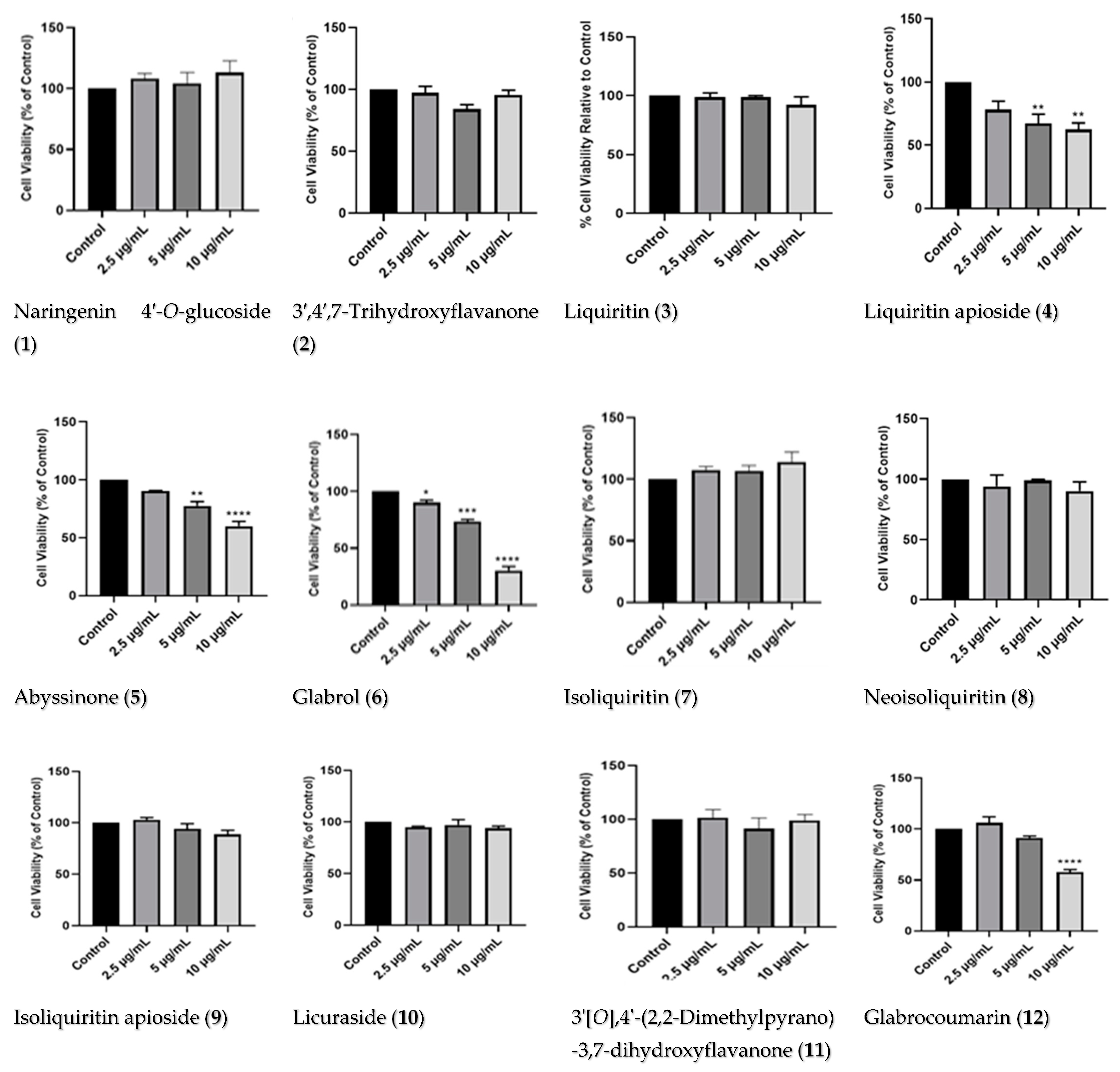
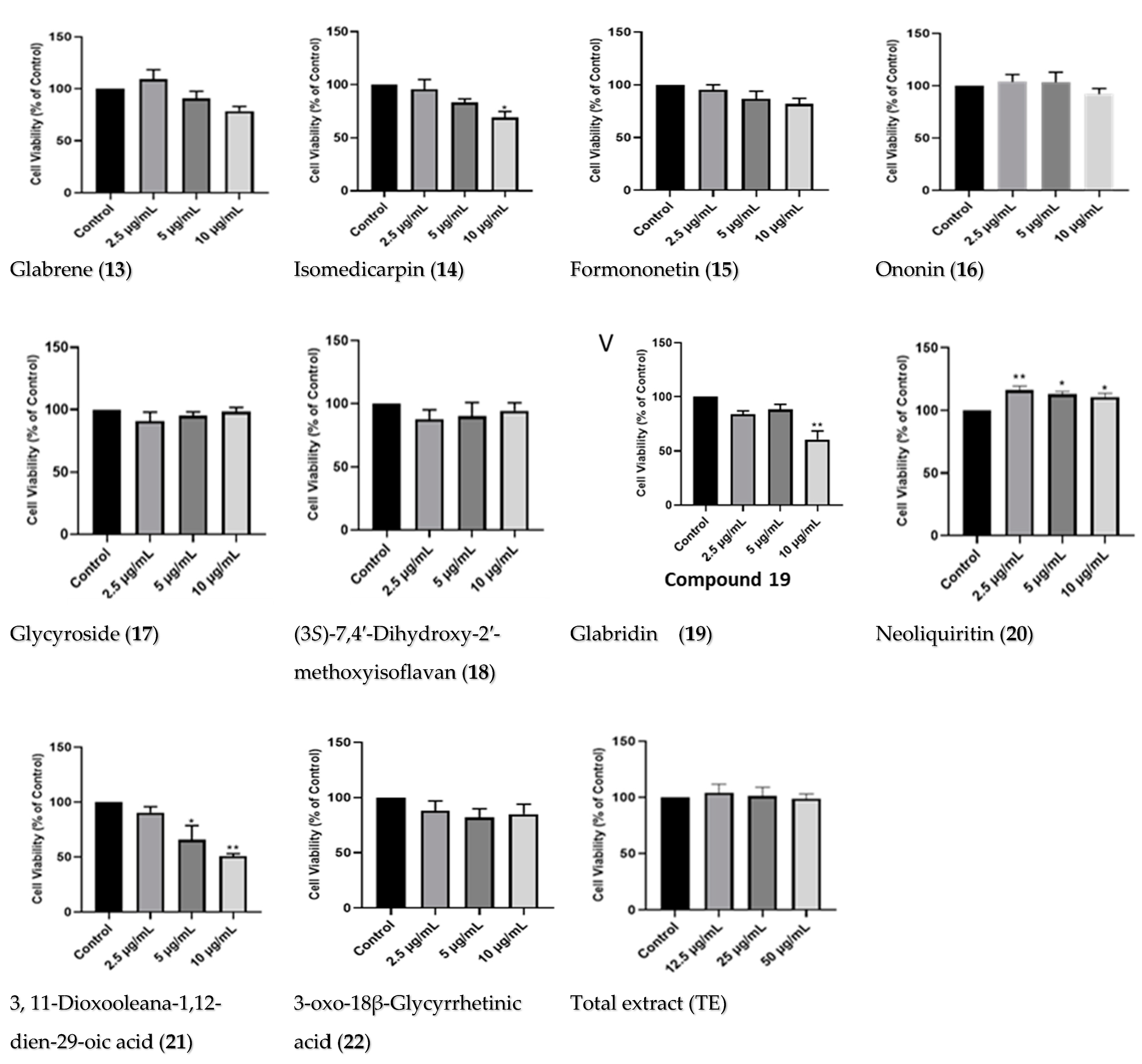
2.2.1. Dose–Response of Licorice TE and Compounds

To investigate the neuroprotective potentials of the TE and compounds, their effects on the cell viability of the SH-SY5Y were first determined. The total extract and 22 compounds were screened for their cytotoxicity using concentrations of 12.5, 25, and 50 μg/mL for the extract, and 2.5, 5, and 10 μg/mL for the compounds. The results show that the TE and compounds had minimal effects on the cell viability of the SH-SY5Y cells, especially at 2.5 μg/mL; however, with increased concentration, some compounds induced toxicity (Figure 3). In addition, the cells treated with compound 20 showed increased cell viability for all concentrations. In contrast, the cells treated with compound 6 showed significant reductions in cell viability at all concentrations tested. Following these results, the TE and five compounds, 1, 7, 11, 16 and 20, were selected to be investigated for their neuroprotective potentials, as they were not cytotoxic, and they improved cell viability the most compared to other compounds. In particular, compound 20 significantly increased cell viability at all concentrations tested.

2.2.2. Effects of Licorice TE and Compounds on MPP+-Induced Toxicity on SH-SY5Y Cells

To investigate the neuroprotective activities of the TE and compounds, their effects on cell viability in the presence of the MPP+ neurotoxin were determined using the MTT cell viability assay. The results show that pretreatment with the TE and compounds resulted in significant improvements in cell viability of MPP+-treated cells, compared to cells treated with MPP alone (Figure 4). While MPP+ reduced cell viability to about 40% when compared to the control cells, pretreatment with the TE increased cell viability to 63, 69, and 73% for 12.5, 25, and 50 μg/mL, respectively (Figure 4A). Although all tested concentrations showed improvement in cell viability, the increase was more in 2.5 μg/mL concentrations for most of the compounds, and in 50 μg/mL for the TE. Considering this, the concentrations of 50 and 2.5 μg/mL were chosen for the TE and compounds, respectively, for further studies. Together, these results show that licorice TE and compounds confer neuroprotection on the SH-SY5Y cells.

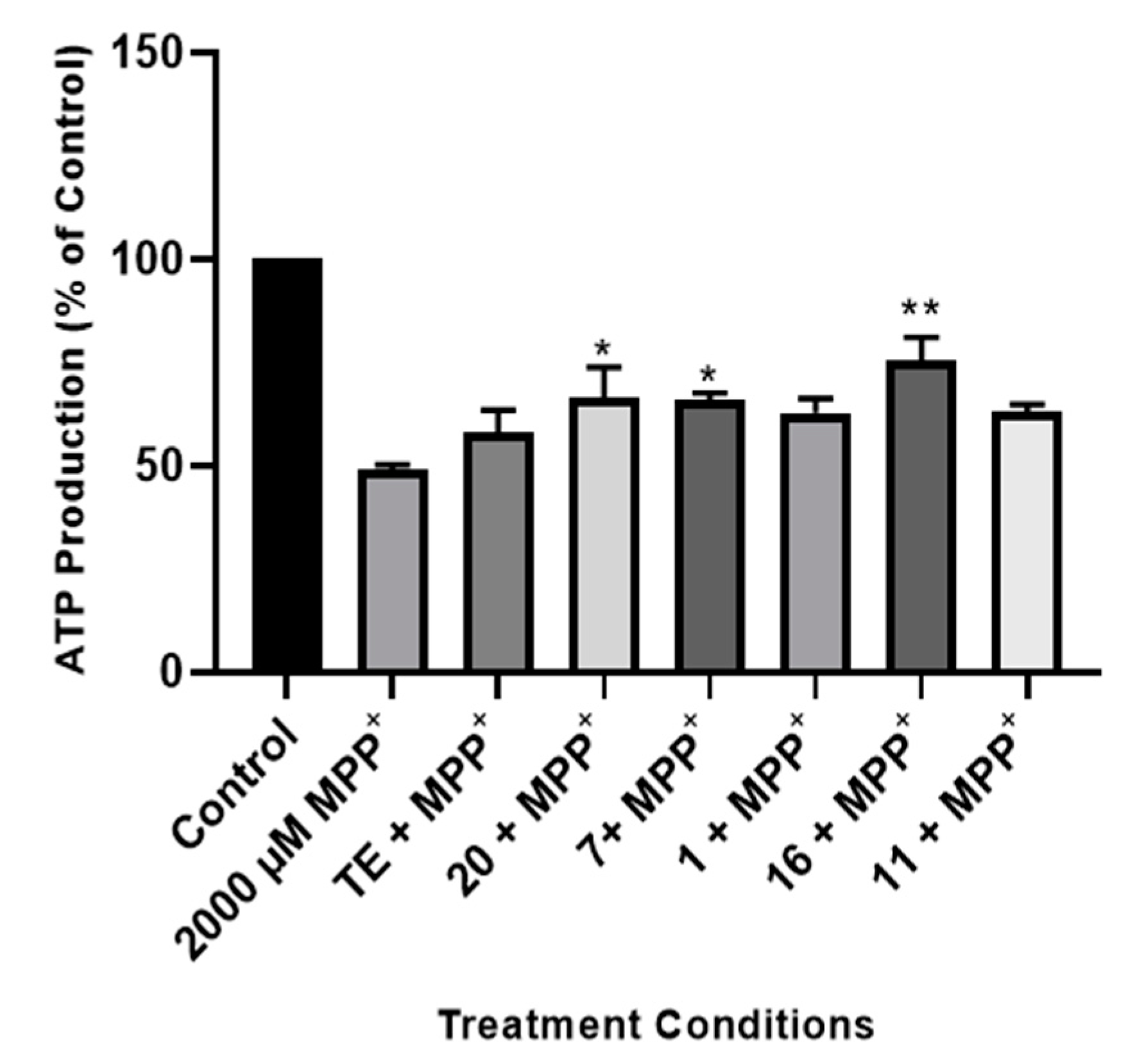
2.2.3. Effect of TE and Compounds on ATP Production in the Cells

As a mechanism of neuronal toxicity, MPP+ induces ATP degeneration in neuronal cells by the inhibition of mitochondrial complex I [52]. To understand the mechanism of action of the licorice TE and compounds 1, 7, 11, 16, and 20, we next investigated their effects on the levels of ATP in the cells following treatment with the MPP+ neurotoxin. Cells were plated in white-walled 96-well plates and treated with 50 μg/mL of the TE and 2.5 μg/mL of the compounds, as these concentrations showed better neuroprotective activity; ATP production was then measured using the MitotoxGlo Promega ATP kit. Figure 5 shows that MPP+ treatment reduced ATP production in the cells to about 49%. However, pretreatment with the licorice TE and compounds improved ATP production in the cells undergoing MPP+ treatment, while this was only significant for the cells pretreated with compounds 20, 7 and 16, with ATP levels of about 66, 65, and 75%, respectively. These results suggest that improving cellular ATP production is part of the mechanism of neuroprotection conferred by the extract and compounds.

2.2.4. Effect of TE and Compounds on Caspase 3/7 Activities in the Cells

To further ascertain the mechanism involved in the neuroprotective aspects of licorice TE and compounds in SH-SY5Y cells, the levels of apoptosis were assessed using caspase 3/7 as a marker. Caspases belong to the family of cysteine proteases, which drive apoptosis in cells and carry out their function by cleavage of substrates; thus, they are used as markers of apoptosis [24,53]. To investigate apoptosis, cells were pretreated with 50 µg/mL of the TE and 2.5 µg/mL of the compounds before exposure to MPP+, and caspase 3/7 activities were measured. Figure 6 shows that TE and compounds mitigate MPP+-increased levels of caspase 3/7 activity in the SH-SY5Y cells. Specifically, MPP+ increased levels of caspase 3/7 to about 3-fold of control and pre-treatment with TE reduced this activity to about 1.8-fold of control. Furthermore, all compounds also protected SH-Y5Y cells from MPP+-induced increase in caspase 3/7 activities. Altogether, these results indicate that inhibition of apoptosis is involved in the neuroprotective aspects of licorice TE and compounds.

3. Discussion

The burden of PD continues to pose a challenge to the ageing population globally [54]. While the exact cause of the disease has yet to be fully elucidated, it has been proposed that PD is caused by a plethora of environmental events and genetic factors [55]. In clinical settings, PD is incurable, as it involves different clinical manifestations, including bradykinesia, resting tremor, and rigidity, along with postural instability; hence, medications are given to relieve symptoms [56]. One notable example of such medication is levodopa, but its prolonged use is associated with neuronal cell toxicity and other serious side effects, such as dyskinesias and psychosis [54]. Delaying the onset of PD is thought to effectively slow down the progression of the disease, so exploiting new approaches to achieve this remains critical.
In the present study, the neuroprotective effects of licorice TE and compounds were evaluated in an MPP+ model of PD. Twenty-two compounds were isolated from the plant, with compound 11 being a new compound. The compounds were screened for their cytotoxicity, and some of them were observed to be cytotoxic to the SH-SY5Y cells. These compounds include glabrol (6), abyssinone (5) [35], liquiritin apioside (4), glabrocoumarin (12), isomedicarpin (14), and 3,11-dioxooleana-1,12-die-29-oic acid (21). In support of these findings, liquiritin apioside has been reported to induce toxicity on human cancer cell lines (HSC-2, HSC-3, HSC-4, and HL-60) which are derived from oral squamous cancers of the tongue [57], while glabrol showed cytotoxicity against C6 rat glioma cells [58]. In addition, 18β-glycyrrhetinic acid and formononetin have been reported to have cytotoxic potential against various cancer cell lines [59,60,61,62,63,64]. Despite the cytotoxicity observed for the compounds mentioned above, other compounds from the Glycyrrhiza genus have been demonstrated to have neuroprotective effects [20,21,22,23]. To this end, our findings show that the compounds naringenin 4′-O-glucoside (1), neoliquiritin (20), glabrol (6), isoliquiritin (7), and 3′[O],4′-(2,2-dimethylpyrano)-3,7-dihydroxyflavanone (11), alongside the TE, when investigated for their neuroprotective potentials, conferred neuroprotection on the cells. Supporting our findings, studies have demonstrated that neoliquiritin (20) protected rat cardiac myocytes from doxorubicin-induced reduction in cell proliferation [65], while naringenin, which is the parent compound from which neoliquiritin (20) and naringenin 4′-O-glucoside (1) were derived, is widely known to have cytoprotective potentials, including protecting neurons from toxin-mediated cell death in Alzheimer’s disease and PD [66,67,68]. Isoliquiritin, on the other hand, has also shown to have neuroprotective potentials, as it inhibits monoamine oxidase and ameliorates depression [69,70], while 7-hydroxy-4′-methoxyflavone (formononetin) (15) was able to alleviate neuroinflammation in lipopolysaccharide-stimulated microglial cells by inhibiting TLR4/MyD88/MAPK signaling and activating the Nrf2/NQO-1 pathway [71].
As a mechanism of action, MPP+ is known to alter cellular energy levels in neuronal cells following the induction of oxidative stress [72]. Oxidative stress leads to impairment of the mitochondrial electron transport chain and, consequently, a decline in ATP production, as well as increased mitochondrial dysfunction [73,74]. Alteration of cellular ATP negatively impacts the ability of cells to carry out their normal metabolic functions, and in relation to humans, this could lead to cognitive decline, as neurotransmission will be affected [75]. Importantly, studies have shown that the ability to ameliorate oxidative stress and improve cellular energy and metabolism counteracts the effects of neurotoxins and, at the same time, improves neurotransmission and neuron function [75,76]. In line with this postulate, we show that the tested compounds and TE improved cellular ATP levels following exposure to MPP+. In support of these findings, it has been suggested that isoliquiritin provides protective action against corticosterone-induced cell damage by reducing oxidative stress and regulating mitochondrial dysfunction by preventing dissipation of mitochondrial membrane potential [77].
Consequently, neuronal cells proceed to programmed cell death following a decline in ATP, and this contributes to the reduction of dopaminergic neuronal cells in the substantia nigra pars compacta of the midbrain [72,78]. Thus, slowing or preventing neurons with respect to cell death via several mechanisms holds a future in the management of neurodegenerative diseases like PD. In the present study, our findings show that G. glabra TE and compounds 20, 7, 1, 16, and 11 inhibited elevated caspase 3/7 activities induced by MPP+. While this was the case for our study, previous studies have demonstrated that prunin (20) exerts its cytoprotective potential by inhibiting apoptosis triggered by cellular toxins in cardiac cells [65], In addition, naringenin showed hepatoprotective and neuroprotective properties against lead-induced oxidative stress, inflammation, and apoptosis in rats [68]. Moreover, isoliquiritin was also demonstrated to attenuate apoptosis by the inhibition of the overload of intracellular calcium Ca2+ ions; down-regulation of Bax, caspase 3, and cytochrome protein expression; and up-regulation of Bcl protein expression [77]. Thus, the findings are sufficient to state that prevention of cellular apoptosis is a critical component of the neuroprotective mechanism of G. glabra and compounds.

4. Materials and Methods

4.1. Chemicals, Materials, and Reagents

Root powder of G. glabra was purchased from the local market in Khartoum, Sudan in May 2020; the specimen (H614) was identified by Prof Hatil Elkamali (Department of Botany, Omdurman Islamic University, Omdurman, Sudan). Organic solvents, including methanol, acetonitrile (HPLC grade, Merck, Cape Town, South Africa), hexane, dichloromethane, ethanol, and ethyl acetate (analytical grade (AR), obtained from a local merchant (Kimix, Cape Town, South Africa)) were acquired. Silica gel 60 (0.063–0.200 mm), Sephadex (LH-20), and Aluminum TLC plate, silica gel PF254 were supplied by Merck (Cape Town, South Africa). The 1D NMR (1H, 13C and DEPT-135) and 2D spectra were measured using a Bruker spectrometer (Rheinstetten, Germany) operating at 400 MHz (for 1H) and 100 MHz (for 13C).

4.2. Method

Extraction and Purification of Compounds

The root powder (0.5 Kg) was extracted with methanol at 60 °C (3 L × 2 h × 2 time). After concentration, it yielded ~104.3 g. A quantity of 50.0 g of the total extract (TE) was applied to the silica gel column and eluted using a gradient of hexane and ethyl acetate with increasing polarity. Collected fractions were pooled together according to their profiles on the TLC to afford 24 major fractions coded as I to XXIV. The fractions were re-chromatographed to yield twenty-three major compounds as follows: Fraction XXIII (3.50 g) underwent chromatography on silica gel using a hexane: EtOAc gradient (80:20 to 0:100). Subsequently, sub-fraction 10 was subjected to purification on Sephadex with an isocratic 80% aqueous ethanol, followed by semi-preparative HPLC (Shimadzu, Kyoto, Japan) utilizing a MeOH and de-ionized water (DIW) gradient (40:60 to 60:80 in 30 min, then to 80:90 in 10 min, and finally to 100% MeOH in 10 min). This process yielded compounds 1 (4.8 mg), 3 (100.8 mg), 7 (45.6 mg), 8 (80.3 mg), and 16 (5.2 mg).
The main fraction, XXIV (4.30 g), was chromatographed on silica gel, and subsequent subfractions 2 and 3 were individually subjected to chromatography on Sephadex and semi-prep HPLC, as described earlier. This resulted in the isolation of compounds 4 (50.3 mg), 9 (39.8 mg), 10 (20 mg), 17 (15.6 mg), and 20 (5.3 mg).
Fraction XV (2.40 g) underwent chromatography on silica gel. Subfraction 16 was subsequently chromatographed on Sephadex and subjected to semi-prep HPLC as outlined before, resulting in the isolation of compound 2 (10.5 mg).
Fraction X (1.40 g) was chromatographed on silica gel using a hexane:EtOAc gradient (80:20 to 0:100), and sub-fraction 7 underwent purification on Sephadex and semi-prep HPLC with a MeOH and DIW mixture (1:1, isocratic) to yield compound 14 (5.8 mg).
Fraction IX (7.30 g) was chromatographed on silica gel using a hexane:EtOAc gradient (80:20 to 0:100), and sub-fraction 6 underwent purification on Sephadex and semi-prep HPLC with a MeOH and DIW mixture (1:1, isocratic) to yield compound 19 (300.8 mg).
Fraction VIII (5.5 g) underwent chromatography on silica gel. Subfractions 4, 5, 8, and 11 were subsequently chromatographed separately on Sephadex and semi-prep HPLC, following the previously mentioned protocol. This led to the isolation of compound 13 (8.4 mg) from sub-fraction 4, the compounds 6 (5.4 mg), 11 (10.8 mg), 12 (5.3 mg), and 18 (9.8 mg) from sub-fraction 5; compounds 5 (10.2 mg) and 15 (5.8 mg) from sub-fraction 8; and compounds 21 (4.4 mg) and 22 (25.2 mg) from sub-fraction 11.

4.3. Compound 11

Whitish yellow powder α 25 D –18.5 (c 0.01, MeOH); UV (MeOH) λmax 315, 277; FTIR (film) 3378, 1677, 1605, 1500, and 1463 cm−1; 1H NMR (DMSO-d6, 400 MHz), and 13C NMR (DMSO-d6, 100 MHz), see Table 1; C20H18O5 by its HRFABMS (positive mode). HRFABMS m/z 339.1232 [M+H]+ (calcd for C20H19O5, 339.1227).

4.4. Cell Culture and Treatments

The human neuroblastoma SH-SY5Y cells were generously donated by the Blackburn Laboratory, University of Cape Town. Cells were grown in Dulbecco’s Modified Eagle’s medium (DMEM, Gibco, Life Technologies Corporation, Paisley, UK), supplemented with 10% fetal bovine serum (FBS, Gibco, Life Technologies Corporation, Paisley, UK), 100 U/mL penicillin, and 100 µg/mL streptomycin (Lonza Group Ltd., Verviers, Belgium). Cultures were incubated at 37 °C in humidified air with 5% CO2, and cell growth medium was routinely changed every three days. Cells were sub-cultured when they attained 70 to 80 percent confluency, using a solution of 0.25% trypsin EDTA (Lonza Group Ltd., Verviers, Belgium).

4.5. Treatments

Stock solutions of 40 mg/mL and 10 mg/mL for total extract and compounds, respectively, were prepared in dimethyl sulfoxide (DMSO) (Sigma-Aldrich, St. Louis, MO, USA), from which final concentrations were made in cell growth media. To determine the optimum concentrations of licorice TE and compounds to be used for neuroprotection studies, SH-SY5Y cells were plated at a density of 10,000 cells/well and treated with concentrations of 12.5, 25, and 50 µg/mL of the TE, and 2.5, 5, and 10 µg/mL of the compounds. The vehicle-treated cells (cells treated with the same concentration of DMSO, similar to that of the highest concentration of extract) were used as control, and the treatments lasted 24 h. For MPP+, 2000 mM was chosen as the concentration used to establish neurotoxicity, which is in accordance with our published works [27,53]. For neuroprotection experiments, cells were plated as above and pre-treated with optimized concentrations of licorice TE and compounds for 2 h prior to the addition of 2000 µM MPP+. Treatments were incubated for 24 h, and the vehicle-treated cells served as control.

4.6. Cell Viability Assays

The MTT (Sigma-Aldrich, St. Louis, MO, USA) cell viability assay was used to determine the viability of cells following treatment with licorice TE- and compounds-only and the pre-treatment of cells with licorice TE and/or compounds and MPP+. Cells were seeded in 96-well plates and treated as stated above, after which the MTT assay was performed. After treatment, 10 or 20 µL (depending on well volume) of the 5 mg/mL MTT solution in phosphate-buffered saline (PBS) (Lonza Group Ltd., Verviers, Belgium) was added to each well and left to incubate in the dark at 37 °C for 4 h. After incubation, the medium containing the MTT dye was discarded, and the MTT formazan was solubilized with 100 μL of DMSO for an absorbance reading using a microplate reader (BMG Labtech Omega® POLARStar, Offenburg, Germany) at a wavelength of 570 nm. Cell viability was calculated and expressed as a percentage of control.

4.7. Adenosine Triphosphate Assay

The Mitochondrial ToxGlo ATP assay kit (Promega, Madison, WI, USA) was used to investigate cell ATP levels. Briefly, cells were plated at a density of 10,000 cells per well in a white 96-well plate, and after attachment, cells were treated as per the neuroprotection assay above. After treatment, cells were processed according to the manufacturer’s protocol by adding a volume of ATP detection reagent appropriate to the contents of each well and leaving the resulting mixture to incubate at 37 °C for 30 min. After incubation, luminescence intensity was read using the Promega GloMax® explorer multimode microplate reader, and luminescence values were expressed as percentages of control.

4.8. Caspase 3/7 Apoptosis Assay

To investigate apoptosis in the cells, the Caspase 3/7 assay kit (Promega, Madison, WI, USA) was used to estimate levels of caspase 3/7 activity in the cells, in a manner according with the manufacturer’s instructions. Briefly, cells were plated in a white 96-well plate at a density of 10,000 cells per well and allowed to attach overnight, after which cells were pre-treated with licorice TE and compounds before the addition of 2000 µM MPP+. Treatments lasted for 24 h, and at the end of the experiments, equal volumes of Caspase 3/7 assay mix were added to each well. The luminescence intensity was read with the Promega GloMax → explorer multimode microplate reader. Luminescence values of the treated cells were expressed as a multiple of the control.

4.9. Statistical Analysis

Data generated from this study were analyzed using GraphPad Prism 6 and expressed as means and standard error values of means of three independent experiments performed in quadruplicate wells. One-way analysis of variance (ANOVA) was used to compare treated cells to either control or MPP+ alone. The significance of difference p < 0.05 was determined using Tukey’s multiple comparisons test and was indicated with * when comparing treatments to control or Φ when comparing licorice and TE and compounds to MPP+ treated cells.

5. Conclusions

This study investigated the neuroprotective potentials of G. glabra TE and isolated compounds in an in vitro model of PD. The results show that some of the compounds were cytotoxic (including glabrol (6), abyssinone (5), liquiritin apioside (4), glabrocoumarin (12), isomedicarpin (14), and 3,11-dioxooleana-1,12-die-29-oic acid (21)), which might have implications for the treatment of cancer. However, some of the compounds (naringenin 4′-O-glucoside (1), isoliquiritin (7), 3′[O],4′-(2,2-dimethylpyrano)-3,7-dihydroxyflavanone (11), ononin (16), and neoliquiritin (20)) were further studied for their neuroprotective potentials. The protective effects were related not only to the inhibition of ATP degeneration, but also to the attenuation of MPP+-induced elevated caspase 3/7 activities. These results indicate that the compounds isolated from G. glabra may provide novel therapeutic strategies for the treatment of PD. One limitation of this study was that the biological characterization was only performed on cell lines; therefore, in the future, it would be interesting to see if these compounds would have similar effects on in vivo models of PD. In addition, we recommend that future studies further elucidate the mechanisms of action, with emphasis on the molecular and genetic events underpinning the actions of these compounds.

Supplementary Materials

The following supporting information can be downloaded at: https://www.mdpi.com/article/10.3390/ph17070852/s1, Table S1. 1H and 13C spectral data of compounds 1-22, Figure S1. 1H and 13C spectra of compounds 1-22 and Scheme S1. The details of the isolation processes for compounds 122 from licorice.

Author Contributions

A.O.E.E., conceptualization; A.A.H., project researcher and supervisor; R.C.L., co-supervisor; S.I.O., investigation, formal analysis, and data curation; T.N.A., funding, investigation, reviewing of draft. All authors have read and agreed to the published version of the manuscript.

Funding

This work was supported by the National Research Foundation, South Africa (grant number 106055). CPUT and WITS are acknowledged for financial and infrastructural support. TNA acknowledges the Friedel Sellschop Fellowship Award and NRF CSUR funding.

Institutional Review Board Statement

Ethics waiver for this study was obtained from the Wits Human Research Ethics Committee with approval number W-CBP-221024-01.

Informed Consent Statement

Not applicable.

Data Availability Statement

The data associated with the isolated compounds, 1H NMR, 13C NMR spectra, and the biological data used to support the findings of this study are available from the corresponding author upon request.

Conflicts of Interest

The authors declare no conflicts of interest.

References

  1. Lamptey, R.N.L.; Chaulagain, B.; Trivedi, R.; Gothwal, A.; Layek, B.; Singh, J. A Review of the Common Neurodegenerative Disorders: Current Therapeutic Approaches and the Potential Role of Nanotherapeutics. Int. J. Mol. Sci. 2022, 23, 1851. [Google Scholar] [CrossRef] [PubMed]
  2. Kalia, L.V.; Lang, A.E. Parkinson’s disease. Lancet Neurol. 2015, 29, 896–912. [Google Scholar] [CrossRef] [PubMed]
  3. Postuma, R.B.; Berg, D.; Stern, M.; Poewe, W.; Olanow, C.W.; Oertel, W.; Obeso, J.; Marek, K.; Litvan, I.; Lang, A.E.; et al. MDS clinical diagnostic criteria for Parkinson’s disease. Mov. Disord. 2015, 30, 1591–1601. [Google Scholar] [CrossRef]
  4. Li, J.-L.; Lin, T.-Y.; Chen, P.-L.; Guo, T.-N.; Huang, S.-Y.; Chen, C.-H.; Lin, C.-H.; Chan, C.-C. Mitochondrial Function and Parkinson’s Disease: From the Perspective of the Electron Transport Chain. Front. Mol. Neurosci. 2021, 14, 797833. [Google Scholar] [CrossRef]
  5. Keane, P.; Kurzawa, M.; Blain, P.; Morris, C. Mitochondrial dysfunction in Parkinson’s disease. Park. Dis. 2011, 2011, 716871. [Google Scholar] [CrossRef]
  6. Nonnekes, J.; Post, B.; Tetrud, J.W.; Langston, J.W.; Bloem, B.R. MPTP-induced parkinsonism: An historical case series. Lancet Neurol. 2018, 17, 300–301. [Google Scholar]
  7. Reeve, A.; Simcox, E.; Turnbull, D. Ageing and Parkinson’s disease: Why is advancing age the biggest risk factor. Ageing Res. Rev. 2014, 14, 19–30. [Google Scholar] [CrossRef] [PubMed]
  8. Thanvi, B.; Lo, N.; Robinson, T. Levodopa-induced dyskinesia in Parkinson’s disease: Clinical features, pathogenesis, prevention and treatment. Postgrad. Med. J. 2007, 83, 384–388. [Google Scholar] [CrossRef]
  9. Brahmachari, G. Discovery and Development of Neuroprotective Agents from Natural Products; Elsevier: Amsterdam, The Netherlands, 2017. [Google Scholar]
  10. Abdolmaleki, A.; Akram, M.; Saeed, M.M.; Asadi, A.; Kajkolah, M. Herbal medicine as neuroprotective potential agent in human and animal models: A historical overview. J. Pharm. Care 2020, 8, 75–82. [Google Scholar] [CrossRef]
  11. Bosch-Morell, F.; Villagrasa, V.; Ortega, T.; Acero, N.; Muñoz-Mingarro, D.; González-Rosende, M.E.; Castillo, E.; Sanahuja, M.A.; Soriano, P.; Martínez-Solís, I.P. Medicinal plants and natural products as neuroprotective agents in age-related macular degeneration. Neural Regen. Res. 2020, 15, 2207–2216. [Google Scholar]
  12. Öztürk, M.; Altay, V.; Hakeem, K.; Akçiçek, E. Liquorice: From Botany to Phytochemistry; Springer: Berlin/Heidelberg, Germany, 2018. [Google Scholar]
  13. Hayashi, H.; Hosono, N.; Kondo, M.; Hiraoka, N.; Ikeshiro, Y.; Shibano, M.; Kusano, G.; Yamamoto, H.; Tanaka, T.; Inoue, K. Phylogenetic relationship of six Glycyrrhiza species based on rbcL sequences and chemical constituents. Biol. Pharm. Bull. 2000, 23, 602–606. [Google Scholar] [CrossRef] [PubMed]
  14. Hayashi, H.; Miwa, E.; Inoue, K. Phylogenetic relationship of Glycyrrhiza lepidota, American licorice, in genus Glycyrrhiza based on rbcL sequences and chemical constituents. Biol. Pharm. Bull. 2005, 28, 161–164. [Google Scholar] [CrossRef] [PubMed]
  15. Avula, B.; Bae, J.-Y.; Chittiboyina, A.G.; Wang, Y.-H.; Wang, M.; Zhao, J.; Ali, Z.; Brinckmann, J.A.; Li, J.; Wu, C.; et al. Chemometric analysis and chemical characterization for the botanical identification of Glycyrrhiza species (G. glabra, G. uralensis, G. inflata, G. echinata and G. lepidota) using liquid chromatography-quadrupole time of flight mass spectrometry (LC-QTof). J. Food Compos. Anal. 2022, 112, 104679. [Google Scholar] [CrossRef]
  16. Akhtar, N.; Ihsan-ul-Haq; Mirza, B. Phytochemical analysis and comprehensive evaluation of antimicrobial and antioxidant properties of 61 medicinal plant species. Arab. J. Chem. 2018, 11, 1223–1235. [Google Scholar] [CrossRef]
  17. Sharma, V.; Katiyar, A.; Agrawal, R.C. Glycyrrhiza Glabra: Chemistry and Pharmacological Activity. In Sweeteners; Mérillon, J., Ramawat, K.G., Eds.; Springer: Cham, The Netherlands, 2018; pp. 87–100. [Google Scholar]
  18. Jiang, M.; Zhao, S.; Yang, S.; Lin, X.; He, X.; Wei, X.; Song, Q.; Li, R.; Fu, C.; Zhang, J.; et al. An “essential herbal medicine” Licorice: A review of phytochemicals and its effects in combination preparations. J. Ethnopharmacol. 2020, 249, 112439. [Google Scholar] [CrossRef] [PubMed]
  19. Hosseini, M.S.; Ebrahimi, M.; Samsampour, D.; Abadía, J.; Khanahmadi, M.; Amirian, R.; Ghafoori, I.N.; Ghaderi-Zefrehei, M.; Gogorcena, Y. Association analysis and molecular tagging of phytochemicals in the endangered medicinal plant licorice (Glycyrrhiza glabra L.). Phytochemistry 2021, 183, 112629. [Google Scholar] [CrossRef]
  20. Chang, K.-H.; Chen, I.-C.; Lin, H.-Y.; Chen, H.-C.; Lin, C.-H.; Lin, T.-H.; Weng, Y.-T.; Chao, C.-Y.; Wu, Y.-R.; Lin, J.-Y.; et al. The aqueous extract of Glycyrrhiza inflata can upregulate unfolded protein response-mediated chaperones to reduce tau misfolding in cell models of Alzheimer’s disease. Drug Des. Dev. Ther. 2016, 10, 885–896. [Google Scholar]
  21. Chen, C.-M.; Weng, Y.-T.; Chen, W.-L.; Lin, T.-H.; Chao, C.-Y.; Lin, C.-H.; Chen, I.-C.; Lee, L.-C.; Lin, H.-Y.; Wu, Y.-R.; et al. Aqueous extract of Glycyrrhiza inflata inhibits aggregation by upregulating PPARGC1A and NFE2L2–ARE pathways in cell models of spinocerebellar ataxia 3. Free Radic. Biol. Med. 2014, 71, 339–350. [Google Scholar] [CrossRef] [PubMed]
  22. Kong, Z.-H.; Chen, X.; Hua, H.-P.; Liang, L.; Liu, L.-J. The oral pretreatment of glycyrrhizin prevents surgery-induced cognitive impairment in aged mice by reducing neuroinflammation and Alzheimer’s-related pathology via HMGB1 inhibition. J. Mol. Neurosci. 2017, 63, 385–395. [Google Scholar] [CrossRef]
  23. Ravanfar, P.; Namazi, G.; Atigh, M.; Zafarmand, S.; Hamedi, A.; Salehi, A.; Izadi, S.; Borhani-Haghighi, A. Efficacy of whole extract of licorice in neurological improvement of patients after acute ischemic stroke. J. Herb. Med. 2016, 6, 12–17. [Google Scholar] [CrossRef]
  24. Hasan, M.K.; Ara, I.; Mondal, M.S.A.; Kabir, Y. Phytochemistry, pharmacological activity, and potential health benefits of Glycyrrhiza glabra. Heliyon 2021, 7, 1–10. [Google Scholar] [CrossRef] [PubMed]
  25. Yi, Y.; Zhang, M.; Xue, H.; Yu, R.; Bao, Y.-O.; Kuang, Y.; Chai, Y.; Ma, W.; Wang, J.; Shi, X.; et al. Schaftoside inhibits 3CLpro and PLpro of SARS-CoV-2 virus and regulates immune response and inflammation of host cells for the treatment of COVID-19. Acta Pharm. Sin. B 2022, 12, 4154–4164. [Google Scholar] [CrossRef] [PubMed]
  26. Xiang, C.; Qiao, X.; Ye, M.; Guo, D. Classification and distribution analysis of components in Glycyrrhiza using licorice compounds database. Acta Pharm. Sin. 2012, 47, 1023–1030. [Google Scholar]
  27. Omoruyi, S.I.; Ibrakaw, A.S.; Ekpo, O.E.; Boatwright, J.S.; Cupido, C.N.; Hussein, A.A. Neuroprotective activities of crossyne flava bulbs and amaryllidaceae alkaloids: Implications for parkinson’s disease. Molecules 2021, 26, 3990. [Google Scholar] [CrossRef] [PubMed]
  28. Egunlusi, A.O.; Malan, S.F.; Omoruyi, S.I.; Ekpo, O.E.; Palchykov, V.A.; Joubert, J. Open and rearranged norbornane derived polycyclic cage molecules as potential neuroprotective agents through attenuation of MPP+-and calcium overload-induced excitotoxicity in neuroblastoma SH-SY5Y cells. Eur. J. Med. Chem. 2020, 204, 112617. [Google Scholar] [CrossRef] [PubMed]
  29. Oyama, K.-i.; Kondo, T. Total synthesis of apigenin 7, 4’-di-O-β-glucopyranoside, a component of blue flower pigment of Salvia patens, and seven chiral analogues. Tetrahedron 2004, 60, 2025–2034. [Google Scholar] [CrossRef]
  30. Chen, K.; Hu, Z.-m.; Song, W.; Wang, Z.-l.; He, J.-b.; Shi, X.-m.; Cui, Q.-h.; Qiao, X.; Ye, M. Diversity of O-glycosyltransferases contributes to the biosynthesis of flavonoid and triterpenoid glycosides in Glycyrrhiza uralensis. Am. Chem. Soc. Synth. Biol. 2019, 8, 1858–1866. [Google Scholar] [CrossRef]
  31. Wu, J.-Y.; Ding, H.-Y.; Wang, T.-Y.; Cai, C.-Z.; Chang, T.-S. Application of Biotransformation-Guided Purification in Chinese Medicine: An Example to Produce Butin from Licorice. Catalysts 2022, 12, 1–10. [Google Scholar] [CrossRef]
  32. Tian, G.; Zhang, U.; Zhang, T.; Yang, F.; Ito, Y. Separation of flavonoids from the seeds of Vernonia anthelmintica Willd by high-speed counter-current chromatography. J. Chromatogr. A 2004, 1049, 219–222. [Google Scholar] [CrossRef]
  33. Ji, S.; Li, Z.; Song, W.; Wang, Y.; Liang, W.; Li, K.; Tang, S.; Wang, Q.; Qiao, X.; Zhou, D.; et al. Bioactive Constituents of Glycyrrhiza uralensis (Licorice): Discovery of the Effective Components of a Traditional Herbal Medicine. J. Nat. Prod. 2016, 79, 281–292. [Google Scholar] [CrossRef]
  34. Pastorino, G.; Cornara, L.; Soares, S.; Rodrigues, F.; Oliveira, M.B.P. Liquorice (Glycyrrhiza glabra): A phytochemical and pharmacological review. Phytother. Res. 2018, 32, 2323–2339. [Google Scholar] [CrossRef] [PubMed]
  35. Park, J.-H.; Wu, Q.; Yoo, K.-H.; Yong, H.-I.; Cho, S.-M.; Chung, I.-S.; Baek, N.-I. Cytotoxic effect of flavonoids from the roots of Glycyrrhiza uralensis on human cancer cell lines. J. Appl. Biol. Chem. 2011, 54, 67–70. [Google Scholar] [CrossRef]
  36. Asada, Y.; Li, W.; Yoshikawa, T. Biosynthesis of the dimethylallyl moiety of glabrol in Glycyrrhiza glabra hairy root cultures via a non-mevalonate pathway. Phytochemistry 2000, 55, 323–326. [Google Scholar] [CrossRef] [PubMed]
  37. Nomura, T.; Fukai, T.; Akiyama, T. Chemistry of phenolic compounds of licorice (Glycyrrhiza species) and their estrogenic and cytotoxic activities. Pure Appl. Chem. 2002, 74, 1199–1206. [Google Scholar] [CrossRef]
  38. Lee, J.E.; Lee, J.Y.; Kim, J.; Lee, K.; Choi, S.U.; Ryu, S.Y. Two minor chalcone acetylglycosides from the roots extract of Glycyrrhiza uralensis. Arch. Pharmacal Res. 2015, 38, 1299–1303. [Google Scholar] [CrossRef]
  39. Wang, D.; Liang, J.; Zhang, J.; Wang, Y.; Chai, X. Natural chalcones in Chinese materia medica: Licorice. Evid.-Based Complement. Altern. Med. 2020, 2020, 3821248. [Google Scholar] [CrossRef] [PubMed]
  40. Kaur, P.; Kaur, S.; Kumar, N.; Singh, B.; Kumar, S. Evaluation of antigenotoxic activity of isoliquiritin apioside from Glycyrrhiza glabra L. Toxicol. Vitr. 2009, 23, 680–686. [Google Scholar] [CrossRef] [PubMed]
  41. Fu, B.; Li, H.; Wang, X.; Lee, F.S.C.; Cui, S. Isolation and Identification of Flavonoids in Licorice and a Study of Their Inhibitory Effects on Tyrosinase. J. Agric. Food Chem. 2005, 53, 7408–7414. [Google Scholar] [CrossRef] [PubMed]
  42. Li, G.; Simmler, C.; Chen, L.; Nikolic, D.; Chen, S.-N.; Pauli, G.F.; van Breemen, R.B. Cytochrome P450 inhibition by three licorice species and fourteen licorice constituents. Eur. J. Pharm. Sci. 2017, 109, 182–190. [Google Scholar] [CrossRef]
  43. Montoro, P.; Maldini, M.; Russo, M.; Postorino, S.; Piacente, S.; Pizza, C. Metabolic profiling of roots of liquorice (Glycyrrhiza glabra) from different geographical areas by ESI/MS/MS and determination of major metabolites by LC-ESI/MS and LC-ESI/MS/MS. J. Pharm. Biomed. Anal. 2011, 54, 535–544. [Google Scholar] [CrossRef]
  44. Kinoshit, T.; Tamur, Y.; Mizutani, K. The Isolation and Structure Elucidation of Minor Isoflavonoids from Licorice of Glycyrrhiza glabra Origin. Chem. Pharm. Bull. 2005, 53, 847–849. [Google Scholar] [CrossRef]
  45. Fukai, T.; Sheng, C.-B.; Horikoshi, T.; Nomura, T. Isoprenylated flavonoids from underground parts of Glycyrrhiza glabra. Phytochemistry 1996, 43, 1119–1124. [Google Scholar] [CrossRef]
  46. Adesanya, S.; O’Neill, M.J.; Roberts, M.F. Structure-related fungitoxicity of isoflavonoids. Physiol. Mol. Plant Pathol. 1986, 29, 95–103. [Google Scholar] [CrossRef]
  47. Piccinelli, A.L.; Fernandez, M.C.; Cuesta-Rubio, O.; Hernández, I.M.; Simone, F.D.; Rastrelli, L. Isoflavonoids Isolated from Cuban Propolis. J. Agric. Food Chem. 2005, 53, 9010–9016. [Google Scholar] [CrossRef]
  48. Liaoa, W.C.; Linb, Y.-H.; Changc, T.-M.; Huang, W.-Y. Identification of two licorice species, Glycyrrhiza uralensis and Glycyrrhiza glabra, based on separation and identification of their bioactive components. Food Chem. 2012, 132, 2188–2193. [Google Scholar] [CrossRef]
  49. Chintharlapalli, S.; Papineni, S.; Jutooru, I.; McAlees, A.; Safe, S. Structure-dependent activity of glycyrrhetinic acid derivatives as peroxisome proliferator–activated receptor γ agonists in colon cancer cells. Mol. Cancer Ther. 2007, 6, 1588–1598. [Google Scholar] [CrossRef] [PubMed]
  50. Baltina, L.A.; Budaev, A.S.; Mikhailova, L.R.; Baltina, J.L.A.; Spirikhin, L.V.; Makara, N.S.; Zarudii, F.S. New stereoisomeric glycyrrhetic acid derivatives. Chem. Nat. Compd. 2014, 50, 1042–1046. [Google Scholar] [CrossRef]
  51. Kuroda, M.; Mimakia, Y.; Honda, S.; Tanaka, H.; Yokota, S.; Mae, T. Phenolics from Glycyrrhiza glabra roots and their PPAR-c ligand-binding activity. Bioorganic Med. Chem. 2010, 18, 962–970. [Google Scholar] [CrossRef]
  52. Hoglinger, G.U.; Carrard, G.; Michel, P.P.; Medja, F.; Lombès, A.; Ruberg, M.; Friguet, B.; Hirsch, E.C. Dysfunction of mitochondrial complex I and the proteasome:interactions between two biochemical deficits in a cellular modelof Parkinson’s disease. J. Neurochem. 2003, 86, 1297–1307. [Google Scholar] [CrossRef]
  53. Omoruyi, S.I.; Akinfenwa, A.O.; Ekpo, O.E.; Hussein, A.A. Aspalathin and linearthin from Aspalathus linearis (Rooibos) protect SH-SY5Y cells from MPP+-induced neuronal toxicity. South Afr. J. Bot. 2023, 157, 53–63. [Google Scholar] [CrossRef]
  54. Kouli, A.; Torsney, K.M.; Kuan, W.-L. Parkinson’s disease: Etiology, neuropathology, and pathogenesis. Exon Publ. 2018, 3–26. [Google Scholar] [CrossRef]
  55. Pang, S.Y.-Y.; Ho, P.W.-L.; Liu, H.-F.; Leung, C.-T.; Li, L.; Chang, E.E.S.; Ramsden, D.B.; Ho, S.-L. The interplay of aging, genetics and environmental factors in the pathogenesis of Parkinson’s disease. Transl. Neurodegener. 2019, 8, 23. [Google Scholar] [CrossRef]
  56. Massano, J.; Bhatia, K. Clinical approach to Parkinson’s disease: Features, diagnosis, and principles of management. Cold Spring Harb. Perspect. Med. 2012, 2, a008870. [Google Scholar] [CrossRef] [PubMed]
  57. HOhno; Araho, D.; Uesawa, Y.; Kagaya, H.; Ishihara, M.; Sakagami, H.; Yamamoto, M. Evaluation of Cytotoxiciy and Tumor-specificity of Licorice Flavonoids Based on Chemical Structure. Anticancer Res. 2013, 33, 3061–3068. [Google Scholar]
  58. Goel, B.; Sharma, A.; Tripathi, N.; Bhardwaj, N.; Sahu, B.; Kaur, G.; Singh, B.; Jain, S.K. In-vitro antitumor activity of compounds from Glycyrrhiza glabra against C6 glioma cancer cells: Identification of natural lead for further evaluation. Nat. Prod. Res. 2021, 35, 5489–5492. [Google Scholar] [CrossRef] [PubMed]
  59. Lee, C.S.; Kim, Y.J.; Lee, M.S.; Han, E.S.; Lee, S.J. 18β-Glycyrrhetinic acid induces apoptotic cell death in SiHa cells and exhibits a synergistic effect against antibiotic anti-cancer drug toxicity. Life Sci. 2008, 83, 481–489. [Google Scholar] [CrossRef] [PubMed]
  60. Kowalska, A.; Kalinowska-Lis, U. 18β-Glycyrrhetinic acid: Its core biological properties and dermatological applications. Int. J. Cosmet. Sci. 2019, 41, 325–331. [Google Scholar] [CrossRef] [PubMed]
  61. Tay, K.-C.; Tan, L.T.-H.; Chan, C.K.; Hong, S.L.; Chan, K.-G.; Yap, W.H.; Pusparajah, P.; Lee, L.-H.; Goh, B.-H. Formononetin: A Review of Its Anticancer Potentials and Mechanisms. Front. Pharmacol. 2019, 10, 820. [Google Scholar] [CrossRef] [PubMed]
  62. Oh, J.-S.; Kim, T.-H.; Park, J.-H.; Lim, H.; Cho, I.-A.; You, J.-S.; Lee, G.-J.; Seo, Y.-S.; Kim, D.K.; Kim, C.S.; et al. Formononetin induces apoptotic cell death through the suppression of mitogen activated protein kinase and nuclear factor κB phosphorylation in FaDu human head and neck squamous cell carcinoma cells. Oncol. Rep. 2020, 43, 700–710. [Google Scholar] [CrossRef]
  63. Jiang, D.; Rasul, A.; Batool, R.; Sarfraz, I.; Hussain, G.; Tahir, M.M.; Qin, Z.S.T.; Ali, M.; Li, J.; Li, X. Potential Anticancer Properties and Mechanisms of Action of Formononetin. BioMed Res. Int. 2019, 2019, 854315. [Google Scholar] [CrossRef]
  64. Hsu, Y.-C.; Hsieh, W.-C.; Chen, S.-H.; Li, Y.-Z.; Liao, H.-F.; Lin, M.-Y.; Sheu, S.-M. 18β-glycyrrhetinic Acid Modulated Autophagy is Cytotoxic to Breast Cancer Cells. Int. J. Med. Sci. 2023, 20, 444–454. [Google Scholar] [CrossRef] [PubMed]
  65. Han, X.; Ren, D.; Fan, P.; Shen, T.; Lou, H. Protective effects of naringenin-7-O-glucoside on doxorubicin-induced apoptosis in H9C2 cells. Eur. J. Pharmacol. 2008, 581, 47–53. [Google Scholar] [CrossRef] [PubMed]
  66. Sugumar, M.; Sevanan, M.; Sekar, S. Neuroprotective effect of naringenin against MPTP-induced oxidative stress. Int. J. Neurosci. 2019, 129, 534–539. [Google Scholar] [CrossRef] [PubMed]
  67. Hassan, H.M.; Elnagar, M.R.; Abdelrazik, E.; Mahdi, M.R.; Hamza, E.; Elattar, E.M.; ElNashar, E.M.; Alghamdi, M.A.; Al-Qahtani, Z.; Al-Khater, K.M.; et al. Neuroprotective effect of naringin against cerebellar changes in Alzheimer’s disease through modulation of autophagy, oxidative stress and tau expression: An experimental study. Front. Neuroanat. 2022, 16, 1012422. [Google Scholar] [CrossRef] [PubMed]
  68. Mansour, L.; Elshopakey, G.; Abdelhamid, F.; Albukhari, T.; Almehmadi, S.; Refaat, B.; El-Boshy, M.; Risha, E. Hepatoprotective and Neuroprotective Effects of Naringenin against Lead-Induced Oxidative Stress, Inflammation, and Apoptosis in Rats. Biomedicines 2023, 11, 1080. [Google Scholar] [CrossRef] [PubMed]
  69. Li, Y.; Song, W.; Tong, Y.; Zhang, X.; Zhao, J.; Gao, X.; Yong, J.; Wang, H. Isoliquiritin ameliorates depression by suppressing NLRP3-mediated pyroptosis via miRNA-27a/SYK/NF-κB axis. J. Neuroinflammation 2021, 18, 1. [Google Scholar] [CrossRef] [PubMed]
  70. Prajapati, R.; Seong, S.H.; Park, S.E.; Paudel, P.; Jung, H.A.; Choi, J.S. Isoliquiritigenin, a potent human monoamine oxidase inhibitor, modulates dopamine D1, D3, and vasopressin V1A receptors. Sci. Rep. 2021, 11, 23528. [Google Scholar] [CrossRef] [PubMed]
  71. Qu, Z.; Chen, Y.; Luo, Z.-H.; Shen, X.-L.; Hu, Y.-J. 7-methoxyflavanone alleviates neuroinflammation in lipopolysaccharide-stimulated microglial cells by inhibiting TLR4/MyD88/MAPK signalling and activating the Nrf2/NQO-1 pathway. J. Pharm. Pharmacol. 2020, 72, 385–395. [Google Scholar] [CrossRef]
  72. Pöltl, D.; Schildknecht, S.; Karreman, C.; Leist, M. Uncoupling of ATP-depletion and cell death in human dopaminergic neurons. NeuroToxicology 2012, 33, 769–779. [Google Scholar] [CrossRef]
  73. Babu, V.; Khurana, N. A review on mitochondrial dysfunction and oxidative stress due to complex-i in Parkinson disease. Res. J. Pharmacol. Pharmacodyn. 2021, 13, 167–170. [Google Scholar] [CrossRef]
  74. Dorszewska, J.D.P.; Kowalska, M.; Prendecki, M.; Piekut, T.; Kozłowska, J.; Kozubski, W. Oxidative stress factors in Parkinson’s disease. Neural Regen. Res. 2021, 16, 1383–1391. [Google Scholar] [CrossRef] [PubMed]
  75. Błaszczyk, J.W. Energy metabolism decline in the aging brain—Pathogenesis of neurodegenerative disorders. Metabolites 2020, 10, 450. [Google Scholar] [CrossRef] [PubMed]
  76. Jiménez-Delgado, A.; Ortiz, G.G.; Delgado-Lara, D.L.; González-Usigli, H.A.; González-Ortiz, L.J.; Cid-Hernández, M.; Cruz-Serrano, J.A.; Pacheco-Moisés, F.P. Effect of Melatonin Administration on Mitochondrial Activity and Oxidative Stress Markers in Patients with Parkinson’s Disease. Oxidative Med. Cell. Longev. 2021, 2021, 1–7. [Google Scholar] [CrossRef] [PubMed]
  77. Zhou, Y.-z.; Li, X.; Gong, W.-x.; Tian, J.-s.; Gao, X.-x.; Gao, L.; Zhang, X.; Du, G.-h.; Qin, X.-m. Protective effect of isoliquiritin against corticosterone-induced neurotoxicity in PC12 cells. Food Funct. 2017, 8, 1235–1244. [Google Scholar] [CrossRef]
  78. Abramov, A.; Angelova, P.R. Mitochondrial dysfunction and energy deprivation in the mechanism of neurodegeneration. Turk. J. Biochem. 2019, 44, 723–729. [Google Scholar] [CrossRef]
Figure 1. Chemical structures of the compounds isolated from G. glabra.
Pharmaceuticals 17 00852 g001
Figure 2. Chemical structure and important HMBC correlations of 11, and the structure of 3,4′,7-trihydroxy-3′-prenylflavanone.
Pharmaceuticals 17 00852 g002
Figure 3. Dose–response of licorice TE and compounds. MTT cytotoxicity assay on SH-SY5Y cells treated with increasing concentrations (12.5, 25, and 50 µg/mL) of licorice TE and compounds (2.5, 5, and 10 µg/mL) for 24 h. After assays, cell viability was expressed as a percentage of control, and each bar represents the mean + SEM of at least three replicate experiments obtained from quadruple wells. Treated cells were compared to control cells; significance levels are indicated. The significance of the difference when control cells were compared to treated cells is indicated by * p< 0.05, ** p < 0.01, *** p < 0.001, and **** p < 0.0001.
Pharmaceuticals 17 00852 g003aPharmaceuticals 17 00852 g003b
Figure 4. Licorice TE and compounds show protection in SH-SY5Y cells. Cells were pre-treated with TE (A) and compounds (BF) before exposure to MPP+ for 24 h. Each bar represents mean percentage cell viability relative to control, and significance of difference is indicated with * p < 0.05, ** p < 0.01, *** p < 0.001, and **** p < 0.0001 when TE/compounds are compared to MPP+, and Φ when MPP+ -treated cells are compared to control.
Pharmaceuticals 17 00852 g004
Figure 5. Effect of licorice TE and compounds on MPP+-induced depletion of ATP. Cells were pre-treated with 50 µg/mL of licorice TE and 2.5 µg/mL of compounds before exposure to 2000 µM of MPP+ for 24 h, and ATP levels were assessed. Each bar represents the mean percentage of ATP production relative to control, and the significance of the difference is indicated as * p < 0.05 and ** p < 0.01 when extract/compounds are compared to MPP+ and when MPP+-treated cells are compared to control.
Pharmaceuticals 17 00852 g005
Figure 6. Licorice TE and compounds attenuate MPP+-induced increase in caspase 3/7 activities. Cells were pre-treated with 50 µg/mL of licorice TE and 2.5 µg/mL of compounds before exposure to 2000 µM of MPP+ for 24 h, and caspase 3/7 activities were determined. Each bar represents the level of caspase 3/7 expressed as a multiple of the control, and significance of difference is indicated by ** p < 0.01 and *** p < 0.001 when TE/compounds were compared to MPP+, and Φ when MPP+-treated cells were compared to control.
Pharmaceuticals 17 00852 g006
Table 1. NMR data of compound 11 (DMSO-d6) and 3,4′,7-trihydroxy-3′-prenylflavanone (acetone-d6).
PositionCompound 113,4′,7-Trihydroxy-3′-prenylflavanone [51]
δCδH, multi, J (Hz)δCδH, multi, J (Hz)
283.7 4.92 (d, 11.8)84.65.03 (d, 11.9)
372.9 4.50 (d, 11.8)73.44.59 (d, 11.9)
4192.7 -192.7-
5128.6 7.72 (d, 8.6)129.37.74 (d, 8.6)
6111.5 6.51 (dd, 8.6, 2.0)111.26.64 (dd, 8.6, 2.2)
7162.7 -165.4-
8102.5 6.30 (d, 2.0)103.16.41 (d, 2.2)
9163.2 -164.1-
10111.9 -112.5-
1′129.6 -128.9-
2′126.2 7.23 (br s)130.07.35 (d, 2.0)
3′120.7 s-128.1-
4′152.6 -155.8-
5′115.5 6.76 (d, 7.9)115.06.90 (d, 8.2)
6′129.1 d7.25 (br d, 7.9)127.17.27 (dd, 8.2, 2.2)
1″121.6 6.43 (d, 9.7)28.73.38 (2H, d, 7.3)
2″131.3 5.58 (d, 9.7)123.15.39 (m)
3″76.3 -132.0-
4″27.7 1.37 (s)25.41.72 (br s)
5″27.7 1.37 (s)17.41.74 (br s)
multi: multiplicity, J values in Hz, s: singlet, br s: broad singlet, br d: broad doublet, d: doublet, dd: doublet of doublet, m: multiple.
Disclaimer/Publisher’s Note: The statements, opinions and data contained in all publications are solely those of the individual author(s) and contributor(s) and not of MDPI and/or the editor(s). MDPI and/or the editor(s) disclaim responsibility for any injury to people or property resulting from any ideas, methods, instructions or products referred to in the content.

