


Hydrogel Loaded with Extracellular Vesicles: An Emerging Strategy for Wound Healing


 ,
, 

Abstract
:1. Introduction
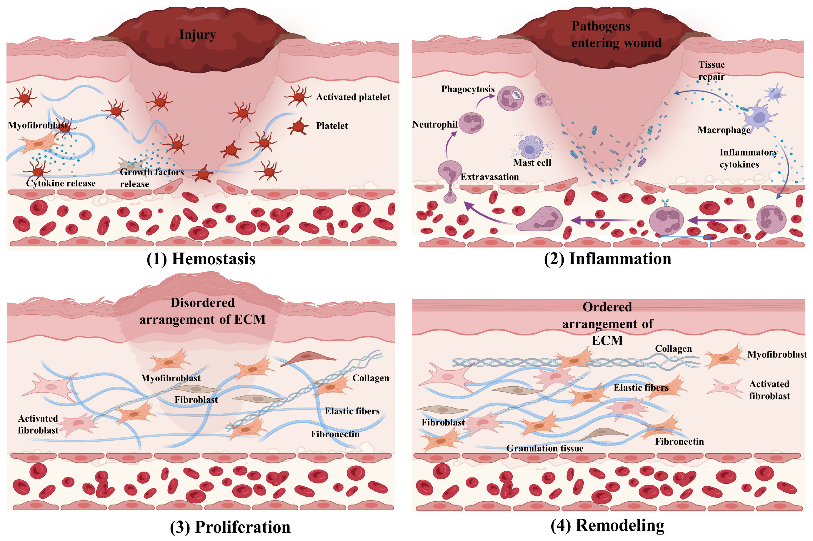
2. Wound Healing
3. Extracellular Vesicles
4. Hydrogels
5. EV-Loaded Hydrogels for Wound Healing Therapies
5.1. Stem Cell-Derived EV-Loaded Hydrogels
5.2. Platelet-Derived EV-Loaded Hydrogels
5.3. Human Umbilical Vein Endothelial Cell-Derived EV-Loaded Hydrogels
5.4. Other Cell-Derived EV-Loaded Hydrogels
5.5. New Strategies for EV-Loaded Hydrogels
6. Limitations and Challenges
7. Conclusions and Prospects
Funding
Conflicts of Interest
Abbreviations
| Abbreviation | Full Name |
| EVs | extracellular vesicles |
| ECM | extracellular matrix |
| MMPs | matrix metalloproteinases |
| VEGF | vascular endothelial growth factor |
| GelMA | gelatin methacryloyl |
| HA | hyaluronic acid |
| F127 | pluronic F-127 |
| hUC-MSCs | human umbilical cord-derived mesenchymal stem cells |
| BMSCs | bone marrow mesenchymal stem cells |
| ADSCs | adipose-derived stem cells |
| HSP90 | heat shock protein 90 |
| α-SMA | α-smooth muscle actin |
| iPSC-MSCs | induced pluripotent stem cells–mesenchymal stem cells |
| PEVs | platelet-derived EVs |
| RGO | graphene oxide |
| ROS | oxygen species |
| ZWP | zedoary turmeric homogeneous polysaccharide |
| TNF-α | tumor necrosis factor-α |
| iNOS | inducible nitric oxide synthase |
| TGF-β | transforming growth factor-β |
| Arg-1 | arginase-1 |
| A2A-R | adenosine receptor 2A |
| HUVEC-EVs | human umbilical vein endothelial cell-derived EVs |
| IL | interleukin |
| NRF2 | nuclear transcription factor 2 |
| ATF3 | transcriptional activator 3 |
| NVs | nanovesicles |
References
- Kabashima, K.; Honda, T.; Ginhoux, F.; Egawa, G. The immunological anatomy of the skin. Nat. Rev. Immunol. 2019, 19, 19–30. [Google Scholar] [CrossRef] [PubMed]
- Lindley, L.E.; Stojadinovic, O.; Pastar, I.; Tomic-Canic, M. Biology and Biomarkers for Wound Healing. Plast. Reconstr. Surg. 2016, 138, 18S–28S. [Google Scholar] [CrossRef] [PubMed]
- Holl, J.; Kowalewski, C.; Zimek, Z.; Fiedor, P.; Kaminski, A.; Oldak, T.; Moniuszko, M.; Eljaszewicz, A. Chronic diabetic wounds and their treatment with skin substitutes. Cells 2021, 10, 655. [Google Scholar] [CrossRef] [PubMed]
- Wilkinson, H.N.; Hardman, M.J. Wound healing: Cellular mechanisms and pathological outcomes. Open Biol. 2020, 10, 200223. [Google Scholar] [CrossRef] [PubMed]
- Rodrigues, M.; Kosaric, N.; Bonham, C.A.; Gurtner, G.C. Wound healing: A cellular perspective. Physiol. Rev. 2019, 99, 665–706. [Google Scholar] [CrossRef] [PubMed]
- Zhao, R.; Liang, H.; Clarke, E.; Jackson, C.; Xue, M. Inflammation in chronic wounds. Int. J. Mol. Sci. 2016, 17, 2085. [Google Scholar] [CrossRef] [PubMed]
- Velnar, T.; Bailey, T.; Smrkolj, V. The wound healing process: An overview of the cellular and molecular mechanisms. J. Int. Med. Res. 2009, 37, 1528–1542. [Google Scholar] [CrossRef] [PubMed]
- Eming, S.A.; Murray, P.J.; Pearce, E.J. Metabolic orchestration of the wound healing response. Cell Metab. 2021, 33, 1726–1743. [Google Scholar] [CrossRef] [PubMed]
- Wynn, T.A.; Vannella, K.M. Macrophages in tissue repair, regeneration, and fibrosis. Immunity 2016, 44, 450–462. [Google Scholar] [CrossRef]
- Talbott, H.E.; Mascharak, S.; Griffin, M.; Wan, D.C.; Longaker, M.T. Wound healing, fibroblast heterogeneity, and fibrosis. Cell Stem Cell 2022, 29, 1161–1180. [Google Scholar] [CrossRef]
- Trimm, E.; Red-Horse, K. Vascular endothelial cell development and diversity. Nat. Rev. Cardiol. 2023, 20, 197–210. [Google Scholar] [CrossRef] [PubMed]
- Oh, E.J.; Gangadaran, P.; Rajendran, R.L.; Kim, H.M.; Oh, J.M.; Choi, K.Y.; Chung, H.Y.; Ahn, B.C. Extracellular vesicles derived from fibroblasts promote wound healing by optimizing fibroblast and endothelial cellular functions. Stem Cells 2021, 39, 266–279. [Google Scholar] [CrossRef]
- Wang, X.; Khalil, R.A. Matrix Metalloproteinases, Vascular Remodeling, and Vascular Disease. Adv. Pharmacol. 2018, 81, 241–330. [Google Scholar] [CrossRef]
- Rousselle, P.; Braye, F.; Dayan, G. Re-epithelialization of adult skin wounds: Cellular mechanisms and therapeutic strategies. Adv. Drug Deliv. Rev. 2019, 146, 344–365. [Google Scholar] [CrossRef] [PubMed]
- Demir, B.; Broughton, R.M.; Qiao, M.Y.; Huang, T.S.; Worley, S.D. N-Halamine biocidal materials with superior antimicrobial efficacies for wound dressings. Molecules 2017, 22, 1582. [Google Scholar] [CrossRef] [PubMed]
- Ibrahim, M.A.; Nasr, G.M.; Ahmed, R.M.; Kelany, N.A. Physical characterization, biocompatibility, and antimicrobial activity of polyvinyl alcohol/sodium alginate blend doped with TiO2 nanoparticles for wound dressing applications. Sci. Rep. 2024, 14, 5391. [Google Scholar] [CrossRef]
- Erdoğmuş, S.F.; Altintaş, Ö.E.; Demirel, H.H.; Okumuş, N. Fabrication of wound dressings: Herbal extract-loaded nanoliposomes embedded in fungal chitosan/polycaprolactone electrospun nanofibers for tissue regeneration. Microsc. Res. Tech. 2023, 87, 360–372. [Google Scholar] [CrossRef] [PubMed]
- Hsu, L.C.; Peng, B.Y.; Chen, M.S.; Thalib, B.; Ruslin, M.; Tung, T.D.X.; Chou, H.H.; Ou, K.L. The potential of the stem cells composite hydrogel wound dressings for promoting wound healing and skin regeneration: In vitro and in vivo evaluation. J. Biomed. Mater. Res. Part B Appl. Biomater. 2019, 107, 278–285. [Google Scholar] [CrossRef]
- Li, D.; Hu, N.; Yu, Y.; Zhou, A.; Li, F.; Jia, J. Trajectories of Multidimensional Caregiver Burden in Chinese Informal Caregivers for Dementia: Evidence from Exploratory and Confirmatory Factor Analysis of the Zarit Burden Interview. J. Alzheimer’s Dis. 2017, 59, 1317–1325. [Google Scholar] [CrossRef]
- Alizadeh, R.; Zarrintaj, P.; Kamrava, S.K.; Bagher, Z.; Farhadi, M.; Heidari, F.; Komeili, A.; Gutiérrez, T.J.; Saeb, M.R. Conductive hydrogels based on agarose/alginate/chitosan for neural disorder therapy. Carbohydr. Polym. 2019, 224, 115161. [Google Scholar] [CrossRef]
- Alizadeh, R.; Bagher, Z.; Kamrava, S.K.; Falah, M.; Ghasemi Hamidabadi, H.; Eskandarian Boroujeni, M.; Mohammadi, F.; Khodaverdi, S.; Zare-Sadeghi, A.; Olya, A.; et al. Differentiation of human mesenchymal stem cells (MSC) to dopaminergic neurons: A comparison between Wharton’s Jelly and olfactory mucosa as sources of MSCs. J. Chem. Neuroanat. 2019, 96, 126–133. [Google Scholar] [CrossRef] [PubMed]
- Nqakala, Z.B.; Sibuyi, N.R.S.; Fadaka, A.O.; Meyer, M.; Onani, M.O.; Madiehe, A.M. Advances in Nanotechnology towards Development of Silver Nanoparticle-Based Wound-Healing Agents. Int. J. Mol. Sci. 2021, 22, 11272. [Google Scholar] [CrossRef] [PubMed]
- Yao, S.; Chi, J.J.; Wang, Y.T.; Zhao, Y.; Luo, Y.; Wang, Y. Zn-MOF Encapsulated Antibacterial and Degradable Microneedles Array for Promoting Wound Healing. Adv. Healthc. Mater. 2021, 10, e2100056. [Google Scholar] [CrossRef]
- Ha, D.H.; Kim, H.K.; Lee, J.; Kwon, H.H.; Park, G.H.; Yang, S.H.; Jung, J.Y.; Choi, H.; Lee, J.H.; Sung, S.; et al. Mesenchymal Stem/Stromal Cell-Derived Exosomes for Immunomodulatory Therapeutics and Skin Regeneration. Cells 2020, 9, 1157. [Google Scholar] [CrossRef] [PubMed]
- Andaloussi, S.E.L.; Mäger, I.; Breakefield, X.O.; Wood, M.J. Extracellular vesicles: Biology and emerging therapeutic opportunities. Nat. Rev. Drug Discov. 2013, 12, 347–357. [Google Scholar] [CrossRef]
- Jeppesen, D.K.; Zhang, Q.; Franklin, J.L.; Coffey, R.J. Extracellular vesicles and nanoparticles: Emerging complexities. Trends Cell Biol. 2023, 33, 667–681. [Google Scholar] [CrossRef]
- Valadi, H.; Ekström, K.; Bossios, A.; Sjöstrand, M.; Lee, J.J.; Lötvall, J.O. Exosome-mediated transfer of mRNAs and microRNAs is a novel mechanism of genetic exchange between cells. Nat. Cell Biol. 2007, 9, 654–659. [Google Scholar] [CrossRef]
- Wan, Y.; Wang, L.; Zhu, C.; Zheng, Q.; Wang, G.; Tong, J.; Fang, Y.; Xia, Y.; Cheng, G.; He, X.; et al. Aptamer-Conjugated Extracellular Nanovesicles for Targeted Drug Delivery. Cancer Res. 2018, 78, 798–808. [Google Scholar] [CrossRef]
- Tenchov, R.; Sasso, J.M.; Wang, X.; Liaw, W.S.; Chen, C.A.; Zhou, Q.A. Exosomes horizontal line Nature’s Lipid Nanoparticles, a Rising Star in Drug Delivery and Diagnostics. ACS Nano 2022, 16, 17802–17846. [Google Scholar] [CrossRef]
- Liu, Z.; Zhuang, Y.; Fang, L.; Yuan, C.; Wang, X.; Lin, K. Breakthrough of extracellular vesicles in pathogenesis, diagnosis and treatment of osteoarthritis. Bioact. Mater. 2023, 22, 423–452. [Google Scholar] [CrossRef]
- Wang, J.; Wu, H.; Peng, Y.X.; Zhao, Y.; Qin, Y.; Zhang, Y.; Xiao, Z. Hypoxia adipose stem cell-derived exosomes promote high-quality healing of diabetic wound involves activation of PI3K/Akt pathways. J. Nanobiotechnol. 2021, 19, 202. [Google Scholar] [CrossRef] [PubMed]
- Ti, D.; Hao, H.; Tong, C.; Liu, J.; Dong, L.; Zheng, J.; Zhao, Y.; Liu, H.; Fu, X.; Han, W. LPS-preconditioned mesenchymal stromal cells modify macrophage polarization for resolution of chronic inflammation via exosome-shuttled let-7b. J. Transl. Med. 2015, 13, 308. [Google Scholar] [CrossRef] [PubMed]
- Antich-Rosselló, M.; Forteza-Genestra, M.A.; Monjo, M.; Ramis, J.M. Platelet-Derived Extracellular Vesicles for Regenerative Medicine. Int. J. Mol. Sci. 2021, 22, 8580. [Google Scholar] [CrossRef] [PubMed]
- Herrmann, I.K.; Wood, M.J.A.; Fuhrmann, G. Extracellular vesicles as a next-generation drug delivery platform. Nat. Nanotechnol. 2021, 16, 748–759. [Google Scholar] [CrossRef] [PubMed]
- Wang, C.; Wang, M.; Xu, T.; Zhang, X.; Lin, C.; Gao, W.; Xu, H.; Lei, B.; Mao, C. Engineering Bioactive Self-Healing Antibacterial Exosomes Hydrogel for Promoting Chronic Diabetic Wound Healing and Complete Skin Regeneration. Theranostics 2019, 9, 65–76. [Google Scholar] [CrossRef] [PubMed]
- Son, Y.J.; Tse, J.W.; Zhou, Y.; Mao, W.; Yim, E.K.F.; Yoo, H.S. Biomaterials and controlled release strategy for epithelial wound healing. Biomater. Sci. 2019, 7, 4444–4471. [Google Scholar] [CrossRef] [PubMed]
- Zhang, L.; Wang, Y.; Yang, M.R.; Yu, W.; Zhao, Z.; Liu, Y. An Injectable, Self-Healing, Adhesive Multifunctional Hydrogel Promotes Bacteria-Infected Wound Healing. Polymers 2024, 16, 1316. [Google Scholar] [CrossRef] [PubMed]
- Kwak, G.; Cheng, J.; Kim, H.; Song, S.; Lee, S.J.; Yang, Y.; Jeong, J.H.; Lee, J.E.; Messersmith, P.B.; Kim, S.H. Sustained Exosome-Guided Macrophage Polarization Using Hydrolytically Degradable PEG Hydrogels for Cutaneous Wound Healing: Identification of Key Proteins and MiRNAs, and Sustained Release Formulation. Small 2022, 18, e2200060. [Google Scholar] [CrossRef] [PubMed]
- Yuan, N.; Shao, K.; Huang, S.; Chen, C. Chitosan, alginate, hyaluronic acid and other novel multifunctional hydrogel dressings for wound healing: A review. Int. J. Biol. Macromol. 2023, 240, 124321. [Google Scholar] [CrossRef]
- Yu, Y.; Jin, H.; Li, L.; Zhang, X.; Zheng, C.; Gao, X.; Yang, Y.; Sun, B. An injectable, activated neutrophil-derived exosome mimetics/extracellular matrix hybrid hydrogel with antibacterial activity and wound healing promotion effect for diabetic wound therapy. J. Nanobiotechnol. 2023, 21, 308. [Google Scholar] [CrossRef]
- Oliva, N.; Conde, J.; Wang, K.; Artzi, N. Designing Hydrogels for On-Demand Therapy. Acc. Chem. Res. 2017, 50, 669–679. [Google Scholar] [CrossRef]
- Luo, L.Q.; Wu, Z.X.; Ding, Q.L.; Wang, H.; Luo, Y.; Yu, J.; Guo, H.; Tao, K.; Zhang, S.; Huo, F.; et al. In Situ Structural Densification of Hydrogel Network and Its Interface with Electrodes for High-Performance Multimodal Artificial Skin. ACS Nano 2024, 18, 15754–15768. [Google Scholar] [CrossRef]
- Tao, K.; Yu, J.; Zhang, J.; Bao, A.; Hu, H.; Ye, T.; Ding, Q.; Wang, Y.; Lin, H.; Wu, J.; et al. Deep-Learning Enabled Active Biomimetic Multifunctional Hydrogel Electronic Skin. ACS Nano 2023, 17, 16160–16173. [Google Scholar] [CrossRef] [PubMed]
- Kang, D.; Wang, W.; Li, Y.; Ma, Y.; Huang, Y.; Wang, J. Biological Macromolecule Hydrogel Based on Recombinant Type I Collagen/Chitosan Scaffold to Accelerate Full-Thickness Healing of Skin Wounds. Polymers 2023, 15, 3919. [Google Scholar] [CrossRef] [PubMed]
- Xu, L.; Liu, Y.; Tang, L.; Xiao, H.; Yang, Z.; Wang, S. Preparation of Recombinant Human Collagen III Protein Hydrogels with Sustained Release of Extracellular Vesicles for Skin Wound Healing. Int. J. Mol. Sci. 2022, 23, 6289. [Google Scholar] [CrossRef] [PubMed]
- Yue, K.; Trujillo-de Santiago, G.; Alvarez, M.M.; Tamayol, A.; Annabi, N.; Khademhosseini, A. Synthesis, properties, and biomedical applications of gelatin methacryloyl (GelMA) hydrogels. Biomaterials 2015, 73, 254–271. [Google Scholar] [CrossRef]
- Liu, Y.; Chan-Park, M.B. A biomimetic hydrogel based on methacrylated dextran-graft-lysine and gelatin for 3D smooth muscle cell culture. Biomaterials 2010, 31, 1158–1170. [Google Scholar] [CrossRef] [PubMed]
- Henriques-Antunes, H.; Cardoso, R.M.S.; Zonari, A.; Correia, J.; Leal, E.C.; Jimenez-Balsa, A.; Lino, M.M.; Barradas, A.; Kostic, I.; Gomes, C.; et al. The Kinetics of Small Extracellular Vesicle Delivery Impacts Skin Tissue Regeneration. ACS Nano 2019, 13, 8694–8707. [Google Scholar] [CrossRef]
- Kuperkar, K.; Atanase, L.I.; Bahadur, A.; Crivei, I.C.; Bahadur, P. Degradable Polymeric Bio(nano)materials and Their Biomedical Applications: A Comprehensive Overview and Recent Updates. Polymers 2024, 16, 206. [Google Scholar] [CrossRef]
- Cabral, J.; Ryan, A.E.; Griffin, M.D.; Ritter, T. Extracellular vesicles as modulators of wound healing. Adv. Drug Deliv. Rev. 2018, 129, 394–406. [Google Scholar] [CrossRef] [PubMed]
- Yang, S.M.; Huang, S.; Feng, C.J.; Fu, X. Umbilical cord-derived mesenchymal stem cells: Strategies, challenges, and potential for cutaneous regeneration. Front. Med. 2012, 6, 41–47. [Google Scholar] [CrossRef]
- Yang, J.; Chen, Z.; Pan, D.; Li, H.; Shen, J. Umbilical cord-derived mesenchymal stem cell-derived exosomes combined Pluronic F127 hydrogel promote chronic diabetic wound healing and complete skin regeneration. Int. J. Nanomed. 2020, 15, 5911–5926. [Google Scholar] [CrossRef] [PubMed]
- Zhang, B.; Wang, M.; Gong, A.H.; Zhang, X.; Wu, X.; Zhu, Y.; Shi, H.; Wu, L.; Zhu, W.; Qian, H.; et al. HucMSC-Exosome Mediated-Wnt4 Signaling Is Required for Cutaneous Wound Healing. Stem Cells 2015, 33, 2158–2168. [Google Scholar] [CrossRef]
- Han, Z.Z.; Dong, L.L.; Li, A.; Li, Z.; Fu, L.; Zhang, Z.; Li, X.; Li, X. Efficient angiogenesis-based wound healing through hydrogel dressing with extracellular vesicles release. Mater. Today Bio 2022, 16, 100427. [Google Scholar] [CrossRef]
- Wang, Y.X.; Song, P.; Wu, L.N.; Su, Z.; Gui, X.; Gao, C.; Zhao, H.; Wang, Y.; Li, Z.; Cen, Y.; et al. In situ photo-crosslinked adhesive hydrogel loaded with mesenchymal stem cell-derived extracellular vesicles promotes diabetic wound healing. J. Mater. Chem. B 2023, 11, 837–851. [Google Scholar] [CrossRef] [PubMed]
- Geng, X.R.; Qi, Y.; Liu, X.T.; Shi, Y.; Li, H.; Zhao, L. A multifunctional antibacterial and self-healing hydrogel laden with bone marrow mesenchymal stem cell-derived exosomes for accelerating diabetic wound healing. Biomater. Adv. 2022, 133, 112613. [Google Scholar] [CrossRef]
- Shi, Y.; Wang, S.; Wang, K.; Yang, R.; Liu, D.; Liao, H.; Qi, Y.; Qiu, K.; Hu, Y.; Wen, H.; et al. Relieving Macrophage Dysfunction by Inhibiting SREBP2 Activity: A Hypoxic Mesenchymal Stem Cells-Derived Exosomes Loaded Multifunctional Hydrogel for Accelerated Diabetic Wound Healing. Small 2024, 20, e2309276. [Google Scholar] [CrossRef] [PubMed]
- Liang, Y.; He, J.; Guo, B. Functional Hydrogels as Wound Dressing to Enhance Wound Healing. ACS Nano 2021, 15, 12687–12722. [Google Scholar] [CrossRef]
- Xiong, Y.; Chen, L.; Liu, P.; Yu, T.; Lin, C.; Yan, C.; Hu, Y.; Zhou, W.; Sun, Y.; Panayi, A.C.; et al. All-in-One: Multifunctional Hydrogel Accelerates Oxidative Diabetic Wound Healing through Timed-Release of Exosome and Fibroblast Growth Factor. Small 2022, 18, e2104229. [Google Scholar] [CrossRef]
- Beheshtizadeh, N.; Gharibshahian, M.; Bayati, M.; Maleki, R.; Strachan, H.; Doughty, S.; Tayebi, L. Vascular endothelial growth factor (VEGF) delivery approaches in regenerative medicine. Biomed. Pharmacother. 2023, 166, 115301. [Google Scholar] [CrossRef]
- Ren, S.; Chen, J.; Guo, J.; Liu, Y.; Xiong, H.; Jing, B.; Yang, X.; Li, G.; Kang, Y.; Wang, C.; et al. Exosomes from Adipose Stem Cells Promote Diabetic Wound Healing through the eHSP90/LRP1/AKT Axis. Cells 2022, 11, 3229. [Google Scholar] [CrossRef] [PubMed]
- Tang, L.Z.; Zhao, C.R.; Liu, Y.F.; Zhou, J.; Dong, Y.; Huang, J.; Yang, T.; Xiao, H.; Liu, D.; Wang, S.; et al. GelMA Hydrogel Loaded with Extracellular Vesicles Derived from Umbilical Cord Mesenchymal Stem Cells for Promoting Cutaneous Diabetic Wound Healing. ACS Omega 2023, 8, 10030–10039. [Google Scholar] [CrossRef] [PubMed]
- You, D.G.; An, J.Y.; Um, W.; Jung, J.M.; Oh, B.H.; Nguyen, V.Q.; Jeon, J.; Lee, J.; Jo, D.G.; Cho, Y.W.; et al. Stem Cell-Derived Extracellular Vesicle-Bearing Dermal Filler Ameliorates the Dermis Microenvironment by Supporting CD301b-Expressing Macrophages. ACS Nano 2022, 16, 251–260. [Google Scholar] [CrossRef]
- Zhou, Y.; Zhang, X.L.; Lu, S.T.; Zhang, N.Y.; Zhang, H.J.; Zhang, J.; Zhang, J. Human adipose-derived mesenchymal stem cells-derived exosomes encapsulated in pluronic F127 hydrogel promote wound healing and regeneration. Stem Cell Res. Ther. 2022, 13, 407. [Google Scholar] [CrossRef] [PubMed]
- Wang, Y.; Cao, Z.; Wei, Q.; Ma, K.; Hu, W.; Huang, Q.; Su, J.; Li, H.; Zhang, C.; Fu, X. VH298-loaded extracellular vesicles released from gelatin methacryloyl hydrogel facilitate diabetic wound healing by HIF-1α-mediated enhancement of angiogenesis. Acta Biomater. 2022, 147, 342–355. [Google Scholar] [CrossRef] [PubMed]
- Tang, Q.M.; Lu, B.; He, J.; Chen, X.; Fu, Q.; Han, H.; Luo, C.; Yin, H.; Qin, Z.; Lyu, D.; et al. Exosomes-loaded thermosensitive hydrogels for corneal epithelium and stroma regeneration. Biomaterials 2022, 280, 121320. [Google Scholar] [CrossRef] [PubMed]
- Hao, P.C.; Burnouf, T.; Chiang, C.W.; Jheng, P.R.; Szunerits, S.; Yang, J.C.; Chuang, E.Y. Enhanced diabetic wound healing using platelet-derived extracellular vesicles and reduced graphene oxide in polymer-coordinated hydrogels. J. Nanobiotechnol. 2023, 21, 318. [Google Scholar] [CrossRef]
- Xu, N.; Wang, L.L.; Guan, J.; Tang, C.; He, N.; Zhang, W.; Fu, S. Wound healing effects of a Curcuma zedoaria polysaccharide with platelet-rich plasma exosomes assembled on chitosan/silk hydrogel sponge in a diabetic rat model. Int. J. Biol. Macromol. 2018, 117, 102–107. [Google Scholar] [CrossRef]
- Zhu, W.; Dong, Y.; Xu, P.; Pan, Q.; Jia, K.; Jin, P.; Zhou, M.; Xu, Y.; Guo, R.; Cheng, B. A composite hydrogel containing resveratrol-laden nanoparticles and platelet-derived extracellular vesicles promotes wound healing in diabetic mice. Acta Biomater. 2022, 154, 212–230. [Google Scholar] [CrossRef] [PubMed]
- Yi, J.R.; Li, Z.N.; Xie, H.Q.; Chen, B.M.; Jiang, L.; Qian, L.X.; Xu, H.G.; Li, S.R.; Lei, Z.Z.; Chen, J.D.; et al. Effects and mechanism of human umbilical vein endothelial cells-derived exosomes on wound healing in diabetic rabbits. Zhonghua Shao Shang Yu Chuang Mian Xiu Fu Za Zhi 2022, 38, 1023–1033. [Google Scholar] [CrossRef]
- Cheng, P.; Xie, X.; Hu, L.; Zhou, W.; Mi, B.; Xiong, Y.; Xue, H.; Zhang, K.; Zhang, Y.; Hu, Y.; et al. Hypoxia endothelial cells-derived exosomes facilitate diabetic wound healing through improving endothelial cell function and promoting M2 macrophages polarization. Bioact. Mater. 2024, 33, 157–173. [Google Scholar] [CrossRef]
- Chen, Y.; Wu, Y.; Guo, L.; Yuan, S.; Sun, J.; Zhao, K.; Wang, J.; An, R. Exosomal Lnc NEAT1 from endothelial cells promote bone regeneration by regulating macrophage polarization via DDX3X/NLRP3 axis. J. Nanobiotechnol. 2023, 21, 98. [Google Scholar] [CrossRef]
- Yuan, M.; Liu, K.; Jiang, T.; Li, S.; Chen, J.; Wu, Z.; Li, W.; Tan, R.; Wei, W.; Yang, X.; et al. GelMA/PEGDA microneedles patch loaded with HUVECs-derived exosomes and Tazarotene promote diabetic wound healing. J. Nanobiotechnol. 2022, 20, 147. [Google Scholar] [CrossRef] [PubMed]
- Shook, B.A.; Wasko, R.R.; Rivera-Gonzalez, G.C.; Salazar-Gatzimas, E.; López-Giráldez, F.; Dash, B.C.; Muñoz-Rojas, A.R.; Aultman, K.D.; Zwick, R.K.; Lei, V.; et al. Myofibroblast proliferation and heterogeneity are supported by macrophages during skin repair. Science 2018, 362, e2971. [Google Scholar] [CrossRef]
- Banerjee, A.; Singh, P.; Sheikh, P.A.; Kumar, A.; Koul, V.; Bhattacharyya, J. A multifunctional silk-hyaluronic acid self-healing hydrogel laden with alternatively activated macrophage-derived exosomes reshape microenvironment of diabetic wound and accelerate healing. Int. J. Biol. Macromol. 2024, 270, 132384. [Google Scholar] [CrossRef]
- Guan, Y.; Niu, H.; Liu, Z.; Dang, Y.; Shen, J.; Zayed, M.; Ma, L.; Guan, J. Sustained oxygenation accelerates diabetic wound healing by promoting epithelialization and angiogenesis and decreasing inflammation. Sci. Adv. 2021, 7, eabj0153. [Google Scholar] [CrossRef] [PubMed]
- Li, Y.J.; Wu, J.Y.; Liu, J.; Xu, W.; Qiu, X.; Huang, S.; Hu, X.B.; Xiang, D.X. Artificial exosomes for translational nanomedicine. J. Nanobiotechnol. 2021, 19, 242. [Google Scholar] [CrossRef]
- Liu, X.; Cao, Z.; Wang, W.; Zou, C.; Wang, Y.; Pan, L.; Jia, B.; Zhang, K.; Zhang, W.; Li, W.; et al. Engineered Extracellular Vesicle-Delivered CRISPR/Cas9 for Radiotherapy Sensitization of Glioblastoma. ACS Nano 2023, 17, 16432–16447. [Google Scholar] [CrossRef] [PubMed]
- Xi, L.; Wang, L.; Zhang, M.; He, C.; Yang, X.; Pang, Y.; Chen, H.; Cheng, F. TNF-R1 Cellular Nanovesicles Loaded on the Thermosensitive F-127 Hydrogel Enhance the Repair of Scalded Skin. ACS Biomater. Sci. Eng. 2023, 9, 5843–5854. [Google Scholar] [CrossRef]
- Wei, Q.; Su, J.; Meng, S.; Wang, Y.; Ma, K.; Li, B.; Chu, Z.; Huang, Q.; Hu, W.; Wang, Z.; et al. MiR-17-5p-engineered sEVs Encapsulated in GelMA Hydrogel Facilitated Diabetic Wound Healing by Targeting PTEN and p21. Adv. Sci. 2024, 11, e2307761. [Google Scholar] [CrossRef]
- Yang, S.; Chen, S.; Zhang, C.; Han, J.; Lin, C.; Zhao, X.; Guo, H.; Tan, Y. Enhanced therapeutic effects of mesenchymal stem cell-derived extracellular vesicles within chitosan hydrogel in the treatment of diabetic foot ulcers. J. Mater. Sci. Mater. Med. 2023, 34, 43. [Google Scholar] [CrossRef]
- Li, Q.J.; Gong, S.Q.; Yao, W.F.; Yang, Z.; Wang, R.; Yu, Z.; Wei, M. Exosome loaded genipin crosslinked hydrogel facilitates full thickness cutaneous wound healing in rat animal model. Drug Deliv. 2021, 28, 884–893. [Google Scholar] [CrossRef] [PubMed]
- Ma, S.; Hu, H.; Wu, J.; Li, X.; Ma, X.; Zhao, Z.; Liu, Z.; Wu, C.; Zhao, B.; Wang, Y.; et al. Functional extracellular matrix hydrogel modified with MSC-derived small extracellular vesicles for chronic wound healing. Cell Prolif. 2022, 55, e13196. [Google Scholar] [CrossRef]
- Xing, Z.; Zhao, C.; Wu, S.; Yang, D.; Zhang, C.; Wei, X.; Wei, X.; Su, H.; Liu, H.; Fan, Y. Hydrogel Loaded with VEGF/TFEB-Engineered Extracellular Vesicles for Rescuing Critical Limb Ischemia by a Dual-Pathway Activation Strategy. Adv. Healthc. Mater. 2022, 11, e2100334. [Google Scholar] [CrossRef] [PubMed]
- Han, X.; Saengow, C.; Ju, L.; Ren, W.; Ewoldt, R.H.; Irudayaraj, J. Exosome-coated oxygen nanobubble-laden hydrogel augments intracellular delivery of exosomes for enhanced wound healing. Nat. Commun. 2024, 15, 3435. [Google Scholar] [CrossRef] [PubMed]
- Tan, D.; Zhu, W.; Liu, L.; Pan, Y.; Xu, Y.; Huang, Q.; Li, L.; Rao, L. In situ formed scaffold with royal jelly-derived extracellular vesicles for wound healing. Theranostics 2023, 13, 2811–2824. [Google Scholar] [CrossRef]
- Wang, C.Y.; Liang, C.Y.; Wang, R.; Yao, X.; Guo, P.; Yuan, W.; Liu, Y.; Song, Y.; Li, Z.; Xie, X. The fabrication of a highly efficient self-healing hydrogel from natural biopolymers loaded with exosomes for the synergistic promotion of severe wound healing. Biomater. Sci. 2019, 8, 313–324. [Google Scholar] [CrossRef] [PubMed]
- Zhong, H.; Huang, J.; Luo, M.; Fang, Y.; Zeng, X.; Wu, J.; Du, J. Near-field electrospun PCL fibers/GelMA hydrogel composite dressing with controlled deferoxamine-release ability and retiform surface for diabetic wound healing. Nano Res. 2022, 16, 599–612. [Google Scholar] [CrossRef]
- Coumans, F.A.W.; Brisson, A.R.; Buzas, E.I.; Dignat-George, F.; Drees, E.E.E.; El-Andaloussi, S.; Emanueli, C.; Gasecka, A.; Hendrix, A.; Hill, A.F.; et al. Methodological Guidelines to Study Extracellular Vesicles. Circ. Res. 2017, 120, 1632–1648. [Google Scholar] [CrossRef]
- Coughlan, C.; Bruce, K.D.; Burgy, O.; Boyd, T.D.; Michel, C.R.; Garcia-Perez, J.E.; Adame, V.; Anton, P.; Bettcher, B.M.; Chial, H.J.; et al. Exosome Isolation by Ultracentrifugation and Precipitation and Techniques for Downstream Analyses. Curr. Protoc. Cell Biol. 2020, 88, e110. [Google Scholar] [CrossRef]
- Visan, K.S.; Lobb, R.J.; Ham, S.; Lima, L.G.; Palma, C.; Edna, C.P.Z.; Wu, L.Y.; Gowda, H.; Datta, K.K.; Hartel, G.; et al. Comparative analysis of tangential flow filtration and ultracentrifugation, both combined with subsequent size exclusion chromatography, for the isolation of small extracellular vesicles. J. Extracell. Vesicles 2022, 11, e12266. [Google Scholar] [CrossRef] [PubMed]
- Heinemann, M.L.; Ilmer, M.; Silva, L.P.; Hawke, D.H.; Recio, A.; Vorontsova, M.A.; Alt, E.; Vykoukal, J. Benchtop isolation and characterization of functional exosomes by sequential filtration. J. Chromatogr. A 2014, 1371, 125–135. [Google Scholar] [CrossRef]
- Li, P.; Kaslan, M.; Lee, S.H.; Yao, J.; Gao, Z. Progress in Exosome Isolation Techniques. Theranostics 2017, 7, 789–804. [Google Scholar] [CrossRef]
- Lee, K.; Shao, H.; Weissleder, R.; Lee, H. Acoustic purification of extracellular microvesicles. ACS Nano 2015, 9, 2321–2327. [Google Scholar] [CrossRef]
- Serrano-Pertierra, E.; Oliveira-Rodriguez, M.; Rivas, M.; Oliva, P.; Villafani, J.; Navarro, A.; Blanco-Lopez, M.C.; Cernuda-Morollon, E. Characterization of Plasma-Derived Extracellular Vesicles Isolated by Different Methods: A Comparison Study. Bioengineering 2019, 6, 8. [Google Scholar] [CrossRef] [PubMed]
- Le, M.N.; Fan, Z.H. Exosome isolation using nanostructures and microfluidic devices. Biomed. Mater. 2021, 16, 022005. [Google Scholar] [CrossRef]
- Liu, Q.L.; Bi, C.; Li, J.L.; Liu, X.; Peng, R.; Jin, C.; Sun, Y.; Lyu, Y.; Liu, H.; Wang, H.; et al. Generating Giant Membrane Vesicles from Live Cells with Preserved Cellular Properties. Research 2019, 2019, 6523970. [Google Scholar] [CrossRef]
- Hu, M.; Zhang, J.; Kong, L.; Yu, Y.; Hu, Q.; Yang, T.; Wang, Y.; Tu, K.; Qiao, Q.; Qin, X.; et al. Immunogenic Hybrid Nanovesicles of Liposomes and Tumor-Derived Nanovesicles for Cancer Immunochemotherapy. ACS Nano 2021, 15, 3123–3138. [Google Scholar] [CrossRef]
- Wu, K.; He, C.; Wu, Y.; Zhou, X.; Liu, P.; Tang, W.; Yu, M.; Tian, W. Preservation of Small Extracellular Vesicle in Gelatin Methacryloyl Hydrogel Through Reduced Particles Aggregation for Therapeutic Applications. Int. J. Nanomed. 2021, 16, 7831–7846. [Google Scholar] [CrossRef] [PubMed]
- Chen, Z.; Yin, Q.; Xu, L.; Guo, W.; Tao, C. Preparation and Photothermal Antimicrobial Performance of Triple Linkage Hydrogels. Coatings 2024, 14, 363. [Google Scholar] [CrossRef]
- Zhong, R.; Talebian, S.; Mendes, B.B.; Wallace, G.; Langer, R.; Conde, J.; Shi, J. Hydrogels for RNA delivery. Nat. Mater. 2023, 22, 818–831. [Google Scholar] [CrossRef] [PubMed]
- Ma, Y.; Sun, L.; Zhang, J.; Chiang, C.l.; Pan, J.; Wang, X.; Kwak, K.J.; Li, H.; Zhao, R.; Rima, X.Y.; et al. Exosomal mRNAs for Angiogenic–Osteogenic Coupled Bone Repair. Adv. Sci. 2023, 10, 2302622. [Google Scholar] [CrossRef] [PubMed]
- Görgens, A.; Corso, G.; Hagey, D.W.; Jawad Wiklander, R.; Gustafsson, M.O.; Felldin, U.; Lee, Y.; Bostancioglu, R.B.; Sork, H.; Liang, X.; et al. Identification of storage conditions stabilizing extracellular vesicles preparations. J. Extracell. Vesicles 2022, 11, e12238. [Google Scholar] [CrossRef] [PubMed]


| EV Source | Biomaterial | Wound Model | Experimental Results or Underlying Mechanism | Reference | |
|---|---|---|---|---|---|
| Umbilical cord | MSC | Chitosan | Diabetic wound | Promote cell migration and angiogenesis | [81] |
| Genipin | Diabetic wound | Accelerate cell proliferation and migration New collagen deposition Reduce inflammation | [82] | ||
| SC | Diabetic wound | Pro-angiogenesis Activation of the HIF-1α/VEGF pathway | [83] | ||
| GelMA | Diabetic wound | Promote the proliferation and migration of fibroblasts Promote collagen deposition in the wound | [62] | ||
| rhCOL III | Diabetic wound | Inhibition of inflammatory responses Promote cell proliferation and angiogenesis CD31↑; α-SMA↑ Ki67↑; IL6↓ | [45] | ||
| F127 | Diabetic wound | CD31↑; Ki67↑ Granulation tissue regeneration↑ VEGF↑; TGFβ-1↑ | [52] | ||
| Umbilical veins | HUVEC | MC | Diabetic wound | Increase angiogenesis Muscle protection Activation of VEGF/VEGFR pathway and autophagy-lysosomal pathway | [84] |
| GelMA/PEGDA microneedles | Diabetic wound | Angiogenesis↑ Collagen deposition↑ Cell migration↑ CD31↑; α-SMA↑ | [73] | ||
| Bone marrow | MSC | CEC DCMC | Diabetic wound | Self-healing properties and mechanical stability Stimulate angiogenesis Promote the transformation of M1 macrophages into M2 macrophages VEGF signaling pathway | [56] |
| NAGA/GelMA/Lapite/glycerin | Acute wound | Cell proliferation↑ Tissue formation, remodeling, and re-epithelialization | [54] | ||
| GelMA-dopamine | Diabetic wound | Cell migration↑ Angiogenesis↑ Collagen deposition↑ Regeneration of skin appendages↑ IL-6↑ CD31↑ TGF-β↑ | [55] | ||
| Adipose | MSC | F127 | Acute wound | Ki67↑ α-SMA↑ CD31↑ Skin barrier proteins (KRT1, AQP3) ↑ | [64] |
| PVA/GA | Acute wound | CD31↑ F4/80↑ CD86↓ miR-192-5p miR-29a Hypertrophic scars↓ | [85] | ||
| HA | Acute wound | CD301b ↑ Proliferation of fibroblasts↑ Regulation of let-7b-5p, miR-24-3p, and other miRNAs in the change of macrophages to the CD301bhi phenotype Collagen↑ | [63] | ||
| Platelets | GelMA SFMA | Diabetic wound | iNOS ↓ Regulate the phenotypic transformation of macrophages TNF-α ↓ Angiogenesis ↑ | [69] | |
| RGO GelAlg | Diabetic wound | Inflammatory biomarkers↓ Heat shock proteins involved in cellular protective pathways ↑ Angiogenesis↑ Hair follicle regeneration | [67] | ||
| Chitosan/silk | Diabetic wound | Wound re-epithelialization↑ Collagen synthesis Skin angiogenesis | [68] | ||
| Macrophage | M2 | HA@MnO2 | Diabetic wound | Antibacterial Catalytic H2O2 Antioxidant properties Angiogenesis Collagen deposition | [59] |
| PEG | Acute wound | Macrophage M1 → M2 iNOS↓, CD206↑, ARG1↑ | [38] | ||
| Fibroblast | Fibrin glue | Acute wound | Cell migration Tube formation VEFG↑, CD31↑, β-catenin↑ Deposition and maturation of collagen protein | [12] | |
| Polymorphonuclear neutrophils | PMN | ECM | Diabetic wound | Antibacterial Angiogenesis | [40] |
| Royal jelly | Serma | Acute wound | Cell proliferation Angiogenesis IL-10/TGF-β1 ↑ TNF-α/IL-6↓ | [86] | |
| Placental | MSC | MC-CS | Diabetic wound | Bcl-2/Bax/VEGF↑ Cell proliferation ECM generation↑ | [87] |
| Human umbilical cord blood (HUCB) | MNC | HA-PCL | Diabetic wound | miRNAs (150-5p, 181a-5p, let-7a-5p, 342-3p, let-7f-5p, 2233p, 142-3p) ↑ Proliferation of epidermal keratinocytes and ECs | [48] |
| Epidermal | ESCs | GelMA | Diabetic wound | Angiogenesis Cell migration HIF-1 α/VEGF-A signaling pathway | [65] |
Disclaimer/Publisher’s Note: The statements, opinions and data contained in all publications are solely those of the individual author(s) and contributor(s) and not of MDPI and/or the editor(s). MDPI and/or the editor(s) disclaim responsibility for any injury to people or property resulting from any ideas, methods, instructions or products referred to in the content. |
© 2024 by the authors. Licensee MDPI, Basel, Switzerland. This article is an open access article distributed under the terms and conditions of the Creative Commons Attribution (CC BY) license (https://creativecommons.org/licenses/by/4.0/).
Share and Cite
Yang, Y.; Chen, H.; Li, Y.; Liang, J.; Huang, F.; Wang, L.; Miao, H.; Nanda, H.S.; Wu, J.; Peng, X.; et al. Hydrogel Loaded with Extracellular Vesicles: An Emerging Strategy for Wound Healing. Pharmaceuticals 2024, 17, 923. https://doi.org/10.3390/ph17070923
Yang Y, Chen H, Li Y, Liang J, Huang F, Wang L, Miao H, Nanda HS, Wu J, Peng X, et al. Hydrogel Loaded with Extracellular Vesicles: An Emerging Strategy for Wound Healing. Pharmaceuticals. 2024; 17(7):923. https://doi.org/10.3390/ph17070923
Chicago/Turabian StyleYang, Yucan, Huizhi Chen, Yunjie Li, Junting Liang, Feng Huang, Liyan Wang, Huilai Miao, Himansu Sekhar Nanda, Jin Wu, Xinsheng Peng, and et al. 2024. "Hydrogel Loaded with Extracellular Vesicles: An Emerging Strategy for Wound Healing" Pharmaceuticals 17, no. 7: 923. https://doi.org/10.3390/ph17070923
APA StyleYang, Y., Chen, H., Li, Y., Liang, J., Huang, F., Wang, L., Miao, H., Nanda, H. S., Wu, J., Peng, X., & Zhou, Y. (2024). Hydrogel Loaded with Extracellular Vesicles: An Emerging Strategy for Wound Healing. Pharmaceuticals, 17(7), 923. https://doi.org/10.3390/ph17070923