Article

Chemical Composition, Antioxidant, and Cytotoxic Effects of Senna rugosa Leaf and Root Extracts on Human Leukemia Cell Lines

by
Cintia Miranda dos Santos
1,
Debora da Silva Baldivia
1,
David Tsuyoshi Hiramatsu de Castro
1,
José Tarciso de Giffoni Carvalho
1,
Alex Santos Oliveira
1,
Paola dos Santos da Rocha
1,
Jaqueline Ferreira Campos
1,
Sikiru Olaitan Balogun
1,2,
Caio Fernando Ramalho de Oliveira
1,
Denise Brentan da Silva
3,
Carlos Alexandre Carollo
3,
Kely de Picoli Souza
1 and
Edson Lucas dos Santos
1,2,*
1
Research Group on Biotechnology and Bioprospecting Applied to Metabolism (GEBBAM), Universidade Federal da Grande Dourados, Dourados 79804-970, MS, Brazil
2
Programa de Pós-Graduação em Ciências de Saúde, Faculdade de Ciências da Saúde, Universidade Federal da Grande Dourados, Dourados 79804-970, MS, Brazil
3
Laboratório de Produtos Naturais e Espectrometria de Massas (LaPNEM), Faculdade de Ciências Farmacêuticas, Alimentos e Nutrição (FACFAN), Universidade Federal de Mato Grosso do Sul (UFMS), Campo Grande 79070-900, MS, Brazil
*
Author to whom correspondence should be addressed.
Pharmaceuticals 2024, 17(8), 974; https://doi.org/10.3390/ph17080974
Submission received: 12 May 2024 / Revised: 13 July 2024 / Accepted: 17 July 2024 / Published: 23 July 2024
(This article belongs to the Special Issue Exploring Natural Products with Antioxidant and Anticancer Properties)

Abstract

:
Senna rugosa is a species found in the Cerrado and used in folk medicine as a vermifuge and in the treatment of poisonous snakebites accidents. In this work, we identified the main secondary metabolites present in ethanolic extracts of the leaves (ELSR) and roots (ERSR) of S. rugosa and evaluated the potential cytoprotective effect against cellular macromolecular damage, as well as the cytotoxic properties of the extracts on the K562 and Jurkat leukemic cell lines. The identification of metabolites was carried out by liquid chromatography coupled with mass spectrometry. The antioxidant activities were investigated by direct ABTS•+ and DPPH radical scavenging methods, protection against oxidative damage in proteins, and DNA. Cytotoxic properties were investigated against healthy cells, isolated from human peripheral blood (PBMC) and leukemic cell lines. The leaf extracts contained catechin, rutin, epigallocatechin derivatives, kaempferol glycosides, luteolin, and dimeric and trimeric procyanidins, while the root extract profile showed obtusichromoneside derivatives, 2-methoxystypandrone, stilbene derivatives, naphthopyranones, and flavanone derivatives. The extracts showed antioxidant activity, with an IC50 of 4.86 ± 0.51 μg/mL and 8.33 ± 0.90 μg/mL in the ABTS assay for ELSR and ERSR, respectively. Furthermore, in the DPPH assay, the IC50 was 19.98 ± 1.96 μg/mL for ELSR and 13.37 ± 1.05 μg/mL for ERSR. The extracts protected macromolecules against oxidative damage at concentrations of 5 μg/mL. The cytotoxicity test against leukemic strains was observed after 24 and 48 h of treatment. After 48 h, results against the K562 cell line demonstrate an IC50 of 242.54 ± 2.38 μg/mL and 223.00 ± 2.34 μg/mL for ELSR and ERSR, respectively. While against the Jurkat cell line, these extracts showed an IC50 of 171.45 ± 2.25 μg/mL and 189.30 ± 2.27 μg/mL, respectively. The results pertaining to PBMC viability demonstrated that the extracts showed selectivity for the leukemic cell lines. Together, our results reveal that the leaves and roots of S. rugosa have completely distinct and complex chemical compositions and expand their significant pharmacological potential in oxidative stress and leukemia conditions.

1. Introduction

Oxidative stress is defined as an inequality between reactive species and the body’s natural antioxidants [1,2]. Oxidative damage to macromolecules, such as proteins and nucleic acids, accelerates cellular aging and impairs DNA repair mechanisms. This increases the buildup of mutations [3,4], which is linked to the development of conditions like cancer and other diseases [5]. The deleterious effects of oxidative stress can be controlled by antioxidants, conferring protection against reactive species of endogenous or exogenous origin [6,7]. The beneficial effects of antioxidants on physiology are widely known [8]. Furthermore, antioxidants can promote pro-oxidant effects in cancer cells by activating different cell death mechanisms [9,10].
Leukemia is a hematologic neoplasm affecting the hematopoietic system’s cells [11]. Four main subtypes of leukemia are known, and diagnosis varies according to the maturity and type of cell lineage. These four main subtypes are acute lymphoblastic leukemia, acute myeloid leukemia, chronic lymphoblastic leukemia, and chronic myeloid leukemia. In addition to these main categories, there exist several less common subtypes [12]. In many cases of leukemia, translocations between chromosomes 9 and 22 are observed, leading to the fusion of the BCR and ABL1 genes. This fusion results in increased tyrosine-kinase activity [13] and the activation of several signaling pathways, such as Jak/Stat and Ras/Raf/Map-kinase, resulting in uncontrolled events during the cell cycle [14]. A lack of control over cell cycle progression can also occur due to the silencing of suppressor (p53 and PTEN) and pro-apoptotic genes (BAX) [15].
Despite advances in the treatment of leukemia, about 20% of patients have a relapse rate of the disease or adverse reactions to treatment, highlighting the importance of developing new therapies [11,16]. Consequently, a high mortality rate is recorded. In 2020, more than 19 million cases occurred worldwide, with a mortality rate of 70% [17]. In this regard, the scientific community has intensified the search for new therapeutic agents with anticancer activity of natural origin. Recent studies have demonstrated the pharmacological potential of plants from the Brazilian Cerrado, a biome with the potential for discovering new phytochemicals [18].
Recent pharmacological studies have shown that the crude extracts or derivatives of the Senna genus possess, among others, antidiabetic, antioxidant, anti-inflammatory, antitumor, and anticancer activities [19,20,21]. Several studies on medicinal plants from Cerrado have presented crude plant extracts with anticancer properties [22,23,24,25]. The extracts of these species presented a varied composition of secondary metabolites, among them gallic acid, catechin, epigallocatechin, epicatechin, kaempferol heteroside, rutin, ellagic acid, anthraquinone, piceatanol, and dimeric and trimeric proanthocyanidins. Several studies have demonstrated the effect of these compounds and their cytotoxic effects on cancer cells [26]. Senna rugosa (Fabaceae) is a plant species popularly known as amendoirana, alcacuz bravo, bico-de-corvo, boi gordo, and paratudo, where its seeds are used to treat parasitic worm infestations [27,28] and its roots are used in the treatment of poisonous snake bites [29]. Recently, Cunha et al. [30] evaluated the chemical composition of the ethanolic extract of S. rugosa leaves and fractions obtained from extracts and identified the presence of mainly phenolic compounds, which include flavonols, anthraquinones, and anthrones derivatives (particularly rutin, emodin, aloe-emodin, and barbaloin/isobarbaloin). In that study, antimicrobial, antifungal, and moderate antitumor activities against breast cancer cell lines were reported. There have been reports that several species of the Senna genus possess anticancer activities against various tumor cell lines [21,31].
As the species has been used by the population in the treatment of pathologies, we proposed to determine the chemical constitution of the metabolites present in the leaves and roots of S. rugosa. In addition, we investigated the antioxidant properties and cytotoxic activities of the ethanolic extracts of S. rugosa leaves (ELSR) and roots (ERSR) against chronic (K562) and acute (Jurkat) human leukemia cell lines. We used human peripheral blood cells (PBMC) in this study to determine the selectivity of the extracts on leukemic cells compared to healthy human cells.

2. Results

2.1. Chemical Composition of ELSR

The detailed phytochemical analysis of S. rugosa, conducted separately on its leaf extracts (Table 1—ELSR) and root extracts (Table 1—ERSR), revealed distinct chemical profiles between these two plant organs. In the leaf extracts, we identified a variety of flavan-3-ol dimers and trimers, epiafzelechin derivatives, and a range of flavonoids, including rutin, luteolin, and kaempferol 3-O-rutinoside, indicating a marked predominance of phenolic and flavonoid derivatives. In contrast, the root extracts presented a widely varied secondary metabolism, ranging from C-glycosylated chromones, notably of the obtusichromoneside class, to glycosylated naphthopyranones with skeletons compatible with rubrofusarin derivatives, stilbenoids, and possible glycosylated and sulfated flavonoids. The chromatographic profile is shown in Figure 1, while the retention times, molecular formula, and M + H are shown in Table 1.

2.2. Antioxidant Activity Assays

Table 2 presents the IC50 values for the ABTS•+ and DPPH radicals, as well as the concentrations at which the ELSR and ERSR showed maximum antioxidant activity. For the ABTS•+ radical, ELSR presented an IC50 1.25 times lower when compared with the reference antioxidant BHT, presenting maximum activity at the same concentrations (50 μg/mL). ERSR presented an IC50 1.37 times higher than BHT, however, its maximum activity was reached at half the concentration of BHT.
In the direct DPPH radical scavenging assay, the IC50 of ELSR and ERSR were 3.5 and 5.3 times lower, respectively, than that calculated for BHT, demonstrating a higher antioxidant activity of the extracts compared to this reference compound. Both extracts showed maximum activity at concentrations 5 times lower than BHT.

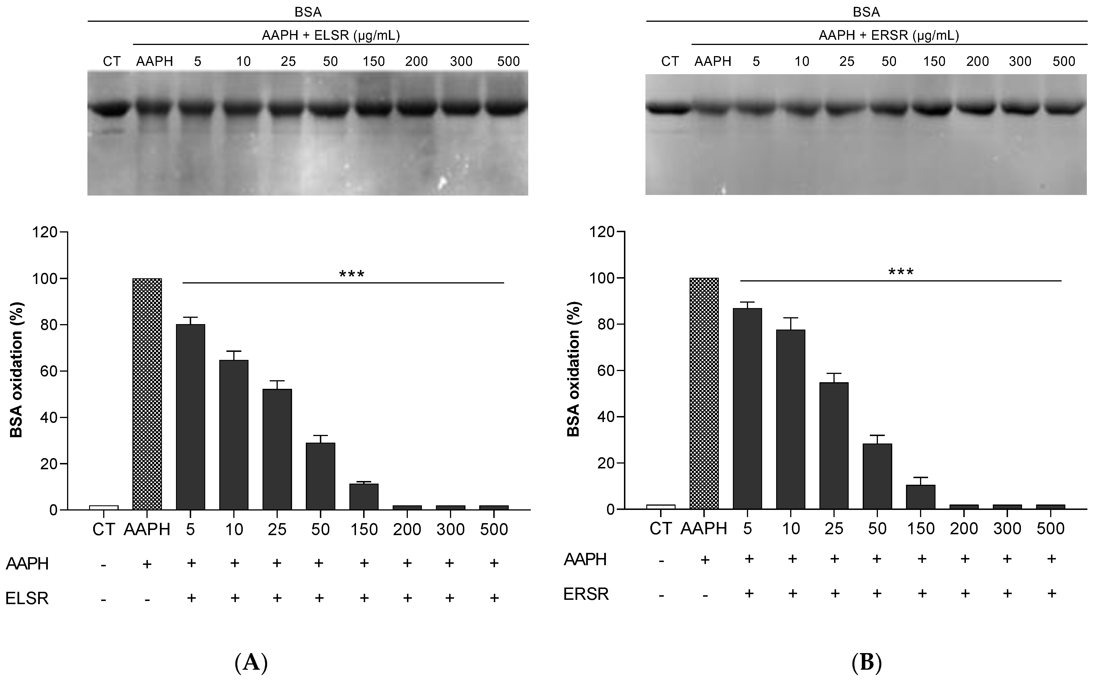
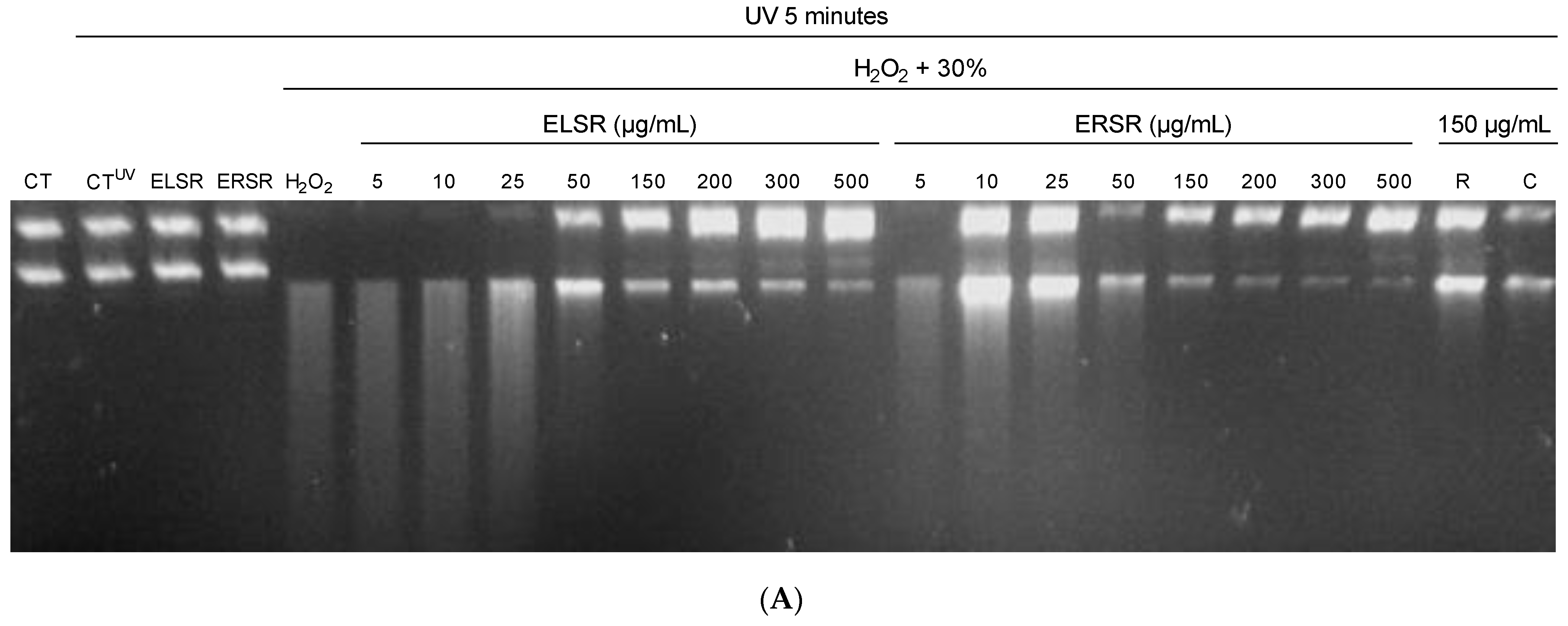
2.3. Protection of ELSR and ERSR against Oxidative Damage in Macromolecules

ELSR and ERSR protected BSA protein against AAPH-induced oxidation at all of the concentrations evaluated (Figure 2). The images of the gels are representative and demonstrate the protection against oxidation after ELSR and ERSR treatments. Both extracts reduced protein oxidation resulting from AAPH exposure when compared to the AAPH control, where BSA was incubated with AAPH alone.
ELSR and ERSR were able to protect DNA from H2O2 UV-induced fragmentation in a concentration-dependent manner (Figure 3B). Figure 3A is a representative picture of the protection against fragmentation after ELSR and ERSR treatments. In the absence of extracts and at low concentrations (up to 25 μg/mL), fragmentation of plasmid DNA is observed. A reduction in fragmentation is visible from 50 μg/mL onward. The reference antioxidants rutin and catechin caused a reduction in DNA fragmentation; however, rutin was more effective in the evaluated concentration.

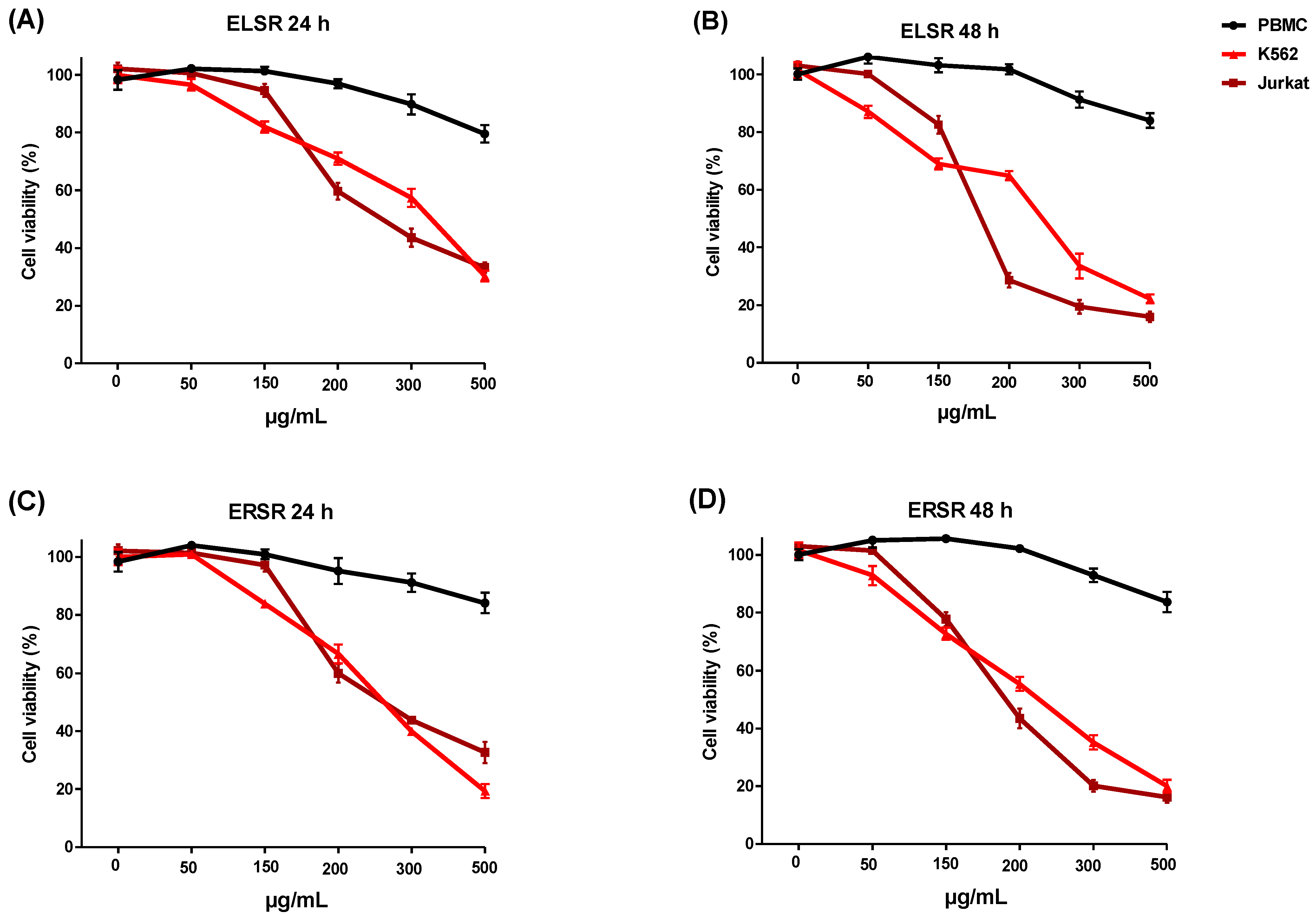
2.4. Cytotoxicity against Leukemic Strains

PBMC cells and K562 and Jurkat leukemic cell lines were treated with different concentrations of the ELSR and ERSR for 24 and 48 h. Incubation of the ELSR and ERSR (Figure 4A–D) with PBMC showed a minimal reduction in cell viability of 17% on average, but only at the highest concentration of the extracts. None of the concentrations of the extracts caused a 50% reduction in PBMC viability, suggesting that the extracts are selective for leukemic cells. The results of the solvent control (0.1% ethanol) demonstrated that the cells’ viability under these conditions was unaffected by the solvent. The results show that the extracts showed a cytotoxic effect against the leukemic cell lines in a concentration-dependent manner, with the K562 cell line being less sensitive than the Jurkat cell line, as observed through the IC50 values presented in Table 3.

3. Discussion

The phytochemical profiles of ELSR and ERSR were examined utilizing HPLC techniques to identify pertinent secondary metabolite groups and valuable chemical markers. These multi-constituent profile markers serve as tools for quality control of herbal preparations and for subsequent validity assessments.
An interesting observation in the present study is the high divergence observed in the phytochemical constituents identified between ELSR and ERSR. Furthermore, the identification for the first time of pharmacologically active metabolites from the roots of S. rugosa. This divergence in phytochemical profiles not only underscores the existence of a unique chemical diversity between the distinct parts of the plant but also reflects the intricate specialized metabolic networks characterizing the leaf and root tissues of S. rugosa.
The chemical profile of ELSR was characterized predominantly by the accumulation of flavan-3-ol derivatives, notably catechin (L-1). This compound, distinguished by its characteristic UV absorption at 280 nm and a molecular ion [M + H]+ at m/z 291.0870, was confirmed through comparison with an authentic standard. Proanthocyanidins, including derivatives such as epiafzelechin-epiafzelechin (L-4, L-5, L-6) and trihydroxyflavan-epiafzelechin derivatives (L-8, L-9, L-10, L-12), highlight the plant’s ability to synthesize complex polymeric flavonoids. These compounds were annotated by their molecular masses and distinctive MS/MS fragmentation patterns.
Flavonols, such as Rutin (L-2) and Kaempferol 3-O-rutinoside (L-7), were identified by their UV spectra, MS/MS fragmentation patterns, and comparison with an authentic standard. Flavones such as Luteolin (L-11) and its methoxylated derivative, 3-methoxyluteolin (L-13), underscore the plant’s ability to synthesize a wide variety of flavonoid structures.
The stilbenoid, 3,3′,5,5′-Tetrahydroxy-4-methoxystilbene (L-3), was detected at a low intensity and annotated by its UV absorption and molecular formula. This class has been reported multiple times within the genus [32].
Furthermore, a tetrahydroanthracene derivative (L-16), representing a less common class, was putatively characterized by its molecular weight and MS/MS fragments. It is possibly novel in the literature; due to the absence of standards, we were unable to determine its chemical structure. We recently identified a compound of this class in S. velutina [20].
The ERSR extract from the roots displayed a metabolite accumulation distinctly different from that observed in the leaves. Chromones prominently featured, evidenced by the annotation of obtusichromoneside derivatives (R-2, R-3, R-7) and a chromone derivative (R-7), characterized by their UV spectra and mass fragmentation patterns. These compounds have previously been reported in other species of Senna and Cassia, a closely related taxon [33].
The analysis also highlighted a significant presence of naphthoquinone and naphthopyrones, with compounds such as 2-methoxystypandrone (R-4), a putative norrubrofusarin gentiobioside (R-8) (a possible novel compound), rubrofusarin gentiobioside (R-10), cassiaside B (R-12), and rubrofusarin-O-glucopyranoside (R-14), demonstrating the root’s capability to synthesize complex naphtho-derived structures. These findings are particularly interesting due to the rare occurrence of such compounds in nature and their potential pharmacological applications.
The only compound observed in both the leaves and roots was R-6 (equivalent to L-3 in the leaf), but it demonstrated low chromatographic intensity, consistent with its observation in leaf extracts. Additionally, the detection of Flavanones and their derivatives, including hexahydroxy Flavanonol pentosyl-hexosyl derivatives (R-9, R-13), a putatively annotated hexahydroxy Flavanonol sulfate (R-11), and hexahydroxy-methoxy flavanonol sulfate (R-16), indicates a distinctive flavonoid accumulation pattern in the leaves and roots of S. rugosa. Some of these compounds, such as R-11 and R-15, do not match any structures described in the literature, suggesting the possibility that they are novel compounds. The structural diversity within this class highlights the intricate enzymatic processes involved in flavonoid modification.
Seven compounds of the roots remained unidentified (R-1, R-15, R-17, R-18, R-20, R-21, R-22), presenting opportunities for further research. These unknown entities, detected through their unique mass spectra and UV profiles, hint at the unexplored chemical space within S. rugosa, suggesting the presence of novel or rare metabolites awaiting discovery.
In summary, this detailed phytochemical investigation significantly enhances our comprehension of the chemical diversity present in S. rugosa, underscoring the plant’s extensive biosynthetic capacities. It sets a foundation for future research aimed at elucidating the pharmacological properties and ecological significance of these compounds, while also indicating the potential for discovering novel metabolites within this species.
Interestingly, the antioxidant and anticancer activities of most of these compounds identified in ELSR, including their molecular mechanisms, have already been proven in several studies. Notably, several in vitro, in vivo, and clinical studies have shown multiple anticancer actions of epigallocatechin, and a review of their potential therapeutic targets and their role in the therapy of various cancers has been the motive of several studies [34,35,36]. These include anti-proliferative, pro-apoptotic, anti-angiogenic, and anti-invasive activities. Another compound identified in ELSR is rutin, which exhibits a wide range of pharmacological properties. These include, among others, antiangiogenic, pro-apoptotic, antioxidant, antiproliferative, and anti-inflammatory activities. These properties may contribute to the prevention and treatment of cancer [36].
In the report by Cunha et al. [30], several peaks were observed in the hydroethanolic extract, but only one compound was identified (rutin), making comparison with our results impossible. Thus, we report the presence of different phytochemicals not previously described for this species.
In the ERSR, several pharmacologically important compounds were also identified. Of the 22 peaks, 6 could not be identified based on the current technique employed. Among the identified compounds in ERSR were the chromone derivatives, particularly the obtusichromoneside derivatives. Several chromone derivatives have been credited with significant biological and pharmacological activities, including antioxidant, antiviral, anti-inflammatory, antitumor, antimicrobial, among others [37,38,39]. Duan et al. [38] presented a comprehensive review of the antitumor activities of naturally occurring chromones. The antitumor mechanisms of these chromones include cytotoxicity (cell-cycle arrest and cell death, RNA inhibition, and inhibition of signal transducers), antimetastatic, anti-angiogenesis, and immune regulation, among others. Moreover, some have been shown to possess cancer chemoprevention potential. The other equally important secondary metabolites found in ERSR are naphthoquinones. These groups of naturally occurring bioactive compounds have been the subject of intense research on anticancer and antioxidant activities [40,41].
Several studies have demonstrated the effect of compounds present in ELSR and ERSR extracts on increasing caspase 3 activity in cancer cells, a key enzyme in the induction of apoptotic processes. The presence of rutin and luteonin in ELSR indicates the activation of caspase 3, as described by Pandey et al. (2021) [42] and Raina et al. (2021) [43] in HeLa cells, respectively. In ERSR, caspase 3 activation is probably linked to the presence of chromone derivatives (Chu et al., 2021) [44]. Specifically, chromone derivatives have been shown to induce apoptosis through the Bcl-2, Bax, and caspase 3 signaling cascades.
Furthermore, the activation of caspase 3 can occur through the activation of caspase 8 (the extrinsic pathway) and through the activation of caspase 9 (the intrinsic pathway), as observed by Pandey et al. (2021) for rutin [42]. It is therefore plausible to expect that the compounds present in the extracts can activate a single pathway for the induction of apoptosis, such as the activation of caspase 9 by luteolin or the activation of caspase 8 by epiafzelechin and procyanidins present in ELSR (Raina et al., 2021 [43]; Kaur et al., 2020 [45]; Minker et al., 2015 [46]). Thus, it is consistent to suggest that the mechanism by which ELSR and ERSR extracts exerted the final cell death effect observed in our study might be related, at least in part, to these mechanisms. The exploration of the mechanisms involved in these activities, together with in vivo antitumor activities, will be our next focus for future research.
The radical scavenging, antioxidant, and anticancer activities of these groups of compounds are widely known. These compounds together may act in synergy to account for the antioxidants and prevent oxidative damage to the macromolecules employed in this study.
The genomic mutation of DNA is an essential component in the process of cancer development. Oxidative damage and the consequent modification of DNA bases can lead to point mutations, deletions, insertions, or chromosomal translocations that, in turn, can produce oncogene activation or tumor suppressor gene inactivation [47,48]. In this context, we evaluated the potential for reactive species sequestration, antioxidant activity, DNA, and protein protective activity against oxidative damage induced by oxidizing agents.
DNA mutation is a critical step in carcinogenesis, and elevated levels of oxidative DNA damage (8-hydroxyguanosine) have been noted in various tumors, which strongly implicates such damage in cancer etiology [49]. Furthermore, the increased production of reactive oxygen species (ROS) in various cancers has been shown to play several roles, such as in the activation of pro-tumourigenic signaling, an increase in cell survival and proliferation, and driving DNA damage and genetic instability [50,51,52].
To evaluate the potential protective effect of ELSR and ERSR in mitigating oxidative damage to macromolecules, models of oxidative damage induced by hydroxide peroxide were used, and ELSR and ERSR exhibited antioxidant activity, acting in the direct capture of reactive species. Furthermore, we demonstrated that the extracts were able to promote the protection of macromolecules against oxidative damage. These results indicate that the extracts can stabilize reactive species through electron or H+ donation, preventing nonspecific oxidoreduction reactions that propagate in the intracellular environment [6]. The antioxidant activity of ELSR and ERSR may be related to the presence of biologically active metabolites identified in both extracts. These metabolites are widely recognized for stabilizing reactive oxygen species through hydrogen donation and binding with metal ions [53,54,55].
In addition to the effect of direct radical scavenging, ELSR and ERSR were able to protect proteins and DNA from oxidative damage. The oxidation of macromolecules by reactive species is an important deleterious effect observed at the cellular level. Protein oxidation promotes the loss of function of receptors, enzymes, and other intracellular proteins, the formation of protein aggregates, and proteolysis.
The protective effect of S. rugosa extracts against the oxidation of macromolecules suggests that intracellular oxidative damage can be reduced by ingesting the extracts and could present beneficial effects for human health. The intake of natural antioxidants is recognized for promoting a double beneficial effect on the organism: the protection of healthy cells against the deleterious action of reactive species and their anticancer properties. In addition to conferring protection to macromolecules, flavonoids, and naphthoquinones, among others, they can regulate the expression of oncogenes and tumor suppressor genes, control cell proliferation, metastasis, and angiogenesis, induce cell cycle arrest, and promote apoptosis in cancer cells [36,40,41,55,56,57].
Thus, based on the results obtained, ELSR and ERSR may modulate the cellular environment, promoting apoptosis activation in K562 and Jurkat cells. From this perspective, further studies are necessary to gain a better understanding of the mechanisms through which the extracts promote the death of leukemic cells.
The search for cytotoxic agents that exhibit selectivity in cancer treatment is increasing [58,59]. In this study, we found that ELSR and ERSR do not affect the viability of healthy cells using PBMC as a model, cells that could be affected during treatment with chemotherapeutic agents during the treatment of leukemias. Since IC50 was not obtained for this cell type, it is possible to infer that ELSR and ERSR exhibit at least 2-3-fold minimal selectivity for leukemic cells. This effect is desirable because the chemotherapeutic agents currently in use are not selective in their actions, causing side effects in the body, such as high toxicity in healthy cells [60]. Thus, we suggest that S. rugosa extracts possess potential therapeutic and chemopreventive effects.
Our results demonstrated that the extracts are more effective on the Jurkat cell line, a model of T-cell acute lymphoblastic leukemia. The differences found in our assays among the cell lines utilized might be attributable to mutations in each of the lines investigated. In acute lymphoblastic leukemias, such as Jurkat, the silencing of tumor suppressor and pro-apoptotic genes such as p53, PTEN, and BAX is common [15,61]. BCR-ABL1-positive leukemias, as in the K562 cell model, exhibit increased tyrosine kinase activity [13].

4. Materials and Methods

4.1. Chemicals and Reagents

The reagents 2,2-diphenyl-1-picrylhydrazyl (DPPH), 2,2′-azinobis 3-ethylbenzthiazoline-6-sulfonic acid (ABTS), 2,2′-Azobis(2-methylpropionamidine) dihydrochloride (AAPH), ascorbic acid (AA), bovine serum albumin (BSA), hydroxytoluene butylate (BHT), 3-(4,5-dimethylthiazol-2-yl)-2,5-diphenyltetrazolium bromide (MTT), ammonium persulfate, and potassium persulfate were purchased from Sigma-Aldrich (São Paulo, Brazil). Electrophoresis reagents were purchased from Bio-Rad Laboratories (São Paulo, Brazil). RPMI-1640 culture medium, antibiotics penicillin, streptomycin, neomycin, and fetal bovine serum (FBS) were purchased from Gibco/Invitrogen (Minneapolis, MN, USA). The other reagents used in this work were of analytical grade.

4.2. Plant Material

The leaves and roots of S. rugosa were collected in the city of Dourados, MS, Brazil (22°05′45″ S 55°20′46″ W), and permission to access genetic heritage (CTA) was registered in the Heritage SisGen under access code AA6FADF. An exsiccate specimen of the species was deposited in the Herbarium of the Universidade Federal da Grande Dourados with the registration number 4664. The leaves and roots of S. rugosa were washed with running water, dried in an oven with air circulation at 36 °C for 10 days, and ground in a knife mill. The leaf (ELSR) and root extracts of S. rugosa (ERSR) were prepared from 200 g of leaf or root powder macerated in 95% ethanol at a 1:7 (w/v) ratio at room temperature for 21 days.
Every seven days, the extract was filtered, the solvent was reserved, and a new aliquot of 95% ethanol was added to perform an exhaustive extraction of the compounds. The filtrates were pooled and concentrated in a rotary vacuum evaporator (Gehaka, São Paulo, SP, Brazil) at 37 °C. The filtrates were frozen and lyophilized to obtain the powders denoted as ELSR and ERSR. The yield of ELSR was 16.61%, and that of ERSR was 1%.

Phytochemical Analyses

The ELSR and ERSR were analyzed in a liquid chromatograph (UFLC, Shimadz, Kyoto, Japan) coupled to a diode array detector (DAD, Shimadzu) and to an electrospray ionization time-of-flight mass spectrometer (ESI-QTOF-micrOTOF QII, Bruker Daltonics, Billerica, MA, USA); positive ionization mode, with detection between 120–1200 Da. The sample was injected into a column C-18 (Kinetex, 2.6 μm, Phenomenex, Torrance, CA, USA). The chromatographic and mass spectrometric parameters applied were the same as those used by Castro et al. [19]. We identified the metabolites in the ELSR and ERSR by analyzing their molecular mass, fragmentation patterns, and ultraviolet (UV) absorption spectra, comparing these characteristics with the existing literature data. Whenever possible, we confirmed the identities of these compounds through comparative analysis with authentic standards.

4.3. Antioxidant Activity Assays

4.3.1. Direct ABTS•+ Radical Scavenging Assay

The antioxidant capacity of ELSR and ERSR was assessed using an ABTS•+ assay [62]. A volume of 5 mL of ABTS solution (7 mM) and 88 μL of potassium persulfate solution (140 mM) were incubated for 12–16 h at room temperature in the dark. The ABTS•+ working solution was obtained by diluting the stock solution in distilled water, and its absorbance was determined (0.70 ± 0.05 units) at 734 nm in a spectrophotometer. ELSR or ERSR were prepared at different concentrations (0.1–100 μg/mL), and 20 μL was mixed with 1980 μL of the ABTS•+ working solution. The mixture was incubated for 6 min and absorbance was measured at 734 nm. Ascorbic acid (AA) and butylated hydroxytoluene (BHT) were used as reference antioxidants. A volume of 20 μL of distilled water was used for the determination of the absorbance of the negative control. The percentage of ABTS•+ radical scavenging was calculated using the following Equation (1):
Inhibition   of   ABTS % = Abs   control - Abs   sample Abs   control   ×   100
Three independent experiments were performed in triplicate and the concentration of the extracts needed for the capture of 50% of the ABTS•+ radical (IC50) was determined, as well as the reference antioxidants.

4.3.2. Direct DPPH Radical Scavenging Assay

The direct capture of the DPPH radical by ELSR and ERSR was evaluated according to Gupta and Gupta [63]. For this, 200 μL of the ELSRs and ERSRs at different concentrations (0.1–100 μg/mL) were diluted in 1800 μL of 0.11 mM DPPH solution prepared in 80% ethanol. The mixture was incubated for 30 min at room temperature in the dark. The absorbance at 517 nm was determined. Ascorbic acid and BHT were used as reference antioxidants. A 0.11 mM DPPH solution plus 200 μL of 80% ethanol was used as the negative control. Three independent experiments were performed in triplicate and the percentage of direct DPPH radical scavenging was calculated with Equation (2) as follows:
I n h i b i t i o n   o f   D P P H % = 1 s a m p l e n e g a t i v e c o n t r o l × 100
The concentration of the extracts required for the capture of 50% of the DPPH radical (IC50) was determined, as well as for the reference antioxidants.
The antioxidant activity assays were performed by diluting the extracts in a 10 mg/mL stock solution with 80% ethanol. From the stock solution, working solutions containing between 5 and 500 μg/mL were prepared in the solvents of each assay.

4.4. Protection against Oxidative Damage to the Macromolecules by ELSRs and ERSRs

4.4.1. Azo Initiator 2,2′-Azobis-(2-amidinopropane) dihydrochloride AAPH-Induced Oxidation of Proteins

The protective effect of the extracts against AAPH-induced protein oxidation was evaluated according to the methodology of Mayo et al. [64], with minor modifications. A 3 μL aliquot of BSA (3 mg/mL, prepared in PBS) was pre-incubated with 3 μL of the ELSRs or ERSRs at different concentrations (5–500 μg/mL) for 30 min at 37 °C. Then, 3 μL of AAPH (120 mM, prepared in water) was added to the respective treatments, followed by further incubation for 120 min at 37 °C.
Then, 11 μL of sample buffer was added to each tube. The samples were incubated for 5 min at 95 °C and applied to a 12% SDS-PAGE polyacrylamide gel, separated at 200 V on the Mini-PROTEAN Tetra Cell system (Bio-Rad Laboratories). At the end of electrophoresis, the gels were stained, destained, and scanned in a Gel Doc EZ Imager (Bio-Rad Laboratories). The volume of the BSA bands was determined with the help of Image Lab software. An increased volume of the BSA bands was considered oxidative damage. BSA incubated with only AAPH was used as a positive control. The protection conferred by the different concentrations of the extracts was calculated by averaging four gels.

4.4.2. Assay on Protection against DNA Damages (H2O2)

The protective effect of ELSR and ERSR against DNA fragmentation was evaluated using the method described by Kumar and Chattopadhyay [65] with modifications. A 4 μL aliquot of plasmid DNA (50 ng/μL) was incubated with 4 μL of ELSR or ERSR at different concentrations, diluted in PBS (5–500 μg/mL). Then, 4 μL of the oxidizing agent H2O2 at 30% was added to the respective treatments. The plasmid in the absence of UV light (CT), the plasmid in the presence of UV light (CTUV), the plasmid in the presence of the extracts plus UV light (ELSR or ERSR), and the control plasmid incubated only with hydrogen peroxide (H2O2) was used as the negative control. The effect of other reference antioxidants (rutin and catechin—150 μg/mL) on protecting DNA fragmentation was analyzed. The samples were incubated in a UVT-312 transilluminator at 302 nm at room temperature for 5 min. Then, the samples were applied to a 2% agarose gel containing ethidium bromide (10 mg/mL) and subjected to electrophoresis. Images of the gels were scanned using the Gel Doc™ EZ Imager photodocumentator (Bio-Rad Laboratories, Hercules, CA, USA) and analyzed using Image Lab™ software (version 6.0.1). The results of 3 gels were expressed as a percentage of DNA fragmentation.
The oxidative damage assays were performed by diluting the extracts in a stock solution containing 0.01% of 80% ethanol supplemented with PBS. From the stock solution, working solutions containing between 5 and 500 μg/mL were prepared in the solvents of each assay.

4.5. Cell-Based Assays

4.5.1. Cell Culture

The human leukemia cell lines Jurkat (Human Acute Lymphoblastic Leukemia) and K562 (Human Chronic Myelogenous Leukemia) were kind donations by Professor Dr. Edgar J. Paredes-Gamero of the Department of Biochemistry, Universidade Federal de Sao Paulo, São Paulo, Brazil. The K562 and Jurkat were grown in suspension in RPMI 1640 media supplemented with 10 nM 4-(2-hydroxyethyl) piperazine-1-ethanesulfonic acid (HEPES), 24 nM sodium bicarbonate, 10% fetal bovine serum (FBS), and antibiotic Penicillin-streptomycin (10,000 U/mL), all from Gibco/Invitrogen, Minneapolis, MN, USA. All cells were kept in an incubator with a humidified atmosphere containing 5% CO2 at 37 °C.

4.5.2. Isolation of Mononuclear Cells from Human Peripheral Blood

The Research Ethics Committee of the Centro Universitário da Grande Dourados (UNIGRAN) approved blood collection from a healthy non-smoking donor, process number 123/12. Human peripheral blood mononuclear cells (PBMC) were obtained by centrifugation using Ficoll-Paque-1077 Premium reagent (1.073 g/cm3). PBMC were suspended in RPMI-1640 medium supplemented with 20% SFB and 1% antibiotic and maintained at 37 °C in a humidified atmosphere containing 5% CO2.

4.5.3. Evaluation of Cytotoxicity of ELSR and ERSR

The potential cytotoxicity of ELSR and ERSR was evaluated with the MTT reagent against leukemic cells (K562 and Jurkat) and PBMC. The K562, Jurkat (2 × 10⁴ cells/well), and PBMC (12 × 104 cells/well) cells were plated in 96-well microplates and treated with different concentrations of the ELSR and ERSR (0–500 μg/mL solubilized in RPMI-1640 medium) for 24 and 48 h. After treatment, the microplates were centrifuged at 1200 rpm for 5 min and the culture medium was removed. A volume of 100 μL of MTT (0.5 mg/mL, prepared in RPMI-1640 medium) was added to the wells, and a 4 h incubation at 37 °C was performed. The plates were centrifuged, the medium was removed, and 100 μL of dimethyl sulfoxide (DMSO) was added to solubilize the formazan crystals. A control treatment was performed with 0.1% ethanol, the volume of solvent used for preparing the extracts. The absorbances were determined at 630 nm in a microplate reader (TP READER NM, Thermo Plate, Beijing, China) and cell viability was calculated using the following Equation (3):
Cell   viability   % = t r e a t e d c e l l s c o n t r o l t r e a t e d c e l l s × 100
The cell viability assay was performed by diluting the extracts in a stock solution containing 0.1% ethanol supplemented with culture medium.

4.6. Statistical Analyses

Data were presented as mean ± standard error of the mean (SEM). Statistically significant differences between groups were analyzed using the Student t-test for comparison between two groups and analysis of variance (ANOVA), followed by Dunnett’s test, for comparison of two or more groups, using Prism 7 Software (GraphPad Software, San Diego, CA, USA). The results were considered significant at p < 0.05. To establish the half-maximal inhibitory concentration (IC50) of DPPH and ABTS free radical scavenging, the samples were tested in serial dilutions (0.1–100 μg/mL) and to test the cytotoxicity against leukemic strains with different concentrations of the ELSR and ERSR (0–500 μg/mL), both were analyzed using nonlinear regression using GraphPad Prism 7 software. (GraphPad Software, San Diego, CA, USA). The results were considered significant at p < 0.05.

5. Conclusions

In this study, we demonstrated that ELSR and ERSR present flavonoid and naphthopyranone derivatives related to the observed antioxidant activities, capable of promoting direct radical scavenging and the protection of macromolecules against oxidative damage. In addition, the ELSR and ERSR exhibited cytotoxicity to human leukemia cell lines without affecting the viability of healthy cells. Our results expand the chemical compounds identified in the leaves and roots of S. rugosa and their pharmacological potential in the management of conditions related to oxidative stress and leukemia.

Author Contributions

Conceptualization, C.M.d.S., J.F.C., K.d.P.S. and E.L.d.S.; Methodology and validation, C.M.d.S., J.T.d.G.C., A.S.O., P.d.S.d.R., J.F.C. and C.F.R.d.O.; Formal analysis, C.M.d.S., J.T.d.G.C., K.d.P.S. and E.L.d.S.; Investigation, C.M.d.S., D.d.S.B., D.T.H.d.C., P.d.S.d.R. and A.S.O.; Resources, C.A.C., D.B.d.S., K.d.P.S. and E.L.d.S.; Data curation, C.M.d.S., C.A.C., D.B.d.S., K.d.P.S. and E.L.d.S.; Writing—original draft preparation, C.M.d.S. and E.L.d.S.; Writing—review and editing, D.d.S.B., D.T.H.d.C., A.S.O., S.O.B., C.A.C., D.B.d.S., K.d.P.S. and E.L.d.S.; Conceptualization and supervision, K.d.P.S. and E.L.d.S.; Project administration, K.d.P.S. and E.L.d.S.; Funding acquisition, C.A.C., D.B.d.S., K.d.P.S. and E.L.d.S. All authors have read and agreed to the published version of the manuscript.

Funding

This work was supported by institutional funding from the Fundação de Apoio ao Desenvolvimento do Ensino, Ciência e Tecnologia do Estado de Mato Grosso do Sul (FUNDECT, Brazil), Coordenação de Aperfeiçoamento de Pessoal de Nível Superior (CAPES, Brazil) and Conselho Nacional de Desenvolvimento Científico e Tecnológico (CNPq, Brazil).

Institutional Review Board Statement

Not applicable.

Informed Consent Statement

Not applicable.

Data Availability Statement

The experimental data used to support the results of this study are included in the article.

Acknowledgments

This work was funded by grants from the Fundação de Apoio ao Desenvolvimento do Ensino, Ciência e Tecnologia do Estado de Mato Grosso do Sul (Foundation for Support to the Development of Education, Science and Technology of the State of Mato Grosso do Sul, FUNDECT); Coordenação de Aperfeiçoamento de Pessoal de Nível Superior (CAPES); and Conselho Nacional de Desenvolvimento Científico e Tecnológico (CNPq); Instituto de Ciência e Tecnologia em Áreas Úmidas (INAU).

Conflicts of Interest

The authors declare no conflicts of interest.

References

  1. Nogueira, V.; Hay, N. Molecular Pathways: Reactive Oxygen Species Homeostasis in Cancer Cells and Implications for Cancer Therapy. Clin. Cancer Res. 2013, 19, 4309. [Google Scholar] [CrossRef] [PubMed]
  2. Jomova, K.; Alomar, S.Y.; Alwasel, S.H.; Nepovimova, E.; Kuca, K.; Valko, M. Several lines of antioxidant defense against oxidative stress: Antioxidant enzymes, nanomaterials with multiple enzyme-mimicking activities, and low-molecular-weight antioxidants. Arch. Toxicol. 2024, 98, 1323–1367. [Google Scholar] [CrossRef] [PubMed]
  3. Irwin, M.E.; Rivera-Del Valle, N.; Chandra, J. Redox Control of Leukemia: From Molecular Mechanisms to Therapeutic Opportunities. Antioxid. Redox Signal. 2013, 18, 1349. [Google Scholar] [CrossRef] [PubMed]
  4. Xie, W.; Ma, W.; Liu, P.; Zhou, F. Overview of thioredoxin system and targeted therapies for acute leukemia. Mitochondrion 2019, 47, 38–46. [Google Scholar] [CrossRef] [PubMed]
  5. Kumari, S.; Badana, A.K.; Murali Mohan, G.; Shailender, G.; Malla, R.R. Reactive Oxygen Species: A Key Constituent in Cancer Survival. Biomark. Insights 2018, 13, 1177271918755391. [Google Scholar] [CrossRef] [PubMed]
  6. Ighodaro, O.M.; Akinloye, O.A. First line defence antioxidants-superoxide dismutase (SOD), catalase (CAT) and glutathione peroxidase (GPX): Their fundamental role in the entire antioxidant defence grid. Alex. J. Med. 2018, 54, 287–293. [Google Scholar] [CrossRef]
  7. Pisoschi, A.M.; Pop, A. The role of antioxidants in the chemistry of oxidative stress: A review. Eur. J. Med. Chem. 2015, 97, 55–74. [Google Scholar] [CrossRef] [PubMed]
  8. Thyagarajan, A.; Sahu, R.P. Potential Contributions of Antioxidants to Cancer Therapy: Immunomodulation and Radiosensitization. Integr. Cancer Ther. 2018, 17, 210. [Google Scholar] [CrossRef] [PubMed]
  9. León-González, A.J.; Auger, C.; Schini-Kerth, V.B. Pro-oxidant activity of polyphenols and its implication on cancer chemoprevention and chemotherapy. Biochem. Pharmacol. 2015, 98, 371–380. [Google Scholar] [CrossRef]
  10. Muchtaridi, M.; Az-Zahra, F.; Wongso, H.; Setyawati, L.U.; Novitasari, D.; Ikram, E.H.K. Molecular Mechanism of Natural Food Antioxidants to Regulate ROS in Treating Cancer: A Review. Antioxidants 2024, 13, 207. [Google Scholar] [CrossRef]
  11. Bhatnagar, N.; Qureshi, A.; Hall, G. Leukaemias: A review. Paediatr. Child Health 2017, 27, 489–494. [Google Scholar] [CrossRef]
  12. Zhao, Y.; Wang, Y.; Ma, S. Racial Differences in Four Leukemia Subtypes: Comprehensive Descriptive Epidemiology. Sci. Rep. 2018, 8, 548. [Google Scholar] [CrossRef] [PubMed]
  13. Mwirigi, A.; Dillon, R.; Raj, K. Acute leukaemia. Medicine 2017, 45, 280–286. [Google Scholar] [CrossRef]
  14. O’Shea, J.J.; Schwartz, D.M.; Villarino, A.V.; Gadina, M.; McInnes, I.B.; Laurence, A. The JAK-STAT Pathway: Impact on Human Disease and Therapeutic Intervention. Annu. Rev. Med. 2015, 66, 311. [Google Scholar] [CrossRef] [PubMed]
  15. Gioia, L.; Siddique, A.; Head, S.R.; Salomon, D.R.; Su, A.I. A genome-wide survey of mutations in the Jurkat cell line. BMC Genom. 2018, 19, 334. [Google Scholar] [CrossRef] [PubMed]
  16. Rose-Inman, H.; Kuehl, D. Acute Leukemia. Hematol. Oncol. Clin. N. Am. 2017, 31, 1011–1028. [Google Scholar] [CrossRef] [PubMed]
  17. World Health Organization Estimated Number of New Cases in 2020, Worldwide, Both Sexes, All Ages. Available online: https://gco.iarc.fr/today/online-analysis-table?v=2020&mode=cancer&mode_population=countries&population=900&populations=900&key=asr&sex=0&cancer=39&type=0&statistic=5&prevalence=0&population_group=0&ages_group%5B%5D=0&ages_group%5B%5D=17&group_cancer=1&in (accessed on 10 June 2022).
  18. De Giffoni De Carvalho, J.T.; Da Silva Baldivia, D.; Leite, D.F.; De Araújo, L.C.A.; De Toledo Espindola, P.P.; Antunes, K.A.; Rocha, P.S.; De Picoli Souza, K.; Dos Santos, E.L. Medicinal Plants from Brazilian Cerrado: Antioxidant and Anticancer Potential and Protection against Chemotherapy Toxicity. Oxidative Med. Cell. Longev. 2019, 2019, 3685264. [Google Scholar] [CrossRef] [PubMed]
  19. Castro, D.T.H.; Campos, J.F.; Damião, M.J.; Torquato, H.F.V.; Paredes-Gamero, E.J.; Carollo, C.A.; Rodrigues, E.G.; de Picoli Souza, K.; dos Santos, E.L. Ethanolic Extract of Senna velutina Roots: Chemical Composition, In Vitro and In Vivo Antitumor Effects, and B16F10-Nex2 Melanoma Cell Death Mechanisms. Oxidative Med. Cell. Longev. 2019, 2019, 5719483. [Google Scholar] [CrossRef] [PubMed]
  20. Castro, D.T.H.; Leite, D.F.; da Silva Baldivia, D.; dos Santos, H.F.; Balogun, S.O.; da Silva, D.B.; Carollo, C.A.; de Picoli Souza, K.; dos Santos, E.L. Structural Characterization and Anticancer Activity of a New Anthraquinone from Senna velutina (Fabaceae). Pharmaceuticals 2023, 16, 951. [Google Scholar] [CrossRef] [PubMed]
  21. Oladeji, O.S.; Adelowo, F.E.; Oluyori, A.P. The genus Senna (Fabaceae): A review on its traditional uses, botany, phytochemistry, pharmacology and toxicology. S. Afr. J. Bot. 2021, 138, 1–32. [Google Scholar] [CrossRef]
  22. Baldivia, D.d.S.; Leite, D.F.; de Castro, D.T.H.; Campos, J.F.; Dos Santos, U.P.; Paredes-Gamero, E.J.; Carollo, C.A.; Silva, D.B.; Souza, K.d.P.; Dos Santos, E.L. Evaluation of In Vitro Antioxidant and Anticancer Properties of the Aqueous Extract from the Stem Bark of Stryphnodendron adstringens. Int. J. Mol. Sci. 2018, 19, 2432. [Google Scholar] [CrossRef] [PubMed]
  23. Campos, J.F.; De Castro, D.T.H.; Damiaõ, M.J.; Vieira Torquato, H.F.; Paredes-Gamero, E.J.; Carollo, C.A.; Estevinho, L.M.; De Picoli Souza, K.; Santos, E.L. Dos The Chemical Profile of Senna velutina Leaves and Their Antioxidant and Cytotoxic Effects. Oxidative Med. Cell. Longev. 2016, 2016, 8405957. [Google Scholar] [CrossRef] [PubMed]
  24. Campos, J.F.; Espindola, P.P.d.T.; Torquato, H.F.V.; Vital, W.D.; Justo, G.Z.; Silva, D.B.; Carollo, C.A.; de Picoli Souza, K.; Paredes-Gamero, E.J.; dos Santos, E.L. Leaf and Root Extracts from Campomanesia adamantium (Myrtaceae) Promote Apoptotic Death of Leukemic Cells via Activation of Intracellular Calcium and Caspase-3. Front. Pharmacol. 2017, 8, 466. [Google Scholar] [CrossRef]
  25. Casagrande, J.C.; Macorini, L.F.B.; Antunes, K.A.; Dos Santos, U.P.; Campos, J.F.; Dias-Júnior, N.M.; Sangalli, A.; Cardoso, C.A.L.; Do Carmo Vieira, M.; Rabelo, L.A.; et al. Antioxidant and cytotoxic activity of hydroethanolic extract from Jacaranda decurrens leaves. PLoS ONE 2014, 9, e112748. [Google Scholar] [CrossRef] [PubMed]
  26. Yang, C.S.; Wang, H. Cancer Preventive Activities of Tea Catechins. Molecules 2016, 21, 1679. [Google Scholar] [CrossRef]
  27. Farias, D.F.; Cavalheiro, M.G.; Viana, M.P.; Queiroz, V.A.; Rocha-Bezerra, L.C.B.; Vasconcelos, I.M.; Morais, S.M.; Carvalho, A.F.U. Water extracts of Brazilian leguminous seeds as rich sources of larvicidal compounds against Aedes aegypti L. An. Acad. Bras. Cienc. 2010, 82, 585–594. [Google Scholar] [CrossRef] [PubMed]
  28. Ricardo, L.M.; De Paula-Souza, J.; Andrade, A.; Brandão, M.G.L. Plants from the Brazilian Traditional Medicine: Species from the books of the Polish physician Piotr Czerniewicz (Pedro Luiz Napoleão Chernoviz, 1812–1881). Rev. Bras. Farmacogn. 2017, 27, 388–400. [Google Scholar] [CrossRef]
  29. Rodrigues, V.E.G.; Carvalho, D.A. de Levantamento etnobotânico de plantas medicinais no domínio do Cerrado na região do Alto Rio Grande—Minas Gerais. Ciência Agrotecnol. 2001, 25, 102–123. [Google Scholar]
  30. Cunha, L.F.; Costa, C.M.; Barroso, P.R.; Kato, K.C.; Oliveira, F.D.; Victor, C.; Filho, M.; Fernanda, C.; Grael, F.; Gregório, L.E.; et al. Pharmacognosy Phytochemical screening and biological assays of ethanolic leaf extract of Senna rugosa widely used in the popular medicine although not yet adequately investigated as to its phytoconstituents and Abstract Resumo Senna rugosa (Fabaceae) é. Rodriguésia 2020, 7, 2–15. [Google Scholar]
  31. Silva, J.G.A.; Silva, A.A.; Coutinho, I.D.; Pessoa, C.O.; Cavalheiro, A.J.; Silva, M.G.V. Chemical Profile and Cytotoxic Activity of Leaf Extracts from Senna spp. from Northeast of Brazil. J. Braz. Chem. Soc. 2016, 27, 1872–1880. [Google Scholar]
  32. Maia, I.R.D.O.; Trevisan, M.T.S.; Silva, M.G.D.V.; Breuer, A.; Owen, R.W. Characterization and quantitation of polyphenolic compounds in senna macranthera var pudibunda from the Northeast of Brazil. Nat. Prod. Commun. 2019, 14, 1934578X19851704. [Google Scholar] [CrossRef]
  33. Wang, S.; Feng, K.; Han, L.; Fang, X.; Zhang, Y.; Yu, H.; Pang, X. Glycosidic compounds from Cassia obtusifolia seeds and their inhibitory effects on OATs, OCTs and OATPs. Phytochem. Lett. 2019, 32, 105–109. [Google Scholar] [CrossRef]
  34. Negri, A.; Naponelli, V.; Rizzi, F.; Bettuzzi, S. Molecular targets of epigallocatechin—Gallate (EGCG): A special focus on signal transduction and cancer. Nutrients 2018, 10, 1936. [Google Scholar] [CrossRef] [PubMed]
  35. Almatrood, S.A.; Almatroudi, A.; Khan, A.A.; Alhumaydh, F.A.; Alsahl, M.A.; Rahmani, A.H. Potential therapeutic targets of epigallocatechin gallate (EGCG), the most abundant catechin in green tea, and its role in the therapy of various types of cancer. Molecules 2020, 25, 3146. [Google Scholar] [CrossRef] [PubMed]
  36. Nouri, Z.; Fakhri, S.; Nouri, K.; Wallace, C.E.; Farzaei, M.H.; Bishayee, A. Targeting Multiple Signaling Pathways in Cancer: The Rutin Therapeutic Approach. Cancers 2020, 12, 2276. [Google Scholar] [CrossRef]
  37. Yadav, P.; Parshad, B.; Manchanda, P.; Sharma, S.K. Chromones and their derivatives as radical scavengers: A remedy for cell impairment. Curr. Top. Med. Chem. 2014, 14, 2552–2575. [Google Scholar] [CrossRef]
  38. Duan, Y.; Jiang, Y.Y.; Guo, F.X.; Chen, L.X.; Xu, L.L.; Zhang, W.; Liu, B. The antitumor activity of naturally occurring chromones: A review. Fitoterapia 2019, 135, 114–129. [Google Scholar] [CrossRef]
  39. Amen, Y.; Elsbaey, M.; Othman, A.; Sallam, M.; Shimizu, K. Naturally occurring chromone glycosides: Sources, bioactivities, and spectroscopic features. Molecules 2021, 26, 7646. [Google Scholar] [CrossRef]
  40. Qiu, H.; Wang, P.; Lin, H.; Tang, C.; Zhu, H.; Yang, Y. Naphthoquinones: A continuing source for discovery of therapeutic antineoplastic agents. Chem. Biol. Drug Des. 2018, 91, 681–690. [Google Scholar] [CrossRef] [PubMed]
  41. Wellington, K.W. Understanding cancer and the anticancer activities of naphthoquinones—A review. RSC Adv. 2015, 5, 20309–20338. [Google Scholar] [CrossRef]
  42. Pandey, P.; Khan, F.; Farhan, M.; Jafri, A. Elucidation of Rutin’s Role in Inducing Caspase-Dependent Apoptosis via HPV-E6 and E7 down-Regulation in Cervical Cancer HeLa Cells. Biosci. Rep. 2021, 41, BSR20210670. [Google Scholar] [CrossRef] [PubMed]
  43. Raina, R.; Pramodh, S.; Rais, N.; Haque, S.; Shafarin, J.; Bajbouj, K.; Hamad, M.; Hussain, A. Luteolin Inhibits Proliferation, Triggers Apoptosis and Modulates Akt/MTOR and MAP Kinase Pathways in HeLa Cells. Oncol. Lett. 2021, 21, 192. [Google Scholar] [CrossRef] [PubMed]
  44. Chu, Y.C.; Chang, C.H.; Liao, H.R.; Cheng, M.J.; Wu, M.D.; Fu, S.L.; Chen, J.J. Rare Chromone Derivatives from the Marine-Derived Penicillium citrinum with Anti-Cancer and Anti-Inflammatory Activities. Mar. Drugs 2021, 19, 25. [Google Scholar] [CrossRef]
  45. Kaur, S.; Kumar, A.; Thakur, S.; Kumar, K.; Sharma, R.; Sharma, A.; Singh, P.; Sharma, U.; Kumar, S.; Landi, M.; et al. Antioxidant, Antiproliferative and Apoptosis-Inducing Efficacy of Fractions from Cassia fistula L. Leaves. Antioxidants 2020, 9, 173. [Google Scholar] [CrossRef]
  46. Minker, C.; Duban, L.; Karas, D.; Järvinen, P.; Lobstein, A.; Muller, C.D. Impact of Procyanidins from Different Berries on Caspase 8 Activation in Colon Cancer. Oxidative Med. Cell. Longev. 2015, 2015, 154164. [Google Scholar] [CrossRef]
  47. Klaunig, J.E. Oxidative Stress and Cancer. Curr. Pharm. Des. 2019, 24, 4771–4778. [Google Scholar] [CrossRef] [PubMed]
  48. Toyokuni, S. Novel aspects of oxidative stress-associated carcinogenesis. Antioxid. Redox Signal. 2006, 8, 1373–1377. [Google Scholar] [CrossRef]
  49. Valko, M.; Rhodes, C.J.; Moncol, J.; Izakovic, M.; Mazur, M. Free radicals, metals and antioxidants in oxidative stress-induced cancer. Chem. Biol. Interact. 2006, 160, 1–40. [Google Scholar] [CrossRef]
  50. Moloney, J.N.; Cotter, T.G. ROS signalling in the biology of cancer. Semin. Cell Dev. Biol. 2018, 80, 50–64. [Google Scholar] [CrossRef]
  51. George, S.; Abrahamse, H. Redox potential of antioxidants in cancer progression and prevention. Antioxidants 2020, 9, 1156. [Google Scholar] [CrossRef]
  52. Prasad, S.; Gupta, S.C.; Tyagi, A.K. Reactive oxygen species (ROS) and cancer: Role of antioxidative nutraceuticals. Cancer Lett. 2017, 387, 95–105. [Google Scholar] [CrossRef] [PubMed]
  53. Kaurinovic, B.; Vastag, D. Flavonoids and Phenolic Acids as Potential Natural Antioxidants; IntechOpen: London, UK, 2019. [Google Scholar]
  54. Nijveldt, R.J.; Van Nood, E.; Van Hoorn, D.E.C.; Boelens, P.G.; Van Norren, K.; Van Leeuwen, P.A.M. Flavonoids: A review of probable mechanisms of action and potential applications. Am. J. Clin. Nutr. 2001, 74, 418–425. [Google Scholar] [CrossRef] [PubMed]
  55. Panche, A.N.; Diwan, A.D.; Chandra, S.R. Flavonoids: An overview. J. Nutr. Sci. 2016, 5, e47. [Google Scholar] [CrossRef] [PubMed]
  56. Abotaleb, M.; Samuel, S.M.; Varghese, E.; Varghese, S.; Kubatka, P.; Liskova, A.; Büsselberg, D. Flavonoids in Cancer and Apoptosis. Cancers 2019, 11, 28. [Google Scholar] [CrossRef] [PubMed]
  57. Farha, A.K.; Gan, R.Y.; Li, H.B.; Wu, D.T.; Atanasov, A.G.; Gul, K.; Zhang, J.R.; Yang, Q.Q.; Corke, H. The anticancer potential of the dietary polyphenol rutin: Current status, challenges, and perspectives. Crit. Rev. Food Sci. Nutr. 2022, 62, 832–859. [Google Scholar] [CrossRef] [PubMed]
  58. Lucas, D.M.; Still, P.C.; Pérez, L.B.; Grever, M.R.; Kinghorn, A.D. Potential of Plant-Derived Natural Products in the Treatment of Leukemia and Lymphoma. Curr. Drug Targets 2010, 11, 812. [Google Scholar] [CrossRef] [PubMed]
  59. Arévalo, C.M.; Cruz-Rodriguez, N.; Quijano, S.; Fiorentino, S. Plant-derived extracts and metabolic modulation in leukemia: A promising approach to overcome treatment resistance. Front. Mol. Biosci. 2023, 10, 1229760. [Google Scholar] [CrossRef] [PubMed]
  60. Senapati, S.; Mahanta, A.K.; Kumar, S.; Maiti, P. Controlled drug delivery vehicles for cancer treatment and their performance. Signal Transduct. Target. Ther. 2018, 3, 7. [Google Scholar] [CrossRef] [PubMed]
  61. Mócsai, A.; Ruland, J.; Tybulewicz, V.L.J. The SYK tyrosine kinase: A crucial player in diverse biological functions. Nat. Rev. Immunol. 2010, 10, 387. [Google Scholar] [CrossRef]
  62. Re, R.; Pellegrini, N.; Proteggente, A.; Pannala, A.; Yang, M.; Rice-Evans, C. Antioxidant activity applying an improved ABTS radical cation decolorization assay. Free Radic. Biol. Med. 1999, 26, 1231–1237. [Google Scholar] [CrossRef]
  63. Gupta, D.; Gupta, R.K. Bioprotective properties of Dragon’s blood resin: In vitro evaluation of antioxidant activity and antimicrobial activity. BMC Complement. Altern. Med. 2011, 11, 13. [Google Scholar] [CrossRef] [PubMed]
  64. Mayo, J.C.; Tan, D.X.; Sainz, R.M.; Lopez-Burillo, S.; Reiter, R.J. Oxidative damage to catalase induced by peroxyl radicals: Functional protection by melatonin and other antioxidants. Free Radic. Res. 2003, 37, 543–553. [Google Scholar] [CrossRef] [PubMed]
  65. Kumar, A.; Chattopadhyay, S. DNA damage protecting activity and antioxidant potential of pudina extract. Food Chem. 2007, 100, 1377–1384. [Google Scholar] [CrossRef]
Figure 1. Chemical profile of ethanolic extracts of the leaves (ELSR) and roots (ERSR) of S. rugosa represented by chromatogram in column C-18. * Column contaminants.
Pharmaceuticals 17 00974 g001
Figure 2. Effect of the protection promoted by the extracts against AAPH-induced oxidation of BSA. BSA was incubated with different concentrations of ELSR (A) and ERSR (B). Data present results from four independent experiments, *** p < 0.0001 compared to control BSA incubated with AAPH alone (AAPH). CT: BSA control.
Pharmaceuticals 17 00974 g002
Figure 3. Effect of ELSR and ERSR protection against DNA fragmentation induced by H2O2 and UV radiation. Representative image of plasmid DNA bands (A). Percent fragmentation of plasmid DNA incubated with different concentrations (5–500 μg/mL) of ELSR and ERSR (B). Results expressed a percentage of DNA fragmentation. PC: Plasmid control; PCUV: Plasmid + UV control; ELSR: Plasmid + ELSR 500 μg/mL; ERSR: Plasmid + ERSR 500 μg/mL; H2O2: plasmid + H2O2 30%; R: rutin; C: catechin. Data present results from three independent experiments,* p < 0.05; ** p < 0.01, and *** p < 0.001 compared to control DNA incubated with H2O2 and subjected to UV.
Pharmaceuticals 17 00974 g003aPharmaceuticals 17 00974 g003b
Figure 4. Cell viability of PBMC, K562, and Jurkat cell lines after 24 (A,C) and 48 h (B,D) of treatment with different concentrations of the ELSR and ERSR (0–500 μg/mL). Data were expressed as the means ± SEM of three independent experiments.
Pharmaceuticals 17 00974 g004
Table 1. Chemical profile of ethanolic extracts of the leaves (ELSR) and roots (ERSR) of S. rugosa analyzed using UFLC-MS.
ELSR
CompoundClassesMS/MS[M + H]+FormulaUVTime (min)Peak
CatechinFlavan-3-ol-291.087C15H14O628010.6L-1
RutinFlavonol465 (C21H21O12), 449 (C21H21O11), 303 (C15H11O7)611.1599C27H30O16256/35319.4L-2
3,3′,5,5′-Tetrahydroxy-4-methoxystilbeneStilbene225 (C14H9O3), 213 (C13H9O3), 197 (C13H9O2), 185 (C12H9O2), 157 (C11H9O)275.0914C15H14O531319.8L-3
Epiafzelechin-epiafzelechin derivativeProanthocyanidin393 (C22H17O7), 285 (C16H13O5), 271 (C15H11O5), 259 (C14H11O5), 241 (C14H9O4), 147 (C9H7O2)547.158C30H26O1028020.1L-4
Epiafzelechin-epiafzelechin derivativeProanthocyanidin393 (C22H17O7), 271 (C15H11O5), 241 (C14H9O4)547.1582C30H26O1028020.3L-5
Epiafzelechin-epiafzelechin derivativeProanthocyanidin409 (C22H17O8), 269 (C16H13O4), 243 (C14H11O4), 163 (C9H7O3)547.1589C30H26O1028020.7L-6
Kaempferol 3-O-rutinosideFlavonol449 (C21H21O11), 287 (C15H11O6)595.1651C27H30O15265/34721.1L-7
Trihydroxyflavan-epiafzelechin derivativeProanthocyanidin393 (C22H17O7), 269 (C16H13O4)531.1644C30H26O928022.6L-8
Trihydroxyflavan-epiafzelechin derivativeProanthocyanidin393 (C22H17O7), 269 (C16H13O4)531.1648C30H26O928024.9L-9
Trihydroxyflavan-epiafzelechin derivativeProanthocyanidin393 (C22H17O7), 269 (C16H13O4), 243 (C14H11O4), 207 (C11H11O4)531.1648C30H26O928025.6L-10
LuteolinFlavone241 (C14H9O4), 153 (C7H5O4)287.0546C15H10O6277/34525.9L-11
Trihydroxyflavan-epiafzelechin derivativeProanthocyanidin-531.1651C30H26O928026.3L-12
3-metoxyluteolin -317.0662C16H12O7279/35027.2L-13
Trimeric procyanidinsProanthocyanidin-787.236C45H38O1328029.2–30.1L-14
Trimeric procyanidinsProanthocyanidin-771.2412C45H38O1228030.2–30.9L-15
Dimeric tetrahydroanthracene derivativeTetrahydroanthracene533 (C32H37O7), 461 (C29H33O5)593.275C34H40O940643.2L-16
ERSR
CompoundClassesMS/MS[M + H]+FormulaUVTime (min)Peak
unknown-489 (C24H25O11), 423 (C20H23O10), 345 (C18H17O7), 291 (C15H15O6)561.1791C24H32O15253/2989.8R-1
Obtusichromoneside derivativeChromone323 (C16H19O7), 293 (C15H17O6), 235 (C12H11O5), 205 (C11H9O4)443.1542C20H26O11254/29912.7R-2
Obtusichromoneside derivativeChromone371 (C20H19O7), 259 (C14H11O5), 235 (C12H11O5), 205 (C11H9O4)443.1548C20H26O11252/29513R-3
2-MethoxystypandroneNaphthoquinone197 (C13H9O2), 169 (C12H9O)261.0757C14H12O532014.9R-4
Tetrahydroxy-methoxy stilbene derivativeStilbene213 (C13H9O3), 197 (C13H9O2), 169 (C12H9O)275.091C15H14O531316.2R-5
3,3′,5,5′-Tetrahydroxy-4-methoxystilbeneStilbene225 (C14H9O3), 213 (C13H9O3), 197 (C13H9O2), 185 (C12H9O2), 157 (C11H9O)275.0914C15H14O531319.8R-6
chromone deriativeChromone271 (C15H11O5), 243 (C14H11O4), 203 (C11H7O4)303.0867C16H14O6280/33422R-7
Putative norrubrofusarin gentiobiosideNaphthopyrone259 (C14H11O5)553.1554C25H28O14278/326/40022.3R-8
Putative hexahydroxy Flavanonol pentosyl-hexosylFlavanone273 (C15H13O5)567.1699C26H30O14279/312/36622.9R-9
Rubrofusarin gentiobiosideNaphthopyrone273 (C15H13O5)597.18C27H32O15277/324/39924.2R-10
Putative hexahydroxy Flavanonol sulfateFlavanone273 (C15H13O5), 230 (C13H10O4)353.0314C15H12O8S276/310/36925.1R-11
Cassiaside BNaphthopyrone273 (C15H13O5)567.1709C26H30O14277/324/39526.4R-12
Putative hexahydroxy-methoxy Flavanonol pentosyl-hexosylFlavanone449 (C22H25O10), 287 (C16H15O5)581.1864C27H32O14285/320/37926.7R-13
Rubrofusarin-O-glucopyranosideNaphthopyrone273 (C15H13O5)435.1286C21H22O10277/324/40127.5R-14
unknown-449 (C22H25O10), 287 (C16H15O5)611.1963C28H34O15279/330/41328.6R-15
Putative hexahydroxy-methoxy Flavanonol sulfateFlavanone287 (C16H15O5)367.0472C16H14O8S285/321/37728.9R-16
unknown-449 (C22H25O10), 419 (C21H23O9), 287 (C16H15O5)581.1861C27H32O14258/278/331/41130.1R-17
unknown-287 (C16H15O5)449.1433C22H24O10279/330/41130.8R-18
RubrofusarinNaphthopyrone230 (C13H10O4)273.0758C15H12O5277/325/40235.7R-19
unknown-272 (C15H12O5), 254 (C15H10O4), 244 (C14H12O4), 226 (C15H10O3), 198 (C13H10O2)287.0917C16H15O5281/337/42537.7R-20
unknown-561 (C34H41O7)621.308C36H44O941043.4R-21
unknown-561 (C34H41O7)621.3077C36H44O941044.5R-22
Table 2. IC50 values and maximum activity of the ELSR, ERSR, AA, and BHT.
SamplesABTS•+DPPH
IC50
(µg/mL)
Maximum Activity
(%)
(µg/mL)IC50
(µg/mL)
Maximum Activity
(%)
(µg/mL)
AA1.32 ± 0.0499.28 ± 0.1652.35 ± 0.3494.88 ± 0.1610
BHT6.08 ± 0.7798.98 ± 0.245071.86 ± 2.3293.16 ± 0.23500
ELSR4.86 ± 0.5198.15 ± 0.535019.98 ± 1.9695.24 ± 0.20100
ERSR8.33 ± 0.9098.40 ± 0.332513.37 ± 1.0594.98 ± 0.27100
Values are expressed as mean ± SEM (n = 3). AA = ascorbic acid; BHT = butylated hydroxytoluene.
Table 3. IC50 values were found for human leukemia cell lines K562 and Jurkat after treatment with ELSR and ERSR.
Cell LineELSR
IC50 (μg/mL)
ERSR
IC50 (μg/mL)
24 h48 h24 h48 h
PBMCNDNDNDND
K562345.01 ± 2.53242.54 ± 2.38257.49 ± 2.41223.00 ± 2.34
Jurkat255.33 ± 2.40171.45 ± 2.25256.65 ± 2.40189.30 ± 2.27
Values are expressed as mean ± SEM (n = 3). ND = IC50 could not be determined.
Disclaimer/Publisher’s Note: The statements, opinions and data contained in all publications are solely those of the individual author(s) and contributor(s) and not of MDPI and/or the editor(s). MDPI and/or the editor(s) disclaim responsibility for any injury to people or property resulting from any ideas, methods, instructions or products referred to in the content.

