Error in Figure
In the original publication [1], there were two mistakes in Figures 7 and 8 as published. Figure 7G included an incorrect image due to file misplacement. Figure 8G incorrectly displayed the dataset intended for Figure 8B during the data import process. The corrected Figure 7 and Figure 8 appear below. The authors state that the scientific conclusions are unaffected. These corrections were approved by the Academic Editor. The original publication has also been updated.
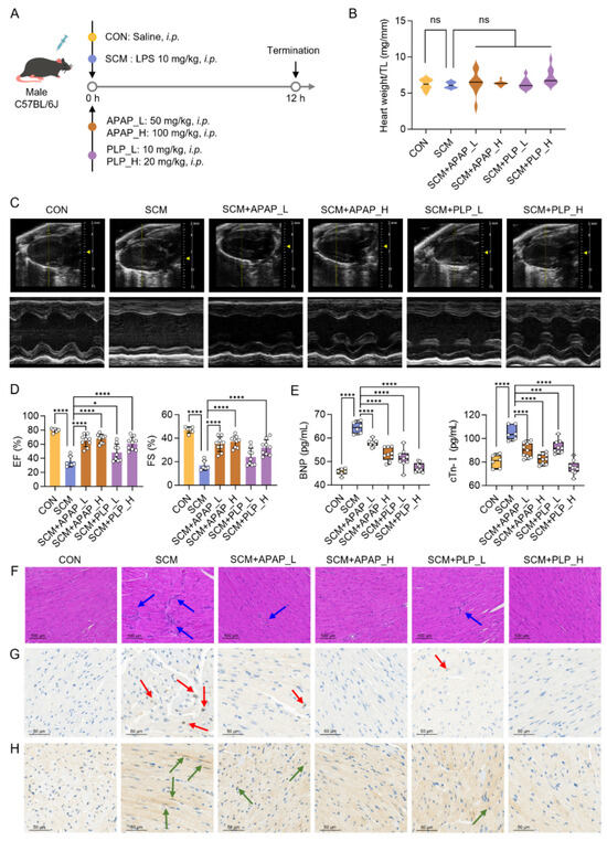
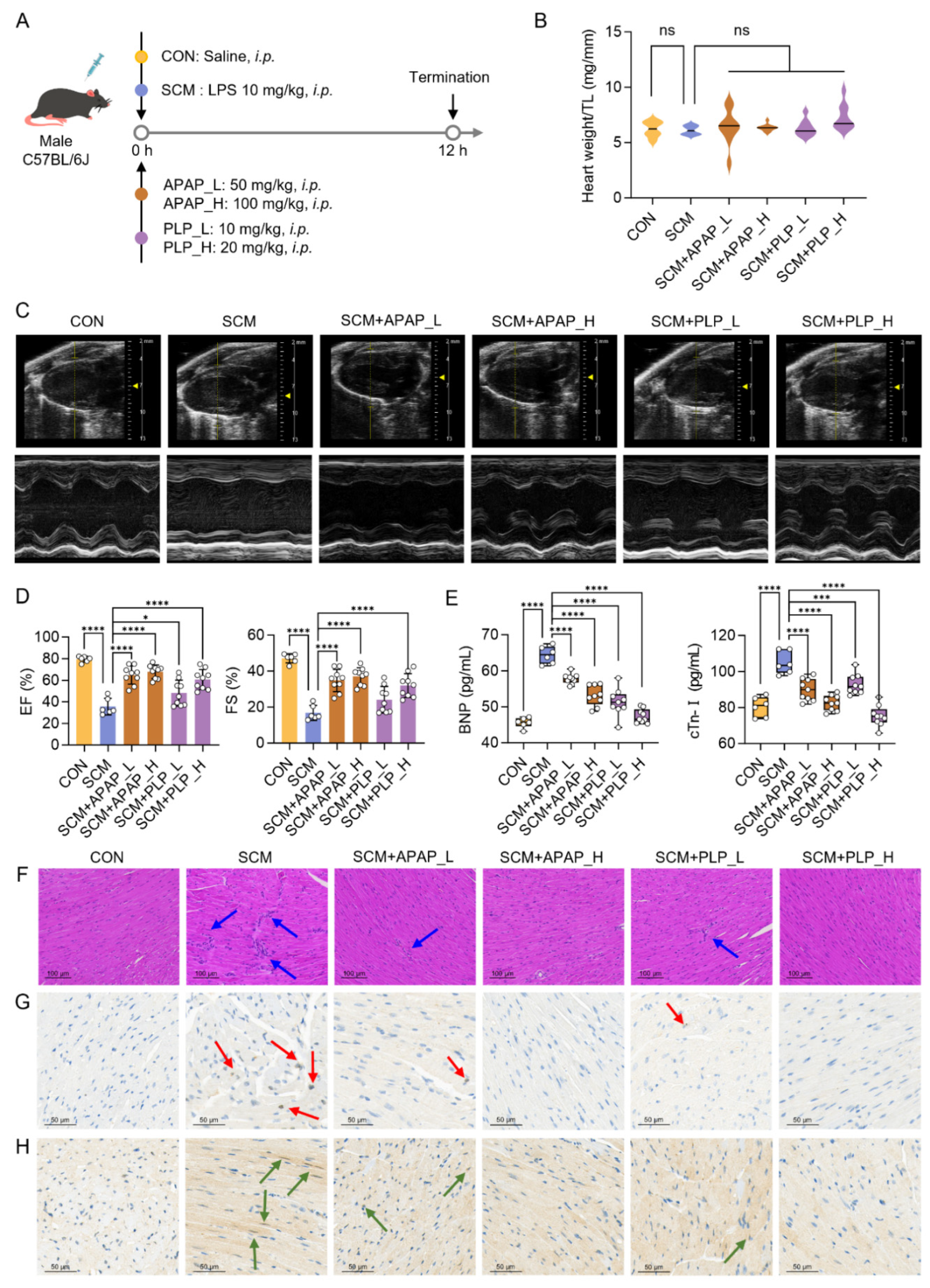
Figure 7.
Acetaminophen (APAP) and pyridoxal phosphate (PLP) protect against cardiac injury in septic cardiomyopathy (SCM) mice. (A) Schematic of SCM mouse model and intervention; (B) heart weight/tibia length ratio (heart weight/TL) in each group (CON, n = 6; SCM, n = 6; SCM+APAP_L, n = 9; SCM+APAP_H, n = 9; SCM+PLP_L, n = 9; SCM+PLP_H, n = 9); (C) representative conventional echocardiography images; (D) echocardiographic analysis of mouse heart function (EF: ejection fraction; FS: fractional shortening); (E) serum levels of brain natriuretic peptide (BNP) and cardiac troponin I (cTn-I); (F) histological examination of mouse hearts with hematoxylin–eosin (H&E) staining (n = 3) (blue arrows: inflammatory cell infiltration); (G,H) the immunohistochemical staining of CD45 (G) and CD68 (H) in mouse hearts (red arrows: CD45-positive cells, green arrows: CD68-positive cells). One-way ANOVA, * p < 0.05, *** p < 0.001, **** p < 0.0001, ns: no significant difference.
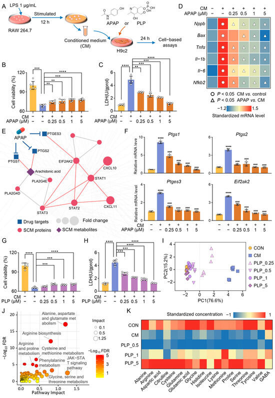
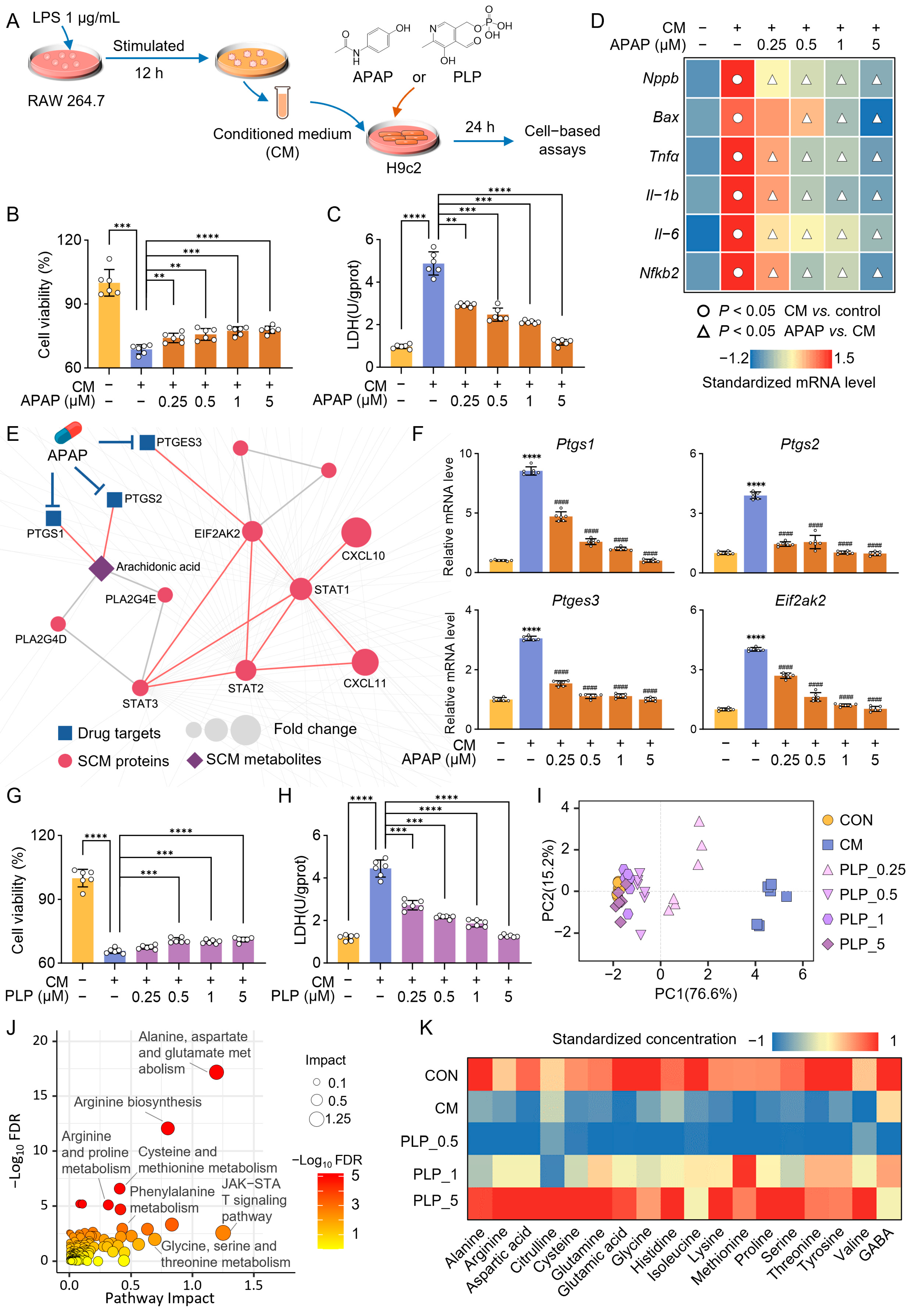
Figure 8.
Acetaminophen (APAP) and pyridoxal phosphate (PLP) protect the heart against sepsis by regulating inflammation-related pathways and amino acid metabolism pathways, respectively. (A) The workflow of cell experiments. (B,C) Cell viability (B) and lactate dehydrogenase (LDH) activity (C) of H9c2 cells following vehicle, conditioned-medium (CM), and APAP treatment (n = 6). (D) Heatmap of normalized expression of genes involved in apoptosis (Bax), cardiac injury (Nppa), and inflammation (Tnfα, Il-1b, Il-6, and Nfkb2) (n = 6). (E) The highlighted subnetwork shows the inferred mechanism of action for APAP’s protective effect in septic cardiomyopathy (SCM). (F) Gene expression levels of key targets of APAP in the treatment of SCM (n = 6), CM vs. control: **** p < 0.0001, APAP vs. CM: #### p < 0.0001. (G,H) Cell viability (G) and LDH activity (H) of H9c2 cells following vehicle, CM, and PLP treatment (n = 6). (I) Principal component analysis (PCA) scatter plot based on the expression of genes involved in apoptosis (Bax), cardiac injury (Nppa), and inflammation (Tnfα, Il-1b, Il-6, and Nfkb2) (n = 6). (J) Joint pathway analysis of PLP–SCM interaction network nodes. (K) Normalized concentrations of 18 amino acids in H9c2 (n = 3). One-way ANOVA, ** p < 0.01, *** p < 0.001, **** p < 0.0001.
Reference
- Liu, P.-P.; Yu, X.-Y.; Pan, Q.-Q.; Ren, J.-J.; Han, Y.-X.; Zhang, K.; Wang, Y.; Huang, Y.; Ban, T. Multi-Omics and Network-Based Drug Repurposing for Septic Cardiomyopathy. Pharmaceuticals 2025, 18, 43. [Google Scholar] [CrossRef] [PubMed]
Disclaimer/Publisher’s Note: The statements, opinions and data contained in all publications are solely those of the individual author(s) and contributor(s) and not of MDPI and/or the editor(s). MDPI and/or the editor(s) disclaim responsibility for any injury to people or property resulting from any ideas, methods, instructions or products referred to in the content. |
© 2025 by the authors. Licensee MDPI, Basel, Switzerland. This article is an open access article distributed under the terms and conditions of the Creative Commons Attribution (CC BY) license (https://creativecommons.org/licenses/by/4.0/).