Review

Perspectives on Light-Based Disinfection to Reduce the Risk of COVID-19 Transmission during Dental Care

by
Abdulrahman A. Balhaddad
1,2,
Lamia Mokeem
1,
Sharukh S. Khajotia
3,
Fernando L. Esteban Florez
3 and
Mary A. S. Melo
1,4,*
1
Dental Biomedical Sciences, University of Maryland School of Dentistry, Baltimore, MD 21201, USA
2
Department of Restorative Dental Sciences, College of Dentistry, Imam Abdulrahman Bin Faisal University, Dammam 31441, Saudi Arabia
3
Department of Restorative Sciences, Division of Dental Biomaterials, College of Dentistry, The University of Oklahoma Health Sciences Center, Oklahoma City, OK 73117, USA
4
Division of Operative Dentistry, Department of General Dentistry, University of Maryland Dental School, Baltimore, MD 21201, USA
*
Author to whom correspondence should be addressed.
BioMed 2022, 2(1), 27-36; https://doi.org/10.3390/biomed2010003
Submission received: 20 December 2021 / Revised: 7 January 2022 / Accepted: 7 January 2022 / Published: 10 January 2022
(This article belongs to the Topic Photodynamic Therapy)

Abstract

:
Severe Acute Respiratory Syndrome 2 (SARS-CoV-2) is a positive-sense single-stranded RNA coronavirus capable of causing potentially lethal pneumonia-like infectious diseases in mammals and birds. The main mechanisms by which SARS-CoV-2 spreads include airborne transmission (aerosols and droplets) and the direct exposure of tissues (conjunctival, nasal, and oral mucosa) to contaminated fluids. The aerosol formation is universal in dentistry due to the use of rotary instruments (handpieces), ultrasonic scalers, and air–water syringes. Several layers of infection control should protect key stakeholders such as dentists, dental staff, and patients. These include the utilization of personal protective equipment, high-volume evacuation systems, pre-procedural mouthwashes, rubber dam, and more recently, antimicrobial photodynamic therapy and intra-oral visible light irradiation. These non-specific light-based approaches are relatively simple, inexpensive, and effective against viruses, bacteria, and fungi. Therefore, the present perspective review discusses the current efforts and limitations on utilizing biophotonic approaches as adjunct infection control methods to prevent the transmission of SARS-CoV-2 in dental settings. In addition, the present perspective review may positively impact subsequent developments in the field, as it offers relevant information regarding the intricacies and complexities of infection control in dental settings.

1. Introduction

Coronaviruses are a large family of viruses that include the Middle East Respiratory Syndrome (MERS-CoV), Severe Acute Respiratory Syndrome (SARS-CoV), and Severe Acute Respiratory Syndrome 2 (SARS-CoV-2), which is responsible for the ongoing coronavirus disease (COVID-19) pandemic. These are known to cause infectious respiratory diseases in birds and mammals that range from the common cold to more severe and potentially lethal pneumonia-like diseases. SARS-CoV-2, a positive-sense single-stranded RNA virus, was transferred from bats (original host) and pangolins (intermediate host) to humans in wet markets in Wuhan [1]. According to Chinese health authorities, COVID-19 is clinically translated in patients in the form of flu-like symptoms, including fever (low to mild), runny nose, sputum production, abnormal CT scans of the lungs (opaque glass), and respiratory dysfunction [2]. Even though SARS-CoV-2 incubation times are restricted to between 5 and 6 days, 14 days of quarantine are typically recommended to individuals with suspected COVID-19 exposure, [3] as recent reports indicate that infected patients may continuously shed significant SARS-CoV-2 quanta (viral dose required to cause infection).
The highly contagious characteristics of SARS-CoV-2, its airborne transmission mechanisms, the occurrence of asymptomatic [4] and/or super-spreaders associated with intense intercontinental air travel have significantly favored the exponential spread of this virus amongst humans. These combined factors resulted in global proportions (5) characterized by unprecedented occupational challenges for healthcare workers, including physicians, nurses, dentists, and staff members. Such an event has overwhelmed the healthcare system of numerous countries and was shown to affect people independently of their socioeconomic status, gender, color, or age [5]. Despite the concerning mortality rates previously reported (2–15%), most cases are associated with mild symptoms that do not require any medical intervention or emergency care. However, in severe cases, affected patients display life-threatening conditions, such as dyspnea, acute respiratory distress syndrome (ARDS), acute cardiac injury, acute hepatic injury, acute kidney injury, and multi-organ failure. [6]
SARS-CoV-2 is transmitted via aerosols (<50 μm) and droplets (>50 μm) [7] formed during breathing, speaking, sneezing, and coughing [8,9]. Generated aerosols have been shown to remain suspended in air (still or turbulent) for long periods while maintaining their infectious nature for up to 3 h [10]. The settling of aerosols with potential pathogenic behavior on surfaces (polymeric, ceramic, or metallic) is also of concern for health care settings because contaminated surfaces may act as transmission sources [11].
Once a significant amount of virus has been inhaled by the host, primary viral replication occurs at the upper and lower respiratory tracts. [7] According to Ni et al., the Angiotensin Convertase Enzyme 2 (ACE2), which is a homolog of the angiotensin-converting enzyme (ACE), mediates SARS-CoV-2 infection by allowing the viral spike glycoprotein (two-part protein; S1—primary attachment, S2—viral infusion) to specifically bind to host cells’ membrane receptors. After initial attachment, virus’ and host cells’ membranes fuse, and viral RNA is then released into the cytoplasm, thereby establishing the COVID-19 infection. Even though varying levels of ACE2 expression have been found in different tissues and organs, recent studies have demonstrated that lungs display the highest levels of ACE2 expression in the human body. Other studies have found that SARS-CoV-2 display binding affinities to ACE2 10 to 20 times higher than those previously reported for SARS-CoV-1 [12], which indicates a multifactorial process (aerosol transmission, ACE2 expression, and affinity) by which SARS-CoV-2 preferentially attacks the lungs [13].
ACE2 has important and broad biological functionalities in negating the renin–angiotensin system’s role (RAS) in numerous diseases, such as hypertension, myocardial infarction, and heart failure. In addition, ACE2 has also been identified in multiple sites within the oral cavity, including the tongue, gingival tissues, and buccal mucosa, which indicates that the oral cavity could work as a port of entry for SARS-CoV-2. [14]

2. SARS-CoV-2 Transmission in Dental Settings

Even though the current scientific evidence is not conclusive in demonstrating the transmission of SARS-CoV-2 in dental settings due to routine dental procedures (e.g., prophylaxis, cavity preparation, and ultrasonic scaling), it has been shown that SARS-CoV-2 is present in saliva. According to recent studies, SARS-CoV-2 is transferred from the respiratory tract to the oral cavity via liquid droplets and blood from gingival cervical fluids or salivary glands [15]. Despite this knowledge gap, it is well known that aerosol generation is ubiquitous in dentistry, which is considered critical for transmitting SARS-CoV-2 [16]. Previous reports have indicated that the amount of aerosol generated is procedure-specific. The amount of aerosols in dental prophylaxis, cavity preparation, root planing and scaling is dependent on the type of handpiece used (low- and high-speed rotary instruments, ultrasonic scalers, and air/water syringes) (Figure 1). The hands-on nature and limited field of view characteristic in dentistry practice obligate both dentists and dental assistants to work nearby (almost face-to-face) to the source where aerosols with potential pathogenic behavior are generated (e.g., the oral cavity).
In this critical scenario, several manuscripts have indicated that dental settings are particularly susceptible to transmitting different types of microorganisms, including bacteria, fungi, and viruses (Figure 2). In addition, other viruses (noroviruses, rabies, hepatitis B and C, human papillomavirus, Epstein–Barr, herpes simplex, HIV, etc.) [17,18] are present in the oral cavity. These can be efficiently transmitted during dental procedures because aerosols typically get in touch with saliva, dental plaque, and blood [19]. Therefore, based on the context presented and considering the intrinsic characteristics of SARS-CoV-2′s and its transmission mechanisms, it is possible to affirm that the oral cavity is a potential source for the transmission of SARS-CoV-2. In a recent report conducted among 2195 dentists in the United States, approximately 0.9% of them were tested positive for SARS-CoV-2 [20]. As aerosols are associated with higher risks for SARS-CoV-2 transmission, many dental organizations have released new infection control protocols in dental settings [21,22].

3. Infection Control in Dental Settings

Oral care providers must be familiar with SARS-CoV-2 transmission routes in dental settings. Recently proposed approaches are implementing remote screening procedures (Zoom, Skype, and FaceTime) before the dental visit (24) and limiting the number of key stakeholders in the building to allow for social distancing and the maintenance of fundamental clinical skills operations. It is also essential to frequently disinfect surfaces in clinical (dental chair, cabinets, floors, etc.) and non-clinical areas (restrooms, chairs, sofas, front desk, etc.) that are directly or indirectly exposed to aerosols generated during routine clinical care. Besides, oral care providers should recognize patients with suspected exposure to SARS-CoV-2 or those displaying symptoms of COVID-19 and refer them for further testing and potential treatment, as necessary [23]. To prevent any physical contact with patients during screening procedures and before granting them access to the building, it is advisable that the dental staff use free-hand forehead thermometers to measure patients’ temperatures. However, even though laser-based thermometers offer the advantage of a contactless screening procedure, some reports have indicated that these devices cannot accurately determine patients’ temperatures when the weather is cold [24]. Consequently, repeated temperature measurements in non-clinical areas (e.g., waiting room) are necessary before executing any dental procedure [25].
Based on the highly infectious nature of SARS-CoV-2, its transmission routes, and the clinical practice of dentistry, which generates large volumes of aerosols with potential pathogenic behavior, it is recommended that three levels of protective measures (primary, secondary, and tertiary) be used [19]. The primary protection level is intended to create a physical barrier that protects the operator and the supporting staff and includes disposable gloves, gowns, surgical masks, N95 masks, goggles, and face shields. Secondary protection involves using disposable isolation clothing over primary protection to further prevent the operator’s exposure and shield staff from exposure to highly contagious organic contaminants. Finally, the tertiary level of protection is based on the utilization of containment clothing (also known as Haz-suits) and P95 respirators to fully isolate the operator and supporting staff when a COVID-19 positive patient must be admitted into the dental clinics for emergency dental care [19].
Recent reports have suggested using high-volume saliva evacuators, aerosol containment devices, and four-hand techniques to minimize aerosols’ generation with pathogenic behavior in dentistry [26,27]. Despite this critical scenario, elective dental procedures are being conducted in many countries without proper mitigation strategies and independently of the risks of the transmission of SARS-CoV-2. According to Samaranayake, Reid, and Evans (33), dental dams’ intraoral utilization can efficiently control approximately 70% of all airborne particles generated during routine dental treatment. Therefore, it should be used as a mechanism to diminish the transmission of SARS-CoV-2 in dental settings. Anti-retraction handpieces are another critical layer of protection in clinical dentistry. These prevent the backflow of liquids containing microorganisms commonly found in the oral cavity into the handpiece’s tubbings [28] and has been shown to decrease the potential for hepatitis B transmission. Detailed guidelines for the safe treatment of patients in dental offices during the COVID-19 pandemics can be accessed at the Centers for Disease Control and Prevention (CDC) [22].

4. Preprocedural Disinfection to Prevent the Spreading of SARS-CoV-2

The use of antiseptic mouth rinses before dental procedures has been shown to reduce the microbial load inside the oral cavity, thereby diminishing the risk for the transmission of infectious diseases in dental settings. Chlorohexidine, the most commonly used mouth rinse in dentistry, was demonstrated not to be effective against SARS-CoV-2 [9,29]. However, ongoing investigations have shown possibilities of contributions to reduce viral load. One study has shown that using 0.12% chlorhexidine as a mouth rinse could suppress the activity of SARS-CoV-2 inside the oral cavity for 2 h [30]. Another study has indicated that combining ethanol with chlorohexidine increases the chances for the inactivation of SARS-CoV-2 [31]. Another recently tested approach involved the utilization of povidone-iodine (PVP-I) as a rinse to inactivate SARS-CoV-2 [32,33].
The efficacy of hydrogen peroxide-containing mouth rinses has also been tested, and reported results have demonstrated that these materials only displayed limited inactivation levels against SARS-CoV-2 [33,34]. Therefore, it can be concluded that different mouth rinses display varying inactivation levels against SARS-CoV-2 and that their utilization should not be focused on preventing SARS-CoV-2 transmission in dental settings. Further investigations are necessary to elucidate the mechanisms of action by which these materials may exert unintended antiviral actions against SARS-CoV-2 and determine whether their long-term utilization may increase the risks associated with developing multi-drug resistant bacteria in the oral cavity. In this context, non-specific, broad range, and inexpensive disinfection strategies must be developed, investigated, and validated to mitigate the potential for transmitting infectious diseases in dental settings.

5. Current Trends in Light-Based Oral Disinfection

The use of antimicrobial photodynamic therapy (aPDT) to disinfect dental tissues (soft and hard) has been reported in previous studies [35,36]. aPDT has been shown to effectively reduce oral cariogenic bacteria’s viability and those implicated in gingivitis, periodontitis, peri-implantitis, and root canal infections [37]. In this technique, a photosensitizer is typically used in combination with a specific wavelength to generate large quantities of different reactive oxygen species (ROS) such as singlet and triplet oxygen [38]. and These highly oxidative and short-lived species can oxidize, in a non-specific manner, the membranes of numerous microorganisms present in the oral cavity [39]. aPDT is a minimally invasive and ultraconservative technique with several advantages compared to traditional antibiotic disinfection approaches, including its broad-spectrum efficacy against bacteria, fungi, and viruses and its implementation is fast and inexpensive [40,41]. Several studies have also demonstrated that the long-term utilization of aPDT does not induce the development of resistant bacteria because its mechanism of action does not depend on a structure or metabolic pathway [42]. The vast majority of studies investigating the efficacy of aPDT against oral microorganisms have focused on specific target sites such as teeth, periodontal tissues, root canals, or alveolar bones. However, a few reports are available regarding the use of aPDT as a disinfecting approach for the entire oral cavity [43]. In the disinfection approach, the patient is asked to rinse his/her mouth with a photosensitizer’s solution, and then, a light irradiation is used to activate the photosensitizer and disinfect the oral cavity (Figure 3).
In 2012, Araujo et al. investigated the efficacy of aPDT to promote oral cavity disinfection in a pilot clinical trial for the first time in dentistry [44]. In that study, participants were instructed not to use any antibiotics or be subjected to professional dental hygiene before the study. Participants fasted for 12 h before collecting saliva samples (before aPDT). After baseline saliva collection, participants were required to swish the curcumin photosensitizer for 5 min forcefully. After that, a visible wavelength (457 nm ± 15 nm) emitted from a light-emitting diode (LED, 67 mW/cm2) was used to irradiate the oral cavity for 5 min (20.1 J/cm2). Participants in the control group swished the photosensitizer for 5 min, but the light was not used. Immediately after the intervention (aPDT), saliva samples were collected to assess the presence and quantity of common oral microorganisms. Results reported lower microbial loads in participants treated with aPDT than those observed in the control group.
Even though all participants in the aPDT group have had microbial loads lower than baseline levels, 50% of those participants displayed microbial reductions higher than 80%. However, in the control group, most participants (>50%) did not show any reduction in the observed microbial load. In combination, the findings reported by Araujo et al. in 2012 indicate that aPDT has a solid potential to be used as an adjunct preprocedural disinfection technique to diminish the transmission of airborne infectious diseases. However, despite these promising results, the study cited did not investigate the efficacy of protocols proposed in controlling the oral microbial load 30 to 60 min after the intervention. Such determination is critically important in today’s day and age because most dental procedures last for almost an hour and generate large quantities of aerosols with pathogenic behavior [44]. Based on a substantial amount of promising results, [43] the research group led by Dr. Bagnato in Brazil advances the clinical investigation on the efficacy of aPDT approaches to prevent oral infections by conducting a two-arm-randomized clinical trial.
Leite et al. [43] conducted a study under similar conditions but with higher clinical relevancy because saliva collections took place before disinfection, immediately after aPDT, and 1 and 2 h after that [43]. In that study, participants were then divided into three groups (aPDT, light irradiation only, and curcumin only) and were asked to use 20 mL of curcumin (30 mg/L) for 5 min, followed by visible light irradiation (455 nm ± 15 nm) of the oral cavity for 5 min (≅200 J/cm2). Time-dependent (immediate, 1 h and 2 h after aPDT) analysis of results demonstrated that aPDT could exert significant and sustainable microbial load reductions in the order of 1-log (90%) compared to baseline values. These promising results were not observed in groups subjected to either light irradiation or curcumin alone, thereby underscoring the necessity to combine light irradiation with a photosensitizer to promote ROS and the consequent death of oral microorganisms. These are critical results because most routine dental visits are usually restricted to a couple of hours. Therefore, determining aPDT’s long-term efficacy to disinfect the oral cavity is of fundamental importance to diminish the chances of transmitting infectious diseases in dental settings (Box 1) [43].
Box 1. Summary of the clinical studies concerning the use of antimicrobial photodynamic therapy (aPDT) to disinfect the oral cavity prior to dental procedures.
  • The most frequently used photosensitizer to disinfect the oral cavity is curcumin at the following concentrations: 25 mg/L; 30 mg/L; 100 mg/L; 1 g/L, and 1.5 g/L.
  • Typical doses of energy used include 20.1 J/cm2, 85 J/cm2, 100 J/cm2 and 200 J/cm2.
  • Concentration-dependent mechanism (higher concentration = higher reductions).
  • High curcumin concentrations were found to exert long-term (24 h) effects.
  • Future studies should consider different photosensitizers and energy doses.
  • aPDT disinfection approaches cited should be investigated against SARS-CoV-2.
In 2016, Panhóca et al. tested the efficacy of aPDT mediated by visible light and curcumin combined or not to sodium dodecyl sulfate (surfactant with known antibacterial properties) as an oral decontamination approach for patients with fixed orthodontic appliances. In that study, participants were randomly distributed into four groups: aPDT (curcumin (1 g/L) + light irradiation (450 nm ± 10 nm)), PDT (curcumin (1 g/L)+ sodium dodecyl sulfate (SDS, 0.1%)) + light irradiation (450 nm ± 10 nm)), light irradiation alone (450 nm ± 10 nm), and 0.12% chlorhexidine gluconate (CHX) [45]. Participants were then requested to forcefully swish the photosensitizer solution with and without the surfactant for 2 min. The intraoral irradiation was then performed for 5 min, with a dose of energy of approximately 100 J/cm2. Participants in the group subjected to CHX forcefully swished the solution for 30 s.
In all groups, saliva collection occurred immediately after swishing (curcumin alone, curcumin + surfactant or 5% DMSO in water), and immediately after the aPDT. The observed microbial load reductions indicated that the tested protocols established a decreasing efficacy order by which the investigated treatments could be rank-ordered in terms of their antimicrobial potential, as follows: CHX > aPDT + SDS > aPDT > light only. The reported results not only confirm that aPDT can reduce the oral microbial load in orthodontic patients with fixed appliances but have also indicated that surfactants such as SDS may be used in combination to improve the efficacy of aPDT. Therefore, additional studies should be conducted to expand the parameters tested and investigate the effectiveness of light-based approaches against SARS-CoV-2. In addition, it is recommended that follow-up studies consider collecting saliva after 1 and 2 h to explore the intervention’s sustainability.
The previous study concerning the use of PDT for oral cavity disinfection was conducted among 50 healthy participants. Visible light (either 450 nm ± 10 nm or 630 nm ± 10 nm) emitted from LED light sources were used to irradiate either curcumin (25 and 100 mg/L) or Photogem (25 and 100 µg/L) [46]. Saliva collection was performed immediately following aPDT and 24 h after. Even though aPDT mediated by curcumin (25 mg/mL) and blue light was shown to reduce the microbial load by 1-log immediately after the intervention, a similar trend could not be observed after 24 h. However, when the aPDT was mediated by curcumin at higher concentrations (100 mg/mL), the immediate microbial load reduction of 1-log was observed after the intervention and maintained for up to 24 h, suggesting a concentration-dependent mechanism, by which sustainability may be achieved through biophotonic approaches. For Photogem, both concentrations tested (25 and 100 µg/mL) immediately induced significant microbial reductions (1-log and 1.5-log, respectively). Furthermore, the results at 24 h indicated microbial loads similar to baseline levels and those on control groups.
In combination, aPDT mediated by curcumin (100 mg/mL) and blue light irradiation may exert microbial load reductions that last for up to 24 h, which indicates that these techniques could be used to significantly decrease the microbial load in the oral cavity when lengthy procedures (orthognathic surgeries, facial reconstruction, and full mouth rehabilitation) are to be performed. On the other hand, aPDT mediated by Photogem and red light irradiation could be efficiently used to significantly decrease the oral cavity’s microbial load for short periods. Additional studies are made necessary to further explore the long-term efficacy of Photogem against the oral cavity microbial load [46].
The current scientific evidence on biophotonic techniques suggests that aPDT can be safely and effectively used as adjunct preprocedural disinfection protocols to decrease the microbial load in the oral cavity and potentially reduce the spreading of highly infectious viral diseases in dental settings [47]. Despite these indications, it is crucial to understand previous reports’ limitations when using published results (from in vitro, in situ, and in vivo studies) to prove safety and clinical efficacy. The significant restriction relies on reducing oral bacteria, and no reports are available to substantiate the efficacy of biophotonics techniques against SARS-CoV-2. Even though it has been documented that aPDT is effective against several oral and respiratory viruses, including herpes simplex and oral human papilloma, the effect of aPDT against SARS-CoV-2 in saliva has not been established to date.
Therefore, studies should be conducted using similar experimental designs to address the viral load and viability of SARS-CoV-2 in saliva following aPDT protocols. Indocyanine green (ICG) irradiated by a diode laser was found helpful in inactivating herpes simplex types 1 and 2 isolated from the oral cavity in two studies [48,49]. In addition, several studies have reported that ultraviolet irradiation can efficiently inactivate different types of coronaviruses [50].

6. Perspective and Future Outlook

Based on the reported evidence, light-based approaches to disinfect the oral cavity before regular dental exams or treatments are promising but not thoroughly investigated. Studies on the efficacy of aPDT in reducing SARS-CoV-2 levels in the oral cavity and suspended in saliva are lacking. Some points should be considered when designing future clinical trials in this area. Optimum dosimetry (wavelength, power intensity, time, continuous, pulsed, etc.), photosensitizer type and concentration, pre-irradiation time, and the use of surfactants are some of the relevant parameters. For intra-oral applications, it is crucial to determine the effect of influencing factors, such as the position of the light source concerning the oral cavity, irradiation angle (to control non-linear scattering and reflection phenomena), and distance light tip-target area, to name a few. Light-based disinfection approaches that seek to reduce viral and bacterial load can help prevent transmission during dental care, but must be placed in a broader oral health context, recognizing that a single approach using light is insufficient to address the need to prevent the spreading of SARS-CoV-2 in dental settings.

Author Contributions

A.A.B., L.M., S.S.K., F.L.E.F. and M.A.S.M. contributed to the design and the writing of the manuscript. S.S.K., F.L.E.F. and M.A.S.M. contributed to the critical review of the manuscript. All authors have read and agreed to the published version of the manuscript.

Funding

This research received no external funding.

Institutional Review Board Statement

Not applicable.

Informed Consent Statement

Not applicable.

Data Availability Statement

Not applicable.

Acknowledgments

A.A.B. acknowledges the scholarship during his Ph.D. studies from the Imam Abdulrahman bin Faisal University, Dammam, Saudi Arabia, and the Saudi Arabian Cultural Mission.

Conflicts of Interest

The authors declare no conflict of interest.

References

  1. Zhu, H.; Wei, L.; Niu, P. The Novel Coronavirus Outbreak in Wuhan, China. Glob. Health Res. Policy 2020, 5, 6. [Google Scholar] [CrossRef] [Green Version]
  2. Song, F.; Shi, N.; Shan, F.; Zhang, Z.; Shen, J.; Lu, H.; Ling, Y.; Jiang, Y.; Shi, Y. Emerging 2019 Novel Coronavirus (2019-NCoV) Pneumonia. Radiology 2020, 295, 210–217. [Google Scholar] [CrossRef] [Green Version]
  3. Backer, J.A.; Klinkenberg, D.; Wallinga, J. Incubation Period of 2019 Novel Coronavirus (2019-NCoV) Infections among Travellers from Wuhan, China, 20-28 January 2020. Eurosurveillance 2020, 25, 2000062. [Google Scholar] [CrossRef] [PubMed] [Green Version]
  4. Li, Q.; Guan, X.; Wu, P.; Wang, X.; Zhou, L.; Tong, Y.; Ren, R.; Leung, K.S.M.; Lau, E.H.Y.; Wong, J.Y.; et al. Early Transmission Dynamics in Wuhan, China, of Novel Coronavirus-Infected Pneumonia. N. Engl. J. Med. 2020, 382, 1199–1207. [Google Scholar] [CrossRef]
  5. Sohrabi, C.; Alsafi, Z.; O’Neill, N.; Khan, M.; Kerwan, A.; Al-Jabir, A.; Iosifidis, C.; Agha, R. World Health Organization Declares Global Emergency: A Review of the 2019 Novel Coronavirus (COVID-19). Int. J. Surg. 2020, 76, 71–76. [Google Scholar] [CrossRef]
  6. Zaim, S.; Chong, J.H.; Sankaranarayanan, V.; Harky, A. COVID-19 and Multiorgan Response. Curr. Probl. Cardiol. 2020, 45, 100618. [Google Scholar] [CrossRef] [PubMed]
  7. Subbarao, K.; Mahanty, S. Respiratory Virus Infections: Understanding COVID-19. Immunity 2020, 52, 905–909. [Google Scholar] [CrossRef] [PubMed]
  8. Chen, J. Pathogenicity and Transmissibility of 2019-NCoV-A Quick Overview and Comparison with Other Emerging Viruses. Microbes Infect. 2020, 22, 69–71. [Google Scholar] [CrossRef]
  9. Kampf, G.; Todt, D.; Pfaender, S.; Steinmann, E. Persistence of Coronaviruses on Inanimate Surfaces and Their Inactivation with Biocidal Agents. J. Hosp. Infect. 2020, 104, 246–251. [Google Scholar] [CrossRef] [Green Version]
  10. Harrel, S.K.; Molinari, J. Aerosols and Splatter in Dentistry: A Brief Review of the Literature and Infection Control Implications. J. Am. Dent. Assoc. 2004, 135, 429–437. [Google Scholar] [CrossRef]
  11. Chia, P.Y.; Coleman, K.K.; Tan, Y.K.; Ong, S.W.X.; Gum, M.; Lau, S.K.; Lim, X.F.; Lim, A.S.; Sutjipto, S.; Lee, P.H.; et al. Detection of Air and Surface Contamination by SARS-CoV-2 in Hospital Rooms of Infected Patients. Nat. Commun. 2020, 11, 2800. [Google Scholar] [CrossRef] [PubMed]
  12. Wrapp, D.; Wang, N.; Corbett, K.S.; Goldsmith, J.A.; Hsieh, C.-L.; Abiona, O.; Graham, B.S.; McLellan, J.S. Cryo-EM Structure of the 2019-NCoV Spike in the Prefusion Conformation. Science 2020, 367, 1260–1263. [Google Scholar] [CrossRef] [Green Version]
  13. Hamming, I.; Timens, W.; Bulthuis, M.L.C.; Lely, A.T.; Navis, G.J.; van Goor, H. Tissue Distribution of ACE2 Protein, the Functional Receptor for SARS Coronavirus. A First Step in Understanding SARS Pathogenesis. J. Pathol. 2004, 203, 631–637. [Google Scholar] [CrossRef]
  14. Xu, H.; Zhong, L.; Deng, J.; Peng, J.; Dan, H.; Zeng, X.; Li, T.; Chen, Q. High Expression of ACE2 Receptor of 2019-NCoV on the Epithelial Cells of Oral Mucosa. Int. J. Oral Sci. 2020, 12, 8. [Google Scholar] [CrossRef]
  15. Sabino-Silva, R.; Jardim, A.C.G.; Siqueira, W.L. Coronavirus COVID-19 Impacts to Dentistry and Potential Salivary Diagnosis. Clin. Oral Investig. 2020, 24, 1619–1621. [Google Scholar] [CrossRef] [Green Version]
  16. Banakar, M.; Bagheri Lankarani, K.; Jafarpour, D.; Moayedi, S.; Banakar, M.H.; MohammadSadeghi, A. COVID-19 Transmission Risk and Protective Protocols in Dentistry: A Systematic Review. BMC Oral Health 2020, 20, 275. [Google Scholar] [CrossRef]
  17. Hindson, J. COVID-19: Faecal-Oral Transmission? Nat. Rev. Gastroenterol. Hepatol. 2020, 17, 259. [Google Scholar] [CrossRef]
  18. De Graaf, M.; Beck, R.; Caccio, S.M.; Duim, B.; Fraaij, P.L.; Le Guyader, F.S.; Lecuit, M.; Le Pendu, J.; de Wit, E.; Schultsz, C. Sustained Fecal-Oral Human-to-Human Transmission Following a Zoonotic Event. Curr. Opin. Virol. 2017, 22, 1–6. [Google Scholar] [CrossRef] [PubMed]
  19. Peng, X.; Xu, X.; Li, Y.; Cheng, L.; Zhou, X.; Ren, B. Transmission Routes of 2019-NCoV and Controls in Dental Practice. Int. J. Oral Sci. 2020, 12, 9. [Google Scholar] [CrossRef]
  20. Estrich, C.G.; Mikkelsen, M.; Morrissey, R.; Geisinger, M.L.; Ioannidou, E.; Vujicic, M.; Araujo, M.W.B. Estimating COVID-19 Prevalence and Infection Control Practices among US Dentists. J. Am. Dent. Assoc. 2020, 151, 815–824. [Google Scholar] [CrossRef] [PubMed]
  21. American Dental Association. ADA Interim Guidance for Minimizing Risk of COVID-19 Transmission Risk when Treating Dental Emergencies. Available online: https://www.ada.org/publications/ada-news/2020/april/ada-releases-interim-guidance-on-minimizing-covid-19-transmission-risk-when-treating-emergencies (accessed on 1 April 2020).
  22. Centers for Disease and Prevention. Guidance for Dental Settings: Interim Infection Prevention and Control Guidance for Dental Settings during the Coronavirus Disease (COVID-19) Pandemic. Available online: Https://Www.Cdc.Gov/Coronavirus/2019-Ncov/Hcp/Dental-Settings.Html (accessed on 29 June 2020).
  23. World Health Organization. Clinical Management of Severe Acute Respiratory Infection When Novel Coronavirus (2019-NCoV) Infection Is Suspected: Interim Guidance. 2020. Available online: https://Www.Who.Int/Publications-Detail/Clinical-Management-of-Severe-Acute-Respiratory-InfecTion-When-Novel-Coronavirus-(Ncov)-Infection-Is-Suspected (accessed on 17 February 2020).
  24. Dzien, C.; Halder, W.; Winner, H.; Lechleitner, M. Covid-19 Screening: Are Forehead Temperature Measurements during Cold Outdoor Temperatures Really Helpful? Wien. Klin. Wochenschr. 2021, 133, 331–335. [Google Scholar] [CrossRef] [PubMed]
  25. Hsiao, S.-H.; Chen, T.-C.; Chien, H.-C.; Yang, C.-J.; Chen, Y.-H. Measurement of Body Temperature to Prevent Pandemic COVID-19 in Hospitals in Taiwan: Repeated Measurement Is Necessary. J. Hosp. Infect. 2020, 105, 360–361. [Google Scholar] [CrossRef]
  26. Bescos, R.; Casas-Agustench, P.; Belfield, L.; Brookes, Z.; Gabaldón, T. Coronavirus Disease 2019 (COVID-19): Emerging and Future Challenges for Dental and Oral Medicine. J. Dent. Res. 2020, 99, 1113. [Google Scholar] [CrossRef]
  27. Samaranayake, L.P.; Peiris, M. Severe Acute Respiratory Syndrome and Dentistry: A Retrospective View. J. Am. Dent. Assoc. 2004, 135, 1292–1302. [Google Scholar] [CrossRef] [PubMed] [Green Version]
  28. Hu, T.; Li, G.; Zuo, Y.; Zhou, X. Risk of Hepatitis B Virus Transmission via Dental Handpieces and Evaluation of an Anti-Suction Device for Prevention of Transmission. Infect. Control. Hosp. Epidemiol. 2007, 28, 80–82. [Google Scholar] [CrossRef]
  29. Fehr, A.R.; Perlman, S. Coronaviruses: An Overview of Their Replication and Pathogenesis. Methods Mol. Biol. 2015, 1282, 1–23. [Google Scholar] [CrossRef] [Green Version]
  30. Yoon, J.G.; Yoon, J.; Song, J.Y.; Yoon, S.-Y.; Lim, C.S.; Seong, H.; Noh, J.Y.; Cheong, H.J.; Kim, W.J. Clinical Significance of a High SARS-CoV-2 Viral Load in the Saliva. J. Korean Med. Sci. 2020, 35, e195. [Google Scholar] [CrossRef] [PubMed]
  31. Kelly, N.; Nic Íomhair, A.; McKenna, G. Can Oral Rinses Play a Role in Preventing Transmission of Covid 19 Infection? Evid. Based Dent. 2020, 21, 42–43. [Google Scholar] [CrossRef]
  32. Bidra, A.S.; Pelletier, J.S.; Westover, J.B.; Frank, S.; Brown, S.M.; Tessema, B. Rapid In-Vitro Inactivation of Severe Acute Respiratory Syndrome Coronavirus 2 (SARS-CoV-2) Using Povidone-Iodine Oral Antiseptic Rinse. J. Prosthodont. 2020, 29, 529–533. [Google Scholar] [CrossRef]
  33. Bidra, A.S.; Pelletier, J.S.; Westover, J.B.; Frank, S.; Brown, S.M.; Tessema, B. Comparison of In Vitro Inactivation of SARS CoV-2 with Hydrogen Peroxide and Povidone-Iodine Oral Antiseptic Rinses. J Prosthodont 2020, 29, 599–603. [Google Scholar] [CrossRef]
  34. Gottsauner, M.J.; Michaelides, I.; Schmidt, B.; Scholz, K.J.; Buchalla, W.; Widbiller, M.; Hitzenbichler, F.; Ettl, T.; Reichert, T.E.; Bohr, C.; et al. A Prospective Clinical Pilot Study on the Effects of a Hydrogen Peroxide Mouthrinse on the Intraoral Viral Load of SARS-CoV-2. Clin. Oral Investig. 2020, 24, 3707–3713. [Google Scholar] [CrossRef]
  35. Melo, M.a.S.; Rolim, J.P.M.L.; Passos, V.F.; Lima, R.A.; Zanin, I.C.J.; Codes, B.M.; Rocha, S.S.; Rodrigues, L.K.A. Photodynamic Antimicrobial Chemotherapy and Ultraconservative Caries Removal Linked for Management of Deep Caries Lesions. Photodiagn. Photodyn. Ther. 2015, 12, 581–586. [Google Scholar] [CrossRef] [Green Version]
  36. Melo, M.A.S. Photodynamic Antimicrobial Chemotherapy as a Strategy for Dental Caries: Building a More Conservative Therapy in Restorative Dentistry. Photomed. Laser Surg. 2014, 32, 589–591. [Google Scholar] [CrossRef]
  37. Balhaddad, A.A.; Garcia, I.M.; Ibrahim, M.S.; Rolim, J.P.M.L.; Gomes, E.A.B.; Martinho, F.C.; Collares, F.M.; Xu, H.; Melo, M.A.S. Prospects on Nano-Based Platforms for Antimicrobial Photodynamic Therapy Against Oral Biofilms. Photobiomodul. Photomed. Laser Surg. 2020, 38, 481–496. [Google Scholar] [CrossRef]
  38. Rolim, J.P.M.L.; de-Melo, M.A.S.; Guedes, S.F.; Albuquerque-Filho, F.B.; de Souza, J.R.; Nogueira, N.A.P.; Zanin, I.C.J.; Rodrigues, L.K.A. The Antimicrobial Activity of Photodynamic Therapy against Streptococcus Mutans Using Different Photosensitizers. J. Photochem. Photobiol. B 2012, 106, 40–46. [Google Scholar] [CrossRef]
  39. Balhaddad, A.A.; AlQranei, M.S.; Ibrahim, M.S.; Weir, M.D.; Martinho, F.C.; Xu, H.H.K.; Melo, M.A.S. Light Energy Dose and Photosensitizer Concentration Are Determinants of Effective Photo-Killing against Caries-Related Biofilms. Int. J. Mol. Sci. 2020, 21, 7612. [Google Scholar] [CrossRef] [PubMed]
  40. Alfirdous, R.A.; Garcia, I.M.; Balhaddad, A.A.; Collares, F.M.; Martinho, F.C.; Melo, M.A.S. Advancing Photodynamic Therapy for Endodontic Disinfection with Nanoparticles: Present Evidence and Upcoming Approaches. Appl. Sci. 2021, 11, 4759. [Google Scholar] [CrossRef]
  41. Balhaddad, A.A.; Xia, Y.; Lan, Y.; Mokeem, L.; Ibrahim, M.S.; Weir, M.D.; Xu, H.H.K.; Melo, M.A.S. Magnetic-Responsive Photosensitizer Nanoplatform for Optimized Inactivation of Dental Caries-Related Biofilms: Technology Development and Proof of Principle. ACS Nano 2021, 15, 19888–19904. [Google Scholar] [CrossRef] [PubMed]
  42. Teófilo, M.Í.S.; de Carvalho Russi, T.M.A.Z.; de Barros Silva, P.G.; Balhaddad, A.A.; Melo, M.A.S.; Rolim, J.P.M.L. The Impact of Photosensitizers Selection on Bactericidal Efficacy of PDT against Cariogenic Biofilms: A Systematic Review and Meta-Analysis. Photodiagn. Photodyn. Ther. 2020, 33, 102046. [Google Scholar] [CrossRef]
  43. Leite, D.P.V.; Paolillo, F.R.; Parmesano, T.N.; Fontana, C.R.; Bagnato, V.S. Effects of Photodynamic Therapy with Blue Light and Curcumin as Mouth Rinse for Oral Disinfection: A Randomized Controlled Trial. Photomed. Laser Surg. 2014, 32, 627–632. [Google Scholar] [CrossRef] [PubMed] [Green Version]
  44. Araújo, N.C.; Fontana, C.R.; Gerbi, M.E.M.; Bagnato, V.S. Overall-Mouth Disinfection by Photodynamic Therapy Using Curcumin. Photomed. Laser Surg. 2012, 30, 96–101. [Google Scholar] [CrossRef]
  45. Panhóca, V.H.; Esteban Florez, F.L.; Corrêa, T.Q.; Paolillo, F.R.; de Souza, C.W.O.; Bagnato, V.S. Oral Decontamination of Orthodontic Patients Using Photodynamic Therapy Mediated by Blue-Light Irradiation and Curcumin Associated with Sodium Dodecyl Sulfate. Photomed. Laser Surg. 2016, 34, 411–417. [Google Scholar] [CrossRef] [PubMed]
  46. Ricci Donato, H.A.; Pratavieira, S.; Grecco, C.; Brugnera-Júnior, A.; Bagnato, V.S.; Kurachi, C. Clinical Comparison of Two Photosensitizers for Oral Cavity Decontamination. Photomed. Laser Surg. 2017, 35, 105–110. [Google Scholar] [CrossRef] [PubMed] [Green Version]
  47. Kellesarian, S.V.; Qayyum, F.; de Freitas, P.C.; Akram, Z.; Javed, F. Is Antimicrobial Photodynamic Therapy a Useful Therapeutic Protocol for Oral Decontamination? A Systematic Review and Meta-Analysis. Photodiagn. Photodyn. Ther. 2017, 20, 55–61. [Google Scholar] [CrossRef] [PubMed] [Green Version]
  48. Namvar, M.A.; Vahedi, M.; Abdolsamadi, H.-R.; Mirzaei, A.; Mohammadi, Y.; Azizi Jalilian, F. Effect of Photodynamic Therapy by 810 and 940 Nm Diode Laser on Herpes Simplex Virus 1: An in Vitro Study. Photodiagn. Photodyn. Ther. 2019, 25, 87–91. [Google Scholar] [CrossRef]
  49. Jin, J.; Zhang, Y.; Zhiyue, L. Successful Treatment of Oral Human Papilloma by Local Injection 5-Aminolevulinic Acid-Mediated Photodynamic Therapy: A Case Report. Photodiagn. Photodyn. Ther. 2019, 26, 134–136. [Google Scholar] [CrossRef]
  50. Heßling, M.; Hönes, K.; Vatter, P.; Lingenfelder, C. Ultraviolet Irradiation Doses for Coronavirus Inactivation—Review and Analysis of Coronavirus Photoinactivation Studies. GMS Hyg. Infect. Control. 2020, 15, Doc08. [Google Scholar] [CrossRef]
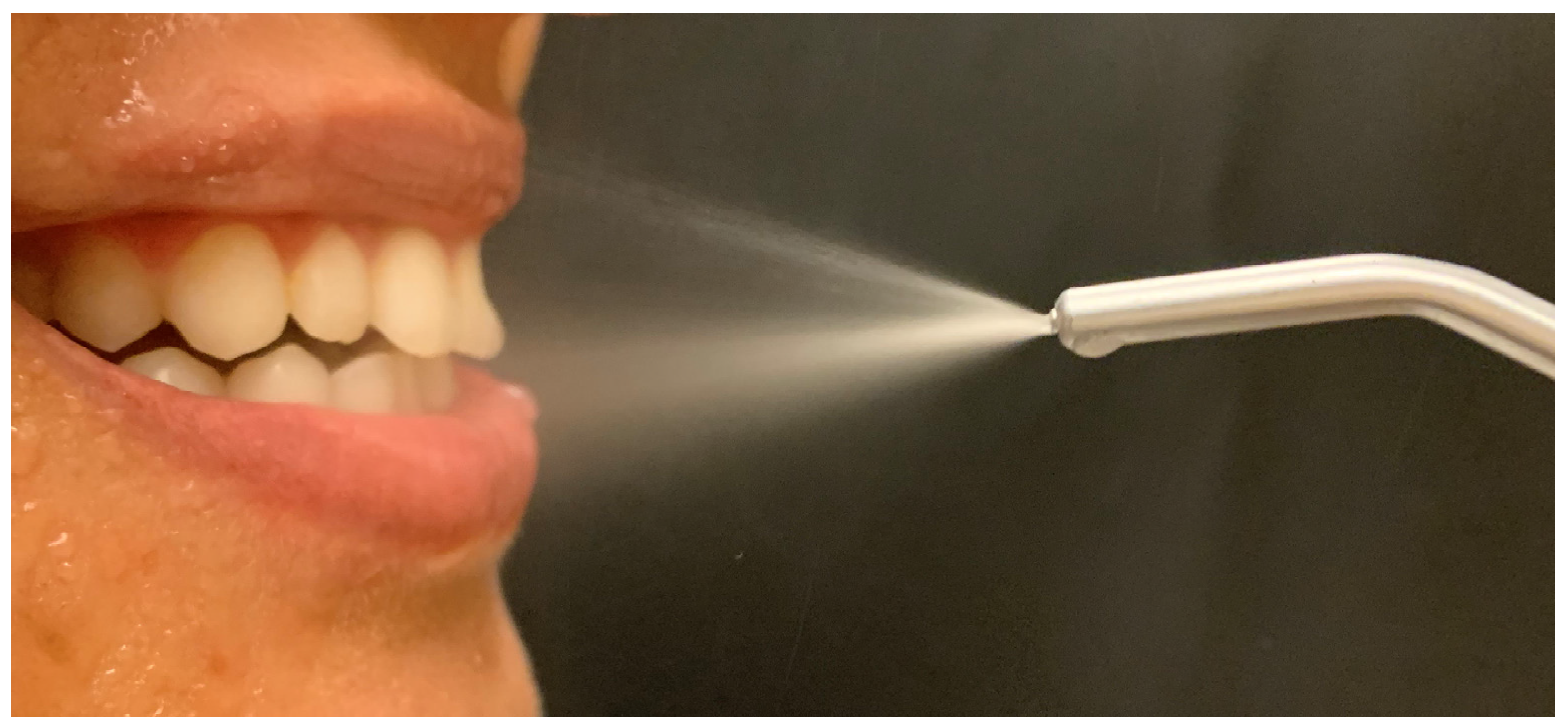
Figure 1. Aerosols generated by air–water syringes hit the hard and soft oral tissues, causing contaminated aerosols, which could be a source of transmitted infection.
Biomed 02 00003 g001
Figure 2. Infectious diseases, including COVID-19, can be directly or indirectly transmitted.
Biomed 02 00003 g002
Figure 3. Antimicrobial blue light therapy (aBLT) is a possible alternative way to control COVID-19 transmission during dental care. Patients are asked to rinse their mouth with a photosensitizer-containing solution. Then, the entire oral cavity is irradiated with light in a specific wavelength to activate the photosensitizer (A). aBLT can also be applied to other areas at risk of infection or to be treated which can also be disinfected in this intended approach (B,C).
Biomed 02 00003 g003
Publisher’s Note: MDPI stays neutral with regard to jurisdictional claims in published maps and institutional affiliations.