Review

Common Denominator of MASLD and Some Non-Communicable Diseases

by
Katarzyna Ferenc
1,
Sara Jarmakiewicz-Czaja
2,
Aneta Sokal-Dembowska
2,
Katarzyna Stasik
1,3 and
Rafał Filip
1,3,*
1
Institute of Medicine, Medical College of Rzeszow University, 35-959 Rzeszow, Poland
2
Institute of Health Sciences, Medical College of Rzeszow University, 35-959 Rzeszow, Poland
3
IBD Unit, Department of Gastroenterology, Clinical Hospital No. 2, 35-301 Rzeszow, Poland
*
Author to whom correspondence should be addressed.
Curr. Issues Mol. Biol. 2024, 46(7), 6690-6709; https://doi.org/10.3390/cimb46070399
Submission received: 28 May 2024 / Revised: 25 June 2024 / Accepted: 27 June 2024 / Published: 29 June 2024
(This article belongs to the Special Issue Cellular and Molecular Mechanisms of Nonalcoholic Fatty Liver Disease)

Abstract

:
Currently, steatohepatitis has been designated as metabolic dysfunction-associated steatohepatitis (MASLD). MASLD risk factors mainly include metabolic disorders but can also include genetic, epigenetic, and environmental factors. Disease entities such as obesity, diabetes, cardiovascular disease, and MASLD share similar pathomechanisms and risk factors. Moreover, a bidirectional relationship is observed between the occurrence of certain chronic diseases and MASLD. These conditions represent a global public health problem that is responsible for poor quality of life and high mortality. It seems that paying holistic attention to these problems will not only help increase the chances of reducing the incidence of these diseases but also assist in the prevention, treatment, and support of patients.

1. Introduction

Metabolic dysfunction-associated steatohepatitis (MASLD) is the latest definition and evidence of interest in the development and treatment of steatohepatitis, which is associated with the metabolic syndrome. Undoubtedly, the incidence of MASLD has been increasing recently. In turn, the prevalence of MASLD is strongly correlated with the incidence of obesity, diabetes, cardiovascular disease (CVD), and liver cancer, among others. Between these disease entities, there are strong common risk factors and biochemical mechanisms, as well as similarities in the direction of their development and complications [1].
Obesity is a growing problem for the health care systems of many countries. It is a disease characterized by excessive fat accumulation and abnormal fat distribution, with negative effects on health [2,3]. The causes of obesity are multifactorial and bidirectional and include a predisposition to many diseases, such as type 2 diabetes, high blood pressure, lipid disorders, and many others [4,5,6]. According to the World Health Organization (WHO), in 2019, type 2 diabetes (T2DM) directly caused 1.5 million deaths, with 460,000 deaths attributed to renal complications. Additionally, elevated glucose levels are believed to be the cause of 20% of deaths from cardiovascular issues [7]. Both in developed and developing nations, there is a continuous rise in the prevalence of chronic conditions, such as MASLD and T2DM. It is estimated that MASLD affects roughly 70–80% of individuals with diabetes [8]. Furthermore, correlations between diabetes, cardiovascular disease, and MASLD show that the risk of death in these patients increases as the severity of liver fibrosis increases [9,10]. In addition, end-stage liver disease, along with all of the above contributing factors, can lead to the development of hepatocellular carcinoma (HCC), which is among the most common cancers worldwide [10,11,12]. Therefore, it is important and vital to understand the mechanisms of MASLD and its relationship to other diseases, not only to detect it early but also to assess its risk. MASLD is a heterogeneous disease. Risk factors include gender, age, ethnicity, metabolic status, and genetic predisposition [8]. In addition, the pathogenesis of MASLD has been attributed to lipotoxicity, mitohondrial dysfunction, lysosome dysfunction, intestinal dysbiosis, or ferroptosis. Currently, drug treatment is under investigation. It is lifestyle changes that are of major importance in the prevention and treatment of MASLD and any comorbidities [10]. Therefore, the purpose of this review is to highlight the role of common mechanisms linking MASLD with obesity, T2DM, CVD, and liver cancer.

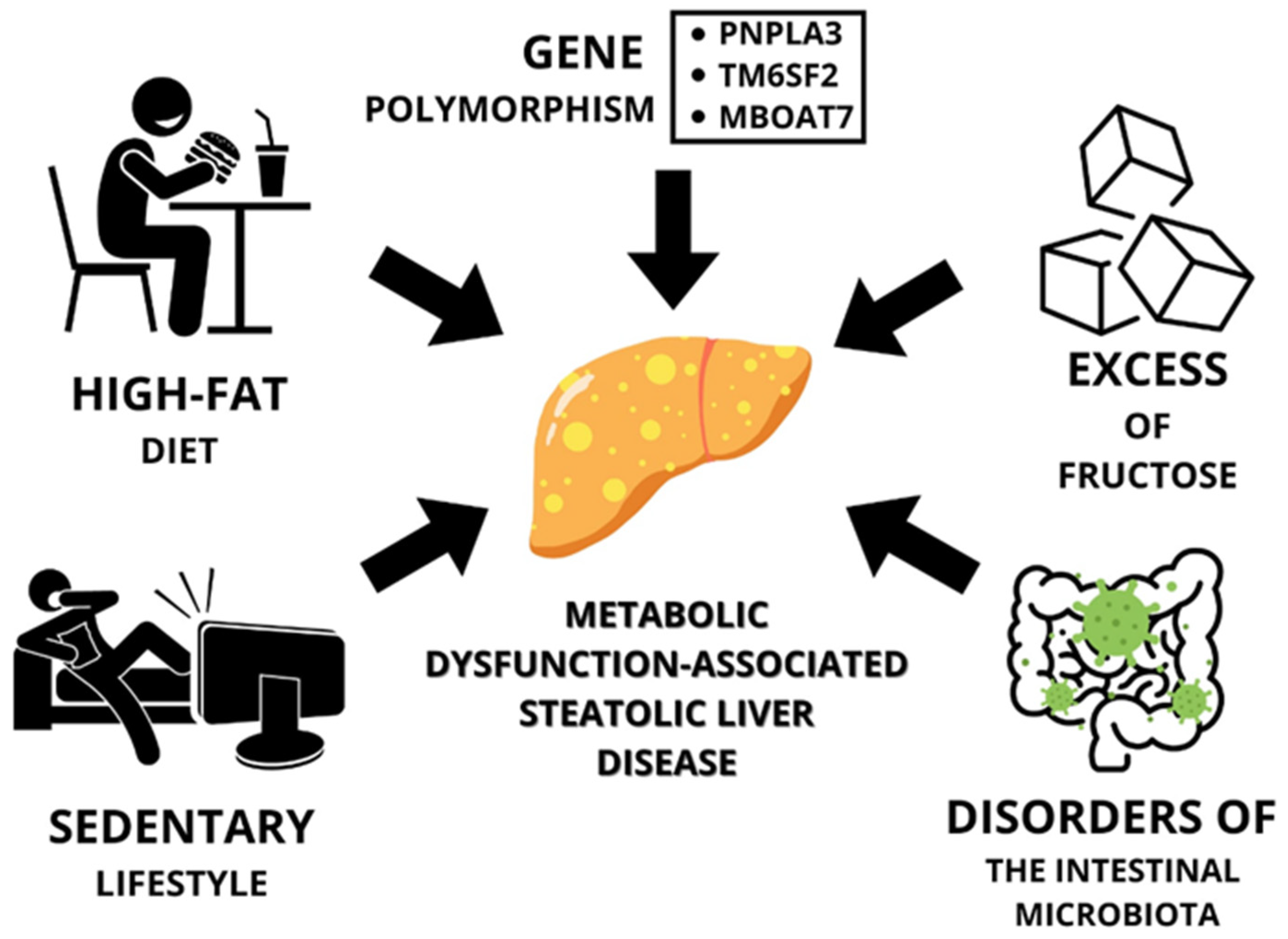
2. Obesity and Metabolic Dysfunction-Associated Steatotic Liver Disease

Quek et al. estimate that the incidence of MASLD has increased significantly over the past 20 years. Furthermore, the incidence of the disease has increased in parallel with an increase in the prevalence of obesity. It has been shown that 75% of people with obesity may have MASLD [13]. Therefore, one of the diagnostic criteria for MASLD is the presence of overweight or obesity and the presence of one of the metabolic risk abnormalities, which include a waist circumference ≥102/88 cm in Caucasian men and women (or ≥90/80 cm in Asian men and women) [14].
One of the factors predisposing to MASLD in obese patients is genetic [15,16,17,18,19,20]. Another is lifestyle, including diet and a lack of physical activity.

2.1. Lifestyle

Another component of the link between obesity and MASLD is lifestyle, which consists of improper eating habits such as a diet high in fat, mainly saturated fatty acids and trans-isomeric fatty acids, fructose, and a lack of or low levels of physical activity.

2.1.1. High-Fat Diet

High-fat diets, in which fat accounts for 45–75% of daily energy needs, have been shown to have an association with the occurrence of hepatic steatosis [21]. In their study on animal models, which included a diet high in 60% fat lasting 80 weeks, Velázquez et al. observed an increase in body weight in mice and hepatic steatosis with portal inflammation and progressive organ fibrosis. In addition, the authors of the study point to the presence of endoplasmic reticulum (ER) stress associated with obesity, which can lead to organ steatosis by altering lipid metabolism in the liver [22]. Dobbie et al. studied the effect of a low-calorie diet (LCD) on the efficacy of treatment for people with MASLD and comorbid obesity. The LCD used was characterized by three variations: an intake of 800–1500 kcal/day, a caloric reduction of 500 kcal/day, or a caloric reduction of 25% of daily energy requirements. The researchers showed that this type of diet reduced the body weight of obese patients, as well as the liver fat content, and also improved serum transaminase values [23]. In another study, De Nucci et al. used a very low-calorie ketogenic diet (VLCKD), which was carried out for 8 weeks. These were adult overweight or obese patients, in whom the amount of liver fat was also examined. They observed no reduction in inflammation and a reduction in the degree of organ steatosis [24]. However, it should be noted that it is primarily the combination of excess fat and carbohydrates in the diet that is wrong.

2.1.2. A Diet High in Fructose

The intestinal catabolism of fructose may have a protective effect on the liver, while overexposure and its metabolism in the liver may predispose to MASLD [25]. In a study by Shapiro et al., the authors observed that chronic intake of significant amounts of fructose can predispose to resistance to leptin, with an indirect effect on weight gain, which in turn can induce the cascade of liver steatosis [26]. Conversely, removing fructose from the diet can reverse leptin resistance [27]. Excess fructose consumption can also lead to the induction of excessive oxidative stress, inflammation, or even liver fibrosis [28]. Fructose is an inducer of de novo lipogenesis (DNL). Through DNL, fructose can contribute to increased oxidative stress, which then contributes to inflammation. Furthermore, visceral fat mass may increase with a high fructose diet, so it may indirectly alter the profile of cytokines and adipokines [29]. In addition, intermediate fructose metabolites may act as regulators of certain transcription factors that control lipogenesis or gluconeogenesis [30]. Fructose also affects molecular determinants of liver steatosis (animal model studies), e.g., fatty acid synthase (Fasn), acetyl-coenzyme carboxylase (Acaca), L-type pyruvate kinase (Pklr), and transcription factor binding sterol regulatory element 1 (Srebp1c) [31].

2.1.3. Sedentary Lifestyle

Low or no physical activity predisposes to excessive body weight, which, in turn, predisposes to MASLD. Romero-Gómez et al. indicate that different types of exercise (aerobic, resistance, and others) show similar effects on liver fat burning (about 20–30%). However, by introducing physical activity, weight reduction is more effective, and this in turn reduces liver fat (up to 80%) [32]. Patients are recommended to introduce more than 150 min per week of moderate-intensity aerobic training in 3–5 sessions [33]. Physical activity and dietary interventions are considered effective therapies for MASLD, and they can upregulate lipophagy in the liver. Due to different molecular pathways, a combination of two therapies (diet and physical activity) can intensify the effectiveness of treatment [34]. In addition, exercise is associated with a reduced risk of advanced liver fibrosis, associated with MASLD [35]. In addition to reducing organ steatosis, the introduction of physical activity can also reduce the levels of inflammatory markers and visceral adipose tissue [36].

2.2. Gut Microbiota Disorders vs. MASLD

Another factor predisposing to MASLD in obesity is the disruption of the gut microbiota, but this is strongly associated with a poor diet and a sedentary lifestyle. Liver steatosis may be associated with a change in the gut microbiota, both quantitatively and qualitatively. An increase in Proteobacteria and Firmicutes is observed, while a decrease in Bacteroidetes is observed [37]. In addition, at each stage of organ steatosis and at later stages, the composition of the gut microbiota changed [38]. Furthermore, Tilg et al. concluded in their paper that intestinal barrier dysfunction, which can affect the translocation of pathogenic bacteria or their metabolites, affects intestinal–liver communication. Endotoxin, which can enhance the release of pro-inflammatory cytokines, may be associated with low-grade inflammation. Endotoxemia shows an association with the severity of liver disease [39]. In addition to endotoxins, peptidoglycans and bacterial DNA can also accelerate the development of liver disease [40]. Therefore, intestinal dysbiosis can promote the progression of liver steatosis to the next stages, such as inflammation or organ fibrosis [41]. Leung et al., in their article, concluded that intestinal dysbiosis may be one of the main factors associated with the heterogeneous phenotype of MAFLD (metabolic-associated fatty liver disease) [42]. In contrast, Hu et al. pointed out that it would be appropriate to consider and direct research to viruses, fungi, or intestinal archaeons, not just intestinal bacteria, in the context of the link to liver disease [43]. In turn, Lang et al. stated that the therapeutic approach should be individualized with respect to the introduction of specific bacterial strains to intensify drug therapy [44]. A summary of obesity-related predisposing factors for MASLD is shown in Figure 1.

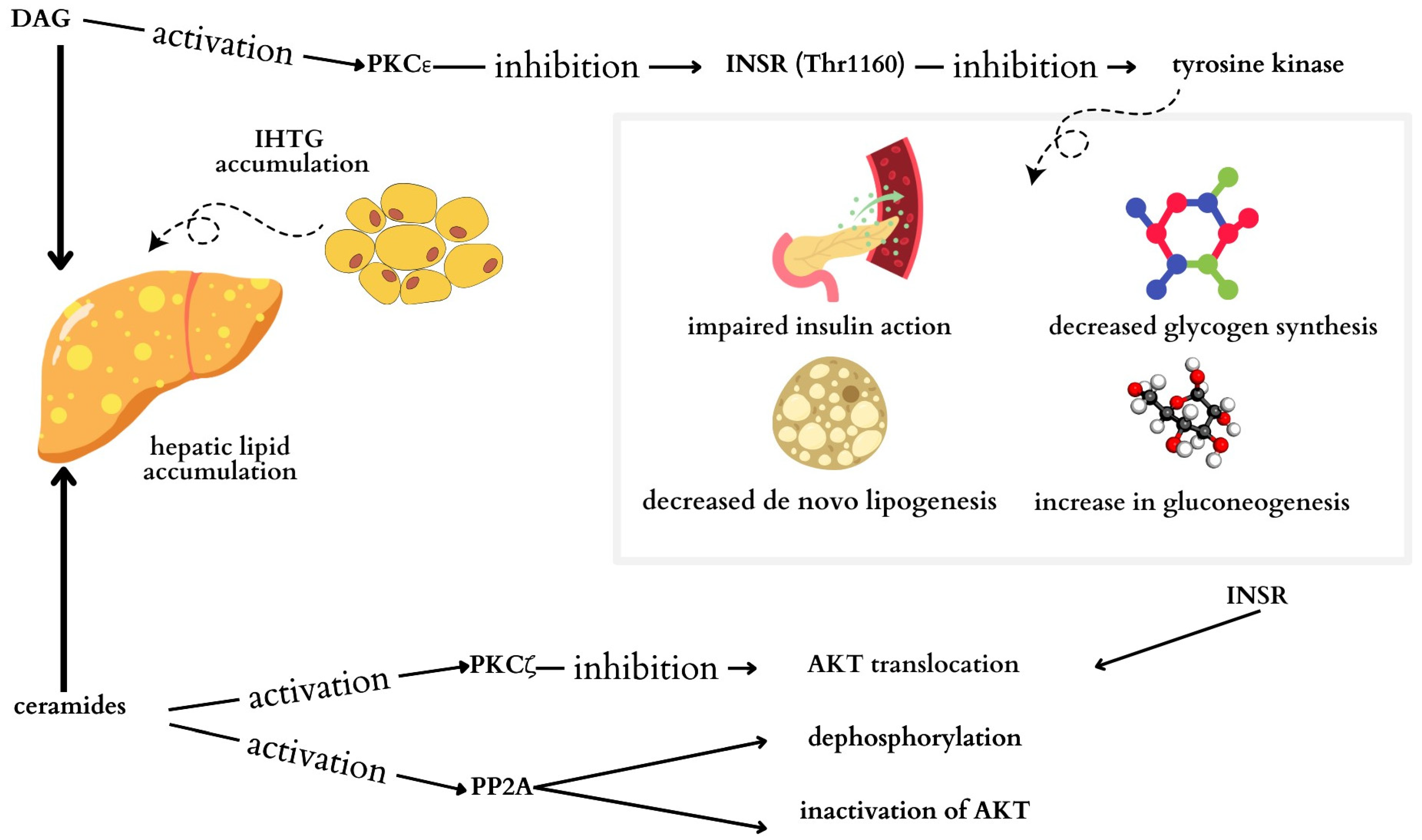
3. Type 2 Diabetes and Metabolic Dysfunction-Associated Steatotic Liver Disease

Available data suggest a strong link between the presence of insulin resistance (IR) and MASLD, affecting both obese and non-obese populations [45]. Research by Jäger et al. has shown a positive correlation between the fatty liver index (FLI) and the risk of type 2 diabetes in both genders, with FLI values associated with markers of dyslipidemia, liver enzymes, fetuin A, and CRP levels [46]. While both MASLD and T2DM are less common in women compared to men, MASLD can contribute to the development of T2DM regardless of gender [47]. Studies by Muzic et al. highlighted a bidirectional relationship between MASLD and T2DM, where MASLD can lead to insulin resistance and an increased risk of T2DM, while T2DM may worsen the progression to MASLD [48]. The presence of MASLD is considered a predictor of the risk of type 2 diabetes [47]. Zeng et al. have shown a positive association between MASLD and increased insulin resistance markers, such as insulin resistance index (HOMA-IR) and TyG-BMI (triglyceride-glucose index with body mass index), within the non-diabetic population [45]. Conversely, improving MASLD can reduce the future risk of T2DM, emphasizing the importance of implementing lifestyle changes and dietary interventions to mitigate the severity of fatty liver disease [47].

3.1. High Supply of Fats and Carbohydrates in the Diet

There are at least two mechanisms that may contribute to the development of insulin resistance and MASLD. One of these mechanisms is closely tied to the dietary intake of fats and carbohydrates. High consumption of dietary fats can lead to elevated levels of free fatty acids, increased de novo lipogenesis (DNL), and consequently reduced secretion of triglycerides (TGs) from the liver. Similarly, excessive intake of carbohydrates can stimulate the DNL pathway, ultimately leading to the onset of fatty liver disease (Figure 2) [49].
Within the DNL pathway, certain lipogenic molecules like acetyl-CoA carboxylase (ACC), stearoyl CoA-desaturase-1 (SCD-1), and fatty acid synthesis (FAS) are produced [49]. Insulin and glucose primarily regulate the DNL pathway, with glucose activating DNL via the transcription factor carbohydrate response element-binding protein (ChREBP) and insulin promoting DNL by upregulating sterol regulating element-binding protein 1c (SREBP-1c), the master transcription factor for lipogenesis [50]. SREBP-1c is implicated in the generation of diacylglycerol (DAG) and ceramides, molecules that induce inflammation by interacting with tumor necrosis factor-α (TNF-α), thereby exacerbating insulin resistance [49].
Additionally, two classes of lipids, DAG and ceramide, play crucial roles in the development of insulin resistance (Figure 3) [51]. Studies have shown a direct correlation between total DAG levels and the HOMA-IR [52,53]. In patients with increased intrahepatic triglycerides (IHTGs), sn-1,2-diacylglycerol accumulates, which is responsible for the activation of protein kinase C (PKC) isoforms, especially the ε isoform (PKCε). As a result of phosphorylation of the insulin receptor (INSR) Thr1160 by PKCε, INSR tyrosine kinase is inhibited, and insulin action is impaired [54].
It has been observed that the presence of ceramides in the liver is associated with hepatic IR in humans. Ceramide activation of protein kinase C-ζ (PKCζ) probably impairs the translocation of protein kinase B to the cell membrane, preventing its participation in the action of insulin [54]. Insulin signaling may also be dependent on protein phosphatase 2A through the inactivation of certain kinases [55]. Ceramide activation of protein phosphatase 2A may lead to dephosphorylation and inactivation of protein kinase B [54].
In a state of insulin resistance, the liver is unable to suppress hepatic glucose production in response to insulin, thus leading to the severity of DNL [56]. The inability to inhibit glucose production in the liver and promote lipid synthesis leads to the development of hyperglycemia and hypertriglyceridemia [57]. Increased hepatic gluconeogenesis is likely the main cause of fasting hyperglycemia in T2DM. In turn, increased lipid accumulation in the liver and skeletal muscles may impair insulin signaling and cause insulin resistance. Additionally, overexpression of lipoprotein lipase in the liver and skeletal muscles may induce peripheral insulin resistance [58].
In the second mechanism, attention is drawn to oxidative stress, inflammatory mediators, lipid peroxidation, and mitochondrial dysfunction [49]. IR is associated with chronic low-grade inflammation seen in patients with diabetes and MASLD [59]. Pro-inflammatory cytokines and transcription factors are highly expressed, among others, in the liver and adipose tissue [60]. It has been observed that some pro-inflammatory cytokines, such as TNF-α and interleukin-6 (Il-6), can activate nuclear factor-κB kinase (IKK), complex IKKβ, and c-Jun N-terminal kinase (JNK). Additionally, Il-6 induces suppressors of cytokine signaling (SOCS) 1 and 3, which may result in impaired insulin signaling and the development of IR. Moreover, IKKβ and JNK can activate nuclear factor kappa-B (NF-κB) and cause its translocation to the nucleus, and endoplasmic reticulum (ER) stress can additionally activate the JNK pathway [61].
Presumably immune-related inflammatory changes, insufficient inhibition of lipolysis, and excess FFA cause ectopic lipid deposition, and pro-inflammatory factors secreted by adipose tissue, such as TNF-α, IL6, and IL-1β, may impair insulin signaling [58,61].

3.2. Genetic Factors

Dongiovanni et al. demonstrated that hepatic steatosis is causally related to insulin resistance in patients at risk of metabolic dysfunction-associated steatohepatitis (MASLD). The authors indicated that some genetic variants, TM6SF2 E167K (rs58542926) and GCKR P446L (rs1260326), increase liver fat content and may be associated with a moderate increase in insulin resistance and the risk of T2DM [62].

4. Cardiovascular Disease and Metabolic-Related Steatohepatitis

Hepatic steatosis is an independent risk factor for the development of cardiovascular disease [11]. In addition, the risk increases when patients have comorbidities such as T2DM or hypertriglyceridemia [11,63]. The pathophysiological mechanisms responsible for MASLD are considered to be an altered lipid profile, insulin resistance, endothelial dysfunction, and chronic inflammation [63]. A 2021 meta-analysis showed that MASLD is associated with a moderately increased risk of fatal or non-fatal CVD events [64]. In contrast, a population-based evaluation in the Framingham study showed that hepatic steatosis is associated with coronary artery and abdominal aortic calcification, regardless of the presence of other conditions that increase cardiovascular risk [65].

4.1. Risk Factors

4.1.1. Chronic Inflammation

MASLD is associated with increased production of pro-inflammatory cytokines and increased levels of serum liver enzymes. Patients with MASLD have an increase in the production of CRP protein, oxidized LDL, and plasminogen activator inhibitor-1, as well as other inflammatory proteins mediated by IL-6 and TNF-α These factors in patients with MASLD are associated with an increased risk of CVD and vascular atherosclerosis [66,67].

4.1.2. Insulin Resistance

MASLD is associated with peripheral and hepatic insulin resistance. Disorders in the course of tissue insulin resistance and an increase in peripheral and hepatic insulin secretion also lead to atherogenic dyslipidemia and the release of pro-inflammatory factors, vasoactive factors, and thrombogenic factors, which promote the development of hypertension and cardiovascular disease [68]. Insulin resistance increases de novo lipogenesis in MASLD. In the course of insulin resistance, there is a persistence of high levels of insulin in the blood, which leads to an abnormal mechanism. It is characterized by an increase in circulating blood levels of glucose and free fatty acids, stimulating the release of triacylglycerol from hepatocytes, which can lead to the development of atherosclerotic disease [69]. High glucose levels induce oxidative stress and contribute to the inflammatory response, leading to cellular damage. In addition, hyperglycemia affects coronary artery disease by increasing endothelial monocyte adhesion, enhancing vascular smooth muscle cell proliferation, and indirectly causing endothelial dysfunction [68]. MASLD patients also experience increased platelet activity, elevated levels of clotting factors, and fibrinolysis inhibitors, which may contribute to the development of atherosclerotic thrombosis [70,71].

4.1.3. Abnormal Lipid Profile

Patients with MASLD exhibit a proatherogenic lipid profile characterized by high triglycerides, low-density lipoprotein cholesterol (LDL-C), reduced high-density lipoprotein (HDL-C) levels, and high apolipoprotein B (apoB) levels [72]. Studies indicate that plasma apoB concentration is the strongest predictor of CVD risk [68]. Another mechanism explaining the increase in CVD risk in patients with MASLD and coexisting obesity is a disruption of reverse cholesterol transport. There is reduced uptake and excretion of cholesterol from plasma by hepatocytes and adipocytes. The reverse transport of cholesterol is also responsible for HDL; if its levels are low, disruption occurs, so HDL is a marker of MASLD and a risk factor for CVD. However, it is not only HDL levels alone that are important; in the course of MASLD, there is also lipoprotein dysfunction, and antioxidant activity is reduced [73]. The increase in triglycerides depends on a decrease in lipoprotein lipase, which is associated with an increase in angiopoietin-like protein ANGPTL 3 and protein activity ANGPTL 4. Levels of these proteins are increased in patients with obesity, type 2 diabetes, and MASLD. This mechanism causes lipid accumulation in the liver parenchyma and underlies the development of atherosclerosis [74].

4.2. Complications Associated with Cardiovascular Disease

4.2.1. MASLD and Hypertension

Due to the frequent co-occurrence of MASLD and obesity, the risk of developing hypertension is very high. Hypertension occurs in about 40% of patients with MASLD [67]. One of the mechanisms explaining this condition is increased activation of the sympathetic nervous system. This is indicated by an increase in neurotransmitters such as norepinephrine and neuropeptide Y [75]. As a result of activation of the sympathetic nervous system, there is increased production of LDL by the liver, which leads to the deposition of atherosclerotic plaques in the vessels, resulting in flow restriction and increased blood pressure. In addition, due to persistent activation of the sympathetic nervous system, there is increased lipolysis in adipocytes, impaired glucose uptake, and insulin resistance, which leads to increased plasma levels of free fatty acids and triglycerides, resulting in visceral obesity [67,75]. In the course of MASLD, there is also a decrease in renal blood flow, glomerular filtration, an increase in sodium retention, and increased activation of the renin–angiotensin–aldosterone system (RAAS)—an increase in activation of the sympathetic nervous system in the liver, a decrease in blood flow, and an increase in hepatic arterial resistance, which activates the renal sympathetic nervous system and, consequently, stimulation of the RAAS [75,76].

4.2.2. MASLD and Structural Abnormalities of the Heart

Studies show that patients with MASLD and coexisting obesity develop concentric left ventricular hypertrophy. Due to increased epicardial adipose tissue, cardiac diastolic function is altered. Numerous available studies have described altered myocardial structure, early left ventricular (LV) systolic, and diastolic dysfunction [67]. Patients with advanced fibrosis (F3–F4) had more epicardial fat than those with milder fibrosis (F0–F2). In addition, other echocardiographic indices, such as posterior diastolic wall thickness, left ventricular mass, relative wall thickness, ejection fraction (EF), and left atrial volume, depended on the severity of hepatic fibrosis [77]. Patients with MASLD exhibit increased LV mass, decreased LV ejection fraction, lower early diastolic relaxation velocity, and increased LV filling pressure, which can lead to heart failure. The mechanisms responsible for these pathologies are the previously mentioned insulin resistance, chronic inflammation, mitochondrial dysfunction, oxidative stress, activation of the sympathetic nervous system, and the RAAS [67].

4.2.3. MASLD and Cardiac Arrhythmias

Many available studies have shown an increased risk of developing cardiac arrhythmias in patients with MASLD, such as atrial fibrillation, ventricular arrhythmias, and QTc interval prolongation. MASLD is associated with cardiac remodeling, which leads to changes in the conduction tissue and induces arrhythmias. In addition, atrial fibrillation (AF), premature ventricular beats, and non-sustained ventricular tachycardia may also occur [78]. Several studies have shown that there is an increased incidence of AF in patients with MASLD, especially with the coexistence of T2DM [68]. This is most likely related to the increased propensity in this group of patients for fat deposition and higher levels of pro-inflammatory, vasoactive, and pro-thrombotic mediators, which can lead to structural and functional disruption of the myocardium and consequently trigger pro-arrhythmogenic effects [79,80].

4.2.4. Ischemic Stroke

The above-described disorders are associated with an increased risk of vascular atherosclerosis, and cardiac arrhythmias can directly lead to ischemic stroke. Patients with MASLD have an increased risk of ischemic stroke [63]. The incidence of stroke in patients with steatohepatic disease in the studies ranged from about 18 to 41% [81,82]. In patients with MASLD, ischemic stroke was more common in middle age [82]. However, biochemical test markers such as alanine aminotransferase (ALT), aspartate aminotransferase (AST), and gamma-glutamyltranspeptidase (GGTP) are not prognostic factors for ischemic stroke [83].

4.3. Pharmacological Therapeutic Strategies

Optimal therapeutic management should include both cardiovascular drugs and drugs that improve liver function (Table 1). The drugs used should primarily reduce fibrosis, normalize hepatocyte metabolism, have antioxidant effects, and counteract apoptosis. These mechanisms include the action of vitamin E and ursodeoxycholic acid (UDCA) [72]. Vitamin E reduces steatosis and inflammation of the liver but does not significantly affect the fibrosis process. In addition, vitamin E may increase the risk of hemorrhagic stroke as well as heart failure [72,84]. UDCA improves the secretory function of hepatocytes and inhibits the activity of pro-inflammatory cytokines. The use of UDCA reduces the activity of aminotransferases and reduces hepatic steatosis. However, there is no evidence that it reduces cardiovascular risk [72].
Given the above, specific treatments for MASLD are still lacking. Patients are primarily treated for concomitant diseases. In clinical practice, statins are recommended to reduce the risk of cardiovascular disease complications such as ischemic stroke and myocardial infarction [67]. Statins are inhibitors of 3-hydroxy-3-methylglutaryl coenzyme A reductase (HMGCoA reductase), an enzyme that limits the rate of cholesterol biosynthesis [68]. Their action also involves reducing triglyceride levels in the liver, and they have anti-inflammatory, antiproliferative, anticoagulant, and antioxidant effects [67]. There is no confirmation that fibrates are effective in the treatment of hepatic steatosis. However, combination therapy of ezetimibe and statin has shown a positive therapeutic effect of, among other things, lowering LDL cholesterol levels and improving therapeutic outcomes in patients with MASLD and acute coronary syndrome [63].
Hypoglycemic medications are another point of interest. Examples include sodium-glucose transporter 2 (SGLT2) inhibitors and glucagon-like peptide 1 (GLP-1) receptor agonists approved for the treatment of type 2 diabetes. It appears that these drugs can prevent and delay the progression of MASLD [85]. GLP-1 receptor agonists lower body weight, reduce insulin resistance, and improve plasma lipid levels, all of which can lower the risk of developing CVD [63,85,86]. The ability of SGLT2 inhibitors to exert cardioprotective effects may involve several important pathways beyond their effects on glucose transport, such as reducing oxidative stress, stimulating lipolysis, improving mitochondrial function, suppressing apoptosis, and altering the renin-angiotensin system [85,87]. They have a protective effect on the cardiovascular system and favorable effects on liver function in MASLD, and they may be promising therapeutic options for CVD in patients with MASLD.
Given that one of the mechanisms leading to hypertension in MASLD is activation of the RAAS, the drugs with the greatest potential for treating CVD in MASLD are inhibitors of the RAAS. In addition to the blood pressure-lowering effects of angiotensin II receptor antagonists, studies have demonstrated the ability of these antagonists to improve plasma triglyceride levels, markers of liver damage, and hepatic lipid accumulation [67,88]. Effects on metabolic pathways may be beneficial in patients with MASLD to prevent CVD.
Table 1. Mechanisms of selected drug groups having an effect on steatohepatitis associated with metabolic dysfunction (MASLD) and cardiovascular disease (CVD).
Drug/Group of DrugsMechanism of Action in MASLDMechanism of Action in CVD
Statins [67]Reduces cholesterol synthesis and liver triglyceride levelsAnti-inflammatory, antiproliferative, anticoagulant,
antioxidant
Vitamin E [67,72]Reduces steatosis and hepatitisNo evidence of reducing cardiovascular risk. May increase the risk of hemorrhagic stroke and heart failure.
Ursodeoxycholic acid [72,89]Improves hepatocyte secretory function and inhibits pro-inflammatory cytokine activity. Reduces aminotransferase activity and inhibits hepatic steatosis.No evidence of CVD risk reduction
Hypotensive drug angiotensin II receptor antagonists [89]Inhibition of the renin–angiotensin–aldosterone system (RAAS), which leads to activation of PPAR-γ, resulting in better control of adipose tissue proliferation and adipokine productionInhibition of the RAAS lowering blood pressure
Hypoglycemic drugs [90]Reduces insulin resistance and improves plasma lipid levelsReduces oxidative stress, stimulates lipolysis, improves mitochondrial function, suppresses apoptosis, and alters the renin-angiotensin system

5. Liver Cancer and Metabolic-Related Steatohepatitis

HCC is one of the most common cancers worldwide. HCC is induced in patients with chronic liver disease (CLD), where processes related to damage, inflammation, and regeneration are intertwined [12]. NAFLD, which has recently been given the new name MASLD, is considered the most prevalent liver disease but also one of the main inducers of HCC [91]. MASLD seems to carry a moderate risk of developing HCC; however, due to its prevalence, it has a large proportion of cancer [12,91]. In contrast, a population-based study in the US found that MASLD was the most common risk factor for HCC induction, at 59% [92]. HCC ranks as the fourth most common cause of cancer death worldwide. In addition, it ranks second in loss of life due to cancer. In recent years, an increase in the incidence of HCC has been found. This appears to be due to a combination of aging and population growth, as well as an increase in the prevalence of metabolic syndrome. The highest incidence of HCC and the burden of death from it are found in East Asia [93]. HCC, which is associated with MASLD, is found in the elderly. Importantly, HCC is diagnosed at later stages in these individuals, and they also show lower survival rates compared to patients with HCC associated with viral hepatitis [94]. It also appears that HCC with co-occurring MASLD can develop in the absence of cirrhosis, which in clinical practice potentially contributes to delaying diagnosis [95].

5.1. Genetic Factors

In recent years, genes that are associated with MASLD and HCC have been discovered. A nucleotide polymorphism (SNP) in the patatin-like phospholipase domain containing 3 (PNPLA3) gene is responsible for encoding adiponutrin triglyceride lipase. As a result, it is associated with fat deposition in the liver. The PNPLA3 rs738409 [G] risk allele may increase the risk of progression of hepatic steatosis to hepatic inflammation, but importantly, it potentially promotes a 12-fold increased risk of HCC induction. It is worth mentioning that this allele is found in about 40% of the European population [96]. Ertle et al. showed that this allele is closely associated with hepatocancerogenesis in obesity, especially in patients with alcoholic cirrhosis [97]. In addition, there are two genetic variants, which include the c.6718A>T mutation in the apolipoprotein B (ApoB) gene and the c.6718>T mutation in the human telomerase reverse transcriptase (TERT) gene. Both of these variants influence the induction of HCC in the steatotic liver [98].

5.2. Gut Microbiota

The gut microbiota, through several different mechanisms, can influence both the development of MASLD and HCC [12]. The mechanisms linking the two disease entities are impaired intestinal barrier permeability and intestinal dysbiosis. It has been shown that high levels of lipopolysaccharide (LPS) are found in patients with CLD and hepatocarcinogenesis. It belongs to the components of the cell wall but is also one of the more potent promoters of inflammation [99]. Studies have shown that intestinal exposure to dextran sulfate sodium (DSS) can contribute to increased LPS levels and systemic inflammation, thereby increasing the risk of HCC in mice. In addition, a high-fat diet in such cases can further increase portal LPS levels and induce inflammation and associated liver fibrosis [100,101]. It seems that it is the process of LPS binding to the Toll-like receptor (TLR) transmembrane receptor that is most important in the induction of HCC. It appears that TLR4 inhibition in mice and rats can reduce inflammation and fibrosis, but also the occurrence of HCC. In turn, TLR4 expression can promote fibrogenesis and the process of hepatocarcinogenesis [102,103]. This can happen at the level of different cells. TLR4 activation in HSCs leads to an increase in epiregulin, which has strong mitogenic effects on hepatocytes. In addition, the LPS–TLR4 axis can reduce hepatocyte apoptosis by utilizing NF-κB. Also, activation of this axis in HCC cells potentially increases epiregulin transition and enhances the invasiveness of these cells [102,103,104]. Intestinal dysbiosis is closely associated with changes in intestinal barrier permeability. Studies have shown changes in the gut microbiota in both CLD and cirrhotic patients. The similarities were characterized by a decrease in beneficial bacteria and an increase in potentially pathogenic bacteria. It seems that these changes are the result of characteristics caused by liver impairment, that is, a decrease in the secretion of intestinal antimicrobial peptides or a reduction in bile secretion [105]. Importantly, not only are changes in the microbiota noted, but also bacterial overgrowth, which is associated with increased amounts of circulating LPS [106]. In addition, LPS acts on liver progenitor cells (HPCs), which it induces to differentiate into myofibroblasts. Through the secretion of TNF-α and IL-6, it can promote inactivation of the p53 tumor suppressor signaling pathway in HPCs and activation of the Ras pathway. As a result, it initiates uncontrolled proliferation and transformation of HPCs, and consequently carcinogenesis [107]. It is also important that the gut microbiota is responsible for the aggregation of liver Natural Killer T cells, and it is these cells, among others, that are responsible for immune mechanisms, which include the production of IFN-γ [108]. Yoshimoto et al. showed that in mice with HCC induced by artificially induced NASH, there was a large increase in Gram-positive bacteria, particularly Clostridium. At the same time, they showed an increase in serum deoxycholic acid (DCA), which is a secondary bile acid. It seems that DCA may also be involved in hepatocarcinogenesis [109]. In addition, there are bacterial species, e.g., Bacteroides fragilis, that have endotoxic effects. As a result of their action on the Stat3 and Th17 pathways, they can induce the development of HCC [110]. Further research is needed to elucidate the mechanisms involved in the gut–hepatic axis and to uncover new issues with the microbiota that could potentially influence the induction of both MASLD and HCC. Ultimately, the targeting of the gut–liver axis could potentially affect not only the quality of life of patients but also their survival.

5.3. Adipose Tissue and Associated Pro-Inflammatory Cytokines

Insulin resistance, which is characterized by chronic low-grade inflammation, promotes macrophage recruitment as well as the induction of pro-inflammatory cytokines. Pro-inflammatory cytokines such as TNF-α and IL-6 are associated with MASLD and the progression of hepatitis to HCC. This appears to happen through mechanisms of action on the signaling pathways IKK and JNK. These pathways are crucial in promoting inflammation induced by excess body weight and liver tumorigenesis [111]. In addition, in obese individuals, high leptin levels are involved in immune regulation and angiogenesis. In turn, leptin binding to the Ob receptor can induce activation of the PI-3K/Akt pathway. Subsequently, mTOR, as one of the Akt effectors, is associated with cell proliferation and growth. It appears that Akt/mTOR activation is confirmed in about 30–40% of HCC patients [112]. In addition, people with obesity have low levels of adiponectin, which may reduce its anti-inflammatory effect [111].

5.4. Diet

A high-fat diet can affect the expression of both TNF-α and IL-6 as well as increase NF-κB activation [113]. Zhang et al. showed that a cholesterol-rich diet induces an increased risk of MASLD progression to HCC in mice. This effect appears to arise as a consequence of changes in the gut microbiota and its metabolites [38]. In addition, carbohydrates, especially excess fructose, could potentially activate the transcription factor ChREBP. This factor contributes to the co-occurrence of chronic hyperinsulinemia. Consequently, it induces hepatic lipogenesis de novo. It turns out that it is increased several-fold in obese subjects and MASLD, but importantly, in its process, only saturated fatty acids are produced, and they play a significant role in lipotoxic damage. It appears that fructose may affect the induction of HCC by several mechanisms. These include an increase in lipoperoxidation, which decreases sirtuin-1 expression but may also affect changes in the gut microbiota [114,115].

5.5. Other Factors

In addition to the mechanisms linking MASLD to HCC described above, there are other risk factors. These include male gender, older age, and ethnicity [116]. In addition, common risk factors are the presence of obesity and diabetes [117]. Dyson et al. showed that older people with established diabetes or metabolic syndrome had the highest mortality associated with HCC [118]. In turn, Marrero et al. observed that patients with cirrhosis and comorbid obesity were 47 times more at risk for HCC compared to those without liver disease [119].
A summary of factors common to MASDL and other conditions is shown in Table 2.

6. Conclusions

In recent years, the prevalence of diseases closely related to MASLD, such as liver cancer, diabetes, obesity, and CVD, has increased significantly. MASLD is part of a major global problem that is now challenging health care. For the treatment of MASLD, most recommendations for “liver detoxification” include a 7–10% weight reduction, in addition to a Mediterranean diet (exclusion of highly processed foods and foods high in fat and fructose), the reduction and preferably elimination of alcohol, and the introduction of physical activity. This last factor should be tailored to the patient’s preferences so as to maintain the conduct of physical activity systematically and in the long term. On the one hand, MASLD contributes to insulin resistance and increased risk of T2DM, and on the other hand, T2DM may lead to the progression of MASLD. In particular, immune-related inflammatory changes, inefficient inhibition of lipolysis, and excess free fatty acids contribute to ectopic lipid deposition and impair insulin signaling in adipose tissue. To counteract unfavorable metabolic changes, all preventive measures should focus on reducing inflammation and oxidative stress in patients. Typical disorders for MASLD, i.e., chronic low-grade inflammation, insulin resistance, or lipid disorders, contribute to cardiovascular disease. In addition, often the final stage of liver disease becomes liver cancer. These risks suggest that MASLD is a multidisciplinary disease, and both cardiologists and gastroenterologists should take extensive preventive measures based on screening high-risk patients.

Author Contributions

Conceptualization, K.F., S.J.-C., A.S.-D., K.S. and R.F.; writing—original draft preparation, K.F., S.J.-C., A.S.-D. and K.S.; writing—review and editing, K.F., S.J.-C., A.S.-D., K.S. and R.F.; supervision, R.F. All authors have read and agreed to the published version of the manuscript.

Funding

This research received no external funding.

Conflicts of Interest

The authors declare no conflicts of interest.

Abbreviations

ACCacetyl-CoA carboxylase
AFatrial fibrillation
ALTalanine aminotransferase
apoBapolipoprotein B
ASTaspartate aminotransferase
ChREBPcarbohydrate response element-binding protein
CLDchronic liver disease
CVDcardiovascular disease
DAGdiacylglycerol
DCAdeoxycholic acid
DNLde novo lipogenesis
DSSdextran sulfate sodium
EFejection fraction
ERendoplasmic reticulum
FASfatty acid synthesis
FLIfatty liver index
GGTPgamma-glutamyltranspeptidase
GLP-1glucagon-like peptide 1
HCChepatocellular carcinoma
HMGCoAreductase 3-hydroxy-3-methylglutaryl coenzyme A reductase
HOMA-IRinsulin resistance index
HPCsliver progenitor cells
IHTGintrahepatic triglycerides
IKKinhibitor κB kinase
IL-6interleukin-6
INSRinsulin receptor
IRinsulin resistance
JNKc-Jun N-terminal kinase
LCDlow-calorie diet
LDL-Clow-density lipoprotein cholesterol
LPSlipopolysaccharide
LVleft ventricular
MAFLDmetabolic-associated fatty liver disease
MASLDmetabolic dysfunction-associated steatohepatitis
NF-κBnuclear factor kappa-B
PKCprotein kinase C
PKCζprotein kinase C-ζ
Pklrl-type pyruvate kinase
PNPLA3patatin-like phospholipase domain containing 3
PPAR-αperoxisome proliferator-activated receptor-α
RAASrenin–angiotensin–aldosterone system
SCD-1stearoyl CoA-desaturase-1
SGLT2sodium-glucose transporter 2
SNPnucleotide polymorphism
SOCSsuppressor of cytokine signaling
Srebp1cfactor binding sterol regulatory element 1
SREBP-1csterol regulatory element-binding protein 1c
T2DMtype 2 diabetes
TERTtelomerase reverse transcriptase
TGtriglycerides
TLRtoll-like receptor
TM6SF2transmembrane 6 superfamily member 2
TNF-αtumor necrosis factor-α
UDCAursodeoxycholic acid
VLKCDvery low-calorie ketogenic diet
WHOWorld Health Organization

References

  1. Eslam, M.; Sarin, S.K.; Wong, V.W.; Fan, J.G.; Kawaguchi, T.; Ahn, S.H.; Zheng, M.H.; Shiha, G.; Yilmaz, Y.; Gani, R.; et al. The Asian Pacific Association for the Study of the Liver clinical practice guidelines for the diagnosis and management of metabolic associated fatty liver disease. Hepatol. Int. 2020, 14, 889–919. [Google Scholar] [CrossRef] [PubMed]
  2. Bray, G.A. Evaluation of obesity. Who are the obese? Postgrad. Med. 2003, 114, 19–27. [Google Scholar] [CrossRef] [PubMed]
  3. Mayoral, L.P.; Andrade, G.M.; Mayoral, E.P.; Huerta, T.H.; Canseco, S.P.; Rodal Canales, F.J.; Cabrera-Fuentes, H.A.; Cruz, M.M.; Pérez Santiago, A.D.; Alpuche, J.J.; et al. Obesity subtypes, related biomarkers & heterogeneity. Indian J. Med. Res. 2020, 151, 11–21. [Google Scholar] [CrossRef] [PubMed]
  4. Stadler, J.T.; Marsche, G. Obesity-Related Changes in High-Density Lipoprotein Metabolism and Function. Int. J. Mol. Sci. 2020, 21, 8985. [Google Scholar] [CrossRef] [PubMed]
  5. Litwin, M.; Kułaga, Z. Obesity, metabolic syndrome, and primary hypertension. Pediatr. Nephrol. 2021, 36, 825–837. [Google Scholar] [CrossRef] [PubMed]
  6. Piché, M.E.; Tchernof, A.; Després, J.P. Obesity Phenotypes, Diabetes, and Cardiovascular Diseases. Circ. Res. 2020, 126, 1477–1500. [Google Scholar] [CrossRef] [PubMed]
  7. World Health Organization. Diabetes. Available online: https://www.who.int/news-room/fact-sheets/detail/diabetes (accessed on 15 May 2024).
  8. Xia, M.F.; Bian, H.; Gao, X. NAFLD and Diabetes: Two Sides of the Same Coin? Rationale for Gene-Based Personalized NAFLD Treatment. Front. Pharmacol. 2019, 10, 877. [Google Scholar] [CrossRef] [PubMed]
  9. Han, E.; Lee, Y.H.; Lee, J.S.; Lee, H.W.; Kim, B.K.; Park, J.Y.; Kim, D.Y.; Ahn, S.H.; Lee, B.W.; Kang, E.S. Fibrotic Burden Determines Cardiovascular Risk among Subjects with Metabolic Dysfunction-Associated Fatty Liver Disease. Gut Liver 2022, 16, 786–797. [Google Scholar] [CrossRef] [PubMed]
  10. Li, Y.; Yang, P.; Ye, J.; Xu, Q.; Wu, J.; Wang, Y. Updated mechanisms of MASLD pathogenesis. Lipids Health Dis. 2024, 23, 117. [Google Scholar] [CrossRef]
  11. Yoneda, M.; Yamamoto, T.; Honda, Y.; Imajo, K.; Ogawa, Y.; Kessoku, T.; Kobayashi, T.; Nogami, A.; Higurashi, T.; Kato, S.; et al. Risk of cardiovascular disease in patients with fatty liver disease as defined from the metabolic dysfunction associated fatty liver disease or nonalcoholic fatty liver disease point of view: A retrospective nationwide claims database study in Japan. J. Gastroenterol. 2021, 56, 1022–1032. [Google Scholar] [CrossRef] [PubMed]
  12. Yu, L.X.; Schwabe, R.F. The gut microbiome and liver cancer: Mechanisms and clinical translation. Nat. Rev. Gastroenterol. Hepatol. 2017, 14, 527–539. [Google Scholar] [CrossRef] [PubMed]
  13. Quek, J.; Chan, K.E.; Wong, Z.Y.; Tan, C.; Tan, B.; Lim, W.H.; Tan, D.J.H.; Tang, A.S.P.; Tay, P.; Xiao, J.; et al. Global prevalence of non-alcoholic fatty liver disease and non-alcoholic steatohepatitis in the overweight and obese population: A systematic review and meta-analysis. Lancet Gastroenterol. Hepatol. 2023, 8, 20–30. [Google Scholar] [CrossRef] [PubMed]
  14. Chan, W.K.; Chuah, K.H.; Rajaram, R.B.; Lim, L.L.; Ratnasingam, J.; Vethakkan, S.R. Metabolic Dysfunction-Associated Steatotic Liver Disease (MASLD): A State-of-the-Art Review. J. Obes. Metab. Syndr. 2023, 32, 197–213. [Google Scholar] [CrossRef] [PubMed]
  15. Lonardo, A.; Mantovani, A.; Lugari, S.; Targher, G. Epidemiology and pathophysiology of the association between NAFLD and metabolically healthy or metabolically unhealthy obesity. Ann. Hepatol. 2020, 19, 359–366. [Google Scholar] [CrossRef] [PubMed]
  16. Xia, M.; Zeng, H.; Wang, S.; Tang, H.; Gao, X. Insights into contribution of genetic variants towards the susceptibility of MAFLD revealed by the NMR-based lipoprotein profiling. J. Hepatol. 2021, 74, 974–977. [Google Scholar] [CrossRef] [PubMed]
  17. Meroni, M.; Longo, M.; Rustichelli, A.; Dongiovanni, P. Nutrition and Genetics in NAFLD: The Perfect Binomium. Int. J. Mol. Sci. 2020, 21, 2986. [Google Scholar] [CrossRef] [PubMed]
  18. Chen, H.; Tan, H.; Wan, J.; Zeng, Y.; Wang, J.; Wang, H.; Lu, X. PPAR-γ signaling in nonalcoholic fatty liver disease: Pathogenesis and therapeutic targets. Pharmacol. Ther. 2023, 245, 108391. [Google Scholar] [CrossRef] [PubMed]
  19. Trépo, E.; Valenti, L. Update on NAFLD genetics: From new variants to the clinic. J. Hepatol. 2020, 72, 1196–1209. [Google Scholar] [CrossRef] [PubMed]
  20. Nevzorova, Y.A.; Cubero, F.J. Obesity under the moonlight of c-MYC. Front. Cell Dev. Biol. 2023, 11, 1293218. [Google Scholar] [CrossRef] [PubMed]
  21. Flessa, C.M.; Nasiri-Ansari, N.; Kyrou, I.; Leca, B.M.; Lianou, M.; Chatzigeorgiou, A.; Kaltsas, G.; Kassi, E.; Randeva, H.S. Genetic and Diet-Induced Animal Models for Non-Alcoholic Fatty Liver Disease (NAFLD) Research. Int. J. Mol. Sci. 2022, 23, 15791. [Google Scholar] [CrossRef] [PubMed]
  22. Velázquez, K.T.; Enos, R.T.; Bader, J.E.; Sougiannis, A.T.; Carson, M.S.; Chatzistamou, I.; Carson, J.A.; Nagarkatti, P.S.; Nagarkatti, M.; Murphy, E.A. Prolonged high-fat-diet feeding promotes non-alcoholic fatty liver disease and alters gut microbiota in mice. World J. Hepatol. 2019, 11, 619–637. [Google Scholar] [CrossRef] [PubMed]
  23. Dobbie, L.J.; Burgess, J.; Hamid, A.; Nevitt, S.J.; Hydes, T.J.; Alam, U.; Cuthbertson, D.J. Effect of a Low-Calorie Dietary Intervention on Liver Health and Body Weight in Adults with Metabolic-Dysfunction Associated Steatotic Liver Disease (MASLD) and Overweight/Obesity: A Systematic Review and Meta-Analysis. Nutrients 2024, 16, 1030. [Google Scholar] [CrossRef] [PubMed]
  24. De Nucci, S.; Bonfiglio, C.; Donvito, R.; Di Chito, M.; Cerabino, N.; Rinaldi, R.; Sila, A.; Shahini, E.; Giannuzzi, V.; Pesole, P.L.; et al. Effects of an Eight Week Very Low-Calorie Ketogenic Diet (VLCKD) on White Blood Cell and Platelet Counts in Relation to Metabolic Dysfunction-Associated Steatotic Liver Disease (MASLD) in Subjects with Overweight and Obesity. Nutrients 2023, 15, 4468. [Google Scholar] [CrossRef] [PubMed]
  25. Jung, S.; Bae, H.; Song, W.S.; Jang, C. Dietary Fructose and Fructose-Induced Pathologies. Annu. Rev. Nutr. 2022, 42, 45–66. [Google Scholar] [CrossRef] [PubMed]
  26. Shapiro, A.; Mu, W.; Roncal, C.; Cheng, K.Y.; Johnson, R.J.; Scarpace, P.J. Fructose-induced leptin resistance exacerbates weight gain in response to subsequent high-fat feeding. Am. J. Physiol. Regul. Integr. Comp. Physiol. 2008, 295, R1370–R1375. [Google Scholar] [CrossRef] [PubMed]
  27. Shapiro, A.; Tümer, N.; Gao, Y.; Cheng, K.Y.; Scarpace, P.J. Prevention and reversal of diet-induced leptin resistance with a sugar-free diet despite high fat content. Br. J. Nutr. 2011, 106, 390–397. [Google Scholar] [CrossRef] [PubMed]
  28. Muriel, P.; López-Sánchez, P.; Ramos-Tovar, E. Fructose and the Liver. Int. J. Mol. Sci. 2021, 22, 6969. [Google Scholar] [CrossRef] [PubMed]
  29. Jegatheesan, P.; De Bandt, J.P. Fructose and NAFLD: The Multifaceted Aspects of Fructose Metabolism. Nutrients 2017, 9, 230. [Google Scholar] [CrossRef] [PubMed]
  30. Ter Horst, K.W.; Serlie, M.J. Fructose Consumption, Lipogenesis, and Non-Alcoholic Fatty Liver Disease. Nutrients 2017, 9, 981. [Google Scholar] [CrossRef] [PubMed]
  31. DiStefano, J.K. Fructose-mediated effects on gene expression and epigenetic mechanisms associated with NAFLD pathogenesis. Cell. Mol. Life Sci. 2020, 77, 2079–2090. [Google Scholar] [CrossRef] [PubMed]
  32. Romero-Gómez, M.; Zelber-Sagi, S.; Trenell, M. Treatment of NAFLD with diet, physical activity and exercise. J. Hepatol. 2017, 67, 829–846. [Google Scholar] [CrossRef] [PubMed]
  33. Semmler, G.; Datz, C.; Reiberger, T.; Trauner, M. Diet and exercise in NAFLD/NASH: Beyond the obvious. Liver Int. 2021, 41, 2249–2268. [Google Scholar] [CrossRef] [PubMed]
  34. Gao, Y.; Zhang, W.; Zeng, L.Q.; Bai, H.; Li, J.; Zhou, J.; Zhou, G.Y.; Fang, C.W.; Wang, F.; Qin, X.J. Exercise and dietary intervention ameliorate high-fat diet-induced NAFLD and liver aging by inducing lipophagy. Redox Biol. 2020, 36, 101635. [Google Scholar] [CrossRef] [PubMed]
  35. Heredia, N.I.; Zhang, X.; Balakrishnan, M.; Daniel, C.R.; Hwang, J.P.; McNeill, L.H.; Thrift, A.P. Physical activity and diet quality in relation to non-alcoholic fatty liver disease: A cross-sectional study in a representative sample of U.S. adults using NHANES 2017–2018. Prev. Med. 2022, 154, 106903. [Google Scholar] [CrossRef] [PubMed]
  36. Rong, L.; Zou, J.; Ran, W.; Qi, X.; Chen, Y.; Cui, H.; Guo, J. Advancements in the treatment of non-alcoholic fatty liver disease (NAFLD). Front. Endocrinol. 2023, 13, 1087260. [Google Scholar] [CrossRef] [PubMed]
  37. Chen, J.; Vitetta, L. Gut Microbiota Metabolites in NAFLD Pathogenesis and Therapeutic Implications. Int. J. Mol. Sci. 2020, 21, 5214. [Google Scholar] [CrossRef] [PubMed]
  38. Zhang, X.; Coker, O.O.; Chu, E.S.; Fu, K.; Lau, H.C.H.; Wang, Y.X.; Chan, A.W.H.; Wei, H.; Yang, X.; Sung, J.J.Y.; et al. Dietary cholesterol drives fatty liver-associated liver cancer by modulating gut microbiota and metabolites. Gut 2021, 70, 761–774. [Google Scholar] [CrossRef] [PubMed]
  39. Tilg, H.; Adolph, T.E.; Trauner, M. Gut-liver axis: Pathophysiological concepts and clinical implications. Cell Metab. 2022, 34, 1700–1718. [Google Scholar] [CrossRef] [PubMed]
  40. Ji, Y.; Yin, Y.; Li, Z.; Zhang, W. Gut Microbiota-Derived Components and Metabolites in the Progression of Non-Alcoholic Fatty Liver Disease (NAFLD). Nutrients 2019, 11, 1712. [Google Scholar] [CrossRef] [PubMed]
  41. Fang, J.; Yu, C.H.; Li, X.J.; Yao, J.M.; Fang, Z.Y.; Yoon, S.H.; Yu, W.Y. Gut dysbiosis in nonalcoholic fatty liver disease: Pathogenesis, diagnosis, and therapeutic implications. Front. Cell. Infect. Microbiol. 2022, 12, 997018. [Google Scholar] [CrossRef] [PubMed]
  42. Leung, H.; Long, X.; Ni, Y.; Qian, L.; Nychas, E.; Siliceo, S.L.; Pohl, D.; Hanhineva, K.; Liu, Y.; Xu, A.; et al. Risk assessment with gut microbiome and metabolite markers in NAFLD development. Sci. Transl. Med. 2022, 14, eabk0855. [Google Scholar] [CrossRef] [PubMed]
  43. Hu, H.; Lin, A.; Kong, M.; Yao, X.; Yin, M.; Xia, H.; Ma, J.; Liu, H. Intestinal microbiome and NAFLD: Molecular insights and therapeutic perspectives. J. Gastroenterol. 2020, 55, 142–158. [Google Scholar] [CrossRef] [PubMed]
  44. Lang, S.; Schnabl, B. Microbiota and Fatty Liver Disease-the Known, the Unknown, and the Future. Cell Host Microbe 2020, 28, 233–244. [Google Scholar] [CrossRef] [PubMed]
  45. Zeng, P.; Cai, X.; Yu, X.; Gong, L. Markers of insulin resistance associated with non-alcoholic fatty liver disease in non-diabetic population. Sci. Rep. 2023, 13, 20470. [Google Scholar] [CrossRef] [PubMed]
  46. Jäger, S.; Jacobs, S.; Kröger, J.; Stefan, N.; Fritsche, A.; Weikert, C.; Boeing, H.; Schulze, M.B.; Kröger, J. Association between the Fatty Liver Index and Risk of Type 2 Diabetes in the EPIC-Potsdam Study. PLoS ONE 2015, 10, e0124749. [Google Scholar] [CrossRef]
  47. Yamazaki, H.; Tsuboya, T.; Tsuji, K.; Dohke, M.; Maguchi, H. Independent Association Between Improvement of Nonalcoholic Fatty Liver Disease and Reduced Incidence of Type 2 Diabetes. Diabetes Care 2015, 38, 1673–1679. [Google Scholar] [CrossRef] [PubMed]
  48. Muzica, C.M.; Sfarti, C.; Trifan, A.; Zenovia, S.; Cuciureanu, T.; Nastasa, R.; Huiban, L.; Cojocariu, C.; Singeap, A.M.; Girleanu, I.; et al. Nonalcoholic Fatty Liver Disease and Type 2 Diabetes Mellitus: A Bidirectional Relationship. Can. J. Gastroenterol. Hepatol. 2020, 28, 6638306. [Google Scholar] [CrossRef] [PubMed]
  49. Tanase, D.M.; Gosav, E.M.; Costea, C.F.; Ciocoiu, M.; Lacatusu, C.M.; Maranduca, M.A.; Ouatu, A.; Floria, M. The Intricate Relationship between Type 2 Diabetes Mellitus (T2DM), Insulin Resistance (IR), and Nonalcoholic Fatty Liver Disease (NAFLD). J. Diabetes Res. 2020, 31, 3920196. [Google Scholar] [CrossRef] [PubMed]
  50. Liu, W.; Baker, R.D.; Bhatia, T.; Zhu, L.; Baker, S.S. Pathogenesis of nonalcoholic steatohepatitis. Cell. Mol. Life Sci. 2016, 73, 1969–1987. [Google Scholar] [CrossRef] [PubMed]
  51. Sokolowska, E.; Blachnio-Zabielska, A. The Role of Ceramides in Insulin Resistance. Front. Endocrinol. 2019, 10, 577. [Google Scholar] [CrossRef]
  52. Kumashiro, N.; Erion, D.M.; Zhang, D.; Kahn, M.; Beddow, S.A.; Chu, X.; Still, C.D.; Gerhard, G.S.; Han, X.; Dziura, J.; et al. Cellular mechanism of insulin resistance in nonalcoholic fatty liver disease. Proc. Natl. Acad. Sci. USA 2011, 108, 16381–16385. [Google Scholar] [CrossRef]
  53. Luukkonen, P.K.; Zhou, Y.; Sädevirta, S.; Leivonen, M.; Arola, J.; Orešič, M.; Hyötyläinen, T.; Yki-Järvinen, H. Hepatic ceramides dissociate steatosis and insulin resistance in patients with non-alcoholic fatty liver disease. J. Hepatol. 2016, 64, 1167–1175. [Google Scholar] [CrossRef] [PubMed]
  54. Petersen, M.C.; Shulman, G.I. Roles of Diacylglycerols and Ceramides in Hepatic Insulin Resistance. Trends Pharmacol. Sci. 2017, 38, 649–665. [Google Scholar] [CrossRef] [PubMed]
  55. Alghanem, L.; Zhang, X.; Jaiswal, R.; Seyoum, B.; Mallisho, A.; Msallaty, Z.; Yi, Z. Effect of Insulin and Pioglitazone on Protein Phosphatase 2A Interaction Partners in Primary Human Skeletal Muscle Cells Derived from Obese Insulin-Resistant Participants. ACS Omega 2022, 7, 42763–42773. [Google Scholar] [CrossRef] [PubMed]
  56. Armandi, A.; Rosso, C.; Caviglia, G.P.; Bugianesi, E. Insulin Resistance across the Spectrum of Nonalcoholic Fatty Liver Disease. Metabolites 2021, 11, 155. [Google Scholar] [CrossRef] [PubMed]
  57. Titchenell, P.M.; Lazar, M.A.; Birnbaum, M.J. Unraveling the Regulation of Hepatic Metabolism by Insulin. Trends Endocrinol. Metab. 2017, 28, 497–505. [Google Scholar] [CrossRef] [PubMed]
  58. Lee, S.H.; Park, S.Y.; Choi, C.S. Insulin Resistance: From Mechanisms to Therapeutic Strategies. Diabetes Metab. J. 2022, 46, 15–37. [Google Scholar] [CrossRef] [PubMed]
  59. Wu, H.; Ballantyne, C.M. Skeletal muscle inflammation and insulin resistance in obesity. J. Clin. Investig. 2017, 127, 43–54. [Google Scholar] [CrossRef] [PubMed]
  60. Chait, A.; den Hartigh, L.J. Adipose Tissue Distribution, Inflammation and Its Metabolic Consequences, Including Diabetes and Cardiovascular Disease. Front. Cardiovasc. Med. 2020, 7, 22. [Google Scholar] [CrossRef] [PubMed]
  61. Fujii, H.; Kawada, N.; Japan Study Group of NAFLD (JSG-NAFLD). The Role of Insulin Resistance and Diabetes in Nonalcoholic Fatty Liver Disease. Int. J. Mol. Sci. 2020, 21, 3863. [Google Scholar] [CrossRef] [PubMed]
  62. Dongiovanni, P.; Stender, S.; Pietrelli, A.; Mancina, R.M.; Cespiati, A.; Petta, S.; Pelusi, S.; Pingitore, P.; Badiali, S.; Maggioni, M.; et al. Causal relationship of hepatic fat with liver damage and insulin resistance in nonalcoholic fatty liver. J. Intern. Med. 2018, 283, 356–370. [Google Scholar] [CrossRef] [PubMed]
  63. Boccatonda, A.; Andreetto, L.; D’Ardes, D.; Cocco, G.; Rossi, I.; Vicari, S.; Schiavone, C.; Cipollone, F.; Guagnano, M.T. From NAFLD to MAFLD: Definition, Pathophysiological Basis and Cardiovascular Implications. Biomedicines 2023, 11, 883. [Google Scholar] [CrossRef]
  64. Mantovani, A.; Csermely, A.; Petracca, G.; Beatrice, G.; Corey, K.E.; Simon, T.G.; Byrne, C.D.; Targher, G. Non-alcoholic fatty liver disease and risk of fatal and non-fatal cardiovascular events: An updated systematic review and meta-analysis. Lancet Gastroenterol. Hepatol. 2021, 6, 903–913. [Google Scholar] [CrossRef] [PubMed]
  65. Mellinger, J.L.; Pencina, K.M.; Massaro, J.M.; Hoffmann, U.; Seshadri, S.; Fox, C.S.; O’Donnell, C.J.; Speliotes, E.K. Hepatic steatosis and cardiovascular disease outcomes: An analysis of the Framingham Heart Study. J. Hepatol. 2015, 63, 470–746. [Google Scholar] [CrossRef] [PubMed]
  66. Shoelson, S.E.; Lee, J.; Goldfine, A.B. Inflammation and insulin resistance. J. Clin. Investig. 2006, 116, 1793–1801. [Google Scholar] [CrossRef] [PubMed]
  67. Badmus, O.O.; Hinds, T.D., Jr.; Stec, D.E. Mechanisms Linking Metabolic-Associated Fatty Liver Disease (MAFLD) to Cardiovascular Disease. Curr. Hypertens. Rep. 2023, 25, 151–162. [Google Scholar] [CrossRef]
  68. Gutiérrez-Cuevas, J.; Santos, A.; Armendariz-Borunda, J. Pathophysiological Molecular Mechanisms of Obesity: A Link between MAFLD and NASH with Cardiovascular Diseases. Int. J. Mol. Sci. 2021, 22, 11629. [Google Scholar] [CrossRef] [PubMed]
  69. Postic, C.; Girard, J. Contribution of de novo fatty acid synthesis to hepatic steatosis and insulin resistance: Lessons from genetically engineered mice. J. Clin. Investig. 2008, 118, 829–838. [Google Scholar] [CrossRef] [PubMed]
  70. Tripodi, A.; Fracanzani, A.L.; Primignani, M.; Chantarangkul, V.; Clerici, M.; Mannucci, P.M.; Peyvandi, F.; Bertelli, C.; Valenti, L.; Fargion, S. Procoagulant imbalance in patients with non-alcoholic fatty liver disease. J. Hepatol. 2014, 61, 148–154. [Google Scholar] [CrossRef]
  71. Platek, A.E.; Szymanska, A. Metabolic dysfunction-associated steatotic liver disease as a cardiovascular risk factor. Clin. Exp. Hepatol. 2023, 9, 187–192. [Google Scholar] [CrossRef] [PubMed]
  72. Gorczyca-Głowacka, I.; Wełnicki, M.; Mamcarz, A.; Filipiak, K.J.; Wożakowska-Kapłon, B.; Barylski, M.; Szymański, F.M.; Kasprzak, J.D.; Tomasiewcz, K. The expert opinion of the Working Group on Cardiovascular Pharmacotherapy of the Polish Cardiac Society. Kardiol. Pol. 2023, 81, 207–214. [Google Scholar] [CrossRef] [PubMed]
  73. Dabravolski, S.A.; Bezsonov, E.E.; Baig, M.S.; Popkova, T.V.; Orekhov, A.N. Mitochondrial Lipid Homeostasis at the Crossroads of Liver and Heart Diseases. Int. J. Mol. Sci. 2021, 22, 6949. [Google Scholar] [CrossRef] [PubMed]
  74. Bini, S.; D’Erasmo, L.; Di Costanzo, A.; Minicocci, I.; Pecce, V.; Arca, M. The Interplay between Angiopoietin-Like Proteins and Adipose Tissue: Another Piece of the Relationship between Adiposopathy and Cardiometabolic Diseases? Int. J. Mol. Sci. 2021, 22, 742. [Google Scholar] [CrossRef] [PubMed]
  75. Carnagarin, R.; Tan, K.; Adams, L.; Matthews, V.B.; Kiuchi, M.G.; Marisol Lugo Gavidia, L.; Lambert, G.W.; Lambert, E.A.; Herat, L.Y.; Schlaich, M.P. Metabolic Dysfunction-Associated Fatty Liver Disease (MAFLD)-A Condition Associated with Heightened Sympathetic Activation. Int. J. Mol. Sci. 2021, 22, 4241. [Google Scholar] [CrossRef] [PubMed]
  76. Wider, M.D. Metabolic syndrome and the hepatorenal reflex. Surg. Neurol. Int. 2016, 7, 99. [Google Scholar] [CrossRef] [PubMed]
  77. Petta, S.; Argano, C.; Colomba, D.; Cammà, C.; Di Marco, V.; Cabibi, D.; Tuttolomondo, A.; Marchesini, G.; Pinto, A.; Licata, G.; et al. Epicardial fat, cardiac geometry and cardiac function in patients with non-alcoholic fatty liver disease: Association with the severity of liver disease. J. Hepatol. 2015, 62, 928–933. [Google Scholar] [CrossRef] [PubMed]
  78. Stahl, E.P.; Dhindsa, D.S.; Lee, S.K.; Sandesara, P.B.; Chalasani, N.P.; Sperling, L.S. Nonalcoholic Fatty Liver Disease and the Heart: JACC State-of-the-Art Review. J. Am. Coll. Cardiol. 2019, 73, 948–963. [Google Scholar] [CrossRef] [PubMed]
  79. Lei, F.; Qin, J.J.; Song, X.; Liu, Y.M.; Chen, M.M.; Sun, T.; Huang, X.; Deng, K.Q.; Zuo, X.; Yao, D.; et al. The prevalence of MAFLD and its association with atrial fibrillation in a nationwide health check-up population in China. Front. Endocrinol. 2022, 13, 1007171. [Google Scholar] [CrossRef] [PubMed]
  80. Zhou, B.G.; Ju, S.Y.; Mei, Y.Z.; Jiang, X.; Wang, M.; Zheng, A.J.; Ding, Y.B. A systematic review and meta-analysis of cohort studies on the potential association between NAFLD/MAFLD and risk of incident atrial fibrillation. Front. Endocrinol. 2023, 14, 11605. [Google Scholar] [CrossRef] [PubMed]
  81. Lombardi, R.; Fargion, S.; Fracanzani, A.L. Brain involvement in non-alcoholic fatty liver disease (NAFLD): A systematic review. Dig. Liver Dis. 2019, 51, 1214–1222. [Google Scholar] [CrossRef] [PubMed]
  82. Canillas, L.; Soriano-Varela, A.; Rodríguez-Campello, A.; Giralt-Steinhauer, E.; Cuadrado-Godia, E.; Broquetas, T. High prevalence of non-alcoholic fatty liver disease in patients with a first episode of acute ischemic stroke. Impact on disability and death. Front. Endocrinol. 2022, 13, 1003878. [Google Scholar] [CrossRef] [PubMed]
  83. Estes, C.; Anstee, Q.M.; Arias-Loste, M.T.; Bantel, H.; Bellentani, S.; Caballeria, J.; Colombo, M.; Craxi, A.; Crespo, J.; Day, C.P.; et al. Modeling NAFLD disease burden in China, France, Germany, Italy, Japan, Spain, United Kingdom, and United States for the period 2016–2030. J. Hepatol. 2018, 69, 896–904. [Google Scholar] [CrossRef] [PubMed]
  84. Duell, P.B.; Welty, F.K.; Miller, M.; Chait, A.; Hammond, G.; Ahmad, Z.; Cohen, D.E.; Horton, J.D.; Pressman, G.S.; Toth, P.P. Nonalcoholic Fatty Liver Disease and Cardiovascular Risk: A Scientific Statement from the American Heart Association. Arterioscler. Thromb. Vasc. Biol. 2022, 42, e168–e185. [Google Scholar] [CrossRef] [PubMed]
  85. Caussy, C.; Aubin, A.; Loomba, R. The Relationship Between Type 2 Diabetes, NAFLD, and Cardiovascular Risk. Curr. Diab. Rep. 2021, 21, 15. [Google Scholar] [CrossRef] [PubMed]
  86. Androutsakos, T.; Nasiri-Ansari, N.; Bakasis, A.D.; Kyrou, I.; Efstathopoulos, E.; Randeva, H.S.; Kassi, E. SGLT-2 Inhibitors in NAFLD: Expanding Their Role beyond Diabetes and Cardioprotection. Int. J. Mol. Sci. 2022, 23, 3107. [Google Scholar] [CrossRef] [PubMed]
  87. Lopaschuk, G.D.; Verma, S. Mechanisms of cardiovascular benefits of sodium glucose co-transporter 2 (SGLT2) inhibitors: A state-of-the-art review. JACC Basic Transl. Sci. 2020, 5, 632–644. [Google Scholar] [CrossRef] [PubMed]
  88. Rajapaksha, I.G.; Gunarathne, L.S.; Asadi, K.; Laybutt, R.; Andrikopoulous, S.; Alexander, I.E.; Watt, M.J.; Angus, P.W.; Herath, C.B. Angiotensin Converting Enzyme-2 Therapy Improves Liver Fibrosis and Glycemic Control in Diabetic Mice with Fatty Liver. Hepatol. Commun. 2022, 6, 1056–1072. [Google Scholar] [CrossRef] [PubMed]
  89. Milic, S.; Mikolasevic, I.; Krznaric-Zrnic, I.; Stanic, M.; Poropat, G.; Stimac, D.; Vlahovic-Palcevski, V.; Orlic, L. Nonalcoholic steatohepatitis: Emerging targeted therapies to optimize treatment options. Drug Des. Dev. Ther. 2015, 9, 4835–4845. [Google Scholar] [CrossRef] [PubMed]
  90. Mantovani, A.; Dalbeni, A. Treatments for NAFLD: State of Art. Int. J. Mol. Sci. 2021, 22, 2350. [Google Scholar] [CrossRef] [PubMed]
  91. Michelotti, G.A.; Machado, M.V.; Diehl, A.M. NAFLD, NASH and liver cancer. Nat. Rev. Gastroenterol. Hepatol. 2013, 10, 656–665. [Google Scholar] [CrossRef] [PubMed]
  92. Sanyal, A.; Poklepovic, A.; Moyneur, E.; Barghout, V. Population-based risk factors and resource utilization for HCC: US perspective. Curr. Med. Res. Opin. 2010, 26, 2183–2191. [Google Scholar] [CrossRef] [PubMed]
  93. Huang, D.Q.; El-Serag, H.B.; Loomba, R. Global epidemiology of NAFLD-related HCC: Trends, predictions, risk factors and prevention. Nat. Rev. Gastroenterol. Hepatol. 2021, 18, 223–238. [Google Scholar] [CrossRef] [PubMed]
  94. Younossi, Z.M.; Otgonsuren, M.; Henry, L.; Venkatesan, C.; Mishra, A.; Erario, M.; Hunt, S. Association of nonalcoholic fatty liver disease (NAFLD) with hepatocellular carcinoma (HCC) in the United States from 2004 to 2009. Hepatology 2015, 62, 1723–1730. [Google Scholar] [CrossRef]
  95. Stine, J.G.; Wentworth, B.J.; Zimmet, A.; Rinella, M.E.; Loomba, R.; Caldwell, S.H.; Argo, C.K. Systematic review with meta-analysis: Risk of hepatocellular carcinoma in non-alcoholic steatohepatitis without cirrhosis compared to other liver diseases. Aliment. Pharmacol. Ther. 2018, 48, 696–703. [Google Scholar] [CrossRef] [PubMed]
  96. Romeo, S.; Kozlitina, J.; Xing, C.; Pertsemlidis, A.; Cox, D.; Pennacchio, L.A.; Boerwinkle, E.; Cohen, J.C.; Hobbs, H.H. Genetic variation in PNPLA3 confers susceptibility to nonalcoholic fatty liver disease. Nat. Genet. 2008, 40, 1461–1465. [Google Scholar] [CrossRef] [PubMed]
  97. Ertle, J.; Dechêne, A.; Sowa, J.P.; Penndorf, V.; Herzer, K.; Kaiser, G.; Schlaak, J.F.; Gerken, G.; Syn, W.K.; Canbay, A. Non-alcoholic fatty liver disease progresses to hepatocellular carcinoma in the absence of apparent cirrhosis. Int. J. Cancer 2011, 15, 2436–2443. [Google Scholar] [CrossRef] [PubMed]
  98. Dongiovanni, P.; Romeo, S.; Valenti, L. Hepatocellular carcinoma in nonalcoholic fatty liver: Role of environmental and genetic factors. World J. Gastroenterol. 2014, 20, 12945–12955. [Google Scholar] [CrossRef] [PubMed]
  99. Dapito, D.H.; Mencin, A.; Gwak, G.Y.; Pradere, J.P.; Jang, M.K.; Mederacke, I.; Caviglia, J.M.; Khiabanian, H.; Adeyemi, A.; Bataller, R.; et al. Promotion of hepatocellular carcinoma by the intestinal microbiota and TLR4. Cancer Cell 2012, 21, 504–516. [Google Scholar] [CrossRef] [PubMed]
  100. Gäbele, E.; Dostert, K.; Hofmann, C.; Wiest, R.; Schölmerich, J.; Hellerbrand, C.; Obermeier, F. DSS induced colitis increases portal LPS levels and enhances hepatic inflammation and fibrogenesis in experimental NASH. J. Hepatol. 2011, 55, 1391–1399. [Google Scholar] [CrossRef]
  101. Achiwa, K.; Ishigami, M.; Ishizu, Y.; Kuzuya, T.; Honda, T.; Hayashi, K.; Hirooka, Y.; Katano, Y.; Goto, H. DSS colitis promotes tumorigenesis and fibrogenesis in a choline-deficient high-fat diet-induced NASH mouse model. Biochem. Biophys. Res. Commun. 2016, 470, 15–21. [Google Scholar] [CrossRef] [PubMed]
  102. Ryu, J.K.; Kim, S.J.; Rah, S.H.; Kang, J.I.; Jung, H.E.; Lee, D.; Lee, H.K.; Lee, J.O.; Park, B.S.; Yoon, T.Y.; et al. Reconstruction of LPS Transfer Cascade Reveals Structural Determinants within LBP, CD14, and TLR4-MD2 for Efficient LPS Recognition and Transfer. Immunity 2017, 46, 38–50. [Google Scholar] [CrossRef] [PubMed]
  103. Yu, L.X.; Yan, H.X.; Liu, Q.; Yang, W.; Wu, H.P.; Dong, W.; Tang, L.; Lin, Y.; He, Y.Q.; Zou, S.S.; et al. Endotoxin accumulation prevents carcinogen-induced apoptosis and promotes liver tumorigenesis in rodents. Hepatology 2010, 52, 1322–1333. [Google Scholar] [CrossRef] [PubMed]
  104. Jing, Y.Y.; Han, Z.P.; Sun, K.; Zhang, S.S.; Hou, J.; Liu, Y.; Li, R.; Gao, L.; Zhao, X.; Zhao, Q.D.; et al. Toll-like receptor 4 signaling promotes epithelial-mesenchymal transition in human hepatocellular carcinoma induced by lipopolysaccharide. BMC Med. 2012, 31, 98. [Google Scholar] [CrossRef] [PubMed]
  105. Qin, N.; Yang, F.; Li, A.; Prifti, E.; Chen, Y.; Shao, L.; Guo, J.; Le Chatelier, E.; Yao, J.; Wu, L.; et al. Alterations of the human gut microbiome in liver cirrhosis. Nature 2014, 513, 59–64. [Google Scholar] [CrossRef] [PubMed]
  106. Bauer, T.M.; Schwacha, H.; Steinbrückner, B.; Brinkmann, F.E.; Ditzen, A.K.; Aponte, J.J.; Pelz, K.; Berger, D.; Kist, M.; Blum, H.E. Small intestinal bacterial overgrowth in human cirrhosis is associated with systemic endotoxemia. Am. J. Gastroenterol. 2002, 97, 2364–2370. [Google Scholar] [CrossRef]
  107. Gram, A.; Kowalewski, M.P. Molecular Mechanisms of Lipopolysaccharide (LPS) Induced Inflammation in an Immortalized Ovine Luteal Endothelial Cell Line (OLENDO). Vet. Sci. 2022, 9, 99. [Google Scholar] [CrossRef] [PubMed]
  108. Zhang, Z.; Tang, H.; Chen, P.; Xie, H.; Tao, Y. Demystifying the manipulation of host immunity, metabolism, and extraintestinal tumors by the gut microbiome. Signal Transduct. Target. Ther. 2019, 4, 41. [Google Scholar] [CrossRef] [PubMed]
  109. Yoshimoto, S.; Loo, T.; Atarashi, K.; Kanda, H.; Sato, S.; Oyadomari, S.; Iwakura, Y.; Oshima, K.; Morita, H.; Hattori, M.; et al. Obesity-induced gut microbial metabolite promotes liver cancer through senescence secretome. Nature 2013, 499, 97–101. [Google Scholar] [CrossRef]
  110. Boleij, A.; Hechenbleikner, E.M.; Goodwin, A.C.; Badani, R.; Stein, E.M.; Lazarev, M.G.; Ellis, B.; Carroll, K.C.; Albesiano, E.; Wick, E.C.; et al. The Bacteroides fragilis toxin gene is prevalent in the colon mucosa of colorectal cancer patients. Clin. Infect. Dis. 2015, 60, 208–215. [Google Scholar] [CrossRef] [PubMed]
  111. Marengo, A.; Rosso, C.; Bugianesi, E. Liver Cancer: Connections with Obesity, Fatty Liver, and Cirrhosis. Annu. Rev. Med. 2016, 67, 103–117. [Google Scholar] [CrossRef] [PubMed]
  112. Villanueva, A.; Chiang, D.Y.; Newell, P.; Peix, J.; Thung, S.; Alsinet, C.; Tovar, V.; Roayaie, S.; Minguez, B.; Sole, M.; et al. Pivotal role of mTOR signaling in hepatocellular carcinoma. Gastroenterology 2008, 135, 1972–1983.e11. [Google Scholar] [CrossRef] [PubMed]
  113. Vanni, E.; Bugianesi, E. Obesity and liver cancer. Clin. Liver Dis. 2014, 18, 191–203. [Google Scholar] [CrossRef] [PubMed]
  114. Donnelly, K.L.; Smith, C.I.; Schwarzenberg, S.J.; Jessurun, J.; Boldt, M.D.; Parks, E.J. Sources of fatty acids stored in liver and secreted via lipoproteins in patients with nonalcoholic fatty liver disease. J. Clin. Investig. 2005, 115, 1343–1351. [Google Scholar] [CrossRef] [PubMed]
  115. Vos, M.B.; Lavine, J.E. Dietary fructose in nonalcoholic fatty liver disease. Hepatology 2013, 57, 5525–5531. [Google Scholar] [CrossRef]
  116. Kanwal, F.; Kramer, J.R.; Mapakshi, S.; Natarajan, Y.; Chayanupatkul, M.; Richardson, P.A.; Li, L.; Desiderio, R.; Thrift, A.P.; Asch, S.M.; et al. Risk of Hepatocellular Cancer in Patients with Non-Alcoholic Fatty Liver Disease. Gastroenterology 2018, 155, 1828–1837.e2. [Google Scholar] [CrossRef] [PubMed]
  117. Younossi, Z.M.; Golabi, P.; de Avila, L.; Paik, J.M.; Srishord, M.; Fukui, N.; Qiu, Y.; Burns, L.; Afendy, A.; Nader, F. The global epidemiology of NAFLD and NASH in patients with type 2 diabetes: A systematic review and meta-analysis. J. Hepatol. 2019, 71, 793–801. [Google Scholar] [CrossRef] [PubMed]
  118. Dyson, J.; Jaques, B.; Chattopadyhay, D.; Lochan, R.; Graham, J.; Das, D.; Aslam, T.; Patanwala, I.; Gaggar, S.; Cole, M.; et al. Hepatocellular cancer: The impact of obesity, type 2 diabetes and a multidisciplinary team. J. Hepatol. 2014, 60, 110–117. [Google Scholar] [CrossRef] [PubMed]
  119. Marrero, J.A.; Fontana, R.J.; Fu, S.; Conjeevaram, H.S.; Su, G.L.; Lok, A.S. Alcohol, tobacco and obesity are synergistic risk factors for hepatocellular carcinoma. J. Hepatol. 2005, 42, 218–224. [Google Scholar] [CrossRef] [PubMed]
Figure 1. Summary of predisposing factors for obesity-related MASLD.
Cimb 46 00399 g001
Figure 2. Abnormal hepatic lipid profiles and decreased hepatic triglyceride (TG) secretion lead to fat accumulation, insulin resistance, and β-cell dysfunction, increasing the risk of type 2 diabetes. MAFLD: metabolic dysfunction-associated fatty liver disease; FFAs: free fatty acids; TG: triglyceride; ChREBP: carbohydrate response element-binding protein; SREBP1c: sterol regulating element-binding protein 1c; DNL: de novo lipogenesis; DAE: diacylglycerols.
Cimb 46 00399 g002
Figure 3. Potential mechanism leading to the development of hepatic insulin resistance mediated by diacylglycerols (DAGs) and ceramides. Increased intrahepatic triglycerides (IHTGs) lead to the accumulation of sn-1,2-diacylglycerol. DAG activates protein kinase C (PKC) isoforms; activation of the ε isoform (PKCε) occurs in the insulin-resistant liver. PKCε phosphorylates the insulin receptor (INSR) Thr1160, leading to inhibition of INSR tyrosine kinase activity. Ceramide activation of protein kinase C-ζ (PKCζ) results in impaired translocation of AKT to the cell membrane, thereby interfering with the involvement of AKT in insulin action. Additionally, ceramide activation of protein phosphatase 2A may lead to dephosphorylation and inactivation of AKT. PKCε: protein kinase Cϵ; INSR: receptor insulin; PKCζ: protein kinase C zeta; AKT: protein kinase B; PP2A: protein phosphatase 2A.
Cimb 46 00399 g003
Table 2. Factors common to MASLD and other conditions.
Obesity [15,16,18,19,21,26,28,32,37]Type 2 Diabetes [49,50,51,52,53,54,55,56,57,58,59,60,61,62]Cardiovascular Disease [64,65,66,67,68,69,70,71,72]Hepatocellular Carcinoma [38,96,97,98,106,110,111,112,113,114]
Common factorsGenetic factors:
  • polymorphisms of genes, e.g., patatin-like phospholipase domain containing 3 (PNPLA3) and transmembrane 6 superfamily member 2 (TM6SF2), may increase the production of hepatic triglyceride-rich lipoproteins, which may be associated with higher liver fat content;
  • PPAR-γ is one of the factors that regulates the processes of lipid metabolism and adipocyte differentiation. Therefore, inadequate PPAR-γ function may also be an indirect factor in the association between obesity and MASLD;
  • downregulation of hepatic MBOAT7.
Lifestyle:
  • high-fat diet (high-fat diets, in which fat accounts for 45–75% of daily energy needs, have been shown to have an association with the occurrence of hepatic steatosis);
  • high-fructose diet (chronic consumption of large amounts of fructose can predispose to leptin resistance, with indirect effects on weight gain, which in turn can induce the hepatic steatosis cascade and also lead to the induction of excessive oxidative stress and inflammation);
  • sedentary lifestyle (low or no physical activity predisposes to excessive body weight, which, in turn, predisposes to MASLD).
Disorders of the intestinal microbiota:
  • increase in Proteobacteria and Firmicutes; decrease in Bacteroidetes.
Chronic inflammation:
  • oxidative stress, inflammatory mediators, lipid peroxidation, and mitochondrial dysfunction-impaired insulin signaling; increase in pro-inflammatory cytokines;
  • insufficient inhibition of lipolysis and excess free fatty acids (FFAs) due to inflammation.
Genetic factors:
  • genetic variants (rs58542926 C>T SNP, which encodes the E167K variant of TM6SF2 and GCKR (P446L)).
Lifestyle:
  • high dietary intake of fats and carbohydrates can lead to;
  • increased levels of free fatty acids, stimulation of de novo lipogenesis (DNL) leading to reduced secretion of triglycerides (TG) from the liver;
  • increased concentrations of diacylglycerols and ceramides in the liver; hepatic DAG-PKCε-INSR axis
Chronic inflammation: increased production of pro-inflammatory cytokines; increase in the
  • production of CRP protein, oxidized LDL, and plasminogen activator inhibitor-1;
  • insulin resistance by atherogenic dyslipidemia; release of pro-inflammatory factors and vasoactive factors;
  • thrombogenic factors;
  • abnormal lipid profile: high levels triglycerides, low-density lipoprotein cholesterol (LDL-C), reduced HDL-C levels, and high apolipoprotein B (apoB) levels.
Genetic factors:
  • Nucleotide; polymorphism in patatin-like phospholipase domain containing 3 gene
  • allel risk PNPLA3 rs738409;
  • the c.6718A>T mutation in the apolipoprotein B (ApoB) gene and the c.6718>T mutation in the human telomerase reverse transcriptase (TERT) gene.
Disorders of the intestinal microbiota:
  • reduced number of beneficial bacteria and an increase in potentially pathogenic bacteria;
  • changes in the permeability of the intestinal barrier;
  • high levels of lipopolysaccharide;
Insulin resistance, which promotes macrophage recruitment and induction of pro-inflammatory cytokines;
High leptin levels;
Low adiponectin levels.
Lifestyle:
  • a high-fat diet, which affects the expression of both TNF-α and IL-6 but may also have the effect of increasing NF-κB activation;
  • excess fructose.
Disclaimer/Publisher’s Note: The statements, opinions and data contained in all publications are solely those of the individual author(s) and contributor(s) and not of MDPI and/or the editor(s). MDPI and/or the editor(s) disclaim responsibility for any injury to people or property resulting from any ideas, methods, instructions or products referred to in the content.

