Review

Ashwagandha-Induced Programmed Cell Death in the Treatment of Breast Cancer

by
Renata Kołodziejska
1,*,
Agnieszka Tafelska-Kaczmarek
2,
Mateusz Pawluk
1,
Krzysztof Sergot
3,
Lucyna Pisarska
1,
Alina Woźniak
1,* and
Hanna Pawluk
1
1
Department of Medical Biology and Biochemistry, Faculty of Medicine, Collegium Medicum in Bydgoszcz, Nicolaus Copernicus University in Toruń, Karłowicza 24, 85-092 Bydgoszcz, Poland
2
Department of Organic Chemistry, Faculty of Chemistry, Nicolaus Copernicus University, Gagarina 7, 87-100 Toruń, Poland
3
Laboratory of Laser Molecular Spectroscopy, Institute of Applied Radiation Chemistry, Faculty of Chemistry, Lodz University of Technology, Wroblewskiego 15, 93-590 Lodz, Poland
*
Authors to whom correspondence should be addressed.
Curr. Issues Mol. Biol. 2024, 46(7), 7668-7685; https://doi.org/10.3390/cimb46070454
Submission received: 21 June 2024 / Revised: 15 July 2024 / Accepted: 17 July 2024 / Published: 18 July 2024
(This article belongs to the Special Issue Phytochemicals in Cancer Chemoprevention and Treatment)

Abstract

:
The aim of this review is to provide experimental evidence for the programmed-death activity of Ashwagandha (Withania somnifera) in the anti-cancer therapy of breast cancer. The literature search was conducted using online electronic databases (Google Scholar, PubMed, Scopus). Collection schedule data for the review article covered the years 2004–2024. Ashwagandha active substances, especially Withaferin A (WA), are the most promising anti-cancer compounds. WS exerts its effect on breast cancer cells by inducing programmed cell death, especially apoptosis, at the molecular level. Ashwagandha has been found to possess a potential for treating breast cancer, especially estrogen receptor/progesterone receptor (ER/PR)-positive and triple-negative breast cancer.

1. Introduction

Cancer affects both women and men and constitutes a serious clinical problem, as cancer incidence and mortality rates are increasing every year. According to the International Agency for Research on Cancer (IARC), almost 20 million new cancer cases and 9.7 million cancer-related deaths were reported in 2022. Estimates show that approximately 1 in 9 men and 1 in 12 women die from cancer. In 2022, the most frequently diagnosed cancer was lung cancer, corresponding to 12.4% of all cancers in the world, followed by breast cancer in women (11.6%), colorectal cancer (9.6%), prostate cancer (7.3%), and stomach (4.9%) [1].
The 2024 Cancer Statistics Update from the American Cancer Society estimates that 2,001,140 new cases of cancer and 611,720 cancer deaths are projected to occur in the United States. Incidence rates from 2015 to 2019 increased by 0.6–1% per year for breast, pancreatic, and endometrial cancers and by 2–3% per year for papillomavirus-related prostate, liver, kidney, and oral cancers and melanoma [2,3].
The main causes of cancer, apart from genetics, may be increasing environmental pollution, improper diet, low physical activity, alcohol abuse or smoking. Exposure of ancestors to toxic substances, stress, or improper nutrition may influence the occurrence of epigenetic intergenerational inheritance of cancer and the emergence of a variable patient phenotype. Breast cancer (BC) is the most common malignant tumor and the main cause of death in women, and pathogenic variants in BC susceptibility genes constitute the strongest hereditary risk factor for the development of the disease, especially in the context of early-onset breast cancer (EOBC), which is inherited in approximately 10–20% [4,5]. BC is a heterogeneous group of tumors that differ in their morphological appearance, clinical course, and prognosis. The following molecular subtypes of breast cancer are distinguished: luminal A (60–70%), luminal B (10–20%), human epidermal growth factor (HER2) positive (13–15%), and triple-negative breast cancer (TNBC; 10–15%). In routine practice, molecular forms of breast cancer are defined on the basis of immunohistochemical examination, assessing the expression of the following proteins: estrogen receptor (ER), progesterone receptor (PR), and HER2 receptor [6,7].
There is a great need to develop effective and easily accessible methods for early detection of this disease. The mammography examination (MMG) used so far is not sensitive enough to detect all cases of breast cancer, although it is still a standard examination and the most frequently used in diagnostics. In addition to MMG, other imaging methods are used to diagnose breast cancer, ultrasound (USG), magnetic resonance imaging (MRI), and positron emission tomography (PET) [8,9,10].
The treatment system is based on clinical and pathological assessment, taking into account the histological type and degree of cancer malignancy, biomarker expression, advancement of the primary tumor location, and the extent of metastases. Apart from invasive treatment and chemotherapy, pharmacological treatment is used. The development of personalized medicine and immunotherapy in recent years has resulted in significant progress in breast cancer treatment [11].
Apart from conventional medicine, natural medicine is becoming more and more popular due to its lower toxicity and selectivity compared to conventional therapies. One of the medicinal plants used in traditional medicine is Withania somnifera (WS) [12]. Ashwagandha is mostly considered safe, but side effects are possible [13].
Preclinical experimental evidence shows that WS leaf and root extracts inhibit cancer [14,15,16,17,18,19]. Methanol and ethanol extracts of WS stems have been shown to be highly cytotoxic and inhibit the growth of the human breast cancer cell line [20]. Experimental studies on animals have proven that the use of WS root extract and intermittent fasting are a promising solution in the treatment of breast cancer to overcome cisplatin resistance [21]. In turn, a standardized Ashwagandha extract (Oncowithanib) showed effective therapeutic activity in MCF7 cells and is associated with suppression of the expression of key cellular kinases, such as RSK1, AKT1, and mTOR [22].
It is known that WS has anticancer properties, but the mechanism of its action is not fully understood. The inhibition of cancer cells by WA may be the result of the following molecular mechanisms: induction of apoptosis; induction of oxidative stress; reduction of NF-κB, STAT3, and estrogen receptor expression; inhibition of the cell cycle in the G2-M phase, proteasomes, and processes important for the spread of cancer cell metastases [16,23,24,25,26,27,28,29,30,31,32,33,34,35,36,37,38,39,40,41,42,43,44,45,46,47,48,49,50,51]. WA inhibits cancer metastasis by partially reversing the epithelial-to-mesenchymal pathway, targeting the urokinase-type plasminogen activator (uPA) and activating the transcription factors Notch2 and Notch4 and reducing Notch1 [39,41,43].
This review assessed the anti-tumor role of WS in breast cancer based on the available scientific literature. We tried to determine the involvement of one of the most active components of Ahwagandha, withaferin A (WA), in the induction of programmed cell death, one of the mechanisms of inhibiting cancer cell proliferation and metastasis.

2. Data Collection Methodology

This review is narrative in nature and presents the collected literature on Ashwagan-dha-induced programmed tumor-cell death in breast cancer. A comprehensive search strategy was employed across international databases like Google Scholar, PubMed, and Scopus using keywords and phrases, namely Withania somnifera L. Dunal in breast cancer; Withaferin A, Ashwagandha, Ashwaganddha in breast cancer; apoptosis by Ashwagandha in breast cancer; and programmatic death of breast cancer. The collection schedule data for the review article covered the years 2004–2024, with a total of 242 articles. The publication inclusion criteria were as follows: (1) articles on Ashwagandha in the treatment of breast cancer, with particular emphasis on the mechanism of promoted cancer cell death; (2) only article and review document types (preclinical and clinical research) and; and (3) only English-language articles. The exclusion criteria were as follows: (1) articles that did not concern the use of Ashwagandha in the treatment of breast cancer; (2) articles that have not been peer-reviewed.

3. Withania somnifera

Ashwagandha (Withania somnifera L. Dunal) is a species of plant from the Solanaceae family. It naturally occurs in dry regions of tropical and subtropical climates, i.e., in Africa, Asia, and southern Europe. The name “Ashwagandha” was derived from the Sanskrit terms “ashva”, which means “horse,” and “gandha”, which means “smell”, and denotes the root’s aroma, which is similar to that of a horse. Synonyms of the name Ashwagandha are Indian winter cherry, Indian ginseng, and poison gooseberry. The plant has derived its names due to its certain features. Because of its pronounced anti-stress effects, the plant was ascribed its species name “somnifera”, meaning “sleep-inducer” in Latin [52]. The roots, leaves, flowers, and fruits of this plant are used in Indian folk medicine (Ayurveda) and as a dietary supplement [53,54].
WS and its roots have a wide range of pharmacological effects due to their medically useful chemical composition. It is active, among others, with the following properties: anti-inflammatory, anti-arthritic, anti-cancer, anti-epileptic, anti-depressant, anti-Alzheimer’s disease, anti-Parkinson’s disease, analgesic, cardioprotective, neuroprotective, anti-microbial, anti-fungal, anti-oxidant, anti-diabetic agent, etc. [44,55].
Various chemical components are found in the different parts of the plant. The roots of WS contain alkaloids (withananine, withanine, somniferine, somnine, somniferinine), amino acids (alanine, cysteine, glycine, tyrosine), steroidal lactones (withaferin A, withanolides A, B, D, E, F, G, H, I, J, K, L, M), steroids (sitoindosides VII, VIII, IX, X), volatile oil, starch, reducing sugars, glycosides, hentriacontane, dulcitol, flavonoids, withaniol, withanicil, iron, withasomnine, withanosides I-VII, and many others [52,56,57,58,59]. Typically, WS roots are used for medicinal purposes in the form of root extract as well as powder. Ashwagandha leaves contain withaferin A; 12 withanolides, including withanolide A, B, D, and E; withanone, flavonoids; free amino acids; and also condensed tannins, chlorogenic acid, N-heterocyclic compounds, and others [52,57,58]. Fruits, flowers, and seeds contain compounds like withanolides, withanolide glycosides, withanone, amino acids, flavonoids, condensed tannins, proteolytic eznyme, and psoralen [52,57,58,60].
The major chemical constituents of Ashwagandha are withanolides, C28-steroidal lactones, having an ergostane skeleton, in which C-22 and C-26 are appropriately oxidized to form five- or six-membered lactone rings [57]. Moreover, various compounds were found in WS, which are the modifications or structural variants of withanolides such as withaferin A, withanone, ashwagandhanolide, and sitoindosides. The main components of Withania somnifera are shown in Figure 1.
Withania somnifera extract has anti-oxidant and anti-microbial properties [61]. It has been found that WS shows activity against a variety of bacteria, viruses, and fungi, mainly due to the presence of withaferin A, withantolide A, and withanone [62,63,64]. Moreover, anti-inflammatory activity against protein denaturation in vitro results from Ashwagandha’s alkaloids and withanolides [65]. Many studies have also confirmed that the main components of Ashwagandha are responsible for the neuroprotective and anti-neurodegenerative effects in diseases such as Alzheimer’s, Parkinson’s, Huntington’s, and epilepsy [66,67,68,69,70,71,72]. Withaferin A from WS has been shown to help treat diabetes [73,74]. In turn, the alkaloids present in the WS plant have anti-stress activity [75,76]. Extensive research is also being conducted on the anti-cancer properties of Ashwagandha extract, especially its phytochemicals withanolides and withaferin A [17]. Activity has been found against cancer of prostate, kidney, bladder, gastric, colon, lung, breast, leukemia, and others [12,77,78].

4. Mechanism of Breast Cancer Cell Death by Ashwagandha

WS is involved in many biochemical processes, including apoptosis, which is a form of cell suicide that occurs not only as a result of cell damage or external stress but also during normal development and morphogenesis.
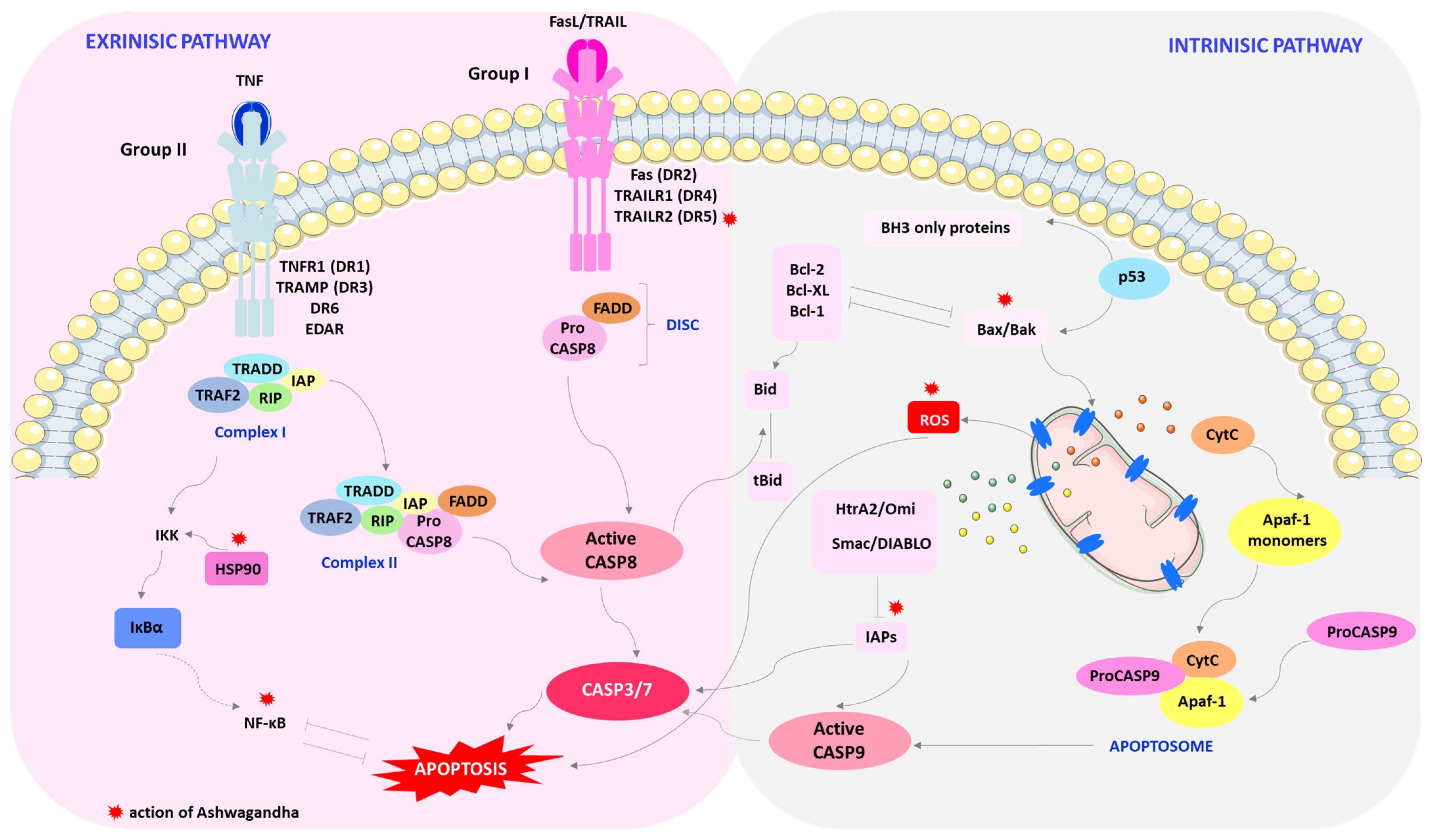
The mechanism of apoptosis mainly consists of two basic pathways involved in inducing apoptosis, intracellular (intrinsic apoptosis) or extracellular (extrinsic apoptosis) (Figure 2) [79].
In the intrinsic pathway, apoptosis occurs via mitochondrial mediation. Apoptotic signaling in the extrinsic pathway involves extracellular ligands such as tumor necrosis factor (TNF), Fas ligand (Fas-L), and TNF-related apoptosis-inducing ligand (TRAIL). These ligands are attached to the extracellular domain of transmembrane receptors (DRs), which include TNF type 1 receptor (TNFR1), Fas (also called CD95/Apo-1), and TRAIL receptors [80].
The intrinsic, mitochondrial pathway of apoptosis is triggered by various intracellular stressors. Proteins from the Bcl-2 family are responsible for regulating this pathway, including pro-apoptotic proteins (Bax, Bak, Bok, Bim, Bid, Bik, Bad, Bmf, Hrk, Noxa, Puma, Blk) and anti-apoptotic proteins (Bcl-2, Bcl-XL, Bcl-w, A1, Mcl-1). Internal cellular stress activates Bcl-2 proteins that have only one domain, the so-called BH3-only proteins (Bcl-2 homology 3), containing Bid, Bim, Bad, Bik, Noxa, Puma, and Hrk. These proteins neutralize the action of anti-apoptotic proteins or, acting directly, activate multi-domain apoptosis-inducing proteins, i.e., Bax or Bak [81,82,83,84,85].
Oligomerizing Bax or Bak create pores in the outer mitochondrial membrane, which results in its destabilization. Disruption of the integrity of mitochondrial potential results in the release of pro-apoptotic proteins into the cytosol. Proteins that are generally involved in intrinsic pathway include cytochrome C (called apoptotic protease-activating factor 1 (Apaf-2)), Smac/DIABLO, HtrA2/Omi and AIF, Endonuclease G and CAD. Cytochrome C (CytC) combines with Apaf-1 and procaspase-9 to form an apoptosome that releases caspase-9 (CASP9). In turn, the CASP9 initiator activates caspase-3 (CASP3) and caspase-7 (CASP7). Smac/DIABLO and HtrA2/Omi indirectly promote apoptosis by affecting caspase-3/7 activation through inhibition of inhibitor of apoptosis proteins (IAPs). Whereas, AIF, Endonuclease G, and CAD proteins travel directly to the cell nucleus, cutting DNA into short fragments [86].
In the intrinsic apoptosis pathway, the element of pro-apoptotic regulation is the p53 protein, the so-called guardian of the genome. Under stress conditions, the p53 protein moves to the mitochondria, where it forms a complex with Bcl-2 and Bcl-XL, causing their inactivation. As a result, the permeability of the mitochondrial membrane increases and CytC is released [87].
In the extrinsic apoptosis pathway, the pro-apoptotic signal is transmitted via receptors from the tumor necrosis factor receptor TNFR family (Fas (DR2) TRAILR1 (DR4), TRAILR2 (DR5), TNFR1 (DR1), TRAMP (DR3), DR6 and EDAR). A feature of death receptors is the presence of an intracellular domain called the death domain (DD). After attaching the ligand to the receptor, it is activated through oligomerization and conformational changes, which then attaches an adapter protein such as Fas-associated death domain (FADD), tumor necrosis factor receptor-1-associated death domain (TRADD), or death domain-associated protein (Daxx)-containing death domains. The adapter protein further combines with procaspase-8 to form the death-inducing signaling complex (DISC). Attachment of an appropriate ligand to the receptor induces signal transmission through the death domain of the adapter protein to caspase-8 (CASP8). The consequence of this is the activation by autoproteolysis of procaspase-8, which is a direct activator of caspase 3/7 [80,81,84,88].
Fas (DR2), TRAILR1 (DR4), and TRAILR2 (DR5) receptors belong to the first group of death receptors activated by FasL and TRAIL. Once activated, these receptors recruit the DISC complex, which consists of FADD and CASP8. Due to the amount of activated CASP8, we can divide this phase into two types. In the type of stage when the amount of active CASP8 is higher, direct activation of CASP3 occurs. However, in the second type, when we are dealing with a low level of CASP3 activation via CASP8, caspase-8 activates the pro-apoptotic Bid protein into the active form tBid, which interacts with Bax/Bak proteins, and CytC is released [84,89,90].
The second group of death receptors includes the TNFR1 (DR1), TRAMP (DR3), DR6, and EDAR receptors. The apoptosis-inducing ligand is TNF. Once activated, the receptors recruit the TNF-related death domain (TRADD) as an adapter protein and bind to TNF receptor-related factors-2.5 (TRAF2.5), receptor-interacting protein kinase (RIP1 or RIPK1), and cellular inhibitors of apoptosis proteins (cIAP) [84].
Two distinct complexes may be formed for TNFR1. Complex I is the TNFR1 complex consisting of proteins containing DD TRADD, TNF-2 receptor-associated factor (TRAF2 (TNF)), cIAP1, and the receptor serine/threonine protein kinase 1 (RIP1). This complex activates nuclear factor-κB (NF-κB) and the c-Jun NH2-terminalkinase/Jun proto-oncogene/AP-1 transcription factor subunit (JNK/AP-1). NF-κB inhibits TNF-induced apoptosis. Complex II arises after the TRADD-based complex cleaves from the receptor and recruits FADD and the initiator CASP8. The balance between these complexes is dependent on the protein FLIP, a CASP8 inhibitor. Complex II mediates apoptosis when activation of the NF-κB pathway is inadequate [84].
In the classic way of NF-κB activation, after the action of an appropriate stimulating factor, e.g., pro-inflammatory cytokines (TNF), the IKK kinase complex is activated. The active IKK complex catalyzes the phosphorylation of IκBα, which leads to its degradation and the release of the NF-κB dimer. Then, the NF-κB dimer is translocated to the cell nucleus and activates the transcription of appropriate genes, the end products of which have the ability to inhibit apoptosis [91,92,93].
In addition to DR, apoptosis is influenced by growth factors through the phosphatidinositide-3-kinase (PI3K) and murine thymus virus oncogene homolog v-AKT (AKT) pathways. Growth factors bind to receptors and activate PI3K, which activates AKT. AKT is important in the regulation of Bad. Moreover, protein kinase-C (PKC) influences apoptosis by activating ribosomal S6 kinase (p90RSK) [94].
In healthy breast cells, a balance is maintained between cell proliferation and death. Once a balance is disrupted, an activated anti-apoptotic signaling pathway or a deficiency in the pro-apoptosis pathway can lead to uncontrolled cell proliferation [95,96].
Withaferin A and extracts from the Ayurvedic medicinal plant Withania somnifera can inhibit the proliferation of breast cancer cells by inducing apoptosis in both the intrinsic and extrinsic pathways. WA and/or WS root protein extract (WSPF) induces death in cultured MDA-MB-231 and MCF-7 cancer cells and MDA-MB-231 xenografts in vivo by mediating the production of reactive oxygen species (ROS) in the intrinsic apoptosis pathway (Figure 2) [28,97,98]. Selective killing of MCF-7 and MDA-MB-231 cancer cells by WA and/or WSPF is mediated by the induction of oxidative stress via ROS levels, DNA damage, mitochondrial structure, and membrane potential [97,98,99]. WA inhibits oxidative phosphorylation (OXPHOS) in Complex III, accompanied by apoptotic release of DNA fragments associated with histones in the cytosol [28,100,101]. However, the anti-cancer effect of WA was significantly attenuated in the presence of anti-oxidants, as it has been shown that ectopic expression of Cu and Zn-superoxide dismutase (SOD) significantly weakens its apoptotic properties [28]. Moreover, WA shows high selectivity, causing ROS production only in MDA-MB-231 and MCF-7 cells, but not in the normal human mammary epithelial cell line (HMEC) [28].
WA is involved in the regulation of the expression of Bcl-2 proteins, which are critical regulators of cell death, acting either as inhibitors or facilitators of apoptosis. Both Bax and Bak appear to contribute to the induction of apoptosis by WA (Figure 2). The active substance contained in Ashwagandha enhances the activation of these pro-apoptotic proteins in MDA-MB-231 and MCF-7 cells [28]. WA inhibits the survival of MCF-7 and MDA-MB-231 breast cancer cell lines in culture and delays the growth of MDA-MB-231 xenografts in vivo due to reduced cell proliferation and increased apoptosis. According to Stan et al., apoptosis by WA is probably mediated by forkhead box O3 (FOXO3a) and Bim proteins in both cancer cell lines. WA-induced apoptosis was accompanied by the induction of Bim-s and Bim-L in MCF-7 cells and the induction of Bim-s and Bim-EL isoforms in MDA-MB-231 cells [25].
WA also results in suppression of these IAP family proteins: XIAP, cIAP-2, and survivin in MDA-MB-231 and MCF-7 human breast cancer cells (Figure 2). IAP family proteins (IAP, cIAP1, cIAP2, NAIP, livin, and survivin) act as endogenous inhibitors of apoptosis and are involved in the cell’s adaptive response to stress, differentiation, motility, and immune response. IAPs can inhibit both the intrinsic and extrinsic apoptosis pathways. The overexpression of IAP family proteins is observed in breast cancer, which results in improved survival of cancer cells, increased tumor growth and, consequently, metastases. Therefore, it seems that the reduction in the expression of these proteins by WA indicates its significant role in apoptosis [102].
In the extrinsic apoptosis pathway, the TRAIL pathway is considered the most attractive cancer therapy agent, which is non-toxic compared to the FasL and TNF-α pathways. Its safety in cancer patients has been confirmed in phase I and II clinical trials [103,104,105,106]. TRAIL is expressed mainly on the surface of immune cells and induces apoptosis in various cancer cell lines. In a group of patients with triple-negative breast cancer (TNBC), pre-clinical models have demonstrated the effectiveness of therapy targeting the TRAIL DR pathway [107]. WA inhibits breast tumor growth by upregulation of death receptor 5 (DR5, also known as TRAIL-R2/TRICK-2/KILLER/TNFRSF10B) (Figure 2) [107]. WA has been shown to activate phosphorylation of S6RSK in breast cancer cells via activation of cell signal-regulated kinase (ERK) [108]. Both RSK and ERK play a key role in breast cancer progression and metastasis [109]. WA induced a feed-forward loop of ERK and RSK, causing the simultaneous upregulation/activation of homology protein/C-EBP axis (CHOP) and ETS-like transcription factor 1 (Elk1). The recruitment of CHOP and Elk1 to the DR5 promoter leads to activation of the death receptor, DR5, which is responsible for the aggregation of proteins promoting apoptotic signal transduction [108].
The heat shock protein (HSP) transcription factor may also play an important role in apoptosis through WA by regulating survivin expression. In many types of cancer, heat shock factor 1 (HSF1) is a transcription factor activated under environmental stress, which leads to increased expression of HSP proteins. HSF1 is increased in expression and facilitates cancer transformation by modulating signaling pathways related to growth and proliferation, apoptosis, metabolism, and motility. WA induced HSF1 phosphorylation, which is probably due to a transient cellular defense response to WA-induced stress [110]. The level of HSP expression determines the fate of cells, because these biomolecules can direct them to the apoptosis or survival pathway. One of the HSP subfamily proteins, heat shock chaperone 90 (HSP90), has become an exciting target for cancer therapy due to its role in regulating cell proliferation, survival, and apoptosis. HSP90 can modulate the activity and stability of many transcription factors and kinases related to apoptosis, including NF-κB, p53, protein kinase B (PKB/AKT), Proto-Oncogene, Serine/Threonine Kinase (Raf-1), and stress-activated protein/kinase/Jun N-terminal kinase (SAPK/JAK). In the pathway involving NF-κB, cell survival is conditioned by the formation of an NF-κB complex with the kinase of the κB inhibitor kinase complex (IKK). KK is composed of IKKα and IKKβ and NF-κB essential modulator (NEMO) or IKKγ. IKKβ IKK kinase phosphorylates two specific serine residues in the N-terminal region of IκBα, causing proteasomal degradation of IκBα and nuclear translocation of canonical NF-κB members, mainly p50/RelA and p50/c-Rel dimers [111,112]. The active substances of Ashwagandha, 2,3-unsaturated double bond-containing withanolides, exhibit potent inhibitory effects on the activity of the Hsp90 chaperone by depleting several important signaling molecules involved in cell survival (Figure 2). Withanolides inhibited the IKK/NF-κB pathway in MDA-MB-23, an ER-negative human breast cancer cell line. They reduced the activity of anti-apoptotic proteins (Bcl-2, Bcl-xL, and c-FLIP) that are regulated by NF-κB [113].
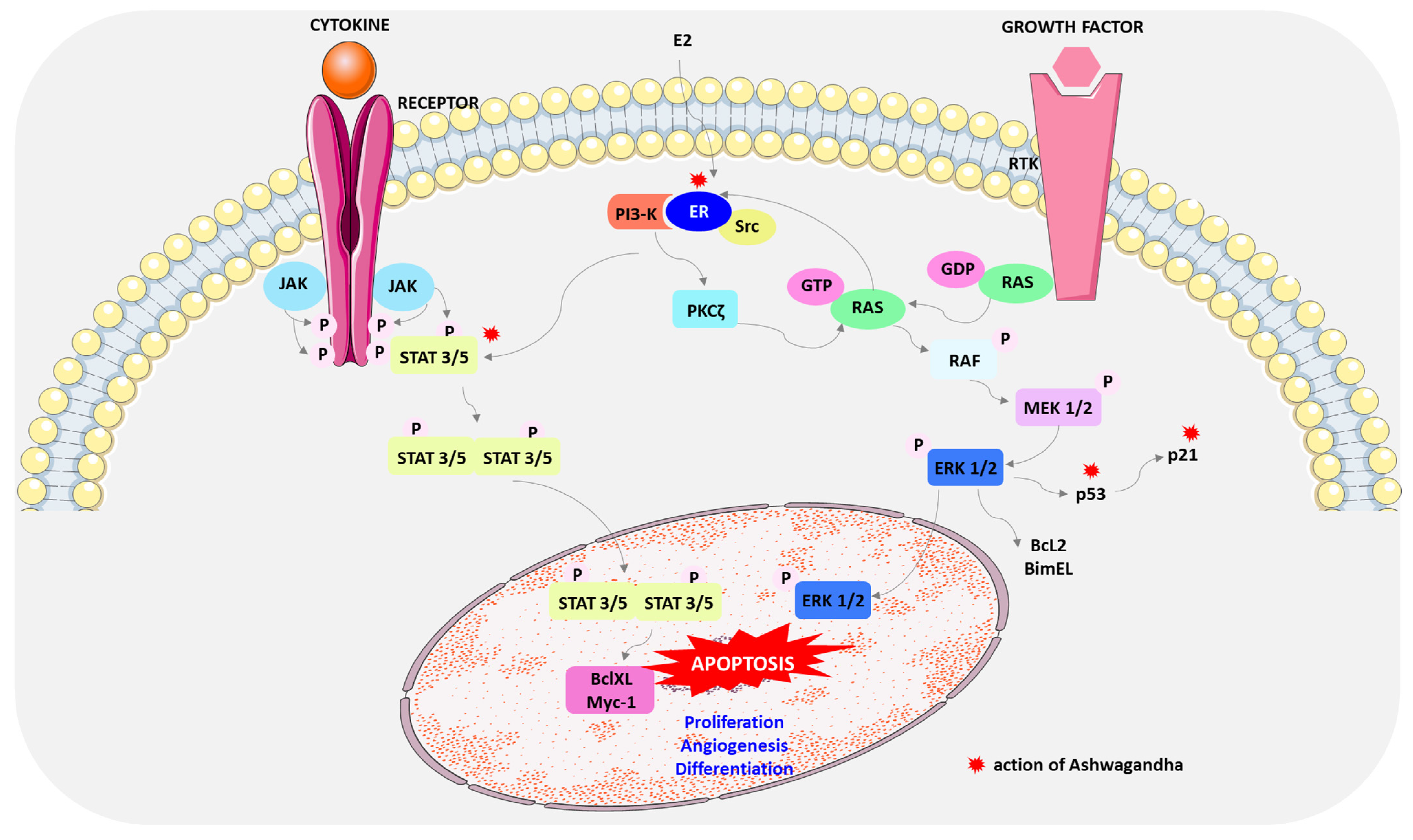
Janus kinase (JAK)-signal transducer and activator of transcription (STAT) signaling also mediates apoptosis. STAT proteins (STAT 1A, 1B, 2, 3, 4, 5A, 5B, and 6) are a family of transcription factors, each of which performs a unique function in the transmission of extracellular and direct signals regulating the transcription of genes that are involved in cell survival, proliferation, chemoresistance, and angiogenesis. Signal transducers and activators of transcription proteins (STATs) are activated by binding to their receptors on the cell membrane. The phosphorylated receptor-kinase complex is the site of STAT attachment, which is then phosphorylated on tyrosine residues necessary for activation. Then, after dimerization, STAT translocated to the nucleus can influence cell apoptosis by regulating the transcription of target genes such as Bcl-xL, p21, and Myc [114,115,116].
In many human cancers, including breast cancer, STAT3 and STAT5 are persistently phosphorylated and overactivated [117]. STAT3 increases the expression of c-Myc and the metastasis regulator Twist genes and is, therefore, thought to induce breast cancer [118]. Aberrant STAT5 signaling promotes cyclin D, Bcl-2, and matrix metalloproteinase-2 (MMP-2) gene expression, resulting in increased cell proliferation, survival, and metastasis [119,120]. WA causes inactivation of STAT3 and STAT5 by inhibiting their recruitment to growth factor and cytokine receptors as well as tyrosine phosphorylation, nuclear translocation, and DNA binding. Furthermore, STAT3 inhibits Bcl-xL and Mcl-1. (Figure 3) [117].
Other studies have demonstrated the role of mitogen-activated protein kinases (MAPKs) in the regulation of apoptosis by WA (Figure 3). MCF-7 cell death resulted from the action of WA via pharmacological inhibition of ERK kinase and p38 MAPK [121]. The MAPK pathway, often called the RAS-RAF-MEK-ERK signaling cascade, serves to transmit upstream signals to downstream effectors to regulate physiological processes such as cell proliferation, differentiation, survival, and death [122]. It is the most frequently mutated signaling pathway in human cancers; therefore, this pathway may be a promising strategy in cancer therapy [123,124].
The mitogen-activated protein kinase (MAPK) cascade consists of serine/threonine kinases that convert extracellular molecules such as growth factors (EGF), insulin-like growth factor I (IGF-I), hormones, and differentiation factors into intracellular signals [123,124]. There are four core protein kinases in the Ras/Raf/MEK/ERK-signaling pathway: Ras, Raf, MEK, and ERK. Once the signaling molecules are attached, the receptors dimerize, which activates RAS proteins. Activated RAS protein recruits RAF protein from the cytosol to the cell membrane, leading to its activation. Active RAF kinase phosphorylates MEK1 and 2 proteins, which activate ERK1 and 2 proteins. After transmitting the signal to the cell nucleus, ERK kinases phosphorylate and activate many target molecules responsible for cell growth, migration, and survival [125,126].
WA also acts to reduce the level of estrogen receptor alpha (Erα), which consequently affects the induction of apoptosis and inhibition of the growth of ER-α-positive MCF-7 and T47D breast cancer cells [33].
17β-estradiol (E2) exerts its effects on proliferation mainly through rapid non-genomic mechanisms derived from the binding of the hormone to Erα. Non-genomic signals are transmitted to the nucleus via various intracellular-signaling pathways such as MAPK/ERK and Pl3K/AKT [127,128,129]. E2 induces the association of ERα with Src and the PI 3-kinase (PI3K) adapter subunit p85a. Estradiol-stimulated PI 3-kinase/PDK1 pathway activates PKCζ in MCF-7 cells, which controls Ras-dependent ERK activation. Furthermore, E2 phosphorylates STAT3 and STAT5 by an ER-dependent mechanism [130]. The ERβ isoform has also been shown to play an important role in the proliferative action of E2, which can act as a tumor suppressor by modulating the proliferative action of ERα [129]. The E2-ERβ complex activates p38/MAPK leading to apoptosis while E2-ERα activates transduction pathways involved in cell cycle progression. The E2-ERβ complex activates p38/MAPK leading to apoptosis while E2-ERα activates transduction pathways involved in cell cycle progression [129,131,132]. WA reduces ERα levels post-translationally, inducing ERα protein aggregation and degradation. The anti-cancer activity of WA is mediated by RET tyrosine kinase (RET) and p53, which inhibits growth and apoptosis [110,133]. While RET is overexpressed in ERα-positive breast cancer, its activation stimulates the proliferation, survival, and dispersal of MCF-7 breast cancer cells [134]. WA had anti-tumor effects through the downregulation of RET protein with parallel depletion of Erα coupled with increases in the expression of phosphorylated mitogen-activated protein kinase p38 (phospho-p38 MAPK), p53, and p21 (Figure 3) [14,110].
In addition to apoptosis, there are also other non-apoptotic modes of cell death including autophagy, necroptosis, paraptosis, and apoptosis-like programmed cell death.
Autophagy or autophagic cell death are referred to as type II cell death. This process plays an important role in the degradation of cellular components inside the dying cell in autophagic vacuoles. It helps maintain cellular energy supply and homeostasis in rapidly proliferating cancer cells. Cancer cells use this process to respond to various environmental stimuli and avoid anti-cancer therapy [135]. The anti-tumor effect of WA in many breast cancer subtypes, including luminal A, luminal B, basal, claudin-low, and HER2W subtypes, is related to impairment of lysosomal activity, causing blockage of autophagic flux, which results in energy depletion leading to growth inhibition and induction of apoptosis. WA is an activator of 5′AMP-activated protein kinase AMP (AMPK), which works synergistically with 2-deoxy-d-glucose (2-DG) to inhibit breast cancer growth and is also an inhibitor of lactate dehydrogenase (LDHA), a key enzyme catalyzing the conversion of pyruvate to lactate [136].
Inhibition of the WA-mediated proteasome degradation system and perturbation of autophagy causes the accumulation of ubiquitinated proteins, which in turn results in unfolded protein responses and ER stress-mediated proteotoxicity in the human breast cancer cell lines MCF-7 and MDA-MB-231 [137].
Another process involved in programmed cell death that is morphologically different from apoptosis and autophagy is paraptosis. WA acts on the mitochondrial membrane potential, causing its hyperpolarization and the formation of many cytoplasmic vesicles. In the human breast cancer cell lines MCF-7 and MDA-MB-231, mitochondrial swelling and fusion occur, and the endoplasmic reticulum (ER) expands. WA reduces the level of the native paraptosis inhibitor, actin-interacting protein-1 (Alix/AIP-1), indicating that WA promotes death in both MCF-7 and MDA-MB-231 cell lines through paraptosis through the action of ROS [138].
Summarizing pre-clinical studies, it can be concluded that Ashwagandha has anti-cancer activity in various experimental models. WS acts at many stages, leading to apoptosis, both in the extrinsic and intrinsic pathways. WA and/or its extracts induce death in cancer cell cultures in vivo by mediating ROS production [28,97,98,99]. WA regulates the expression of Bcl-2 proteins, reduces the expression of IAP proteins XIAP, cIAP-2, and survivin and apoptosis inhibitors [25,97,102]. It inhibits the growth of breast cancer cells by activating DR5 [108]. It affects the activity of the Hsp90 protein, the recruitment of STAT3 and STAT5, and the regulation of the MAPK pathway [107,117,121]. It influences the reduction of the level of estrogen receptor alpha [33,110]. WA inhibits autophagic flux and reduces the level of paraptosis inhibitor, inducing death in breast cancer cells via ROS [137,138].
Information regarding the mechanism of action of Ashwagandha on various cell lines, taking into account the concentrations of active components of the plant extract used, is presented in Table 1.
There is little information about clinical trials confirming the use of WS in the treatment of breast cancer, although they are crucial to confirm its effectiveness and safety. It has only been shown that WS reduces chemotherapy-induced fatigue and improves the quality of life after administering 2 g of WS root extract every 8 h, throughout the course of chemotherapy. Similarly, survival analysis showed that patients in the WS treatment group had a better 24-month survival rate of 76% compared to the control group, which had a survival rate of 56% [47,139,140].
Although Ashwagandha was considered safe in clinical trials, improper dosage or failure to take into account individual contraindications may lead to a number of health problems, ranging from nausea and vomiting to more serious complications, such as hypertension, liver dysfunction, and hyperbilirubinemia [141,142].
Additionally, people taking anti-diabetic, anti-hypertensive, or central nervous system medications should be careful, because WS may interact with these medications, enhancing or changing their effects. Ashwagandha may also affect thyroid hormone and testosterone levels [143,144,145,146].
There is also a lack of clear evidence on the safety of the long-term use of WS over many months or years. Ashwagandha was well tolerated at a dose of 300 mg per day in an eight-week study [147]. For insomnia, a dose of 600 milligrams per day has been found to be safe and effective [148]. This dosage has also been associated with improved memory [149].
The extensive exploratory studies demonstrate the potential of Ashwagandha in the treatment of breast cancer through its anti-cancer activity, safety profile, and combination therapy possibilities. However, due to inconsistent therapeutic results resulting from the widely varying composition of active components in the plant extract, the actual use of WS in the treatment of cancer is limited. Further research and standardization are needed to harness its full therapeutic potential.

5. Conclusions

Withaferin A (WA) and withanolides are the most promising anti-cancer compounds of Ashwagandha, which play a major role in the induction of the apoptosis of cancer cells. This review attempts to confirm its therapeutic properties, with a particular emphasis on its role in the treatment of breast cancer. The literature data indicate that compounds isolated from various parts of this plant, such as the root, stem, and leaves, have significant anti-cancer and immunomodulatory properties; therefore, they can be used alone or in combination with other chemotherapy drugs in the treatment of breast cancer. Reaching for natural medicine can complement the standard treatment of cancer patients without undesirable side effects and may also have a positive impact on reducing the feeling of fatigue and improving their quality of life [139].

Author Contributions

Conceptualization, R.K. and H.P.; writing—original draft preparation, R.K., H.P. and A.T.-K.; writing—review and editing, R.K., H.P., A.T.-K., M.P., K.S., L.P. and A.W.; visualization, R.K., K.S. and M.P.; supervision, R.K. and H.P.; project administration, R.K. and H.P.; funding acquisition, A.W. All authors have read and agreed to the published version of the manuscript.

Funding

This research received no external funding.

Conflicts of Interest

The authors declare no conflicts of interest.

References

  1. Bray, F.; Laversanne, M.; Sung, H.; Jacques Ferlay, J.; Siegel, R.L.; Soerjomataram, I.; Jemal, A. Global cancer statistics 2022: GLOBOCAN estimates of incidence and mortality worldwide for 36 cancers in 185 countries. CA Cancer J. Clin. 2024, 74, 229–263. [Google Scholar] [CrossRef] [PubMed]
  2. Siegel, R.L.; Giaquinto, A.N.; Jemal, A. Cancer statistics. CA Cancer J. Clin. 2024, 74, 12–49. [Google Scholar] [CrossRef] [PubMed]
  3. Dizon, D.S.; Kamal, A.H. Cancer statistics 2024: All hands on deck. CA Cancer J. Clin. 2024, 74, 8–9. [Google Scholar] [CrossRef] [PubMed]
  4. Siddig, A.; Tengku Din, T.A.D.A.; Mohd Nafi, S.N.; Yahya, M.M.; Sulong, S.; Wan Abdul Rahman, W.F. The unique biology behind the early onset of breast cancer. Genes 2021, 12, 372. [Google Scholar] [CrossRef] [PubMed]
  5. Carmen Criscitiello, C.; Corti, C. Breast cancer genetics: Diagnostics and treatment. Genes 2022, 13, 1593. [Google Scholar] [CrossRef] [PubMed]
  6. Harbeck, N.; Penault-Llorca, F.; Cortes, J.; Gnant, M.; Houssami, N.; Poortmans, P.; Ruddy, K.; Tsang, J.; Cardoso, F. Breast cancer. Nat. Rev. Dis. Primers 2019, 5, 1–31. [Google Scholar] [CrossRef] [PubMed]
  7. Łukasiewicz, S.; Czeczelewski, M.; Forma, A.; Baj, J.; Sitarz, R.; Stanisławek, A. Breast cancer—Epidemiology, risk factors, classification, prognostic markers, and current treatment strategies—An updated review. Cancers 2021, 13, 4287. [Google Scholar] [CrossRef] [PubMed]
  8. Mohi ud din, N.; Ahmad Dar, R.; Rasool, M.; Assad, A. Breast cancer detection using deep learning: Datasets, methods, and challenges ahead. Comput. Biol. Med. 2022, 149, 106073. [Google Scholar] [CrossRef]
  9. Bhimani, C.; Matta, D.; Roth, R.G.; Liao, L.; Tinney, E.; Brill, K.; Germaine, P. Contrast-enhanced spectral mammography: Technique, indications, and clinical applications. Acad. Radiol. 2017, 24, 84–88. [Google Scholar] [CrossRef]
  10. Krzakowski, M.; Rutkowski, P.; Jassem, J.; Zaucha, R.; Fijuth, J.; Słuszniak, J.; Jarząb, B.; Zegarski, W.; Małkowski, B.; Kawecki, A.; et al. Recommendations on the application of positron emission tomography in oncology. Oncol. Clin. Pract. 2015, 11, 155–171. [Google Scholar]
  11. Debela, D.T.; Muzazu, S.G.Y.; Heraro, K.D.; Ndalama, M.N.; Mesele, B.W.; Haile, D.C.; Sophia Khalayi, S. New approaches and procedures for cancer treatment: Current perspectives. SAGE Open Med. 2021, 9, 20503121211034366. [Google Scholar] [CrossRef] [PubMed]
  12. Mehta, V.; Chander, H.; Munshi, A. Mechanisms of anti-tumor activity of Withania somnifera (Ashwagandha). Nutr. Cancer 2021, 73, 914–926. [Google Scholar] [CrossRef] [PubMed]
  13. National Center for Complementary and Integrative Health. Ashwagandha. Available online: https://www.nccih.nih.gov/health/ashwagandha#:~:text=What%20Have%20We%20Learned%3F,testosterone%20levels%20and%20sperm%20quality (accessed on 12 July 2024).
  14. Widodo, N.; Kaur, K.; Shrestha, B.G.; Takagi, Y.; Ishii, T.; Wadhwa, R.; Kaul, S.C. Selective killing of cancer cells by leaf extract of ashwagandha: Identification of a tumor-inhibitory factor and the first molecular insights to its effect. Clin. Cancer Res. 2007, 13, 2298–2306. [Google Scholar] [CrossRef] [PubMed]
  15. Padmavathi, B.; Rath, P.C.; Rao, A.R.; Singh, R.P. Roots of Withania somnifera inhibit forestomach and skin carcinogenesis in mice. Evid. Based Complement. Alternat. Med. 2005, 2, 99–105. [Google Scholar] [CrossRef] [PubMed]
  16. Antony, M.L.; Lee, J.; Hahm, E.-R.; Kim, S.-H.; Marcus, A.I.; Kumari, V.; Ji, X.; Yang, Z.; Vowell, C.L.; Wipf, P.; et al. Growth arrest by the antitumor steroidal lactone withaferin A in human breast cancer cells is associated with down-regulation and covalent binding at cysteine 303 of β-tubulin. J. Biol. Chem. 2014, 289, 1852–1865. [Google Scholar] [CrossRef] [PubMed]
  17. Dutta, R.; Khalil, R.; Green, R.; Mohapatra, S.S.; Mohapatra, S. Withania somnifera (Ashwagandha) and Withaferin A: Potential in integrative oncology. Int. J. Mol. Sci. 2019, 20, 5310. [Google Scholar] [CrossRef]
  18. Singh, N.; Yadav, S.S.; Rao, A.S.; Nandal, A.; Kumar, S.; Ganaie, S.A.; Narasihman, B. Review on anticancerous therapeutic potential of Withania somnifera (L.) Dunal. J. Ethnopharmacol. 2021, 270, 113704. [Google Scholar] [CrossRef] [PubMed]
  19. Govindaram, L.K.; Al Bratty, M.; Alhazmi, H.A.; Kandasamy, R.; Thangavel, N.; Ibrahim, A.M.; Mariya, G.A.; Kumar, P. Formulation, biopharmaceutical evaluation and in-vitro screening of polyherbal phytosomes for breast cancer therapy. Drug Dev. Ind. Pharm. 2022, 48, 552–565. [Google Scholar] [CrossRef]
  20. Srivastava, A.N.; Ahmad, R.; Khan, M.A. Evaluation and comparison of the in vitro cytotoxic activity of Withania somnifera methanolic and ethanolic extracts against MDA-MB-231 and vero cell lines. Sci. Pharm. 2015, 84, 41–59. [Google Scholar] [CrossRef]
  21. Jawarneh, S.; Talib, W.H. Combination of Ashwagandha water extract and intermittent fasting as a therapy to overcome cisplatin resistance in breast cancer: An in vitro and in vivo study. Front. Nutr. 2022, 9, 863619. [Google Scholar] [CrossRef]
  22. Cavaleri, F.; Chattopadhyay, S.; Palsule, V.; Kar, P.K.; Chatterjee, R. Study of drug targets associated with oncogenesis and cancer cell survival and the therapeutic activity of engineered Ashwagandha extract having differential Withanolide constitutions. Integr. Cancer Ther. 2024, 23, 15347354231223499. [Google Scholar] [CrossRef]
  23. Srinivasan, S.; Ranga, R.S.; Burikhanov, R.; Han, S.S.; Chendil, D. Par-4-dependent apoptosis by the dietary compound withaferin A in prostate cancer cells. Cancer Res. 2007, 67, 246–253. [Google Scholar] [CrossRef]
  24. Ahmad, R.; Fatima, A.; Srivastava, A.N.; Ali Khan, M. Evaluation of apoptotic activity of Withania coagulans methanolic extract against human breast cancer and Vero cell lines. J. Ayurveda Integr. Med. 2017, 8, 177–183. [Google Scholar] [CrossRef]
  25. Stan, S.D.; Hahm, E.R.; Warin, R.; Singh, S.V. Withaferin A causes FOXO3a- and Bim-dependent apoptosis and inhibits growth of human breast cancer cells in vivo. Cancer Res. 2008, 68, 7661–7669. [Google Scholar] [CrossRef] [PubMed]
  26. Grover, A.; Singh, R.; Shandilya, A.; Priyandoko, D.; Agrawal, V.; Bisaria, V.S.; Wadhwa, R.; Kaul, S.C.; Sundar, D. Ashwagandha derived withanone targets TPX2-aurora A complex: Computational and experimental evidence to its anticancer activity. PLoS ONE 2012, 7, e30890. [Google Scholar] [CrossRef] [PubMed]
  27. Samanta, S.K.; Lee, J.; Hahm, E.-R.; Singh, S.V. Peptidyl-prolyl cis/trans isomerase Pin1 regulates withaferin A-mediated cell cycle arrest in human breast cancer cells. Mol. Carcinog. 2018, 57, 936–946. [Google Scholar] [CrossRef]
  28. Hahm, E.R.; Moura, M.B.; Kelley, E.E.; Van Houten, B.; Shiva, S.; Singh, S.V. Withaferin A-induced apoptosis in human breast cancer cells is mediated by reactive oxygen species. PLoS ONE 2011, 6, e23354. [Google Scholar] [CrossRef]
  29. Gambhir, L.; Checker, R.; Sharma, D.; Thoh, M.; Patil, A.; Degani, M.; Vikram Gota, V.; Sandur, S.K. Thiol dependent NF-κB suppression and inhibition of T-cell mediated adaptive immune responses by a naturally occurring steroidal lactone Withaferin A. Toxicol. Appl. Pharmacol. 2015, 289, 297–312. [Google Scholar] [CrossRef]
  30. Ichikawa, H.; Takada, Y.; Shishodia, S.; Jayaprakasam, B.; Nair, M.G.; Aggarwal, B.B. Withanolides potentiate apoptosis, inhibit invasion, and abolish osteoclastogenesis through suppression of nuclear factor-κB (NF-κB) activation and NF-κB-regulated gene expression. Mol. Cancer Ther. 2006, 5, 1434–1445. [Google Scholar] [CrossRef] [PubMed]
  31. Kaileh, M.; Vanden Berghe, W.; Heyerick, A.; Horion, J.; Piette, J.; Libert, C.; De Keukeleire, D.; Essawi, T.; Haegeman, G. Withaferin a strongly elicits IκB kinase β hyperphosphorylation concomitant with potent inhibition of its kinase activity. J. Biol. Chem. 2007, 282, 4253–4264. [Google Scholar] [CrossRef]
  32. Lee, J.; Hahm, E.R.; Singh, S.V. Withaferin A inhibits activation of signal transducer and activator of transcription 3 in human breast cancer cells. Carcinogenesis 2010, 31, 1991–1998. [Google Scholar] [CrossRef] [PubMed]
  33. Hahm, E.R.; Lee, J.; Huang, Y.; Singh, S.V. Withaferin A suppresses estrogen receptor-α expression in human breast cancer cells. Mol. Carcinog. 2011, 50, 614–624. [Google Scholar] [CrossRef] [PubMed]
  34. Stan, S.D.; Zeng, Y.; Singh, S.V. Ayurvedic medicine constituent withaferin A causes G2 and M phase cell cycle arrest in human breast cancer cells. Nutr. Cancer 2008, 60, 51–60. [Google Scholar] [CrossRef]
  35. Meher, K.; Paithankar, H.; Hosur, R.V.; Lopus, M. Ashwagandha-polyphenols-functionalized gold nanoparticles facilitate apoptosis by perturbing microtubule assembly dynamics in breast cancer cells. J. Drug Deliv. Sci. Technol. 2022, 70, 103225. [Google Scholar] [CrossRef]
  36. Yang, H.; Shi, G.; Dou, Q.P. The tumor proteasome is a primary target for the natural anticancer compound withaferin A isolated from “Indian winter cherry”. Mol. Pharmacol. 2007, 71, 426–437. [Google Scholar] [CrossRef]
  37. Mohan, R.; Hammers, H.J.; Bargagna-Mohan, P.; Zhan, X.H.; Herbstritt, C.J.; Ruiz, A.; Zhang, L.; Hanson, A.D.; Conner, B.P.; Rougas, J.; et al. Withaferin A is a potent inhibitor of angiogenesis. Angiogenesis 2004, 7, 115–122. [Google Scholar] [CrossRef]
  38. Lee, J.; Hahm, E.-R.; Marcus, A.I.; Singh, S.V. Withaferin A inhibits experimental epithelial-mesenchymal transition in MCF-10a cells and suppresses vimentin protein level in vivo in breast tumors. Mol. Carcinog. 2015, 54, 417–429. [Google Scholar] [CrossRef]
  39. Yang, Z.; Garcia, A.; Xu, S.; Powell, D.R.; Vertino, P.M.; Singh, S.; Marcus, A.I. Withania somnifera root extract inhibits mammary cancer metastasis and epithelial to mesenchymal transition. PLoS ONE 2013, 8, e75069. [Google Scholar] [CrossRef] [PubMed]
  40. Thaiparambil, J.T.; Bender, L.; Ganesh, T.; Kline, E.; Patel, P.; Liu, Y.; Tighiouart, M.; Vertino, P.M.; Harvey, R.D.; Garcia, A.; et al. Withaferin A inhibits breast cancer invasion and metastasis at sub-cytotoxic doses by inducing vimentin disassembly and serine 56 phosphorylation. Int. J. Cancer 2011, 129, 2744–2755. [Google Scholar] [CrossRef]
  41. Szarc vel Szic, K.; Op de Beeck, K.; Ratman, D.; Wouters, A.; Beck, I.M.; Declerck, K.; Heyninck, K.; Fransen, E.; Bracke, M.; De Bosscher, K.; et al. Pharmacological levels of withaferin A (Withania somnifera) trigger clinically relevant anticancer effects specific to triple negative breast cancer cells. PLoS ONE 2014, 9, e87850. [Google Scholar] [CrossRef]
  42. Gao, R.; Shah, N.; Lee, J.-S.; Katiyar, S.P.; Li, L.; Oh, E.; Sundar, D.; Yun, C.-O.; Wadhwa, R.; Kaul, S.C. Withanone-rich combination of Ashwagandha withanolides restricts metastasis and angiogenesis through hnRNP-K. Mol. Cancer Ther. 2014, 13, 2930–2940. [Google Scholar] [CrossRef]
  43. Lee, J.; Sehrawat, A.; Singh, S.V. Withaferin A causes activation of Notch2 and Notch4 in human breast cancer cells. Breast Cancer Res. Treat. 2012, 136, 45–56. [Google Scholar] [CrossRef] [PubMed]
  44. Bashir, A.; Nabi, M.; Tabassum, N.; Afzal, S.; Ayoub, M. An updated review on phytochemistry and molecular targets of Withania somnifera (L.) Dunal (Ashwagandha). Front. Pharmacol. 2023, 14, 1049334. [Google Scholar] [CrossRef] [PubMed]
  45. Ali, M.A.; Farah, M.A.; Al-Anazi, K.M.; Basha, S.H.; Bai, F.; Lee, J.; Al-Hemaid, F.M.A.; Mahmoud, A.H.; Hailan, W.A.Q. In silico elucidation of the plausible inhibitory potential of Withaferin A of Withania somnifera medicinal herb against Breast cancer targeting estrogen receptor. Curr. Pharm. Biotechnol. 2020, 21, 842–851. [Google Scholar] [CrossRef]
  46. Hahm, E.-R.; Kim, S.-H.; Singh, K.B.; Singh, K.; Singh, S.V. A comprehensive review and perspective on anticancer mechanisms of withaferin A in breast cancer. Cancer Prev. Res. 2020, 13, 721–734. [Google Scholar] [CrossRef] [PubMed]
  47. Vashi, R.; Patel, B.M.; Goyal, R.K. Keeping abreast about ashwagandha in breast cancer. J. Ethnopharmacol. 2021, 269, 113759. [Google Scholar] [CrossRef] [PubMed]
  48. Mallipeddi, H.; Thyagarajan, A.; Sahu, R.P. Implications of Withaferin-A for triple-negative breast cancer chemoprevention. Biomed. Pharmacother. 2021, 134, 111124. [Google Scholar] [CrossRef] [PubMed]
  49. Prasad, K.; Saggam, A.; Guruprasad, K.P.; Tillu, G.; Patwardhan, B.; Satyamoorthy, K. Molecular mechanisms of Asparagus racemosus willd. and Withania somnifera (L.) Dunal as chemotherapeutic adjuvants for breast cancer treatment. J. Ethnopharmacol. 2024, 331, 118261. [Google Scholar] [CrossRef] [PubMed]
  50. Sivasankarapillai, V.S.; Nair, R.M.K.; Rahdar, A.; Bungau, S.; Zaha, D.C.; Aleya, L.; Tit, D.M. Overview of the anticancer activity of withaferin A, an active constituent of the Indian ginseng Withania somnifera. Environ. Sci. Pollut. Res. Int. 2020, 27, 26025–26035. [Google Scholar] [CrossRef]
  51. Rahman, M.M.; Huixin Wu, H.; Tollefsbol, T.O. A novel combinatorial approach using sulforaphane- and withaferin A-rich extracts for prevention of estrogen receptor-negative breast cancer through epigenetic and gut microbial mechanisms. Sci. Rep. 2024, 14, 12091. [Google Scholar] [CrossRef]
  52. Ahmad, M.; Dar, N.J. Withania somnifera: Ethnobotany, Pharmacology, and Therapeutic Functions. In Sustained Energy for Enhanced Human Functions and Activity; Academic Press: Cambridge, MA, USA, 2017; pp. 137–154. [Google Scholar] [CrossRef]
  53. Kulkarni, S.K.; Dhir, A. Withania somnifera: An Indian ginseng. Prog. Neuro-Psychopharmacol. Biol. Psychiatry 2008, 32, 1093–1105. [Google Scholar] [CrossRef] [PubMed]
  54. Winters, M. Ancient medicine, modern use: Withania somnifera and its potential role in integrative oncology. Altern. Med. Rev. 2006, 11, 269–277. [Google Scholar] [PubMed]
  55. Bhat, J.A.; Akther, T.; Najar, R.A.; Rasool, F.; Hamid, A. Withania somnifera (L.) Dunal (Ashwagandha); current understanding and future prospect as a potential drug candidate. Front. Pharmacol. 2022, 13, 1029123. [Google Scholar] [CrossRef] [PubMed]
  56. Sharifi-Rad, J.; Quispe, C.; Ayatollahi, S.A.; Kobarfard, F.; Staniak, M.; Stępień, A.; Czopek, K.; Sen, S.; Acharya, K.; Matthews, K.R.; et al. Chemical composition, biological activity, and health-promoting effects of Withania somnifera for pharma-food industry applications. J. Food Qual. 2021, 2021, 8985179. [Google Scholar] [CrossRef]
  57. Gaurav, H.; Yadav, D.; Maurya, A.; Yadav, H.; Yadav, R.; Shukla, A.C.; Sharma, M.; Gupta, V.K.; Palazon, J. Biodiversity, biochemical profiling, and pharmaco-commercial applications of Withania somnifera: A Review. Molecules 2023, 28, 1208. [Google Scholar] [CrossRef] [PubMed]
  58. Uddin, Q.; Samiulla, L.; Singh, V.K.; Jamil, S.S. Phytochemical and pharmacological profile of Withania somnifera Dunal: A Review. J. Appl. Pharm. Sci. 2012, 2, 170–175. [Google Scholar]
  59. Visweswari, G.; Christopher, R.; Rajendra, W. Phytochemical screening of active secondary metabolites present in Withania somnifera root: Role in traditional medicine. Int. J. Pharm. Sci. Res. 2013, 4, 2770–2776. [Google Scholar] [CrossRef]
  60. Bhatia, A.; Bharti, S.K.; Tewari, S.K.; Sidhu, O.P.; Roy, R. Metabolic profiling for studying chemotype variations in Withania somnifera (L.) Dunal fruits using GC–MS and NMR spectroscopy. Phytochemistry 2013, 93, 105–115. [Google Scholar] [CrossRef] [PubMed]
  61. Alam, N.; Hossain, M.; Mottalib, M.A.; Sulaiman, S.A.; Gan, S.H.; Khalil, M.I. Methanolic extracts of Withania somnifera leaves, fruits and roots possess antioxidant properties and antibacterial activities. BMC Complement. Altern. Med. 2012, 12, 175. [Google Scholar] [CrossRef]
  62. Ozeer, F.Z.; Nagandran, S.; Wu, Y.S.; Wong, L.S.; Stephen, A.; Lee, M.F.; Kijsomporn, J.; Guad, R.M.; Batumalaie, K.; Oyewusi, H.A.; et al. A comprehensive review of phytochemicals of Withania somnifera (L.) Dunal (Solanaceae) as antiviral therapeutics. Discov. Appl. Sci. 2024, 6, 187. [Google Scholar] [CrossRef]
  63. Saleem, S.; Muhammad, G.; Hussain, M.A.; Altaf, M.; Bukhari, S.N.A. Withania somnifera L.: Insights into the phytochemical profile, therapeutic potential, clinical trials, and future prospective. Iran J. Basic Med. Sci. 2020, 23, 1501–1526. [Google Scholar] [CrossRef]
  64. Bahira, S.; Moharana, T.; Sahu, R.R.; Raut, S.; Behera, S. In vitro study of the antibacterial activities of Withania somnifera leaf extract against human pathogenic bacteria. Int. J. Pharm. Sci. Rev. Res. 2019, 56, 139–145. [Google Scholar]
  65. Chandra, S.; Chatterjee, P.; Dey, P.; Bhattacharya, S. Evaluation of anti-inflammatory effect of Ashwagandha: A preliminary study in vitro. Pharmacogn. J. 2012, 4, 47–49. [Google Scholar] [CrossRef]
  66. Pandey, A.; Bani, S.; Dutt, P.; Satti, N.K.; Suri, K.A.; Qazi, G.N. Multifunctional neuroprotective effect of withanone, a compound from Withania somnifera roots in alleviating cognitive dysfunction. Cytokine 2018, 102, 211–221. [Google Scholar] [CrossRef] [PubMed]
  67. Das, R.; Rauf, A.; Akhter, S.; Islam, M.N.; Emran, T.B.; Mitra, S.; Khan, I.N.; Mubarak, M.S. Role of withaferin A and its derivatives in the management of Alzheimer’s disease: Recent trends and future perspectives. Molecules 2021, 26, 3696. [Google Scholar] [CrossRef]
  68. Singh, M.; Ramassamy, C. In vitro screening of neuroprotective activity of Indian medicinal plant Withania somnifera. J. Nutr. Sci. 2017, 6, e54. [Google Scholar] [CrossRef]
  69. Wongtrakul, J.; Thongtan, T.; Kumrapich, B.; Saisawang, C.; Ketterman, A.J. Neuroprotective effects of Withania somnifera in the SH-SY5Y Parkinson cell model. Heliyon 2021, 7, e08172. [Google Scholar] [CrossRef]
  70. Kumar, P.; Kumar, A. Possible neuroprotective effect of Withania somnifera root extract against 3-nitropropionic acid-induced behavioral, biochemical, and mitochondrial dysfunction in an animal model of Huntington’s disease. J. Med. Food 2009, 12, 591–600. [Google Scholar] [CrossRef] [PubMed]
  71. Joshi, T.; Kumar, V.; Kaznacheyeva, E.V.; Jana, N.R. Withaferin A induces heat shock response and ameliorates disease progression in a mouse model of Huntington’s disease. Mol. Neurobiol. 2021, 58, 3992–4006. [Google Scholar] [CrossRef] [PubMed]
  72. Anju, T.R.; Smijin, S.; Jobin, M.; Paulose, C.S. Altered muscarinic receptor expression in the cerebral cortex of epileptic rats: Restorative role of Withania somnifera. Biochem. Cell Biol. 2018, 96, 433–440. [Google Scholar] [CrossRef]
  73. SoRelle, J.A.; Itoh, T.; Peng, H.; Kanak, M.A.; Sugimoto, K.; Matsumoto, S.; Levy, M.S.; Lawrence, M.C.; Naziruddin, B. Withaferin A inhibits pro-inflammatory cytokine-induced damage to islets in culture and following transplantation. Diabetologia 2013, 56, 814–824. [Google Scholar] [CrossRef] [PubMed]
  74. Gorelick, J.; Rosenberg, R.; Smotrich, A.; Hanuš, L.; Bernstein, N. Hypoglycemic activity of withanolides and elicitated Withania somnifera. Phytochemistry 2015, 116, 283–289. [Google Scholar] [CrossRef] [PubMed]
  75. Auddy, B.; Hazra, J.; Mitra, A.; Abedon, B. A standardized Withania somnifera extract significantly reduces stress-related parameters in chronically stressed humans: A double-blind, randomized, placebo-controlled study. J. Am. Neutraceut. Assoc. 2008, 11, 50–56. [Google Scholar]
  76. Majeed, M.; Nagabhushanam, K.; Mundkur, L. A standardized Ashwagandha root extract alleviates stress, anxiety, and improves quality of life in healthy adults by modulating stress hormones: Results from a randomized, double-blind, placebo-controlled study. Medicine 2023, 102, 41. [Google Scholar] [CrossRef] [PubMed]
  77. Kashyap, V.K.; Peasah-Darkwah, G.; Dhasmana, A.; Jaggi, M.; Yallapu, M.M.; Chauhan, S.C. Withania somnifera: Progress towards a pharmaceutical agent for immunomodulation and cancer therapeutics. Pharmaceutics 2022, 14, 611. [Google Scholar] [CrossRef] [PubMed]
  78. Palliyaguru, D.L.; Singh, S.V.; Kensler, T.W. Withania somnifera: From prevention to treatment of cancer. Mol. Nutr. Food Res. 2016, 60, 1342–1353. [Google Scholar] [CrossRef] [PubMed]
  79. Suraweera, C.D.; Hinds, M.G.; Kvansakul, M. Poxviral strategies to overcome host cell ppoptosis. Pathogens 2020, 10, 6. [Google Scholar] [CrossRef] [PubMed]
  80. Jan, R.; Chaudhry, G.-e.-S. Understanding apoptosis and apoptotic pathways targeted cancer herapeutics. Adv. Pharm. Bull. 2019, 9, 205–218. [Google Scholar] [CrossRef] [PubMed]
  81. Elmore, S. Apoptosis: A review of programmed cell death. Toxicol. Pathol. 2007, 35, 495–516. [Google Scholar] [CrossRef]
  82. Wang, C.; Youle, R.J. The Role of mitochondria in apoptosis. Annu. Rev. Genet. 2009, 43, 95–118. [Google Scholar] [CrossRef]
  83. Galluzzi, L.; Kepp, O.; Trojel-Hansen, C.; Kroemer, G. Mitochondrial control of cellular life, stress, and death. Circ. Res. 2012, 111, 1198–1207. [Google Scholar] [CrossRef] [PubMed]
  84. Cavalcante, G.C.; Schaan, A.P.; Cabral, G.F.; Santana-da-Silva, M.N.; Pinto, P.; Vidal, A.F.; Ribeiro-dos-Santos, Â.; Cell’s Fate, A. An overview of the molecular biology and genetics of apoptosis. Int. J. Mol. Sci. 2019, 20, 4133. [Google Scholar] [CrossRef] [PubMed]
  85. Lomonosova, E.; Chinnadurai, G. BH3-only proteins in apoptosis and beyond: An overview. Oncogene 2008, 27, 2–19. [Google Scholar] [CrossRef] [PubMed]
  86. Yuan, S.; Akey, C.W. Apoptosome structure, assembly, and procaspase activation. Structure 2013, 21, 501–515. [Google Scholar] [CrossRef]
  87. Schlereth, K.; Charles, J.P.; Bretz, A.C.; Stiewe, T. Life or death: p53-induced apoptosis requires DNA binding cooperativity. Cell Cycle 2010, 15, 4068–4076. [Google Scholar] [CrossRef]
  88. Jin, Z.; El-Deiry, W.S. Overview of cell death signaling pathways. Cancer Biol. Ther. 2005, 4, 139–163. [Google Scholar] [CrossRef] [PubMed]
  89. Mahmood, Z.; Shukla, Y. Death receptors: Targets for cancer therapy. Exp. Cell Res. 2010, 316, 887–899. [Google Scholar] [CrossRef] [PubMed]
  90. Lee, E.W.; Seo, J.; Jeong, M.; Lee, S.; Song, J. The roles of FADD in extrinsic apoptosis and necroptosis. BMB Rep. 2012, 45, 496–508. [Google Scholar] [CrossRef] [PubMed]
  91. Kim, H.J.; Hawke, N.; Baldwin, A.S. NF-kappaB and IKK as therapeutic targets in cancer. Cell Death Differ. 2006, 13, 738–747. [Google Scholar] [CrossRef]
  92. Nakajima, S.; Kitamura, M. Bidirectional regulation of NF-κB by reactive oxygen species: A role of unfolded protein response. Free Radic. Biol. Med. 2013, 65, 162–174. [Google Scholar] [CrossRef]
  93. Napetschnig, J.; Wu, H. Molecul ar basis of NF-κB signaling. Annu. Rev. Biophys. 2013, 42, 443–468. [Google Scholar] [CrossRef] [PubMed]
  94. Rascio, F.; Spadaccino, F.; Rocchetti, M.T.; Castellano, G.; Stallone, G.; Netti, G.S.; Ranieri, E. The pathogenic role of PI3K/AKT pathway in cancer onset and drug resistance: An updated review. Cancers 2021, 13, 3949. [Google Scholar] [CrossRef] [PubMed]
  95. Mohammad, R.M.; Muqbil, I.; Lowe, L.; Yedjou, C.; Hsu, H.Y.; Lin, L.T.; Siegelin, M.D.; Fimognari, C.; Kumar, N.B.; Dou, Q.P.; et al. Broad targeting of resistance to apoptosis in cancer. Semin. Cancer Biol. 2015, 35, 78–103. [Google Scholar] [CrossRef] [PubMed]
  96. Yuan, L.; Cai, Y.; Zhang, L.; Liu, S.; Li, P.; Li, X. Promoting apoptosis, a promising way to treat breast cancer with natural products: A comprehensive review. Front. Pharmacol. 2022, 12, 801662. [Google Scholar] [CrossRef]
  97. Dar, P.A.; Mir, S.A.; Bhat, J.A.; Hamid, A.; Singh, L.R.; Malik, F.; Dar, T.A. Ananti-cancerous protein fraction from Withania somnifera induces ROS dependent mitochondria-mediated apoptosis in human MDA-MB-231 breast cancer cells. Int. J. Biol. Macromol. 2019, 135, 77–87. [Google Scholar] [CrossRef] [PubMed]
  98. Widodo, N.; Priyandoko, D.; Shah, N.; Wadhwa, R.; Kaul, S.C. Selective killing of cancer cells by Ashwagandha leaf extract and its component Withanone involves ROS signaling. PLoS ONE 2010, 5, e13536. [Google Scholar] [CrossRef] [PubMed]
  99. Sehrawat, A.; Samanta, S.K.; Hahm, E.R.; St Croix, C.; Watkins, S.; Singh, S.V. Withaferin A-mediated apoptosis in breast cancer cells is associated with alterations in mitochondrial dynamics. Mitochondrion 2019, 47, 282–293. [Google Scholar] [CrossRef] [PubMed]
  100. Hahm, E.-R.; Singh, S.V. Autophagy fails to alter withaferin A-mediated lethality in human breast cancer cells. Curr. Cancer Drug Targets 2013, 13, 640–650. [Google Scholar] [CrossRef]
  101. Jamalzadeh, L.; Ghafoori, H.; Aghamaali, M.; Sariri, R. Induction of apoptosis in human breast cancer MCF-7 cells by a semi-synthetic derivative of artemisinin: A caspase-related mechanism. Iran. J. Biotechnol. 2017, 15, 157–165. [Google Scholar] [CrossRef]
  102. Hahm, E.-R.; Singh, S.V. Withaferin A-induced apoptosis in human breast cancer cells is associated with suppression of inhibitor of apoptosis family protein expression. Cancer Lett. 2013, 334, 101–108. [Google Scholar] [CrossRef]
  103. Pimentel, J.M.; Zhou, J.Y.; Wu, G.S. The role of TRAIL in apoptosis and immunosurveillance in cancer. Cancers 2023, 15, 2752. [Google Scholar] [CrossRef] [PubMed]
  104. Ralff, M.D.; El-Deiry, W.S. TRAIL pathway targeting therapeutics. Expert. Rev. Precis. Med. Drug Dev. 2018, 3, 197–204. [Google Scholar] [CrossRef]
  105. Yuan, X.; Gajan, A.; Chu, Q.; Xiong, H.; Wu, K.; Wu, G.S. Developing TRAIL/TRAIL death receptor-based cancer therapies. Cancer Metastasis Rev. 2018, 37, 733–748. [Google Scholar] [CrossRef] [PubMed]
  106. Snajdauf, M.; Havlova, K.; Vachtenheim, J., Jr.; Ozaniak, A.; Lischke, R.; Bartunkova, J.; Smrz, D.; Strizova, Z. The TRAIL in the treatment of human cancer: An update on clinical trials. Front. Mol. Biosci. 2021, 8, 628332. [Google Scholar] [CrossRef] [PubMed]
  107. Kundu, M.; Endo, Y.; Dine, J.L.; Lipkowitz, S. Targeting TRAIL death receptors in triple-negative breast cancers: Challenges and strategies for cancer therap. Cells 2022, 11, 3717. [Google Scholar] [CrossRef] [PubMed]
  108. Nagalingam, A.; Kuppusamy, P.; Singh, S.V.; Sharma, D.; Saxena, N.K. Mechanistic elucidation of the antitumor properties of withaferin a in breast cancer. Cancer Res. 2014, 74, 2617–2629. [Google Scholar] [CrossRef] [PubMed]
  109. Zhao, H.; Martin, T.A.; Davies, E.L.; Ruge, F.; Yu, H.; Zhang, Y.; Teng, X.; Jiang, W.G. The clinical implications of RSK1-3 in human breast cancer. Anticancer Res. 2016, 36, 1267–1274. [Google Scholar] [CrossRef] [PubMed]
  110. Zhang, X.; Mukerji, R.; Samadi, A.K.; Cohen, M.S. Down-regulation of estrogen receptor-alpha and rearranged during transfection tyrosine kinase is associated with withaferin a-induced apoptosis in MCF-7 breast cancer cells. BMC Compl. Altern. Med. 2011, 11, 84. [Google Scholar] [CrossRef]
  111. Hayden, M.S.; Ghosh, S. Signalling to NF-κB. Genes Dev. 2004, 18, 2195–2224. [Google Scholar] [CrossRef]
  112. Powers, M.V.; Workman, P. Targeting of multiple signaling pathways by heat shock protein 90 molecular chaperone inhibitors. Endocr. Relat. Cancer 2006, 13, 125–135. [Google Scholar] [CrossRef]
  113. Wang, H.-C.; Tsai, Y.-L.; Wu, Y.-C.; Chang, F.-R.; Liu, M.-H.; Chen, W.-Y.; Wu, C.-C. Withanolides-induced breast cancer cell death is correlated with their ability to inhibit heat protein 90. PLoS ONE 2012, 27, 1984–2009. [Google Scholar] [CrossRef]
  114. Bar-Natan, M.; Nelson, E.A.; Xiang, M.; Frank, D.A. STAT signaling in the pathogenesis and treatment of myeloid malignancies. JAKSTAT 2012, 1, 55–64. [Google Scholar] [CrossRef] [PubMed]
  115. Morris, R.; Kershaw, N.J.; Babon, J.J. The molecular details of cytokine signaling via the JAK/STAT pathway. Protein Sci. 2018, 27, 1984–2009. [Google Scholar] [CrossRef] [PubMed]
  116. Yu, H.; Pardoll, D.; Jove, R. STATs in cancer inflammation and immunity: A leading role for STAT3. Nat. Rev. Cancer 2009, 9, 798–809. [Google Scholar] [CrossRef] [PubMed]
  117. Zhang, X.; Blaskovich, M.A.; Forinash, K.D.; Sebti, S.M. Withacnistin inhibits recruitment of STAT3 and STAT5 to growth factor and cytokine receptors and induces regression of breast tumours. Br. J. Cancer 2014, 26, 894–902. [Google Scholar] [CrossRef] [PubMed]
  118. Ling, X.; Arlinghaus, R.B. Knockdown of STAT3 expression by RNA interference inhibits the induction of breast tumors in immunocompetent mice. Cancer Res. 2005, 65, 2532–2536. [Google Scholar] [CrossRef] [PubMed]
  119. Halim, C.E.; Deng, S.; Ong, M.S.; Yap, C.T. Involvement of STAT5 in oncogenesis. Biomedicines 2020, 8, 316. [Google Scholar] [CrossRef]
  120. Igelmann, S.; Neubauer, H.A.; Ferbeyre, G. STAT3 and STAT5 activation in solid cancers. Cancers 2019, 11, 1428. [Google Scholar] [CrossRef] [PubMed]
  121. Hahm, E.R.; Lee, J.; Singh, S.V. Role of mitogen-activated protein kinases and mcl-1 in apoptosis induction by withaferin a in human breast cancer cells. Mol. Carcinog. 2014, 53, 907–916. [Google Scholar] [CrossRef]
  122. Yang, S.; Liu, G. Targeting the Ras/Raf/MEK/ERK pathway in hepatocellular carcinoma. Oncol. Lett. 2017, 13, 1041–1047. [Google Scholar] [CrossRef]
  123. Jiang, M.; Wen, F.; Cao, J.; Li, P.; She, J.; Chu, Z. Genome-wide exploration of the molecular evolution and regulatory network of mitogen-activated protein kinase cascades upon multiple stresses in Brachypodium distachyon. BMC Genom. 2015, 16, 228. [Google Scholar] [CrossRef] [PubMed]
  124. Ward, A.F.; Braun, B.S.; Shannon, K.M. Targeting oncogenic Ras signaling in hematologic malignancies. Blood 2012, 120, 3397–3406. [Google Scholar] [CrossRef] [PubMed]
  125. Liu, F.; Yang, X.; Geng, M.; Huang, M. Targeting ERK, an Achilles’ Heel of the MAPK pathway, in cancer therapy. Acta Pharm. Sin. B 2018, 8, 552–562. [Google Scholar] [CrossRef] [PubMed]
  126. Yue, J.; López, J.M. Understanding MAPK signaling pathways in apoptosis. Int. J. Mol. Sci. 2020, 21, 2346. [Google Scholar] [CrossRef] [PubMed]
  127. Castoria, G.; Migliaccio, A.; Di Domenico, M.; Lombardi, M.; de Falco, A.; Varricchio, L.; Bilancio, A.; Barone, M.V.; Auricchio, F. Role of atypical protein kinase C in estradiol-triggered G1/S progression of MCF-7 cells. Mol. Cell Biol. 2004, 24, 7643–7653. [Google Scholar] [CrossRef] [PubMed]
  128. Hong, K.; Choi, Y. Role of estrogen and RAS signaling in repeated implantation failure. BMB Rep. 2018, 51, 225–229. [Google Scholar] [CrossRef] [PubMed]
  129. Acconcia, F.; Totta, P.; Ogawa, S.; Cardillo, I.; Inoue, S.; Leone, S.; Trentalance, A.; Muramatsu, M.; Marino, M. Survival versus apoptotic 17β-estradiol effect: Role of ERα and ERβ activated non-genomic signaling. J. Cell Physiol. 2005, 203, 193–201. [Google Scholar] [CrossRef] [PubMed]
  130. Björnström, L.; Sjöberg, M. Mechanisms of estrogen receptor signaling: Convergence of genomic and nongenomic actions on target genes. Mol. Endocrinol. 2005, 19, 833–842. [Google Scholar] [CrossRef] [PubMed]
  131. Cheng, J.; Lee, E.J.; Madison, L.D.; Lazennec, G. Expression of estrogen receptor b in prostate carcinoma cells inhibits invasion and proliferation and triggers apoptosis. FEBS Lett. 2004, 566, 169–172. [Google Scholar] [CrossRef]
  132. Paruthiyil, S.; Parmar, H.; Kerekatte, V.; Cunha, G.R.; Firestone, G.L.; Leitman, D.C. Estrogen receptor b inhibits human breast cancer cell proliferation and tumor formation by causing a G2 cell cycle arrest. Cancer Res. 2004, 64, 423–428. [Google Scholar] [CrossRef]
  133. Sayeed, A.; Konduri, S.D.; Liu, W.; Bansal, S.; Li, F.; Das, G.M. Estrogen receptor alpha inhibits p53-mediated transcriptional repression: Implications for the regulation of apoptosis. Cancer Res. 2007, 67, 7746–7755. [Google Scholar] [CrossRef] [PubMed]
  134. Esseghir, S.; Todd, S.K.; Hunt, T.; Poulsom, R.; Plaza-Menacho, I.; Reis-Filho, J.S.; Isacke, C.M. A role for glial cell derived neurotrophic factor induced expression by inflammatory cytokines and RET/GFR alpha 1 receptor upregulation in breast cancer. Cancer Res. 2007, 67, 11732–11741. [Google Scholar] [CrossRef]
  135. Janku, F.; McConkeya, D.J.; Hong, D.S.; Kurzrock, R. Autophagy as a target for anticancer therapy. Nat. Rev. Clin. Oncol. 2011, 8, 528–539. [Google Scholar] [CrossRef] [PubMed]
  136. Muniraj, N.; Siddharth, S.; Nagalingam, A.; Walker, A.; Woo, J.; Gyorffy, B.; Gabrielsona, E.; Saxena, N.K.; Sharma, D. Withaferin A inhibits lysosomal activity to block autophagic flux and induces apoptosis via energetic impairment in breast cancer cells. Carcinogenesis 2019, 40, 1110–1120. [Google Scholar] [CrossRef] [PubMed]
  137. Ghosh, K.; De, S.; Mukherjee, S.; Das, S.; Ghosh, A.N.; Sengupta, S.B. Withaferin A induced impaired autophagy and unfolded protein response in human breast cancer cell-lines MCF-7 and MDA-MB-231. Toxicol. Vitr. 2017, 44, 330–338. [Google Scholar] [CrossRef] [PubMed]
  138. Ghosh, K.; De, S.; Das, S.; Mukherjee, S.; Bandyopadhyay, S. Withaferin A induces ROS-mediated paraptosis in human breast cancer cell-lines MCF-7 and MDA-MB-231. PLoS ONE 2016, 11, e168488. [Google Scholar] [CrossRef]
  139. Biswal, B.M.; Sulaiman, A.M.; Ismail, H.C.; Zakaria, H.; Jalil Abdul, M.I.; Muhammad, K.I. AOS14 Phase II clinical study of combination chemotherapy with herb Withania somnifera (ashwagandha) in breast cancer. Eur. J. Cancer 2012, 48, 8–9. [Google Scholar] [CrossRef]
  140. Biswal, B.M.; Sulaiman, S.A.; Ismail, H.C.; Zakaria, H.; Musa, K.I. Effect of Withania somnifera (Ashwagandha) on the development of chemotherapy-induced fatigue and quality of life in breast cancer patients. Integr. Cancer Ther. 2013, 12, 312–322. [Google Scholar] [CrossRef] [PubMed]
  141. Afewerky, H.K.; Ayodeji, A.E.; Tiamiyu, B.B.; Orege, J.I.; Okeke, E.S.; Oyejobi, A.O.; Bate, P.N.N.; Adeyemi, S.B. Critical review of the Withania somnifera (L.) Dunal: Ethnobotany, pharmacological efficacy, and commercialization significance in Africa. Bull. Natl. Res. Cent. 2021, 45, 176. [Google Scholar] [CrossRef]
  142. Björnsson, H.K.; Björnsson, E.S.; Avula, B.; Khan, I.A.; Jonasson, J.C.; Ghabril, M.; Hayashi, P.H.; Navarro, V. Ashwagandha-induced liver injury: A case series from Iceland and the US Drug-Induced Liver Injury Network. Liver Int. 2020, 40, 825–829. [Google Scholar] [CrossRef]
  143. Lopresti, A.L.; Smith, S.J. Ashwagandha (Withania somnifera) for the treatment and enhancement of mental and physical conditions: A systematic review of human trialsexternal link disclaimer. J. Herb. Med. 2021, 28, 100434. [Google Scholar] [CrossRef]
  144. Gannon, J.M.; Forrest, P.E.; Roy Chengappa, K.N. Subtle changes in thyroid indices during a placebo-controlled study of an extract of Withania somnifera in persons with bipolar disorder. J. Ayurveda Integr. Med. 2014, 5, 241–245. [Google Scholar] [CrossRef]
  145. Smith, S.J.; Lopresti, A.L.; Teo, S.Y.M.; Fairchild, T.J. Examining the effects of herbs on testosterone concentrations in men: A systematic review. Adv. Nutr. 2021, 12, 744–765. [Google Scholar] [CrossRef] [PubMed]
  146. Lopresti, A.L.; Drummond, P.D.; Smith, S.J. A randomized, double-blind, placebo-controlled, crossover study examining the hormonal and vitality effects of Ashwagandha (Withania somnifera) in aging, overweight males. Am. J. Mens. Health 2019, 13, 1557988319835985. [Google Scholar] [CrossRef] [PubMed]
  147. Cheah, K.L.; Norhayati, M.N.; Husniati Yaacob, L.; Abdul Rahman, R. Effect of ashwagandha (Withania somnifera) extract on sleep: A systematic review and meta-analysis. PLoS ONE 2021, 16, e0257843. [Google Scholar] [CrossRef]
  148. Verma, N.; Gupta, S.K.; Tiwari, S.; Mishra, A.K. Safety of ashwagandha root extract: A randomized, placebo-controlled, study in healthy volunteers. Complement. Ther. Med. 2021, 57, 102642. [Google Scholar] [CrossRef]
  149. Choudhary, D.; Bhattacharyya, S.; Bose, S. Efficacy and safety of Ashwagandha (Withania somnifera (L.) Dunal) root extract in improving memory and cognitive functions. J. Diet Suppl. 2017, 14, 599–612. [Google Scholar] [CrossRef]
Figure 1. Structures of key components present in Withania somnifera.
Cimb 46 00454 g001
Figure 2. Internal and external pathways of apoptosis: the action of Ashwagandha. The effects of Ashwagandha on pathways of apoptosis are marked with a red asterisk. Active components of Ashwagandha induce apoptosis in both the internal and external pathways by mediating the production of reactive oxygen species and regulating the expression of Bcl-2 and IAP family proteins and the heat shock protein HSP90, as well as activating Death Receptor 5 (DR5) and inhibiting the IKK/NF-κB pathway. Abbreviations: Apaf-1—apoptotic protease activating factor 1; Bak, Bax, Bcl-1, Bcl-XL, Bid, tBid—Bcl-2 family proteins; BH3-only proteins—Bcl-2 homology 3; CASP3/7/8/9—caspase-3/7/8/9; CytC—cytochrome C; FADD—Fas-associated death domain; Fas-L—Fas ligand; IAPs—inhibitors of apoptosis proteins; HSP90—heat shock protein 90; HtrA2/Omi—apoptosis proteins; IκBα—nuclear factor of kappa light polypeptide gene enhancer in B-cells inhibitor alpha; IKK—kinase complex; NF-κB—nuclear factor-κB; ProCASP9—procaspase-9; Fas (DR2) TRAILR1 (DR4), TRAILR2 (DR5), TNFR1 (DR1), TRAMP (DR3), DR6 and EDAR—tumor necrosis factor receptor TNFR family; RIP—receptor-interacting protein kinase; ROS—reactive oxygen species; Smac/DIABLO—apoptosis proteins; TRAF2—TNF receptor associated factor-2; TRAIL—TNF-related apoptosis-inducing ligand; TNF—tumor necrosis factor; TRADD—TNF-related death domain. This figure was created using Servier Medical Art (available at https://smart.servier.com/, accessed on 12 April 2024).
Cimb 46 00454 g002
Figure 3. Mechanism of action of Ashwagandha. The effects of Ashwagandha on pathways of apoptosis is marked with a red asterisk. Active components of Ashwagandha induce apoptosis by inhibiting the recruitment of STAT3 and STAT5, regulating the MAPK pathway and the expression of p53 and p21 proteins and reducing the level of estrogen receptor alpha (Erα). Abbreviations: AKT—protein kinase B; Bcl-XL, BimEL, Myc-1—Bcl-2 family proteins; E2—17β-estradiol; ER—estrogen receptor; ERK—extracellular signal-regulated kinase; FOXO3a—forkhead transcription factor; GDP—guanosine diphosphate; GTP—guanosine-5′-triphosphate; JAK—janus kinase; MEK—mitogen-activated extracellular signal-regulated kinase; NF-κB—nuclear factor-κB; PI3-K—PI 3-kinase; p21 protein—Cyclin-dependent kinase inhibitor; p53 protein—transcription factor with tumor suppressor properties; RAF—rapidly accelerated fibrosarcoma kinase; RAS—cellular signal transduction protein; RTK—receptor tyrosine kinase; STAT—signal transducer and activator of transcription. This figure was created using Servier Medical Art (available at https://smart.servier.com/, accessed on 12 April 2024).
Cimb 46 00454 g003
Table 1. Effect of WA and Ashwagandha extract on various breast cancer cell lines.
Phytochemicals/
Concentration
Cell LinesMechanism of Action of AshwagandhaReference
WA/2.5 μMMDA-MB-23
MCF-7
MDA-MB-231
ROS-mediated apoptotic induction due to inhibition of mitochondrial respiration.[28]
WA/3.0 mg/mLMCF-7
MDA-MB-231
ROS-mediated apoptotic induction.
Dysregulation of Bax/Bcl-2, loss of mitochondrial membrane potential and caspase-3 activation.
[97]
Extract/6 μg/mL; WA/1 μM, witanonem/25 μg/mLMCF-7ROS-mediated apoptotic induction. DNA damage, mitochondrial structure, and membrane potential.[98]
WA/2 μMMCF-7 i SUM159Changes the assembly of complex III. Inhibition of mitochondrial dynamics.[99]
WA/IC50 < 2 μMMCF-7
MDA-MB-231
Regulation of apoptosis involving FOXO3a and Bim.
Induction of Bim-s and Bim-L in MCF-7 cells. Induction of Bim-s and Bim-EL isoforms in MDA-MB-231 cells.
[25]
WA/2.5 and 5 μMMDA-MB-231
MCF-7
Decrease in the expression of XIAP, cIAP-2 and survivin proteins, apoptosis inhibitors.[102]
WFA/5 μMMCF7
MDA-MB-231
DR5 upregulation.
Increased nuclear accumulation of Elk1 and CHOP.
[108]
WA/5 μM,
4β-Hydroxywithanolide (HW)/20 μM,
Anomanolide A (AA)/20 μM, Peruvianolide H (PH)/20 μM)
MDA-MB-231
MCF-7
Inhibition of Hsp90.[107]
WA/3 μMMDA-MB-468Inhibition of STAT3 and STAT5 recruitment.[117]
WA/2.5 μMMCF-7Inhibition of ERK and p38 MAPK.[121]
MCF-7
T47D
Inhibition of estrogen receptor α expression.[33]
WA/2.5, 5 μMMCF-7[110]
WA/5 μMMCF7, MDA-MB-231, MDA-MB-468, T47D, SUM149, SUM159, SKBR3Blocking the flow of autophagy and lysosomal proteolytic activity.[136]
WA/4 µMMCF-7
MDA-MB-231
Inhibition of the proteasome degradation system and disruption of autophagy.[137]
MCF-7
MDA-MB-231
ROS-mediated paraptosis induction.[138]
Disclaimer/Publisher’s Note: The statements, opinions and data contained in all publications are solely those of the individual author(s) and contributor(s) and not of MDPI and/or the editor(s). MDPI and/or the editor(s) disclaim responsibility for any injury to people or property resulting from any ideas, methods, instructions or products referred to in the content.