Review

Recent Advances in Understanding the Molecular Mechanisms of SGLT2 Inhibitors in Atrial Remodeling

1
5th Department of Internal Medicine, Faculty of Medicine, “Iuliu Hațieganu” University of Medicine and Pharmacy, 400347 Cluj-Napoca, Romania
2
Cardiology Department, Rehabilitation Hospital, 400066 Cluj-Napoca, Romania
*
Author to whom correspondence should be addressed.
Curr. Issues Mol. Biol. 2024, 46(9), 9607-9623; https://doi.org/10.3390/cimb46090571
Submission received: 1 August 2024 / Revised: 26 August 2024 / Accepted: 30 August 2024 / Published: 31 August 2024
(This article belongs to the Special Issue A Focus on the Molecular Basis of Cardiovascular Diseases)

Abstract

:
Atrial cardiomyopathy and remodeling play pivotal roles in the development of atrial fibrillation (AF) and heart failure (HF), involving complex changes in atrial structure and function. These changes facilitate the progression of AF and HF by creating a dynamic interplay between mechanical stress and electrical disturbances in the heart. Sodium–glucose cotransporter 2 inhibitors (SGLT2is), initially developed for the management of type 2 diabetes, have demonstrated promising cardiovascular benefits, being currently one of the cornerstone treatments in HF management. Despite recent data from randomized clinical trials indicating that SGLT2is may significantly influence atrial remodeling, their overall effectiveness in this context is still under debate. Given the emerging evidence, this review examines the molecular mechanisms through which SGLT2is exert their effects on atrial remodeling, aiming to clarify their potential benefits and limitations. By exploring these mechanisms, this review aims to provide insights into how SGLT2is can be integrated into strategies for preventing the progression of atrial remodeling and HF, as well as the development of AF.

1. Introduction

Atrial cardiomyopathy and remodeling are key features that contribute to the development of atrial fibrillation (AF) and heart failure (HF). While initially adaptative, these changes encompass a range of pathophysiological alterations in atrial structure and mechanical and electrical function [1,2,3,4]. Conversely, atrial remodeling can be triggered by various stimuli, including sustained high atrial rates and volume or pressure overload. Persistently high atrial rates, such as those observed in tachyarrhythmias and AF, induce alterations in atrial tissue due to continuous electrical and mechanical stress. This results in structural and functional changes in the atrial myocardium, contributing to the remodeling process. Also, volume and pressure overload, resulting from conditions such as mitral valve disease or HF, play a significant role in atrial remodeling by inducing atrial hypertrophy and changes in atrial mechanics. These processes collectively drive the progression of AF and HF, highlighting the complex interplay between mechanical stress and electrical disturbances in atrial remodeling [5,6].
Current therapies targeting cardiac remodeling in HF and AF face several challenges, including suboptimal efficacy and the considerable risk of adverse effects [5]. Originally developed for the management of type 2 diabetes, sodium–glucose cotransporter 2 inhibitors (SGLT2is) are currently recognized for their substantial cardiovascular protective effects in addition to their role in glycemic control [4,7]. Recent large-scale randomized clinical trials—including DEFINE-HF [8], PRESERVED-HF [9], EMPA-REG OUTCOME [10], DAPA-HF [11], DELIVER [12], EMPEROR-REDUCED [13], EMPEROR-PRESERVED [14], and EMPULSE [15]—have confirmed the cardiovascular benefits of SGLT2is. As a result, the latest European and American HF guidelines [16,17,18,19] now recommend the use of SGLT2is for both diabetic and non-diabetic HF patients, regardless of left ventricular ejection fraction.
Recent research published in the last five years has identified key mechanisms involved in atrial remodeling targeted by SGLT2is, including inflammation, oxidative stress, mitochondrial dysfunction, and the dysregulation of the autonomic nervous system [2,3,20,21]. These factors collectively contribute to disruptions in energy production and expenditure [22,23,24], lead to imbalances in atrial hormones and molecules, and cause impairments in sodium and calcium homeostasis [25,26]. Additionally, SGLT2is may influence processes such as fatty accumulation [23,24,26,27], apoptosis, and fibrosis [3,20,21,28], which are critical processes leading to irreversible structural changes in the atrium. SGLT2is exert their effects through several molecular pathways: they modulate critical inflammatory pathways and macrophage activity, reduce oxidative stress by lowering reactive oxygen species (ROS) levels, and enhance mitochondrial function. SGLT2is also address autonomic dysfunction by reducing sympathetic nerve activation by the lowering of circulating catecholamine levels. They inhibit fibroblast activation and alter fibrosis-associated signaling pathways. Additionally, SGLT2is improve myocardial energy efficiency by reducing epicardial adipose tissue and enhance calcium handling and mitochondrial function by lowering cytosolic sodium and calcium levels. A comprehensive understanding of these complex molecular and cellular mechanisms is crucial for developing future strategies and integrating novel therapeutic agents designed to effectively reduce cardiovascular events and mortality associated with atrial remodeling.
Despite recent randomized trials demonstrating that SGLT2is reduce the incidence of AF and prevent its recurrence following rhythm control therapy, the role of these agents on atrial remodeling remains a subject of debate. To address this issue, the present review examines the molecular mechanisms by which SGLT2is influence atrial remodeling. This review aims to enhance the understanding of the potential benefits and limitations of SGLT2is in the management of AF and HF, ultimately offering valuable insights for guiding future research and clinical practice.

2. Methods

To identify relevant publications, the authors conducted a search on PubMed using the following keyword combinations: “SGLT2” AND “atrial remodeling,” “SGLT2” AND “atrial fibrosis,” and “SGLT2” AND “atrial cardiomyopathy.” Articles addressing SGLT2 in relation to atrial remodeling, atrial fibrosis, or atrial cardiomyopathy were considered. Both human and animal studies were included. The initial search, performed in June 2024, yielded 51 studies published between 2017 and 2024, of which 2 were duplicates, 2 editorials, and 3 guidelines, resulting in a final selection of 44 articles (refer to Table 1 for the detailed selection process). A narrative review was subsequently created to summarize the findings. All selected studies, involving either human or animal subjects, adhered to research protocol approval requirements, with human studies obtaining informed consent where applicable.

3. Results and Discussions

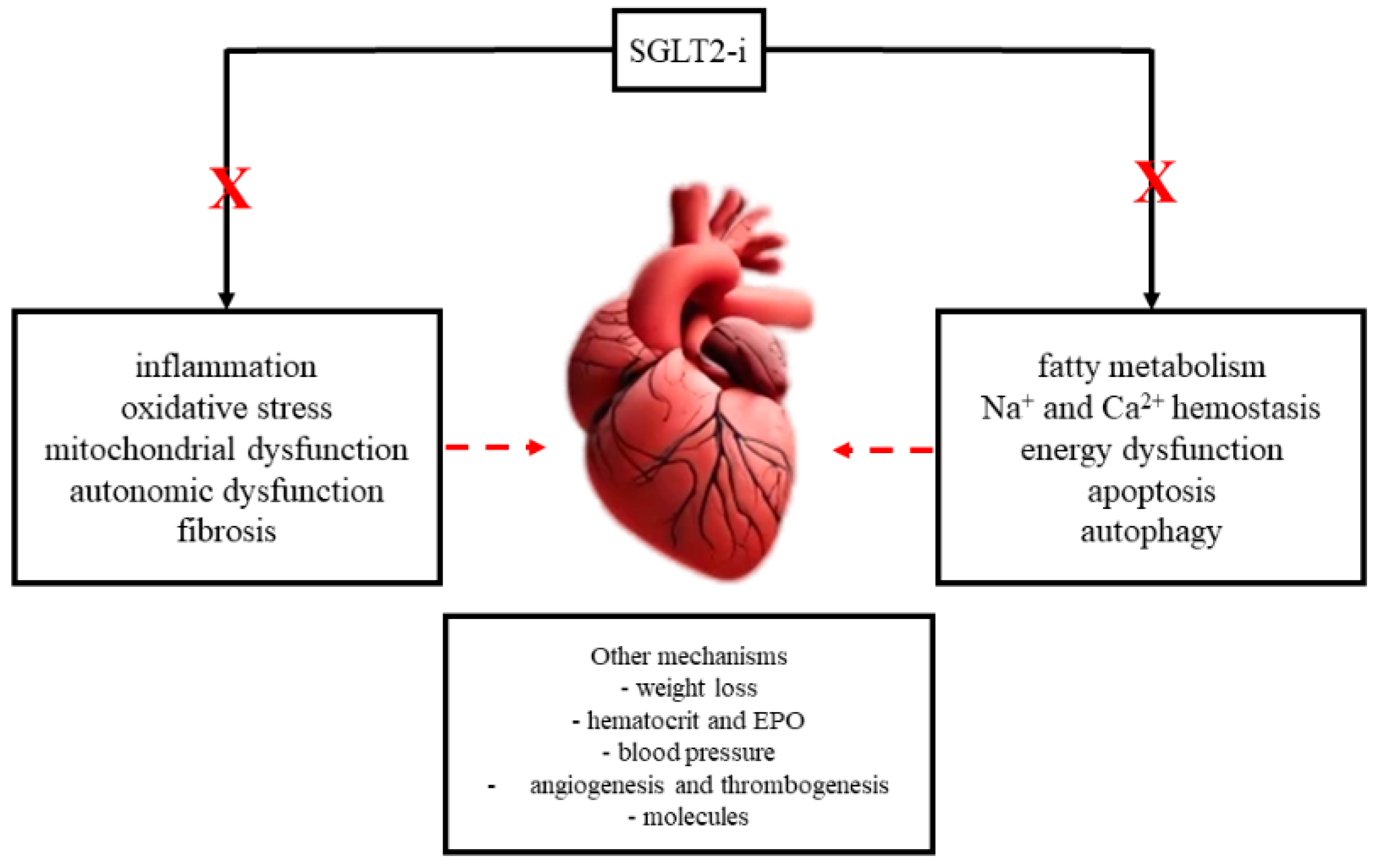
3.1. Molecular Mechanisms of SGLT2is and Atrial Remodeling

Originally, it was believed that SGLT2is primarily exerted their effects through diuretic or natriuretic mechanisms. However, current evidence indicates that these effects are only temporary and not central to their action [46]. Instead, SGLT2is influence multiple systems, including the nervous, cardiovascular, and endocrine systems, resulting in intricate metabolic effects that go beyond simple glycemic control and contribute to mitigating myocardial remodeling and enhancing cardiovascular protection [48]. Figure 1 illustrates the various pathways through which SGLT2is exert their effects on atrial remodeling, including their impact on myocardial fibrosis, atrial dilation, electrophysiological properties, and molecular and cellular mechanisms.

3.1.1. Inflammation

HF is often associated with elevated levels of circulating pro-inflammatory cytokines such as interleukin (IL)-1β, IL-6, IL-8, and tumor necrosis factor (TNF)-α, which contribute to adverse cardiac remodeling. Inflammation in HF is mediated by pattern recognition receptors (PRRs) like nucleotide-binding domain, leucine-rich–containing family, pyrin domain–containing-3 (NLRP3) and Toll-like receptor 4 (TLR4), which activate inflammatory pathways leading to cardiac hypertrophy and fibrosis. These cytokines promote immune cell activation and additional cytokine release, exacerbating the inflammatory and fibrotic processes in the heart [24]. The inflammatory biomarkers’ soluble suppression of tumorigenesis-2 (sST2) and galectin-3 are particularly noteworthy, as they provide prognostic information related to fibrosis and are more accurate than N-terminal pro-B-type natriuretic peptide (NT-proBNP) in assessing inflammation in HF [42].
SGLT2is have been shown to exert anti-inflammatory effects, which contribute to their cardioprotective benefits. Clinical meta-analyses indicate that SGLT2is reduce the levels of inflammatory markers such as IL-6, C-reactive protein, TNF-α, and monocyte chemotactic protein 1 (MCP-1), which are linked to systemic inflammation (28). Animal studies also support these findings, revealing that SGLT2is decrease cardiac inflammation by inhibiting inflammasome activation [19,20,26,38,39]. This action shifts macrophage polarization from the pro-inflammatory M1 phenotype to the anti-inflammatory M2 phenotype, thereby reducing inflammatory markers and improving cardiac function [28].
Myocardial infarction and ischemia–reperfusion injury trigger an inflammatory response that can worsen myocardial damage and remodeling. Macrophages play a crucial role in this context, with M1 macrophages driving inflammation and M2 macrophages aiding repair. SGLT2is, including empagliflozin and dapagliflozin, are noted for their ability to modulate macrophage activity, thereby reducing inflammation and enhancing cardiac repair processes, both in the atria and ventricles [28,39]. Atrial remodeling and AF are both associated with heightened levels of inflammation. Several preclinical [20,21,26,38,39,50] and clinical [3,4,54,60] studies have highlighted the role of SGLT2is in mitigating this inflammation, identifying it as a crucial mechanism underlying their cardioprotective effects.

3.1.2. Oxidative Stress and Mitochondrial Dysfunction

Oxidative stress, characterized by an imbalance between ROS production and the cell’s antioxidant defenses, leads to significant cardiomyocyte damage. Excessive ROS, primarily generated by mitochondria, nicotinamide adenine dinucleotide phosphate oxidase (NADPH) oxidases, and nitric oxide synthase (NOS) uncoupling, can adversely affect proteins, lipids, and DNA [50]. Mitochondria play a crucial role in adenosine triphosphate (ATP) production and cellular homeostasis. They are particularly abundant in myocardial cells to support heart contraction, and their quality control involves processes such as fusion, fission, biogenesis, and mitophagy. Mitophagy, which can be receptor-dependent (e.g., BNIP3, BNIP3L) or ubiquitin-dependent (e.g., PINK/Parkin pathway) is essential for removing damaged mitochondria and maintaining cardiovascular health [28].
In diabetic models, mitochondrial dysfunction is notably prominent, contributing to the development of AF. Diabetic cardiomyocytes exhibit increased ROS production and mitochondrial membrane hyperpolarization, leading to mitochondrial damage and oxidative stress. This dysfunction impairs cardiac efficiency, promoting structural and electrical remodeling and increasing susceptibility to AF [8]. Both empagliflozin and dapagliflozin have been demonstrated to improve mitochondrial function and reduce ROS production [21,35]. In studies involving rats, empagliflozin enhanced mitochondrial function, improved electrophysiological abnormalities, and reduced atrial remodeling and AF inducibility [21].
The role of ROS in myocardial diseases such as AF and HF is significant. ROS production from sources like NADPH oxidases, mitochondria, and xanthine oxidases contributes to tissue damage, fibrosis, and impaired conduction, promoting atrial remodeling and arrhythmias. Mitochondrial dysfunction exacerbates these processes by reducing ATP synthesis and perpetuating the oxidative damage cycle. In a study by Nishinarita et al., the authors have shown that canagliflozin reduces oxidative stress and AF inducibility in animal models [38]. Similarly, research by Kondo et al. highlights that canagliflozin suppresses myocardial NADPH oxidase activity and improves NOS coupling, providing anti-inflammatory and anti-apoptotic benefits [41].
Adenosine monophosphate-activated protein kinase (AMPK), a key regulator of cellular energy, plays a role in mitochondrial health by promoting biogenesis, reducing ROS generation, and managing mitochondrial dynamics. In DM, AMPK activation is often impaired, but activators like empagliflozin can help manage cardiac issues and AF by enhancing AMPK-mediated mitochondrial dynamics [50]. This aligns with findings that show that AMPK activation through SGLT2is could protect against atrial remodeling in DM.
Overall, oxidative stress and mitochondrial dysfunction are pivotal in atrial remodeling and AF progression, in both diabetic and non-diabetic subjects. SGLT2is offer potential therapeutic benefits by improving mitochondrial function, reducing oxidative stress, and mitigating structural and electrical alterations in the atria [4].

3.1.3. Autonomic Dysfunction

Autonomic dysfunction plays a pivotal role in the development of atrial remodeling with both sympathetic and parasympathetic imbalances contributing to increased AF risk. In patients with diabetes mellitus (DM), this dysfunction is characterized by heightened sympathetic and reduced parasympathetic activity, often observable through heart rate variability studies [35]. Autonomic remodeling in AF progresses through three stages: initial parasympathetic denervation, followed by sympathetic hyperactivation, and finally sympathetic denervation. Parasympathetic stimuli can lead to macro-re-entry phenomena, while sympathetic stimuli drive abnormal automaticity and trigger activity. In DM, autonomic dysfunction may precede the diagnosis, manifesting as impaired vagal responses and decreased acetylcholine release, which further influences AF by altering cardiac electrical activity [4].
SGLT2is have been shown to influence sympathetic nerve activation, which is crucial in cardiac remodeling. These inhibitors can reduce sympathetic nerve activation, as evidenced by lower tyrosine hydroxylase levels in the kidney and decreased circulating catecholamines [27]. The EMBODY trial, which investigated empagliflozin’s effects on cardiac nerve activity in type 2 DM and acute myocardial infarction patients, found that SGLT2is significantly enhance cardiac nerve activity while maintaining a favorable safety profile. Additionally, these inhibitors may lower blood pressure and sympathetic nerve activity, suggesting that SGLT2is effectively modulate sympathetic activation, although the exact mechanisms are still under investigation [28]. Clinical trials, including the EMBODY and EMPYREAN trials, demonstrate that SGLT2is such as empagliflozin can reduce sympathetic nerve activity and improve cardiac remodeling [3].
Interestingly, while increased heart rates typically exacerbate HF in patients with reduced ejection fraction, a recent case report by Zlahtic et al. proposes that in HF with preserved ejection fraction (HFpEF), higher heart rates combined with guideline-directed therapies, including SGLT2is, may positively affect atrial and ventricular structural and functional remodeling [58]. Thus, SGLT2is not only impact autonomic dysfunction but also hold potential benefits in mitigating adverse atrial and ventricular remodeling associated with various HF types.

3.1.4. Fibrosis

Fibrosis plays a fundamental role in the pathophysiology of many cardiovascular diseases. It is characterized by the excessive accumulation of extracellular matrix (ECM) components, which leads to increased tissue stiffness and alters normal cardiac structure and function. The primary drivers of cardiac fibrosis are myofibroblasts, which are activated following myocardial injury. These cells, in conjunction with macrophages, lymphocytes, and mast cells, contribute to fibrosis by producing ECM proteins and promoting inflammation. Excessive ECM deposition further exacerbates cardiac dysfunction. The interplay between inflammation and fibrosis is particularly pronounced in conditions such as AF and DM. Inflammation-induced fibrosis involves abnormal ECM remodeling, which affects matrix composition and cardiac muscle function. In AF, atrial fibrosis is crucial for maintaining the condition, with epicardial cells differentiating into myofibroblasts. Angiotensin-II and oxidative stress further contribute to the arrhythmogenic remodeling of the atrium by activating the NF-κB pathway, promoting myofibroblast differentiation, and overexpressing ECM proteins, thereby increasing AF susceptibility in diabetic models [4].
Recent studies highlight the potential of SGLT2is in addressing myocardial fibrosis. Clinical research indicates that SGLT2is such as empagliflozin and dapagliflozin can improve cardiac function and reduce fibrosis, both in diabetic and non-diabetic patients. These drugs appear to influence fibrosis through the modulation of calcium and sodium levels in heart cells, reductions in inflammation, and alterations in various signaling pathways associated with fibrosis. Despite these promising findings, the precise mechanisms through which SGLT2is exert their cardioprotective effects are not yet fully understood [28].
For instance, dapagliflozin reduces myocardial fibrosis by inhibiting fibroblast activation and proliferation, primarily through the AMPK α-mediated suppression of TGF-β/Smad signaling. It also lowers oxidative stress and affects macrophage polarization, which helps improve cardiac remodeling and reduce fibrosis following myocardial infarction [3]. Similarly, in insulin-resistant female diabetic db/db mice, empagliflozin improved the expression of profibrotic proteins, such as serum/glucocorticoid-regulated kinase 1 (SGK1) and the epithelial sodium channel (ENaC), leading to reduced interstitial fibrosis in both atrial and ventricular myocardium [20]. Empagliflozin has been shown to directly influence human cardiac myofibroblast activity and ECM remodeling, indicating that it targets key profibrotic mechanisms involved in the progression of HF [33]. Additionally, Lin et al. demonstrated that empagliflozin could suppress cardiac fibrosis and endoplasmic reticulum stress while improving hemodynamics in a rat model of mitral regurgitation-induced HF [39]. Also, recent findings by Scatularo et al. suggest that endomyocardial fibrosis significantly contributes to the development of AF, and its reduction may be a crucial mechanism underlying the cardioprotective effects of SGLT2is [32,52].
Thereby, SGLT2is demonstrate significant potential in reducing myocardial fibrosis and atrial remodeling. By modulating various molecular and cellular pathways, these new agents can improve cardiac function, offering valuable cardioprotective benefits in conditions such as HF and AF.

3.1.5. Myocardial Energy Metabolism

Fatty acids and glucose are the primary energy sources for myocardial metabolism. Under normal conditions, fatty acid metabolism, despite being more oxygen-demanding and less efficient for ATP production compared to glucose metabolism, serves as the main energy source. However, during pathological conditions such as AF, glycolysis becomes the predominant metabolic pathway. The role of AMPK is crucial in cardiac metabolism by enhancing fatty acid and glucose uptake to boost ATP production during energy shortages. During AF-induced metabolic stress, AMPK activation may help resist AF progression by restoring atrial calcium balance [24,35].
SGLT2is promote a metabolic shift from glucose to fatty acids, ketone bodies, and branched-chain amino acids, thereby improving myocardial energy efficiency. This metabolic switch has been observed across various species and was shown to enhance cardiac energetics in both in vitro and human studies [23,24,25,26,27]. Thirumatyam et al. demonstrated that SGLT2is increase lipid mobilization and oxidation, elevate plasma ketone body levels, and reduce tissue glucose uptake, thereby linking metabolism to improved cardiac function [47]. Correspondingly, the metabolic/myocardial fuel-supply hypothesis suggests that increased ketone body production from SGLT2is enhances energy supply efficiency to the heart, particularly beneficial under metabolic stress conditions like HF and diabetes [34]. The positive impact of SGLT2is on cardiac energetics and efficiency through ketone body production is highlighted by Subramanian et al. in patients with nonobstructive hypertrophic cardiomyopathy [49]. Additionally, Mantovani et al., in their comprehensive review, demonstrated the beneficial effects of SGLT2is on myocardial and liver fibrosis in individuals with non-alcoholic steatohepatitis and HF, irrespective of diabetes status [44]. Furthermore, Dong et al. provided evidence that SGLT2is also offer neurovascular protective benefits [48].
Obesity and type 2 DM contribute to HFpEF through the enlargement of visceral adipose tissue, which promotes inflammation and fibrosis in the heart. Enlarged epicardial adipose tissue restricts heart function and releases pro-inflammatory adipokines, increasing the risk of AF and impaired ventricular contraction. In their study, Tschöpe et al. showed that SGLT2is can reduce epicardial adipose tissue, lower cardiovascular risk, and potentially mitigate atrial fibrosis and serve as a treatment for AF and HF in obese HFpEF patients [43]. Also, epicardial adipose tissue around the atria is associated with a higher risk of AF. In type 2 DM and obesity, increased visceral fat creates a hypoxic and inflammatory environment, worsening HFpEF and promoting arrhythmias by disrupting electrical activity. The thickness of epicardial adipose tissue correlates with higher arrhythmogenesis and adverse outcomes [4].
Overall, SGLT2is enhance myocardial energy efficiency and decrease epicardial adipose tissue, which may help reduce atrial remodeling and lower the risk of AF in patients with obesity and type 2 DM.

3.1.6. Sodium and Calcium Metabolism

During AF, excessive cytosolic calcium release from the sarcoplasmic reticulum activates calmodulin kinase type II (CaMKII) and ryanodine receptors, leading to increased cytosolic Ca2+. This overactivation of CaMKII and sodium/calcium exchangers results in delayed afterdepolarizations and spontaneous ectopic activity, contributing to stable AF circuits. CaMKII further supports AF by activating the calcineurin/nuclear factor of the activated T-cells (NFAT) pathway, which reduces L-type Ca2+ current and shortens action potential duration. DM exacerbates these processes by upregulating certain ion channels and altering gap junction proteins, thereby worsening arrhythmias and impairing cardiac function [4].
Chronic treatment with the dual SGLT-1&2 inhibitor sotagliflozin has demonstrated significant benefits in a rat model of HFpEF. Sotagliflozin improved left atrial cardiomyopathy by reducing cytosolic calcium ([Ca2+]) levels and enhancing mitochondrial calcium buffering, which mitigated mitochondrial swelling and decreased ROS production. These effects are linked to increased Na+/Ca2+ exchanger forward-mode activity, which alleviates cytosolic Ca2+ overload and reduces arrhythmia susceptibility. By normalizing mitochondrial function and improving calcium handling, sotagliflozin addresses the adverse effects of elevated cytosolic Ca2+ and ROS, making it a promising option for managing atrial remodeling and related arrhythmias in HFpEF [26].
In diabetic hearts, excessive sodium (Na⁺) influx through SGLT can lead to Na⁺ overload, contributing to arrhythmias and oxidative stress. SGLT2is such as empagliflozin counteract this by reducing Na⁺ influx and inhibiting the late sodium current (late-Iₙₐ), which is associated with arrhythmias and prolonged QT syndrome. Empagliflozin also improves calcium handling, mitochondrial function, and connexin expression, thereby enhancing cardiac function and reducing arrhythmias [3].
Empagliflozin’s protective effects against HF and related complications are partly due to its ability to reduce cytoplasmic sodium and calcium levels while increasing mitochondrial calcium. This likely results from the direct inhibition of the Na⁺/H⁺ exchanger [23]. Moreover, SGLT2is may mitigate cardiac injury, hypertrophy, fibrosis, and remodeling by directly inhibiting the myocardial Na+/H+ exchanger [34].
To summarize, SGLT2is, by modulating sodium and calcium metabolism, improve calcium handling and mitochondrial function, thus mitigating atrial remodeling and reducing arrhythmia risk in conditions like HF and DM.

3.1.7. Other Mechanisms

Weight Loss

Obesity is associated with increased cardiac arrhythmias such as AF and sudden cardiac death, largely due to the abnormal electrical and structural changes in the heart linked to excess adiposity, as discussed earlier in this review [28,57]. SGLT2is effectively reduce body weight, fat mass, and visceral fat in patients with type 2 DM by promoting urinary glucose excretion and enhancing lipolysis through lower insulin and elevated glucagon levels [28]. Studies involving over 10,000 patients with type 2 DM have demonstrated that a weight loss of at least 5% achieved with SGLT2is correlates with a reduced risk of new-onset AF [3]. By decreasing excess adiposity and its adverse effects on cardiac structure and function, SGLT2is may reduce the risk of atrial remodeling and associated arrhythmias.

Hematocrit and Erythropoietin

The EMPA-REG OUTCOME trial indicates that SGLT2is such as dapagliflozin can enhance hematocrit levels and reduce cardiovascular events [20,28], potentially through their diuretic effects and influence on erythropoiesis. They achieve this by increasing erythropoietin production and inhibiting hepcidin, which regulates iron absorption and mobilization. In animal models, dapagliflozin’s cardioprotective effects appear to involve increasing erythropoietin levels and activating signaling pathways such as phosphorylated protein kinase B (p-Akt), phosphorylated Janus kinase 2 (p-JAK2), and phosphorylated mitogen-activated protein kinase (p-MAPK), which help reduce apoptosis. Thus, the therapeutic benefits of SGLT2is might be linked to their ability to elevate erythropoietin levels [28].

Blood Pressure

Previously published data indicate that SGLT2is reduce blood pressure by about 3.62/1.70 mmHg, primarily through initial fluid and sodium loss, followed by reductions in body fat and the inhibition of the renin–angiotensin–aldosterone system [20]. Additionally, a study of dapagliflozin shows a modest decrease in systolic blood pressure by 2.54 mmHg. The DAPA-HF trial confirmed that the observed benefits of the treatment are consistent across different blood pressure levels, although no significant interaction was detected (p = 0.78) [54]. Given that hypertension is a major risk factor for left atrial remodeling and AF, reducing blood pressure using SGLT2is may improve atrial remodeling and lead to better cardiovascular outcomes.

Angio- and Thrombogenesis

Atrial remodeling, marked by a granular and wrinkled endocardium with edema, fibrosis, and thrombotic aggregates, heightens the risk of thrombus formation in AF patients. This disruption in normal blood flow creates a pro-thrombotic state characterized by abnormal coagulation factors and increased platelet activation. DM worsens this condition by promoting inflammation, atrial myopathy, and epicardial adipose tissue expansion, which increases stroke risk through enhanced thrombus formation and altered coagulation [4].
Additionally, Fakih et al. suggest that components of the coagulation cascade produced by pathological endothelium contribute to atrial remodeling, creating a substrate for AF. Thrombin and Factor Xa (FXa) are implicated in maladaptive cardiac hypertrophy, inflammation, and fibrosis, influencing these processes beyond their roles in blood clotting. Targeting the angiotensin II type 1 receptor (AT1R)/NADPH oxidases/SGLT1/2 pathway could be a promising approach to prevent atrial remodeling and mitigate atrial cardiomyopathy in high-risk patients [57].

Molecules

Impaired myokine production and dysfunction within the muscle–adipose tissue axis lead to collagen accumulation in the heart, exacerbating atherosclerosis, endothelial dysfunction, oxidative stress, and mitochondrial dysfunction. These changes drive adverse cardiac remodeling, reduced angiogenesis, and muscle weakness, significantly affecting HF in type 2 DM. Myokines, which are produced by muscles, the heart, and adipose tissue, play a crucial role in regulating energy metabolism and inflammation. Some myokines, such as adropin, apelin, and irisin, offer protective effects, while others like myostatin can induce maladaptive changes. Adropin affects oxidative stress and autophagy through the lysosome-dependent pathway, a mechanism potentially targeted by SGLT2is. In their study, Berezin et al. found that in type 2 DM patients with HF, especially females, elevated adropin levels independently predicted improved hemodynamic performance with long-term treatment using the SGLT2i dapagliflozin, regardless of NT-proBNP levels [27]. Thus, SGLT2is may alleviate atrial remodeling and enhance cardiac function in type 2 DM by modulating myokines like adropin.

Epigenetic Effects

Recent translational studies have highlighted that SGLT2is may modulate atrial and ventricular remodeling by targeting epigenetic regulators involved in inflammation, oxidative stress, and profibrotic processes [28,38,39,42,50]. Notable effects include the downregulation of genes related to inflammatory responses, such as inflammasome mRNA [42,50], the improvement of mitochondrial function through the regulation of mRNA in adipocytes [38,39,42], and reductions in cardiac fibrosis by targeting collagen mRNAs [42,50].

4. Clinical Implications

4.1. Atrial Fibrillation

SGLT2is are already used as first-line treatments in all types of HF, their benefits being undeniable. Current data show their cardiovascular protection roles as positively influencing cardiac remodeling and consequently stopping the progression of clinical HF [9] or even regressing cardiac remodeling [14,50] in both diabetic and non-diabetic patients. Speculated mechanisms involved in this are their direct effect on profibrotic pathways [2], ventricular unloading and afterload reduction, and improvement in cardiac metabolism and energetics, among several other proposed mechanisms [36,47,54]. Moreover, recent translational studies highlight the potential benefits of SGLT2is in targeting mRNAs, which are well-established biomarkers for AF [28,38,39,42,50]. However, the role of SGLT2is in the current management strategies for patients with AF remains controversial.
The existing evidence, while not yet definitive, suggests a promising rationale for considering SGLT2is as a potential therapeutic approach for patients with AF in both diabetic and non-diabetic patients. Preclinical studies [20,21,26,38,39,50,57] indicate that SGLT2is may positively influence AF mechanisms through antifibrotic effects and improved electrical remodeling. While SGLT2is enhance cardiac bioenergetics by improving mitochondrial function and calcium homeostasis, their anti-arrhythmic potential is controversial [2,3,25,42,53,54].
SGLT2is have significantly impacted the prognosis of patients with HF according to the results of landmark trials on the topic [31,40,45,46,55,61]. Data from randomized clinical trials, including EMPA-REG, DECLARE-TIMI [1], DEFINE-HF, and PRESERVED-HF [8,9], along with several other studies [37,42,51,53], indicate a reduction in AF incidence and recurrence. Also, several other studies imply their potential efficacy in this regard [3,4,39,50]. These studies have collectively indicated that SGLT2is could be beneficial in managing AF through their molecular and cellular mechanisms, having a positive effect on both triggering and maintaining AF.
However, other research, including the CANVAS trial, did not consistently support this benefit. For instance, the CANVAS trial and additional studies [29,30] failed to show a significant reduction in AF associated with SGLT2is. The contrasting results from these studies underscore the complexity of AF and the multifaceted effects of SGLT2is.
Despite the limitations of current studies, which may not fully establish causality or the extent of the benefits, the observed effects of SGLT2is in related cardiovascular conditions provide a compelling basis for further investigation. The potential for SGLT2is to influence the progression of AF suggests that they could become an important component of comprehensive AF management strategies. Overall, while preliminary findings are promising, further research is needed to confirm the role of SGLT2is in AF.

4.2. Other Cardiac Conditions

SGLT2is are demonstrating promising results in various cardiac conditions beyond HF. Recent studies have explored the impact of SGLT2is following acute coronary syndrome. Lan et al. reported that in patients with both acute coronary syndrome and type 2 diabetes (T2D), adding empagliflozin to the standard acute coronary syndrome treatment at hospital discharge led to a reduction in left ventricular mass and improvements in diastolic function over a period of 3–6 months, compared to those with acute coronary syndrome and well-controlled type 2 DM who did not receive an SGLT2i [34]. Additionally, the ongoing EMPRESS-MI trial is expected to yield valuable data on the effects of empagliflozin through advanced cardiac and kidney imaging, as well as circulating biomarkers, in individuals at high risk of developing HF following acute myocardial infarction [56]. In a comprehensive review conducted by Patoulias et al., the authors evaluated the potential benefits of SGLT2is in acute HF. Their findings indicated that the current evidence is inadequate to support the use of SGLT2is in the treatment of acute HF [25]. SGLT2is have also been evaluated in patients with cancer therapy-related cardiac dysfunction, demonstrating improved outcomes in this population [53]. Lastly, in a separate study by Subramanian et al., the use of SGLT2is was shown to enhance diastolic function and functional capacity in patients with diabetes and nonobstructive hypertrophic cardiomyopathy with left ventricular function [49]. As new randomized trials continue to emerge, SGLT2is are expected to provide more comprehensive data, potentially expanding the indications to include a wider range of cardiac conditions, thereby broadening their therapeutic applications.

4.3. Other Non-Cardiac Conditions

Numerous emerging studies are exploring the potential benefits of SGLT2is beyond their established effects on cardiac conditions and diabetes [59]. Starting from their cardiovascular clearly proven benefits, Dong et al. studied the effects of SGLT2is on the central nervous system, suggesting neuroprotective actions [48]. Additionally, Mantovani et al., in their extensive review, highlighted the beneficial effects of SGLT2is on myocardial and liver fibrosis in individuals with non-alcoholic steatohepatitis and HF, regardless of their diabetes status [44]. However, additional prospective research is necessary to assess the efficacy of SGLT2is in these broader patient populations.

4.4. Cardiovascular Aging

Cardiovascular aging is one of the major risk factors associated with multi-morbidity and HF [24,44,54], particularly HFpEF. Key mechanisms in cardiovascular aging, which overlap with those involved in atrial and ventricular remodeling and targeted by SGLT2is, include inflammation, mitochondrial dysfunction, oxidative stress, and autophagy [24]. Additionally, in DM patients, advanced glycation endproducts (AGEs) accumulate during hyperglycemic states, promoting cardiovascular aging. Also, by binding to their receptors, AGEs promote inflammatory and oxidative responses, increasing the risk of AF development in these patients, thus showing a possible link with atrial remodeling [35]. Regarding vascular aging, recent data failed to show a major effect of SGLT2is [25]. Although current findings are promising, further large-scale studies are necessary to demonstrate a clear relationship between SGLT2is, atrial remodeling, and cardiovascular aging.

5. Conclusions and Future Perspectives

SGLT2si have emerged as powerful therapeutic agents that extend their benefits far beyond glucose control. The ability of SGLT2is to influence multiple molecular pathways involved in atrial remodeling presents significant implications for the treatment and prevention of AF and HF. This review underscores several key mechanisms through which SGLT2is contribute to mitigating atrial remodeling: reducing inflammation and oxidative stress, improving mitochondrial function, modulating autonomic and myocardial energy metabolism, and reducing fibrosis. Current European and American HF guidelines recommend SGLT2is as first-line treatment for HF patients, independent of their phenotype. Recent clinical and preclinical data support the multifaceted actions of SGLT2is, ranging from metabolic effects to molecular and cellular regulation, making them promising agents in altering the course of atrial remodeling. Further research is essential to better understand the clinical impact of SGLT2is on atrial remodeling in patients with AF and/or HF and to refine their therapeutic use for optimal patient outcomes.

Author Contributions

Conceptualization—I.-A.M., R.T., and G.C.; Writing—Original Draft Preparation—I.-A.M., D.M., and G.S.; Writing—Review and Editing—F.F., D.A.I., G.S., M.P., R.R., and B.C.; Visualization—D.Z.; Supervision—D.P. All authors have read and agreed to the published version of the manuscript.

Funding

This research received no external funding.

Conflicts of Interest

The authors report no conflicts of interest.

References

  1. Zelniker, T.A.; Bonaca, M.P.; Furtado, R.H.; Mosenzon, O.; Kuder, J.F.; Murphy, S.A.; Bhatt, D.L.; Leiter, L.A.; McGuire, D.K.; Wilding, J.P.; et al. Effect of Dapagliflozin on Atrial Fibrillation in Patients with Type 2 Diabetes Mellitus: Insights from the DECLARE-TIMI 58 Trial. Circulation 2020, 141, 1227–1234. [Google Scholar] [CrossRef]
  2. von Lewinski, D.; Tripolt, N.J.; Sourij, H.; Pferschy, P.N.; Oulhaj, A.; Alber, H.; Gwechenberger, M.; Martinek, M.; Seidl, S.; Moertl, D.; et al. Ertugliflozin to reduce arrhythmic burden in ICD/CRT patients (ERASe-trial)—A phase III study. Am. Heart J. 2022, 246, 152–160. [Google Scholar] [CrossRef] [PubMed]
  3. Manolis, T.A.; Melita, H.; Manolis, A.S. Sodium-glucose cotransporter type 2 inhibitors and cardiac arrhythmias. J. Clin. Med. 2023, 12, 2868. [Google Scholar] [CrossRef] [PubMed]
  4. Lorenzo-Almorós, A.; Cerrada, J.C.; Walther, L.-A.-S.; Bailón, M.M.; González, L. Atrial Fibrillation and Diabetes Mellitus: Dangerous Liaisons or Innocent Bystanders? J. Clin. Med. 2023, 12, 2868. [Google Scholar] [CrossRef] [PubMed]
  5. Al Ghamdi, B.; Hassan, W. Atrial Remodeling and Atrial Fibrillation: Mechanistic Interactions And Clinical Implications. J. Atr. Fibrillation 2009, 2, 125. [Google Scholar] [PubMed] [PubMed Central]
  6. Hoit, B.D. Left Atrial Remodeling: More Than Just Left Atrial Enlargement. Circ. Cardiovasc. Imaging 2017, 10, e006036. [Google Scholar] [CrossRef] [PubMed]
  7. Sabouret, P.; Attias, D.; Beauvais, C.; Berthelot, E.; Bouleti, C.; Genty, G.G.; Galat, A.; Hanon, O.; Hulot, J.; Isnard, R.; et al. Diagnosis and management of heart failure from hospital admission to discharge: A practical expert guidance. Ann. Cardiol. d’Angéiologie 2022, 71, 41–52. [Google Scholar] [CrossRef] [PubMed]
  8. Nassif, M.E.; Windsor, S.L.; Tang, F.; Khariton, Y.; Husain, M.; Inzucchi, S.E.; Mc-Guire, D.K.; Pitt, B.; Scirica, B.M.; Austin, B.; et al. Dapagliflozin effects on biomarkers, symptoms, and functional status in patients with heart failure with reduced ejection fraction. Circulation 2019, 140, 1463–1476. [Google Scholar] [CrossRef]
  9. Nassif, M.E.; Windsor, S.L.; Borlaug, B.A.; Kitzman, D.W.; Shah, S.J.; Tang, F.; Khariton, Y.; Malik, A.O.; Khumri, T.; Umpierrez, G.; et al. The SGLT2 inhibitor dapagliflozin in heart failure with preserved ejection fraction: A multicenter randomized trial. Nat. Med. 2021, 27, 1954–1960. [Google Scholar] [CrossRef]
  10. Zinman, B.; Wanner, C.; Lachin, J.M.; Fitchett, D.; Bluhmki, E.; Hantel, S.; Mattheus, M.; Devins, T.; Johansen, O.E.; Woerle, H.J.; et al. Empagliflozin, cardiovascular outcomes, and mortality in type 2 diabetes. N. Engl. J. Med. 2015, 373, 2117–2128. [Google Scholar] [CrossRef]
  11. McMurray, J.J.V.; Solomon, S.D.; Inzucchi, S.E.; Køber, L.; Kosiborod, M.N.; Martinez, F.A.; Ponikowski, P.; Sabatine, M.S.; Anand, I.S.; Bělohlávek, J.; et al. Dapagliflozin in Patients with Heart Failure and Reduced Ejection Fraction. N. Engl. J. Med. 2019, 381, 1995–2008. [Google Scholar] [CrossRef] [PubMed]
  12. Solomon, S.D.; McMurray, J.J.; Claggett, B.; de Boer, R.A.; DeMets, D.; Hernandez, A.F.; Inzucchi, S.E.; Kosiborod, M.N.; Lam, C.S.; Martinez, F.; et al. Dapagliflozin in Heart Failure with Mildly Reduced or Preserved Ejection Fraction. N. Engl. J. Med. 2022, 387, 1089–1098. [Google Scholar] [CrossRef]
  13. Packer, M.; Anker, S.D.; Butler, J.; Filippatos, G.; Pocock, S.J.; Carson, P.; Januzzi, J.; Verma, S.; Tsutsui, H.; Brueckmann, M.; et al. Cardiovascular and Renal Outcomes with Empagliflozin in Heart Failure. N. Engl. J. Med. 2020, 383, 1413–1424. [Google Scholar] [CrossRef] [PubMed]
  14. Anker, S.D.; Butler, J.; Filippatos, G.; Ferreira, J.P.; Bocchi, E.; Böhm, M.; Brunner–La Rocca, H.-P.; Choi, D.-J.; Chopra, V.; Chuquiure-Valenzuela, E.; et al. Empagliflozin in Heart Failure with a Preserved Ejection Fraction. N. Engl. J. Med. 2021, 385, 1451–1461. [Google Scholar] [CrossRef]
  15. Voors, A.A.; Angermann, C.E.; Teerlink, J.R.; Collins, S.P.; Kosiborod, M.; Biegus, J.; Ferreira, J.P.; Nassif, M.E.; Psotka, M.A.; Tromp, J.; et al. The SGLT2 inhibitor empagliflozin in patients hospitalized for acute heart failure: A multinational randomized trial. Nat. Med. 2022, 28, 568–574. [Google Scholar] [CrossRef] [PubMed]
  16. McDonagh, T.A.; Metra, M.; Adamo, M.; Gardner, R.S.; Baumbach, A.; Böhm, M.; Burri, H.; Butler, J.; Čelutkienė, J.; Chioncel, O.; et al. 2021 ESC Guidelines for the diagnosis and treatment of acute and chronic heart failure. Eur. Heart J. 2021, 42, 3599–3726. [Google Scholar] [CrossRef]
  17. McDonagh, T.A.; Metra, M.; Adamo, M.; Gardner, R.S.; Baumbach, A.; Böhm, M.; Burri, H.; Butler, J.; Čelutkienė, J.; Chioncel, O.; et al. 2023 Focused Update of the 2021 ESC Guidelines for the diagnosis and treatment of acute and chronic heart failure Developed by the task force for the diagnosis and treatment of acute and chronic heart failure of the European Society of Cardiology (ESC) With the special contribution of the Heart Failure Association (HFA) of the ESC. Eur. Heart J. 2023, 44, 3627–3639. [Google Scholar] [CrossRef]
  18. Heidenreich, P.A.; Bozkurt, B.; Aguilar, D.; Allen, L.A.; Byun, J.J.; Colvin, M.M.; Deswal, A.; Drazner, M.H.; Dunlay, S.M.; Evers, L.R.; et al. 2022 AHA/ACC/HFSA Guideline for the Management of Heart Failure: A Report of the American College of Cardiology/American Heart Association Joint Committee on Clinical Practice Guidelines; Lippincott Williams and Wilkins: Philadelphia, PA, USA, 2022. [Google Scholar] [CrossRef]
  19. Heidenreich, P.A.; Bozkurt, B.; Aguilar, D.; Allen, L.A.; Byun, J.J.; Colvin, M.M.; Deswal, A.; Drazner, M.H.; Dunlay, S.M.; Evers, L.R.; et al. 2022 AHA/ACC/HFSA Guideline for the Management of Heart Failure: Executive Summary: A Report of the American College of Cardiology/American Heart Association Joint Committee on Clinical Practice Guidelines. Circulation; Lippincott Williams and Wilkins: Philadelphia, PA, USA, 2022. [Google Scholar] [CrossRef]
  20. Lee, H.-C.; Shiou, Y.-L.; Jhuo, S.-J.; Chang, C.-Y.; Liu, P.-L.; Jhuang, W.-J.; Dai, Z.-K.; Chen, W.-Y.; Chen, Y.-F.; Lee, A.-S. The sodium–glucose co-transporter 2 inhibitor empagliflozin attenuates cardiac fibrosis and improves ventricular hemodynamics in hypertensive heart failure rats. Cardiovasc. Diabetol. 2019, 18, 45. [Google Scholar] [CrossRef]
  21. Shao, Q.; Meng, L.; Lee, S.; Tse, G.; Gong, M.; Zhang, Z.; Zhao, J.; Zhao, Y.; Li, G.; Liu, T. Empagliflozin, a sodium glucose co-transporter-2 inhibitor, alleviates atrial remodeling and improves mitochondrial function in high-fat diet/streptozotocin-induced diabetic rats. Cardiovasc. Diabetol. 2019, 18, 165. [Google Scholar] [CrossRef]
  22. Ferrini, M.; Johansson, I.; Aboyans, V. Heart failure and its complications in patients with diabetes: Mounting evidence for a growing burden. Eur. J. Prev. Cardiol. 2019, 26, 106–113. [Google Scholar] [CrossRef]
  23. Natali, A.; Nesti, L.; Fabiani, I.; Calogero, E.; Di Bello, V. Impact of empagliflozin on subclinical left ventricular dysfunctions and on the mechanisms involved in myocardial disease progression in type 2 diabetes: Rationale and design of the EMPA-HEART trial. Cardiovasc. Diabetol. 2017, 16, 130. [Google Scholar] [CrossRef] [PubMed]
  24. Li, Z.; Zhao, H.; Wang, J. Metabolism and Chronic Inflammation: The Links Between Chronic Heart Failure and Comorbidities. Front. Cardiovasc. Med. 2021, 8, 650278. [Google Scholar] [CrossRef] [PubMed]
  25. Patoulias, D.; Fragakis, N.; Rizzo, M. The Therapeutic Role of SGLT-2 Inhibitors in Acute Heart Failure: From Pathophysiologic Mechanisms to Clinical Evidence with Pooled Analysis of Relevant Studies across Safety and Efficacy Endpoints of Interest. Life 2022, 12, 2062. [Google Scholar] [CrossRef] [PubMed]
  26. Bode, D.; Semmler, L.; Wakula, P.; Hegemann, N.; Primessnig, U.; Beindorff, N.; Powell, D.; Dahmen, R.; Ruetten, H.; Oeing, C.; et al. Dual SGLT-1 and SGLT-2 inhibition improves left atrial dysfunction in HFpEF. Cardiovasc. Diabetol. 2021, 20, 7. [Google Scholar] [CrossRef] [PubMed]
  27. Berezin, A.A.; Obradovic, Z.; Fushtey, I.M.; Berezina, T.A.; Novikov, E.V.; Schmidbauer, L.; Lichtenauer, M.; Berezin, A.E. The Impact of SGLT2 Inhibitor Dapagliflozin on Adropin Serum Levels in Men and Women with Type 2 Diabetes Mellitus and Chronic Heart Failure. Biomedicines 2023, 11, 457. [Google Scholar] [CrossRef]
  28. Chen, Y.; Peng, D. New insights into the molecular mechanisms of SGLT2 inhibitors on ventricular remodeling. Int. Immunopharmacol. 2023, 118, 110072. [Google Scholar] [CrossRef]
  29. Persson, F.; Nyström, T.; Jørgensen, M.E.; Carstensen, B.; Gulseth, H.L.; Thuresson, M.; Fenici, P.; Nathanson, D.; Eriksson, J.W.; Norhammar, A.; et al. Dapagliflozin is associated with lower risk of cardiovascular events and all-cause mortality in people with type 2 diabetes (CVD-REAL Nordic) when compared with dipeptidyl peptidase-4 inhibitor therapy: A multinational observational study. Diabetes Obes. Metab. 2018, 20, 344–351. [Google Scholar] [CrossRef]
  30. Bell, D.S.H.; Goncalves, E. Atrial fibrillation and type 2 diabetes: Prevalence, etiology, pathophysiology and effect of anti-diabetic therapies. Diabetes Obes. Metab. 2019, 21, 210–217. [Google Scholar] [CrossRef]
  31. Bell, D.S.H.; Goncalves, E. Heart failure in the patient with diabetes: Epidemiology, aetiology, prognosis, therapy and the effect of glucose-lowering medications. Diabetes Obes. Metab. 2019, 21, 1277–1290. [Google Scholar] [CrossRef]
  32. Iwakura, K. Heart failure in patients with type 2 diabetes mellitus: Assessment with echocardiography and effects of antihyperglycemic treatments. J. Echocardiogr. 2019, 17, 177–186. [Google Scholar] [CrossRef]
  33. Kang, S.; Verma, S.; Hassanabad, A.F.; Teng, G.; Belke, D.D.; Dundas, J.A.; Guzzardi, D.G.; Svystonyuk, D.A.; Pattar, S.S.; Park, D.S.; et al. Direct Effects of Empagliflozin on Extracellular Matrix Remodelling in Human Cardiac Myofibroblasts: Novel Translational Clues to Explain EMPA-REG OUTCOME Results. Can. J. Cardiol. 2020, 36, 543–553. [Google Scholar] [CrossRef] [PubMed]
  34. Lan, N.S.R.; Yeap, B.B.; Fegan, P.G.; Green, G.; Rankin, J.M.; Dwivedi, G. Empagliflozin and left ventricular diastolic function following an acute coronary syndrome in patients with type 2 diabetes. Int. J. Cardiovasc. Imaging 2021, 37, 517–527. [Google Scholar] [CrossRef] [PubMed]
  35. Lee, T.-W.; Lin, Y.-K.; Chen, Y.-C.; Kao, Y.-H.; Chen, Y.-J. Effect of antidiabetic drugs on the risk of atrial fibrillation: Mechanistic insights from clinical evidence and translational studies. Cell. Mol. Life Sci. 2021, 78, 923–934. [Google Scholar] [CrossRef] [PubMed]
  36. Ibrahim, N.E.; Januzzi, J.L. Sodium-Glucose Co-Transporter 2 Inhibitors and Insights from Biomarker Measurement in Heart Failure Patients. Clin. Chem. 2021, 67, 79–86. [Google Scholar] [CrossRef]
  37. Tanaka, H.; Tatsumi, K.; Matsuzoe, H.; Soga, F.; Matsumoto, K.; Hirata, K.-I. Association of type 2 diabetes mellitus with the development of new-onset atrial fibrillation in patients with non-ischemic dilated cardiomyopathy: Impact of SGLT2 inhibitors. Int. J. Cardiovasc. Imaging 2021, 37, 1333–1341. [Google Scholar] [CrossRef]
  38. Nishinarita, R.; Niwano, S.; Niwano, H.; Nakamura, H.; Saito, D.; Sato, T.; Matsuura, G.; Arakawa, Y.; Kobayashi, S.; Shirakawa, Y.; et al. Canagliflozin suppresses atrial remodeling in a canine atrial fibrillation model. J. Am. Heart Assoc. 2021, 10, e017483. [Google Scholar] [CrossRef] [PubMed]
  39. Lin, Y.; Chen, C.; Shih, J.; Cheng, B.; Chang, C.; Lin, M.; Ho, C.; Chen, Z.; Fisch, S.; Chang, W. Dapagliflozin improves cardiac hemodynamics and mitigates arrhythmogenesis in mitral regurgitation-induced myocardial dysfunction. J. Am. Heart Assoc. 2021, 10, e019274. [Google Scholar] [CrossRef]
  40. Kearney, A.; Linden, K.; Savage, P.; Menown, I.B.A. Advances in Clinical Cardiology 2020: A Summary of Key Clinical Trials. Adv. Ther. 2021, 38, 2170–2200. [Google Scholar] [CrossRef]
  41. Kondo, H.; Akoumianakis, I.; Badi, I.; Akawi, N.; Kotanidis, C.P.; Polkinghorne, M.; Stadiotti, I.; Sommariva, E.; Antonopoulos, A.S.; Carena, M.C.; et al. Effects of canagliflozin on human myocardial redox signalling: Clinical implications. Eur. Heart J. 2021, 42, 4947–4960. [Google Scholar] [CrossRef] [PubMed] [PubMed Central]
  42. Vrachatis, D.A.; Papathanasiou, K.A.; Iliodromitis, K.E.; Giotaki, S.G.; Kossyvakis, C.; Raisakis, K.; Kaoukis, A.; Lambadiari, V.; Avramides, D.; Reimers, B.; et al. Could Sodium/Glucose Co-Transporter-2 Inhibitors Have Antiarrhythmic Potential in Atrial Fibrillation? Literature Review and Future Considerations. Drugs 2021, 81, 1381–1395. [Google Scholar] [CrossRef]
  43. Tschöpe, C.; Elsanhoury, A.; Nelki, V.; Van Linthout, S.; Kelle, S.; Remppis, A. Heart failure with preserved ejection fraction as a model disease for the cardio-pulmonary-renal syndrome: Importance of visceral fat expansion as central pathomechanism. Die Inn. Med. 2021, 62, 1141–1152. [Google Scholar] [CrossRef]
  44. Mantovani, A.; Byrne, C.D.; Benfari, G.; Bonapace, S.; Simon, T.G.; Targher, G. Risk of Heart Failure in Patients With Nonalcoholic Fatty Liver Disease. J. Am. Coll. Cardiol. 2022, 79, 180–191. [Google Scholar] [CrossRef] [PubMed]
  45. Savage, P.; Cox, B.; Linden, K.; Coburn, J.; Shahmohammadi, M.; Menown, I. Advances in Clinical Cardiology 2021: A Summary of Key Clinical Trials. Adv. Ther. 2022, 39, 2398–2437. [Google Scholar] [CrossRef]
  46. Theofilis, P.; Antonopoulos, A.S.; Katsimichas, T.; Oikonomou, E.; Siasos, G.; Aggeli, C.; Tsioufis, K.; Tousoulis, D. The impact of SGLT2 inhibition on imaging markers of cardiac function: A systematic review and meta-analysis. Pharmacol. Res. 2022, 180, 106243. [Google Scholar] [CrossRef]
  47. Thirumathyam, R.; Richter, E.A.; Goetze, J.P.; Fenger, M.; Van Hall, G.; Dixen, U.; Holst, J.J.; Madsbad, S.; Vejlstrup, N.; Madsen, P.L.; et al. Investigating the roles of hyperglycaemia, hyperinsulinaemia and elevated free fatty acids in cardiac function in patients with type 2 diabetes via treatment with insulin compared with empagliflozin: Protocol for the HyperCarD2 randomised, crossover trial. BMJ Open 2022, 12, e054100. [Google Scholar] [CrossRef] [PubMed]
  48. Dong, M.; Wen, S.; Zhou, L. The Relationship Between the Blood-Brain-Barrier and the Central Effects of Glucagon-Like Peptide-1 Receptor Agonists and Sodium-Glucose Cotransporter-2 Inhibitors. Diabetes Metab. Syndr. Obesity 2022, 15, 2583–2597. [Google Scholar] [CrossRef]
  49. Subramanian, M.; Sravani, V.; Krishna, S.P.; Bijjam, S.; Sunehra, C.; Yalagudri, S.; Saggu, D.K.; Narasimhan, C. Efficacy of SGLT2 Inhibitors in Patients With Diabetes and Nonobstructive Hypertrophic Cardiomyopathy. Am. J. Cardiol. 2021, 188, 80–86. [Google Scholar] [CrossRef] [PubMed]
  50. Koizumi, T.; Watanabe, M.; Yokota, T.; Tsuda, M.; Handa, H.; Koya, J.; Nishino, K.; Tatsuta, D.; Natsui, H.; Kadosaka, T.; et al. Empagliflozin suppresses mitochondrial reactive oxygen species generation and mitigates the inducibility of atrial fibrillation in diabetic rats. Front. Cardiovasc. Med. 2023, 10, 1005408. [Google Scholar] [CrossRef] [PubMed] [PubMed Central]
  51. Nassif, M.E.; Windsor, S.L.; Gosch, K.; Borlaug, B.A.; Husain, M.; Inzucchi, S.E.; Kitzman, D.W.; McGuire, D.K.; Pitt, B.; Scirica, B.M.; et al. Dapagliflozin Improves Heart Failure Symptoms and Physical Limitations Across the Full Range of Ejection Fraction: Pooled Patient-Level Analysis From DEFINE-HF and PRESERVED-HF Trials. Circ. Heart Fail. 2023, 16, e009837. [Google Scholar] [CrossRef] [PubMed]
  52. Scatularo, C.E.; Martínez, E.L.P.; Alba, A.C.; Renedo, M.F.; Llober, M.N.; Elfman, M.; de Arenaza, D.P.; Diez, M.; Saldarriaga, C.; Cingolani, E.; et al. Endomyocardiofibrosis in the Americas Collaborative Study: The EMF-SIAC Registry. Curr. Probl. Cardiol. 2023, 48, 101995. [Google Scholar] [CrossRef]
  53. Avula, V.; Sharma, G.; Kosiborod, M.N.; Vaduganathan, M.; Neilan, T.G.; Lopez, T.; Dent, S.; Baldassarre, L.; Scherrer-Crosbie, M.; Barac, A.; et al. SGLT2 Inhibitor Use and Risk of Clinical Events in Patients With Cancer Therapy–Related Cardiac Dysfunction. JACC Heart Fail. 2024, 12, 67–78. [Google Scholar] [CrossRef] [PubMed]
  54. Escobar, C.; Pascual-Figal, D.; Manzano, L.; Nuñez, J.; Camafort, M. Current Role of SLGT2 Inhibitors in the Management of the Whole Spectrum of Heart Failure: Focus on Dapagliflozin. J. Clin. Med. 2023, 12, 6798. [Google Scholar] [CrossRef]
  55. El-Saied, S.B.; El-Sherbeny, W.S.; El-Sharkawy, S.I. Impact of sodium glucose co-transporter-2 inhibitors on left atrial functions in patients with type-2 diabetes and heart failure with mildly reduced ejection fraction. IJC Heart Vasc. 2024, 50, 101329. [Google Scholar] [CrossRef]
  56. Carberry, J.; Petrie, M.C.; Lee, M.M.; Brooksbank, K.; Campbell, R.T.; Good, R.; Jhund, P.S.; Kellman, P.; Lang, N.N.; Mangion, K.; et al. Empagliflozin to prevent progressive adverse remodelling after myocardial infarction (EMPRESS-MI): Rationale and design. ESC Heart Fail. 2024, 11, 2001–2012. [Google Scholar] [CrossRef] [PubMed]
  57. Fakih, W.; Mroueh, A.; Gong, D.-S.; Kikuchi, S.; Pieper, M.P.; Kindo, M.; Mazzucottelli, J.-P.; Mommerot, A.; Kanso, M.; Ohlmann, P.; et al. Activated factor X stimulates atrial endothelial cells and tissues to promote remodelling responses through AT1R/NADPH oxidases/SGLT1/2. Cardiovasc. Res. 2024, 0, 1–17. [Google Scholar] [CrossRef] [PubMed]
  58. Žlahtič, T.; Mrak, M.; Žižek, D. Complexities of treating co-morbidities in heart failure with preserved ejection fraction. ESC Heart Fail. 2024. [Google Scholar] [CrossRef]
  59. Kodur, N.; Tang, W.H.W. Non-cardiac comorbidities in heart failure: An update on diagnostic and management strategies. Minerva Med. 2024, 115, 337–353. [Google Scholar] [CrossRef]
  60. Connelly, K.; Cai, S. Softening the Stiff Heart. JACC Cardiovasc. Imaging 2021, 14, 408–410. [Google Scholar] [CrossRef]
  61. Hitzeroth, J.; Mpe, M.; Klug, E.; Ranjith, N.; Sliwa, K.; Steingo, L.; Lachman, L.; Tsabedze, N.; Ntusi, N.A.B.; Society of South Africa HF. 2020 Heart Failure Society of South Africa perspective on the 2016 Eu-ropean Society of Cardiology Chronic Heart Failure Guidelines. S. Afr. Med. J. 2020, 110, 13057. [Google Scholar] [PubMed]
Figure 1. Mechanisms of SGLT2is on Atrial Remodeling.
Cimb 46 00571 g001
Table 1. Characteristics of the studies included in the review.
No.YearStudyRef. No.No. of SubjectsSubject TypeComparisonEndpointsConclusions
12017Persson F et al.[29]40.980Human, type 2 diabetesDapagliflozin vs. DPP-4 inhibitors (dose not specified)Major adverse cardiovascular events; non-fatal MI, non-fatal stroke, cardiovascular mortality, all-cause mortality, hospitalization for HF, atrial fibrillation, and severe hypoglycemia Lower risk of major adverse cardiovascular events, hospitalization for HF and all-cause mortality
22017Natali A et al.[23]75Human, type 2 diabetesEmpagliflozin 10 mg/day vs. Satagliptin 100 mg/dayGlobal longitudinal strain, left ventricle ejection fraction, left atrial volume, E/E′, VO2max, cardiac autonomic function tests, and plasma biomarkers
32018David S et al.[30]reviewvGLP-1 agonists vs. SGLT2i vs. DPP-4 inhibitors Structural and electrical atrial remodeling GLP-1 agonists and SGLT2 and DPP-4 inhibitors do not accelerate or decelerate the development of new-onset AF
42018David S et al.[31]reviewHumanSeveral antidiabetic drugs Reversal of cardiac remodelingMetformin and SGLT2is can prevent or ameliorate HF
52019Lee HC et al.[20]13Spontaneous hypertensive ratsEmpagliflozin 20 mg/day vs. controlElectrocardiography, echocardiography, invasive hemodynamic study, biomarkers, and tissue analyses Beneficial effects on systemic blood pressure, renal function, ameliorated left atrial dilatation, intra-cardiac fibrosis, contraction, and relaxation dysfunction
62019Iwakura K et al.[32]reviewHumanSeveral antidiabetic drugsLeft ventricle hypertrophy, left atrium dilatation, systolic and diastolic function SGLT2is decrease the incidence of hospitalization for HF, cardiovascular death, and left ventricle mass
72019Ferrini M et al.[22]reviewHumanSeveral antidiabetic drugsMajor adverse cardiovascular events, HF outcome, sudden cardiac death, AFGlucose-lowering drugs may reduce the progression and avoid the onset of HF
82019Shao Q et al.[21]96Diabetic ratsEmpagliflozin 10 mg/kg/day vs. empagliflozin 30 mg/kg/day vs. controlLeft atrium diameter, interstitial fibrosis AF, atrial mitochondrial respiratory function, mitochondrial membrane potential, and mitochondrial biogenesis Empagliflozin can ameliorate atrial structural and electrical remodeling and improve mitochondrial function and mitochondrial biogenesis in type 2 DM
92020Kang S et al.[33]11Cardiac fibroblasts from human atrial tissueEmpagliflozin 10/25 mg/day vs. controlMyofibroblast activity, cell morphology, and cell-mediated extracellular matrix remodeling Empagliflozin significantly attenuated TGF-b1e-induced fibroblast activation
102020Lan NSR et al.[34]44Human, acute coronary syndrome, and type 2 diabetesEmpagliflozin 10/25 mg/day vs. controlE/e′ ratio, left ventricle mass index, and left ventricle diastolic function Empagliflozin is associated with a reduction in left ventricle mass and favorable changes in diastolic function
112021Lee TW et al.[35]reviewHumanSeveral antidiabetic drugsNew-onset AF, atrial dilatation and fibrosis, mitochondrial function, and mitochondrial biogenesisEmpagliflozin and canagliflozin—no effect on the incidence of AF in patients with DM, dapagliflozin reduced AF risk and atrial flutter events
122021Ibrahim NE et al.[36]reviewHumanSGLT2is vs. controlConcentrations of atrial natriuretic peptide, B-type natriuretic peptide, and N-terminal pro-B-type natriuretic peptide, and reduction in high-sensitivity troponin Reduction in concentrations of atrial natriuretic peptide, B-type natriuretic peptide, and N-terminal pro-B-type natriuretic peptide and high-sensitivity Troponin C in patients receiving SLGT2is
132021Tanaka H et al.[37]210Human, non-ischemic diabetic cardiomyopathySGLT2is vs. controlNew-onset AFSGLT2is can significantly reduce new-onset AF
142021Nishinarita R et al.[38]12Beagle dogs, induced AFCanagliflozin (3 mg/kg/day) vs. controlAtrial effective refractory period, conduction velocity, and AF inducibility Canagliflozin
and possibly other SGLT2is might be useful for preventing AF and suppressing the promotion of atrial remodeling
152021Bode D et al.[26]-Obese ratsDual SGLT-1&2 inhibitors (sotagliflozin 30 mg/day) vs. controlLA remodelingDual SGLT-1&2 inhibitors ameliorated left atrial remodeling and exerted an anti-arrhythmic effect on left atrial cardiomyocytes
162021Lin YW et al.[39]32Rats, induced left heart dilatationDapagliflozin 10 mg/kg/day vs. controlElectrocardiography and echocardiography, hemodynamic studies, and postmortem tissue analyses Dapagliflozin suppresses cardiac fibrosis and endoplasmic reticulum stress
172021Kearney A et al.[40]reviewHumanSGLT2is-Reduction in left ventricle end-systolic volume, left ventricle end-diastolic volume, and N-terminal pro-B-type natriuretic peptide, suggesting reverse left ventricle remodeling
182021Lee Z et al.[24]reviewHumanSGLT2is-SGLT2is are beneficial for HF regardless of diabetic status
192021Damy T et al.[7]reviewHumanSGLT2is-SGLT2is are beneficial for HF regardless of diabetic status
202021Kondo H et al.[41]364HumanCanagliflozin 10 µmol/L (cell culture) vs controlSuperoxide sources and the expression of inflammation, fibrosis, and myocardial stretch genes Canagliflozin has global anti-inflammatory and anti-apoptotic effects in the human myocardium
212021Vrachatis DA et al.[42]reviewHumanSGLT2is-Pathophysiologic mechanisms involved in AF seem to be favorably affected by SGLT2 inhibition
222021Tschope C et al.[43]reviewHumanSeveral antidiabetic drugs-SGLT2is are beneficial for HF regardless of diabetic status and LVEF
232022Mantovani A et al.[44]reviewHumanSeveral antidiabetic drugs-SGLT2is reduce major adverse cardiovascular events, all-cause mortality, and hospitalization for HF regardless of the presence or absence of type 2 DM and left ventricle ejection fraction
242022Von Lewinski D et al.[2]402Human, HF, with reduced or midrange ejection fractionErtugliflozin 5 mg/day vs. controlTotal burden of ventricular arrhythmiasTrial design
252023Manolis AA et al.[3]reviewHumanSGLT2is-SGLT2is can reverse atrial and ventricular remodeling and ameliorate mitochondrial dysfunction
262022Savage P et al.[45]reviewHumanSGLT2is-SGLT2is are associated with a greater incidence of clinical benefits (as defined by rates of death, HF events, time to first HF event)
272022Theofilis P et al.[46]reviewHumanSGLT2is-SGLT2ihave beneficial effects on cardiac remodeling and attenuate
HF progression
282022Thirumathyam R et al.[47]20Human, type 2 diabetes,
body mass index ≥ 28 kg/m2, glycated hemoglobin ≤ 9%, fasting C-peptide > 500 pmol/L
Empagliflozin 25 mg/day vs. insulinChange in myocardial diastolic function Study protocol
292022Dong M et al.[48]reviewHumanSGLT2is vs. GLP-1 agonists-GLP-1RAs and SGLT2is cross the blood–brain barrier and can be applied in the treatment of central nervous system diseases
3020222Subramanian M et al.[49]48Human, nonobstructive hypertrophic cardiomyopathyEmpagliflozin 10/25 mg/day or dapagliflozin 5/10 mg/day vs. controlImprovement of at least 1.5 in the E/e ratio and a reduction of >1 NYHA functional classSGLT2is have favorable effects on diastolic function and functional capacity
312022Patoulias D et al.[25]reviewHumanSGLT2isRole of SGLT2i in acute HFInsufficient data to substantiate the use of SGLT2is in acute HF
322023Koizumi T et al.[50]48Diabetic ratsEmpagliflozin 30 mg/kg/day vs. controlExamine whether empagliflozin suppresses mitochondrial-ROS generation and mitigates fibrosis. Empagliflozin reduced mitochondrial oxidative stress and prevented atrial remodeling in a murine model of type 2 DM
332023Berezin AA et al.[27]417Human, HF with preserved, midrange, and reduced ejection fraction Dapagliflozin 10 mg/day vs. controlModulation of adropin levels The levels of adropin seem to be a predictor for the favorable modification of hemodynamic performances during SGLT2 inhibition
342023Chen Y et al.[28]reviewHumanSGLT2iMolecular mechanisms of SGLT2i on ventricular remodelingSGLT2 receptor is almost not expressed in the heart, so its target is difficult to determine at present
352023Lorenzo-Almorós A et al.[4]reviewHumanSeveral antidiabetic drugs-Structural, electrical, and autonomic remodeling may lead to AF
362023Nassif ME et al.[51]587Human, HF with preserved or reduced ejection fractionDapagliflozin 10 mg vs. controlChange in the Kansas City Cardiomyopathy Questionnaire Clinical Summary Score at 12 weeks Dapagliflozin improved Kansas City Cardiomyopathy Questionnaire Clinical Summary Score at 12 weeks by 5.0 points
372023Scatularo CE et al.[52]54Human, endomyocardial fibrosis-Demographic, clinical, biochemical, and imaging
variables
7.4% of patients with endomyocardiofibrosis were treated with SGLT2is
382024Avula V et al.[53]1280Human, cancer therapy-related cardiac dysfunctionSGLT2is vs. controlAcute HF exacerbation,
All-cause mortality
Patients on SGLT2is had a lower risk of acute HF exacerbation
392023Escobar C et al.[54]reviewHumanDapagliflozin 10 mg/day-Dapagliflozin has effects on reversing cardiac remodeling, reducing cardiac fibrosis and inflammation, and improving endothelial dysfunction
402024El-Saied SB et al.[55]70Human, HF with mildly reduced ejection fraction, and DMEmpagliflozin 10 mg/day or dapagliflozin 10 mg/day vs. controlDemographic, clinical, biochemical, and imaging
variables
SGLT2is cause significant improvement of left atrium volume and functions, with
further improvement of left ventricle diastolic and longitudinal functions
412024Carberry J et al.[56]100Human, left ventricular systolic dysfunction after myocardial infarctionEmpagliflozin 10 mg/day vs. controlChange in left ventricle end-systolic volume indexed to body surface area over 24 weeks from randomization Study design
422024Fakih W et al.[57]20Human and porcine modelSGLT2iExamined whether activated factor X induces pro-remodeling and profibrotic responses in atrial endothelial cellsSotagliflozin and
empagliflozin prevented the FXa-induced eNOS-NO/ROS imbalance and the induction of endothelial senescence
432024Žlahtič T et al.[58]case reportHuman--SGLT2is are a cornerstone in the management of HF patients
442024Kodur N et al.[59]reviewHuman--SGLT2is have shown promise in reducing HF-related morbidity and mortality
AF, atrial fibrillation; DM, diabetes mellitus; DPP-4, dipeptidyl peptidase-4; GLP-1, glucagon-like peptide 4; SGLT2i, sodium–glucose cotransporter 2 inhibitor; HF, heart failure; TGF, transforming growth factor; ROS, reactive oxygen species.
Disclaimer/Publisher’s Note: The statements, opinions and data contained in all publications are solely those of the individual author(s) and contributor(s) and not of MDPI and/or the editor(s). MDPI and/or the editor(s) disclaim responsibility for any injury to people or property resulting from any ideas, methods, instructions or products referred to in the content.

