Article

Foliar Silicon Spray before Summer Cutting Propagation Enhances Resistance to Powdery Mildew of Daughter Plants

1
Department of Horticulture, Division of Applied Life Science (BK21 Four), Graduate School of Gyeongsang National University, Jinju 52828, Korea
2
Institute of Agriculture and Life Science, Gyeongsang National University, Jinju 52828, Korea
3
Research Institute of Life Science, Gyeongsang National University, Jinju 52828, Korea
*
Author to whom correspondence should be addressed.
Int. J. Mol. Sci. 2022, 23(7), 3803; https://doi.org/10.3390/ijms23073803
Submission received: 4 March 2022 / Revised: 29 March 2022 / Accepted: 29 March 2022 / Published: 30 March 2022
(This article belongs to the Special Issue Role of Silicon and Biochar in Plant Stress Tolerance)

Abstract

:
Silicon (Si) has beneficial effects on not only plant growth but also against biotic and abiotic stresses. However, a few studies focus on how Si application helps strawberry (Fragaria × ananassa Duch.) resist powdery mildew. The aim of this work was to find out the optimal Si application method before cutting propagation to enhance the resistance to powdery mildew in strawberry “daughter” plants. Naturally infected “mother” plants of ‘Sulhyang’, ‘Maehyang’, and ‘Kuemsil’ strawberries were supplied with Si. Potassium silicate (K2SiO3) at a final concentration of 75 mg·L−1 Si was either added to the medium for drenching or sprayed to the leaves of the “mother” or “daughter” plant, or soluble Si fertilizer was used to dress the “mother” plant. The Si application significantly increased the shoot fresh weight of the “daughter” plants. Supplemental Si also increased the contents of phosphorus (P), potassium (K), and magnesium (Mg). In addition, the Si treatment decreased the damage of powdery mildew by increased level of proline content and suppressive reactive oxygen species. After applying Si, the length and density of hyphae on the leaf surface decreased. In addition, the infected area of “daughter” plant leaves covered with powdery mildew decreased. This study also demonstrated that Si increased the expression of resistance-gene and decreased the expression of susceptibility-gene of strawberry. Overall, Si application promoted the growth of the “daughter” plants regardless of the application method. Direct foliar Si spray to the “daughter” plants before cutting propagation is recommended to increase their resistance to powdery mildew.

1. Introduction

Strawberry (Fragaria × ananassa Duch.) is a major horticultural crop in Korea [1]. These plants are propagated with runners instead of seeds in commercial farms because the asexual propagation by runners maintains unique characteristics and gives greater yields [2]. The axillary buds of “mother” plants start developing into runners in March and gradually grow into “daughter” plants, and growers prepare them by cutting or pinning in June [3]. During this period, high humidity and closed culture systems facilitate rapid growth and spread of diseases, especially powdery mildew (Podosphaera aphanis syn. Sphaerotheca macularis f. sp. Fragariae) [4]. Leaves infected with powdery mildew display white patches of mycelium, experience severely reduced photosynthesis, and eventually die [5].
Strawberries vary in their susceptibility to powdery mildew, and different genes may confer susceptibility (S-gene) or resistance (R-gene) to different diseases [6]. Mildew resistance locus o (MLO) is a seven transmembrane protein and contains the C-terminal calmodulin-binding domain that is required for powdery mildew infection [7]. Therefore, mutants without the gene MLO function can resist powdery mildew [8]. Conversely, the R-gene plays a key role in plant defense against pathogens. Resistance to powdery mildew 8.1 (RPW8.1) and RPW8.2 genes in Arabidopsis thaliana confer broad resistance to powdery mildew. The resistance to powdery mildew of the two genes is conferred by the salicylic acid (SA) and enhanced disease susceptibility (EDS1) dependent signaling pathways [9]. Moreover, the R-gene mediates the expression of pathogenesis-related (PR) proteins, including all microbe-induced proteins and their homologs, such as chitinase (belonging to PR3), peroxidase (belonging to PR9), and lipid-transfer proteins (belonging to PR14) [10].
Traditionally, the disease control of strawberry plants depended on fungicides and soil fumigation [11]. However, with the enhancement of fungicide-resistance in fungi along with environmental concerns, researchers have been on the lookout for environmentally safe alternatives or supplements to traditional fungicides [12,13]. Silicon (Si) is the second most abundant element in the earth’s crust, accounting for 28.8% of the total weight [14]. Different plants contain varying Si levels in their shoots; plants that contain less than 0.5% of Si in their shoots are classified as Si excluders, plants that contain between 0.5% and 1.0% of Si in their shoots are classified as Si intermediates, and plants that contain over 1.0% in their shoots are classified as Si accumulators [15]. The difference in the Si accumulation of different plants stems from the ability of the roots to absorb Si. Plants absorb Si from the soil in the form of H4SiO4, where the Si is transported into the cortical cells from the external solution, and subsequently released to the xylem [16]. The two processes are based on the transport mediate proteins called low silicon rice 1 (Lsi1) and Lsi2; the Lsi1 and Lsi2 are, respectively, localized at the exodermal and endodermal cells of roots, and cooperate to uptake Si from the soil; the rice mutant lsi2 results in a defect of Si absorption [17]. Then, Si is transferred into the xylem as silicic acid by Lsi6, which is localized in the adaxial side of the xylem parenchyma cells in the leaf sheaths and blades [18]. Lastly, silicic acid is gradually transformed into amorphous silica and deposited inside cells of the leaf epidermis [19]. It remains unclear which species possess adequate Si transport systems. Strawberry is a Si accumulator species, in which Lsi1 and Lsi2 were first identified in 2017 [15,20]. These genes provide new insight into optimizing Si absorption in strawberry. Additionally, the application method also influences how Si affects strawberries as its transport in strawberries is unidirectional [3]. Si has benefits on the photosynthesis, growth, and development, and especially on the resistance to abiotic and biotic stresses in strawberry [21,22,23,24,25,26]. It has yet to be clearly determined how silicon affects specific internal mechanisms to increase such stress resistance in strawberry.
Among strawberry cultivars investigated in this study, ‘Sulhyang’ is the most widely planted cultivar with a high yield [27]; ‘Maehyang’ is a highly firm cultivar that was released by Nonsan Strawberry Experiment Station of Chungnam ARES in 2002 [28]; ‘Kuemsil’ was derived as an artificial cross between ‘Sulhyang’ and ‘Maehyang’ and selected in 2011 [29]. Although powdery mildew is one of the most pervasive and serious diseases for strawberry, the assessment of the severity is generally difficult [30]. Carisse [31] built a model to predict powdery mildew in strawberry and suggested that the severity can be estimated according to the number of diseased leaves. However, it is inapplicable for strawberry “daughter” plants because they only possess two or three compound leaves.
The purpose of this work is to find out the optimal Si application method before cutting propagation to enhance the resistance to powdery mildew in strawberry “daughter” plants. We provide a method for estimating the disease severity (score and the area) in strawberry leaves. The growth and powdery mildew occurrence of strawberry “daughter” plants were evaluated after Si application. In addition, the antioxidant enzyme and reactive oxygen species levels were measured. Lastly, we studied the relative expressions of Si transfer, PR, and powdery mildew S- and R-genes, to preliminarily dissect the response of resistance to powdery mildew incurred by Si, and to find the optimal method of Si application to promote powdery mildew resistance in strawberry “daughter” plants.

2. Results

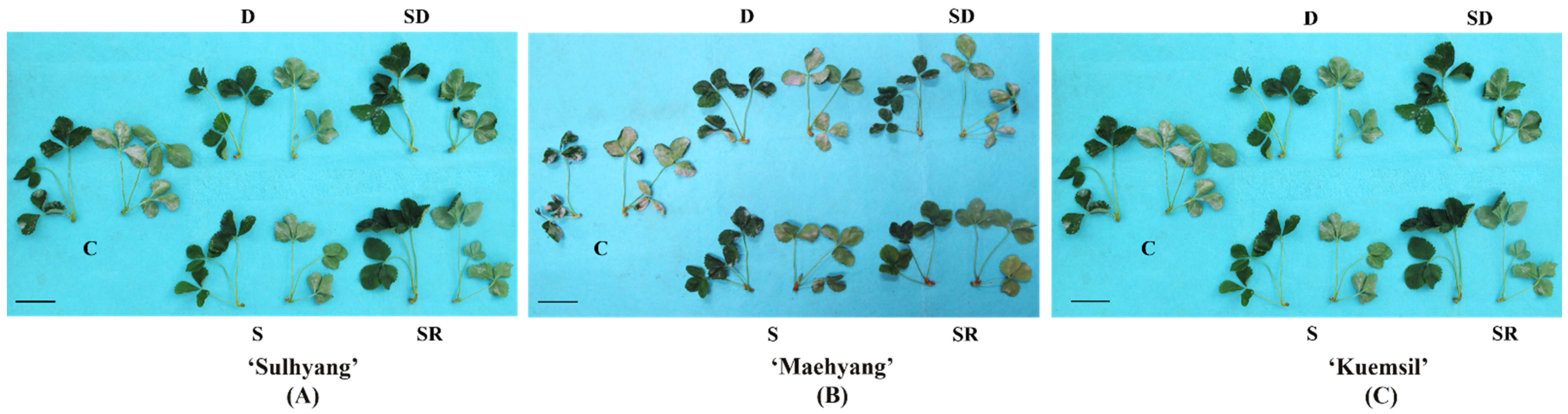
2.1. Growth and Powdery Mildew Severity

After 8 weeks of treatments, the growth parameters of strawberry plants were measured, as shown in Table 1. The shoot fresh weight of “mother” plants increased with the Si treatments, especially when dressed in a soluble Si fertilizer. However, sprayed Si to “daughter” plants did not significantly increase the fresh weight of the “mother” plants. Similarly, strawberry “daughter” plants treated with Si were bigger compared to the control. Regardless of the cultivar, sprayed Si to “daughter” plants resulted in the greatest shoot fresh weight of the “daughter” plants.
Varying degrees of white patches were observed on the leaves of strawberry “daughter” plants (Figure 1 and Figure 2). ‘Maehyang’ suffered the most severe among the three cultivars, where the leaves had visibly more white hyphae, and appeared to have shriveled compared to the control (Figure 1B). ‘Kuemsil’ was the second most severely affected by the powdery mildew (Figure 1C), while ‘Sulhyang’ leaves had no obvious white mycelia (Figure 1A). Similarly, the score of powdery mildew severity for ‘Maehyang’ was the highest. Although Si application reduced the powdery mildew severity, ‘Maehyang’ treated with Si had a disease severity score four times that of ‘Sulhyang’ (Figure 2). Thus, the powdery mildew severity was associated with the Si application and strawberry cultivar.

2.2. Scanning Electron Microscopy (SEM) and Chlorophyll Fluorescence Characteristics

The disease severity can also be reflected by the condition of the mycelia through SEM (Figure 3). Firstly, SEM observation of Si-treated leaves revealed the change of the hyphae morphology, and the hyphae of the control group were more even and smoother than those treated with Si. Si application inhibited hyphae growth and led to a rope-like structure in the hyphae. In the control group, the hyphae covered a greater area and were dense, and except for ‘Sulhyang’, the stomata were completely covered with hyphae. In contrast, Si application limited the hyphae coverage of leaves, and made the stomata visible on the leaves. Foliar Si sprayed of the “daughter” plants resulted in the greatest folding degree of hyphae and the lowest hyphae coverage.
Hyphae coverage of the leaves also affects their light capture capacity and gas exchange. The stomata are closely related to the chlorophyll fluorescence parameters of the photosystem II (PS II). The chlorophyll fluorescence parameters of the maximum primary yield of PSII photochemistry (Fv/F0) and the maximum/potential quantum efficiency of PSII (Fv/Fm) are shown in Figure 4. The Fv/F0 of leaves in the control group and SR-MP decreased except for ‘Sulhyang’, and there were no significant differences in the Fv/F0 values among Si-treated leaves.

2.3. Contents of Micro- and Macro-Nutrients

The macro- and micro-nutrient contents are shown in Table 2. The Si content in strawberry “mother” and “daughter” plants were increased with supplemental Si in the form of foliar spray or medium drench. Furthermore, foliar Si sprayed to the “mother” plants resulted in a higher Si content in “daughter” plants than in “mother” plants. However, sprayed Si on “daughter” plants only increased the Si content in “daughter” plants. Si supplementation also significantly affected the macro- and micro-nutrient contents in strawberry. Si application significantly improved the tissue contents of phosphorus (P), potassium (K), and magnesium (Mg), and the increasing trend of these nutrient levels coincided with the Si supplementation levels.

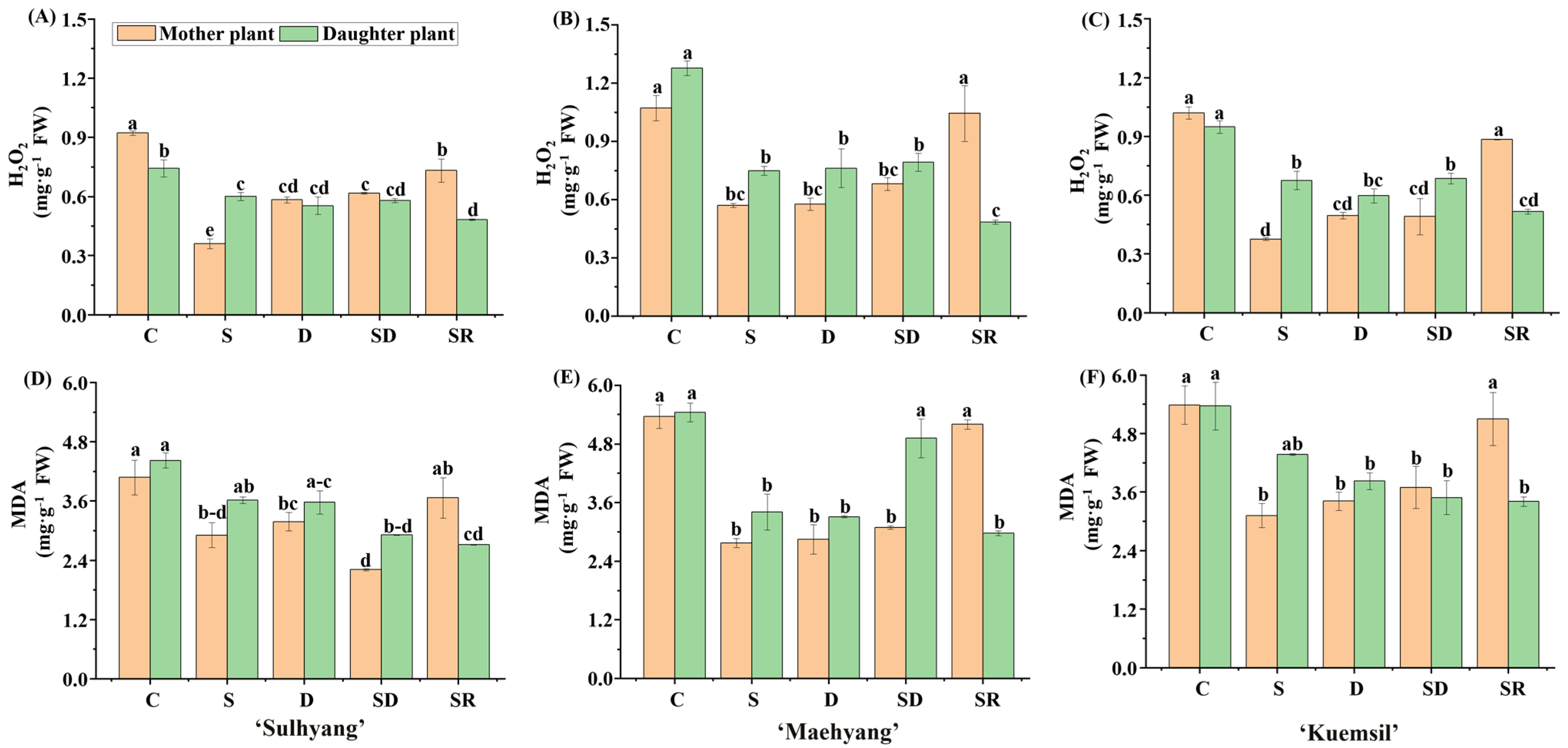
2.4. Hydrogen Peroxide (H2O2) and Lipid Peroxidation

The results of H2O2 and malondialdehyde (MDA) analyses are shown in Figure 5. The higher levels of H2O2 and MDA observed in the control indicate cell membrane damage. The cell membrane of ‘Sulhyang’ suffered less damage than that of the other two cultivars. The H2O2 and MDA contents of Si-treated strawberry, respectively, decreased by 47% and 34% in ‘Sulhyang’, 73% and 46% in ‘Maehyang’, and 56% and 60% in ‘Kuemsil’. This indicated that Si supplementation reduced damages caused by powdery mildew.

2.5. Proline Content

The trend of proline content was opposite to that of H2O2 and MDA contents (Figure 6). In the control leaves, the proline level in ‘Maehyang’ was 50% and 71% of that in ‘Sulhyang’ and ‘Kuemsil’, respectively. The proline accumulation increased in Si-treated strawberry, and the proline levels in ‘Sulhyang’ (2.1 mg·g−1), ‘Maehyang’ (1.5 mg·g−1), and ‘Kuemsil’ (1.2 mg·g−1) were, respectively, 2.1, 2.5, and 1.7 times that in the control (1.1, 0.58, and 0.71 mg·g−1). Furthermore, the proline content of ‘Sulhyang’ “daughter” plants treated with Si greatly increased. When “mother” plants were drenched or dressed with soluble Si fertilizers, or when the leaves of “daughter” plants were sprayed, the proline content in the “daughter” plants was 2.0, 2.8, and 1.5 times that in their “mother” plants.

2.6. Soluble Sugars and Soluble Proteins

There were significant differences in the soluble sugar contents between the “mother” and “daughter” plants (Figure 7). Among ‘Sulhyang’, ‘Maehyang’, and ‘Kuemsil’, the soluble sugar contents in “mother” plants (11.6, 11.4, and 8.7 mg·g−1) were, respectively, 37%, 27%, and 18% higher than that in “daughter” plants (8.4, 8.9, and 7.3 mg·g−1). Furthermore, the soluble sugar contents of the control group were slightly higher than that of the Si-treated “mother” and “daughter” plants. Si-treated plants (62 mg·g−1) had significantly higher soluble protein contents compared to the control (56 mg·g−1) in ‘Maehyang’. For ‘Sulhyang’ and ‘Kuemsil’, no significant differences were observed in the soluble protein content with Si supplementation.

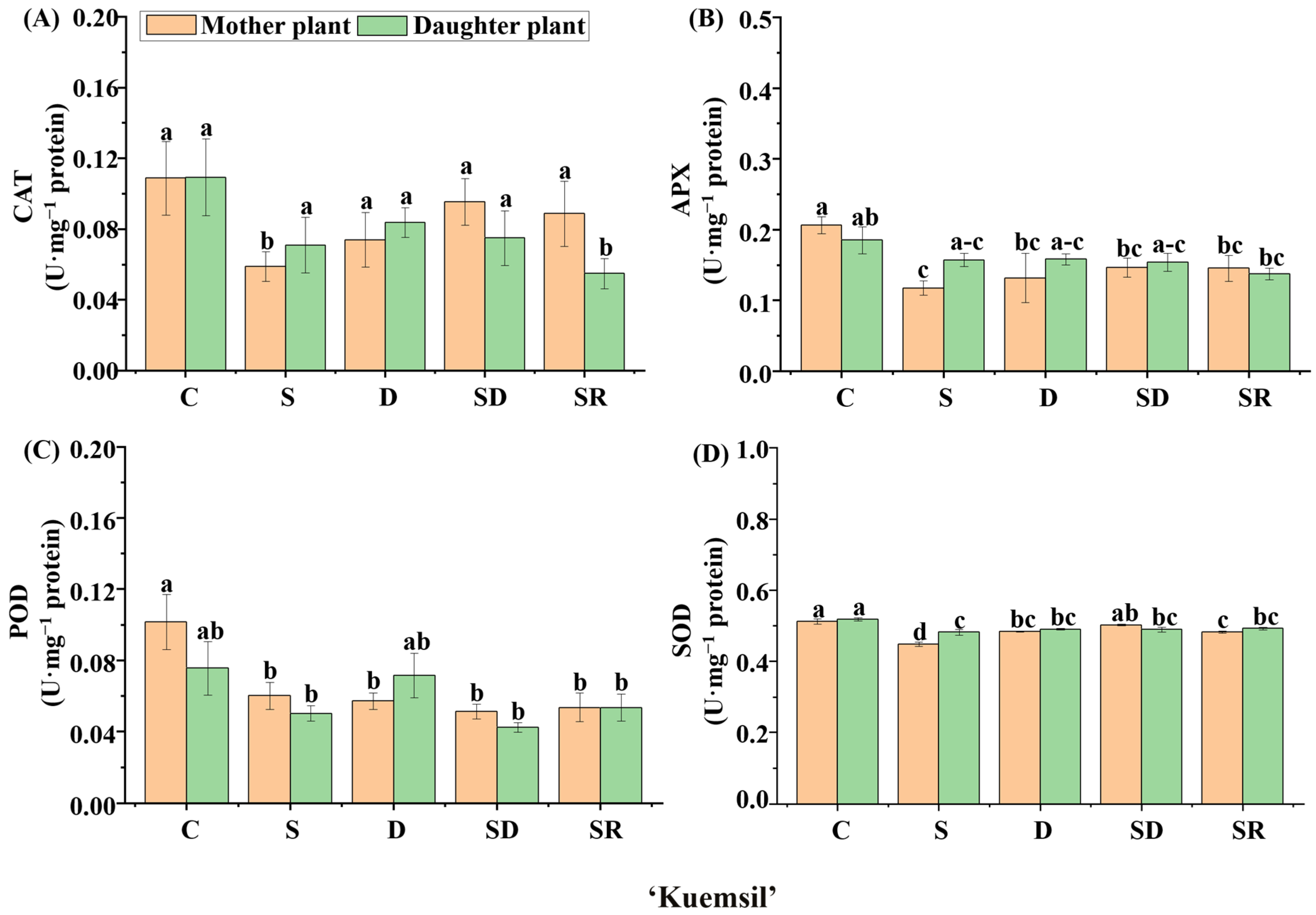
2.7. Antioxidant Enzyme Activities

We further explored the enzymatic activities in ‘Sulhyang’, ‘Maehyang’, and ‘Kuemsil’ leaves (Figure 8, Figure 9 and Figure 10). Significant differences were observed in the catalase (CAT) (Figure 8A, Figure 9A and Figure 10A), ascorbate peroxidase (APX) (Figure 8B, Figure 9B and Figure 10B), guaiacol peroxidase (POD) (Figure 8C, Figure 9C and Figure 10C), and superoxide peroxidase (SOD) (Figure 8D, Figure 9D and Figure 10D) activities. In ‘Sulhyang’, the CAT activities were the greatest without Si application. However, for ‘Maehyang’ and ‘Kuemsil’, Si application resulted in no significant differences in the CAT activities. Similarly, Si application resulted in no significant differences in the SOD activities for all three cultivars. The maximum value of APX (0.34 U·mg−1) activities was found in the control ‘Maehyang’ group, although there were no significant differences among the different treatments. Interestingly, Si application slightly decreased the APX activities in both ‘Sulhyang’ and ‘Kuemsil’ which were lower than 0.3 U·mg−1. On the other hand, the POD activities were significantly increased with Si application for all three cultivars.

2.8. Quantitative Real-Time RT-PCR

The relative expression levels of FaPR3, FaPR5, FaRPW8, FaMLO10, FaLsi2, and FaLsi3 showed significant differences in response to the different Si applications (Figure 11). The expression of FaPR3, FaPR5, FaLsi2, and FaLsi3 were significantly increased in response to Si application. However, the expression of FaRPW8 was only up-regulated markedly in ‘Sulhyang’ in response to the Si treatments. On the other hand, the expression of FaMLO10 significantly decreased in response to Si treatments regardless of the cultivar, and the control ‘Maehyang’ had the maximum expression.

3. Discussion

3.1. Growth and Disease Severity

In this study, the disease severity was assessed according to the area covered by white hyphae. On this basis, the physiological metabolism and expression of related genes were measured. Researchers found that powdery mildew led to premature senescence and reduced the photosynthesis in leaves [32]. This is mainly because the hyphae coverage reduces the functional leaf area and decreases the assimilation rate of the remaining leaf area [33], and limits the leaf gas exchange [34]. In photosynthesis, the CO2 transport from the mesophyll cell surface to the sites of carboxylation is a physical process, and interestingly, there is no difference in this process between healthy and diseased leaves [35]. Therefore, the reduced CO2 exchange caused by powdery mildew was associated with stomatal closure [33]. In this study, almost all the stomata in the strawberry leaves were closed (Figure 3). However, Si-treated strawberry “daughter” plants had higher Fv/Fm values than the control, because Si increased the feed-forward stimulation of the photosynthesis rate instead of affecting the photosynthetic gas exchange [36]. Similar observations were reported in zucchini (Cucurbita pepo L.) and melon (C. melo L.) [37,38]. Nevertheless, the soluble sugar contents did not increase with the increase in photosynthesis. It is reported that pathogens can absorb nutrients from leaves, especially glucose, as the major carbon energy source [39]. Infection affects the source-sink distribution of plants, which results in an enhanced sugar uptake capacity of the leaves and the activity of sucrose degradation enzymes [40]. In this situation, Si maintains the alteration balance of the sugar production and preserves the source-sink relationship. Similarly, it was reported that Si was beneficial in improving the source-sink relationship between leaves and spikes in rice infected with blast (Pyricularia oryzae) by reducing acid invertase activity and content of fructose and glucose [41].
In addition, Si application influenced the nutrient levels in the strawberry plants. It is reported that Si increases P absorption by reducing the soil P sorption [42]; the P content in strawberry tissues increased with Si application, when applied to either the “mother” or “daughter” plants in this study. Our results did not always agree with those of other researchers. In potato, although both soil and foliar Si application increased the Si content in leaves, only soil Si application increased the tissue P content [43]. On the other hand, Si does not affect the soil K and Mg availability [44], but Si application can still increase the K and Mg contents [45,46]. It is likely that Si can affect the uptake, distribution, and status of various nutrients through unknown pathways.
However, the demand of Si fertilizers varies in different agricultural environments [47]. For liquid Si, soil addition was the most effective in alleviating the cadmium (Cd) stresses in wheat [48]. Some researchers on the other hand reported that both soil and foliar Si application are more effective than only soil or spray Si application on their own [49]. In this study, four different Si application methods all increased the growth of “daughter” plants and reduced the disease severity; the greatest resistance to powdery mildew was obtained by spraying the “daughter” plants. Similar results were found on that Si can direct affect the pathogen, and it led to a significant reduction of powdery mildew severity by as much as 80% in wheat plants [50].

3.2. Analysis of the Biochemical Mechanisms

There are two opinions on the resistance to biotic stress: From the onset, it was reported that deposition of amorphous silica in the leaves plays a major role in resistance to biological stresses. This sedimentation can form a cuticle-Si double layer to prevent pathogen penetration and to decrease disease incidence [51]. Furthermore, environmental scanning electron microscopy and transmission electron microscopy found that Si led to more Si-papilla on the guard cell of stoma [52,53], which acts as a mechanical barrier against fungal pathogens [54]. Moreover, Si-complexes have been identified in cell walls including hemicellulose, pectin, and lignin, and they accelerate cell wall synthesis and remodeling [55]. The pectinase can’t cleave the C-O-Si bonds of Si-pectin complexes [56]. Therefore, Si maintained mesophyll cells relatively intact under biological stresses. Another suggestion is that Si mediates plant resistance to pathogens through changes in the primary metabolism and molecular aspects, e.g., increasing the contents of chlorogenic acids and activities of antioxidant enzymes [57,58,59,60]. Interestingly, it was reported that the phenomenon of Si accumulation at infection sites is probably due to a higher transpiration rate, rather than serving as an active defensive mechanism [61]. An increasing number of studies focus on the exact nature of how Si interacts with the biochemical pathways leading to disease resistance [62]. In general, SOD defends against reactive oxygen species (ROS) by disproportionation reaction, and APX, CAT, and POD reduce H2O2 to H2O. These enzymes increased in response to mild powdery mildew but decreased in response to severe powdery mildew [63]. However, the antioxidative enzyme activities were greater in seedlings under Cd toxicity, than in seedlings affected by Cd and Si toxicity [64]. Similar results were found in this study, where the Si application increased the POD activity but decreased the APX and CAT activities. It has been reported that POD has a higher tendency to reduce H2O2 than CAT does [65]. Furthermore, POD also participates in the oxidation of phenolic compounds [66], and Si supplementation can improve stress tolerance by affecting the secondary metabolism [67]. Our study corroborates these findings, demonstrating that Si supplementation significantly increased the proline content. Increasing evidence also showed that Si plays a role in numerous key components in plant signaling systems [68,69]. It was reported that Si treatment contributed to the accumulation of total soluble phenolic and lignin-thioglycolic acid derivatives by increasing the activity of phenylalanine ammonia-lyase (PAL) in banana and coffee plants [70,71]. Moreover, Si stimulated the production of phytoalexins which is critical to defend against pathogen infection in plants [72].

3.3. Gene Expressions

To explore how Si affects powdery mildew resistance, it is important to investigate the variation in the genes related to Si transport and strawberry powdery mildew. In this study, there were significant differences in the area of powdery mildew infection among the three cultivars. It is reported that the powdery mildew incidence of ‘Sulhyang’ is lower than that of other cultivars [73]. Our study corroborates this observation with various degrees of powdery mildew severity in the “daughter” plants according to the cultivar. Furthermore, compared to the control, ‘Maehyang’ had the minimum relative expression of FaRPW8 while that of FaMLO10 was maximum. This indicates that the ability of ‘Sulhyang’ to tolerate powdery mildew was stronger than that of ‘Maehyang’ and ‘Kuemsil’. Several reports have highlighted the negative regular function of MLO proteins in plant immunity, and that loss-of-function of MLO can confer durable and broad-spectrum resistance to diseases [74,75]. Differently, the R product of RPW genes acts as a specific receptor of powdery mildew, and activate the subsequent defense responses [76,77].
However, the R-gene is not always expressed, or the resistance is ephemeral because their effects are specific to certain species [78,79]. In this study, Si application increased the expression of FaRPW8 and simultaneously decreased that of FaMLO10. It is reported that Si probably induces the impact of pathogen infection on the transcriptome of host plants, by preventing the virulence of the pathogen [80]. Interestingly, the FaPR3 expression level in Si-sprayed “daughter” plants of ‘Sulhyang’ only slightly increased, whereas that of FaPR5 was markedly up-regulated. It is speculated that the resistance of PR5-regulate occupies the main position by spraying Si to ‘Sulhyang’ “daughter” plants. Similar results were obtained Si corresponded with PR1, PR2, and PR5, showed higher expression and lower infected area in Arabidopsis [81]. Moreover, in this study, the expression of FaLsi2 slightly increased, while that of FaLsi3 significantly increased. It is speculated that the Lsi2 protein acted as a Si efflux transporter and was affected by Si influx transporters. Furthermore, in rice, both Lsi2 and Lsi3 were found to be involved in the nodes, but only one efflux transporter, Lsi2, was involved in the roots [18]. Interestingly, the expression of FaLsi3 was increased by Si spraying on both the “mother” and “daughter” plants, compared to other Si treatments. It is speculated that the Lsi3 protein is probably associated with the Si uptake in leaves.

3.4. Application Prospects of Si in Plant Stress

The Si has been demonstrated to benefit growth of rice plants since 1924 [82], and commonly used for improvement of crop production [83]. However, research on the application of Si for plant disease suppression is its infancy. Especially in the context of sustainable agriculture, and greenhouses as an intensive cultivation system, it is necessary to use the environmentally beneficial and non-chemical fungicidal agents [84]. The Si is a crossover element as both fertilizer and plant protection. There is at least 1-year residual effect on disease control of Si applications [85], and more profitable and effective methods of Si application should be explored.

4. Materials and Methods

4.1. Plant Materials and Estimation of Powdery Mildew Severity

The strawberry “daughter” plants ‘Sulhyang’, ‘Maehyang’, and ‘Kuemsil’ were purchased from a strawberry farm (Sugok-myeon, Jinju, Gyeongsangnam-do, Korea) on 10 January 2021, and planted in a hydroponic gutter system filled with the BVB medium (Bas Van Buuren Substrate, EN-12580, De Lier, Westland, The Netherlands) in a glasshouse at Gyeongsang National University, Jinju, Korea (35°09′ N, 128°05′ E) for two months.
The ‘image processing of conversion’ found in a study [86] was used a reference, and 85,000 pixels were extracted from compound leaves free of powdery mildew infection. The powdery mildew severity was scored from 1 to 5 depending on the number of pixels infected: 17,000 (0~20%), 34,000 (21~40%), 51,000 (41~60%), 68,000 (61~80%), and 85,000 (81~100%) pixels (Figure 12), using the ImageJ graphical analysis software version 1.48v (ImageJ US National Institutes of Health, Bethesda, MD, USA, http://imagej.nih.gov/ij/ accessed on 12 February 2021).

4.2. Silicon Treatments

Naturally infected “mother” plants of ‘Sulhyang’, ‘Maehyang’, and ‘Kuemsil’ strawberries with a disease score of 1 were subjected to the Si applications on 10 March 2021. The Si solution was supplied from potassium silicate (K2SiO3) at a final concentration of 75 mg·L−1 was either added to the medium for drenching or sprayed to the leaves of the “mother” or “daughter” plant, or dress 0.5 g of the commercial soluble silicon fertilizer ‘Keunson’ (equivalent to 0.075 g of pure Na2SiO3) (Saturn Bio Tech Co., Ltd., Gangwon-do, Korea) to the “mother” plant weekly.

4.3. Measurement of Growth Parameters

After 8 weeks, the disease severity of the “daughter” plants was estimated and the plant height, crown diameter, chlorophyll fluorescence, chlorophyll level (SPAD), and fresh and dry weights of shoots were examined.
A chlorophyll meter (SPAD-502, Konica Minolta Inc., Osaka, Japan) measured the SPAD. The chlorophyll fluorescence parameters were measured using a portable fluorometer (FluorPen FP110, Photon Systems Instruments, Drásov, Czech Republic). The fresh weight was measured with an electronic scale (EW 220-3NM, Kern and Sohn GmbH., Balingen, Germany). The samples were rinsed with distilled water and dried using a forced air-dry oven (Venticell-222, MMM Medcenter Einrichtungen GmbH., Munich, Germany) at 70 °C for 72 h before the dry weights were recorded. The dried leaves were further used to determine the nutrient contents.

4.4. Scanning Electron Microscopy (SEM)

Leaf samples were cut into 0.5 mm2 pieces and fixed in 3.0% (v/v) glutaraldehyde (pH 7.5) for 12 h at 4 °C. Staining was carried out in a 1.0% (v/v) osmium tetroxide solution for 2 h at 4 °C. The samples were subsequently dehydrated in a graded series of 20, 40, 60, 80, and 100% (v/v) ethanol, and finally immersed in 80% acetone. The samples were then dried for 2 h at 70 °C and positioned on aluminum stubs with double-stick tape, and gold-coated in a sputter coater (SC7640; Polaron, Sussex, UK). A field emission scanning electron microscope II (SEM/EDS, JSM-7610F, JEOL Ltd., Tokyo, Japan) was used to observe the stomata and mycelium [87].

4.5. Determination of Contents of Macro- and Micro-Nutrients

The macro- and micro-nutrient contents were measured according to the method of Jeon [88]. Briefly, 0.5 g dried samples were ashed in a Nabertherm muffle furnace (Model LV 5/11/B180, Lilienthal, Breman, Germany) at 525 °C for 4 h. Then the ash was dissolved in 5 mL 25% (v/v) HCl, and subsequently diluted with 15 mL of warm distilled water and 10 mL of room-temperature distilled water. The filtrate was finally diluted three times. The macro- and micro-nutrient contents were measured using an inductively coupled plasma (ICP) spectrometer (Optima 4300DV/5300DV, Perkin Elmer, Germany).

4.6. Determination of Hydrogen Peroxide (H2O2) and Lipid Peroxidation

The H2O2 content was measured according to the method of Junglee [89]. Homogenized 0.1 g leaf samples were extracted in 5 mL 10 mM phosphate buffer (pH 5.8) containing 0.1% (w/v) trichloroacetic acid (TCA). After centrifugation at 12,000· g for 30 min at 4 °C, 5 mL of the supernatant was mixed with 0.5 mL 10 mM phosphate buffer (pH 5.8) and 0.5 mL 1 M Potassium iodide (KI). The absorbance was measured at 350 nm after incubation in the dark for 30 min.
Lipid peroxidation was estimated by measuring the malondialdehyde (MDA) level. Homogenized 0.5 g leaf samples were extracted in 5 mL 0.1% (w/v) TCA and centrifuged at 10,000· g for 5 min. Then 1 mL supernatant was added to 1 mL mixture of 0.6% (w/v) thiobarbituric acid and 10% (w/v) TCA. They were then left to react in a boiling water bath for 30 min and rapidly cooled with an ice bath. The absorbance was measured at 450, 532, and 600 nm, and MDA content was determined using the following formula [90]:
The   MDA   content   ( μ mol · g 1   FW ) = 6.45   ×   A 532 A 600     0.56   ×   A 450

4.7. Determination of Contents of Proline and Solulel Sugar

The leaf proline content was measured according to Bates et al. [91]. Homogenized 1 g leaf samples were extracted in 10 mL 3% (w/v) sulfosalicylic acid. After filtration, 1 mL of the extracting solution was added to 1 mL acid ninhydrin and 1 mL glacial acetic acid, and kept at 95 °C for 1 h in a water bath. Finally, 2 mL cold toluene was added to extract the chromophore and the absorbance was measured at 520 nm.
The soluble sugar content was measured according to the method of Sun [90]. 0.5 g leaf samples were mixed in 10 mL 20 mM phosphate buffer (pH 7.0), then centrifuged at 10,000 rpm for 30 min at 4 °C, then 5 mL H2SO4 and 1.8 mL distilled H2O were added to a 0.2 mL supernatant. Finally, the mixture was kept for 10 min in a boiling water bath, and the absorbance was measured at 620 nm after cooling to room temperature.

4.8. Analysis of Contents of Total Soluble Proteins and Activities of Antioxidant Enzymes Activities

The soluble protein content and the activities of superoxide dismutase (SOD), ascorbate peroxidase (APX), catalase (CAT), and peroxidase (POD) were measured according to the established protocols of Soundararajan et al. [92,93]. 0.5 g leaf samples were homogenized with liquid nitrogen and extracted in a 1.5 mL ice-cold 50 mM phosphate buffer (pH 7.0) containing 1 mM ethylenediaminetetraacetic acid (EDTA), 0.05% (v/v) Triton X-100, and 1 mM polyvinylpyrrolidone (PVP). The extracts were centrifuged at 13,000 rpm for 20 min at 4 °C, and the supernatant was used immediately to determine the soluble protein contents and activities of antioxidant enzymes.

4.9. Quantitative Real-Time PCR Analysis

The CTAB method was adopted for the total RNA extraction [94]. Briefly, 0.1 g frozen leaf tissues were homogenized with liquid nitrogen and mixed with 0.6 mL CTAB extraction buffer, then kept in a 65 °C water bath for 30 min. The supernatant was extracted three times with chloroform after centrifugation (13,000 rpm for 20 min at 4 °C). Afterwards, an equal volume of 6 M lithium chloride (LiCl) was added to the mixture at 4 °C for 75 min. The supernatant was discarded, and the pellet was washed three times in 1.0 mL 75% (v/v) ethanol. Finally, the pellets were air-dried and dissolved in 20 μL RNase-free diethyl pyrocarbonate water. The quality of the RNAs was determined with the NanoDrop 2000C Spectrophotometer (Thermo Fisher Scientific, Waltham, MA, USA), then reverse transcribed to cDNA using the PrimeScript RT Reagent Kit (Takara, Shiga, Japan). A total of 20 µL reaction volume was constructed with 2 µL each of forward and reverse primers, 2 µL cDNA, 4 µL of RNase-free water, and 10 µL SYBR green.
The FaActin was selected as the housekeeping gene. All primers used in this study are shown in Table 3. The CDS sequences were found at Strawberry GARDEN: http://strawberry-garden.kazusa.or.jp/index.html (accessed on 4 February 2022). The running procedure was set to 95 °C for 3 min, followed by 40 cycles of 95 °C for 30 s and 55 °C for 30 s on the CFX96 real-time PCR system (Bio-Rad, Hercules, CA, USA). Three biological replicates were adopted for each treatment.

4.10. Data Collection and Analysis

The statistical analysis was carried out using the Statistical Analysis Program (SAS 9.1, SAS Institute Inc., Cary, NC, USA). The experimental results were subjected to an analysis of variance (ANOVA) (p ≤ 0.05) and Duncan’s multiple range test (p ≤ 0.05). The F-test was also calculated based on Fisher’s least significant difference test at a threshold of p = 0.05. Pearson’s correlation coefficient was calculated with the SPSS 17.0 software (SPSS Inc., Chicago, IL, USA). Graphing was performed with the OriginPro software (version 9.0). The relative expression levels were calculated using the 2−∆∆Ct method and using the control as a reference (value = 1).

5. Conclusions

Our results showed that the growth and disease severity of strawberry plants were affected by supplementary Si. As expected, powdery mildew inhibited the photosynthesis, and the sensitivity to powdery mildew of ‘Maehyang’ was higher than that of ‘Sulhyang’ and ‘Kuemsil’. Si application promoted the contents of K, P, and Mg, and increased the biomass of the “daughter” plants. Moreover, supplemental Si reduced the contents of H2O2 and MDA, decreased the cell damage, and increased the resistance to powdery mildew. Among all the Si treatments studied, direct foliar Si spray to the “daughter” plants before cutting propagation was found to be the most effective in promoting their resistance to powdery mildew.

Author Contributions

Conceptualization, B.R.J.; methodology, B.R.J., J.X. and Y.L.; software, J.X.; validation, B.R.J.; formal analysis, B.R.J. and J.X.; investigation, J.X.; resources, B.R.J.; data curation, J.X.; writing—original draft preparation, J.X.; writing, review and editing, B.R.J., J.X. and Y.L.; visualization, J.X.; supervision, B.R.J.; project administration, B.R.J. and J.X.; funding acquisition, B.R.J. and J.X. All authors have read and agreed to the published version of the manuscript.

Funding

This research received no external funding. Jie Xiao and Yali Li were supported by the BK21 Four Program, Ministry of Education, Republic of Korea.

Institutional Review Board Statement

Not applicable.

Informed Consent Statement

Not applicable.

Data Availability Statement

Not applicable.

Conflicts of Interest

The authors declare no conflict of interest.

References

  1. Choi, H.G.; Moon, B.Y.; Kang, N.J.; Kwon, J.K.; Bekhzod, K.; Park, K.S.; Lee, S.Y. Yield loss and quality degradation of strawberry fruits cultivated under the deficient insolation conditions by shading. Hortic. Environ. Biotechnol. 2014, 55, 263–270. [Google Scholar] [CrossRef]
  2. Caruana, J.C.; Sittmann, J.W.; Wang, W.; Liu, Z. Suppressor of runnerless encodes a DELLA protein that controls runner formation for asexual reproduction in strawberry. Mol. Plant 2018, 11, 230–233. [Google Scholar] [CrossRef] [PubMed] [Green Version]
  3. Li, Y.; Xiao, J.; Hu, J.; Jeong, B.R. Method of silicon application affects quality of strawberry daughter plants during cutting propagation in hydroponic substrate system. Agronomy 2020, 10, 1753. [Google Scholar] [CrossRef]
  4. Maas, J.L. Strawberry disease management. In Diseases of Fruits and Vegetables: Volume II; Springer: New York, NY, USA, 2004; pp. 441–483. [Google Scholar]
  5. Nellist, C.F. Disease resistance in polyploid strawberry. In The Genomes of Rosaceous Berries and Their Wild Relatives; Springer: New York, NY, USA, 2018; pp. 79–94. [Google Scholar]
  6. Nelson, M.; Gubler, W.; Shaw, D.V. Relative resistance of 47 strawberry cultivars to powdery mildew in California greenhouse and field environments. In Plant Disease; Springer: New York, NY, USA, 1996; pp. 326–328. [Google Scholar]
  7. Kim, M.C.; Panstruga, R.; Elliott, C.; Müller, J.; Devoto, A.; Yoon, H.W.; Park, H.C.; Cho, M.J.; Schulze, L.P. Calmodulin interacts with MLO protein to regulate defence against mildew in barley. Nature 2002, 416, 447–451. [Google Scholar] [CrossRef] [PubMed]
  8. Jorgensen, J.H.; Wolfe, M. Genetics of powdery mildew resistance in barley. Crit. Rev. Plant Sci. 1994, 13, 97–119. [Google Scholar] [CrossRef]
  9. Dangl, J.L.; Jones, J.D. Plant pathogens and integrated defence responses to infection. Nature 2001, 411, 826–833. [Google Scholar] [CrossRef] [PubMed]
  10. Amil, R.F.; Blanco, P.R.; Munoz, B.J.; Caballero, J.L. The strawberry plant defense mechanism: A molecular review. Plant Cell Physiol. 2011, 52, 1873–1903. [Google Scholar] [CrossRef] [PubMed] [Green Version]
  11. Maas, J. VII International Strawberry Symposium. In Strawberry Diseases and Pests-Progress and Problems; Springer: New York, NY, USA, 2012; pp. 133–142. [Google Scholar]
  12. McGrath, M.T. Fungicide resistance in cucurbit powdery mildew: Experiences and challenges. Plant Dis. 2001, 85, 236–245. [Google Scholar] [CrossRef] [Green Version]
  13. Shishkoff, N.; McGrath, M. AQ10 biofungicide combined with chemical fungicides or AddQ spray adjuvant for control of cucurbit powdery mildew in detached leaf culture. Plant Dis. 2002, 86, 915–918. [Google Scholar] [CrossRef] [Green Version]
  14. Wedepohl, K.H. The composition of the continental crust. Geochim. Cosmochim. Acta 1995, 59, 1217–1232. [Google Scholar] [CrossRef]
  15. Ma, J.F.; Yamaji, N.; Tamai, K.; Mitani, N. Genotypic difference in silicon uptake and expression of silicon transporter genes in rice. Plant Physiol. 2007, 145, 919–924. [Google Scholar] [CrossRef] [Green Version]
  16. Mitani, N.; Ma, J.F. Uptake system of silicon in different plant species. J. Exp. Bot. 2005, 56, 1255–1261. [Google Scholar] [CrossRef] [Green Version]
  17. Ma, J.F.; Yamaji, N.; Mitani, N.; Tamai, K.; Konishi, S.; Fujiwara, T.; Katsuhara, M.; Yano, M. An efflux transporter of silicon in rice. Nature 2007, 448, 209–212. [Google Scholar] [CrossRef]
  18. Yamaji, N.; Sakurai, G.; Mitani, U.N.; Ma, J.F. Orchestration of three transporters and distinct vascular structures in node for intervascular transfer of silicon in rice. Proc. Natl. Acad. Sci. USA 2015, 112, 11401–11406. [Google Scholar] [CrossRef] [Green Version]
  19. Ma, J.F.; Takahashi, E. Chapter 7, Function of silicon in plant growth. In Soil, Fertilizer, and Plant Silicon Research in Japan; Elsevier: Amsterdam, The Netherlands, 2002; pp. 107–118. [Google Scholar]
  20. Ouellette, S.; Goyette, M.H.; Labbé, C.; Laur, J.; Gaudreau, L.; Gosselin, A.; Dorais, M.; Deshmukh, R.K.; Bélanger, R.R. Silicon transporters and effects of silicon amendments in strawberry under high tunnel and field conditions. Front. Plant Sci. 2017, 8, 949. [Google Scholar] [CrossRef]
  21. Park, Y.G.; Muneer, S.; Kim, S.; Hwang, S.J.; Jeong, B.R. Silicon application during vegetative propagation affects photosynthetic protein expression in strawberry. Hortic. Environ. Biotechnol. 2018, 59, 167–177. [Google Scholar] [CrossRef]
  22. Epstein, E. Silicon in plants: Facts vs. Concepts. In Studies in Plant Science; Elsevier: Amsterdam, The Netherlands, 2001; pp. 1–15. [Google Scholar]
  23. Coskun, D.; Britto, D.T.; Huynh, W.Q.; Kronzucker, H.J. The role of silicon in higher plants under salinity and drought stress. Front. Plant Sci. 2016, 7, 1072. [Google Scholar] [CrossRef] [Green Version]
  24. Muneer, S.; Park, Y.G.; Kim, S.; Jeong, B.R. Foliar or subirrigation silicon supply mitigates high temperature stress in strawberry by maintaining photosynthetic and stress-responsive proteins. J. Plant Growth Regul. 2017, 36, 836–845. [Google Scholar] [CrossRef]
  25. Park, Y.G.; Muneer, S.; Kim, S.; Hwang, S.J.; Jeong, B.R. Foliar or subirrigational silicon supply modulates salt stress in strawberry during vegetative propagation. Hortic. Environ. Biotechnol. 2018, 59, 11–18. [Google Scholar] [CrossRef]
  26. Liu, B.; Davies, K.; Hall, A. Silicon builds resilience in strawberry plants against both strawberry powdery mildew Podosphaera aphanis and two-spotted spider mites Tetranychus urticae. PLoS ONE 2020, 15, e0241151. [Google Scholar] [CrossRef]
  27. Jun, H.; Jung, H.; Imai, K. Gas exchange characteristics of a leading cultivar of Korean strawberry (Fragaria × ananassa, ‘Sulhyang’). Sci. Hortic. 2017, 221, 10–15. [Google Scholar] [CrossRef]
  28. Kim, T.I.; Jang, W.S.; Choi, J.H.; Nam, M.H.; Kim, W.S.; Lee, S.S. Breeding of strawberry ‘Maehyang’ for forcing culture. Hortic. Sci. Technol. 2004, 22, 434–437. [Google Scholar]
  29. Yoon, H.S.; Jin, H.J.; Oh, J.Y. ‘Kuemsil’, a strawberry variety suitable for forcing culture. Korean Soc. Breed. Sci. 2020, 52, 184–189. (In Korean) [Google Scholar] [CrossRef]
  30. Jambagi, S.; Dunwell, J.M. Global transcriptome analysis and identification of differentially expressed genes after infection of Fragaria vesca with powdery mildew (Podosphaera aphanis). Transcr. Open Access 2015, 3, 1–10. [Google Scholar] [CrossRef] [Green Version]
  31. Carisse, O.; Lefebvre, A.; Vander, H.H.; Roberge, L.; Brodeur, L. Analysis of incidence-severity relationships for strawberry powdery mildew as influenced by cultivar, cultivar type, and production systems. Plant Dis. 2013, 97, 354–362. [Google Scholar] [CrossRef] [Green Version]
  32. Saharan, G.S.; Mehta, N.K.; Meena, P.D. The Disease: Powdery Mildew. In Powdery Mildew Disease of Crucifers: Biology, Ecology and Disease Management; Springer: New York, NY, USA, 2019; pp. 17–51. [Google Scholar]
  33. Moriondo, M.; Orlandini, S.; Giuntoli, A.; Bindi, M. The effect of downy and powdery mildew on grapevine (Vitis vinifera L.) leaf gas exchange. J. Phytopathol. 2005, 153, 350–357. [Google Scholar] [CrossRef]
  34. Shtienberg, D. Effects of foliar diseases on gas exchange processes: A comparative study. Phytopathology 1992, 82, 760–765. [Google Scholar] [CrossRef]
  35. Gordon, T.; Duniway, J. Photosynthesis in powdery mildewed sugar beet leaves. Phytopathology 1982, 72, 718–723. [Google Scholar] [CrossRef]
  36. Detmann, K.C.; Araújo, W.L.; Martins, S.C.; Sanglard, L.M.; Reis, J.V.; Detmann, E.; Rodrigues, F.Á.; Nunes, N.A.; Fernie, A.R.; DaMatta, F.M. Silicon nutrition increases grain yield, which, in turn, exerts a feed-forward stimulation of photosynthetic rates via enhanced mesophyll conductance and alters primary metabolism in rice. New Phytol. 2012, 196, 752–762. [Google Scholar] [CrossRef] [Green Version]
  37. Savvas, D.; Giotis, D.; Chatzieustratiou, E.; Bakea, M.; Patakioutas, G. Silicon supply in soilless cultivations of zucchini alleviates stress induced by salinity and powdery mildew infections. Environ. Exp. Bot. 2009, 65, 11–17. [Google Scholar] [CrossRef]
  38. Dallagnol, L.; Rodrigues, F.; Tanaka, F.; Amorim, L.; Camargo, L. Effect of potassium silicate on epidemic components of powdery mildew on melon. Plant Pathol. 2012, 61, 323–330. [Google Scholar] [CrossRef] [Green Version]
  39. Hall, J.L.; Williams, L.E. Assimilate transport and partitioning in fungal biotrophic interactions. Funct. Plant Biol. 2000, 27, 549–560. [Google Scholar] [CrossRef]
  40. Sutton, P.N.; Gilbert, M.J.; Williams, L.E.; Hall, J. Powdery mildew infection of wheat leaves changes host solute transport and invertase activity. Physiol. Plant. 2007, 129, 787–795. [Google Scholar] [CrossRef]
  41. Araújo, M.U.P.; Rios, J.A.; Silva, E.T.; Rodrigues, F.Á. Silicon alleviates changes in the source-sink relationship of wheat plants infected by Pyricularia oryzae. Phytopathology 2019, 109, 1129–1140. [Google Scholar] [CrossRef]
  42. Jian, F.M.; Takahashi, E. Effect of silicate on phosphate availability for rice in a P-deficient soil. Plant Soil 1991, 133, 151–155. [Google Scholar]
  43. Pilon, C.; Soratto, R.P.; Moreno, L.A. Effects of soil and foliar application of soluble silicon on mineral nutrition, gas exchange, and growth of potato plants. Crop Sci. 2013, 53, 1605–1614. [Google Scholar] [CrossRef]
  44. Greger, M.; Landberg, T.; Vaculík, M. Silicon influences soil availability and accumulation of mineral nutrients in various plant species. Plants 2018, 7, 41. [Google Scholar] [CrossRef] [Green Version]
  45. Do, N.C.W.A.; Souza, N.G.H.; Preston, H.A.F.; Da, S.F.B.V.; Preston, W.; Loureiro, F.L.C. Influence of silicon fertilization on nutrient accumulation, yield and fruit quality of melon grown in northeastern Brazil. Silicon 2020, 12, 937–943. [Google Scholar]
  46. Swain, R.; Rout, G.R. Effect of silicon interaction with nutrients of rice. J. Exp. Biol. Agric. Sci. 2018, 6, 717–731. [Google Scholar] [CrossRef]
  47. Dallagnol, L.J.; Ramos, A.E.R.; Rosa, D.K. Silicon use in the integrated disease management of wheat: Current knowledge. In Current Trends in Wheat Research; IntechOpen: London, UK, 2020; pp. 5–26. [Google Scholar]
  48. Howladar, S.M.; Al, R.S.A.; Al, Z.F.S.; Howladar, M.M.; Aldhebiani, A.Y. Silicon and its application method effects on modulation of cadmium stress responses in Triticum aestivum (L.) through improving the antioxidative defense system and polyamine gene expression. Ecotoxicol. Environ. Saf. 2018, 159, 143–152. [Google Scholar] [CrossRef]
  49. Kowalska, J.; Tyburski, J.; Jakubowska, M.; Krzymińska, J. Effect of different forms of silicon on growth of spring wheat cultivated in organic farming system. Silicon 2021, 13, 211–217. [Google Scholar] [CrossRef] [Green Version]
  50. Guével, M.H.; Menzies, J.G.; Bélanger, R.R. Effect of root and foliar applications of soluble silicon on powdery mildew control and growth of wheat plants. Eur. J. Plant Pathol. 2007, 119, 429–436. [Google Scholar] [CrossRef]
  51. Wang, M.; Gao, L.; Dong, S.; Sun, Y.; Shen, Q.; Guo, S. Role of silicon on plant–pathogen interactions. Front. Plant Sci. 2017, 8, 701. [Google Scholar] [CrossRef] [Green Version]
  52. Cai, K.; Gao, D.; Luo, S.; Zeng, R.; Yang, J.; Zhu, X. Physiological and cytological mechanisms of silicon-induced resistance in rice against blast disease. Physiol. Plant. 2008, 134, 324–333. [Google Scholar] [CrossRef]
  53. Ning, D.; Song, A.; Fan, F.; Li, Z.; Liang, Y. Effects of slag-based silicon fertilizer on rice growth and brown-spot resistance. PLoS ONE 2014, 9, e102681. [Google Scholar] [CrossRef] [Green Version]
  54. Winslow, M.D.; Okada, K.; Correa, V.F. Silicon deficiency and the adaptation of tropical rice ecotypes. Plant Soil 1997, 188, 239–248. [Google Scholar] [CrossRef]
  55. He, C.; Ma, J.; Wang, L. A hemicellulose-bound form of silicon with potential to improve the mechanical properties and regeneration of the cell wall of rice. New Phytol. 2015, 206, 1051–1062. [Google Scholar] [CrossRef]
  56. Sheng, H.; Chen, S. Plant silicon-cell wall complexes: Identification, model of covalent bond formation and biofunction. Plant Physiol. Biochem. 2020, 155, 13–19. [Google Scholar] [CrossRef]
  57. Bilgin, D.D.; Zavala, J.A.; Zhu, J.; Clough, S.J.; Ort, D.R.; Delucia, E.H. Biotic stress globally downregulates photosynthesis genes. Plant Cell Environ. 2010, 33, 1597–1613. [Google Scholar] [CrossRef] [Green Version]
  58. Rémus, B.W.; Menzies, J.G.; Bélanger, R.R. Aconitate and methyl aconitate are modulated by silicon in powdery mildew-infected wheat plants. J. Plant Physiol. 2009, 166, 1413–1422. [Google Scholar] [CrossRef]
  59. Rojas, C.M.; Senthil, K.M.; Tzin, V.; Mysore, K. Regulation of primary plant metabolism during plant-pathogen interactions and its contribution to plant defense. Front. Plant Sci. 2014, 5, 17. [Google Scholar] [CrossRef] [Green Version]
  60. Rahman, A.; Wallis, C.M.; Uddin, W. Silicon-induced systemic defense responses in perennial ryegrass against infection by Magnaporthe oryzae. Phytopathology 2015, 105, 748–757. [Google Scholar] [CrossRef] [Green Version]
  61. Chérif, M.; Benhamou, N.; Menzies, J.G.; Bélanger, R. Silicon induced resistance in cucumber plants against Pythium ultimum. Physiol. Mol. Plant Pathol. 1992, 41, 411–425. [Google Scholar] [CrossRef]
  62. Bakhat, H.F.; Bibi, N.; Zia, Z.; Abbas, S.; Hammad, H.M.; Fahad, S.; Ashraf, M.R.; Shah, G.M.; Rabbani, F.; Saeed, S. Silicon mitigates biotic stresses in crop plants: A review. Crop Prot. 2018, 104, 21–34. [Google Scholar] [CrossRef]
  63. Gao, H.; Niu, J.; Zhao, W.; Zhang, D.; Li, S.; Liu, Y. Effect of powdery mildew on antioxidant enzymes of wheat grain. Plant Pathol. 2021, 1–16. [Google Scholar] [CrossRef]
  64. Liu, J.; Zhang, H.; Zhang, Y.; Chai, T. Silicon attenuates cadmium toxicity in Solanum nigrum L. by reducing cadmium uptake and oxidative stress. Plant Physiol. Biochem. 2013, 68, 1–7. [Google Scholar] [CrossRef]
  65. Jimenez, A.; Hernandez, J.A.; Del, R.L.A.; Sevilla, F. Evidence for the presence of the ascorbate-glutathione cycle in mitochondria and peroxisomes of pea leaves. Plant Physiol. 1997, 114, 275–284. [Google Scholar] [CrossRef] [Green Version]
  66. Pieczul, K.; Dobrzycka, A.; Wolko, J.; Perek, A.; Zielezińska, M.; Bocianowski, J.; Rybus, Z.M. The activity of β-glucosidase and guaiacol peroxidase in different genotypes of winter oilseed rape (Brassica napus L.) infected by Alternaria black spot fungi. Acta Physiol. Plant. 2020, 42, 142. [Google Scholar] [CrossRef]
  67. Fauteux, F.; Rémus, B.W.; Menzies, J.G.; Bélanger, R.R. Silicon and plant disease resistance against pathogenic fungi. FEMS Microbiol. Lett. 2005, 249, 1–6. [Google Scholar] [CrossRef] [Green Version]
  68. Bélanger, R.; Benhamou, N.; Menzies, J. Cytological evidence of an active role of silicon in wheat resistance to powdery mildew (Flumeria graminis f. Sp. Tritici). Phytopathology 2003, 93, 402–412. [Google Scholar] [CrossRef] [Green Version]
  69. Ma, J.F. Role of silicon in enhancing the resistance of plants to biotic and abiotic stresses. Soil Sci. Plant Nutr. 2004, 50, 11–18. [Google Scholar] [CrossRef]
  70. Fortunato, A.A.; Rodrigues, F.A.; Nascimento, K.J.T. Physiological and biochemical aspects of the resistance of banana plants to fusarium wilt potentiated by silicon. Phytopathology 2012, 102, 957–966. [Google Scholar] [CrossRef] [PubMed] [Green Version]
  71. Silva, R.; Oliveira, R.; Nascimento, K.; Rodrigues, F. Biochemical responses of coffee resistance against Meloidogyne exigua mediated by silicon. Plant Pathol. 2010, 59, 586–593. [Google Scholar] [CrossRef]
  72. Rodrigues, F.A.; Resende, R.S.; Dallagnol, L.J.; Datnoff, L.E. Silicon potentiates host defense mechanisms against infection by plant pathogens. In Silicon and Plant Diseases; Springer: New York, NY, USA, 2015; pp. 109–138. [Google Scholar]
  73. Nam, M.H.; Jeon, Y.N.; Lee, H.C.; Lee, H.D.; Kang, H.K. Comparative analysis between healthy and powdery mildew-infected plants of strawberry cultivar ‘Seolhyang’. Res. Plant Dis. 2012, 18, 80–85. [Google Scholar] [CrossRef] [Green Version]
  74. Kusch, S.; Panstruga, R. Mlo-based resistance: An apparently universal “weapon” to defeat powdery mildew disease. Mol. Plant Microbe Interact. 2017, 30, 179–189. [Google Scholar] [CrossRef] [Green Version]
  75. Tapia, R.R.; Barbey, C.R.; Chandra, S.; Folta, K.M.; Whitaker, V.M.; Lee, S. Evolution of the MLO gene families in octoploid strawberry (Fragaria × ananassa) and progenitor diploid species identified potential genes for strawberry powdery mildew resistance. Hortic. Res. 2021, 8, 153. [Google Scholar] [CrossRef]
  76. Xiao, S.; Ellwood, S.; Calis, O.; Patrick, E.; Li, T.; Coleman, M.; Turner, J.G. Broad-spectrum mildew resistance in Arabidopsis thaliana mediated by RPW8. Science 2001, 291, 118–120. [Google Scholar] [CrossRef]
  77. Kim, H.; O’Connell, R.; Maekawa, Y.M.; Uemura, T.; Neumann, U.; Schulze, L.P. The powdery mildew resistance protein RPW8.2 is carried on VAMP721/722 vesicles to the extrahaustorial membrane of haustorial complexes. Plant J. 2014, 79, 835–847. [Google Scholar] [CrossRef]
  78. Bai, G.; Su, Z.; Cai, J. Wheat resistance to Fusarium head blight. Can. J. Plant Pathol. 2018, 40, 336–346. [Google Scholar] [CrossRef]
  79. Goddard, R.; Steed, A.; Chinoy, C.; Ferreira, J.R.; Scheeren, P.L.; Maciel, J.L.N.; Caierão, E.; Torres, G.A.M.; Consoli, L.; Santana, F.M. Dissecting the genetic basis of wheat blast resistance in the Brazilian wheat cultivar BR 18-Terena. BMC Plant Biol. 2020, 20, 398. [Google Scholar] [CrossRef]
  80. Van, B.J.; Steppe, K.; Bauweraerts, I.; Kikuchi, S.; Asano, T.; Höfte, M.; De, V.D. Primary metabolism plays a central role in moulding silicon-inducible brown spot resistance in rice. Mol. Plant Pathol. 2015, 16, 811–824. [Google Scholar]
  81. Vivancos, J.; Labbé, C.; Menzies, J.G.; Bélanger, R.R. Silicon-mediated resistance of Arabidopsis against powdery mildew involves mechanisms other than the salicylic acid (SA)-dependent defence pathway. Mol. Plant Pathol. 2015, 16, 572–582. [Google Scholar] [CrossRef]
  82. Sommer, A.L. Studies concerning the essential nature of aluminum and silicon for plant growth. In Agriculture Sciences; University of California Press: Berkeley, CA, USA, 1924; pp. 58–81. [Google Scholar]
  83. Liang, Y.C.; Ma, T.S.; Li, F.J.; Feng, Y.J. Silicon availability and response of rice and wheat to silicon in calcareous soils. Commun. Soil Sci. Plant Anal. 1994, 25, 2285–2297. [Google Scholar] [CrossRef]
  84. Van, L.J.C. A greenhouse without pesticides: Fact or fantasy? Crop Prot. 2000, 19, 375–384. [Google Scholar]
  85. Zellner, W.; Tubaña, B.; Rodrigues, F.A.; Datnoff, L.E. Silicon’s role in plant stress reduction and why this element is not used routinely for managing plant health. Plant Dis. 2021, 105, 2033–2049. [Google Scholar] [CrossRef]
  86. Shin, J.; Chang, Y.K.; Heung, B.; Nguyen, Q.T.; Price, G.W.; Al, M.A. Effect of directional augmentation using supervised machine learning technologies: A case study of strawberry powdery mildew detection. Biosyst. Eng. 2020, 194, 49–60. [Google Scholar] [CrossRef]
  87. Rodrigues, F.A.; Silva, I.T.; Antunes, C.M.F.; Carré, M.V. The infection process of Pestalotiopsis longisetula leaf spot on strawberry leaves. J. Phytopathol. 2014, 162, 690–692. [Google Scholar] [CrossRef]
  88. Jeon, S.H.; Kuppusamy, S.; Yoon, Y.E.; Kim, H.T.; Lee, Y.B. Are there as many essential and non-essential minerals in hydroponic strawberry (Fragaria ananassa L.) compared to those grown in soil? Biol. Trace Elem. Res. 2019, 187, 562–567. [Google Scholar] [CrossRef]
  89. Junglee, S.; Urban, L.; Sallanon, H.; Lopez, L.F. Optimized assay for hydrogen peroxide determination in plant tissue using potassium iodide. Am. J. Anal. Chem. 2014, 5, 730. [Google Scholar] [CrossRef] [Green Version]
  90. Sun, C.; Li, X.; Hu, Y.; Zhao, P.; Xu, T.; Sun, J.; Gao, X. Proline, sugars, and antioxidant enzymes respond to drought stress in the leaves of strawberry plants. Hortic. Sci. Technol. 2015, 33, 625–632. [Google Scholar] [CrossRef] [Green Version]
  91. Bates, L.S.; Waldren, R.P.; Teare, I. Rapid determination of free proline for water-stress studies. Plant Soil 1973, 39, 205–207. [Google Scholar] [CrossRef]
  92. Soundararajan, P.; Manivannan, A.; Park, Y.G.; Muneer, S.; Jeong, B.R. Silicon alleviates salt stress by modulating antioxidant enzyme activities in Dianthus caryophyllus ‘Tula’. Hortic. Environ. Biotechnol. 2015, 56, 233–239. [Google Scholar] [CrossRef]
  93. Soundararajan, P.; Manivannan, A.; Ko, C.H.; Park, J.E.; Jeong, B.R. Evaluation of relative toxicity caused by deicing agents on photosynthesis, redox homeostasis, and the osmoregulatory system in creeper-type plants. Hortic. Environ. Biotechnol. 2019, 60, 175–186. [Google Scholar] [CrossRef]
  94. Chen, Q.; Yu, H.; Wang, X.; Xie, X.; Yue, X.; Tang, H. An alternative cetyltrimethylammonium bromide-based protocol for RNA isolation from blackberry (Rubus L.). GMR Genet. Mol. Res. 2012, 11, 1773–1782. [Google Scholar] [CrossRef]
Figure 1. Photographs showing the front and back of leaves of the “daughter” plant (A) ‘Sulhyang’; (B) ‘Maehyang’; and (C) ‘Kuemsil’ infected by the powdery mildew: C, control; S, sprayed “mother” plant; D, drenched “mother” plant; SD, dressed “mother” plant; and SR, sprayed “daughter” plant. Bars indicate 10 cm.
Ijms 23 03803 g001
Figure 2. The powdery mildew severity in strawberry “daughter” plants after Si treatments. Lowercase letters indicate significant differences calculated by the Duncan’s multiple range test at p ≤ 0.05: C, control; S, sprayed “mother” plant; D, drenched “mother” plant; SD, dressed “mother” plant; and SR, sprayed “daughter” plant.
Ijms 23 03803 g002
Figure 3. The powdery mildew hyphae on the leaf surface of “daughter” plant (A) ‘Sulhyang’; (B) ‘Maehyang’; and (C) ‘Kuemsil’: C, control; S, sprayed “mother” plant; D, drenched “mother” plant; SD, dressed “mother” plant; and SR, sprayed “daughter” plant. Bars indicate 50 μm.
Ijms 23 03803 g003
Figure 4. The chlorophyll fluorescence parameters Fv/Fm and Fv/F0 of strawberry (A) ‘Sulhyang’; (B) ‘Maehyang’; and (C) ‘Kuemsil’ as affected by the Si treatments. Lowercase letters indicate significant differences calculated by the Duncan’s multiple range test at p ≤ 0.05: C, control; S, sprayed “mother” plant; D, drenched “mother” plant; SD, dressed “mother” plant; SR, sprayed “daughter” plant; MP, “mother” plant; and DP, “daughter” plant.
Ijms 23 03803 g004
Figure 5. The contents of H2O2 and MDA of strawberry ‘Sulhyang’ (A,D); ‘Maehyang’ (B,E); and ‘Kuemsil’ (C,F) as affected by the Si treatments. Lowercase letters indicate significant differences calculated by the Duncan’s multiple range test at p ≤ 0.05: C, control; S, sprayed “mother” plant; D, drenched “mother” plant; SD, dressed “mother” plant; and SR, sprayed “daughter” plant.
Ijms 23 03803 g005
Figure 6. The proline content of strawberry ‘Sulhyang’ (A), ‘Maehyang’ (B), and ‘Kuemsil’ (C) as affected by the Si treatments. Lowercase letters indicate significant differences calculated by the Duncan’s multiple range test at p ≤ 0.05: C, control; S, sprayed “mother” plant; D, drenched “mother” plant; SD, dressed “mother” plant; and SR, sprayed “daughter” plant.
Ijms 23 03803 g006
Figure 7. The contents of soluble sugars and proteins of strawberry ‘Sulhyang’ (A,D), ‘Maehyang’ (B,E), and ‘Kuemsil’ (C,F) as affected by the Si treatments. Lowercase letters indicate significant differences calculated by the Duncan’s multiple range test at p ≤ 0.05: C, control; S, sprayed “mother” plant; D, drenched “mother” plant; SD, dressed “mother” plant; and SR, sprayed “daughter” plant.
Ijms 23 03803 g007
Figure 8. The activities of catalase (CAT) (A), ascorbate peroxidase (APX) (B), guaiacol peroxidase (POD) (C), and superoxide dismutase (SOD) (D) in ‘Sulhyang’ as affected by the Si treatments. Lowercase letters indicate significant differences calculated by the Duncan’s multiple range test at p ≤ 0.05: C, control; S, sprayed “mother” plant; D, drenched “mother” plant; SD, dressed “mother” plant; and SR, sprayed “daughter” plant.
Ijms 23 03803 g008
Figure 9. The activities of catalase (CAT) (A), ascorbate peroxidase (APX) (B), guaiacol peroxidase (POD) (C), and superoxide dismutase (SOD) (D) in ‘Maehyang’ as affected by the Si treatments. Lowercase letters indicate significant differences calculated by the Duncan’s multiple range test at p ≤ 0.05: C, control; S, sprayed “mother” plant; D, drenched “mother” plant; SD, dressed “mother” plant; and SR, sprayed “daughter” plant.
Ijms 23 03803 g009
Figure 10. The activities of catalase (CAT) (A), ascorbate peroxidase (APX) (B), guaiacol peroxidase (POD) (C), and superoxide dismutase (SOD) (D) in ‘Kuemsil’ as affected by the Si treatments. Lowercase letters indicate significant differences calculated by the Duncan’s multiple range test at p ≤ 0.05: C, control; S, sprayed “mother” plant; D, drenched “mother” plant; SD, dressed “mother” plant; and SR, sprayed “daughter” plant.
Ijms 23 03803 g010
Figure 11. Expression profile heat map of pathogenesis-related 3 (PR3), PR5, resistance to powdery mildew 8 (RPW8), mildew resistance locus o 10 (MLO10), low silicon rice 2 (Lsi2) and Lsi3 in strawberry “daughter” plants ‘Sulhyang’, ‘Maehyang’, and ‘Kuemsil’. The gene expression is presented with a scale of fold change calculated by 2−ΔΔCT. Lowercase letters indicate significant differences calculated by the Duncan’s multiple range test at p ≤ 0.05: C, control; S, sprayed “mother” plant; D, drenched “mother” plant; SD, dressed “mother” plant; and SR, sprayed “daughter” plant.
Ijms 23 03803 g011
Figure 12. Standard area diagrams for powdery mildew severity on strawberry leaves.
Ijms 23 03803 g012
Table 1. The growth parameters of strawberry plants under different Si treatments.
Cultivar (A)Treatment (B)“Mother” Plant“Daughter” Plant
Plant Height
(cm)
Crown
Diameter (mm)
Fresh Weight (g)Leaf Length (cm)Leaf Width (cm)SPADPlant Height
(cm)
Crown
Diameter (mm)
Fresh Weight (g)Leaf Length (cm)Leaf Width (cm)SPAD
‘Sulhyang’C44.55 de z18.84 e–g66.01 c11.00 d7.63 c–e39.55 d21.52 f6.06 h5.12 f5.65 g4.7 bc44.23 ab
S46.62 a–d21.85 b–d107.04 ab13.27 a10.78 a46.57 ab26.15 c–e7.60 e–g9.95 b–d6.75 c–f5.3 a–c45.52 ab
D48.03 a–c22.14 b–d90.44 a–c13.07 ab10.97 a45.12 ab29.53 ab7.51 fg11.83 ab7.43 bc5.5 ab37.40 c
SD45.05 c–e23.11 bc116.24 ab11.98 a–d9.20 b46.43 ab28.05 a–d8.18 d–f11.35 ab7.58 ab5.5 ab39.47 a–c
SR44.73 de19.39 d–g66.85 b11.57 b–d7.97 b–e46.05 ab26.30 b–e9.80 ab13.99 a7.18 b–d5.1 a–c43.03 a–c
‘Maehyang’C43.77 de17.15 g63.09 c11.40 cd7.15 e40.48 cd24.73 d–f6.67 gh5.86 ef6.35 ef4.3 c38.88 bc
S45.85 b–e21.18 c–e86.72 bc12.53 a–d7.97 b–e43.18 a–c27.20 b–d6.88 gh5.81 ef7.40 bc6.0 a42.32 a–c
D45.88 b–e20.54 c–f82.47 bc12.93 a–c8.45 b–d44.35 ab24.65 d–f7.19 fg9.94 b–d6.47 d–f4.6 bc42.68 a–c
SD44.37 de21.32 c–e105.33 ab12.33 a–d8.78 bc43.75 a–c28.77 a–c8.60 c–e10.62 a–c7.60 ab5.3 a–c42.28 a–c
SR42.87 e17.96 fg67.18 c11.97 a–d7.32 de44.28 ab25.68 c–e8.57 c–e11.88 ab7.55 ab5.2 a–c45.03 ab
‘Kuemsil’C44.47 de18.07 fg94.82 a–c11.17 d7.90 b–e45.33 ab25.55 c–e7.08 g6.80 c–f6.93 b–e5.3 a–c45.90 a
S48.45 ab24.29 b121.28 a11.70 a–d8.03 b–e45.05 ab22.90 ef8.99 b–d6.48 d–f6.12 fg4.7 bc42.83 a–c
D49.80 a26.93 a115.25 ab11.82 a–d8.47 b–d43.63 a–c25.95 c–e9.65 ab9.33 b–e7.20 b–d5.4 ab39.23 a–c
SD49.38 a21.90 b–d121.33 ab12.53 a–d8.58 b–d47.03 a24.68 d–f10.11 a8.22 b–f6.60 d–f5.0 bc41.60 a–c
SR48.60 ab20.20 d–f93.56 a–c12.18 a–d8.22 b–e42.80 b–d30.75 a9.41 a–c9.38 b–e8.18 a6.0 a44.55 ab
F-test yA*********NS***NSNS***NSNSNSNS
B******************************NS
A × B************************************
z Lowercase letters indicate significant differences calculated by the Duncan’s multiple range test at p ≤ 0.05; y NS, *, **, and *** represent non-significant or significant at p ≤ 0.05, 0.01, and 0.001, respectively: C, control; S, sprayed “mother” plant; D, drenched “mother” plant; SD, dressed “mother” plant; and SR, sprayed “daughter” plant.
Table 2. Mineral contents in strawberry plants as affected by different Si treatments.
Cultivar (A)Treatment (B)TissueSi
(mg·g−1 DW)
Macro-NutrientMicro-Nutrient
Ca (g·g−1 DW)K (g·g−1 DW)P (mg·g−1 DW)Mg (mg·g−1 DW)S (mg·g−1 DW)Fe (mg·g−1 DW)Mn (mg·g−1 DW)Zn (mg·g−1 DW)Cu (mg·g−1 DW)
‘Sulhyang’C“Mother” plant1.15 j–l z0.18 b–d0.10 gh14.99 d–i20.53 k–m1.97 i–m0.75 e–j0.69 i–l0.17 b–f0.10 e–g
“Daughter” plant0.83 l0.08 ij0.10 f–h12.18 ij18.53 m1.80 j–m0.66 h–k0.41 mn0.14 e–j0.11 d–f
S“Mother” plant1.65 f–k0.15 c–f0.12 c–g16.56 a–h29.06 b–g2.18 h–l0.65 h–k0.47 l–n0.16 b–h0.12 a–c
“Daughter” plant1.69 f–j0.10 g–j0.15 a–d17.34 a–f19.53 lm1.81 j–m0.85 c–g0.67 i–l0.10 j0.10 e–g
D“Mother” plant2.08 d–h0.16 c–f0.17 ab20.19 a28.03 b–h3.09 c–g0.90 b–e0.72 i–l0.20 ab0.12 a–c
“Daughter” plant1.92 e–i0.10 g–j0.12 c–h14.63 e–i20.24 k–m2.94 c–h0.65 h–k0.56 j–n0.12 h–j0.10 e–g
SD“Mother” plant1.71 f–j0.11 g–i0.12 c–g15.01 d–i20.72 j–m1.14 m0.62 i–l0.51 k–n0.12 h–j0.10 f–h
“Daughter” plant1.16 j–l0.15 d–f0.10 f–h13.63 e–j25.93 c–k2.12 h–l0.92 b–e0.60 j–n0.13 f–j0.12 ab
SR“Mother” plant1.92 e–i0.10 g–j0.12 c–h14.63 e–i20.24 k–m2.94 c–h0.65 h–k0.56 j–n0.12 h–j0.10 e–g
“Daughter” plant2.24 d–g0.11 g–i0.13 c–f15.03 d–i21.65 i–m1.83 j–m0.72 f–j0.77 h–j0.13 f–j0.12 a–c
‘Maehyang’C“Mother” plant1.38 h–l0.13 f–h0.10 f–h10.41 j25.11 d–l2.70 d–i0.59 j–l0.75 h–k0.12 g–j0.10 e–g
“Daughter” plant0.97 kl0.11 g–i0.09 h16.75 a–g23.57 f–m3.47 b–d0.71 f–j0.87 g–i0.18 b–e0.11 c–e
S“Mother” plant1.63 f–k0.18 b–d0.13 b–f12.37 h–j27.59 b–h2.58 e–k0.66 h–k1.28 c–e0.13 g–j0.09 gh
“Daughter” plant1.78 f–j0.13 e–g0.14 a–e17.86 a–e29.68 b–e4.16 b0.62 i–l1.07 e–g0.19 a–c0.09 hi
D“Mother” plant2.55 c–e0.22 a0.17 a14.72 e–i30.76 a–d3.33 b–e0.78 d–i1.70 b0.13 f–j0.11 c–e
“Daughter” plant1.77 f–j0.13 e–g0.15 a–c19.09 a–d36.21 a3.69 bc0.70 g–j0.98 f–h0.18 a–d0.10 f–h
SD“Mother” plant2.28 d–f0.15 d–f0.14 b–e12.99 g–j29.25 b–f2.18 h–l0.88 b–f1.34 cd0.13 g–j0.13 a
“Daughter” plant1.63 f–k0.10 g–j0.16 ab19.22 a–d27.25 b–i4.94 a0.59 j–l0.80 h–j0.22 a0.12 b–d
SR“Mother” plant1.52 g–l0.21 ab0.10 f–h13.48 f–j31.55 a–c2.34 f–l0.94 b–d1.99 a0.11 ij0.10 e–g
“Daughter” plant3.10 c0.11 g–i0.14 a–e19.49 a–c22.88 h–m2.64 d–k0.47 l0.99 f–h0.15 c–h0.03 k
‘Kuemsil’C“Mother” plant1.20 i–l0.18 a–d0.11 d–h15.77 b–i26.46 c–j2.93 c–h1.05 b1.67 b0.15 c–h0.12 ab
“Daughter” plant0.95 kl0.07 j0.10 f–h14.73 e–i18.56 m3.15 c–f0.50 kl0.37 n0.18 a–d0.04 k
S“Mother” plant1.70 f–j0.19 a–c0.12 c–h16.74 a–g28.40 b–h2.68 d–j0.90 b–e1.27 c–e0.15 c–h0.12 a–c
“Daughter” plant1.83 f–j0.09 h–j0.13 c–g16.49 a–h20.20 k–m3.38 b–e0.63 i–l0.65 i–m0.18 a–d0.03 k
D“Mother” plant3.87 b0.17 cd0.12 c–h19.08 a–d35.54 ab2.24 g–l1.32 a1.49 bc0.12 h–j0.11 b–d
“Daughter” plant2.73 cd0.13 f–h0.13 c–g16.84 a–g20.06 k–m3.70 bc0.82 c–h0.48 l–n0.13 f–j0.04 k
SD“Mother” plant2.94 c0.16 c–f0.13 c–f16.16 a–i30.87 a–d2.80 d–i1.20 a1.48 bc0.16 b–g0.11 b–d
“Daughter” plant2.07 d–h0.08 ij0.12 c–h19.59 ab23.19 g–m3.79 bc0.96 bc0.66 i–m0.20 ab0.05 j
SR“Mother” plant1.62 f–k0.18 b–d0.11 e–h15.26 c–i20.31 k–m4.07 b0.66 h–k1.66 b0.16 b–h0.09 hi
“Daughter” plant4.94 a0.11 g–i0.13 c–g19.08 a–d24.19 e–m1.79 k–m0.50 kl1.15 d–f0.14 d–i0.03 k
F-test yA***************************
B****************************
A × B****NS*********************
z Lowercase letters indicate significant differences calculated by the Duncan’s multiple range test at p ≤ 0.05; y NS, *, **, and *** represent non-significant or significant at p ≤ 0.05, 0.01, and 0.001, respectively: C, control; S, sprayed “mother” plant; D, drenched “mother” plant; SD, dressed “mother” plant; SR, sprayed “daughter” plant; MP, “mother” plant; and DP, “daughter” plant.
Table 3. List of qPCR primers used in this study.
Gene NameForward Primer (5’ to 3’)Reverse Primer (5’ to 3’)
FaPR3ACAAACCATCAAGCCACGACGTTGTCCACGCCCACATTCAAGTC
FaPR5AGGTCCAGTGCAGCAATACCTGGATTGTCGGACCTCTACCTGCA
FaRPW8CTCTACAACCACGAATCGCTCAACGCTCATTCGTATGTCTCTCTTCCTG
FaMLO10GATTATTCACCTGGTCGGACATTGGATGGTAAGGACAAGGCAACATCGTA
FaLsi2GCTCTTTTCACCAATGACACCTCGCATTCACACAAACTCCTACAAGC
FaLsi3AACTGTTCTCTTGCTTGGAGGACGCCCAAACTTGAGATGGCTCCAGAAT
FaActinTTCACGAGACCACCTATAACTCGCTCATCCTATCAGCGATT
Publisher’s Note: MDPI stays neutral with regard to jurisdictional claims in published maps and institutional affiliations.

