Next Article in Journal
Oral bony outgrowths: Prevalence and genetic factor influence. Study of twins
Previous Article in Journal
Interaction of ethidium and tetraphenylphosphonium cations with Salmonella enterica cells
 
 
Medicina is published by MDPI from Volume 54 Issue 1 (2018). Previous articles were published by another publisher in Open Access under a CC-BY (or CC-BY-NC-ND) licence, and they are hosted by MDPI on mdpi.com as a courtesy and upon agreement with Lithuanian Medical Association, Lithuanian University of Health Sciences, and Vilnius University.
Case Report

Pathologic fracture of the thoracic spine in a male master ultra-marathoner due to the combination of a vertebral hemangioma and osteopenia

by
Beat Knechtle
1,2,*,
Pantelis T. Nikolaidis
3,
Bruno Lutz
4,
Thomas Rosemann
2 and
Christian B. Baerlocher
5
1
Gesundheitszentrum St. Gallen, St. Gallen, Switzerland
2
Institute of Primary Care, University of Zurich, Zurich, Switzerland
3
Exercise Physiology Laboratory, Nikaia, Greece
4
RODIAG Diagnostic Centers, St. Gallen, Switzerland
5
Wirbelsäulenchirurgie Ostschweiz, Klinik Stephanshorn, St. Gallen, Switzerland
*
Author to whom correspondence should be addressed.
Medicina 2017, 53(2), 131-137; https://doi.org/10.1016/j.medici.2017.02.003
Submission received: 12 May 2016 / Revised: 11 February 2017 / Accepted: 20 February 2017 / Published: 27 March 2017

Abstract

:
Vertebral hemangiomas are the most common benign vertebral neoplasms and are generally asymptomatic. In the present study, we report the case of a 52-year-old male master ultra-marathoner suffering from a pathologic fracture of the thoracic spine due to a vertebral hemangioma. A further examination in the athlete revealed an accompanying osteopenia, which was most likely due to a deficiency in both vitamin D and testosterone. The treatment of the fracture consisted of percutaneous vertebroplasty. Shortly after the operation the athlete was able to continue running. The most likely reason for the pathologic fracture of the vertebral body was the combination of the vertebral hemangioma and osteopenia. The further treatment consisted of supplementation of both vitamin D and testosterone. Athletes and physicians should be aware that male master ultra-marathoners older than 50 years might suffer from osteopenia, where a deficiency in vitamin D and testosterone could be contributing factors for osteopenia development in general.

1. Introduction

Primary intraosseous hemangiomas are most frequently seen in the vertebrae or in the skull [1,2]. Vertebral hemangiomas are the most common benign vertebral neoplasms and incidentally detected due to their characteristic features on imaging for other reasons. They usually occur in the lower thoracic and upper lumbar region.
We present the case of a 52-year-old male master ultra-marathoner who suffered from a pathologic fracture of his thoracic spine while running. Radiological imagining revealed an intraosseous hemangioma as the main reason for the fracture. The most likely reason for the fracture of the vertebral body with the hemangioma was an accompanying osteopenia most likely due to a combination of low vitamin D and low testosterone.

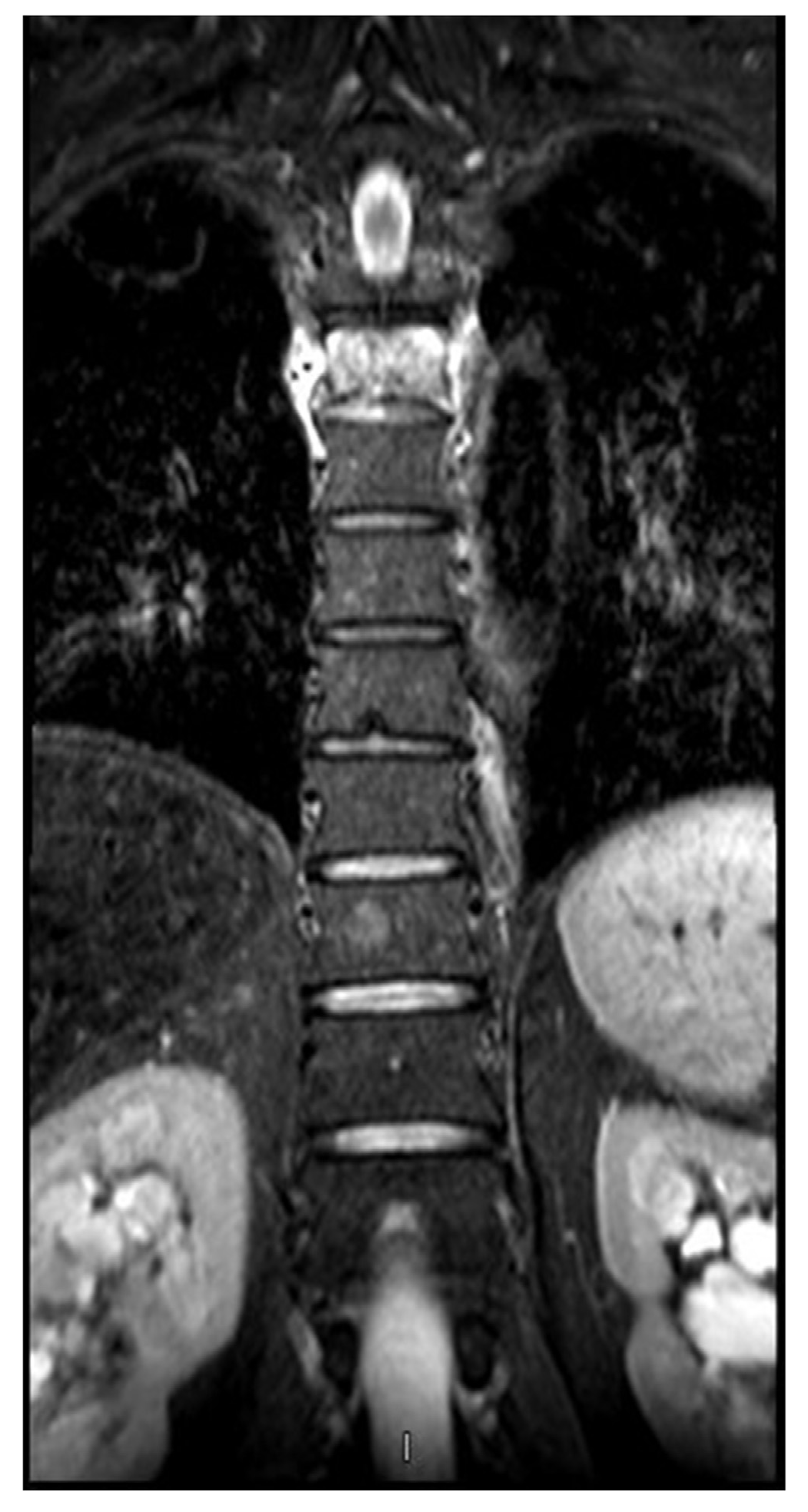
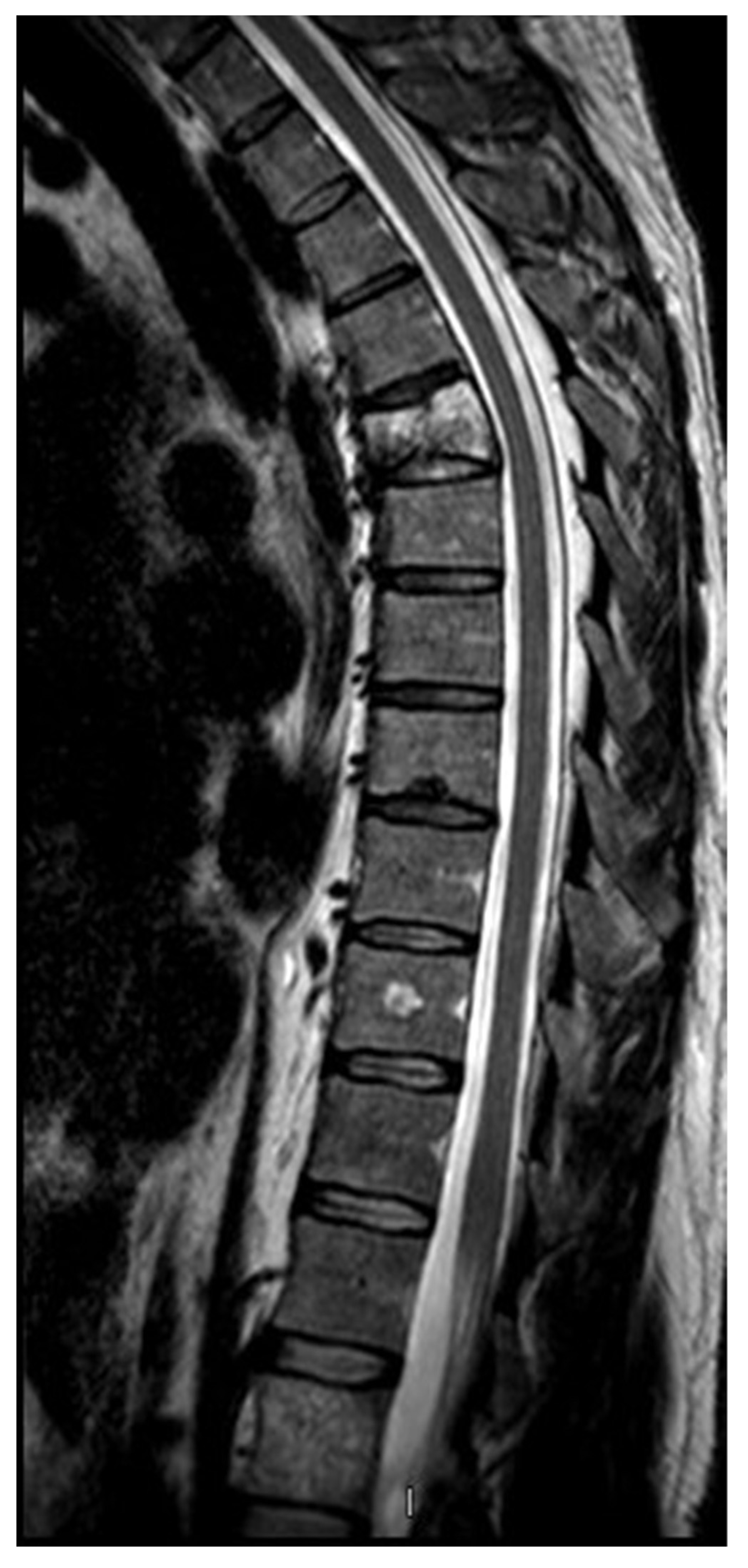
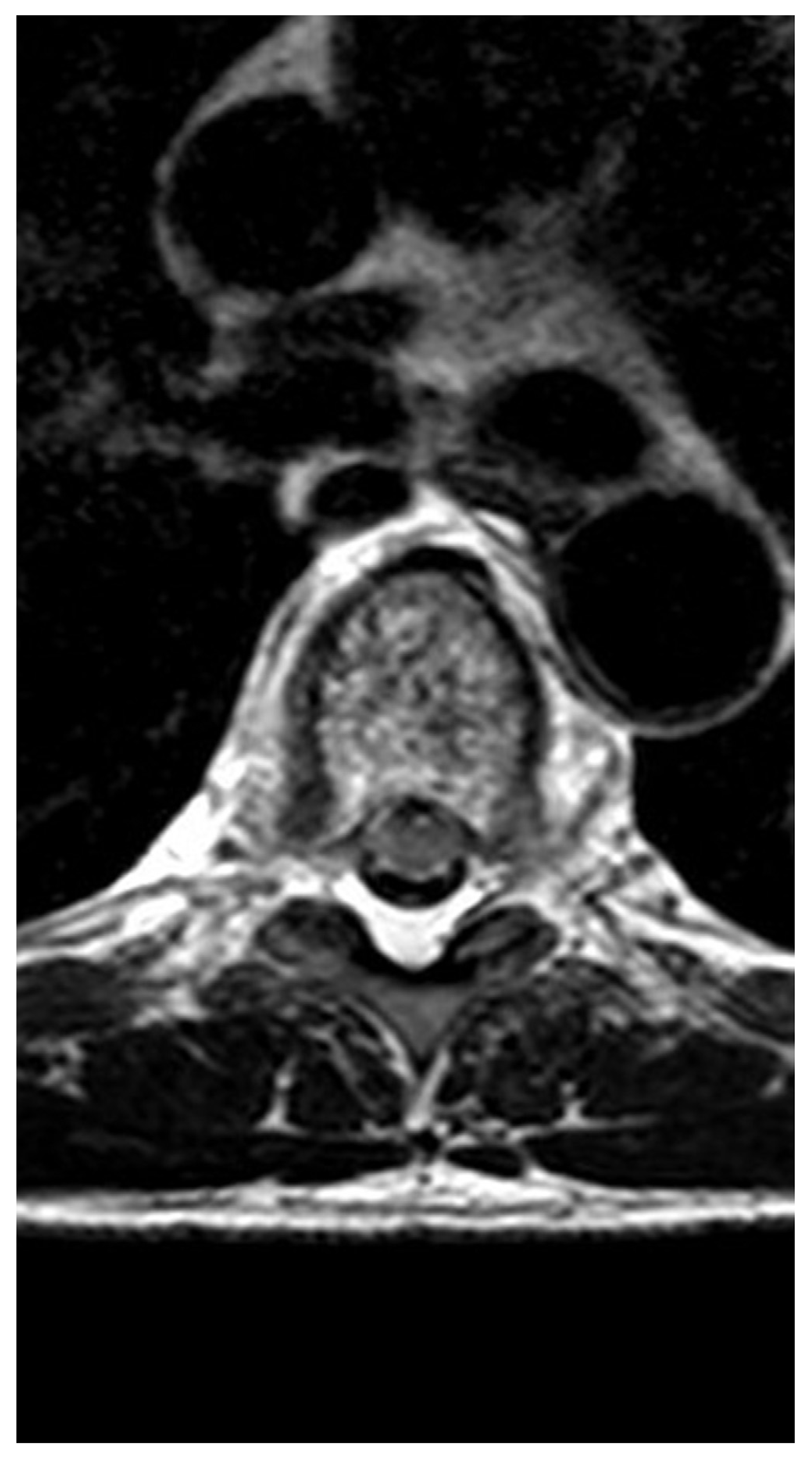
2. Case presentation

The 52-year-old highly experienced male ultra-marathoner went with his 64-year-old run comrade on a routine long training run. The two athletes had planned to complete their usual running distance of 40 km. Shortly after takeoff, the athlete under examination felt a sharp pain between his shoulder blades. He ignored the pain and thought it was a muscular strain after he had changed from the sidewalk across the street. Over the kilometers the pain became stronger and by the time he could hardly breathe and had to let his colleague running alone to the turning point (~20 km). On the way back both ran slowly together to the starting point. Back home, breathing became more severe and the pain increased steadily. From a recent operation he had available some painkillers and could sleep for a while after high-dose ingestion.
Over the next few weeks the pain between the shoulder blades remained and each step caused pain. While the pain remained for several weeks, the athlete visited his primary care physician who performed a radiograph of the thoracic spine showing a compression fracture in the mid thoracic spine (Fig. 1). For a more precise clarification, a magnetic resonance imaging (MRI) scan (3.0 Tesla, Achieva, Release 3.2.3.2; Philips Medical System, Best, the Netherlands) of the thoracic spine was performed using coronal, sagittal, and axial T1-weighted and T2-weighted sequences. Since the suspicion had been expressed on osteopenia in the X-ray of the spine, a DEXA (Dual-Energy X-ray Absorptiometry) followed the MRI.
In the MRI of the thoracic spine (Fig. 2, Fig. 3 and Fig. 4), the vertebral body 5 showed a reduction in height in the anterior part with an increase in signal in the whole vertebra in all sequences with a loss of signal in the caudal part of the vertebra in the T1 and T2 sequences so that a relatively fresh fracture of this vertebra was diagnosed with an underlying vertebral hemangioma. Apart from vertebral body 5, further hemangiomas were found in vertebral bodies 6 and 10 of the thoracic spine and in vertebral body 1 of the lumbar spine.
In the DEXA, bone density showed a transition to osteopenia (T-score of −1.0 and lower) at the left wrist (T-score of −1.0), at the left femoral neck (T-score of −1.1) and at the lumbar spine (T-score of −1.6). A laboratory examination showed a decreased total testosterone (7 nmol/L, reference 9.5–30 nmol/L), a decreased free testosterone (20.8 pmol/L, reference range 22.9–104.1 pmol/L), an increased estradiol (181 pmol/L, reference 40–162 pmol/L), a decreased vitamin D (42 nmol/L, reference 50–100 nmol/L), an increased bone specific alkaline phosphatase (35.5 μg/L, reference 5.7–32.9 μg/L), and a decreased phosphate (0.67 mmol/L, reference 0.87–1.45 mmol/L). Sex-hormone binding globulin (31.7 nmol/ L, reference 13.1–49.4 nmol/L), calcium (2.49 mmol/L, reference 2.1–2.6 mmol/L), and parathyroid hormone (2.0 pmol/L, reference 1.0–6.8 pmol/L) were within the reference range. Immunoassays analyzed the concentrations of sex hormones. Serum concentrations of testosterone and sex hormone-binding globulin were obtained using radioimmunoassay.
The fracture of the vertebral body was treated with vertebroplasty (Fig. 5) and the deficiency of testosterone and vitamin D was treated with parental supplementation of testosterone (Testosteronienantas, Testoviron®, 250 mg every 3 weeks) and vitamin D (Calciicarbonas et Cholecalciferolum, Calcimagon®-D3/-Forte, 1 tablet per day with 1000 mg calcium and 800 I.U. cholecalciferol), respectively. A few days after the operation, the athlete started again his running training without pain or any other discomfort.
The family history of this athlete showed that his aunt (i.e., sister of his father) and his great aunt (i.e., sister of the mother of his father) suffered from severe osteoporosis with multiple fractures. His aunt had a fracture of the hip and the forearm, and his great aunt died of multiple fractures of the thoracic spine. An actual DEXA of the mother of the athlete showed also severe osteoporosis but without fractures.

3. Discussion

The most likely reason for the pathologic fracture of the vertebral body in the athlete was the combination of the vertebral hemangioma and the osteopenia due to deficiency of both vitamin D and testosterone.

3.1. Vertebral hemangiomas and potential symptoms

The athlete suffered from a fracture of a vertebral hemangioma of the spine. The MRI of the spine showed four hemangiomas. Previous studies have shown that spinal hemangiomas are the most common benign spinal neoplasms, whereas vertebral hemangiomas are very frequent and often incidental findings on computed tomography and MRI of the spine [3,4]. The prevalence of vertebral hemangiomas is 26%, sex-independent, appearing in 28.6% of females and 23.5% of males, and age-dependent where the mean age of affected individuals (65.8 years) is higher than that of unaffected individuals (56.2 years) [4]. The size of vertebral hemangiomas is age-dependent. No vertebra seems significantly more prone to be affected by a hemangioma; however, T11 and T12 show the highest prevalence of vertebral hemangiomas (3.57% of vertebrae affected) [4]. Vertebral hemangiomas are found in similar percentages in the anterior and posterior parts of the vertebral body, in its center and periphery [4]. Vertebral hemangiomas usually appear in the mid-height of the vertebral body or slightly higher [4].
Most hemangiomas of the spine are asymptomatic. Sixty percent of the spinal hemangiomas are asymptomatic without sex preference [5]. Symptomatic hemangiomas are more common in women and are accompanied with back pain, radicular pain, or spinal cord compression. Acute symptoms result from compression fracture, epidural extension and sudden mass effect, and bleeding. Common causes of pain due to a vertebral hemangioma could be a collapse of a vertebral body or encroachment into the neural canal [6]. An increase in physical activity can cause the vertebral hemangioma to become painful, such as starting to exercise or housework. This is most likely due to axial loading through the body of the vertebra. Symptomatic hemangiomas may cause vertebral compression fractures or vertebral collapses [7]. Generally, vertebral hemangiomas are not associated with osteopenia or osteoporosis [8,9] where only medium to large size vertebral hemangiomas are associated with osteoporosis [9].
In a symptomatic vertebral hemangioma, percutaneous cement vertebroplasty is an effective treatment technique, which is a valuable, minimally invasive and quick method that allows a complete and lasting resolution of painful vertebral symptoms [6,10]. Moreover, percutaneous vertebroplasty is the treatment of choice for osteoporotic fractures [11]. Therefore, in the present case, vertebroplasty was the treatment of choice for both the hemangioma and the osteoporotic fracture.

3.2. Master athlete and osteopenia

The athlete of the present case study was shortly after the age of 50 years. At that age, the prevalence of osteopenia and osteoporosis is increased not only in women, but also in men. For instance, in a sample of 346 elderly healthy men aged 50 years and above, 18.5% had osteoporosis, 55.5% had osteopenia and 26.0% had neither osteopenia nor osteoporosis [12].
Elderly men are at substantial risk for fractures, for which low bone mineral density is an important risk factor. Low bone mineral density is associated with an increased risk of vertebral fractures in both men and women [13] and vertebral deformities in men are at least as common as in women suggesting that vertebral osteoporotic fractures are overlooked in men [14]. In middle-aged men, age, femoral and spine bone mineral density were significant predictors of the presence of a vertebral fracture [15]. The site of low bone mineral density seems to be important for the risk of an osteoporotic fracture in men. A study investigating bone mineral density in 402 men aged 45–92 years (mean 70 years) showed that low bone mineral density at the spine was associated with vertebral fracture [16]. Men with a vertebral fracture had lower bone mineral density than those men without fracture [17].

3.3. Osteopenia and sex hormones

Osteopenia in the present athlete might also be due to low testosterone due to his training. It has been shown that ultra-endurance performance leads to a suppression of testosterone [18,19]. The athlete competed since more than 20 years in endurance and ultra-endurance races and has a mean running training volume of about 330 kilometers per month, equal to more than 80 km per week. A study investigating high volume runners with 64–80 km running kilometer per week and very high volume runners running more than 95 km a week showed that long term distance running with training volumes less than 80 km a week had a positive effect on bone mineral density of the proximal femur. With running volumes greater than 64 km a week, training was inversely related to testosterone levels, but levels remained within the normal range [20]. A recent review summarized adverse effects of the extremes of physical loading as a function of duration of exposure [21]. After years of physical load the risk for bone demineralization and osteoporotic fractures is increased [21]. Endurance athletes are known to have decreased levels of sex hormones, which can cause physiologic changes leading to bone loss. This may result in relative osteoporosis despite the loading of the bone during exercise, which would normally increase bone mineral density. Premature osteoporosis may be irreversible, causing young athletes to become osteoporotic at an earlier age and have an increased risk of fracture later in life [22].
Testosterone and estradiol levels in this athlete were only marginally out of the reference range. These hormone levels were determined using immunoassays. However, this method is not the gold-standard [23] and most likely the values of testosterone and estradiol are not the real reason for osteopenia in this athlete.
Osteopenia can be found in long-distance runners but it is rather due to the high volume training in these athletes than to a reduction in testosterone [24]. In a study investigating runners older than 50 years with high volume training, no case of exercise-induced hypogonadotropic hypogonadism could be identified [25].
In this athlete, estradiol was marginally increased. Estradiol seems to be more important for bone mineral density in male athletes than testosterone. Indeed, an increased estradiol is rather protective for bone mineral density than the opposite. A study investigating the effect of testosterone and estradiol on bone mineral density in male athletes showed that free and total levels of estradiol, but not of testosterone, were important significant predictors of bone mineral density [26]. Sex hormone binding globin might be of more importance for premature fractures of vertebral bodies than testosterone [27,28]. It has been shown that a higher level of sex hormone binding globin, but not testosterone or estradiol, is an independent risk factor for vertebral fractures in older men [29,30,31,32]. In the present case, however, sex hormone binding globulin was within the reference but estradiol was elevated above the reference range. In men with idiopathic osteoporosis, estradiol may be decreased, but not increased [33,34].

3.4. Osteopenia and age

Also advanced age might be responsible for low testosterone and consecutive osteopenia in male athletes. In a study investigating 183 Caucasian male athletes older than 50 years, severe or mild testosterone deficiency was observed in 12% and 18%, respectively, of overall athletes, with the highest prevalence in athletes older than 70 years (27.5% and 25.0%, respectively). Interestingly, total testosterone did not correlate with training duration [35]. In 734 men aged 43–87 years, the prevalence of androgen deficiency was 24.1% based on the criterion of total testosterone level <300 ng/dL, and 16.6% based on the criterion of both total testosterone <300 ng/dL and free testosterone <5 ng/dL [36]. Low testosterone is not required for osteoporosis development leading to osteoporotic fractures in men. A study evaluating 38 men with primary osteoporosis and vertebral fractures showed that testosterone levels were similar between osteoporotic and control men [37].
A further aspect is the family history of this male runner. It has been reported that a parental history of fracture (i.e., particularly a family history of hip fracture) confers an increased risk of fracture that is independent of bone mineral density [38].

4. Conclusions

In this athlete, the pathologic fracture of the thoracic spine is most likely due to the vertebral hemangioma but not due to the osteopenia which might be a contributing factor, but not the cause. A pathologic fracture of the spine due to osteopenia is very unlikely; the present case is most probably a very rare exception. Athletes and physicians should, however, be aware that master ultra-marathoners older than 50 years might suffer from osteopenia. For athletes and coaches, master ultramarathoners older than 50 years and training and competing since decades should undergo determination of bone mineral density, testosterone and vitamin D in order to exclude osteopenia and/or osteoporosis and to establish a treatment, if necessary, to continue training and competing without a pathologic fracture. This is especially recommended when the athlete has a positive family history for osteopenia and/or osteoporosis.

Competing interests

The authors declare that they have no competing interests.

Author Contributions

B.K. and P.N. drafted the manuscript, T.R. revised the manuscript critically for important intellectual content, B.L. performed MRI, and C.B. carried out the operation. All the authors read and approved the final manuscript.

Consent to publish

Consent to publish has been obtained from the participant to report individual data.

Funding

The authors received no funding.

References

  1. Sweet, C; Silbergleit, R; Mehta, B. Primary intraosseous hemangioma of the orbit: CT and MR appearance. Am J Neuroradiol 1997, 18(2), 379–81. [Google Scholar]
  2. Murphey, MD; Fairbairn, KJ; Parman, LM. Musculoskeletal angiomatous lesions: radiologic-pathologic correlation. Radiographic 1995, 15(4), 893–917. [Google Scholar] [CrossRef] [PubMed]
  3. Gaudino, S; Martucci, M; Colantonio, R; Lozupone, E; Visconti, E; Leone, A; et al. A systematic approach to vertebral hemangioma. Skelet Radiol 2015, 44(1), 25–36. [Google Scholar]
  4. Slon, V; Peled, N; Abbas, J; Stein, D; Cohen, H; Hershkovitz, I. Vertebral hemangiomas and their correlation with other pathologies. Spine 2016, 41(8), E481–8. [Google Scholar]
  5. Fox, MW; Onofrio, BM. The natural history and management of symptomatic and asymptomatic vertebral hemangiomas. J Neurosurg 1993, 78, 36–45. [Google Scholar]
  6. Hao, J; Hu, Z. Percutaneous cement vertebroplasty in the treatment of symptomatic vertebral hemangiomas. Pain Physician 2012, 15(1), 43–9. [Google Scholar]
  7. Jones, J; Bruel, BM; Vattam, SR. Management of painful vertebral hemangiomas with kyphoplasty: a report of two cases and a literature review. Pain Physician 2009, 12(4), E297–303. [Google Scholar]
  8. Slon, V; Stein, D; Cohen, H; Sella-Tunis, T; May, H; Hershkovitz, I. Vertebral hemangiomas: their demographical characteristics, location along the spine and position within the vertebral body. Eur Spine J 2015, 24(10), 2189–95. [Google Scholar]
  9. Slon, V; Peled, N; Abbas, J; Stein, D; Cohen, H; Hershkovitz, I. Vertebral hemangiomas and their correlation with other pathologies. Spine (Phila Pa 1976) 2016, 41(8), E481–8. [Google Scholar]
  10. Guarnieri, G; Ambrosanio, G; Vassallo, P; Pezzullo, MG; Galasso, R; Lavanga, A; et al. Vertebroplasty as treatment of aggressive and symptomatic vertebral hemangiomas: up to 4 years of follow-up. Neuroradiology 2009, 51(7), 471–6. [Google Scholar]
  11. Rand, T; Lomoschitz, F; Cejna, M; Grohs, A; Kettenbach, J. Percutaneous radiologically-guided vertebroplasty in the treatment of osteoporotic and tumorous spinal body lesions. Radiologe 2003, 43(9), 723–8. [Google Scholar]
  12. Zhang, X; Lin, J; Yang, X; Fei, Q; Wang, B; Yang, Y; et al. Investigation of osteoporosis prevalence and osteoporosis- related clinical risk factors among healthy elderly male. Zhonghua Yi Xue Za Zhi 2015, 95(41), 3366–9. [Google Scholar]
  13. Cauley, JA; Zmuda, JM; Wisniewski, SR; Krishnaswami, S; Palermo, L; Stone, KL; et al. Bone mineral density and prevalent vertebral fractures in men and women. Osteoporos Int 2004, 15(1), 32–7. [Google Scholar]
  14. Jones, G; White, C; Nguyen, T; Sambrook, PN; Kelly, PJ; Eisman, JA. Prevalent vertebral deformities: relationship to bone mineral density and spinal osteophytosis in elderly men and women. Osteoporos Int 1996, 6(3), 233–9. [Google Scholar]
  15. Legrand, E; Chappard, D; Pascaretti, C; Duquenne, M; Rondeau, C; Simon, Y; et al. Bone mineral density and vertebral fractures in men. Osteoporos Int 1999, 10(4), 265–70. [Google Scholar]
  16. Hongsdusit, N; von Mühlen, D; Barrett-Connor, E. A comparison between peripheral BMD and central BMD measurements in the prediction of spine fractures in men. Osteoporos Int 2006, 17(6), 872–7. [Google Scholar]
  17. Kherad, M; Mellström, D; Rosengren, BE; Hasserius, R; Nilsson, JÅ; Redlund-Johnell, I; et al. The number and characteristics of prevalent vertebral fractures in elderly men are associated with low bone mass and osteoporosis. Bone Jt J 2015, 97B(8), 1106–10. [Google Scholar]
  18. Geesmann, B; Gibbs, JC; Mester, J; Koehler, K. Association between energy balance and metabolic hormone suppression during ultra-endurance exercise. Int J Sports Physiol Perform 2016, 1–20, [Epub ahead of print]. [Google Scholar]
  19. Guglielmini, C; Paolini, AR; Conconi, F. Variations of serum testosterone concentrations after physical exercises of different duration. Int J Sports Med 1984, 5(5), 246–9. [Google Scholar]
  20. MacKelvie, KJ; Taunton, JE; McKay, HA; Khan, KM. Bone mineral density and serum testosterone in chronically trained, high mileage 40–55 year old male runners. Br J Sports Med 2000, 34(4), 273–8. [Google Scholar]
  21. Lucas, SJE; Helge, JW; Schütz, UHW; Goldman, RF; Cotter, JD. Moving in extreme environments: extreme loading; carriage versus distance. Extrem Physiol Med 2016, 5(6). [Google Scholar]
  22. Voss, LA; Fadale, PD; Hulstyn, MJ. Exercise-induced loss of bone density in athletes. J Am Acad Orthop Surg 1998, 6(6), 349–57. [Google Scholar]
  23. Auchus, RJ. Steroid assays and endocrinology: best practices for basic scientists. Endocrinology 2014, 155(6), 2049–51. [Google Scholar]
  24. Scofield, KL; Hecht, S. Bone health in endurance athletes: runners, cyclists, and swimmers. Curr Sports Med Rep 2012, 11(6), 328–34. [Google Scholar]
  25. Skarda, ST; Burge, MR. Prospective evaluation of risk factors for exercise-induced hypogonadism in male runners. West J Med 1998, 169(1), 9–12. [Google Scholar]
  26. Ackerman, KE; Skrinar, GS; Medvedova, E; Misra, M; Miller, KK. Estradiol levels predict bone mineral density in male collegiate athletes: a pilot study. Clin Endocrinol (Oxf) 2012, 76(3), 339–45. [Google Scholar]
  27. Mellström, D; Vandenput, L; Mallmin, H; Holmberg, AH; Lorentzon, M; Odén, A; et al. Older men with low serum estradiol and high serum SHBG have an increased risk of fractures. J Bone Miner Res 2008, 23(10), 1552–60. [Google Scholar]
  28. Vandenput, L; Mellström, D; Kindmark, A; Johansson, H; Lorentzon, M; Ohlsson, C. High serum SHBG predicts incident vertebral fractures in elderly men. J Bone Miner Res 2016, 31(3), 683–9. [Google Scholar]
  29. Cawthon, PM; Schousboe, JT; Harrison, SL; Ensrud, KE; Black, D; Cauley, JA; et al. Osteoporotic Fractures in Men (MrOS) Study Research Group. Sex hormones, sex hormone binding globulin, and vertebral fractures in older men. Bone 2016, 84, 271–8. [Google Scholar]
  30. Hoppé, E; Bouvard, B; Royer, M; Audran, M; Legrand, E. Sex hormone-binding globulin in osteoporosis. Jt Bone Spine 2010, 77(4), 306–12. [Google Scholar]
  31. Legrand, E; Hedde, C; Gallois, Y; Degasne, I; Boux de Casson, F; Mathieu, E; et al. Osteoporosis in men: a potential role for the sex hormone binding globulin. Bone 2001, 29(1), 90–5. [Google Scholar]
  32. Lormeau, C; Soudan, B; d’Herbomez, M; Pigny, P; Duquesnoy, B; Cortet, B. Sex hormone-binding globulin, estradiol, and bone turnover markers in male osteoporosis. Bone 2004, 34(6), 933–9. [Google Scholar]
  33. Gillberg, P; Johansson, AG; Ljunghall, S. Decreased estradiol levels and free androgen index and elevated sex hormone-binding globulin levels in male idiopathic osteoporosis. Calcif Tissue Int 1999, 64(3), 209–13. [Google Scholar]
  34. Pietschmann P; Kudlacek, S; Grisar, J; Spitzauer, S; Woloszczuk, W; Willvonseder, R; et al. Bone turnover markers and sex hormones in men with idiopathic osteoporosis. Eur J Clin Investig 2001, 31(5), 444–51. [Google Scholar]
  35. Di Luigi, L; Sgrò, P; Fierro, V; Bianchini, S; Battistini, G; Magini, V; et al. Prevalence of undiagnosed testosterone deficiency in aging athletes: does exercise training influence the symptoms of male hypogonadism? J Sex Med 2010, 7(7), 2591–601. [Google Scholar]
  36. Liu, CC; Wu, WJ; Lee, YC; Wang, CJ; Ke, HL; Li, WM; et al. The prevalence of and risk factors for androgen deficiency in aging Taiwanese men. J Sex Med 2009, 6(4), 936–46. [Google Scholar]
  37. Vega, E; Ghiringhelli, G; Mautalen, C; Rey Valzacchi, G; Scaglia, H; Zylberstein, C. Bone mineral density and bone size in men with primary osteoporosis and vertebral fractures. Calcif Tissue Int 1998, 62(5), 465–9. [Google Scholar]
  38. Kanis, JA; Johansson, H; Oden, A; Johnell, O; De Laet, C; Eisman, JA; et al. A family history of fracture and fracture risk: a meta-analysis. Bone 2004, 35(5), 1029–37. [Google Scholar]
Fig. 1. X-ray of the thoracic spine with compression fracture of vertebral body 5.
Medicina 53 00131 g001
Fig. 2. Coronal T2-weighted MRI of the thoracic spine.
Medicina 53 00131 g002
Fig. 3. Sagittal T2-weighted MRI of the thoracic spine.
Medicina 53 00131 g003
Fig. 4. Transversal T2-weighted MRI of the thoracic spine.
Medicina 53 00131 g004
Fig. 5. X-ray of the thoracic spine after vertebroplasty of vertebral body 5.
Medicina 53 00131 g005

