Next Article in Journal
Relationship between ACTN3 R577X polymorphism and maximal power output in elite Polish athletes
Previous Article in Journal
Insufficient control of heart rate in stable coronary artery disease patients in Latvia
 
 
Medicina is published by MDPI from Volume 54 Issue 1 (2018). Previous articles were published by another publisher in Open Access under a CC-BY (or CC-BY-NC-ND) licence, and they are hosted by MDPI on mdpi.com as a courtesy and upon agreement with Lithuanian Medical Association, Lithuanian University of Health Sciences, and Vilnius University.
Article

Infantile hemangioma: Predicting proliferation by infrared thermography

by
Arūnas Strumila
1,
Vytis Kazlauskas
1,*,
Gintas Pošiūnas
1,
Gilvydas Verkauskas
1 and
Virgilijus Beiša
2
1
Children's Surgery Centre, Faculty of Medicine, Vilnius University, Vilnius, Lithuania
2
Centre of Abdominal Surgery, Faculty of Medicine, Vilnius University, Vilnius, Lithuania
*
Author to whom correspondence should be addressed.
Medicina 2017, 53(2), 85-89; https://doi.org/10.1016/j.medici.2017.04.002
Submission received: 26 January 2016 / Revised: 19 March 2017 / Accepted: 6 April 2017 / Published: 20 April 2017

Abstract

:
Background and objective: Infantile hemangiomas (IHs) are benign lesions found in infants. Predicting the cosmetic outcome of these lesions is very difficult. Therefore, in this prospective study, we assessed whether using an infrared thermometer (IRT) to measure the surface temperature of IHs would help to predict their proliferative potential. Materials and methods: Between January 2012 and March 2014, we prospectively investigated 103 children up to 6 months of age with a diagnosis of IH. None of them required immediate treatment. Two projection plain photographs of the IHs were obtained and the temperature of the IH surface was measured with the IRT at each visit. The IHs in these patients were divided into three groups: stable, slightly growing and growing IHs. We analyzed temperature differences between the groups, relative operating characteristic (ROC) curves, and possible application of this method to clinical practice. Results: The median initial temperatures in the groups were 36.7 °C for the stable group, 37 °C for the slightly growing group, and 37.4 °C for the growing group (P < 0.01). The area under the ROC curve for the temperature values to predict growth was 0.929. Temperatures at or above 37.4 °C showed a specificity of 95%, a sensitivity of 75%, a positive predictive value 81%, and a negative predictive value of 95%. Conclusions: IRT is a time and cost effective tool, and is easy to learn. The surface temperature of IH reflects its remaining growth potential and could be used in the outpatient setting for the evaluation and follow-up of IH.

1. Introduction

Infantile hemangioma (IH) is a benign vascular tumor in infants that is occasionally present at birth, but most often occurs during the first weeks of life. Sometimes IHs are preceded by tiny red papules, telangiectasia, pale macules or pseudoecchymosis, which are present at birth and have a tendency to develop into IH during the first months of life [1,2]. The estimated incidence of IHs varies from 4 to 12% of infants, depending on the studied population. IH is the most common tumor of infancy [3,4]. Unlike other tumor, IH demonstrates a unique natural history. Proliferation and involution have been defined as the main phases of the IH life cycle [5]. Most IHs proliferate until 3–10 months of age and typically start to regress soon after the child’s first birthday. The regression process continues for several subsequent years [6,7]. IH is a benign lesion demonstrating no significant invasion or metastasis, but its course may be compromised by ulcerating, bleeding, scarring, disfigurement, airway, oral or visual obstruction, and even cardiovascular complications [8,9,10]. While most IHs leave little to no residua, some may leave scars, subcutaneous fibro-fatty masses, yellowish discoloration, or telangiectasia [11]. Approximately 10% of the tumor grow rapidly to a significant size and result in the previously listed complications [1]. We have questioned whether it is possible to determine the increased proliferative activity of IH during routine physical examination. Detection of increased proliferative activity is important for physicians to increase their vigilance and consider treatment. Unfortunately, physical examination alone is not objective enough to predict proliferation. Proliferating IH has increased vascularity based on histological examination [5]. As a result, increased microcirculation could be reflected by increased IH surface temperature. We tested the hypothesis that the temperature of the IH surface is indicative of increased proliferative activity.

2. Materials and methods

All patients with IHs between January 2012 and March 2014 were followed up in the Department of Paediatric Surgery. In all cases, IHs were confirmed clinically. Inclusion criteria for the survey were as follows: the patient presented with IHs located along the body axis (head, neck, chest, abdomen, and buttocks), was up to six months of age, had not received previous treatment, and had not been ill for at least 36 h. Informed consent was obtained from all individual participants included in the study.
There were 103 patients who met the inclusion criteria: 63 (61%) were female and 40 (39%) were male. A total of 66 patients had two appointments (the duration of the follow-up was 1 month) and 37 patients had more than two appointments, ranging from three to five appointments (the duration of the follow-ups ranged 2–4 months) accounting for 156 growth evaluations and 260 visits in total. The evaluation of the IHs consisted of a physical examination (palpation), followed by 2 projection plain photographs and measurement of surface temperatures of the IHs after 20 min in the exam room to allow for equilibration. We used the Microlife NC 120 infrared thermometer (IRT) to measure temperatures in centigrade. The environmental temperature was constant at 20 °C–22 °C. The temperature of each IH was measured repeatedly until three consecutive measurements demonstrated a constant value. Growth evaluations were performed by comparing the current picture of IH to the one taken a month previous and categorized as stable, slightly growing or growing.
To avoid bias, all photographs were reviewed by the same two independent evaluators, a pediatric surgeon and pediatric surgery resident, for all cases. Our database was arranged by linking the initial temperature of the lesion to the category of its course, detected on a subsequent series of photos. The slightly growing group consisted of only cases in which the two independent evaluators were not in agreement or the growth was segmental (change of shape at one segment).
The Institutional Review Board and the Independent Ethics Committee of Vilnius University approved all aspects of this study.
The normality of data was verified by the Shapiro–Wilk test and a nonparametric comparison with the Mann–Whitney test was performed. Since almost all the data demonstrated nonnormal distribution, they were expressed as medians with 25th and 75th quartiles. The receiver operating characteristic (ROC) analysis was applied to determine sensitivity and specificity of the test. The same statistical operations were performed in the analysis of age distribution in the respective groups to determine the significance of age in predicting proliferative activity of IH. The results were considered significant at P < 0.05. All calculations (except the P value) were approximated to one decimal. SPSS 16.0 was used for calculations.

3. Results

Of the 156 IHs evaluated, 100 (64%) were assigned to the stable group; 30 (19%), to the slightly growing group; and 26 (17%), to the growing group. Medians with 25th and 75th quartiles of the temperatures measured in the groups are demonstrated in Table 1. There was a statistically significant difference in the variables in all three groups of proliferative activity. A cut-off value of 37.4 °C was extracted from the ROC curve, which is demonstrated in Fig. 1. Temperature at or above 37.4 °C showed a specificity of 95%, a sensitivity of 75%, a positive predictive value of 81%, and a negative predictive value of 95%.
Age also differed significantly between the stable and growing groups and between the growing and slightly growing groups, but it did not differ between the stable and slightly growing groups (Table 1). The area under curve (AUC) for age was smaller than that for the temperature, suggesting lower efficiency of age alone to predict proliferation. The AUC for age was 0.722 ± 0.57.

4. Discussion

Most IHs only cause cosmetic complications and no significant functional complications or disfigurements [12]. There are sparse methodical recommendations on how to monitor the course of IHs and how to recognize rapidly growing lesions that demand active management [13]. The factors necessitating a close follow-up include ongoing proliferation, a larger size, a deep component, and segmental and indeterminate morphologic subtypes [7,14,15]. The proportion of treated vs. expectantly managed IHs depends on local practice, with ~10%–20% being treated [16]. Expectant management continues to be the most common approach, relying only on the natural history of hemangioma [17].
According to the literature, the growing phase may last from 3–10 months of age [6,7]. For this reason, we have selected to investigate IHs in children up to 6 months of age. The younger age cutoff may be more likely to capture the proliferation phase, but age alone does not adequately detect IHs with high proliferative activity. Ongoing proliferation in the absence of any instrumental investigation necessitates at least two subsequent doctor’s examinations, while the use of an IRT enables decision-making during the first examination.
Infrared thermography as a tool to study the relation between the course of IH and temperature differential was previously investigated [18,19,20]. Our study confirms prospectively that even an ordinary inexpensive IRT has the potential to serve as a first line method to detect the increased proliferative activity of IH. The average price of a single device is rather low, so any general practitioner can afford to use it on daily basis.
In the case of proliferation, pharmacological, or surgical (laser or surgical excision) management is advocated in 38% of cases [21,22]. IHs are located on the head in 60% of patients, on the trunk in 25% of patients, and on the extremities in 15% of patients [23]. Special attention should be paid if the IH is located on the face and threatens vital functions. For patients with IHs obstructing visual access or mouth opening, causing hearing loss, or carrying risk for other complications, it is crucial to start the appropriate therapy early to avoid advanced functional and anatomical complications. Therefore, the ability to predict the increased proliferative activity at the moment of examination is crucial in planning IH management [24].
In untreated IHs, involution starts at a median age of 2 years and completes at a median age of 4 years [25,26]. Bauland et al. reported residual skin lesions in 69% of the cases after involution. Superficial nodular hemangiomas showed significantly more residual lesions than the deep hemangiomas. Untreated infection and ulceration were a risk factor for scarring [26].
All IHs that demonstrated extensive proliferation and were placed in the growing group, were managed systematically with oral propranolol. The earlier the treatment of growing IH, the better the cosmetic outcomes can be expected to be [24]. The treatment of IHs typically depends on the characteristics of lesion and comorbidities [27]. In our clinic, the treatment of choice for residual skin changes after involution is a laser therapy.
Unnecessary treatment may subject a patient to the potential side effects and require unnecessary financial costs. Conversely, expectant management in cases of rapidly growing IH may result in a worse cosmetic outcome [7,11,28]. Based on our findings, we provide our center’s algorithm to facilitate decision-making in the management of IHs (Fig. 2).
For the analysis presented here, we have chosen not to analyze the behavior of IHs in the extremities. For this group, it is more difficult to determine the temperature and predict growth, since the proximal part of the limb may demonstrate temperatures approximating core parameters, whereas distally it may be much cooler.
Limitations of our study include the relatively short time that IH courses were followed and the fact that long-term outcomes are unavailable with respect to temperature. Arbitrarily assigning IHs into three different groups was a design flaw of our study. Initially planning to have two groups, we found it necessary to separate uncertain cases into the third group. Additional research is necessary to predict the course of IH on the extremities and to determine the prognostic value of the temperature measured by IRT for long-term outcomes. The correlation between IRT and more sophisticated IR thermographic equipment would also be of interest.

5. Conclusions

The IRT is a time and cost effective tool, and is easy to learn. The measurement of IH surface temperature can predict increased proliferative activity and may be used in the outpatient setting for the evaluation and follow-up of IH. Using IRT, a surface temperature of 36.7 °C or less was associated with no IH progression. The majority of IHs with a temperature of 37.4 °C and higher demonstrated clinically significant proliferation.

Conflict of interest

The authors state no conflict of interest.

Acknowledgments

No financial or other support was received from the third party.

R E F E R E N C E S

  1. Jinnin, M; Ishihara, T; Boye, E; Olsen, BR. Recent progress in studies of infantile hemangioma. J Dermatol 2010, 37, 283–98. [Google Scholar] [CrossRef] [PubMed]
  2. Chiller, KG; Passaro, D; Frieden, IJ. Hemangiomas of infancy: clinical characteristics, morphologic subtypes, and their relationship to race, ethnicity, and sex. Arch Dermatol 2002, 138, 1567–76. [Google Scholar] [CrossRef] [PubMed]
  3. Frieden, IJ; Haggstrom, AN; Drolet, BA; Mancini, AJ; Friedlander, SF; Boon, L; et al. Infantile hemangiomas: current knowledge, future directions. Proceedings of a research workshop on infantile hemangiomas. Pediatr Dermatol 2005, 22, 383–406. [Google Scholar] [CrossRef] [PubMed]
  4. Soltani, AM; Reinisch, JF. Algorithmic approach to the management of hemangiomas. J Craniofac Surg 2011, 22, 585–8. [Google Scholar] [CrossRef] [PubMed]
  5. Greenberger, S; Bischoff, J. Pathogenesis of infantile haemangioma. Br J Dermatol 2013, 169(1), 12–9. [Google Scholar] [CrossRef] [PubMed]
  6. Boscolo, E; Bischoff, J. Vasculogenesis in Infantile Hemangioma. Angiogenesis 2008, 12(2), 197–207. [Google Scholar] [CrossRef] [PubMed]
  7. Chang, LC; Haggstrom, AN; Drolet, BA; Baselga, E; Chamlin, SL; Garzon, MC; et al. Growth characteristics of infantile hemangiomas: implications for management. Pediatrics 2008, 122, 360–7. [Google Scholar] [CrossRef] [PubMed]
  8. Bang, GM; Setabutr, P. Periocular capillary hemangiomas: indications and options for treatment. Middle East Afr J Ophthalmol 2010, 17, 121–8. [Google Scholar] [PubMed]
  9. Léauté-Labrèze, C. Infantile hemangioma: update and treatment. Arch Pediatr 2013, 20, 517–22. [Google Scholar]
  10. Kwon, EK; Seefeldt, M; Drolet, BA. Infantile hemangiomas: an update. Am J Clin Dermatol 2013, 14, 111–23. [Google Scholar] [CrossRef] [PubMed]
  11. Alcantara-Gonzalez, J; Boixeda, P; Truchuelo-Díez, MT; Pérez-García, B; Alonso-Castro, L; Jaén Olasolo, P. Infantile hemangiomas treated by sequential application of pulsed dye laser and Nd:YAG laser radiation: a retrospective study. Actas Dermosifiliogr 2013, 104, 504. [Google Scholar] [CrossRef] [PubMed]
  12. Kessels, JP; Hamers, ET; Ostertag, JU. Superficial hemangioma: pulsed dye laser versus wait-and-see. Dermatol Surg 2013, 39, 414–21. [Google Scholar] [CrossRef] [PubMed]
  13. Talaat, AA; Elbasiouny, MS; Elgendy, DS; Elwakil, TF. Propranolol treatment of infantile hemangioma: clinical and radiologic evaluations. J Pediatr Surg 2012, 47, 707–14. [Google Scholar] [CrossRef] [PubMed]
  14. Akcay, A; Karakas, Z; Saribeyoglu, ET; Unuvar, A; Baykal, C; Garipardic, M; et al. Infantile hemangiomas, complications and follow-up. Indian Pediatr 2012, 49, 805–9. [Google Scholar] [CrossRef] [PubMed]
  15. Tambe, K; Munshi, V; Dewsbery, C; Ainsworth, JR; Willshaw, H; Parulekar, MV. Relationship of infantile periocular hemangioma depth to growth and regression pattern. J AAPOS 2009, 13, 567–70. [Google Scholar] [CrossRef] [PubMed]
  16. Bruckner, AL; Frieden, IJ. Hemangiomas of infancy. J Am Acad Dermatol 2003, 48, 671–82. [Google Scholar] [CrossRef] [PubMed]
  17. Serra, AM; Soares, FM; Cunha Júnior, AG; Costa, IM. Therapeutic management of skin hemangiomas in children. An Bras Dermatol 2010, 85, 307–17. [Google Scholar] [CrossRef] [PubMed]
  18. Saxena, AK; Willital, GH. Infrared thermography: experience from a decade of pediatric imaging. Eur J Pediatr 2008, 167, 757–64. [Google Scholar] [CrossRef] [PubMed]
  19. Mohammed, JA; Balma-Mena, A; Chakkittakandiyil, A; Matea, F; Pope, E. Infrared thermography to assess proliferation and involution of infantile hemangiomas: a prospective cohort study. JAMA Dermatol 2014, 150(9), 964–9. [Google Scholar] [CrossRef] [PubMed]
  20. Garcia-Romero, MT; Chakkittakandiyil, A; Pope, E. The role of infrared thermography in evaluation of proliferative infantile hemangiomas. Results of a pilot study. Int J Dermatol 2014, 53(3), e216–7. [Google Scholar] [CrossRef] [PubMed]
  21. Haggstrom, AN; Drolet, BA; Baselga, E; Chamlin, SL; Garzon, MC; Horii, KA; et al. Prospective study of infantile hemangiomas: clinical characteristics predicting complications and treatment. Pediatrics 2006, 118, 882–7. [Google Scholar] [CrossRef] [PubMed]
  22. Berlien, HP. Principles of therapy of infantile hemangiomas and other congenital vascular tumors of the newborns and infants. In Hemangiomas and Vascular Malformations. An Atlas of Diagnosis and Treatment part II; Mattassi, R, Belov, S, Loose, DA, Vaghi, M, Eds.; Springer Verlag Italia: Milan, 2003; [chapter 4]. [Google Scholar]
  23. Frieden, IJ; Eichenfield, LF; Esterly, NB; Geronemus, R; Mallory, SB. Guidelines of care for hemangiomas of infancy. American Academy of Dermatology Guidelines/Outcomes Committee. J Am Acad Dermatol 1997, 37, 631–7. [Google Scholar] [CrossRef]
  24. Admani, S; Krakovski, AC; Nelson, JS; Eichenfield, LF; Friedlander, SF. Beneficial effects of early pulsed dye laser therapy in individuals with infantile hemangiomas. Dermatol Surg 2012, 38, 1732–8. [Google Scholar] [CrossRef] [PubMed]
  25. Couto, RA; Maclellan, RA; Zurakowski, D; Greene, AK. Infantile hemangioma: clinical assessment of the involuting phase and implications for management. Plast Reconstr Surg 2012, 130(3), 619–24. [Google Scholar] [CrossRef] [PubMed]
  26. Bauland, CG; Lüning, TH; Smit, JM; Zeebregts, CJ; Spauwen, PH. Untreated hemangiomas: growth pattern and residual lesions. Plast Reconstr Surg 2011, 127(4), 1643–8. [Google Scholar] [CrossRef] [PubMed]
  27. Hoeger, PH. An update on infantile haemangiomas. Br J Dermatol 2013, 169, 11. [Google Scholar] [CrossRef] [PubMed]
  28. Jian, D; Chen, X; Babajee, K; Su, J; Li, J; Hu, X; et al. Adverse effects of propranolol treatment for infantile hemangiomas in China. J Dermatol Treat 2014, 25, 388–90. [Google Scholar] [CrossRef] [PubMed]
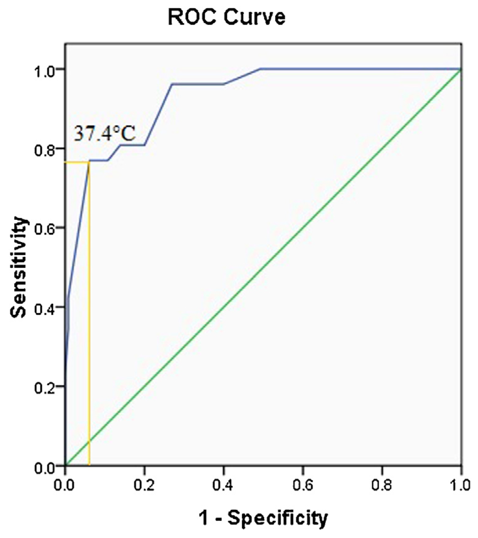
Fig. 1. Temperature ROC curve with area under the curve (AUC) of 0.929 ± 0.25 and a cut-off value of 37.4 °C (copied from SPSS 16.0). Values approaching 1 indicate good efficiency of the test.
Medicina 53 00085 g001
Fig. 2. Algorithm to facilitate decision-making in the management of IHs.
Medicina 53 00085 g002
Table 1. The comparison of temperatures and age between the groups by growing profile.
VariableGrowing profile
Stable IHs
(n = 100)
Slightly growing His
(n = 30)
Growing His
(n = 26)
Temperature (°C)
Age, months
36.7 (36.4; 36.9)*,
3 (2; 4)
37 (36.7; 37.3)*
3 (2; 4)½
37.4 (37.2; 37.6)
2 (1; 3)
Values are median (25th; 75th quartiles).
*P < 0.01 versus growing. P < 0.01 versus slightly growing. P < 0.01 versus growing. ½P = 0.02 versus growing (Mann–Whitney test to compare the groups).