Current Therapeutic Strategies in Diabetic Foot Ulcers


 ,
,  ,
,
Abstract
:1. Introduction
2. Etiology of Diabetic Foot Ulcers
2.1. Diabetic Peripheral Neuropathy
2.2. Peripheral Arterial Disease
2.3. Infection
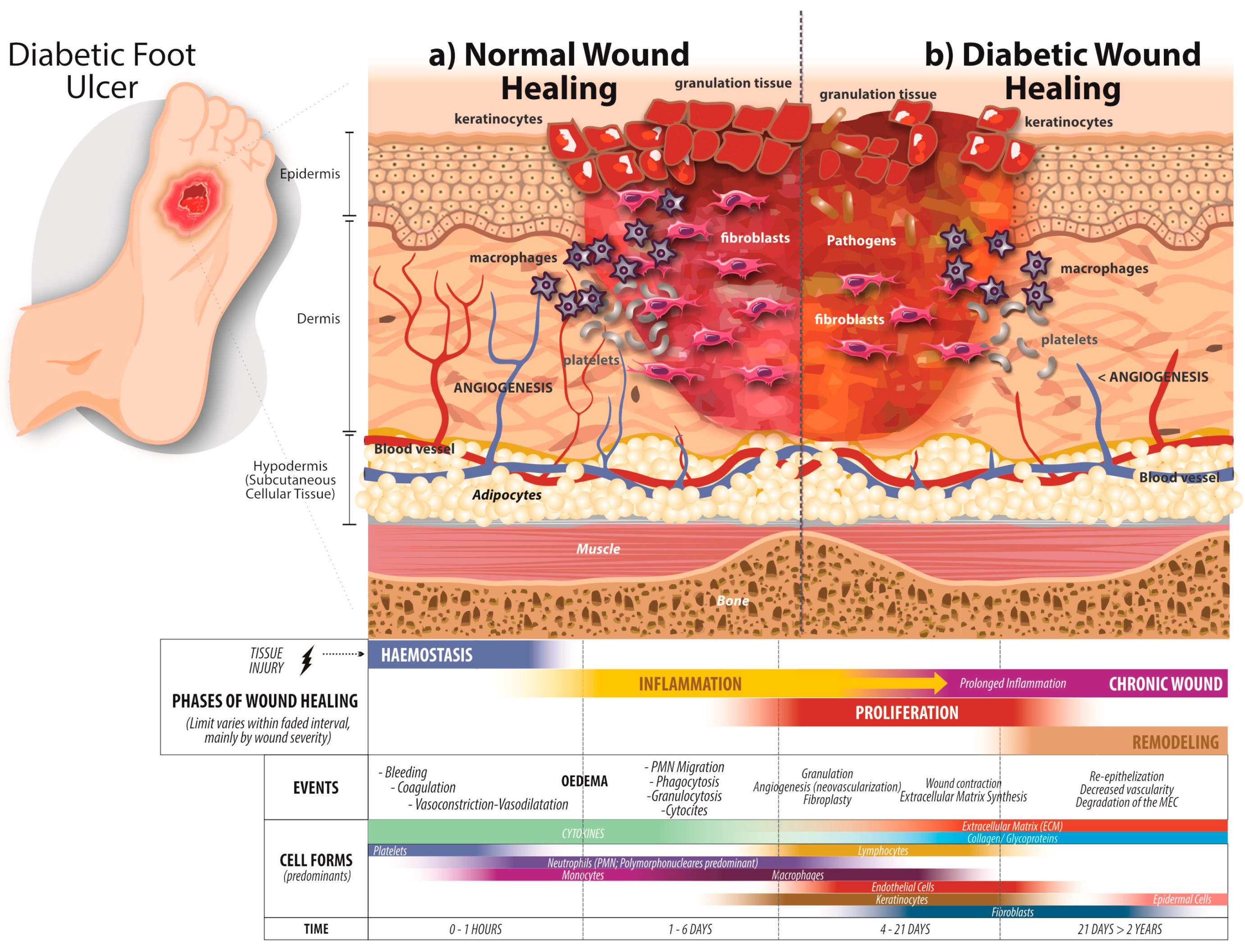
3. Wound Healing Process in Diabetes Mellitus
3.1. Hemostasis
3.2. Inflammation
3.3. Proliferation and Migration
3.4. Remodeling Phase
4. Treatments for Diabetic Foot Ulcers
4.1. Treatments for Diabetic Peripheral Neuropathy
4.2. Treatments for Peripheral Arterial Disease (Ischemia)
4.3. Wound Dressings
4.4. Debridement
4.5. Relief of Pressure
4.6. Infection Treatment
4.7. Antimicrobial Peptides
4.8. Larval Therapy
4.9. Laser Therapy
5. Tissue Engineering Approaches
5.1. Growth Factors
5.2. Cells
5.3. Scaffolds
5.4. Human Skin Substitutes
6. Conclusions
Author Contributions
Funding
Acknowledgments
Conflicts of Interest
References
- American Diabetes Association. Diagnosis and classification of diabetes mellitus. Diabetes Care 2009, 32 (Suppl. 1), S62–S67. [Google Scholar] [CrossRef] [PubMed]
- International Diabetes Federation. IDF Diabetes Atlas; International Diabetes Federation: Brussels, Belgium, 2017; p. 150. [Google Scholar]
- Goday, A. Epidemiology of diabetes and its non-coronary complications. Rev. Esp. Cardiol. 2002, 55, 657–670. [Google Scholar] [CrossRef]
- Lozano-Platonoff, A.; Mejía-Mendoza, M.D.F.; Ibáñez-Doria, M.; Contreras-Ruiz, J. The gold standard in diabetic foot treatment: Total contact cast. Gac. Med. Mex. 2014, 150, 58–64. [Google Scholar] [PubMed]
- Dubsky, M.; Jirkovska, A.; Bem, R.; Fejfarova, V.; Skibova, J.; Schaper, N.C.; Lipsky, B.A. Risk factors for recurrence of diabetic foot ulcers: prospective follow-up analysis in the Eurodiale subgroup. Int. Wound J. 2013, 10, 555–561. [Google Scholar] [CrossRef]
- Buowari, O.Y. Diabetes Mellitus in Developing Countries and Case Series, in Diabetes Mellitus—Insights and Perspectives; Oguntibeju, O.O., Ed.; IntechOpen: London, UK, 2013; p. 19. [Google Scholar]
- Ren, M.; Yang, C.; Lin, D.Z.; Xiao, H.S.; Mai, L.F.; Guo, Y.C.; Yan, L. Effect of intensive nursing education on the prevention of diabetic foot ulceration among patients with high-risk diabetic foot: a follow-up analysis. Diabetes Technol. Ther. 2014, 16, 576–581. [Google Scholar] [CrossRef]
- Boulton, A.J. The Pathway to Foot Ulceration in diabetes. Med. Clin. N. Am. 2013, 97, 775–790. [Google Scholar] [CrossRef]
- Alavi, A.; Sibbald, R.G.; Mayer, D.; Goodman, L.; Botros, M.; Armstrong, D.G.; Woo, K.; Boeni, T.; Ayello, E.A.; Kirsner, R.S. Diabetic foot ulcers: Part I. Pathophysiology and prevention. J. Am. Acad. Dermatol. 2014, 70, 1 e1–1 e18. [Google Scholar] [CrossRef]
- Boulton, A.J.; Gries, F.A.; Jervell, J.A. Guidelines for the diagnosis and outpatient management of diabetic peripheral neuropathy. Diabet. Med. 1998, 15, 508–514. [Google Scholar] [CrossRef]
- Watson, J.C.; Dyck, P.J. Peripheral Neuropathy: A Practical Approach to Diagnosis and Symptom Management. Mayo Clin. Proc. 2015, 90, 940–951. [Google Scholar] [CrossRef] [Green Version]
- Lavery, L.A.; Higgins, K.R.; Lanctot, D.R.; Constantinides, G.P.; Zamorano, R.G.; Athanasiou, K.A.; Armstrong, D.G.; Agrawal, C.M. Preventing diabetic foot ulcer recurrence in high-risk patients: use of temperature monitoring as a self-assessment tool. Diabetes Care 2007, 30, 14–20. [Google Scholar] [CrossRef]
- Forsythe, R.O.; Brownrigg, J.; Hinchliffe, R.J. Peripheral arterial disease and revascularization of the diabetic foot. Diabetes Obes. Metab. 2015, 17, 435–444. [Google Scholar] [CrossRef] [PubMed]
- Dinh, T.L.; Veves, A. A review of the mechanisms implicated in the pathogenesis of the diabetic foot. Int. J. Low Extrem. Wounds 2005, 4, 154–159. [Google Scholar] [CrossRef] [PubMed]
- Lanzer, P. Primary media sclerosis Mönckeberg: Diagnostic criteria. Cor Vasa 2017, 60, e205–e208. [Google Scholar] [CrossRef]
- Glagov, S.; Weisenberg, E.; Zarins, C.K.; Stankunavicius, R.; Kolettis, G.J. Compensatory enlargement of human atherosclerotic coronary arteries. N. Engl. J. Med. 1987, 316, 1371–1375. [Google Scholar] [CrossRef] [PubMed]
- Geerlings, S.E.; Hoepelman, A.I. Immune dysfunction in patients with diabetes mellitus (DM). FEMS Immunol. Med. Microbiol. 1999, 26, 259–265. [Google Scholar] [CrossRef] [PubMed]
- Moutschen, M.P.; Scheen, A.J.; Lefebvre, P.J. Impaired immune responses in diabetes mellitus: Analysis of the factors and mechanisms involved. Relevance to the increased susceptibility of diabetic patients to specific infections. Diabete Metab. 1992, 18, 187–201. [Google Scholar] [PubMed]
- Fejfarova, V.; Jirkovska, A.; Dubsky, M.; Game, F.; Vydlakova, J.; Sekerkova, A.; Franekova, J.; Kucerova, M.; Striz, I.; Petkov, V.; et al. An Alteration of Lymphocytes Subpopulations and Immunoglobulins Levels in Patients with Diabetic Foot Ulcers Infected Particularly by Resistant Pathogens. J. Diabetes Res. 2016, 2016, 2356870. [Google Scholar] [CrossRef]
- Lipsky, B.A.; Berendt, A.R.; Deery, H.G.; Embil, J.M.; Joseph, W.S.; Karchmer, A.W.; LeFrock, J.L.; Lew, D.P.; Mader, J.T.; Norden, C.; et al. Diagnosis and treatment of diabetic foot infections. Clin. Infect. Dis. 2004, 39, 885–910. [Google Scholar] [CrossRef]
- Mendes, J.J.; Leandro, C.; Corte-Real, S.; Barbosa, R.; Cavaco-Silva, P.; Melo-Cristino, J.; Gorski, A.; Garcia, M. Wound healing potential of topical bacteriophage therapy on diabetic cutaneous wounds. Wound Repair. Regen. 2013, 21, 595–603. [Google Scholar] [CrossRef]
- Mendes, J.J.; Leandro, C.I.; Bonaparte, D.P.; Pinto, A.L. A rat model of diabetic wound infection for the evaluation of topical antimicrobial therapies. Comp. Med. 2012, 62, 37–48. [Google Scholar]
- Grigoropoulou, P.; Eleftheriadou, I.; Jude, E.B.; Tentolouris, N. Diabetic Foot Infections: An Update in Diagnosis and Management. Curr. Diabetes Rep. 2017, 17, 3. [Google Scholar] [CrossRef] [PubMed]
- Nelson, E.A.; O’Meara, S.; Craig, D.; Iglesias, C.; Golder, S.; Dalton, J.; Claxton, K.; Bell-Syer, S.E.; Jude, E.; Dowson, C.; et al. A series of systematic reviews to inform a decision analysis for sampling and treating infected diabetic foot ulcers. Health Technol. Assess. 2006, 10, 1–221. [Google Scholar] [CrossRef]
- Moffarah, A.S.; Al Mohajer, M.; Hurwitz, B.L.; Armstrong, D.G. Diagnostic microbiology of the immunocompromised host: skin and soft tissue infection. Microbiol Spectrum. 2016, 4, 1–17. [Google Scholar]
- Rahim, K.; Saleha, S.; Zhu, X.; Huo, L.; Basit, A.; Franco, O.L. Bacterial Contribution in Chronicity of Wounds. Microb. Ecol. 2017, 73, 710–721. [Google Scholar] [CrossRef] [PubMed]
- Santoro, M.M.; Gaudino, G. Cellular and molecular facets of keratinocyte reepithelization during wound healing. Exp. Cell Res. 2005, 304, 274–286. [Google Scholar] [CrossRef]
- Erem, C.; Hacihasanoglu, A.; Celik, S.; Ovali, E.; Ersoz, H.O.; Ukinc, K.; Deger, O.; Telatar, M. Coagulation and fibrinolysis parameters in type 2 diabetic patients with and without diabetic vascular complications. Med. Princ. Pract. 2005, 14, 22–30. [Google Scholar] [CrossRef]
- Chhabra, S.; Chhabra, N.; Kaur, A.; Gupta, N. Wound Healing Concepts in Clinical Practice of OMFS. J. Maxillofac. Oral Surg. 2017, 16, 403–423. [Google Scholar] [CrossRef]
- Werner, S.; Grose, R. Regulation of wound healing by growth factors and cytokines. Physiol. Rev. 2003, 83, 835–870. [Google Scholar] [CrossRef]
- Xiao, J.; Li, J.; Cai, L.; Chakrabarti, S.; Li, X. Cytokines and diabetes research. J. Diabetes Res. 2014, 2014, 920613. [Google Scholar] [CrossRef]
- Pradhan, L.; Nabzdyk, C.; Andersen, N.D.; LoGerfo, F.W.; Veves, A. Inflammation and neuropeptides: The connection in diabetic wound healing. Expert Rev. Mol. Med. 2009, 11, e2. [Google Scholar] [CrossRef]
- Falanga, V. Wound healing and its impairment in the diabetic foot. Lancet 2005, 366, 1736–1743. [Google Scholar] [CrossRef]
- Lan, C.C.; Liu, I.H.; Fang, A.H.; Wen, C.H.; Wu, C.S. Hyperglycaemic conditions decrease cultured keratinocyte mobility: Implications for impaired wound healing in patients with diabetes. Br. J. Dermatol. 2008, 159, 1103–1115. [Google Scholar] [CrossRef] [PubMed]
- Galiano, R.D.; Tepper, O.M.; Pelo, C.R.; Bhatt, K.A.; Callaghan, M.; Bastidas, N.; Bunting, S.; Steinmetz, H.G.; Gurtner, G.C. Topical vascular endothelial growth factor accelerates diabetic wound healing through increased angiogenesis and by mobilizing and recruiting bone marrow-derived cells. Am. J. Pathol. 2004, 164, 1935–1947. [Google Scholar] [CrossRef]
- Lobmann, R.; Schultz, G.; Lehnert, H. Proteases and the diabetic foot syndrome: Mechanisms and therapeutic implications. Diabetes Care 2005, 28, 461–471. [Google Scholar] [CrossRef] [PubMed]
- Maione, A.G.; Smith, A.; Kashpur, O.; Yanez, V.; Knight, E.; Mooney, D.J.; Veves, A.; Tomic-Canic, M.; Garlick, J.A. Altered ECM deposition by diabetic foot ulcer-derived fibroblasts implicates fibronectin in chronic wound repair. Wound Repair Regen. 2016, 24, 630–643. [Google Scholar] [CrossRef]
- Wang, C.; Mai, L.; Yang, C.; Liu, D.; Sun, K.; Song, W.; Luo, B.; Li, Y.; Xu, M.; Zhang, S.; et al. Reducing major lower extremity amputations after the introduction of a multidisciplinary team in patient with diabetes foot ulcer. BMC Endocr. Disord. 2016, 16, 38. [Google Scholar] [CrossRef]
- Blakely, M. The Use of Best Practice in the Treatment of a Complex Diabetic Foot Ulcer: A Case Report. Healthcare (Basel) 2016, 4, 18. [Google Scholar] [CrossRef]
- Wu, S.C.; Wu, S.C.; Driver, V.R.; Wrobel, J.S.; Armstrong, D.G. Foot ulcers in the diabetic patient, prevention and treatment. Vasc. Health Risk Manag. 2007, 3, 65–76. [Google Scholar]
- Brem, H.; Sheehan, P.; Boulton, A.J. Protocol for treatment of diabetic foot ulcers. Am. J. Surg. 2004, 187, 1S–10S. [Google Scholar] [CrossRef]
- Markakis, K.; Bowling, F.L.; Boulton, A.J. The diabetic foot in 2015: An overview. Diabetes Metab. Res. Rev. 2016, 32, 169–178. [Google Scholar] [CrossRef]
- Tang, H.Y.; Jiang, A.J.; Ma, J.L.; Wang, F.J.; Shen, G.M. Understanding the Signaling Pathways Related to the Mechanism and Treatment of Diabetic Peripheral Neuropathy. Endocrinology. 2019, 160, 2119–2127. [Google Scholar] [CrossRef] [PubMed]
- Kennedy, W.R.; Navarro, X.; Goetz, F.C.; Sutherland, D.E.; Najarian, J.S. Effects of pancreatic transplantation on diabetic neuropathy. N. Engl. J. Med. 1990, 322, 1031–1037. [Google Scholar] [CrossRef] [PubMed]
- Games, G.; Hutchison, A. Tapentadol-ER for the treatment of diabetic peripheral neuropathy. Consult. Pharm. 2013, 28, 672–675. [Google Scholar] [CrossRef] [PubMed]
- Attal, N.; Cruccu, G.; Baron, R.; Haanpaa, M.; Hansson, P.; Jensen, T.S.; Nurmikko, T. EFNS guidelines on the pharmacological treatment of neuropathic pain: 2010 revision. Eur. J. Neurol. 2010, 17, 1113–e88. [Google Scholar] [CrossRef] [PubMed]
- Javed, S.; Alam, U.; Malik, R.A. Burning through the pain: Treatments for diabetic neuropathy. Diabetes Obes. Metab. 2015, 17, 1115–1125. [Google Scholar] [CrossRef]
- Bartkoski, S.; Day, M. Alpha-Lipoic Acid for Treatment of Diabetic Peripheral Neuropathy. Am. Fam. Physician 2016, 93, 786. [Google Scholar]
- Oses, C.; Olivares, B.; Ezquer, M.; Acosta, C.; Bosch, P.; Donoso, M.; Leniz, P.; Ezquer, F. Preconditioning of adipose tissue-derived mesenchymal stem cells with deferoxamine increases the production of pro-angiogenic, neuroprotective and anti-inflammatory factors: Potential application in the treatment of diabetic neuropathy. PLoS ONE 2017, 12, e0178011. [Google Scholar] [CrossRef]
- Cox, A.A.; Sagot, Y.; Hedou, G.; Grek, C.; Wilkes, T.; Vinik, A.I.; Ghatnekar, G. Low-Dose Pulsatile Interleukin-6 As a Treatment Option for Diabetic Peripheral Neuropathy. Front. Endocrinol. (Lausanne) 2017, 8, 89. [Google Scholar] [CrossRef]
- Dworkin, R.H.; O’Connor, A.B.; Backonja, M.; Farrar, J.T.; Finnerup, N.B.; Jensen, T.S.; Kalso, E.A.; Loeser, J.D.; Miaskowski, C.; Nurmikko, T.J.; et al. Pharmacologic management of neuropathic pain: Evidence-based recommendations. Pain 2007, 132, 237–251. [Google Scholar] [CrossRef]
- Raskin, P.; Huffman, C.; Yurkewicz, L.; Pauer, L.; Scavone, J.M.; Yang, R.; Parsons, B. Pregabalin in Patients With Painful Diabetic Peripheral Neuropathy Using an NSAID for Other Pain Conditions: A Double-Blind Crossover Study. Clin. J. Pain 2016, 32, 203–210. [Google Scholar] [CrossRef]
- Fernández-Samos Gutiérrez, R. The angiosome model in the revascularization strategy of critical limb ischemia. Angiología 2012, 64, 173–182. [Google Scholar] [CrossRef]
- Taylor, S.M.; Johnson, B.L.; Samies, N.L.; Rawlinson, R.D.; Williamson, L.E.; Davis, S.A.; Kotrady, J.A.; York, J.W.; Langan, E.M., 3rd.; Cull, D.L. Contemporary management of diabetic neuropathic foot ulceration: A study of 917 consecutively treated limbs. J. Am. Coll. Surg. 2011, 212, 532–545. [Google Scholar] [CrossRef] [PubMed]
- Iida, O.; Soga, Y.; Hirano, K.; Kawasaki, D.; Suzuki, K.; Miyashita, Y.; Terashi, H.; Uematsu, M. Long-term results of direct and indirect endovascular revascularization based on the angiosome concept in patients with critical limb ischemia presenting with isolated below-the-knee lesions. J. Vasc. Surg. 2012, 55, 363–370. [Google Scholar] [CrossRef]
- Lee, V.; Singh, G.; Trasatti, J.P.; Bjornsson, C.; Xu, X.; Tran, T.N.; Yoo, S.S.; Dai, G.; Karande, P. Design and fabrication of human skin by three-dimensional bioprinting. Tissue Eng. Part C Methods 2014, 20, 473–484. [Google Scholar] [CrossRef] [PubMed]
- Bachoo, P.; et al. Endovascular stents for intermittent claudication. Cochrane Database Syst. Rev. 2010, CD003228. [Google Scholar] [CrossRef]
- Bavishi, C.; Baber, U.; Panwar, S.; Pirrotta, S.; Dangas, G.D.; Moreno, P.; Tamis-Holland, J.; Kini, A.S.; Sharma, S.K. Efficacy and safety of everolimus and zotarolimus-eluting stents versus first-generation drug-eluting stents in patients with diabetes: A meta-analysis of randomized trials. Int. J. Cardiol. 2017, 230, 310–318. [Google Scholar] [CrossRef]
- Kayssi, A.; Al-Atassi, T.; Oreopoulos, G.; Roche-Nagle, G.; Tan, K.T.; Rajan, D.K. Drug-eluting balloon angioplasty versus uncoated balloon angioplasty for peripheral arterial disease of the lower limbs. Cochrane Database Syst. Rev. 2016, CD011319. [Google Scholar] [CrossRef]
- Ambler, G.K.; Twine, C.P. Graft type for femoro-popliteal bypass surgery. Cochrane Database Syst Rev. 2018, 2, 1–113. [Google Scholar] [CrossRef]
- Albers, M.; Romiti, M.; Brochado-Neto, F.C.; Pereira, C.A.B. Meta-analysis of alternate autologous vein bypass grafts to infrapopliteal arteries. J. Vasc. Surg. 2005, 42, 449–455. [Google Scholar] [CrossRef]
- Palomo, I.; Moore-Carrasco, R.E.; Alarcón, M.A.; Maragaño, P.J. Platelet antiaggregants: Mechanisms of action and use asocied risks. Vitae Revista Facultad de Química Farmacéutica 2008, 16, 133–143. [Google Scholar]
- Nicolai, S.P.; Kruidenier, L.M.; Bendermacher, B.L.; Prins, M.H.; Stokmans, R.A.; Broos, P.P.; Teijink, J.A. Ginkgo biloba for intermittent claudication. Cochrane Database Syst. Rev. 2013, CD006888. [Google Scholar] [CrossRef] [PubMed]
- Kleijnen, J.; Mackerras, D. Vitamin E for intermittent claudication. Cochrane Database Syst. Rev. 2000, 2, 1–13. [Google Scholar] [CrossRef] [PubMed]
- Brass, E.P.; Koster, D.; Hiatt, W.R.; Amato, A. A systematic review and meta-analysis of propionyl-L-carnitine effects on exercise performance in patients with claudication. Vasc. Med. 2013, 18, 3–12. [Google Scholar] [CrossRef] [PubMed]
- Delaney, C.L.; Spark, J.I.; Thomas, J.; Wong, Y.T.; Chan, L.T.; Miller, M.D. A systematic review to evaluate the effectiveness of carnitine supplementation in improving walking performance among individuals with intermittent claudication. Atherosclerosis 2013, 229, 1–9. [Google Scholar] [CrossRef]
- Paravastu, S.C.; Mendonca, D.A.; Da Silva, A. Beta blockers for peripheral arterial disease. Cochrane Database Syst. Rev. 2013, CD005508. [Google Scholar] [CrossRef]
- Bedenis, R.; Stewart, M.; Cleanthis, M.; Robless, P.; Mikhailidis, D.P.; Stansby, G. Cilostazol for intermittent claudication. Cochrane Database Syst. Rev. 2014, CD003748. [Google Scholar] [CrossRef]
- Lauvrak, V.; Fronsdal, K.B.; Ormstad, S.S.; Vaagbo, G.; Fure, B. Effectiveness of Hyperbaric Oxygen Therapy in Patients with Late Radiation Tissue Injury or Diabetic Foot Ulcer; The Norwegian Institute of Public Health: Oslo, Norway, 2015. [Google Scholar]
- Liu, J.; Zhang, P.; Tian, J.; Li, L.; Li, J.; Tian, J.H.; Yang, K. Ozone therapy for treating foot ulcers in people with diabetes. Cochrane Database Syst. Rev. 2015, CD008474. [Google Scholar] [CrossRef]
- Ubbink, D.T.; Vermeulen, H. Spinal cord stimulation for non-reconstructable chronic critical leg ischaemia. Cochrane Database Syst. Rev. 2013, CD004001. [Google Scholar] [CrossRef]
- Simpson, E.L.; Duenas, A.; Holmes, M.W.; Papaioannou, D.; Chilcott, J. Spinal cord stimulation for chronic pain of neuropathic or ischaemic origin: systematic review and economic evaluation. Health Technol. Assess. 2009, 13, 1–154. [Google Scholar] [CrossRef]
- Germán AlbertoTéllez, J.C. Péptidos antimicrobianos-Antimicrobial peptides. Infectio 2010, 14, 55–67. [Google Scholar]
- Gomes, A.; Teixeira, C.; Ferraz, R.; Prudencio, C.; Gomes, P. Wound-Healing Peptides for Treatment of Chronic Diabetic Foot Ulcers and Other Infected Skin Injuries. Molecules 2017, 22, 1743. [Google Scholar] [CrossRef] [PubMed]
- Hinchliffe, R.J.; Brownrigg, J.R.; Apelqvist, J.; Boyko, E.J.; Fitridge, R.; Mills, J.L.; Reekers, J.; Shearman, C.P.; Zierler, R.E.; Schaper, N.C.; et al. IWGDF guidance on the diagnosis, prognosis and management of peripheral artery disease in patients with foot ulcers in diabetes. Diabetes Metab. Res. Rev. 2016, 32, 37–44. [Google Scholar] [CrossRef] [PubMed] [Green Version]
- Hinchliffe, R.J.; Brownrigg, J.R.; Andros, G.; Apelqvist, J.; Boyko, E.J.; Fitridge, R.; Mills, J.L.; Reekers, J.; Shearman, C.P.; Zierler, R.E.; et al. Effectiveness of revascularization of the ulcerated foot in patients with diabetes and peripheral artery disease: a systematic review. Diabetes Metab. Res. Rev. 2016, 32, 136–144. [Google Scholar] [CrossRef] [PubMed]
- Uccioli, L.; Meloni, M.; Izzo, V.; Giurato, L.; Merolla, S.; Gandini, R. Critical limb ischemia: Current challenges and future prospects. Vasc. Health Risk Manag. 2018, 14, 63–74. [Google Scholar] [CrossRef]
- Pomposelli, F.B., Jr.; et al. Dorsalis pedis arterial bypass: Durable limb salvage for foot ischemia in patients with diabetes mellitus. J. Vasc. Surg. 1995, 21, 375–384. [Google Scholar] [CrossRef] [Green Version]
- Gatti, C.; Cecchini, S.; Fabbietti, P.; Romagnoli, F.; Ricci, S. Endovascular treatment of diabetic peripheral arterial disease in older and oldest old patients: A retrospective study. Aging Clin. Exp. Res. 2017, 30, 205–207. [Google Scholar] [CrossRef]
- Hingorani, A.; LaMuraglia, G.M.; Henke, P.; Meissner, M.H.; Loretz, L.; Zinszer, K.M.; Driver, V.R.; Frykberg, R.; Carman, T.L.; Marston, W.; et al. The management of diabetic foot: A clinical practice guideline by the Society for Vascular Surgery in collaboration with the American Podiatric Medical Association and the Society for Vascular Medicine. J. Vasc. Surg. 2016, 63, 3S–21S. [Google Scholar] [CrossRef] [Green Version]
- Steed, D.L.; Attinger, C.; Colaizzi, T.; Crossland, M.; Franz, M.; Harkless, L.; Johnson, A.; Moosa, H.; Robson, M.; Serena, T.; et al. Guidelines for the treatment of diabetic ulcers. Wound Repair Regen. 2006, 14, 680–692. [Google Scholar] [CrossRef] [Green Version]
- Dumville, J.C.; O’Meara, S.; Deshpande, S.; Speak, K. Hydrogel dressings for healing diabetic foot ulcers. Cochrane Database Syst Rev. 2013, 7, 1–54. [Google Scholar] [CrossRef]
- Zhao, L.; Niu, L.; Liang, H.; Tan, H.; Liu, C.; Zhu, F. pH and Glucose Dual-Responsive Injectable Hydrogels with Insulin and Fibroblasts as Bioactive Dressings for Diabetic Wound Healing. ACS Appl. Mater. Interfaces 2017, 9, 37563–37574. [Google Scholar] [CrossRef]
- Dumville, J.C.; Deshpande, S.; O’Meara, S.; Speak, K. Hydrocolloid dressings for healing diabetic foot ulcers. Cochrane Database Syst Rev. 2013, 8, 1–43. [Google Scholar] [CrossRef] [PubMed]
- Registered Nurses’ Association of Ontario. Assessment and Management of Foot Ulcers for People with Diabetes, 2nd ed.; Registered Nurses’ Association of Ontario: Toronto, ON, Canada, 2013; Volume 1, 160 p. [Google Scholar]
- Abouaesha, F.; van Schie, C.H.; Griffths, G.D.; Young, R.J.; Boulton, A.J. Plantar tissue thickness is related to peak plantar pressure in the high-risk diabetic foot. Diabetes Care 2001, 24, 1270–1274. [Google Scholar] [CrossRef] [PubMed]
- Hirche, C.; Citterio, A.; Hoeksema, H.; Koller, J.; Lehner, M.; Martinez, J.R.; Monstrey, S.; Murray, A.; Plock, J.A.; Sander, F.; et al. Eschar removal by bromelain based enzymatic debridement (Nexobrid(R)) in burns: An European consensus. Burns 2017, 43, 1640–1653. [Google Scholar] [CrossRef] [PubMed]
- Frykberg, R.G.; Zgonis, T.; Armstrong, D.G.; Driver, V.R.; Giurini, J.M.; Kravitz, S.R.; Landsman, A.S.; Lavery, L.A.; Moore, J.C.; Schuberth, J.M.; et al. Diabetic foot disorders. A clinical practice guideline (2006 revision). J. Foot Ankle Surg. 2006, 45, S1–S66. [Google Scholar] [CrossRef]
- Fleischli, J.G.; Lavery, L.A.; Vela, S.A.; Ashry, H.; Lavery, D.C. 1997 William J. Stickel Bronze Award. Comparison of strategies for reducing pressure at the site of neuropathic ulcers. J. Am. Podiatr. Med. Assoc. 1997, 87, 466–472. [Google Scholar] [CrossRef] [PubMed]
- Piaggesi, A.; Goretti, C.; Iacopi, E.; Clerici, G.; Romagnoli, F.; Toscanella, F.; Vermigli, C. Comparison of Removable and Irremovable Walking Boot to Total Contact Casting in Offloading the Neuropathic Diabetic Foot Ulceration. Foot Ankle Int. 2016, 37, 855–861. [Google Scholar] [CrossRef]
- Health Quality Ontario. Fibreglass Total Contact Casting, Removable Cast Walkers, and Irremovable Cast Walkers to Treat Diabetic Neuropathic Foot Ulcers: A Health Technology Assessment. Ont. Health Technol. Assess. Ser. 2017, 17, 1–124. [Google Scholar]
- Bergman, S.; Shah, P.J. Diabetic Foot Infections. In Infection Primary Care; Fish, D.N., Taylor, S., Thoennes, M.J., Eds.; Army Center for Substance Abuse Programs: Fort Knox, KY, USA, 2016; p. 26. [Google Scholar]
- Singh, S.K.; Gupta, B. Choices and Challenges of Antibiotics Therapy in Diabetic Foot Infection. Indian J. Endocrinol. Metab. 2017, 21, 647–648. [Google Scholar]
- Filius, P.M.; Gyssens, I.C. Impact of increasing antimicrobial resistance on wound management. Am. J. Clin. Dermatol. 2002, 3, 1–7. [Google Scholar] [CrossRef]
- Meletis, G. Carbapenem resistance: Overview of the problem and future perspectives. Ther. Adv. Infect. Dis. 2016, 3, 15–21. [Google Scholar] [CrossRef]
- Limoli, D.H.; Rockel, A.B.; Host, K.M.; Jha, A.; Kopp, B.T.; Hollis, T.; Wozniak, D.J. Cationic antimicrobial peptides promote microbial mutagenesis and pathoadaptation in chronic infections. PLoS Pathog. 2014, 10, e1004083. [Google Scholar] [CrossRef] [PubMed]
- Carter, V.; Underhill, A.; Baber, I.; Sylla, L.; Baby, M.; Larget-Thiery, I.; Zettor, A.; Bourgouin, C.; Langel, U.; Faye, I.; et al. Killer bee molecules: antimicrobial peptides as effector molecules to target sporogonic stages of Plasmodium. PLoS Pathog. 2013, 9, e1003790. [Google Scholar] [CrossRef] [PubMed]
- Lee, J.K.; Park, Y.J.; Kum, K.Y.; Han, S.H.; Chang, S.W.; Kaufman, B.; Jiang, J.; Zhu, Q.; Safavi, K.; Spangberg, L. Antimicrobial efficacy of a human beta-defensin-3 peptide using an Enterococcus faecalis dentine infection model. Int. Endod. J. 2013, 46, 406–412. [Google Scholar] [CrossRef] [PubMed]
- Santos, R.; Gomes, D.; Macedo, H.; Barros, D.; Tiberio, C.; Veiga, A.S.; Tavares, L.; Castanho, M.; Oliveira, M. Guar gum as a new antimicrobial peptide delivery system against diabetic foot ulcers Staphylococcus aureus isolates. J. Med. Microbiol. 2016, 65, 1092–1099. [Google Scholar] [CrossRef]
- Gawande, P.V.; Leung, K.P.; Madhyastha, S. Antibiofilm and antimicrobial efficacy of DispersinB(R)-KSL-W peptide-based wound gel against chronic wound infection associated bacteria. Curr. Microbiol. 2014, 68, 635–641. [Google Scholar] [CrossRef]
- Saeed, S.; Zafar, J.; Khan, B.; Akhtar, A.; Qurieshi, S.; Fatima, S.; Ahmad, N.; Irfanullah, J. Utility of (9)(9)mTc-labelled antimicrobial peptide ubiquicidin (29-41) in the diagnosis of diabetic foot infection. Eur. J. Nucl. Med. Mol. Imaging 2013, 40, 737–743. [Google Scholar] [CrossRef]
- Galkowska, H.; Olszewski, W.L.; Wojewodzka, U. Expression of natural antimicrobial peptide beta-defensin-2 and Langerhans cell accumulation in epidermis from human non-healing leg ulcers. Folia Histochem. Cytobiol. 2005, 43, 133–136. [Google Scholar]
- Lipsky, B.A.; Holroyd, K.J.; Zasloff, M. Topical versus systemic antimicrobial therapy for treating mildly infected diabetic foot ulcers: a randomized, controlled, double-blinded, multicenter trial of pexiganan cream. Clin. Infect. Dis. 2008, 47, 1537–1545. [Google Scholar] [CrossRef]
- Sherman, R.A. Maggot therapy for treating diabetic foot ulcers unresponsive to conventional therapy. Diabetes Care 2003, 26, 446–451. [Google Scholar] [CrossRef]
- Hassan, M.I.; Hammad, K.M.; Fouda, M.A.; Kamel, M.R. The using of Lucilia cuprina maggots in the treatment of diabetic foot wounds. J. Egypt Soc. Parasitol. 2014, 44, 125–129. [Google Scholar] [CrossRef]
- Gilead, L.; Mumcuoglu, K.Y.; Ingber, A. The use of maggot debridement therapy in the treatment of chronic wounds in hospitalised and ambulatory patients. J. Wound Care 2012, 21, 78–85. [Google Scholar] [CrossRef] [PubMed]
- De Alencar Fonseca Santos, J.; Campelo, M.B.D.; de Oliveira, R.A.; Nicolau, R.A.; Rezende, V.E.A.; Arisawa, E.A.L. Effects of Low-Power Light Therapy on the Tissue Repair Process of Chronic Wounds in Diabetic Feet. Photomed. Laser Surg. 2018, 36, 298–304. [Google Scholar] [CrossRef] [PubMed] [Green Version]
- Kaviani, A.; Djavid, G.E.; Ataie-Fashtami, L.; Fateh, M.; Ghodsi, M.; Salami, M.; Zand, N.; Kashef, N.; Larijani, B. A randomized clinical trial on the effect of low-level laser therapy on chronic diabetic foot wound healing: a preliminary report. Photomed. Laser Surg. 2011, 29, 109–114. [Google Scholar] [CrossRef] [PubMed]
- Katari, R.; Peloso, A.; Orlando, G. Tissue engineering and regenerative medicine: semantic considerations for an evolving paradigm. Front. Bioeng. Biotechnol. 2014, 2, 57. [Google Scholar] [CrossRef] [PubMed]
- Veves, A.; Falanga, V.; Armstrong, D.G.; Sabolinski, M.L.; Apligraf Diabetic Foot Ulcer, S. Graftskin, a human skin equivalent, is effective in the management of noninfected neuropathic diabetic foot ulcers: a prospective randomized multicenter clinical trial. Diabetes Care 2001, 24, 290–295. [Google Scholar] [CrossRef]
- Joao De Masi, E.C.; Campos, A.C.; Joao De Masi, F.D.; Ratti, M.A.; Ike, I.S.; Joao De Masi, R.D. The influence of growth factors on skin wound healing in rats. Braz. J. Otorhinolaryngol. 2016, 82, 512–521. [Google Scholar] [CrossRef] [Green Version]
- Hong, J.P.; Jung, H.D.; Kim, Y.W. Recombinant human epidermal growth factor (EGF) to enhance healing for diabetic foot ulcers. Ann. Plast Surg. 2006, 56, 394–398. [Google Scholar] [CrossRef]
- Laato, M.; Kähäri, V.M.; Niinikoski, J.; Vuorio, E. Epidermal growth factor increases collagen production in granulation tissue by stimulation of fibroblast proliferation and not by activation of procollagen genes. Biochem. J. 1987, 247, 385–388. [Google Scholar] [CrossRef] [Green Version]
- Metcalfe, A.D.; Ferguson, M.W. Harnessing wound healing and regeneration for tissue engineering. Biochem. Soc. Trans. 2005, 33, 413–417. [Google Scholar] [CrossRef] [Green Version]
- Choi, S.M.; Lee, K.M.; Kim, H.J.; Park, I.K.; Kang, H.J.; Shin, H.C.; Baek, D.; Choi, Y.; Park, K.H.; Lee, J.W. Effects of structurally stabilized EGF and bFGF on wound healing in type I and type II diabetic mice. Acta Biomater. 2017, 66, 325–334. [Google Scholar] [CrossRef]
- Sridharan, K.; Sivaramakrishnan, G. Growth factors for diabetic foot ulcers: Mixed treatment comparison analysis of randomized clinical trials. Br. J. Clin. Pharmacol. 2017, 84, 434–444. [Google Scholar] [CrossRef] [PubMed]
- Ertugrul, B.M.; Lipsky, B.A.; Guvenc, U. An Assessment of Intralesional Epidermal Growth Factor for Treating Diabetic Foot WoundsThe First Experiences in Turkey. J. Am. Podiatr. Med. Assoc. 2017, 107, 17–29. [Google Scholar] [CrossRef] [PubMed]
- Marfia, G.; Navone, S.E.; Di Vito, C.; Ughi, N.; Tabano, S.; Miozzo, M.; Tremolada, C.; Bolla, G.; Crotti, C.; Ingegnoli, F.; et al. Mesenchymal stem cells: Potential for therapy and treatment of chronic non-healing skin wounds. Organogenesis 2015, 11, 183–206. [Google Scholar] [CrossRef] [PubMed]
- Cao, Y.; Gang, X.; Sun, C.; Wang, G. Mesenchymal Stem Cells Improve Healing of Diabetic Foot Ulcer. J. Diabetes Res. 2017, 2017, 9328347. [Google Scholar] [CrossRef]
- Seo, E.; Lim, J.S.; Jun, J.B.; Choi, W.; Hong, I.S.; Jun, H.S. Exendin-4 in combination with adipose-derived stem cells promotes angiogenesis and improves diabetic wound healing. J. Transl. Med. 2017, 15, 35. [Google Scholar] [CrossRef]
- Kim, J.W.; Lee, J.H.; Lyoo, Y.S.; Jung, D.I.; Park, H.M. The effects of topical mesenchymal stem cell transplantation in canine experimental cutaneous wounds. Vet. Dermatol. 2013, 24, 242–e53. [Google Scholar] [CrossRef]
- Han, S.K.; Kim, H.S.; Kim, W.K. Efficacy and safety of fresh fibroblast allografts in the treatment of diabetic foot ulcers. Dermatol. Surg. 2009, 35, 1342–1348. [Google Scholar] [CrossRef]
- Werner, S.; Krieg, T.; Smola, H. Keratinocyte-fibroblast interactions in wound healing. J. Invest. Dermatol. 2007, 127, 998–1008. [Google Scholar] [CrossRef]
- Wu, Z.; Tang, Y.; Fang, H.; Su, Z.; Xu, B.; Lin, Y.; Zhang, P.; Wei, X. Decellularized scaffolds containing hyaluronic acid and EGF for promoting the recovery of skin wounds. J. Mater. Sci. Mater. Med. 2015, 26, 5322. [Google Scholar] [CrossRef]
- Gugerell, A.; Pasteiner, W.; Nurnberger, S.; Kober, J.; Meinl, A.; Pfeifer, S.; Hartinger, J.; Wolbank, S.; Goppelt, A.; Redl, H.; et al. Thrombin as important factor for cutaneous wound healing: comparison of fibrin biomatrices in vitro and in a rat excisional wound healing model. Wound Repair Regen. 2014, 22, 740–748. [Google Scholar] [CrossRef]
- Momeni, M.; Zarehaghighi, M.; Hajimiri, M.; Khorasani, G.; Dinarvand, R.; Nekookar, A.; Sodeifi, N.; Khosravani, P.; Shayanasl, N.; Ebrahimi, M. In vitro and in vivo investigation of a novel amniotic-based chitosan dressing for wound healing. Wound Repair Regen. 2018, 26, 87–101. [Google Scholar] [CrossRef] [PubMed]
- Straccia, M.C.; d’Ayala, G.G.; Romano, I.; Oliva, A.; Laurienzo, P. Alginate hydrogels coated with chitosan for wound dressing. Mar. Drugs 2015, 13, 2890–2908. [Google Scholar] [CrossRef] [PubMed]
- Goins, A.; Ramaswamy, V.; Dirr, E.; Dulany, K.; Irby, S.; Webb, A.; Allen, J. Development of poly (1,8 octanediol-co-citrate) and poly (acrylic acid) nanofibrous scaffolds for wound healing applications. Biomed. Mater. 2017, 13, 015002. [Google Scholar] [CrossRef] [PubMed]
- Miyaguchi, S.I.; Horii, A.; Kambara, R.; Takemoto, N.; Akazawa, H.; Takahashi, N.; Baba, H.; Inohara, H. Effects of Covering Surgical Wounds with Polyglycolic Acid Sheets for Posttonsillectomy Pain. Otolaryngol. Head Neck Surg. 2016, 155, 876–878. [Google Scholar] [CrossRef]
- Shin, Y.C.; Shin, D.M.; Lee, E.J.; Lee, J.H.; Kim, J.E.; Song, S.H.; Hwang, D.Y.; Lee, J.J.; Kim, B.; Lim, D.; et al. Hyaluronic Acid/PLGA Core/Shell Fiber Matrices Loaded with EGCG Beneficial to Diabetic Wound Healing. Adv. Healthc. Mater. 2016, 5, 3035–3045. [Google Scholar] [CrossRef]
- Chong, E.J.; Phan, T.T.; Lim, I.J.; Zhang, Y.Z.; Bay, B.H.; Ramakrishna, S.; Lim, C.T. Evaluation of electrospun PCL/gelatin nanofibrous scaffold for wound healing and layered dermal reconstitution. Acta Biomater. 2007, 3, 321–330. [Google Scholar] [CrossRef]
- Koehler, J.; Verheyen, L.; Hedtrich, S.; Brandl, F.P.; Goepferich, A.M. Alkaline poly(ethylene glycol)-based hydrogels for a potential use as bioactive wound dressings. J. Biomed. Mater. Res. A 2017, 105, 3360–3368. [Google Scholar] [CrossRef]
- Kim, J.W.; Kim, M.J.; Ki, C.S.; Kim, H.J.; Park, Y.H. Fabrication of bi-layer scaffold of keratin nanofiber and gelatin-methacrylate hydrogel: Implications for skin graft. Int. J. Biol. Macromol. 2017, 105, 541–548. [Google Scholar] [CrossRef]
- Kaisang, L.; Siyu, W.; Lijun, F.; Daoyan, P.; Xian, C.J.; Jie, S. Adipose-derived stem cells seeded in Pluronic F-127 hydrogel promotes diabetic wound healing. J. Surg. Res. 2017, 217, 63–74. [Google Scholar] [CrossRef]
- Frykberg, R.G.; Cazzell, S.M.; Arroyo-Rivera, J.; Tallis, A.; Reyzelman, A.M.; Saba, F.; Warren, L.; Stouch, B.C.; Gilbert, T.W. Evaluation of tissue engineering products for the management of neuropathic diabetic foot ulcers: an interim analysis. J. Wound Care 2016, 25, S18–S25. [Google Scholar] [CrossRef]
- Falanga, V.; Isaacs, C.; Paquette, D.; Downing, G.; Kouttab, N.; Butmarc, J.; Badiavas, E.; Hardin-Young, J. Wounding of bioengineered skin: Cellular and molecular aspects after injury. J. Invest. Dermatol. 2002, 119, 653–660. [Google Scholar] [CrossRef] [PubMed]
- Gilligan, A.M.; Waycaster, C.R.; Milne, C.T. Cost Effectiveness of Becaplermin Gel on Wound Closure for the Treatment of Pressure Injuries. Wounds 2018, 30, 197–204. [Google Scholar] [PubMed]
- Wainwright, D.J. Use of an acellular allograft dermal matrix (AlloDerm) in the management of full-thickness burns. Burns 1995, 21, 243–248. [Google Scholar] [CrossRef]
- Hart, C.E.; Loewen-Rodriguez, A.; Lessem, J. Dermagraft: Use in the Treatment of Chronic Wounds. Adv. Wound Care (New Rochelle) 2012, 1, 138–141. [Google Scholar] [CrossRef] [Green Version]
- Towler, M.A.; Rush, E.W.; Richardson, M.K.; Williams, C.L. Randomized, Prospective, Blinded-Enrollment, Head-To-Head Venous Leg Ulcer Healing Trial Comparing Living, Bioengineered Skin Graft Substitute (Apligraf) with Living, Cryopreserved, Human Skin Allograft (TheraSkin). Clin. Podiatr. Med. Surg. 2018, 35, 357–365. [Google Scholar] [CrossRef]
- Still, J.; Glat, P.; Silverstein, P.; Griswold, J.; Mozingo, D. The use of a collagen sponge/living cell composite material to treat donor sites in burn patients. Burns 2003, 29, 837–841. [Google Scholar] [CrossRef]
- Carsin, H.; Ainaud, P.; Le Bever, H.; Rives, J.; Lakhel, A.; Stephanazzi, J.; Lambert, F.; Perrot, J. Cultured epithelial autografts in extensive burn coverage of severely traumatized patients: a five year single-center experience with 30 patients. Burns 2000, 26, 379–387. [Google Scholar] [CrossRef]
- Woodroof, A.; Phipps, R.; Woeller, C.; Rodeheaver, G.; Naughton, G.K.; Piney, E.; Hickerson, W.; Branski, L.; Holmes, J.H.T. Evolution of a Biosynthetic Temporary Skin Substitute: A Preliminary Study. Eplasty 2015, 15, e30. [Google Scholar]
- Spater, T.; Frueh, F.S.; Metzger, W.; Menger, M.D.; Laschke, M.W. In vivo biocompatibility, vascularization, and incorporation of Integra((R)) dermal regenerative template and flowable wound matrix. J. Biomed. Mater. Res. B Appl. Biomater. 2018, 106, 52–60. [Google Scholar] [CrossRef]
- Kumar, R.J.; Kimble, R.M.; Boots, R.; Pegg, S.P. Treatment of partial-thickness burns: a prospective, randomized trial using Transcyte. ANZ J. Surg. 2004, 74, 622–626. [Google Scholar] [CrossRef]
- Benbow, M. Oasis: An innovative alternative dressing for chronic wounds. Br. J. Nurs. 2001, 10, 1489–1492. [Google Scholar] [CrossRef] [PubMed]
- Deneve, J.L.; Turaga, K.K.; Marzban, S.S.; Puleo, C.A.; Sarnaik, A.A.; Gonzalez, R.J.; Sondak, V.K.; Zager, J.S. Single-institution outcome experience using AlloDerm(R) as temporary coverage or definitive reconstruction for cutaneous and soft tissue malignancy defects. Am. Surg. 2013, 79, 476–482. [Google Scholar] [PubMed]
- Becker, S.; Saint-Cyr, M.; Wong, C.; Dauwe, P.; Nagarkar, P.; Thornton, J.F.; Peng, Y. AlloDerm versus DermaMatrix in immediate expander-based breast reconstruction: A preliminary comparison of complication profiles and material compliance. Plast. Reconstr. Surg. 2009, 123, 16. [Google Scholar] [CrossRef]
- Derwin, K.A.; Baker, A.R.; Spragg, R.K.; Leigh, D.R.; Iannotti, J.P. Commercial extracellular matrix scaffolds for rotator cuff tendon repair. Biomechanical, biochemical, and cellular properties. J. Bone Joint Surg. Am. 2006, 88, 2665–2672. [Google Scholar] [CrossRef] [PubMed]
- Meaume, S.; Gemmen, E. Cost-effectiveness of wound management in France: Pressure ulcers and venous leg ulcers. J. Wound Care 2002, 11, 219–224. [Google Scholar] [CrossRef]


| Therapy for | Method | Advantage | Disadvantage | Reference |
|---|---|---|---|---|
| Neuropathic Ulcer | Anticonvulsants: Gabapentin, Pregabalin | Neuropathic pain reduction. | Dyspnea, drowsiness, fatigue. The effect occurs after the second week. | [51,52] |
| Antidepressants: Duloxetine, Amitriptyline, Nortriptyline, Venlafaxine | Good effect against the neuropathic pain. Effects similar to gabapentin and pregabalin. | Sleep disturbances, depression, and have muscarinic effects. | [46,47] | |
| Analgesics: Tapentadol, Tramadol, Acetaminophen, Oxycodone | Reduce pain in diabetic polyneuropathy. | Confusion and sedation; opioids can be used inappropriately. | [11,46] | |
| Alpha-lipoic acid | Delay or reverse damages to peripheral nerves. | There is no evidence evaluating long-term treatment. | [48] | |
| Mesenchymal stem cells | Neuroprotective effects. It can be easily isolated from adipose tissue; has cell plasticity. | The number of transplanted cells that reach and are integrated into the functioning of the organ is low. The therapies are expensive. | [49] | |
| Interleukin 6 | Regenerates peripheral nerve fibers. | High doses can cause inflammation. | [50] | |
| Ischemic Ulcer:(a) Endovascular therapy | Angiosomas | Increases arterial flow to the ischemic limb. Get at least one pulsatile flow. Improves the healing of ischemic ulcer. Improves or eliminates pain at rest Reduce the level of amputation. Reduce the duration and number of hospitalizations. Improve mobility. Improves quality of life. Improves survival. | Variability in infrapopliteal arterial distribution. Differences between extension and borders of angiosomes. Difficulties in identifying affected angiosoma. Many lesions depend on several angiosomes. Objective diagnostic angiographic pattern not described. Optimal angiographic end point post endovascular therapy is not known. Differences in collateralization. Very long arterial segments. Diffuse, calcified, and multiple lesions. Small arterial caliber. Slow flow of distal beds. Poor run-off. Instrument handling. Technical difficulties. | [53,54,55] |
| Percutaneous transluminal angioplasty | Technical feasibility reduces the number of complications, and increases the rate of recovery of the limb useful in elderly patient. | Limited scientific evidence. Not suitable for young patients. Requires adjuvant treatment to prevent restenosis with platelet inhibitors or vitamin antagonists K. | [55,56] | |
| Stents | Improves blood flow. | The permeability of the arteries after an angioplasty is the same if this is placed than if it is omitted. | [57,58] | |
| Angioplasty | Increases the primary permeability of the vessel. Revascularization of the target lesion. | High percentage of restenosis. Does not decrease the risk of amputation High cost. | [59] | |
| Bypass: autologous human umbilical vein and synthetic materials with or without heparin | Improve primary permeability. Preservation of the foot. | Lack of scientific evidence. | [60,61] | |
| (b) Anticoagulant Therapy | Plaquetary inhibitors. Antagonists of vitamin K. | Adjuvant after angioplasty. | Hemorrhages. Hypersensitivity. Gastrointestinal disorders. | [62] |
| Ginkgo biloba | Improves intermittent claudication. | Lack of scientific evidence. | [63] | |
| Vitamin E | Improves blood flow. Increases the body’s ability to repair. No adverse effects. Low cost. | Lack of documented scientific evidence. | [64] | |
| Levocarnitine | Improves walking tolerance. Greater effectiveness intravenously. Severe claudication better results. | There are not enough studies documenting their effectiveness in these patients. No dose has been established, and duration of treatment for patient safety. | [65,66] | |
| Beta-blockers | Its use does not affect walking distance, blood flow, the vascular resistance of the leg, or skin temperature. | Lack of scientific evidence. | [67] | |
| Cilostazol | Improve walking distance. | Presents mild and treatable side effects. Lack of scientific evidence. | [68] | |
| Hyperbaric oxygen therapy/ozone | Improves symptoms. Decreases ulcer area. Shorter duration of hospitalization. | The studies found are small, and there is a high risk of bias. It requires adjuvants with antibiotics. | [69,70] | |
| Stimulation of the spinal cord | Decreased pain. Greater limb preservation rate. Regression of the ischemic limb state (Fontaine). Improves the quality of life. | High cost, the risk of complications, such as implantation problems, infections that will eventually require reoperation. | [71,72] | |
| Infection | Antibiotics | Selective. Low cost. Mechanism of specific action. Established doses. Multiple administration routes. | Drug interactions, high resistance potential hypersensitivity. | [73] |
| Antimicrobial peptides of mammals | Multiple mechanisms of action. Broad-spectrum antimicrobial. Low resistance potential. Antiviral, antifungal, antibacterial, antitumor activity. | Its toxicity is unknown; it can only be administered topically. Embryotoxic and paralyzing activity for sperm. Short half-life, for the degradation of proteases, high price. | [73,74] |
| Type of Product | Name/Clinical Phase | Composition |
|---|---|---|
| Cell therapy | Dermagraft/Approved by the FDA. | Dermal substitute derived from cryopreserved human fibroblasts composed of fibroblasts, extracellular matrix, and a bioabsorbable scaffold [139]. |
| Cell therapy | Apligraf/Approved by the FDA in 1998. | Two-layer skin substitute: the epidermal layer is composed of human keratinocytes; the dermal layer is formed by human fibroblasts in a type I bovine collagen matrix [140]. |
| Cell therapy | Becaplermin/Approved by the FDA. | Transparent colorless to straw-colored gel, which contains 0.01% of the active ingredient becaplermin [137]. |
| Cell therapy | OrCel/Approved by the FDA. | The cultivated skin compound is an absorbable bilayer of cellular matrix, made of bovine collagen, in which the dermal cells have been cultivated [141]. |
| Cell therapy | Epicel/Approved by the FDA. | Autograft grew for deep or full-thickness dermal treatment comprising a surface area of greater than 30% [142]. |
| Biosynthetic | Biobrane/Approved by the FDA. | Biosynthetic dressing for wounds, consisting of a single silicon film with a nylon fabric partially embedded in the film. The fabric creates a complex of the three-dimensional structure of thread trifilamento, which chemically binds to the collagen. The blood and serum form a clot in the nylon matrix of the dressing that adheres to the wound until epithelization occurs [143]. |
| Biosynthetic | Integra/Approved by the FDA. | A compound of bovine collagen with dermal glycosaminoglycans coated with a silicone, as a temporary epidermal substitute [144]. |
| Biosynthetic | TansCyte/Approved by the FDA in 1997. | Human dermal fibroblasts cultured in a nylon mesh, combined with a synthetic epidermal layer. Used as a temporary cover for some wounds that heal without autografting [145]. |
| Collagen Support | OASIS/Approved by the FDA. | Support of xenogenic collagen derived from the porcine intestinal mucosa [146]. |
| Acellular Dermal Matrix | AlloDerm/Approved by the FDA. | Acellular dermal matrix dressing (allograft), used as a replacement tissue. The product is created from the native human skin and processed so that the basement membrane and the cellular matrix remain intact [147]. |
| Acellular Dermal Matrix | DermaMatrix/Unknown | Acellular dermal matrix (allograft) from donated human skin tissues; processed by the skeletal muscle [148]. |
| Acellular Dermal Matrix | GraftJacket/Approved by the FDA. | Formed by a matrix of acellular regenerative tissue that has been processed from the donation of human skin; minimally processed to eliminate epidermal and dermal cells, while preserving the skin structure at the same time [149]. |
© 2019 by the authors. Licensee MDPI, Basel, Switzerland. This article is an open access article distributed under the terms and conditions of the Creative Commons Attribution (CC BY) license (http://creativecommons.org/licenses/by/4.0/).
Share and Cite
Perez-Favila, A.; Martinez-Fierro, M.L.; Rodriguez-Lazalde, J.G.; Cid-Baez, M.A.; Zamudio-Osuna, M.d.J.; Martinez-Blanco, M.d.R.; Mollinedo-Montaño, F.E.; Rodriguez-Sanchez, I.P.; Castañeda-Miranda, R.; Garza-Veloz, I. Current Therapeutic Strategies in Diabetic Foot Ulcers. Medicina 2019, 55, 714. https://doi.org/10.3390/medicina55110714
Perez-Favila A, Martinez-Fierro ML, Rodriguez-Lazalde JG, Cid-Baez MA, Zamudio-Osuna MdJ, Martinez-Blanco MdR, Mollinedo-Montaño FE, Rodriguez-Sanchez IP, Castañeda-Miranda R, Garza-Veloz I. Current Therapeutic Strategies in Diabetic Foot Ulcers. Medicina. 2019; 55(11):714. https://doi.org/10.3390/medicina55110714
Chicago/Turabian StylePerez-Favila, Aurelio, Margarita L Martinez-Fierro, Jessica G Rodriguez-Lazalde, Miguel A Cid-Baez, Michelle de J Zamudio-Osuna, Ma. del Rosario Martinez-Blanco, Fabiana E Mollinedo-Montaño, Iram P Rodriguez-Sanchez, Rodrigo Castañeda-Miranda, and Idalia Garza-Veloz. 2019. "Current Therapeutic Strategies in Diabetic Foot Ulcers" Medicina 55, no. 11: 714. https://doi.org/10.3390/medicina55110714
APA StylePerez-Favila, A., Martinez-Fierro, M. L., Rodriguez-Lazalde, J. G., Cid-Baez, M. A., Zamudio-Osuna, M. d. J., Martinez-Blanco, M. d. R., Mollinedo-Montaño, F. E., Rodriguez-Sanchez, I. P., Castañeda-Miranda, R., & Garza-Veloz, I. (2019). Current Therapeutic Strategies in Diabetic Foot Ulcers. Medicina, 55(11), 714. https://doi.org/10.3390/medicina55110714