Article

TET2/IDH1/2/WT1 and NPM1 Mutations Influence the RUNX1 Expression Correlations in Acute Myeloid Leukemia

1
Department of Hematology, Iuliu Hatieganu University of Medicine and Pharmacy, 400012 Cluj Napoca, Romania
2
Medfuture Research Center for Advanced Medicine, Iuliu Hatieganu University of Medicine and Pharmacy, 400349 Cluj Napoca, Romania
3
Research Center for Functional Genomics, Biomedicine and Translational Medicine, Iuliu Hatieganu University of Medicine and Pharmacy, 400337 Cluj-Napoca, Romania
4
Department of Hematology, Ion Chiricuta Clinical Cancer Center, 400124 Cluj Napoca, Romania
*
Author to whom correspondence should be addressed.
Medicina 2020, 56(12), 637; https://doi.org/10.3390/medicina56120637
Submission received: 15 October 2020 / Revised: 4 November 2020 / Accepted: 22 November 2020 / Published: 24 November 2020

Abstract

:
Background and objectives: Mutational analysis has led to a better understanding of acute myeloid leukemia (AML) biology and to an improvement in clinical management. Some of the most important mutations that affect AML biology are represented by mutations in genes related to methylation, more specifically: TET2, IDH1, IDH2 and WT1. Because it has been shown in numerous studies that mutations in these genes lead to similar expression profiles and phenotypes in AML, we decided to assess if mutations in any of those genes interact with other genes important for AML. Materials and Methods: We downloaded the clinical data, mutational profile and expression profile from the TCGA LAML dataset via cBioPortal. Data were analyzed using classical statistical methods and functional enrichment analysis software represented by STRING and GOrilla. Results: The first step we took was to assess the 196 AML cases that had a mutational profile available and observe the mutations that overlapped with TET2/IDH1/2/WT1 mutations. We observed that RUNX1 mutations significantly overlap with TET2/IDH1/2/WT1 mutations. Because of this, we decided to further investigate the role of RUNX1 mutations in modulating the level of RUNX1 mRNA and observed that RUNX1 mutant cases presented higher levels of RUNX1 mRNA. Because there were only 16 cases of RUNX1 mutant samples and that mutations in this gene determined a change in mRNA expression, we further observed the correlation between RUNX1 and other mRNAs in subgroups regarding the presence of hypermethylating mutations and NPM1. Here, we observed that both TET2/IDH1/2/WT1 and NPM1 mutations increase the number of genes negatively correlated with RUNX1 and that these genes were significantly linked to myeloid activation. Conclusions: In the current study, we have shown that NPM1 and TET2/IDH1/2/WT1 mutations increase the number of negative correlations of RUNX1 with other transcripts involved in myeloid differentiation.

1. Introduction

In the last decades, acute myeloid leukemia (AML) has seen an important increase in the use of mutational analysis. This has led to a better understanding of the biology of this disease and, added to this, several hospitals have employed the clinical use of mutational panel for AML, influencing both prognosis and therapeutic management [1]. Nonetheless, there are still more studies to be done so that AML could be completely understood and properly classified in subentities.
Of the mutations that have been shown to be important in AML biology, of particular interest are mutations in TET2, IDH1, IDH2 and WT1, as they are known to influence both the epigenetic and transcriptomic landscape of AML [2]. These mutations are considered as having similar effects regarding the methylation changes, transcriptomic profile and immature phenotype they induce. These effects revolve around the loss of function of the TET2 enzyme. This enzyme catalyzes the oxidation of methyl cytosine to hydroxy methyl cytosine and then takes part in the further sequential oxidation steps of the hydroxy methyl to formil and carboxyl groups, the latter, alongside the cytosine it is attached to being excised and replaced with an unmethylated cytosine [3]. Mutations in TET2 generally lead to the formation of a nonfunctional form of the enzyme, which is unable to catalyze the abovementioned process, leading to the hypermethylation of certain DNA loci [3]. In the case of IDH1 and IDH2, it has been observed that their mutations lead to a gain of function effect that converts the activity of these enzymes from generating α-keto glutarate to generating 2-hydroxy glutarate, which acts as a competitive inhibitor of TET2, thus reducing its activity and leading to a similar effect as in the case of TET2 mutations [4]. Moreover, WT1 has been shown to physically interact with TET2 and to be necessary for TET2 activity on methylated cytosines. Mutations in WT1 have been observed to generate a nonfunctional form of WT1 and to subsequently impair the effects of normal TET2 activity [5].
We, as well as other authors, have shown that, in the case of AML, these mutations have a similar methylation and transcription profile and are associated with more immature forms of AML. Because of this, it can be proposed that these mutations may influence the maturity of AML blasts and potentially form a separate entity of AML [2,6,7].
Further on, we will refer to TET2, IDH1, IDH2 and WT1 mutations as hypermethylating mutations because of the effect that mutations in these genes have on the overall methylation.
Because, as mentioned, the hypermethylating mutations were shown to form one entity in AML, we decided to further investigate if this entity would have specific interactions with gene mutations or expression profiles that are regarded as important for AML.

2. Material and Methods

The results shown here are based upon data generated by the TCGA Research Network: https://www.cancer.gov/tcga. The RNAseq z scores, clinical data and mutational profile from the LAML TCGA cohort were downloaded via cBioPortal [8,9].
Data analysis was performed using R 4.0.1 (R Foundation for Statistical Computing, Vienna, Austria). Contingency tables were analyzed using Fisher’s test. Normality of the distribution was assessed using Shapiro–Wilk’s test and histogram visualization. Spearman’s test was used to determine the association between two non-normally distributed variables. The Benjamini–Hochberg (BH) method was used for adjusting the p value in the case of multiple analyses. Gene Ontology [10] was used as the source for the biological processes. STRING was used to visualize the gene networks [11]. GOrilla was used to visualize the links between biological processes [12]. A p value under 0.05 was considered statistically significant.

3. Results

Our first approach was to assess the 196 LAML TCGA samples that had available mutational profile regarding the overlaps that hypermethylating mutations have with other gene mutations (Figure 1). The overlaps in relative counts of these mutations have been presented in Supplementary Figure S1.
Of the 196 cases, 63 presented any of the hypermethylating gene mutations, with 5 cases presenting two of these mutations co-occurring. Of those, in 4 out of the 5 cases, one of the mutations occurred in IDH1. We observed that hypermethylating mutations overlapped with 11 (64.7%) of the RUNX1 mutated samples. Because of this, we decided to further investigate the dynamics between hypermethylating mutations and RUNX1 mutations. It has to be mentioned that we did not take into consideration RUNX1 fusions but only single nucleotide polymorphisms and indels (insertions and deletions). Of the 196 sequenced LAML samples, 17 presented RUNX1 mutations. In Table 1, we present the importance of the associations between hypermethylating mutations and other mutations occurring in AML.
RUNX1 mutations were in part almost exclusively of the intermediate cytogenetic risk, with 16 cases having intermediate cytogenetic risk and 1 not determined (p < 0.01). Of the 16 cases, 8 had normal karyotype and 8 had intermediate risk cytogenetic abnormalities. The description of RUNX1 mutations is offered in Supplementary Table S1. Effect of different mutations was inferred from the study of Li et al. [13].
The hypermethylating group was also associated with intermediate cytogenetic risk (p < 0.001). Because of this, we decided to further analyze only samples with intermediate cytogenetic risk. Thus, of the 115 intermediate cytogenetic risk AML samples, 101 also had RNAseq data available. Of the 101, 9 had both one of the hypermethylating mutations and a RUNX1 mutation, 32 had only a hypermethylating mutation, 6 had only RUNX1 mutations and 54 had none of the selected mutations. The general characteristics of the selected patients is presented in Supplementary Table S2. After this, we wanted to see if there were any differences regarding the RUNX1 expression between the mentioned groups. There was a statistically significant difference between the 4 groups (p < 0.01). For the post hoc test, we used the pairwise Mann–Whitney–Wilcoxon rank sum test with Benjamini–Hochberg adjustment. The ones that reached statistical significance were HwRw (hypermethylator wild type and RUNX1 wild type) versus HmRm (hypermethylator mutated and RUNX1 mutated) (p < 0.01) and HwRw versus HwRm (hypermethylator wild type and RUNX1 mutated) (p = 0.04). In this case, RUNX1 mutations were associated with a higher RUNX1 mRNA expression (Figure 2).
After this, we wanted to observe if RUNX1 mRNA expression correlates with the expression of other genes and if hypermethylating mutations influence these correlations. We filtered the correlations with a BH p adjusted value < 0.05 and an absolute rho correlation coefficient over 0.5. When no subgrouping based on the hypermethylating status was done, there were 439 altered genes, of which 275 positively correlated and 164 were negatively correlated. We observed that there was a significant number of processes related to myeloid immunity. When analyzing only the hypermethylating mutated group, there were 1398 altered genes of which 635 were positively correlated and 763 negatively correlated. Conversely, when analyzing the hypermethylating wild-type group, there were 228 altered genes, of which 174 positively correlated and 54 negatively correlated. What can be observed is that hypermethylating mutations increase the number of correlations that RUNX1 presents (Figure 3).
Because there were a significant number of genes negatively correlated with the RUNX1 transcript that were related to myeloid activation, we represented the network formed by these genes in Figure 4.
Because of this role in myeloid activation, we further wanted to offer an extension of Figure 1 in which to show the different RUNX1 expression between mature (M4/5) and immature (M1/2) AML (Figure 5).
We observed that, overall, there was a higher expression of RUNX1 in immature AML (p = 0.015). Additionally, there was a higher expression of RUNX1 in Rm mature compared to Rw mature (p < 0.01).
Another gene that is known to interact with RUNX1 is NPM1. Mutations in NPM1 have been shown to change the location of the NPM1 protein from the nucleus to the cytoplasm, taking with it the transcription factor SPI1, an important transcription for myeloid development. Because of this, NPM1 mutations lead to a decoupling of differentiation from proliferation in myeloid blasts [14]. Thus, we decided to assess what would be the effects of the overlap between hypermethylating mutations and NPM1 mutations regarding RUNX1 correlations. In doing so, we observed that most negatively correlated genes were in the HwNm and HmNm groups. Added to this, HwNm presented an important number of the positively correlated genes (Figure 6).
We further assessed the influence that HmNw and HmNm have on myeloid activation considering either positive or negative correlation with RUNX1. Thus, HmNw presented 35 genes related to myeloid activation, while HmNm presented 166 genes related to myeloid activation. When assessing the processes of the negatively correlated genes in HmNm, we observed that there were 157 genes related to myeloid activation, while in the case of positive correlation, myeloid activation was not observed as a significant biological process. Interestingly, this effect was still present in the case of HwNm negative correlation, where we observed 159 genes related to myeloid activation.
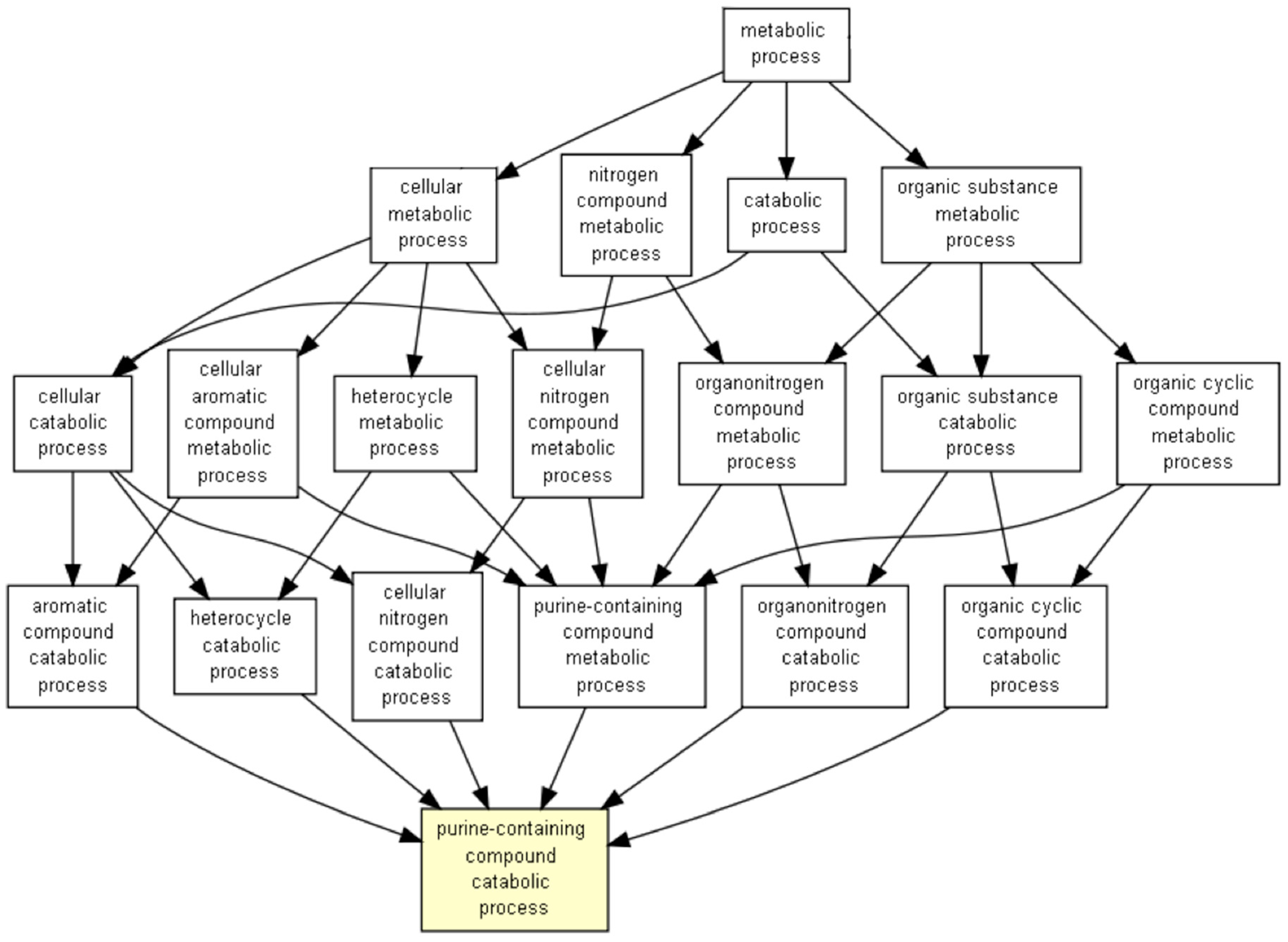
Interestingly, there were an impressive number of positively correlated genes in the case of HwNm, and because of the high number of genes altered, we represented the altered biological processes using the GOrilla software (Figure 7).
After the GOrilla analysis, we observed that the main altered biological processes were related to purine metabolism.

4. Discussions

In the current study, we have shown that RUNX1 mRNA correlations are altered by mutations occurring in NPM1 and TET2/IDH1/2/WT1. Interestingly, we observed that the number of genes negatively correlated with RUNX1 transcript were increased by these mutations. Interestingly, we observed that most of these mutations influence RUNX1 DNA binding. More than this, the most frequent hotspot mutations, as seen in Supplementary Table S1, were located at residues R135 and R174. Our observations regarding the increase in RUNX1 expression could be accounted for either because of a compensatory increase in transcription by the normal allele, the advantageous selection of RUNX1 mutated cells with a higher RUNX1 mutated transcript or because of the general changes that RUNX1 mutations induce that ultimately also lead to RUNX1 overexpression.
Moreover, the negatively correlated genes were observed to be linked to myeloid activation. This observation might add another argument to the importance of several genes like TET2/IDH1/2/WT1, NPM1 and RUNX1 in myeloid activation, as we and other groups have shown that the immature phenotype of AML is associated with TET2/IDH1/2/WT1 mutations [2,3]. This is also supported by the fact that RUNX1 has been shown to induce the expression of IL3 and SPI1, which are known for their importance in myeloid differentiation [15]. Haploinsufficiency of RUNX1 has been shown to induce the alteration of hematopoietic stem cell formation [16], with other studies showing that RUNX1 induces the transcription of IL-3 with importance in the hematopoietic stem cell generation in the aorta-gonad mesonephros region, yolk sac and placenta. Moreover, when IL-3 is replaced in the case of RUNX1 haploinsufficiency, the generation of hematopoietic stem cells was shown to be enhanced [17]. Added to this, IL-3 can not only have an effect in immature myeloid cells, but also in mature ones [18]. This role is important considering stress response of myeloid cells as in the case of sepsis [19], or in the atherogenesis process, as IL-3 stimulates monocytes infiltrating the atherosclerotic plaques [20]. Because of the implication that IL-3 has in stimulating myeloid cells, it is of no surprise that, in AML, IL-3R is of real importance as its upregulation was associated with blast proliferation and reduced overall survival [21,22,23]. Thus, it is possible that the interaction between RUNX1 and IL-3 might have an importance in determining the activity of myeloid blasts and could represent a valuable hypothesis for further studies.
Additionally, it has been observed that the TET2, RUNX1, SPI1, NPM1 complex is disrupted by mutant NPM1, which leads to the translocation of the NPM1 protein to the cytoplasm along with the transcription factor SPI1, leading to an impairment in myeloid differentiation [14]. Moreover, it has been shown that RUNX1 interacts with TET2 and amplifies the demethylating properties of TET2, influencing hematopoietic stem cell development [24]. Another argument for the importance of RUNX1 in myeloid differentiation has been shown in a murine model in which it has been observed that RUNX1 is necessary for hematopoietic lineage commitment [25].
In addition to the role of RUNX1 in myeloid activation, we have also observed that NPM1 mutations associated with TET2/IDH1/2/WT1 wild type determine RUNX1 to be positively correlated with genes related to purine metabolism. Because of this, it can be hypothesized that purine analogs used in AML, like fludarabine and cladribine, could have a different effect on AML based on the mutational status of TET2/IDH1/2/WT1 and NPM1 and on the expression level of RUNX1. There is an important possibility that this is the case, as NPM1 is known to offer a good prognosis in AML [26]. Fludarabine and cladribine are used in the clinical scenario as components of FLAG (fludarabine, cytarabine, granulocyte colony stimulating factor) and of CLAG (cladribine, cytarabine, granulocyte colony stimulating factor), respectively. This is generally the case as the combination between these purine analogues and cytarabine are used to this day because of the synergy between these agents [27]. Most frequently, these regimens have been used in the case of relapse/refractory AML representing viable therapeutic options [28,29,30,31]. Between fludarabine- and cladribine-containing regimens used for relapsed/refractory AML, the complete remission rates, overall survival and relapse free survival are similar. Nonetheless, it has to be mentioned that cladribine-based regimens were shown to be better in patients with favorable prognostic factors like nonpoor cytogenetic risk and the presence of complete remission at the first induction therapy [29]. Thus, these agents might have different efficacy based on disease characteristics. Currently, NPM1 mutations are known for their favorable prognosis role and have been included in the stratification of the World Health Organization from 2016 or myeloid neoplasms. It has to be considered that these conclusions were taken from AML studies using intensive chemotherapy regimens [26,32]. Interestingly, Kantarjian has mentioned that NPM1 mutations might be associated with cladribine sensitivity [33]. More than this, a regimen combining cladribine with low-dose cytarabine alternating with decitabine has been shown to induce either complete remissions or complete remissions with incomplete count recovery in all patients presenting with an NPM1 mutation [34]. Further trials could assess if different combinations between mutational statuses of NPM1 and TET2/IDH1/2/WT1 and RUNX1 expression level have an impact in predicting cladribine or fludarabine sensitivity.
In future studies, we plan to further assess other transcription factors that colocalize with TET2 to determine what would be the effects of the interactions. Nonetheless, for such a study, a larger cohort would be necessary. Some of the important transcription factors that have been shown to colocalize with TET2 are represented by, but not limited to, NANOG [35], EBF1 [36], PPARγ [37] and IDAX [38].
The main limitation of this study is represented by the fact that it has been performed on an online dataset without the use of other experimental data. Nonetheless, we could argue that the results presented in this manuscript add useful information regarding the interactions between NPM1 and TET2/IDH1/2/WT1 mutations and the correlations that RUNX1 presents could lead to a better understanding of myeloid differentiation and, potentially, the formation of novel AML subtypes that would be treated in a personalized manner.

5. Conclusions

In the current study, we have shown that NPM1 and TET2/IDH1/2/WT1 mutations increase the number of negative correlations of RUNX1 with other transcripts, especially with transcripts involved in myeloid differentiation. The results shown here would not only reveal more insights in the biology of AML maturation, but could also lead to better therapeutic management strategies tailored to each AML subtype.

Supplementary Materials

The following are available online at https://www.mdpi.com/1648-9144/56/12/637/s1, Figure S1. Chord diagram presenting the overlaps in relative counts between hypermethylating mutations and other mutations frequently occurring in AML. Table S1. Table of the RUNX1 mutations occurring in the samples presenting intermediate cytogenetic risk. Table S2. Summary table of the cohort analyzed included for the assessment of RUNX1 transcript correlations.

Author Contributions

Conceptualization, S.P., C.T. and M.Z.; investigation, A.J.; methodology, S.P. and A.J.; software, S.P.; supervision, M.Z.; visualization, S.P.; writing—original draft, S.P. and A.J.; writing—review and editing, C.T. and M.Z. All authors have read and agreed to the published version of the manuscript.

Funding

Ancuta Jurj, published under the frame of the European Social Fund, Human Capital Operational Programme 2014–2020, project No. POCU/380/6/13/125171. This work was supported by a grant from the Romanian Ministry of Research and Innovation, CCCDI-UEFISCDI, project No. PN-III-P4-ID-PCCF-2016-0112 within PNCDI III, as well as by an award for Young Research Teams 2020–2022 (Grant No. PN-III-P1-1.1-TE-2019–0271), and by an international collaborative grant of the European Economic Space between Romania and Iceland 2020–2022 (Grant No. 19-COP-0031).

Acknowledgments

M.Z. and C.T. are both considered co-last authors in the present study.

Conflicts of Interest

The authors declare no conflict of interest.

References

  1. Khwaja, A.; Bjorkholm, M.; Gale, R.E.; Levine, R.L.; Jordan, C.T.; Ehninger, G.; Bloomfield, C.D.; Estey, E.; Burnett, A.; Cornelissen, J.J.; et al. Acute myeloid leukaemia. Nat. Rev. Dis. Primers 2016, 2, 16010. [Google Scholar] [CrossRef] [PubMed]
  2. Pasca, S.; Turcas, C.; Jurj, A.; Teodorescu, P.; Iluta, S.; Hotea, I.; Bojan, A.; Selicean, C.; Fetica, B.; Petrushev, B.; et al. The Influence of Methylating Mutations on Acute Myeloid Leukemia: Preliminary Analysis on 56 Patients. Diagnostics 2020, 10, 263. [Google Scholar] [CrossRef] [PubMed]
  3. Bowman, R.L.; Levine, R.L. TET2 in Normal and Malignant Hematopoiesis. Cold Spring Harb. Perspect. Med. 2017, 7, a026518. [Google Scholar] [CrossRef] [PubMed] [Green Version]
  4. Amaya, M.L.; Pollyea, D.A. Targeting the IDH2 Pathway in Acute Myeloid Leukemia. Clin. Cancer Res. 2018, 24, 4931–4936. [Google Scholar] [CrossRef] [Green Version]
  5. Wang, Y.; Xiao, M.; Chen, X.; Chen, L.; Xu, Y.; Lv, L.; Wang, P.; Yang, H.; Ma, S.; Lin, H.; et al. WT1 Recruits TET2 to Regulate Its Target Gene Expression and Suppress Leukemia Cell Proliferation. Mol. Cell 2015, 57, 662–673. [Google Scholar] [CrossRef] [Green Version]
  6. Moran-Crusio, K.; Reavie, L.; Shih, A.; Abdel-Wahab, O.; Ndiaye-Lobry, D.; Lobry, C.; Figueroa, M.E.; Vasanthakumar, A.; Patel, J.; Zhao, X.; et al. Tet2 Loss Leads to Increased Hematopoietic Stem Cell Self-Renewal and Myeloid Transformation. Cancer Cell 2011, 20, 11–24. [Google Scholar] [CrossRef] [Green Version]
  7. Figueroa, M.E.; Lugthart, S.; Li, Y.; Erpelinck-Verschueren, C.; Deng, X.; Christos, P.J.; Schifano, E.; Booth, J.; van Putten, W.; Skrabanek, L.; et al. DNA Methylation Signatures Identify Biologically Distinct Subtypes in Acute Myeloid Leukemia. Cancer Cell 2010, 17, 13–27. [Google Scholar] [CrossRef] [Green Version]
  8. Cerami, E.; Gao, J.; Dogrusoz, U.; Gross, B.E.; Sumer, S.O.; Aksoy, B.A.; Jacobsen, A.; Byrne, C.J.; Heuer, M.L.; Larsson, E.; et al. The cBio Cancer Genomics Portal: An Open Platform for Exploring Multidimensional Cancer Genomics Data: Figure 1. Cancer Dis. 2012, 2, 401–404. [Google Scholar] [CrossRef] [Green Version]
  9. Gao, J.; Aksoy, B.A.; Dogrusoz, U.; Dresdner, G.; Gross, B.; Sumer, S.O.; Sun, Y.; Jacobsen, A.; Sinha, R.; Larsson, E.; et al. Integrative Analysis of Complex Cancer Genomics and Clinical Profiles Using the cBioPortal. Sci. Signal. 2013, 6, pl1. [Google Scholar] [CrossRef] [Green Version]
  10. The Gene Ontology Consortium. The Gene Ontology Resource: 20 years and still GOing strong. Nucleic Acids Res. 2019, 47, D330–D338. [Google Scholar] [CrossRef] [Green Version]
  11. Szklarczyk, D.; Gable, A.L.; Lyon, D.; Junge, A.; Wyder, S.; Huerta-Cepas, J.; Simonovic, M.; Doncheva, N.T.; Morris, J.H.; Bork, P.; et al. STRING v11: Protein–protein association networks with increased coverage, supporting functional discovery in genome-wide experimental datasets. Nucleic Acids Res. 2019, 47, D607–D613. [Google Scholar] [CrossRef] [PubMed] [Green Version]
  12. Eden, E.; Navon, R.; Steinfeld, I.; Lipson, D.; Yakhini, Z. GOrilla: A tool for discovery and visualization of enriched GO terms in ranked gene lists. BMC Bioinform. 2009, 10, 48. [Google Scholar] [CrossRef] [PubMed] [Green Version]
  13. Li, Z.; Yan, J.; Matheny, C.J.; Corpora, T.; Bravo, J.; Warren, A.J.; Bushweller, J.H.; Speck, N.A. Energetic Contribution of Residues in the Runx1 Runt Domain to DNA Binding. J. Biol. Chem. 2003, 278, 33088–33096. [Google Scholar] [CrossRef] [PubMed] [Green Version]
  14. Gu, X.; Ebrahem, Q.; Mahfouz, R.Z.; Hasipek, M.; Enane, F.; Radivoyevitch, T.; Rapin, N.; Przychodzen, B.; Hu, Z.; Balusu, R.; et al. Leukemogenic nucleophosmin mutation disrupts the transcription factor hub that regulates granulomonocytic fates. J. Clin. Investig. 2018, 128, 4260–4279. [Google Scholar] [CrossRef] [Green Version]
  15. Cameron, S.; Taylor, D.S.; TePas, E.C.; Speck, N.A.; Mathey-Prevot, B. Identification of a critical regulatory site in the human interleukin-3 promoter by in vivo footprinting. Blood 1994, 83, 2851–2859. [Google Scholar] [CrossRef]
  16. Cai, Z.; de Bruijn, M.; Ma, X.; Dortland, B.; Luteijn, T.; Downing, J.R.; Dzierzak, E. Haploinsufficiency of AML1 Affects the Temporal and Spatial Generation of Hematopoietic Stem Cells in the Mouse Embryo. Immunity 2000, 13, 423–431. [Google Scholar] [CrossRef] [Green Version]
  17. Robin, C.; Ottersbach, K.; Durand, C.; Peeters, M.; Vanes, L.; Tybulewicz, V.; Dzierzak, E. An Unexpected Role for IL-3 in the Embryonic Development of Hematopoietic Stem Cells. Dev. Cell 2006, 11, 171–180. [Google Scholar] [CrossRef] [Green Version]
  18. Borriello, F.; Galdiero, M.; Varricchi, G.; Loffredo, S.; Spadaro, G.; Marone, G. Innate Immune Modulation by GM-CSF and IL-3 in Health and Disease. Int. J. Mol. Sci. 2019, 20, 834. [Google Scholar] [CrossRef] [Green Version]
  19. Weber, G.F.; Chousterman, B.G.; He, S.; Fenn, A.M.; Nairz, M.; Anzai, A.; Brenner, T.; Uhle, F.; Iwamoto, Y.; Robbins, C.S.; et al. Interleukin-3 amplifies acute inflammation and is a potential therapeutic target in sepsis. Science 2015, 347, 1260–1265. [Google Scholar] [CrossRef] [Green Version]
  20. Robbins, C.S.; Chudnovskiy, A.; Rauch, P.J.; Figueiredo, J.-L.; Iwamoto, Y.; Gorbatov, R.; Etzrodt, M.; Weber, G.F.; Ueno, T.; van Rooijen, N.; et al. Extramedullary Hematopoiesis Generates Ly-6C high Monocytes That Infiltrate Atherosclerotic Lesions. Circulation 2012, 125, 364–374. [Google Scholar] [CrossRef] [Green Version]
  21. Testa, U.; Riccioni, R.; Militi, S.; Coccia, E.; Stellacci, E.; Samoggia, P.; Latagliata, R.; Mariani, G.; Rossini, A.; Battistini, A.; et al. Elevated expression of IL-3Rα in acute myelogenous leukemia is associated with enhanced blast proliferation, increased cellularity, and poor prognosis. Blood 2002, 100, 2980–2988. [Google Scholar] [CrossRef] [PubMed]
  22. Vergez, F.; Green, A.S.; Tamburini, J.; Sarry, J.-E.; Gaillard, B.; Cornillet-Lefebvre, P.; Pannetier, M.; Neyret, A.; Chapuis, N.; Ifrah, N.; et al. High levels of CD34+CD38low/-CD123+ blasts are predictive of an adverse outcome in acute myeloid leukemia: A Groupe Ouest-Est des Leucemies Aigues et Maladies du Sang (GOELAMS) study. Haematologica 2011, 96, 1792–1798. [Google Scholar] [CrossRef] [PubMed]
  23. Jordan, C.; Upchurch, D.; Szilvassy, S.; Guzman, M.; Howard, D.; Pettigrew, A.; Meyerrose, T.; Rossi, R.; Grimes, B.; Rizzieri, D.; et al. The interleukin-3 receptor alpha chain is a unique marker for human acute myelogenous leukemia stem cells. Leukemia 2000, 14, 1777–1784. [Google Scholar] [CrossRef] [PubMed] [Green Version]
  24. Suzuki, T.; Shimizu, Y.; Furuhata, E.; Maeda, S.; Kishima, M.; Nishimura, H.; Enomoto, S.; Hayashizaki, Y.; Suzuki, H. RUNX1 regulates site specificity of DNA demethylation by recruitment of DNA demethylation machineries in hematopoietic cells. Blood Adv. 2017, 1, 1699–1711. [Google Scholar] [CrossRef] [Green Version]
  25. Lacaud, G. Runx1 is essential for hematopoietic commitment at the hemangioblast stage of development in vitro. Blood 2002, 100, 458–466. [Google Scholar] [CrossRef] [PubMed]
  26. Estey, E.H. Acute myeloid leukemia: 2019 update on risk-stratification and management. Am. J. Hematol. 2018, 93, 1267–1291. [Google Scholar] [CrossRef] [PubMed] [Green Version]
  27. Robak, T. Purine Nucleoside Analogues in the Treatment of Myleoid Leukemias. Leuk. Lymphoma 2003, 44, 391–409. [Google Scholar] [CrossRef] [PubMed]
  28. Freyer, C.W.; Gupta, N.; Wetzler, M.; Wang, E.S. Revisiting the role of cladribine in acute myeloid leukemia: An improvement on past accomplishments or more old news? Cladribine in Acute Myeloid Leukemia. Am. J. Hematol. 2015, 90, 62–72. [Google Scholar] [CrossRef] [Green Version]
  29. Park, H.; Youk, J.; Kim, I.; Yoon, S.-S.; Park, S.; Lee, J.-O.; Bang, S.-M.; Koh, Y. Comparison of cladribine- and fludarabine-based induction chemotherapy in relapsed or refractory acute myeloid leukaemia. Ann. Hematol. 2016, 95, 1777–1786. [Google Scholar] [CrossRef]
  30. Wierzbowska, A.; Robak, T.; Pluta, A.; Wawrzyniak, E.; Cebula, B.; Hołowiecki, J.; Kyrcz-Krzemień, S.; Grosicki, S.; Giebel, S.; Skotnicki, A.B.; et al. Cladribine combined with high doses of arabinoside cytosine, mitoxantrone, and G-CSF (CLAG-M) is a highly effective salvage regimen in patients with refractory and relapsed acute myeloid leukemia of the poor risk: A final report of the Polish Adult Leukem: CLAG-M in refractory and relapsed AML. Eur. J. Haematol. 2007, 80, 115–126. [Google Scholar] [CrossRef]
  31. Zhou, A.; Han, Q.; Song, H.; Zi, J.; Ma, J.; Ge, Z. Efficacy and toxicity of cladribine for the treatment of refractory acute myeloid leukemia: A meta-analysis. DDDT 2019, 13, 1867–1878. [Google Scholar] [CrossRef] [PubMed] [Green Version]
  32. Arber, D.A.; Orazi, A.; Hasserjian, R.; Thiele, J.; Borowitz, M.J.; Le Beau, M.M.; Bloomfield, C.D.; Cazzola, M.; Vardiman, J.W. The 2016 revision to the World Health Organization classification of myeloid neoplasms and acute leukemia. Blood 2016, 127, 2391–2405. [Google Scholar] [CrossRef] [PubMed]
  33. Kantarjian, H. Acute myeloid leukemia-Major progress over four decades and glimpses into the future: Progress of acute myeloid leukemia. Am. J. Hematol. 2016, 91, 131–145. [Google Scholar] [CrossRef] [PubMed] [Green Version]
  34. Kadia, T.M.; Cortes, J.; Ravandi, F.; Jabbour, E.; Konopleva, M.; Benton, C.B.; Burger, J.; Sasaki, K.; Borthakur, G.; DiNardo, C.D.; et al. Cladribine and low-dose cytarabine alternating with decitabine as front-line therapy for elderly patients with acute myeloid leukaemia: A phase 2 single-arm trial. Lancet Haematol. 2018, 5, e411–e421. [Google Scholar] [CrossRef]
  35. Costa, Y.; Ding, J.; Theunissen, T.W.; Faiola, F.; Hore, T.A.; Shliaha, P.V.; Fidalgo, M.; Saunders, A.; Lawrence, M.; Dietmann, S.; et al. NANOG-dependent function of TET1 and TET2 in establishment of pluripotency. Nature 2013, 495, 370–374. [Google Scholar] [CrossRef] [PubMed] [Green Version]
  36. Guilhamon, P.; Eskandarpour, M.; Halai, D.; Wilson, G.A.; Feber, A.; Teschendorff, A.E.; Gomez, V.; Hergovich, A.; Tirabosco, R.; Fernanda Amary, M.; et al. Meta-analysis of IDH-mutant cancers identifies EBF1 as an interaction partner for TET2. Nat. Commun. 2013, 4, 2166. [Google Scholar] [CrossRef] [PubMed] [Green Version]
  37. Fujiki, K.; Shinoda, A.; Kano, F.; Sato, R.; Shirahige, K.; Murata, M. PPARγ-induced PARylation promotes local DNA demethylation by production of 5-hydroxymethylcytosine. Nat. Commun. 2013, 4, 2262. [Google Scholar] [CrossRef] [PubMed] [Green Version]
  38. Ko, M.; An, J.; Bandukwala, H.S.; Chavez, L.; Äijö, T.; Pastor, W.A.; Segal, M.F.; Li, H.; Koh, K.P.; Lähdesmäki, H.; et al. Modulation of TET2 expression and 5-methylcytosine oxidation by the CXXC domain protein IDAX. Nature 2013, 497, 122–126. [Google Scholar] [CrossRef] [PubMed]
Figure 1. Chord diagrams showing the association between hypermethylating mutations and other mutations commonly occurring in acute myeloid leukemia (AML) in absolute counts.
Medicina 56 00637 g001
Figure 2. RUNX1 expression considering the hypermethylating and RUNX1 mutation overlaps. Hm = hypermethylating mutation; Rm = RUNX1 mutation; Hw = hypermethylating wild type; Rw = RUNX1 wild type.
Medicina 56 00637 g002
Figure 3. Venn diagram of the overlaps between genes that were significantly positively or negatively correlated with RUNX1 considering hypermethylating mutation or wild type. Hyper mut = hypermethylating mutation; Hyper wt = hypermethylating wild type; Pos = positive correlation with RUNX1; Neg = negative correlation with RUNX1.
Medicina 56 00637 g003
Figure 4. STRING diagram for the genes related to myeloid activation observed to be negatively correlated with RUNX1 expression when hypermethylating mutations were present.
Medicina 56 00637 g004
Figure 5. RUNX1 expression differences between AML with different maturity.
Medicina 56 00637 g005
Figure 6. Venn diagrams representing the effects of hypermethylating and NPM1 mutation overlaps on RUNX1 associations. We represented one Venn diagram only considering the negative correlations and the other only considering the positive correlations. Hw = hypermethylating gene wild type; Hm = hypermethylating gene mutation; Nw = NPM1 wild type; Nm = NPM1 mutation.
Medicina 56 00637 g006
Figure 7. Representation of HwNm positively correlated processes.
Medicina 56 00637 g007
Table 1. The association between hypermethylating mutations and other mutations. OR = odds ratio; CI = confidence interval.
Mutated GeneORLower 95% CIUpper 95% CIp-Value
TP530.0970.0020.6590.006
RUNX13.4281.08911.9610.032
FLT30.4820.2211.0140.043
ASXL16.9040.663346.4970.068
MUC16001.7790.158
STAG23.4310.47539.0060.200
DNMT3A1.3550.6462.8200.387
TTN1.6890.3039.4300.477
KRAS1.6890.3039.4300.477
PHF61.6780.21812.9370.674
U2AF10.5370.0513.1290.711
SMC1A0.6510.0604.1260.711
SMC30.6510.0604.1260.711
NPM10.8520.4071.7530.734
CEBPA0.7170.1542.7120.768
NRAS0.8110.2072.7640.787
PTPN110.8180.1284.0001
KIT0.9900.1485.2991
FCGBP1.1030.0909.9151
BRINP31.1030.0909.9151
Publisher’s Note: MDPI stays neutral with regard to jurisdictional claims in published maps and institutional affiliations.