Evaluation of TP-E Interval and TP-E/QT Ratio in Panic Disorder


Abstract
:1. Introduction
2. Materials and Methods
Statistical Analysis
3. Results
4. Discussion
5. Conclusions
Author Contributions
Funding
Acknowledgments
Conflicts of Interest
References
- Christenson, J.; Innes, G.; McKnight, D.; Boychuk, B.; Grafstein, E.; Thompson, C.; Rosenberg, F.; Anis, A.H.; Gin, K.; Tilley, J.; et al. Safety and efficiency of emergency department assessment of chest discomfort. Can. Med. Assoc. J. 2004, 170, 1803–1807. [Google Scholar] [CrossRef] [Green Version]
- Foldes-Busque, G.; Marchand, A.; Chauny, J.-M.; Poitras, J.; Diodati, J.; Denis, I.; Lessard, M.-J.; Pelland, M.-E.; Fleet, R. Unexplained chest pain in the ED: Could it be panic? Am. J. Emerg. Med. 2011, 29, 743–751. [Google Scholar] [CrossRef]
- De Jonge, P.; Roest, A.M.; Lim, C.; Florescu, S.E.; Bromet, E.J.; Stein, D.J.; Harris, M.; Nakov, V.; De Almeida, J.M.C.; Levinson, D.; et al. Cross-national epidemiology of panic disorder and panic attacks in the world mental health surveys. Depress. Anxiety 2016, 33, 1155–1177. [Google Scholar] [CrossRef] [Green Version]
- McCraty, R.; Atkinson, M.; Tomasino, D.; Stuppy, W.P. Analysis of twenty-four hour heart rate variability in patients with panic disorder. Boil. Psychol. 2001, 56, 131–150. [Google Scholar] [CrossRef] [Green Version]
- Ozturk, M.; Turan, O.E.; Karaman, K.; Bilge, N.; Ceyhun, G.; Aksu, U.; Aksakal, E.; Gulcu, O.; Kalkan, K.; Demirelli, S. Evaluation of ventricular repolarization parameters during migraine attacks. J. Electrocardiol. 2019, 53, 66–70. [Google Scholar] [CrossRef] [PubMed]
- Sauer, A.; Wilcox, J.E.; Andrei, A.C.; Passman, R.; Goldberger, J.J.; Shah, S.J. Association of the ECG T-peak to T-end interval with echocardiographic markers of diastolic dysfunction. Circ. Arrhythm. Electrophysiol. 2012, 5, 537–543. [Google Scholar] [CrossRef] [Green Version]
- Atmaca, M.; Yavuzkir, M.; Izci, F.; Gurok, M.G.; Adiyaman, S. QT wave dispersion in patients with panic disorder. Neurosci. Bull. 2012, 28, 247–252. [Google Scholar] [CrossRef] [PubMed] [Green Version]
- Kors, J.A.; Van Eck, H.J.R.; Van Herpen, G. The meaning of the Tp-Te interval and its diagnostic value. J. Electrocardiol. 2008, 41, 575–580. [Google Scholar] [CrossRef]
- Shu, J.; Li, H.; Yan, G.; Cui, C. Tp-e/QT ratio as a predictive index of sudden cardiac death in patients with ST-segment elevation myocardial infarction. J. Xi’an Jiaotong Univ. Med. Sci. 2010, 31, 441–443. [Google Scholar]
- Walters, K.R.; Rait, G.; Petersen, I.; Williams, R.; Nazareth, I. Panic disorder and risk of new onset coronary heart disease, acute myocardial infarction, and cardiac mortality: Cohort study using the general practice research database. Eur. Heart J. 2008, 29, 2981–2988. [Google Scholar] [CrossRef] [Green Version]
- Balıkçı, K.; Herdem, A.; Aydemir, O.; Grubu, D. Reliability and validity of Turkish form of panic disorder scale. Anatol. J. Psychiatry 2017, 18, 13. [Google Scholar] [CrossRef] [Green Version]
- Hevia, J.C.; Antzelevitch, C.; Bárzaga, F.T.; Sánchez, M.D.; Balea, F.D.; Molina, R.Z.; Pérez, M.A.Q.; Rodríguez, Y.F. Tpeak-tend and tpeak-tend dispersion as risk factors for ventricular tachycardia/ventricular fibrillation in patients with the brugada syndrome. J. Am. Coll. Cardiol. 2006, 47, 1828–1834. [Google Scholar] [CrossRef] [PubMed] [Green Version]
- Lang, R.M.; Badano, L.P.; Mor-Avi, V.; Afilalo, J.; Armstrong, A.; Ernande, L.; Flachskampf, F.A.; Foster, E.; Goldstein, S.A.; Kuznetsova, T. Recommendations for cardiac chamber quantification by echocardiography in adults: An update from the American society of echocardiography and the european association of cardiovascular imaging. J. Am. Soc. Echocardiogr. 2015, 16, 233–271. [Google Scholar]
- Katerndahl, D.A. The association between panic disorder and coronary artery disease among primary care patients presenting with chest pain: An updated literature review. Prim. Care Companion J. Clin. Psychiatry 2008, 10, 276–285. [Google Scholar] [CrossRef]
- Machado, S.; Sancassiani, F.; Paes, F.; Rocha, N.B.; Murillo-Rodríguez, E.; Nardi, A.E. Panic disorder and cardiovascular diseases: An overview. Int. Rev. Psychiatry 2017, 29, 436–444. [Google Scholar] [CrossRef]
- Batelaan, N.M.; Have, M.T.; Van Balkom, A.J.; Tuithof, M.; De Graaf, R. Anxiety disorders and onset of cardiovascular disease: The differential impact of panic, phobias and worry. J. Anxiety Disord. 2014, 28, 252–258. [Google Scholar] [CrossRef]
- Ng, G.A. Neuro-cardiac interaction in malignant ventricular arrhythmia and sudden cardiac death. Auton. Neurosci. 2016, 199, 66–79. [Google Scholar] [CrossRef] [Green Version]
- Wise, V.; McFarlane, A.C.; Clark, C.R.; Battersby, M. An integrative assessment of brain and body function ‘at rest’ in panic disorder: A combined quantitative EEG/autonomic function study. Int. J. Psychophysiol. 2011, 79, 155–165. [Google Scholar] [CrossRef]
- Cohen, H.; Benjamin, J. Power spectrum analysis and cardiovascular morbidity in anxiety disorders. Auton. Neurosci. 2006, 128, 1–8. [Google Scholar] [CrossRef]
- Carney, R.M.; Freedland, K.E.; Stein, P.K.; Skala, J.A.; Hoffman, P.; Jaffe, A.S. Change in heart rate and heart rate variability during treatment for depression in patients with coronary heart disease. Psychosom. Med. 2000, 62, 639–647. [Google Scholar] [CrossRef]
- Chalmers, J.A.; Quintana, D.S.; Abbott, M.J.-A.; Kemp, A.H. Anxiety disorders are associated with reduced heart rate variability: A meta-analysis. Front. Psychol. 2014, 5, 80. [Google Scholar] [CrossRef] [PubMed] [Green Version]
- Thayer, J.F.; Lane, R.D. A model of neurovisceral integration in emotion regulation and dysregulation. J. Affect. Disord. 2000, 61, 201–216. [Google Scholar] [CrossRef] [Green Version]
- Rennie, K.L.; Hemingway, H.; Kumari, M.; Brunner, E.; Malik, M.; Marmot, M. Effects of moderate and vigorous physical activity on heart rate variability in a British study of civil servants. Am. J. Epidemiol. 2003, 158, 135–143. [Google Scholar] [CrossRef] [PubMed]
- Gündüz, N.; Aslan, E.A.; Eren, F.; Turan, H.S.; Öztürk, M.; Tural, U. Psikotrop İlaç Kullanımı, Başka Tıbbi Hastalığı ve Psikiyatrik Eş Tanısı Olmayan Panik Bozukluğu Hastalarında 24 Saatlik Kalp Hızı Değişkenliğinin Değerlendirilmesi. Türk. Psikiyatr. Derg. 2019, 30, 236–244. [Google Scholar]
- Hovland, A.; Pallesen, S.; Hammar, Å.; Hansen, A.L.; Thayer, J.F.; Tarvainen, M.; Nordhus, I.H. The relationships among heart rate variability, executive functions, and clinical variables in patients with panic disorder. Int. J. Psychophysiol. 2012, 86, 269–275. [Google Scholar] [CrossRef]
- Ishida, S.; Nakagawa, M.; Fujino, T.; Yonemochi, H.; Saikawa, T.; Ito, M. Circadian variation of QT interval dispersion: Correlation with heart rate variability. J. Electrocardiol. 1997, 30, 205–210. [Google Scholar] [CrossRef]
- Fossa, A.A. The impact of varying autonomic states on the dynamic beat-to-beat QT-RR and QT-TQ interval relationships. Br. J. Pharmacol. 2008, 154, 1508–1515. [Google Scholar] [CrossRef] [Green Version]
- De Bruyne, M.C.; Hoes, A.W.; Kors, J.A.; Hofman, A.; Van Bemmel, J.H.; Grobbee, D.E. QTc dispersion predicts cardiac mortality in the elderly: The rotterdam study. Circulation 1998, 97, 467–472. [Google Scholar] [CrossRef] [Green Version]
- Stewart, A.; Waterhouse, J.; Howard, P. The QTc interval, autonomic neuropathy and mortality in hypoxaemic COPD. Respir. Med. 1995, 89, 79–84. [Google Scholar] [CrossRef] [Green Version]
- Uyarel, H.; Okmen, E.; Cobanoğlu, N.; Karabulut, A.; Cam, N. Effects of anxiety on QT dispersion in healthy young men. Acta Cardiol. 2006, 61, 83–87. [Google Scholar] [CrossRef]
- Rautaharju, P.M. Why Did QT Dispersion Die? Card. Electrophysiol. Rev. 2002, 6, 295–301. [Google Scholar] [CrossRef] [PubMed]
- Somberg, J.C.; Molnar, J. Usefulness of QT dispersion as an electrocardiographically derived index. Am. J. Cardiol. 2002, 89, 291–294. [Google Scholar] [CrossRef]
- Lubinski, A.; Kempa, M.; Baczynska, A.M.; Romanowska, I.; Świątecka, G.; Lewicka, E. New insight into repolarization abnormalities in patients with congenital long QT syndrome: The increased transmural dispersion of repoiarization. Pacing Clin. Electrophysiol. 1998, 21, 172–175. [Google Scholar] [CrossRef] [PubMed]
- Gupta, P.; Patel, C.; Patel, H.; Narayanaswamy, S.; Malhotra, B.; Green, J.T.; Yan, G.-X. Tp-e/QT ratio as an index of arrhythmogenesis. J. Electrocardiol. 2008, 41, 567–574. [Google Scholar] [CrossRef] [PubMed]
- Zhao, X.; Xie, Z.; Chu, Y.; Yang, L.; Xu, W.; Yang, X.; Liu, X.; Tian, L. Association between Tp-e/QT ratio and prognosis in patients undergoing primary percutaneous coronary intervention for ST-segment elevation myocardial infarction. Clin. Cardiol. 2012, 35, 559–564. [Google Scholar] [CrossRef]
- Içli, A.; Kayrak, M.; Akilli, H.; Aribas, A.; Coskun, M.; Ozer, S.F.; Ozdemir, K. Prognostic value of Tpeak-Tend interval in patients with acute pulmonary embolism. BMC Cardiovasc. Disord. 2015, 15, 99. [Google Scholar] [CrossRef] [Green Version]
- Panikkath, R.; Reinier, K.; Uy-Evanado, A.; Teodorescu, C.; Hattenhauer, J.; Mariani, R.; Gunson, K.; Jui, J.; Chugh, S.S. Prolonged Tpeak-to-tend interval on the resting ECG is associated with increased risk of sudden cardiac death. Circ. Arrhythm. Electrophysiol. 2011, 4, 441–447. [Google Scholar] [CrossRef] [Green Version]
- Tse, G.; Gong, M.; Wong, W.T.; Georgopoulos, S.; Letsas, K.P.; Vassiliou, V.S.; Chan, Y.S.; Yan, B.P.; Wong, S.H.; Wu, K.K.; et al. The Tpeak-Tend interval as an electrocardiographic risk marker of arrhythmic and mortalityoutcomes: A systematic review and meta-analysis. Heart Rhythm. 2017, 14, 1131–1137. [Google Scholar] [CrossRef] [Green Version]
- Perkiömäki, J.S.; Ikäheimo, M.J.; Pikkujämsä, S.M.; Rantala, A.; Lilja, M.; Kesäniemi, Y.A.; Huikuri, H.V. Dispersion of the QT interval and autonomic modulation of heart rate in hypertensive men with and without left ventricular hypertrophy. Hypertension 1996, 28, 16–21. [Google Scholar] [CrossRef]


| Panic Disorder Group (n = 40) | Control Group (n = 50) | p | |
|---|---|---|---|
| Baseline Demographic Parameters | |||
| Age (years) | 34.7 ± 8.7 | 34.5 ± 5.5 | 0.188 |
| Male sex, n (%) | 13 (32.5) | 20 (40%) | 0.139 |
| Married, n (%) | 20 (50%) | 20 (40%) | 0.231 |
| Smoking, n (%) | 12 (30) | 16 (32) | 0.511 |
| Body mass index (kg/m2) | 26.8 ± 2.3 | 25.9 ± 2.6 | 0.099 |
| Systolic BP (mmHg) | 125 (118–130) | 120 (120–125) | 0.077 |
| Diastolic BP (mmHg) | 73.8 ± 7.9 | 70.9 ± 9.1 | 0.120 |
| Severity Measure for Panic Disorder—Adult score | 21.2 ± 5.4 | ||
| Laboratory Parameters | |||
| Fasting glucose (mg/dL) | 96 (86–99) | 90 (86–95) | 0.052 |
| Creatinine (mg/dL) | 0.84 ± 0.15 | 0.82 ± 0.13 | 0.484 |
| Potassium (mmol/L) | 4.2 ± 0.2 | 4.2 ± 0.3 | 0.484 |
| Calcium (mg/dL) | 9.57 ± 0.42 | 9.65 ± 0.47 | 0.434 |
| Magnesium (mg/dL) | 2.1 ± 0.1 | 2.0 ± 0.2 | 0.235 |
| Thyroid-stimulating hormone (µU/mL) | 2.33 ± 1.20 | 2.60 ± 1.12 | 0.055 |
| Echocardiography Parameters | |||
| LV ejection fraction (%) | 55.3 ± 2.8 | 56.4 ± 3.1 | 0.080 |
| LVEDD (mm) | 43.7 ± 5.4 | 42.5 ± 3.6 | 0.190 |
| LVESD (mm) | 30.7 ± 4.2 | 30.9 ± 3.5 | 0.854 |
| Left atrial diameter (cm) | 3.7 ± 0.2 | 3.6 ± 0.3 | 0.197 |
| Panic Disorder Group (n = 40) | Control Group (n = 50) | p | |
|---|---|---|---|
| Heart rate (beats/min) | 80.2 ± 13.6 | 73.7 ± 11.2 | 0.017 |
| QRS duration (ms) | 86.7 ± 6.6 | 89.1 ± 6.3 | 0.094 |
| QT interval (ms) | 362.8 ± 24.9 | 361.9 ± 22.2 | 0.884 |
| QTc interval (ms) | 407.8 ± 21.2 | 405.1 ± 20.9 | 0.645 |
| QTd interval (ms) | 49 [40–60] | 45 [40–60] | 0.010 |
| cQTd interval (ms) | 62.4 ± 11.6 | 52.5 ± 17.3 | 0.004 |
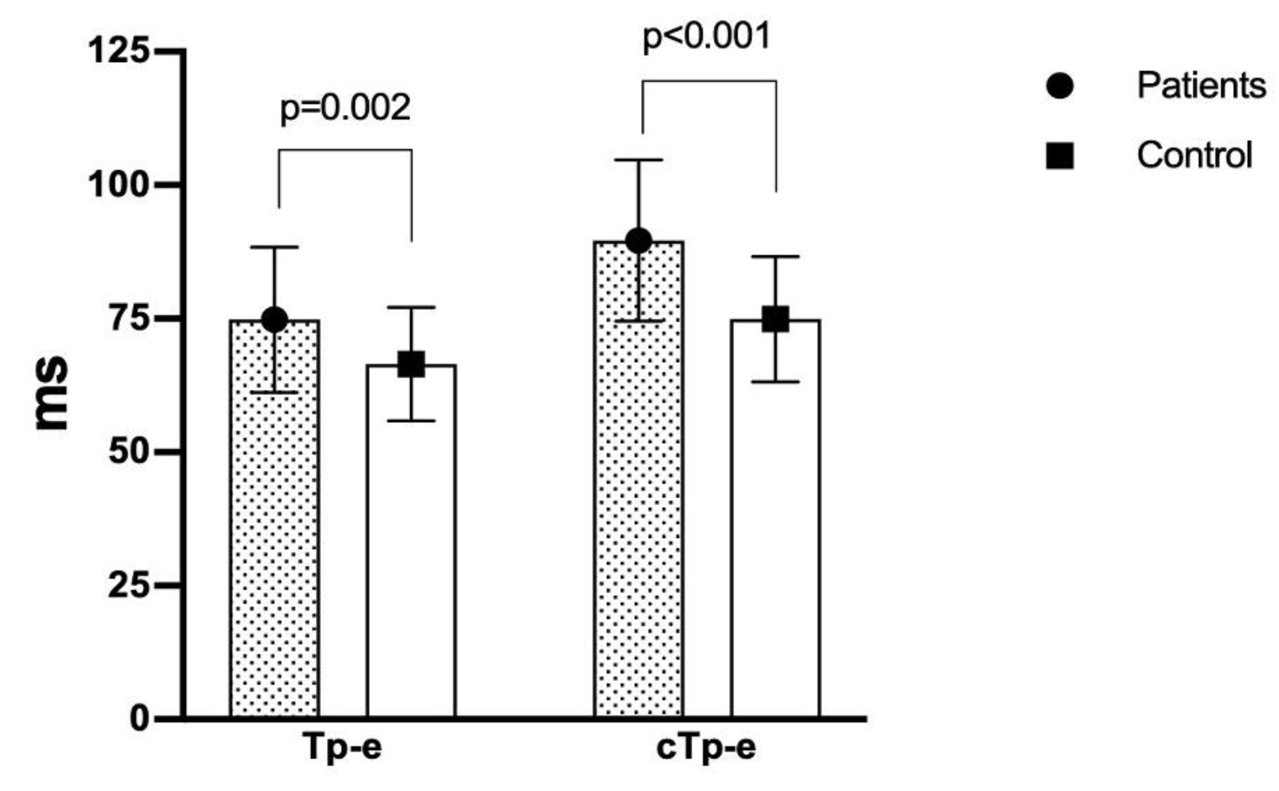
| Tp-e interval (ms) | 74.8 ± 13.6 | 66.5 ± 10.6 | 0.002 |
| cTp-e interval (ms) | 89.6 ± 15.1 | 74.9 ± 11.7 | <0.001 |
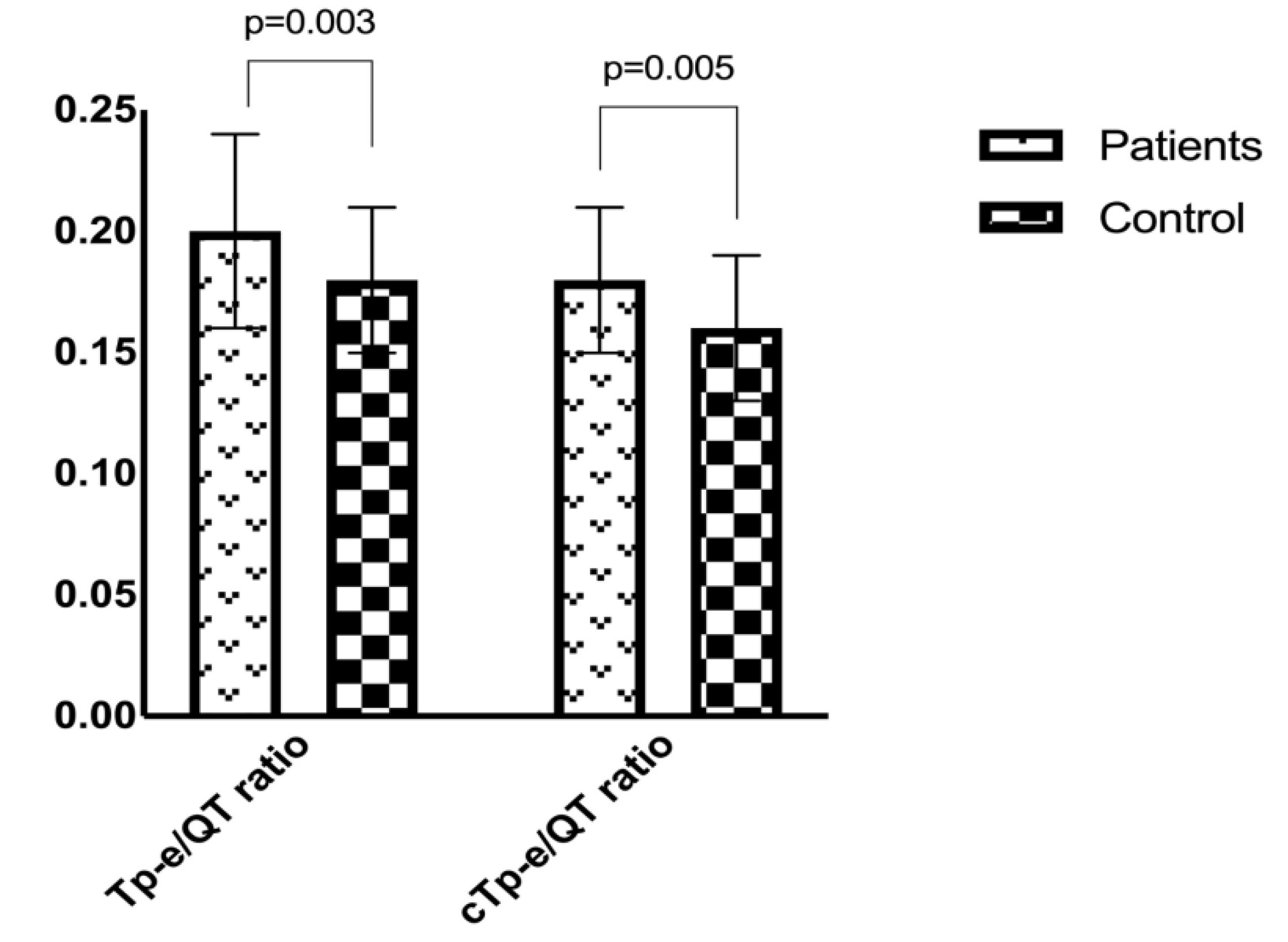
| Tp-e/QT ratio | 0.20 ± 0.04 | 0.18 ± 0.03 | 0.003 |
| Tp-e/QTc ratio | 0.18 ± 0.03 | 0.16 ± 0.03 | 0.005 |
| r | p | |
|---|---|---|
| Heart rate (beats/min) | 0.260 | 0.007 |
| QRS duration | −0.151 | 0.082 |
| QT interval | −0.030 | 0.391 |
| QTc interval | −0.015 | 0.444 |
| QTd interval (ms) | 0.277 | 0.005 |
| cQTd interval (ms) | 0.294 | 0.003 |
| Tp-e interval (ms) | 0.369 | <0.001 |
| cTp-e interval (ms) | 0.531 | <0.001 |
| Tp-e/QT ratio | 0.358 | 0.001 |
| Tp-e/QTc ratio | 0.351 | 0.001 |
| Beta | p-Value | EXP (beta) | 95% CI for EXP (beta) | ||
|---|---|---|---|---|---|
| Lower | Upper | ||||
| BPM (beats/min) | 0.059 | 0.147 | 1.060 | 0.980 | 1.148 |
| QT | 0.040 | 0.944 | 1.004 | 0.891 | 1.131 |
| QTc | −0.109 | 0.166 | 0.897 | 0.769 | 1.046 |
| QTd | 0.056 | 0.071 | 1.057 | 0.995 | 1.123 |
| cQTd | 0.045 | 0.026 | 1.053 | 1.006 | 1.101 |
| Tp-e interval | 0.576 | 0.231 | 1.778 | 0.694 | 4.556 |
| cTp-e interval | 0.217 | <0.001 | 1.242 | 1.103 | 1.400 |
| Tp-e/QT ratio | −25.445 | 0.346 | 0.000 | 0.000 | 9.001 |
| Tp-e/QTc ratio | −252.844 | 0.187 | 0.000 | 0.000 | 2.611 |
| Constant | 32.618 | 0.293 | 1.465 | ||
© 2020 by the authors. Licensee MDPI, Basel, Switzerland. This article is an open access article distributed under the terms and conditions of the Creative Commons Attribution (CC BY) license (http://creativecommons.org/licenses/by/4.0/).
Share and Cite
Afsin, A.; Asoğlu, R.; Orum, M.H.; Cicekci, E. Evaluation of TP-E Interval and TP-E/QT Ratio in Panic Disorder. Medicina 2020, 56, 215. https://doi.org/10.3390/medicina56050215
Afsin A, Asoğlu R, Orum MH, Cicekci E. Evaluation of TP-E Interval and TP-E/QT Ratio in Panic Disorder. Medicina. 2020; 56(5):215. https://doi.org/10.3390/medicina56050215
Chicago/Turabian StyleAfsin, Abdulmecit, Ramazan Asoğlu, Mehmet Hamdi Orum, and Elvan Cicekci. 2020. "Evaluation of TP-E Interval and TP-E/QT Ratio in Panic Disorder" Medicina 56, no. 5: 215. https://doi.org/10.3390/medicina56050215