Review

Fecal Microbiota Transplantation in NAFLD Treatment

1
Department of Health Sciences, University “Magna Græcia”, 88100 Catanzaro, Italy
2
Internal Medicine Residency Program, Università Politecnica delle Marche, 60121 Ancona, Italy
3
Clinical Nutrition Unit, Fondazione Policlinico A. Gemelli IRCCS, 00168 Rome, Italy
4
Department of Translational Medicine and Surgery, Università Cattolica del Sacro Cuore, 00168 Rome, Italy
5
T.A.R.G.I.D., Gasthuisberg University Hospital, KU Leuven, Herestraat 49, 3000 Leuven, Belgium
6
Clinical Nutrition and Internal Medicine Unit, “Madonna del Soccorso” General Hospital, 63074 San Benedetto del Tronto, Italy
7
Hepatology and Internal Medicine Unit, “Madonna del Soccorso” General Hospital, 63074 San Benedetto del Tronto, Italy
8
Healthcare Genetics and Genomics Doctoral Program, School of Nursing, College of Behavioral, Social and Health Sciences, Clemson University, 105 Sikes Hall, Clemson, SC 29631, USA
*
Author to whom correspondence should be addressed.
Medicina 2022, 58(11), 1559; https://doi.org/10.3390/medicina58111559
Submission received: 16 September 2022 / Revised: 26 October 2022 / Accepted: 27 October 2022 / Published: 30 October 2022
(This article belongs to the Special Issue Research Progress of Microbiota and Fecal Transplantation)

Abstract

:
Introduction: Gut microbiota is not only a taxonomic biologic ecosystem but is also involved in human intestinal and extra-intestinal functions such as immune system modulation, nutrient absorption and digestion, as well as metabolism regulation. The latter is strictly linked to non-alcoholic fatty liver disease (NAFLD) pathophysiology. Materials and methods: We reviewed the literature on the definition of gut microbiota, the concepts of “dysbiosis” and “eubiosis”, their role in NAFLD pathogenesis, and the data on fecal microbiota transplantation (FMT) in these patients. We consulted the main medical databases using the following keywords, acronyms, and their associations: gut microbiota, eubiosis, dysbiosis, bile acids, NAFLD, and FMT. Results: Gut microbiota qualitative and quantitative composition is different in healthy subjects vs. NALFD patients. This dysbiosis is associated with and involved in NAFLD pathogenesis and evolution to non-acoholic steatohepatitis (NASH), liver cirrhosis, and hepatocellular carcinoma (HCC). In detail, microbial-driven metabolism of bile acids (BAs) and interaction with hepatic and intestinal farnesoid nuclear X receptor (FXR) have shown a determinant role in liver fat deposition and the development of fibrosis. Over the use of pre- or probiotics, FMT has shown preclinical and initial clinical promising results in NAFLD treatment through re-modulation of microbial dysbiosis. Conclusions: Promising clinical data support a larger investigation of gut microbiota dysbiosis reversion through FMT in NAFLD using randomized clinical trials to design precision-medicine treatments for these patients at different disease stages.

1. Introduction

Gut microbiota can be defined as a complex ecosystem encompassing more than 100 trillion microbes belonging to bacteria, viruses, archaea, protozoa, fungi, and yeasts living in our intestines. However, this taxonomic definition does not describe its functioning, which strictly relies on qualitative and quantitative composition [1]. When the qualitative and quantitative composition of gut microbiota remains stable we can speak about “eubiosis” [1]. When this composition is altered by intestinal and environmental factors (e.g., use of antibiotics, or gastrointestinal and extraintestinal infections), “dysbiosis” is established [2]. There are different dysbioses for different gastrointestinal and extra-intestinal diseases [3,4]. Thus, this significant association is gaining more and more attention from researchers as it can help the development of “personalized” medicine [5]. Non-alcoholic fatty liver disease (NAFLD) is a complex condition characterized by fat deposits within hepatocytes [6]. Liver fat deposition is significantly associated with obesity, dyslipidemia, type 2 diabetes, and insulin resistance as comorbidities [7,8]. Almost one-quarter of the Westernized population is affected by this condition which has a spectrum of stages [9]. Aside from fat deposition, about 10–15% of patients can develop liver fibrosis because of progressive liver inflammation [10]. Liver fibrosis characterizes non-alcoholic steatohepatitis (NASH). If liver fibrosis is not reverted and/or stopped by treatments and lifestyle changes, patients can develop liver cirrhosis as well [11]. Particularly, NAFLD patients can show hepatocellular carcinoma (HCC) also without liver cirrhosis at any stage [12]. In NAFLD, gut microbiota derangement, namely dysbiosis, is resembled by an increased abundance of Bacteroidetes and a decrease of those of Firmicutes [13]. This starts a pathophysiological process through the “gut-liver axis “ where antigens from intestinal pathogens enter the portal blood cycle via a deranged intestinal permeability through damage-associated molecular patterns (DAMPS) and pathogen-associated molecular patterns (PAMPS) [14]. This pathophysiological cascade leads the liver to be exposed to detrimental antigens that start and perpetuate liver damage [14]. Conversely, liver inflammation favors altered intestinal permeability increase [14]. Bile acids (BA) are involved in lipid absorption and metabolism regulation [15]. Gut microbiota is also implicated in such regulation and actively interacts with the free BAs pool and their receptors, particularly the farnesoid X receptor (FXR), present both in enterocytes and hepatocytes, with different and tissue-specific effects on liver fat deposition and fibrosis [16]. The use of antibiotics, pre-, and probiotics to restore gut “eubiosis” in NAFLD patients has shown limitations as well as promising results. Probiotics can protect the liver from toxic substances driven to the liver by altered intestinal permeability. In addition, they can restore altered intestinal permeability, and gut eubiosis and exert a direct anti-inflammatory effect on gut and liver tissue. Antibiotics can restore gut eubiosis and prevent the pathophysiologic cascade characteristic of the “gut-liver axis”. Unfortunately, probiotics efficacy needs larger randomized clincal trials (RCT)s investigating their safety. Antibiotics suffer from the emerging issue of antibiotic resistance development within the gut, with potential deleterious side effects for the whole human body [17]. Direct fecal microbiota transplantation (FMT) is a safe and very successful treatment for GI infections such as those by Clostridium difficile and other Clostridiales [18]. In particular, Clostridium difficile infection (CDI) is a worldwide healthcare problem as it is associated with severe diarrhea until toxic megacolon occurrence. CDI is typically associated with the use of antibiotics, and recurrent CDI (rCDI) has significant mortality. FMT has been seen to confer a high success rate in the treatment of CDI and rCDI as it restores the commensal gut microbiota and re-establish “indirect mechanisms“ of colonization resistance. Indeed, FMT restores microbiome-mediated regulation of the integrity of the gut barrier function to prevent penetration/translocation of potential pathogens (namely, Clostridiales). Several preclinical and some clinical evidence support the use of FMT also in NAFLD patients [19]. However, the role of FMT as a therapeutic choice in NAFLD has not been fully elucidated. Thus, we first review the literature on gut microbiota physiologic composition, the concepts of “dysbiosis” and “eubiosis”, and their role in the pathogenesis and pathophysiology of NAFLD. Finally, we reviewed the data on FMT use in preclinical and initial human studies.

2. Materials and Methods

We conducted a PubMed and Medline search for original articles, reviews, meta-analyses, and case series using the following keywords, their acronyms, and associations: «gut microbiota», «eubiosis», «dysbiosis», «bile acids», «NAFLD», «FMT». When appropriate, preliminary evidence from abstracts belonging to main national and international gastroenterological meetings (e.g., United European Gastroenterology Week, Digestive Disease Week) was also included. The papers found from the above-mentioned sources were reviewed by two of the authors (ES and ER) according to PRISMA guidelines [20]. The last MEDLINE search was dated 31 July 2022.

3. Results

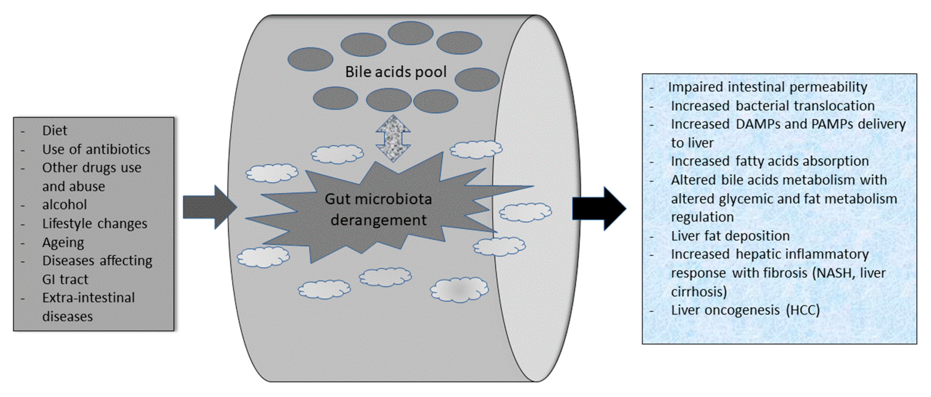
3.1. Gut Microbiota Composition in Health and Its “dysbiosis” in NAFLD Patients

The human intestine hosts over 100 trillion microbes, prevalently bacteria. There are also viruses, fungi, archaea, and protozoa [21]. Gut microbiota participates in nutrient absorption and fermentation, modulates intestinal permeability (IP), and is implicated in host metabolism (e.g., carbohydrates absorption and processing, proteins putrefaction, bile acids formation, insulin sensitivity), and modulation of mucosal and systemic immunity [22]. Firmicutes and Bacteroidetes represent the most abundant phyla of the bacterial microbiota. Their ratio is a fundamental factor in the host’s health [23]. Among the other phyla present, we find Fusobacteria, Actinobacteria, Proteobacteria, and Verrucomicrobia [24]. Eubiosis defines the balanced qualitative and quantitative condition of the intestinal microflora and is essential to preserving the host’s health [22,23,24]. On the contrary, its qualitative/quantitative perturbation–dysbiosis is associated with the development of various diseases such as NAFLD, NASH until HCC, diabetes type 2, cardiovascular disorders, and, undoubtedly, obesity [25,26,27,28]. Patients with NAFLD, during their stages of disease evolution, also have an increased number of Bacteroidetes and a reduction of Firmicutes abundance (a decreased F/B ratio) [29]. However, the F/B ratio may vary across studies mainly because of the gut microbiota studying technique. There are available culture-based and culture-free methods such as 16S rRNA gene sequencing until whole-genome shotgun sequencing. Finally, there are newer metagenomics approaches that allow whole genomic sequence reconstruction starting from a few genetic fragments present in the biological sample in the study. Interestingly, there are diversities in the microorganisms’ abundance within each phylum. NAFLD patients also show an increased proportion of species belonging to Clostridium, Anaerobacter, Streptococcus, Escherichia, and Lactobacillus, whereas Oscillibacter, Flavonifractor, Odoribacter, Alistipes spp. are less abundant [30]. More interestingly, the hallmark of decreased F/B ratio is not typical of patients suffering from HCC. Of interest, in children with NAFLD and NASH, there is a peculiar gut dysbiosis characterized by decreased abundance of Oscillospira spp. and by an increased concentration of Dorea, Blautia, Prevotella copri, and Ruminococcus spp. vs. in healthy children [31]. In terms of diversity, adult NAFLD gut microbiota shows decreased α-diversity (richness and evenness), significantly altered β-diversity, and significant differences in the abundance of bacteria at the phylum, class, family, or genus level vs. healthy subjects. Interestingly, NAFLD children show decreased α-diversity, distinct differences in β-diversity, or differing abundance of bacteria at the phylum or genus levels vs. healthy subjects.
It is worth mentioning the role of other factors in maintaining the dynamic balance of gut eubiosis in the every-day life of humans: diet, use of antibiotics, aging, diseases affecting GI nutrients absorption, motility, and inflammatory state. For example, animal experiments have shown how a high-fat diet is able to give an “obese shape” to our gut microbiota. On the other hand “obese gut microbiota transplantation” is able to change gut microbiota behavior in lean mice [32].
Diet, and related lifestyle, have a particular impact on NAFLD pathogenesis and physiopathologic evolution. However, the gut microbiota is a peculiar actor in NAFLD’s natural history that interacts with other specific actors within the gut, leading to specific liver steatosis and fibrosis features [6] (Figure 1).

3.2. Bile Acids (BA), Gut Microbiota, and NAFLD

The first physiologic interaction between BAs and microbiota happens in the large intestine where the microorganisms carry out several enzymatic reactions (e.g., deconjugation, dihydroxylation, and epimerization) to form secondary BAs such as deoxycholic acid (DCA) from cholic acid, and ursodeoxycholic acid (UDCA) and lithocholic acid (LCA) from chenodeoxycholic acid (CDCA) [33,34]. Indeed, BAs deconjugation is crucial to make BAs more lipophilic. Secondary BAs can be reabsorbed in the large bowel and return to the liver, via systemic circulation, allowing their “entero-hepatic circle” [23,35]. One proof of the strict relationship and evolutive interaction between intestinal microbiota and BAs is the presence of BAs hydrolases (BSH) in these microbes. In particular, BSH is present in most bacterial phyla; in Gram-positive bacteria, like Lactobacillus, Bifidobacterium, Clostridium spp., and Enterococcus, and in some commensal Gram-negative strains such as the Bacteroides spp. [36,37,38,39]. In pathogens such as Listeria monocytogenes, BSH activity probably represents an adaptive quality guaranteeing its gut persistence [40]. Interestingly, this adaptive quality has a horizontal transmission amongst bacteria [41]. More interestingly, the human gut can affect BSH activity in gut bacteria through a “host species-specific selection” of microbial BSH activities according to “species-specific” differences in BAs pools [42]. Indeed, BSH activity is a “protective shield” for bacteria colonizing the human gut [43]. In the frame of NAFLD, the consolidated concept of the gut–liver axis helps the understanding of the multi-directional interaction of BAs, gut microbiota, and liver “through” the intestine [14]. BAs are ligands for several receptors. They include FXR and G-protein-coupled bile acid receptor 1 (or TGR5). They mainly modulate host metabolism and BAs enterohepatic circulation [44]. BAs are natural antibacterial substances and maintain gut microbial eubiosis through the activation of FXR [45]. Physiologically, hepatic FXR activation by BAs induces the expression of atypical nuclear receptors small heterodimer partner (SHP), which promotes the inhibition of the sterol-regulatory element-binding protein-1c (SREBP-1c) with a reduced hepatic synthesis of triglycerides. In addition, FXR physiologically limits fat accumulation in the liver [46,47]. Moreover, FXR activation in the liver results in the inhibition of gluconeogenesis and glycolysis. These two effects have a potential protective role in insulin resistance and type II diabetes [46,47]. Interestingly, in NAFLD patients the BAs-FXR interaction is altered such as the resulting lipid and glucose metabolism [16]. Gut microbiota derangements are dynamically affected by BAs pool changes, and, conversely, BAs pool composition and abundance are affected by gut microbiota eubiosis and dysbiosis. In NAFLD patients, gut dysbiosis can alter the BAs pool and start a pathophysiological cascade that favors liver fat accumulation and microinflammation, which can result in fibrosis. The latter is a typical feature of NASH [48] (Figure 1). Bariatric surgery patients offer an example of FXR regulation by bile acids. In bariatric surgery on obese patients, the metabolic improvements obtained upon the procedure follow both weight-dependent and weight-independent mechanisms. Substantially, increased volumes of digestate-free BAs are delivered to the distal gut, with increased hepatic flux/enterohepatic circulation, increased plasma BA levels and composition, and, subsequent normalization of the blunted postprandial plasma BA concentration typical of obesity. Altogether, this results in modulation of satiety, improvement of lipid and cholesterol metabolism, incretins and glucose homeostasis, and energy storage and distribution via fine FXR and TGR5 interaction of BAs.

3.3. From Probiotics Use to Fecal Microbiota Transplantation (FMT) in NALFD Treatment

Several pieces of evidence from both in vitro and in vivo studies have shown that probiotics—“alive organisms beneficially affecting host (human) health”—can compete with pathogenic bacteria, improve intestinal permeability to pathogens and/or their molecules, exert immune-modulatory effects and gut-brain axis effects with the production of neurotransmitters [49]. In detail, probiotics can significantly decrease endotoxin levels within the gut [50]. Subsequently, the first human studies have revealed how probiotics can also clinically ameliorate NAFLD fat deposition, micro-inflammation, and fibrosis onset and progression. In detail, in overweight NAFLD patients, administration of multistrain probiotic mixtures reduces the systemic inflammatory state and, more interestingly, brings back the gut microbiota towards a “commensal” one [51,52]. However, these promising shreds of evidence have to face a solid Cochrane meta-analysis from 2007 that did not come to a clear conclusion substantially because of the lack of a sufficient number of randomized controlled trials [53]. Thus, RCTs have been doubling in the few last years. The multistrain VSL#3® and a combination of pro/prebiotics as symbiotics were able to reduce levels of TNF-α, liver transaminases, and oxidative stress markers in NAFLD patients for a duration of 2- 3 months [54]. Similar results have been obtained in children [55,56,57,58]. (Table 1).
In more detail, multistrain preparations show a significant gut microbiota reshaping in both NAFLD and NASH patients. In addition, their effect on liver function and biochemical lipid profile is positive: several RCTs report liver transaminases reduction with cholesterol and triglycerides reduction. There is also a significant reduction in inflammatory cytokines. It is important to mention that most of the enrolled patients were also type 2 diabetes subjects (Table 1).
However, dose- and strain(s)-finding studies are still missing and data on adverse effects linked to the use of probiotics in NAFLD patients. Thus, the use of live commensals coming directly from a healthy gut is appealing and may (in theory) guarantee a safer use than probiotics. These issues have paved the road for use of FMT in NAFLD patients. Several animal studies offered the first evidence of FMT in NAFLD. Leroy et al. showed that NAFLD- mice are able to induce the development of NAFLD in the vast majority of recipients of FMT. This finding was confirmed by the species-specific rise of bacteria being predominant colonizers upon FMT [65]. Furthermore, Zhou et al. studied two groups of high-fat diet (HFD)-fed mice; one of those underwent FMT from healthy donors. This group showed a significant reduction in histological findings typical of NAFLD (namely, intracellular hepatic lipid and proinflammatory cytokines concentration) vs. those not receiving healthy FMT [66]. More systematically, FMT has been successfully used in HFD-NAFLD mice. Interestingly, also NASH histological findings (namely, liver fibrosis and inflammatory infiltrates) have been demonstrated to improve after FMT. This healing was correlated with body weight, fat content, and serum reduction of transaminases levels [67]. Anecdotal reports show a fine interaction between gut microbiota and NAFLD in humans, including eighteen patients with metabolic syndrome diagnosis received either self-FMT or from lean healthy subjects. Interestingly, six weeks after FMT, insulin sensitivity significantly improved, and this improvement was correlated with an increase in butyrate-producing bacteria [68]. Phillips et al. confirmed the efficacy of FMT in the treatment of alcoholic hepatitis patients, also describing the gut dysbiosis changes after treatment [69]. In this pilot study, eight male patients with severe acute alcoholic hepatitis (SAH) consented to the FMT procedure. FMT consisted of thirty grams of donor stool samples, obtained after screening from consenting family members, homogenized with 100 mL of sterile normal saline, and filtered using sterile gauze. Therefore, small amounts were infused through a nasoduodenal tube daily for one week. Control subjects were historical patients with SAH under standard-of-care treatment. Samples for the gut microbiota study were collected at baseline, 6-month, and one-year intervals. Interestingly, one week of FMT was able to improve liver disease severity indexes and, more importantly, the survival rate at 1 year after treatment. In addition, FMT was confirmed to be a safe procedure, recording just flatulence as a mild side effect. In detail, and more intriguingly, there was the coexistence of donor and recipient gut microbial species at 6 and 12 months after FMT in treated patients. This finding describes a peculiar behavior of transplanted microbial species from donors: they are less pathogenic and beneficial and coexist with pre-existing bacterial communities instead of replacing those of the recipient. The latter are “beneficially” modified by bacteria from donors, resulting in a new-one “symbiotic coexistence”. This data finds initial agreement in the literature [70]. Very recently, a study by Xue et al. has better highlighted the different impacts of FMT on NAFLD according to the presence or absence of obesity [71]. NAFLD patients were randomized to receive either oral probiotics or FMT. FMT was prepared from donor stool (heterologous), administered through colonoscopy, and followed by three enemas over 3 days. Both groups were required to undergo healthy lifestyle changes (namely, a healthy diet and regular physical exercise for more than 40 min per day). Patients were rechecked 1 month after treatment (e.g., liver fat deposition, fecal microbiota composition). Interestingly, FMT was able to significantly decrease liver fat accumulation and reduce gut microbiota dysbiosis. Importantly, FMT had a significantly higher healing efficacy on lean NAFLD vs. obese patients. This finding was consensual with a more marked restoration of gut eubiosis in lean vs. obese patients. Indeed, this study reinforces evidence from the literature showing a different impact of gut dysbiosis in lean fatty liver, which is not correlated to impaired lipid and glucose metabolism issues such as obese fatty liver disease [72]. Currently, it remains to be investigated whether or not the FMT impact NAFLD and NASH complications. In detail, the RCTs available in the literature have shown a good efficacy of FMT used in NAFLD, NASH diabetic, and non-diabetic patients with improvement of glycemic control, and liver steatosis. However, some trials showed better efficacy in liver fibrosis reversal (NASH feature) vs. liver steatosis (namely, NAFLD subjects). Finally, some promising report confirms FMT safety also in liver cirrhosis patients, considered as an evolution of NASH (Table 2).
Overall, FMT can be considered a therapeutic, safe treatment in NAFLD patients, and perhaps it is a step up from NASH.

4. Conclusions

Gut microbial dysbiosis is a validated pathogenic mechanism connected to the pathophysiology of NAFLD and its stages. The type and severity of dysbiosis differ between patients with NAFLD, NASH, cirrhotic, and HCC. In addition, it is worth mentioning the recent evidence describing the different weights of gut dysbiosis in lean vs. obese NAFLD patients. This evidence is mirrored by the major efficacy of amelioration of liver fat deposition and reduction of dysbiosis of FMT in lean vs. obese NAFLD patients. FMT is gaining more and more evidence for use as a treatment of NAFLD, as probiotics and lifestyle changes have several limits and risks for patients. Probiotics use should be “personalized” for different NAFLD patients; accurate dose-finding and adverse events studies are needed. Lifestyle changes show the best efficacy in the treatment of obese patients. However, this subset of patients shows compliance with short- and long-term difficulties in mining the use of this life asset over the entire lifespan. Thus, FMT seems to be a safe, efficient treatment for NAFLD patients. However, more RCT studies and long-term follow-ups are needed to verify their efficacy, especially in lean NAFLD patients, often not beneficially affected by lifestyle changes, cholesterol-lowering agents and probiotics use.

Author Contributions

Conceptualization, E.S. and L.A.; methodology, V.M., A.D.B., C.R. and P.S.; software, A.C.P.; validation, E.S. and L.A.; formal analysis, E.S. and L.A.; investigation, E.S. and V.M.; data curation, A.C.P.; writing—original draft preparation, E.S., L.A. and A.C.P.; writing—review and editing, E.S., L.A., L.B., P.S., E.R. and J.T.; visualization, A.D.B., V.M., E.R. and J.T.; supervision, E.S. and L.A.; project administration, E.S. and L.A. All authors have read and agreed to the published version of the manuscript.

Funding

This research received no external funding.

Institutional Review Board Statement

Not applicable.

Informed Consent Statement

Not applicable.

Data Availability Statement

All the data reported in this review of literature are available online on PubMed and main national and international gastroenterological meetings websites.

Conflicts of Interest

The authors declare no conflict of interest.

References

  1. Pinart, M.; Dötsch, A.; Schlicht, K.; Laudes, M.; Bouwman, J.; Forslund, S.K.; Pischon, T.; Nimptsch, K. Gut Microbiome Composition in Obese and Non-Obese Persons: A Systematic Review and Meta-Analysis. Nutrients 2021, 14, 12. [Google Scholar] [CrossRef] [PubMed]
  2. Hofman, D.; Kudla, U.; Miqdady, M.; Nguyen, T.V.H.; Morán-Ramos, S.; Vandenplas, Y. Faecal Microbiota in Infants and Young Children with Functional Gastrointestinal Disorders: A Systematic Review. Nutrients 2022, 14, 974. [Google Scholar] [CrossRef] [PubMed]
  3. Van Zyl, K.N.; Matukane, S.R.; Hamman, B.L.; Whitelaw, A.C.; Newton-Foot, M. Effect of antibiotics on the human microbiome: A systematic review. Int. J. Antimicrob. Agents 2021, 59, 106502. [Google Scholar] [CrossRef]
  4. Thrastardottir, T.O.; Copeland, V.J.; Constantinou, C. The Association Between the Gut Microbiome, Nutritional Habits, Antibiotics, and Gastric Cancer: A Scoping Review. Curr. Nutr. Rep. 2022, 11, 19–38. [Google Scholar] [CrossRef]
  5. Amedei, A.; Boem, F. I’ve Gut A Feeling: Microbiota Impacting the Conceptual and Experimental Perspectives of Personalized Medicine. Int. J. Mol. Sci. 2018, 19, 3756. [Google Scholar] [CrossRef] [PubMed] [Green Version]
  6. Yaqub, S.; Ananias, P.; Shah, A.; Luenam, K.; Jose, A.M.; Melo, J.P.; Turkistani, A.; Mohammed, L. Decoding the Pathophysiology of Non-alcoholic Fatty Liver Disease Progressing to Non-alcoholic Steatohepatitis: A Systematic Review. Cureus 2021, 13, e18201. [Google Scholar] [CrossRef]
  7. Younossi, Z.M.; Golabi, P.; de Avila, L.; Paik, J.M.; Srishord, M.; Fukui, N.; Qiu, Y.; Burns, L.; Afendy, A.; Nader, F. The global epidemiology of NAFLD and NASH in patients with type 2 diabetes: A systematic review and meta-analysis. J. Hepatol. 2019, 71, 793–801. [Google Scholar] [CrossRef]
  8. Mascaró, C.M.; Bouzas, C.; A Tur, J. Association between Non-Alcoholic Fatty Liver Disease and Mediterranean Lifestyle: A Systematic Review. Nutrients 2021, 14, 49. [Google Scholar] [CrossRef]
  9. Le, M.H.; Yeo, Y.H.; Li, X.; Li, J.; Zou, B.; Wu, Y.; Ye, Q.; Huang, D.Q.; Zhao, C.; Zhang, J.; et al. 2019 Global NAFLD Prevalence: A Systematic Review and Meta-analysis. Clin. Gastroenterol. Hepatol. 2021, 21, S1542–S3565. [Google Scholar] [CrossRef]
  10. Bifari, F.; Manfrini, R.; Cas, M.D.; Berra, C.; Siano, M.; Zuin, M.; Paroni, R.; Folli, F. Multiple target tissue effects of GLP-1 analogues on non-alcoholic fatty liver disease (NAFLD) and non-alcoholic steatohepatitis (NASH). Pharmacol. Res. 2018, 137, 219–229. [Google Scholar] [CrossRef]
  11. Majzoub, A.M.; Nayfeh, T.; Barnard, A.; Munaganuru, N.; Dave, S.; Singh, S.; Murad, M.H.; Loomba, R. Systematic review with network meta-analysis: Comparative efficacy of pharmacologic therapies for fibrosis improvement and resolution of NASH. Aliment. Pharmacol. Ther. 2021, 54, 880–889. [Google Scholar] [CrossRef] [PubMed]
  12. Petrelli, F.; Manara, M.; Colombo, S.; De Santi, G.; Ghidini, M.; Mariani, M.; Iaculli, A.; Rausa, E.; Rampulla, V.; Arru, M.; et al. Hepatocellular carcinoma in patients with nonalcoholic fatty liver disease: A systematic review and meta-analysis: HCC and Steatosis or Steatohepatitis. Neoplasia 2022, 30, 100809. [Google Scholar] [CrossRef] [PubMed]
  13. Li, F.; Ye, J.; Shao, C.; Zhong, B. Compositional alterations of gut microbiota in nonalcoholic fatty liver disease patients: A systematic review and Meta-analysis. Lipids Health Dis. 2021, 20, 22. [Google Scholar] [CrossRef] [PubMed]
  14. De Munck, T.J.I.; Xu, P.; Verwijs, H.J.A.; Masclee, A.A.M.; Jonkers, D.; Verbeek, J.; Koek, G.H. Intestinal permeability in human nonalcoholic fatty liver disease: A systematic review and meta-analysis. Liver Int. 2020, 40, 2906–2916. [Google Scholar] [CrossRef]
  15. Stofan, M.; Guo, G.L. Bile Acids and FXR: Novel Targets for Liver Diseases. Front. Med. 2020, 7, 544. [Google Scholar] [CrossRef]
  16. Mori, H.; Baroni, G.S.; Marzioni, M.; Di Nicola, F.; Santori, P.; Maroni, L.; Abenavoli, L.; Scarpellini, E. Farnesoid X Receptor, Bile Acid Metabolism, and Gut Microbiota. Metabolites 2022, 12, 647. [Google Scholar] [CrossRef]
  17. Sabirin, F.; Lim, S.M.; Neoh, C.F.; Ramasamy, K. Hepatoprotection of Probiotics Against Non-Alcoholic Fatty Liver Disease in vivo: A Systematic Review. Front. Nutr. 2022, 9, 844374. [Google Scholar] [CrossRef]
  18. Rakotonirina, A.; Galperine, T.; Allémann, E. Fecal microbiota transplantation: A review on current formulations in Clostridioides difficile infection and future outlooks. Expert Opin. Biol. Ther. 2022, 22, 929–944. [Google Scholar] [CrossRef]
  19. Suk, K.T.; Koh, H. New perspective on fecal microbiota transplantation in liver diseases. J. Gastroenterol. Hepatol. 2021, 37, 24–33. [Google Scholar] [CrossRef]
  20. Liberati, A.; Altman, D.G.; Tetzlaff, J.; Mulrow, C.; Gøtzsche, P.C.; Ioannidis, J.P.A.; Clarke, M.; Devereaux, P.J.; Kleijnen, J.; Moher, D. The PRISMA Statement for Reporting Systematic Reviews and Meta-Analyses of Studies That Evaluate Health Care Interventions: Explanation and Elaboration. PLoS Med. 2009, 6, e1000100. [Google Scholar] [CrossRef]
  21. Scarpellini, E.; Fagoonee, S.; Rinninella, E.; Rasetti, C.; Aquila, I.; LaRussa, T.; Ricci, P.; Luzza, F.; Abenavoli, L. Gut Microbiota and Liver Interaction through Immune System Cross-Talk: A Comprehensive Review at the Time of the SARS-CoV-2 Pandemic. J. Clin. Med. 2020, 9, 2488. [Google Scholar] [CrossRef] [PubMed]
  22. Maynard, C.L.; Elson, C.O.; Hatton, R.D.; Weaver, C.T. Reciprocal interactions of the intestinal microbiota and immune system. Nature 2012, 489, 231–241. [Google Scholar] [CrossRef] [PubMed] [Green Version]
  23. Fukuda, S.; Toh, H.; Taylor, T.; Ohno, H.; Hattori, M. Acetate-producing bifidobacteria protect the host from enteropathogenic infection via carbohydrate transporters. Gut Microbes 2012, 3, 449–454. [Google Scholar] [CrossRef] [PubMed] [Green Version]
  24. Scarpellini, E.; Basilico, M.; Rinninella, E.; Carbone, F.; Schol, J.; Rasetti, C.; Abenavoli, L.; Santori, P. Probiotics and gut health. Minerva Gastroenterol. 2022, 67, 314–325. [Google Scholar] [CrossRef] [PubMed]
  25. Tedelind, S.; Westberg, F.; Kjerrulf, M.; Vidal, A. Anti-inflammatory properties of the short-chain fatty acids acetate and propionate: A study with relevance to inflammatory bowel disease. World J. Gastroenterol. 2007, 13, 2826–2832. [Google Scholar] [CrossRef] [PubMed]
  26. Melbye, P.; Olsson, A.; Hansen, T.H.; Søndergaard, H.B.; Oturai, A.B. Short-chain fatty acids and gut microbiota in multiple sclerosis. Acta Neurol. Scand. 2018, 139, 208–219. [Google Scholar] [CrossRef]
  27. Schaad, U.B.; Mütterlein, R.; Goffin, H.; BV-Child Study Group. Immunostimulation with OM-85 in Children with Recurrent Infections of the Upper Respiratory Tract: A Double-Blind, Placebo-Controlled Multicenter Study. Chest 2002, 122, 2042–2049. [Google Scholar] [CrossRef] [PubMed]
  28. Feleszko, W.; Jaworska, J.; Rha, R.-D.; Steinhausen, S.; Avagyan, A.; Jaudszus, A.; Ahrens, B.; Groneberg, D.A.; Wahn, U.; Hamelmann, E. Probiotic-induced suppression of allergic sensitization and airway inflammation is associated with an increase of T regulatory-dependent mechanisms in a murine model of asthma. Clin. Exp. Allergy 2006, 37, 498–505. [Google Scholar] [CrossRef]
  29. Jadhav, K.; Cohen, T.S. Can You Trust Your Gut? Implicating a Disrupted Intestinal Microbiome in the Progression of NAFLD/NASH. Front. Endocrinol. 2020, 11, 592157. [Google Scholar] [CrossRef]
  30. Jiang, W.; Wu, N.; Wang, X.; Chi, Y.; Zhang, Y.; Qiu, X.; Hu, Y.; Li, J.; Liu, Y. Dysbiosis gut microbiota associated with inflammation and impaired mucosal immune function in intestine of humans with non-alcoholic fatty liver disease. Sci. Rep. 2015, 5, 08096. [Google Scholar] [CrossRef] [Green Version]
  31. Del Chierico, F.; Nobili, V.; Vernocchi, P.; Russo, A.; De Stefanis, C.; Gnani, D.; Furlanello, C.; Zandonà, A.; Paci, P.; Capuani, G.; et al. Gut microbiota profiling of pediatric nonalcoholic fatty liver disease and obese patients unveiled by an integrated meta-omics-based approach. Hepatology 2017, 65, 451–464. [Google Scholar] [CrossRef] [PubMed] [Green Version]
  32. Burz, S.; Monnoye, M.; Philippe, C.; Farin, W.; Ratziu, V.; Strozzi, F.; Paillarse, J.-M.; Chêne, L.; Blottière, H.; Gérard, P. Fecal Microbiota Transplant from Human to Mice Gives Insights into the Role of the Gut Microbiota in Non-Alcoholic Fatty Liver Disease (NAFLD). Microorganisms 2021, 9, 199. [Google Scholar] [CrossRef] [PubMed]
  33. Kumar, A.; Al-Hassi, H.O.; Steed, H.; Phipps, O.; Brookes, M.J. Bile Acids and the Microbiome: Making Sense of This Dynamic Relationship in Their Role and Management in Crohn’s Disease. Can. J. Gastroenterol. Hepatol. 2022, 2022, 1–12. [Google Scholar] [CrossRef] [PubMed]
  34. Cowen, A.E.; Κοrman, M.G.; Hofmann, A.F.; Cass, O.W.; Coffin, S.B. Metabolism of Lithocholate in Healthy Man. II. Entero-hepatic circulation. Gastroenterology 1975, 69, 67–76. [Google Scholar] [CrossRef]
  35. Urdaneta, V.; Casadesús, J. Interactions between Bacteria and Bile Salts in the Gastrointestinal and Hepatobiliary Tracts. Front. Med. 2017, 4, 163. [Google Scholar] [CrossRef] [Green Version]
  36. Kumar, R.S.; Brannigan, J.A.; Prabhune, A.; Pundle, A.V.; Dodson, G.G.; Dodson, E.J.; Suresh, C.G. Structural and Functional Analysis of a Conjugated Bile Salt Hydrolase from Bifidobacterium longum Reveals an Evolutionary Relationship with Penicillin V Acylase. J. Biol. Chem. 2006, 281, 32516–32525. [Google Scholar] [CrossRef] [Green Version]
  37. Gopal-Srivastava, R.; Hylemon, P.B. Purification and characterization of bile salt hydrolase from Clostridium perfringens. J. Lipid Res. 1988, 29, 1079–1085. [Google Scholar] [CrossRef]
  38. Chand, D.; Panigrahi, P.; Varshney, N.; Ramasamy, S.; Suresh, C. Structure and function of a highly active Bile Salt Hydrolase (BSH) from Enterococcus faecalis and post-translational processing of BSH enzymes. Biochim. et Biophys. Acta (BBA)-Proteins Proteom. 2018, 1866, 507–518. [Google Scholar] [CrossRef]
  39. Stellwag, E.; Hylemon, P. Purification and characterization of bile salt hydrolase from Bacteroides fragilis subsp. fragilis. Biochim. et Biophys. Acta (BBA)-Enzym. 1976, 452, 165–176. [Google Scholar] [CrossRef]
  40. Begley, M.; Gahan, C.G.; Hill, C. The interaction between bacteria and bile. FEMS Microbiol. Rev. 2005, 29, 625–651. [Google Scholar] [CrossRef]
  41. Dussurget, O.; Cabanes, D.; Dehoux, P.; Lecuit, M.; Buchrieser, C.; Glaser, P.; Cossart, P. The European Listeria Genome Consortium Listeria monocytogenes bile salt hydrolase is a PrfA-regulated virulence factor involved in the intestinal and hepatic phases of listeriosis. Mol. Microbiol. 2002, 45, 1095–1106. [Google Scholar] [CrossRef] [PubMed]
  42. Jia, B.; Park, D.; Chun, B.; Hahn, Y.; Jeon, C. Diet-Related Alterations of Gut Bile Salt Hydrolases Determined Using a Metagenomic Analysis of the Human Microbiome. Int. J. Mol. Sci. 2021, 22, 3652. [Google Scholar] [CrossRef] [PubMed]
  43. De Smet, I.; Van Hoorde, L.; Woestyne, M.V.; Christiaens, H.; Verstraete, W. Significance of bile salt hydrolytic activities of lactobacilli. J. Appl. Bacteriol. 1995, 79, 292–301. [Google Scholar] [CrossRef] [PubMed]
  44. Bertolini, A.; Fiorotto, R.; Strazzabosco, M. Bile acids and their receptors: Modulators and therapeutic targets in liver inflammation. Semin. Immunopathol. 2022, 44, 547–564. [Google Scholar] [CrossRef]
  45. Parséus, A.; Sommer, N.; Sommer, F.; Caesar, R.; Molinaro, A.; Ståhlman, M.; Greiner, T.U.; Perkins, R.; Bäckhed, F. Microbiota-induced obesity requires farnesoid X receptor. Gut 2017, 66, 429–437. [Google Scholar] [CrossRef] [PubMed] [Green Version]
  46. Watanabe, M.; Houten, S.M.; Wang, L.; Moschetta, A.; Mangelsdorf, D.J.; Heyman, R.A.; Moore, D.D.; Auwerx, J. Bile acids lower triglyceride levels via a pathway involving FXR, SHP, and SREBP-1c. J. Clin. Investig. 2004, 113, 1408–1418. [Google Scholar] [CrossRef] [PubMed] [Green Version]
  47. Pineda Torra, I.; Claudel, T.; Duval, C.; Kosykh, V.; Fruchart, J.-C.; Staels, B. Bile Acids Induce the Expression of the Human Peroxisome Proliferator-Activated Receptor Alpha Gene via Activation of the Farnesoid X Receptor. Mol. Endocrinol. 2003, 17, 259–272. [Google Scholar] [CrossRef] [Green Version]
  48. Penney, N.; Kinross, J.; Newton, R.C.; Purkayastha, S. The role of bile acids in reducing the metabolic complications of obesity after bariatric surgery: A systematic review. Int. J. Obes. 2015, 39, 1565–1574. [Google Scholar] [CrossRef]
  49. Sánchez, B.; Delgado, S.; Blanco-Míguez, A.; Lourenço, A.; Gueimonde, M.; Margolles, A. Probiotics, gut microbiota, and their influence on host health and disease. Mol. Nutr. Food Res. 2017, 61, 1600240. [Google Scholar] [CrossRef] [Green Version]
  50. He, S.; Cui, S.; Song, W.; Jiang, Y.; Chen, H.; Liao, D.; Lu, X.; Li, J.; Chen, X.; Peng, L. Interleukin-17 Weakens the NAFLD/NASH Process by Facilitating Intestinal Barrier Restoration Depending on the Gut Microbiota. mBio 2022, 13, e0368821. [Google Scholar] [CrossRef]
  51. Kobyliak, N.; Abenavoli, L.; Mykhalchyshyn, G.; Kononenko, L.; Boccuto, L.; Kyriienko, D.; Dynnyk, O. A Multi-strain Probiotic Reduces the Fatty Liver Index, Cytokines and Aminotransferase levels in NAFLD Patients: Evidence from a Randomized Clinical Trial. J. Gastrointest. Liver Dis. 2018, 27, 41–49. [Google Scholar] [CrossRef] [PubMed] [Green Version]
  52. Derosa, G.; Guasti, L.; D’Angelo, A.; Martinotti, C.; Valentino, M.C.; Di Matteo, S.; Bruno, G.M.; Maresca, A.M.; Gaudio, G.V.; Maffioli, P. Probiotic Therapy With VSL#3® in Patients With NAFLD: A Randomized Clinical Trial. Front Nutr. 2022, 9, 846873. [Google Scholar] [CrossRef] [PubMed]
  53. Lirussi, F.; Mastropasqua, E.; Orando, S.; Orlando, R. Probiotics for non-alcoholic fatty liver disease and/or steatohepatitis. Cochrane Database Syst. Rev. 2007, 2007, CD005165. [Google Scholar] [CrossRef] [PubMed]
  54. Loguercio, C.; Federico, A.; Tuccillo, C.; Terracciano, F.; D’Auria, M.V.; De Simone, C.; Del Vecchio Blanco, C. Beneficial Effects of a Probiotic VSL#3 on Parameters of Liver Dysfunction in Chronic Liver Diseases. J. Clin. Gastroenterol. 2005, 39, 540–543. [Google Scholar] [CrossRef] [Green Version]
  55. Vajro, P.; Mandato, C.; Licenziati, M.R.; Franzese, A.; Vitale, D.F.; Lenta, S.; Caropreso, M.; Vallone, G.; Meli, R. Effects of Lactobacillus rhamnosus Strain GG in Pediatric Obesity-related Liver Disease. J. Pediatr. Gastroenterol. Nutr. 2011, 52, 740–743. [Google Scholar] [CrossRef] [PubMed] [Green Version]
  56. Aller, R.; De Luis, D.A.; Izaola, O.; Conde, R.; Gonzalez Sagrado, M.; Primo, D.; De La Fuente, B.; Gonzalez, J. Effect of a probiotic on liver aminotransferases in nonalcoholic fatty liver disease patients: A double blind randomized clinical trial. Eur. Rev. Med. Pharmacol. Sci. 2011, 15, 1090–1095. [Google Scholar]
  57. Alisi, A.; Bedogni, G.; Baviera, G.; Giorgio, V.; Porro, E.; Paris, C.; Giammaria, P.; Reali, L.; Anania, F.; Nobili, V. Randomised clinical trial: The beneficial effects of VSL#3 in obese children with non-alcoholic steatohepatitis. Aliment. Pharmacol. Ther. 2014, 39, 1276–1285. [Google Scholar] [CrossRef] [Green Version]
  58. Famouri, F.; Shariat, Z.; Hashemipour, M.; Keikha, M.; Kelishadi, R. Effects of Probiotics on Nonalcoholic Fatty Liver Disease in Obese Children and Adolescents. J. Pediatr. Gastroenterol. Nutr. 2017, 64, 413–417. [Google Scholar] [CrossRef]
  59. Wong, V.W.-S.; Tse, C.-H.; Lam, T.-T.Y.; Wong, G.L.-H.; Chim, A.M.-L.; Chu, W.C.-W.; Yeung, D.K.-W.; Law, P.T.-W.; Kwan, H.S.; Yu, J.; et al. Molecular Characterization of the Fecal Microbiota in Patients with Nonalcoholic Steatohepatitis–A Longitudinal Study. PLoS ONE 2013, 8, e62885. [Google Scholar] [CrossRef] [Green Version]
  60. Kobyliak, N.; Abenavoli, L.; Falalyeyeva, T.; Mykhalchyshyn, G.; Boccuto, L.; Kononenko, L.; Kyriienko, D.; Komisarenko, I.; Dynnyk, O. Beneficial effects of probiotic combination with omega-3 fatty acids in NAFLD: A randomized clinical study. Minerva Med. 2018, 109, 418–428. [Google Scholar] [CrossRef]
  61. Wang, W.; Shi, L.P.; Shi, L.; Xu, L. [Efficacy of probiotics on the treatment of non-alcoholic fatty liver disease]. Zhonghua Nei Ke Za Zhi 2018, 57, 101–106. [Google Scholar] [CrossRef] [PubMed]
  62. Manzhalii, E.; Virchenko, O.; Falalyeyeva, T.; Beregova, T.; Stremmel, W. Treatment efficacy of a probiotic preparation for non-alcoholic steatohepatitis: A pilot trial. J. Dig. Dis. 2017, 18, 698–703. [Google Scholar] [CrossRef] [PubMed]
  63. Sepideh, A.; Karim, P.; Hossein, A.; Leila, R.; Hamdollah, M.; Mohammad, E.G.; Mojtaba, S.; Mohammad, S.; Ghader, G.; Moayed, A.S. Effects of Multistrain Probiotic Supplementation on Glycemic and Inflammatory Indices in Patients with Nonalcoholic Fatty Liver Disease: A Double-Blind Randomized Clinical Trial. J. Am. Coll. Nutr. 2015, 35, 500–505. [Google Scholar] [CrossRef] [PubMed]
  64. Nabavi, S.; Rafraf, M.; Somi, M.; Homayouni-Rad, A.; Asghari-Jafarabadi, M. Effects of probiotic yogurt consumption on metabolic factors in individuals with nonalcoholic fatty liver disease. J. Dairy Sci. 2014, 97, 7386–7393. [Google Scholar] [CrossRef]
  65. Le Roy, T.; Llopis, M.; Lepage, P.; Bruneau, A.; Rabot, S.; Bevilacqua, C.; Martin, P.; Philippe, C.; Walker, F.; Bado, A.; et al. Intestinal microbiota determines development of non-alcoholic fatty liver disease in mice. Gut 2012, 62, 1787–1794. [Google Scholar] [CrossRef]
  66. Kelly, C.J.; Zheng, L.; Campbell, E.L.; Saeedi, B.; Scholz, C.C.; Bayless, A.J.; Wilson, K.E.; Glover, L.E.; Kominsky, D.J.; Magnuson, A.; et al. Crosstalk between Microbiota-Derived Short-Chain Fatty Acids and Intestinal Epithelial HIF Augments Tissue Barrier Function. Cell Host Microbe 2015, 17, 662–671. [Google Scholar] [CrossRef] [Green Version]
  67. Zhou, D.; Pan, Q.; Shen, F.; Cao, H.-X.; Ding, W.-J.; Chen, Y.-W.; Fan, J.-G. Total fecal microbiota transplantation alleviates high-fat diet-induced steatohepatitis in mice via beneficial regulation of gut microbiota. Sci. Rep. 2017, 7, 1–11. [Google Scholar] [CrossRef] [Green Version]
  68. Vrieze, A.; Van Nood, E.; Holleman, F.; Salojärvi, J.; Kootte, R.S.; Bartelsman, J.F.; Dallinga-Thie, G.M.; Ackermans, M.T.; Serlie, M.J.; Oozeer, R.; et al. Transfer of Intestinal Microbiota From Lean Donors Increases Insulin Sensitivity in Individuals With Metabolic Syndrome. Gastroenterology 2012, 143, 913–916.e7. [Google Scholar] [CrossRef]
  69. Philips, C.A.; Pande, A.; Shasthry, S.M.; Jamwal, K.D.; Khillan, V.; Chandel, S.S.; Kumar, G.; Sharma, M.K.; Maiwall, R.; Jindal, A.; et al. Healthy Donor Fecal Microbiota Transplantation in Steroid-Ineligible Severe Alcoholic Hepatitis: A Pilot Study. Clin. Gastroenterol. Hepatol. 2016, 15, 600–602. [Google Scholar] [CrossRef]
  70. Song, S.J.; Lauber, C.; Costello, E.K.; Lozupone, C.A.; Humphrey, G.; Berg-Lyons, D.; Caporaso, J.G.; Knights, D.; Clemente, J.C.; Nakielny, S.; et al. Cohabiting family members share microbiota with one another and with their dogs. eLife 2013, 2, e00458. [Google Scholar] [CrossRef]
  71. Xue, L.; Deng, Z.; Luo, W.; He, X.; Chen, Y. Effect of Fecal Microbiota Transplantation on Non-Alcoholic Fatty Liver Disease: A Randomized Clinical Trial. Front. Cell. Infect. Microbiol. 2022, 12, 759306. [Google Scholar] [CrossRef] [PubMed]
  72. Tang, A.; Ng, C.H.; Phang, P.H.; Chan, K.E.; Chin, Y.H.; Fu, C.E.; Zeng, R.W.; Xiao, J.; Tan, D.J.H.; Quek, J.; et al. Comparative Burden of Metabolic Dysfunction in Lean NAFLD vs Non-lean NAFLD-A Systematic Review and Meta-analysis. Clin. Gastroenterol. Hepatol. 2022, S1542–S3565, 00669-3. [Google Scholar] [CrossRef] [PubMed]
Figure 1. The complex interaction between factors affecting gut microbiota composition, its interaction with bile acids pool, and NAFLD pathogenesis. GI: gastrointestinal; DAMPs: damage associated microbial products; PAMPs: pathogens associated microbial products; NASH: non-alcoholic steatohepatitis.
Medicina 58 01559 g001
Table 1. Clinical trials using probiotics for NAFLD patients’ treatment.
PatientsType of StudyProbiotics UsedOutcomesRef
48 children with NAFLDRandomized clinical trials (RCT)VSL#3 supplementation for 4 months vs. placeboNAFLD reversal[57]
64 children with obesity and NAFLDRCT (triple blind)L. acidophilus,
B. lactis,
B. bifidum,
L. rhamnosus
↓ALT, AST, mean
cholesterol, LDL-C, and TG in the probiotic-administered group
[58]
38 patients (16 with NASH diagnosis): 7 treated with
Probiotics, 9 with the standard of care vs. 22 healthy controls
RCTLactobacillus ssp. and Bifidobacterium
bifidum
In NASH patients there was gut microbiota modulation:
↓Faecalibacterium
↓Anaerosporobacter
↑Parabacteroide
↑Allisonella
[59]
48 T2DM NAFLD patientsRCT“Symbiter Omega”
probiotic biomass vs. placebo
↓Fatty acids, ↓serum
gamma-glutamyl
transpeptidase
↓TG ↓TC
[60]
58 patients with NAFLD and T2DM:
30 treated with probiotics vs. 28 receiving a placebo
RCTbiomass
of 14 probiotic
bacterial genera
In NAFLD patients: ↓Liver fat deposition
↓aminotransferase
↓TNF-α and IL-6
[51]
200 patients with NAFLD randomized to control group
(standard of care treatment) and add-on treatments groups A, B, and C.
RCTBifidobacterium,
Lactobacillus,
Enterococcus,
Bacillus subtilis,
and Enterococcus
Amelioration of fatty
liver deposition, ↓ALT, AST and
TNF-α
↑HMW-APN
[61]
75 patients with NASH under a low-fat/low-calorie dietRCTLactobacilli,
Bifidobacteria,
Streptococcus
thermophilus
Gut microbiota modulation towards “healthy” one, ↓BMI, ↓cholesterol in the probiotic-treated group[62]
50 patients (42 NAFLD) were randomized to probiotic or
placebo
RCTL. casei,
L. acidophilus,
L. rhamnosus,
L. bulgaricus,
B. breve, B. longum,
S. thermophilus
↓glycemic,
inflammatory markers, Insulin, insulin
resistance in NAFLD patients
[63]
30 patients with NAFLD RCTL. bulgaricus and
S. thermophilus
↓ALT, AST, GGT in the probiotics group[56]
72 patients with NAFLD RCTProbiotic yogurt↓ALT, AST, TC, LDL-C[64]
NAFLD: non-alcoholic fatty liver disease, NASH: non-alcoholic steatohepatitis, T2DM: type 2 diabetes mellitus, TNF-α tumor necrosis factor-α, IL-6: interleukin 6, BMI: body mass index, LDL: low-density lipoprotein, TC: total cholesterol, LDL-C: LDL cholesterol, HMW-APN high molecular weight adiponectin, LDL-C: low-density lipoprotein cholesterol, TG: triglycerides, GGT: gamma-glutamyl transferase, ALT alanine aminotransferase, AST aspartate aminotransferase, ↑ increase, ↓ decrease.
Table 2. Clinical trials using FMT for NAFLD treatment.
Study TypePatientsOutcomesRCT Number
FMT via nasojejunal tubeDiabetes and NAFLDImproved HOMA indexNCT02469272
FMT, pilot studyNAFLD and NASHImproved degree of liver steatosis as assessed by MRINCT02469272
FMT via duodenal infusionNAFLD and NASHEfficacy in NASH treatment vs. NAFLDNCT03803540
FMT via duodenal infusionNAFLD and NASHReduction of hepatic venous gradient pressureNCT02721264
FMT vs. standard treatment (RCT)Liver cirrhosis derived from NASHSafety (e.g., number of adverse events, complications rate)NCT02868164
NAFLD: non-alcoholic fatty liver disease; NASH: non-alcoholic steatohepatitis; FMT: fecal microbiota transplantation; HOMA: homeostasis model assessment; MRI: magnetic resonance imaging.
Publisher’s Note: MDPI stays neutral with regard to jurisdictional claims in published maps and institutional affiliations.

