Article

Accelerated Oral Healing by Angelica gigas Nakai from Hot Melt Extrusion Technology: An In Vitro Study

1
Department of Pediatric Dentistry, Kyung Hee University Medical Center, College of Dentistry, Kyung Hee University, Seoul 02447, Republic of Korea
2
Department of Bio-Health Convergence, Kangwon National University, Chuncheon 24341, Republic of Korea
3
Department of Rehabilitative Medicine of Korean Medicine and Neuropsychiatry, College of Korean Medicine, Sangji University, Wonju 26339, Republic of Korea
4
Department of Oriental Pharmaceutical Science, College of Pharmacy, Kyung Hee University, Seoul 02447, Republic of Korea
5
BeNatureBioLab, Chuncheon 24206, Republic of Korea
6
Department of Pediatric Dentistry, School of Dentistry, Kyung Hee University, Seoul 02447, Republic of Korea
*
Authors to whom correspondence should be addressed.
These authors contributed equally to this work.
Medicina 2023, 59(12), 2066; https://doi.org/10.3390/medicina59122066
Submission received: 1 November 2023 / Revised: 16 November 2023 / Accepted: 21 November 2023 / Published: 23 November 2023
(This article belongs to the Special Issue Medicine and Dentistry: New Methods and Clinical Approaches)

Abstract

:
Background and Objectives: In spite of the oral environment being healing-prone, its dynamic changes may affect wound healing. The purpose of this study was to assess the oral wound healing effect of Angelica gigas Nakai (AG) prepared by hot-melt extrusion. Materials and Methods: Human gingival fibroblast (HGF) cells were treated with AG or AG via hot-melt extrusion (AGH) for 24 h to determine the optimal concentration. For evaluating the anti-inflammatory effect of AG and AGH, a nitric oxide assay was performed under lipopolysaccharide (LPS) stimulation. The wound-healing effects of AG and AGH were evaluated using cell proliferation/migration assays and wound-healing marker expression through qRT-PCR. Results: Both AG and AGH showed no cytotoxicity on HGH cells. Regarding nitric oxide production, AGH significantly decreased LPS-induced nitric oxide production (p < 0.05). AGH showed a significantly positive result in the cell proliferation/cell migration assay compared with that in AG and the control. Regarding wound healing marker expression, AGH showed significantly greater VEGF and COL1α1 expression levels than those in the others (p < 0.05), whereas α-SMA expression was significantly different among the groups. Conclusions: Within the limits of this study, AGH accelerated oral wound healing in vitro.

1. Introduction

As the oral cavity is lined with mucosal tissue and oral saliva can release various growth hormones and cytokines, oral wound healing is usually rapid [1]. However, the oral cavity is a dynamic environment characterized by constant changes in temperature and oral flora throughout life [2]. Tissues moving during speaking, chewing, and swallowing are key features of the oral cavity that can potentially affect oral wound healing. Infection may occur and impede oral wound healing if the wounded oral tissues are persistently irritated by these features [3]. Moreover, oral wound healing materials are clinically necessary for the dental surgery [4,5]. For example, a free gingival graft is a dental procedure used to induce intraoral soft tissue augmentation when the periodontal support is compromised [6]. The palate is frequently indicated as a donor site for free gingival grafts. The duration of palatal wound healing is reported to be approximately 2–4 weeks after free gingival graft harvesting [7]. However, wound healing of the palate may be delayed after harvesting, because palatal tissue undergoes secondary healing that requires the spontaneous migration of epithelial cells from the margins to the center of the wound [8]. During the healing process, patient discomfort remains a primary concern [9].Therefore, it is critical to develop a wound healing material which protects the wound site and promotes wound healing.
Angelica gigas Nakai (AG) is a medicinal herbal plant that belongs to the genus Angelica L. in the family of Umbelliferae [10] and is commonly found in Korea [11]. AG has traditionally been used in oriental medicine because of its biological benefits. A recent review showed that AG contains several chemical components, such as pyranocoumarins (decursin and decursinol algelate), other coumarins, phthalides, flavonoids, and polysaccharide, and pyranocoumarins mainly produce the biological effects of AG [12]. An in vivo study showed that topical AG treatment reduced collagen destruction and tissue necrosis factor-α and interleukin-1β expression in rats with ultraviolet-induced skin damage [13]. A previous study found that AG showed anti-inflammatory effects on lipopolysaccharide (LPS)-stimulated RAW 264.7 cells [14]. Another in vitro study showed its wound-healing effect in human keratinocytes [15], suggesting a potential contribution of AG to oral wound healing.
The selection of the extraction method is important to maintain the biological properties of medicinal plants. Traditional extraction methods include percolation, ultrasound-assisted extraction, and Soxhlet extraction [16]. Recently, advances in pharmaceutics have led to innovations in drug delivery. Hot melt extrusion (HME) is a technology that produces solid dispersions to improve the bioavailability and solubility of water-insoluble active ingredients [17]. HME technology has several advantages over the traditional extraction methods. As HME is a solvent-free process, immunogenicity or other toxic effects can be minimized [18]. Furthermore, HME is easily customized for pharmaceutical uses [19], including oral delivery [20]. Several studies have demonstrated the biological effects of AG prepared with HME technology [21,22]. A previous study demonstrated that HME increases the water solubility of active ingredients in AG and enhances biological activity [21]. Therefore, this study aimed to evaluate the effects of AG prepared from HME technology on oral wound healing in an in vitro model. The null hypothesis was that oral wound healing is not significantly influenced by AG prepared using HME technology.

2. Materials and Methods

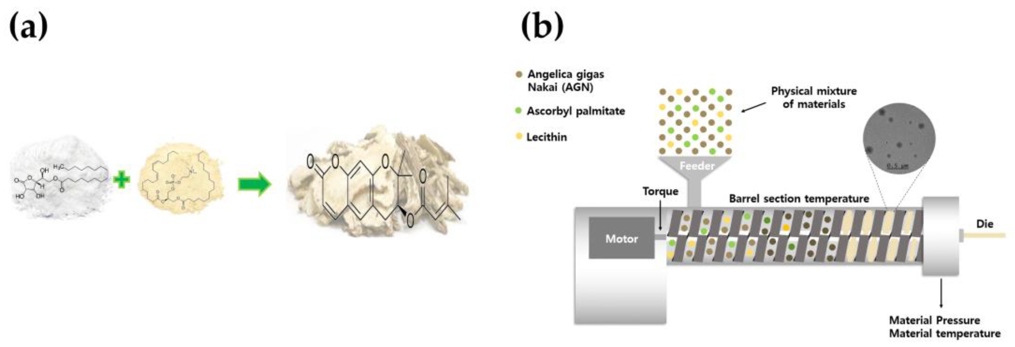
2.1. Preparation of AG by HME Technology

AG powders were processed by the HME process, as previously described, with some modifications [21]. AG was applied to copper metal, and fully loaded into a twin-screw extruder (STS-25HS, co-rotating intermeshing twin-screw extruder, Pyeongtaek, Republic of Korea). A circular die (1 mm diameter) was mounted on the extruder. The powder was fed into the extruder at a feed rate of 150 pm and a pressure of 15 bar. The temperature profile from the feed zone to the injected dye of the extrusion mold was 100 °C. After completing the HME process, the extrudates were dried in an oven (SCOV-150, Sungchan Science, Pocheon, Republic of Korea) at 70 °C for 48 h, and the pulverized powder was stored for further experiments (Figure 1).
To characterize the particles, AG and AG via hot-melt extrusion (AGH) samples were coated with platinum for 60 s using a sample-coating machine (EM ACE600, Leica, Germany) and photographed under a scanning electron microscope (SEM; S-4800, Hitachi, Japan). In addition, dynamic light scattering (DLS) was performed using a ZSP (Malvern Instruments, Malvern, UK) to determine the size distribution (Figure 2).

2.2. Culture of Human Gingival Fibroblasts

Human gingiva fibroblast (HGF; ATCC, PCS-201-018™) cells were purchased and incubated with Dulbecco’s Modified Eagle’s (DMEM; GibcoBRL, Life Technologies, Grand Island, NY, USA) supplemented with 10% fetal bovine serum and 1% antibiotics (100 U/mL penicillin and 100 μg/mL streptomycin) at 37 °C, 5% CO2. The cells were further incubated until confluency. Finally, cells from the 4th to 6th generations were used in this study.

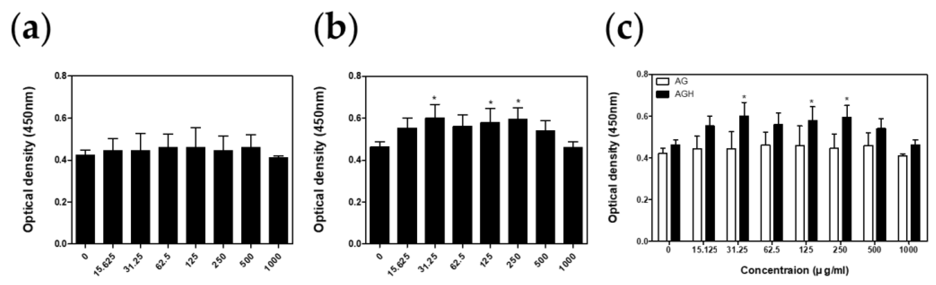
2.3. Cell Viability Test

Cell viability was measured using a cell counting kit-8 assay (CCK-8; Dojindo Molecular Technologies, Rockville, MD, USA). HGF cells were seeded into 96-well plates at a density of 5 × 10³ cells/well and incubated at 37 °C, 5% CO2 for 24 h. The AG and AGH samples (n = 5) were treated with various concentrations for 24 h. The CCK-8 solution (20 μL) was added to the plates and incubated for 2 h at 37 °C. To compare cell viabilities, optical density at 450 nm was measured using a microreader (AMR-100; Allsheng, Hangzhou, Zhejiang, China).

2.4. Nitric Oxide (NO) Assay

For evaluating anti-inflammatory effects of AG and AGH, a NO assay was performed following the standard method with some modifications [23]. The cells (n = 4) were seeded into 6-well plates with a density of 2 × 10⁵ cells/well, incubated for 24 h, and treated with 10 μg/mL of LPS from Porphyromonas gingivalis (InvivoGen, San Diego, CA, USA) for 24 h. AGH solutions (125 μg/mL) were added into the wells 1 h before LPS treatment. NO production was measured using a NO Plus Detection Kit (iNtRON Biotechnology, Inc., Seoul, Republic of Korea). Then, 100 μL of the supernatants were placed in 96-well plates, followed by 50 μL of N1 buffer, and incubated for 10 min at 37 °C. Next, 50 μL of N2 buffer was added and the optical density was measured at 540 nm after 10 min using a microreader (AMR-100; Allsheng, Hangzhou, Zhejiang, China). The quantification of NO production was calculated from an 8-point nitrite standard curve according to the manufacturer’s protocol.

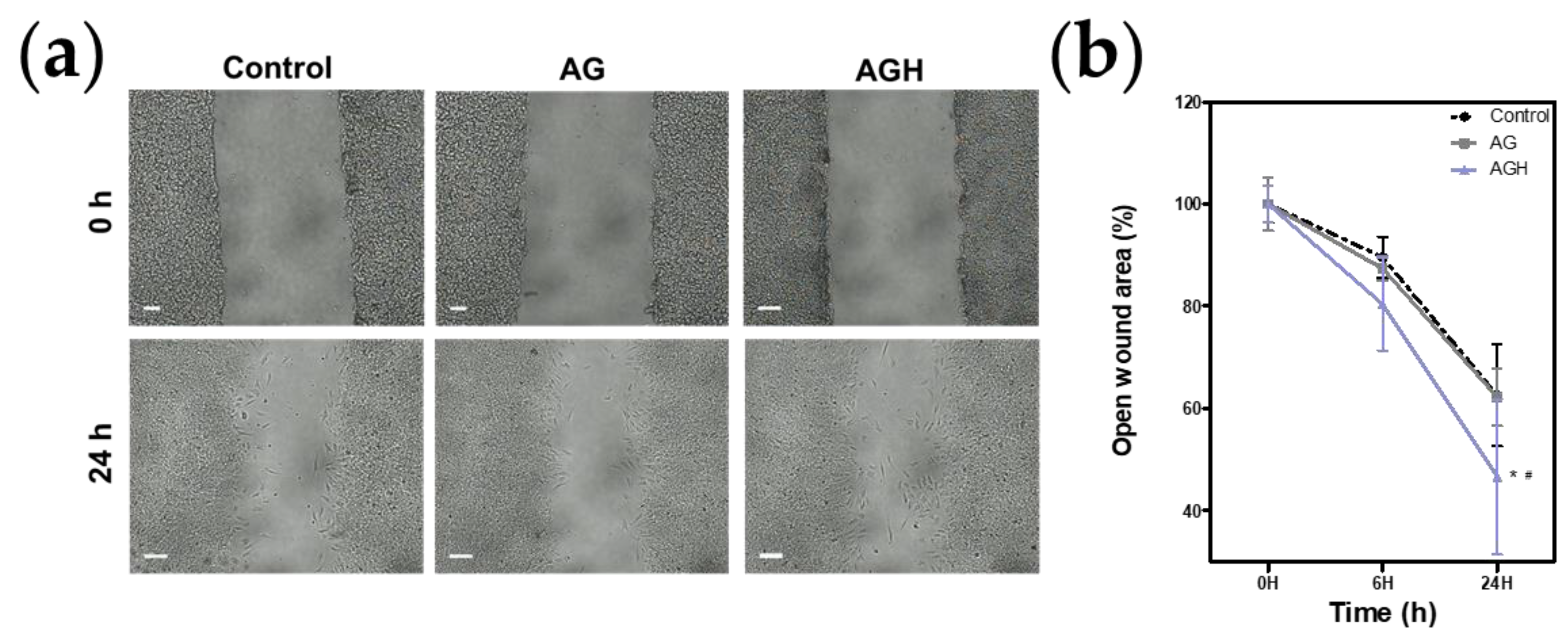
2.5. Cell Proliferation/Cell Migration Assay

For evaluating cell proliferation/cell migration, the cells were seeded on silicone inserts (SPLScar™ Block, SPL Life Sciences, Gyeonggido, Republic of Korea) in 35 mm culture dishes and incubated for 24 h at 37 °C. The silicone was then removed using sterile forceps. Then, either 125 μg/mL of AG or AGH was treated and then incubated. The gap lengths between the margins (n = 8) were imaged and measured with microscopy for 24 h. The open wound area (%) was calculated as the ratio of the gaps between the evaluation time and 0 h.

2.6. Quantitative Real-Time Polymerase Chain Reaction (qRT-PCR)

To assess the expression of biomarkers involved in oral wound healing, HGF cells were seeded in 6-well plates (5 × 10⁵ cells/well) for 24 h. The cells (n = 3) were then exposed to either 125 μg/mL of AG or AGH for 24 h, and RNA was extracted using easy-BLUE™. Total RNA was reverse-transcribed to cDNA using Maxime™ RT PreMix (iNtRON Biotechnology). qPCR was performed on a Step One Plus real-time PCR system (Applied Biosystems, Thermo Fisher Scientific, Inc., Waltham, MA, USA) using Power SYBR™ Green PCR Master Mix (Applied Biosystems). cDNA levels were calculated using the 2–ΔΔCt method and normalized to glyceraldehyde 3-phosphate dehydrogenase (GAPDH) levels.
The following primer sequences were used: vascular endothelial growth factor (VEGF), (F) 5′-TCACAGGTACAGGGATGAGGACAC-3′, (R) 5′-CAAAGCACAGCAATGTCCTGAAG-3′, alpha-smooth muscle actin (α-SMA), (F) 5′-GCCTGATGGGCAAGTGAT-3′, (R) 5′-TGCTGGAAGGTGGACA-3′, alpha 1 type I collagen (COL1α1), (F) 5′-CCCGGGTTTCAGAGACAACTTC-3′, and (R) 5′-TCCACATGCTTTATTCCAGCAATC-3′.

2.7. Statistical Analysis
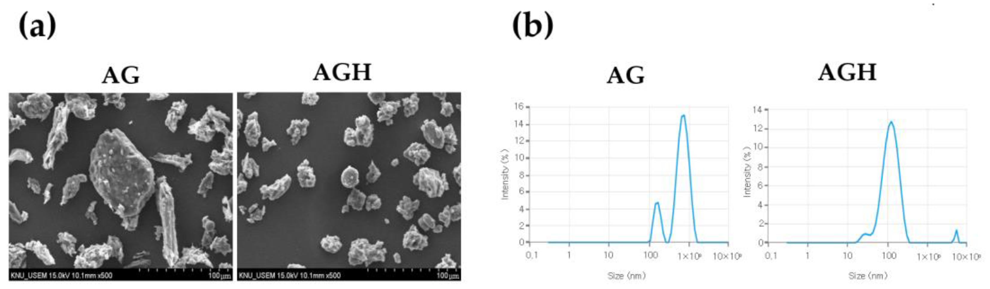
IBM SPSS Statistics (version 20.0; IBM Corp., Armonk, NY, USA) was used to analyze the data. The Shapiro–Wilk test was performed to check the normality of the data. A one-way analysis of variance (ANOVA) was used to determine significance, followed by a post hoc analysis using the Tukey test. p-values < 0.05 were considered statistically significant.

3. Results

Cell viability tests revealed that AGH had no cytotoxic effect on human gingival fibroblasts (Figure 3). The results from the NO assay showed that AGH significantly decreased NO production under LPS stimulation (Figure 4). The cell proliferation/migration assay showed that AGH significantly decreased the open wound area compared to that in the control after 24 h (p = 0.022). However, there were no differences in the open wound areas between the AG and control groups (Figure 5). Figure 6 shows the expression of the biomarkers associated with oral wound healing. We observed a significant upregulation of both VEGF (p = 0.002) and COL1α1 (p = 0.001) in AGH compared to that in the control and AG. However, there were no significant differences in α-SMA expression among the materials.

4. Discussion

This study evaluated the wound healing effects of AGH in vitro. AGH significantly decreased the open wound area and upregulated the expression of biomarkers involved in oral wound healing. Thus, the null hypothesis was rejected.
In this study, HME technology was used. Recently, HME has been considered a convenient processing technology for use in various industries such as pharmaceuticals, polymer science, and food science [24]. HME has the potential to enhance the water solubility and oral bioavailability of the active molecule [25]. Moreover, HME allows for the attainment of distinct particle characteristics, including the uniform distribution of processed extrudates and consistency in particle size [26]. HME is a sustainable processing system that decreases organic solvent usage, minimizes the risk of toxicity, and provides advantages of a single and continuous process [27]. A previous study regarding HME-orientated antihypertensive molecules revealed high levels of bioavailability in a rat model [28]. Another study on HME-orientated resveratrol confirmed an improvement in the solubility and oral bioavailability of resveratrol [29]. A previous animal study also confirmed improvements in the oral bioavailability and biological effects of cyclosporine through HME [30]. Together with these findings, utilizing HME technology for drug delivery in oral environment will be beneficial.
In this study, AGH treatment significantly reduced LPS-induced NO production. NO is a major signaling molecule that participates in the inflammation process [31]. Previous evidence has suggested that NO production is positively related to pro-inflammatory cytokine expression [32,33]. Based on these findings, it may be assumed that AGH improves oral wound healing by inhibiting inflammation. Inflammation is fundamental for oral wound healing in the early phase of oral wound healing because it brings macrophages and neutrophils to the wound site and thereby induces the release of cytokines associated with wound healing [34]. However, prolonged inflammation inhibits wound healing by producing toxic free radicals and reactive oxygen species, as well as increasing the risk of infection [4].
After the inflammation subsides, the oral wound healing process progresses through two phases: proliferation and remodeling. During the proliferation stage, various types of cells, including endothelial cells, fibroblasts, and epithelial cells, migrate to the wound site and induce angiogenesis and re-epithelialization [1]. Briefly, fibroblasts produce granulation tissue, with highly vascularized, endothelial cells building structural support for the wound, and epithelial cells induce re-epithelialization, in turn [35]. Thus, cell migration is fundamental in the process of oral wound healing.
In this study, the cell migration assay confirmed that AGH accelerated HGF migration; however, AG did not accelerate HGF migration compared to the control. This finding indicates that AGH may produce a favorable environment for facilitating oral wound healing. The sustained release of the biological effect of AG through HME technology could be a possible explanation. HME technology aims to facilitate targeted drug delivery. Recent studies have demonstrated that the HME technology is a feasible vehicle for effective drug release [21,36].
In this study, AGH upregulated the expression of biomarkers associated with oral wound healing. VEGF expression was significantly higher in AGH. Considering that VEGF is a key factor in angiogenesis during the proliferation stage [5], this finding indicated that drug delivery by AGH was steadier and more effective. VEGF is associated with angiogenesis, epithelialization, and collagen synthesis [37]. Similarly, COL1α1 expression was also highlighted in AGH, which was correlated with the results of previous studies which reported that AG increases type I collagen synthesis in human dermal fibroblasts [38]. This finding indicates that AGH may act as a collagen synthesis stimulator and facilitate oral tissue regeneration.
Our results showed that AGH may accelerate the oral wound healing process. However, this study has some limitations. First, the biological effects of AGH during the inflammatory phase were not determined. The precise mechanism by which AGH affects wound healing has not yet been fully elucidated. Further evaluation of the protein levels is needed.

5. Conclusions

In conclusion, oral wound healing effects of AGH were assessed in this study. Within the limits of this study, AGH accelerated the oral wound healing process and showed significantly upregulated expressions of VEGF and COL1α1. This finding suggests that AG delivery via HEM technology holds benefits.

Author Contributions

Conceptualization, H.-J.A., J.-S.B. and O.H.N.; investigation, J.R.Y., H.Y.L. and Y.-J.P.; formal analysis, J.R.Y., H.Y.L. and Y.-J.P.; data curation, J.R.Y., H.Y.L., Y.-J.P. and Y.K.C.; writing—original draft preparation, J.R.Y., H.Y.L. and Y.-J.P.; writing—review and editing, J.-S.B. and O.H.N.; supervision, H.-J.A.; funding acquisition, J.-S.B. and O.H.N. All authors have read and agreed to the published version of the manuscript.

Funding

This work was supported by the project for Collabo R&D between Industry, University, and Research Institute funded by the Korea Ministry of SMEs and Startups in 2023 (RS-2023-00225738).

Institutional Review Board Statement

Not applicable.

Informed Consent Statement

Not applicable.

Data Availability Statement

All data analyzed during this study are included in this published article.

Conflicts of Interest

The authors declare no conflict of interest.

References

  1. Waasdorp, M.; Krom, B.P.; Bikker, F.J.; van Zuijlen, P.P.M.; Niessen, F.B.; Gibbs, S. The Bigger Picture: Why Oral Mucosa Heals Better Than Skin. Biomolecules 2021, 11, 1165. [Google Scholar] [CrossRef] [PubMed]
  2. Sedghi, L.; DiMassa, V.; Harrington, A.; Lynch, S.V.; Kapila, Y.L. The oral microbiome: Role of key organisms and complex networks in oral health and disease. Periodontol. 2000 2021, 87, 107–131. [Google Scholar] [CrossRef] [PubMed]
  3. Graça, M.F.P.; Miguel, S.P.; Cabral, C.S.D.; Correia, I.J. Hyaluronic acid-Based wound dressings: A review. Carbohydr. Polym. 2020, 241, 116364. [Google Scholar] [CrossRef] [PubMed]
  4. Lee, J.H.; Lee, K.E.; Nam, O.H.; Chae, Y.K.; Lee, M.H.; Kweon, D.K.; Kim, M.S.; Lee, H.S.; Choi, S.C. Orodispersible hyaluronic acid film delivery for oral wound healing in rats. J. Dent. Sci. 2022, 17, 1595–1603. [Google Scholar] [CrossRef] [PubMed]
  5. Lee, J.H.; Lee, K.E.; Kang, S.W.; Park, S.H.; Chae, Y.K.; Lee, M.H.; Kweon, D.K.; Choi, S.C.; Nam, O.H. Effect of orodispersible hyaluronic acid film on palatal mucosa wound healing. Oral Dis. 2023, 1–10. [Google Scholar] [CrossRef] [PubMed]
  6. Agarwal, C.; Tarun Kumar, A.B.; Mehta, D.S. Comparative evaluation of free gingival graft and AlloDerm(®) in enhancing the width of attached gingival: A clinical study. Contemp. Clin. Dent. 2015, 6, 483–488. [Google Scholar] [CrossRef] [PubMed]
  7. Bosco, A.F.; Bosco, J.M. An alternative technique to the harvesting of a connective tissue graft from a thin palate: Enhanced wound healing. Int. J. Periodontics Restor. Dent. 2007, 27, 133–139. [Google Scholar]
  8. Hämmerle, C.H.; Giannobile, W.V. Biology of soft tissue wound healing and regeneration--consensus report of Group 1 of the 10th European Workshop on Periodontology. J. Clin. Periodontol. 2014, 41 (Suppl. S15), S1–S5. [Google Scholar] [CrossRef]
  9. Del Pizzo, M.; Modica, F.; Bethaz, N.; Priotto, P.; Romagnoli, R. The connective tissue graft: A comparative clinical evaluation of wound healing at the palatal donor site. A preliminary study. J. Clin. Periodontol. 2002, 29, 848–854. [Google Scholar] [CrossRef]
  10. He, Z.; Wang, Y.; Chen, Y.; Geng, F.; Jiang, Z.; Li, X. Angelica gigas Nakai: An overview on its chemical composition and pharmacological activity. Biochem. Syst. Ecol. 2023, 111, 104717. [Google Scholar] [CrossRef]
  11. Sarker, S.D.; Nahar, L. Natural medicine: The genus Angelica. Curr Med Chem 2004, 11, 1479–1500. [Google Scholar] [CrossRef] [PubMed]
  12. Zhang, J.; Li, L.; Jiang, C.; Xing, C.; Kim, S.H.; Lü, J. Anti-cancer and other bioactivities of Korean Angelica gigas Nakai (AGN) and its major pyranocoumarin compounds. Anticancer Agents Med. Chem. 2012, 12, 1239–1254. [Google Scholar] [CrossRef] [PubMed]
  13. Her, Y.; Lee, T.K.; Ahn, J.H.; Lim, S.S.; Kang, B.G.; Park, J.S.; Kim, B.; Sim, H.; Lee, J.C.; Kim, H.S.; et al. Chemical Composition of a Novel Distillate from Fermented Mixture of Nine Anti-Inflammatory Herbs and Its UVB-Protective Efficacy in Mouse Dorsal Skin via Attenuating Collagen Disruption and Inflammation. Molecules 2020, 26, 124. [Google Scholar] [CrossRef] [PubMed]
  14. Shin, J.Y.; Kang, E.S.; Park, J.H.; Cho, B.O.; Jang, S.I. Anti-inflammatory effect of red ginseng marc, Artemisia scoparia, Paeonia japonica and Angelica gigas extract mixture in LPS-stimulated RAW 264.7 cells. Biomed. Rep. 2022, 17, 63. [Google Scholar] [CrossRef]
  15. Han, J.; Jin, W.; Ho, N.A.; Hong, J.; Kim, Y.J.; Shin, Y.; Lee, H.; Suh, J.W. Decursin and decursinol angelate improve wound healing by upregulating transcription of genes encoding extracellular matrix remodeling proteins, inflammatory cytokines, and growth factors in human keratinocytes. Biochem. Biophys. Res. Commun. 2018, 499, 979–984. [Google Scholar] [CrossRef] [PubMed]
  16. Abubakar, A.R.; Haque, M. Preparation of Medicinal Plants: Basic Extraction and Fractionation Procedures for Experimental Purposes. J. Pharm. Bioallied Sci. 2020, 12, 1–10. [Google Scholar] [CrossRef]
  17. Ashour, E.A.; Majumdar, S.; Alsheteli, A.; Alshehri, S.; Alsulays, B.; Feng, X.; Gryczke, A.; Kolter, K.; Langley, N.; Repka, M.A. Hot melt extrusion as an approach to improve solubility, permeability and oral absorption of a psychoactive natural product, piperine. J. Pharm. Pharmacol. 2016, 68, 989–998. [Google Scholar] [CrossRef]
  18. Makadia, H.K.; Siegel, S.J. Poly Lactic-co-Glycolic Acid (PLGA) as Biodegradable Controlled Drug Delivery Carrier. Polymers 2011, 3, 1377–1397. [Google Scholar] [CrossRef]
  19. Smith, M.K.; French, J.L.; Kowalski, K.G.; Hutmacher, M.M.; Ewy, W. Decision-Making in Drug development: Application of a model based framework for assessing trial performance. In Clinical Trial Simulations; Springer: Cham, Switzerland, 2011; pp. 61–83. [Google Scholar]
  20. Pimparade, M.B.; Vo, A.; Maurya, A.S.; Bae, J.; Morott, J.T.; Feng, X.; Kim, D.W.; Kulkarni, V.I.; Tiwari, R.; Vanaja, K.; et al. Development and evaluation of an oral fast disintegrating anti-allergic film using hot-melt extrusion technology. Eur. J. Pharm. Biopharm. 2017, 119, 81–90. [Google Scholar] [CrossRef]
  21. Ryu, S.; Lee, H.Y.; Nam, S.-H.; Baek, J.-S. Antifungal Activity of Angelica gigas with Enhanced Water Solubility of Decursin and Decursinol Angelate by Hot-Melt Extrusion Technology against Candida albicans. Int. J. Transl. Med. 2022, 2, 515–521. [Google Scholar] [CrossRef]
  22. Jiang, Y.; Piao, J.; Cho, H.J.; Kang, W.S.; Kim, H.Y. Improvement in antiproliferative activity of Angelica gigas Nakai by solid dispersion formation via hot-melt extrusion and induction of cell cycle arrest and apoptosis in HeLa cells. Biosci. Biotechnol. Biochem. 2015, 79, 1635–1643. [Google Scholar] [CrossRef]
  23. Nam, O.H.; Ro, S.T.; Lee, H.W.; Jeong, J.; Chae, Y.K.; Lee, K.E.; Choi, S.C.; Kang, S.W. Evaluation of delphinidin as a storage medium for avulsed teeth. BMC Oral Health 2023, 23, 21. [Google Scholar] [CrossRef]
  24. Kim, H.B.; Ryu, S.; Baek, J.S. The Effect of Hot-Melt Extrusion of Mulberry Leaf on the Number of Active Compounds and Antioxidant Activity. Plants 2022, 11, 3019. [Google Scholar] [CrossRef]
  25. Wang, W.; Kang, Q.; Liu, N.; Zhang, Q.; Zhang, Y.; Li, H.; Zhao, B.; Chen, Y.; Lan, Y.; Ma, Q.; et al. Enhanced dissolution rate and oral bioavailability of Ginkgo biloba extract by preparing solid dispersion via hot-melt extrusion. Fitoterapia 2015, 102, 189–197. [Google Scholar] [CrossRef]
  26. Tambe, S.M.; Jain, D.D.; Mehta, C.H.; Ashwini, T.; Yogendra Nayak, U.; Amin, P.D. Hot-melt extruded in situ gelling systems (MeltDrops Technology): Formulation development, in silico modelling and in vivo studies. Eur. J. Pharm. Biopharm. 2023, 188, 108–124. [Google Scholar] [CrossRef]
  27. Hwang, I.; Kang, C.-Y.; Park, J.-B. Advances in hot-melt extrusion technology toward pharmaceutical objectives. J. Pharm. Investig. 2017, 47, 123–132. [Google Scholar] [CrossRef]
  28. Jadhav, P.; Gokarna, V.; Deshpande, V.; Vavia, P. Bioavailability Enhancement of Olmesartan Medoxomil Using Hot-Melt Extrusion: In-Silico, In-Vitro, and In-Vivo Evaluation. AAPS PharmSciTech 2020, 21, 254. [Google Scholar] [CrossRef]
  29. Fan, W.; Wu, J.; Gao, M.; Zhang, X.; Zhu, W. Preparation of Solid Dispersion of Polygonum Cuspidatum Extract by Hot Melt Extrusion to Enhance Oral Bioavailability of Resveratrol. Molecules 2023, 28, 737. [Google Scholar] [CrossRef]
  30. Jiang, X.; Zhao, Y.; Guan, Q.; Xiao, S.; Dong, W.; Lian, S.; Zhang, H.; Liu, M.; Wang, Z.; Han, J. Amorphous solid dispersions of cyclosporine A with improved bioavailability prepared via hot melt extrusion: Formulation, physicochemical characterization, and in vivo evaluation. Eur. J. Pharm. Sci. 2022, 168, 106036. [Google Scholar] [CrossRef]
  31. Sharma, J.N.; Al-Omran, A.; Parvathy, S.S. Role of nitric oxide in inflammatory diseases. Inflammopharmacology 2007, 15, 252–259. [Google Scholar] [CrossRef]
  32. Rafa, H.; Saoula, H.; Belkhelfa, M.; Medjeber, O.; Soufli, I.; Toumi, R.; de Launoit, Y.; Moralès, O.; Nakmouche, M.; Delhem, N.; et al. IL-23/IL-17A axis correlates with the nitric oxide pathway in inflammatory bowel disease: Immunomodulatory effect of retinoic acid. J. Interferon Cytokine Res. 2013, 33, 355–368. [Google Scholar] [CrossRef]
  33. Rafa, H.; Amri, M.; Saoula, H.; Belkhelfa, M.; Medjeber, O.; Boutaleb, A.; Aftis, S.; Nakmouche, M.; Touil-Boukoffa, C. Involvement of interferon-γ in bowel disease pathogenesis by nitric oxide pathway: A study in Algerian patients. J. Interferon Cytokine Res. 2010, 30, 691–697. [Google Scholar] [CrossRef]
  34. Pereira, L.M.; Hatanaka, E.; Martins, E.F.; Oliveira, F.; Liberti, E.A.; Farsky, S.H.; Curi, R.; Pithon-Curi, T.C. Effect of oleic and linoleic acids on the inflammatory phase of wound healing in rats. Cell Biochem. Funct. 2008, 26, 197–204. [Google Scholar] [CrossRef]
  35. Pastar, I.; Stojadinovic, O.; Yin, N.C.; Ramirez, H.; Nusbaum, A.G.; Sawaya, A.; Patel, S.B.; Khalid, L.; Isseroff, R.R.; Tomic-Canic, M. Epithelialization in Wound Healing: A Comprehensive Review. Adv. Wound Care 2014, 3, 445–464. [Google Scholar] [CrossRef]
  36. Cassidy, C.M.; Tunney, M.M.; Caldwell, D.L.; Andrews, G.P.; Donnelly, R.F. Development of novel oral formulations prepared via hot melt extrusion for targeted delivery of photosensitizer to the colon. Photochem. Photobiol. 2011, 87, 867–876. [Google Scholar] [CrossRef]
  37. Bao, P.; Kodra, A.; Tomic-Canic, M.; Golinko, M.S.; Ehrlich, H.P.; Brem, H. The role of vascular endothelial growth factor in wound healing. J. Surg. Res. 2009, 153, 347–358. [Google Scholar] [CrossRef]
  38. Lee, J.-H.; Lee, S.-N.; Kim, M.-G.; Kim, M.-H.; Kim, H.-J.; Jo, H.-J.; Leem, K.-H. Effects of Angelica gigantis Radix extracts on the collagenase activity and procollagen synthesis in HS68 human fibroblasts and tyrosinase activity. Korea J. Herbol. 2011, 26, 29–33. [Google Scholar]
Figure 1. Preparation of AG by HME technology. (a) AG with added excipients; (b) a schematic design of the HME process.
Medicina 59 02066 g001
Figure 2. Confirmation of the particle characterization. (a) Scanning electron microscopy (SEM) images. SEM images at 500× magnification show the homogeneity of AGH particles. (b) Dynamic light scattering (DLS). The results show that AGH is more homogeneous in size distribution and smaller in size than AG.
Medicina 59 02066 g002
Figure 3. Cell viability tests. (a) Cell viability test of AG (n = 5). (b) Cell viability test of AGH (n = 5). The cell viabilities were compared to that in the control (* p < 0.05 vs. control). (c) Comparison of cell viabilities between AG and AGH (* p < 0.05 vs. AG).
Medicina 59 02066 g003
Figure 4. NO production (n = 4). AGH significantly decreased NO production from LPS stimulation. * p < 0.05 vs. control, # p < 0.05 vs. control + LPS.
Medicina 59 02066 g004
Figure 5. Cell proliferation/cell migration assay (n = 8). (a) Microscopic view. (b) Analysis of open wound area (%). The open wound area (%) was calculated as the ratio of the gaps between the evaluation time and 0 h in each group. The results show that AGH significantly decreased in the open wound area (* p < 0.05 vs. control, # p < 0.05 vs. AG).
Medicina 59 02066 g005
Figure 6. Expression of wound healing-related biomarkers (n = 3). (a) VEGF. (b) α-SMA. (c) COL1α1. The results show that AGH significantly upregulates the VEGF (p = 0.002 vs. control, p = 0.002 vs. AG) and COL1α1 (p = 0.001 vs. control, p = 0.001 vs. AG) expression levels (* p < 0.05 vs. control, # p < 0.05 vs. AG). CON, control (no treatment); AG, Angelica gigas Nakai; AGH, Angelica gigas Nakai from hot-melt extrusion technology.
Medicina 59 02066 g006
Disclaimer/Publisher’s Note: The statements, opinions and data contained in all publications are solely those of the individual author(s) and contributor(s) and not of MDPI and/or the editor(s). MDPI and/or the editor(s) disclaim responsibility for any injury to people or property resulting from any ideas, methods, instructions or products referred to in the content.

