Article

Evaluation of Wear on Primary Tooth Enamel and Fracture Resistance of Esthetic Pediatric Crowns Manufactured from Different Materials

by
Nagehan Aktaş
1,* and
Merve Bankoğlu Güngör
2
1
Department of Pediatric Dentistry, Faculty of Dentistry, Gazi University, 06490 Ankara, Türkiye
2
Department of Prosthetic Dentistry, Faculty of Dentistry, Gazi University, 06490 Ankara, Türkiye
*
Author to whom correspondence should be addressed.
Medicina 2024, 60(10), 1678; https://doi.org/10.3390/medicina60101678
Submission received: 23 September 2024 / Revised: 3 October 2024 / Accepted: 11 October 2024 / Published: 13 October 2024
(This article belongs to the Special Issue Recent Advances in Pediatric Oral Health)

Abstract

:
Background and Objectives: Advances in dental materials and CAD-CAM technology have expanded crown options in primary teeth due to their improved appearance and mechanical properties. Thus, this study aimed to assess the enamel wear and fracture resistance of prefabricated, milled, and 3D-printed esthetic pediatric crowns. Materials and Methods: The study involved 60 extracted maxillary second primary molars and 60 3D-printed resin dies, divided into six groups based on different crown materials (n = 10): prefabricated zirconia, prefabricated composite, milled composite, milled resin matrix ceramic, milled PEEK, and 3D-printed resin. Prefabricated crowns were selected after the preparation of the typodont mandibular second primary molar tooth, while milled and 3D-printed crowns were custom produced. The specimens underwent mechanical loading of 50 N at 1.6 Hz for 250,000 cycles with simultaneous thermal cycling. The 3D and 2D wear amounts were evaluated by scanning the specimens before and after aging. Then, the fracture resistance and failure types of the restorations were recorded. Results: The results showed that the milled PEEK group had superior fracture resistance compared to the other groups, while prefabricated zirconia crown group had the lowest value. Milled resin matrix ceramic crown group displayed the lowest 3D wear volume, while 3D-printed crown group showed the highest 2D wear. Conclusions: The restorative material type did not have a significant effect on the wear of primary tooth enamel. The fracture resistance of the tested materials differed according to the material type. Although the milled PEEK group showed the highest fracture resistance, all tested materials can withstand chewing forces in children.

1. Introduction

The restoration of primary teeth is fundamental in pediatric dentistry for maintaining space, ensuring proper chewing, and guiding the eruption of permanent teeth. Failure to restore damaged or decayed primary teeth can result in early tooth loss, malocclusion, and complex orthodontic issues that may require extensive treatment later [1,2]. Crowns are one of the preferred treatment options for restoring function and esthetics in primary teeth, especially in cases of severe caries, developmental defects, or after pulp therapy [3].
The primary indications for using crowns in pediatric dentistry include extensive carious lesions that cannot be restored with conventional fillings, developmental anomalies such as hypoplasia, teeth that have undergone pulpotomy or pulpectomy, and structurally compromised teeth. Crowns are contraindicated in cases where there is insufficient remaining tooth structure for retention, severe periodontal involvement, or when the child’s behavior prevents effective treatment [3,4].
Pediatric crowns offer several advantages for primary teeth, including superior strength, durability, and complete coverage, which provides enhanced protection against further caries. They also help maintain occlusal function and prevent space loss, which is critical for the proper eruption of permanent teeth. Indeed, they enable effective and durable therapeutic outcomes, making them a favorable option for restoring primary teeth. Compared to traditional manual restorations, they often allow for quicker and more efficient treatment, particularly in cases involving extensive multi-surface carious lesions [3,4,5]. However, there are some limitations. Although stainless steel crowns (SSCs) are the first option for crown restorations on primary teeth, they may not provide esthetic appeal, and inadequately contoured crowns are the reason for gingivitis associated with SSCs. These crowns contain very low levels of nickel and are associated with a low risk of nickel allergy and hypersensitivity [3,5].
Recent advancements in technology and the increasing demand from both parents and children for more esthetic solutions have shifted the focus toward alternative materials instead of SSCs [6,7]. Prefabricated zirconia crowns have been developed to meet these esthetic expectations while providing high strength and durability for both anterior and posterior primary teeth [8,9,10]. However, prefabricated zirconia crowns have drawbacks, such as the inability to be modified and the need for passive seating during cementation, which may require more aggressive tooth preparation. They also cannot withstand flexure, increasing the risk of fracture during cementation, and if any part fractures, the entire crown must be replaced [11,12]. Laser-sintered, pre-cured nano-hybrid composite crowns have recently been introduced and expand the range of prefabricated pediatric crowns. Designed to replicate natural primary teeth, they offer excellent physical and esthetic properties for both anterior and posterior use. Cured through light, heat, or pressure, these crowns have homogeneous, non-porous surfaces, improving wear resistance and ensuring proper adaptation [13].
The use of computer-aided design and computer-aided manufacturing (CAD-CAM) in dentistry has advanced, utilizing either subtractive manufacturing with milling technology or additive manufacturing through three-dimensional (3D) printing [14,15]. CAD-CAM systems have enabled the production of customized esthetic and functional crowns for pediatric patients. Milling-based systems can create crowns from materials such as zirconia ceramics, resin-based ceramics, and composite resins [16,17,18]. Resin ceramic and composite blocks are ideal for primary teeth, as their lower hardness reduces wear on opposing teeth, and their low modulus of elasticity helps absorb functional stresses [16,17,18,19,20]. The introduction of 3D-printed crowns has opened new possibilities for dental restorations and is becoming a preferred alternative in pediatric dentistry [21,22]. The key advantages of 3D-printing technology include the ability to produce multiple objects in a single process, the reproduction of complex geometries, and its cost effectiveness [22,23]. The development of 3D-printing technology has led to the introduction of various innovative materials in pediatric dentistry. These materials include resin-based composites and high-performance polymers such as polyetheretherketone (PEEK). Resin-based materials are preferred for their esthetic properties, ease of customization, and elasticity moduli that closely resemble those of natural dentin, making them suitable for both anterior and posterior restorations [23,24]. Permanent crown resins from different companies for 3D printers have been introduced to the market for mid-to-long-term use in the oral cavity [25,26,27]. The successful use of these permanent resins in pediatric patients has been documented in a case report, which restored a severely damaged premolar tooth using a 3D-printed endocrown following coronal pulpotomy [28]. PEEK, a high-performance polymer, stands out for its superior biocompatibility and mechanical properties comparable to natural dentin, making it a promising material for long-term restorations [24,29]. The versatility of these 3D-printing materials allows for more precise and conservative tooth preparation, ultimately improving treatment outcomes in pediatric dentistry. Due to the wide variety of restorative materials, selecting the most suitable material for a given case can often be challenging for pediatric dentists. Factors such as enamel wear properties and fracture resistance, which can impact the long-term success of the restoration, play a crucial role in the selection of restorative materials for pediatric patients [13,30,31].
Therefore, the purpose of this study was to assess crown restorations in pediatric dentistry by utilizing the latest technology and materials that address the limitations of existing materials used in the restoration of primary molars. For this purpose, the wear properties of the primary tooth enamel and fracture resistances of different esthetic pediatric crowns used in primary molars were compared. The null hypothesis was that the type of restorative material would not impact the wear of primary teeth and fracture resistance of the restoration.

2. Materials and Methods

Ethics approval for this study was obtained from the Clinical Research Ethics Committee of Gazi University, Faculty of Dentistry, under approval number 2023.18/01 and date 21 September 2023.

2.1. Sample Size Calculation

The required sample size for this test was calculated using the “pwr” package in the R version 3.1.9.4. programming language. The effect size for comparing the groups in terms of wear was calculated as 0.61 using the mean and standard deviation values obtained from previous studies [29,32,33,34]. Based on this effect size, with a Type I error rate of 0.05 and a power of 0.95, it was calculated that 60 samples (at least 10 per group) were required for the study. For the compared groups in terms of fracture resistance, the effect size was calculated as 0.8 using the mean and standard deviation values obtained from previous studies [19,35,36]. Based on this effect size, with a Type I error rate of 0.05 and a power of 0.95, the minimum sample size per group was calculated as 7.

2.2. Study Design

Six different experimental groups were generated using restorative materials for pediatric crowns (Table 1). The mandibular second primary molar (85) on a pediatric dental model (AK-6/2M, Frasaco, Germany) was prepared with a 1 mm reduction on the axial surfaces, 1.5 mm on the occlusal surface, and a 1 mm chamfer margin. Sharp angles and edges were rounded. After tooth preparation, gypsum study models were fabricated to replicate the prepared tooth accurately. These models were then positioned and secured on a semi-adjustable articulator (Stratos 100, Ivoclar Vivadent, Schaan, Liechtenstein), simulating the natural occlusal relationships for further procedures. Sixty 3D-printed resin dies (n = 10) were then produced and embedded vertically in cylindrical molds with self-curing acrylic resin. Prefabricated zirconia and composite crowns of appropriate sizes were selected by fitting them on the prepared tooth.

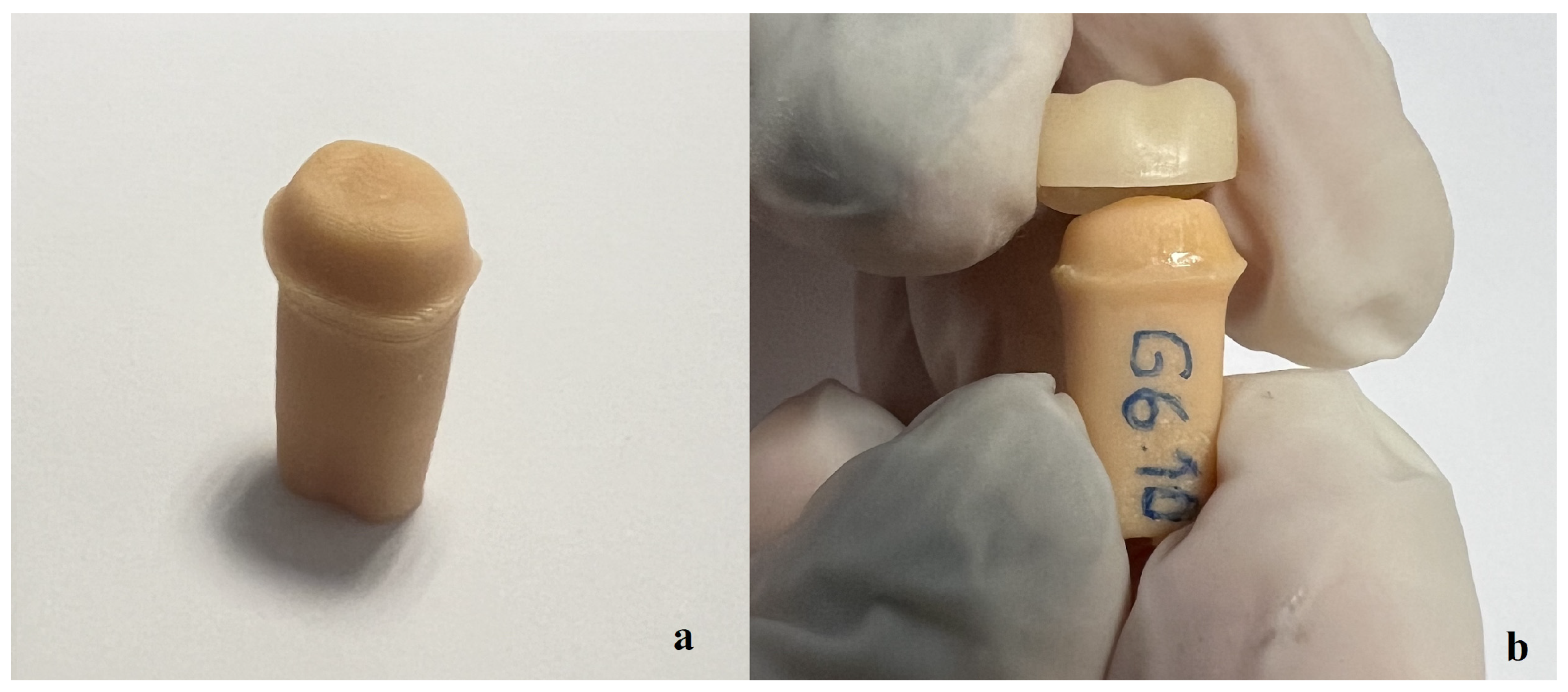
2.3. Crown Manufacturing and Cementation

For milled and 3D-printed crowns, the digital model was transferred to the design software (DentalCad®, Exocad GmbH, Darmstadt, Germany). On the digital model, margins were defined, the cement space was set to 50 microns, and the crowns were designed to match the anatomical structure. The designs of the crowns were saved in STL file format. The composite block (Tetric CAD, Ivoclar Vivadent, Schaan, Liechtenstein), nanoceramic block (GC Cerasmart, GC Corporation, Tokyo, Japan), and PEEK (White Peaks Dental Systems GmbH, Essen, Germany) disc were milled using a milling unit (Redon, Hybrid Dental CNC, Redon Technology, İstanbul, Türkiye). For the 3D-printed crown production, the STL files were transferred to PreForm software (Formlabs Inc., Somerville, MA, USA), where the printing parameters were set and the file converted to a “.form” file. The crowns were printed using a Formlabs Form 3 printer with permanent crown resin (Formlabs, Somerville, MA, USA). After printing, the crowns were washed in 95% isopropyl alcohol for 3 min using the Form Wash&Cure unit (Formlabs Inc., Somerville, MA, USA), dried with compressed air, and then cured for 20 min to ensure dimensional stability. All produced specimens were mechanically polished (OptraPOL; Ivoclar Vivadent).
Before cementation, the 3D-printed resin dies were sandblasted with 50 µm Al2O3 particles at 2 bar pressure from a distance of 10 mm for 20 s. Subsequently, they were cleaned in an ultrasonic cleaner and air-dried. A bonding agent (Scotchbond Universal Plus; 3M ESPE, Seefeld, Germany) was applied to the resin dies for 20 s, dried with air, and surface contamination was prevented. Additional procedures were performed on the inner surface of the restorations according to the manufacturers’ recommendations for cementation (Table 2). All crowns were cemented using resin cement (RelyX Universal Resin Cement, 3M ESPE, Seefeld, Germany). The crowns were placed onto the resin dies under finger pressure. The crowns were then cured for 2–3 s using an LED curing device (Valo Grand; Ultradent Products, South Jordan, UT, USA). After removing excess cement, the crowns were cured for 10 s from each side of the crown. The cementation process was completed by waiting 5 min (Figure 1).

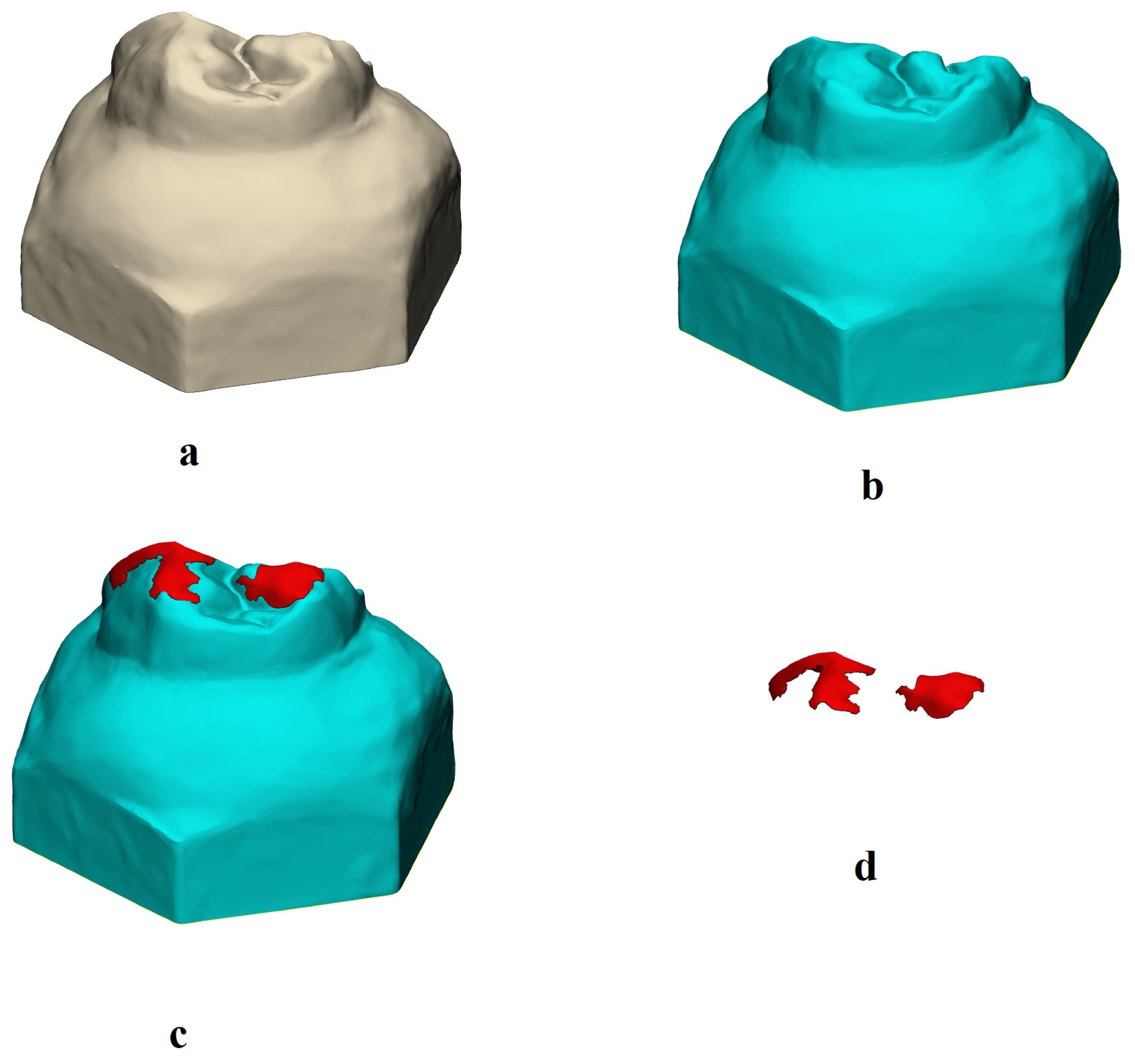
2.4. Wear Evaluation

For evaluating the amount of wear of the crowns on opposing primary molar enamel, 60 caries-free maxillary primary second molar teeth (55) were used. Teeth extracted from children whose parents gave verbal consent and signed an informed consent form before the procedure at the Pediatric Dentistry Clinic between February 2024 and May 2024 were included in the study. Teeth were selected based on the following inclusion criteria: absence of caries, structural defects, or abnormal morphology. The exclusion criteria included teeth with extensive caries, fractures, or teeth showing abnormal wear patterns. The teeth were extracted for orthodontic reasons, pathological causes, or physiological root resorption. The roots of the extracted teeth were retained only to the extent necessary to ensure proper retention during embedding in acrylic resin. The extracted teeth were cleaned and disinfected using a 0.5% chloramine-T solution for 48 hours and then stored in distilled water at room temperature for further use. These teeth were mounted on the loading jigs of the chewing simulator (Esetron Robotechnologies, Ankara, Türkiye). The specimens (teeth with crowns and natural antagonist upper teeth) underwent thermodynamic aging with mechanical loading in a dynamic loading device (chewing simulator) at 50 N and a frequency of 1.6 Hz for 250,000 cycles (approximately equivalent to one year of aging) [7,12]. The loading was applied with the palatal cusp tips of the maxillary primary second molar teeth in contact with the occlusal central fossa of the crowns. Simultaneously, the specimens were subjected to thermal cycling between temperatures of 50 °C and 55 °C.
All tooth specimens were scanned with a 3D intraoral dental scanner (Trios 5, 3Shape, Copenhagen, Denmark) before and after the wear test, and the scans were saved in STL file format (Figure 2). For vertical (2D) and volumetric (3D) wear measurements, the STL files obtained from the scans were imported into the software (3-matic 18.0, Mimics Innovation Suite 26.0, Materialise, Leuven, Belgium). The aged models were subtracted using a digital 3D superimposition technique. The STL files of the specimens before and after aging were aligned using the 3-matic software (Materialise, Belgium). The non-aged models were set as the reference, and the aged models were superimposed on them. The files were aligned, and after alignment, wear models were created by subtracting the aged models from the non-aged ones in their aligned positions (Figure 2). A subtraction algorithm within the software was then applied to calculate the volumetric (3D) and vertical (2D) differences between the two models, thereby quantifying the amount of wear. Vertical wear of maxillary primary second molar teeth was recorded in millimeters (mm), and volume loss was recorded in cubic millimeters (mm3).

2.5. Fracture Resistance Test

After thermomechanical aging, the teeth and crowns were inspected, and any cracks, micro-perforations, fractures, or losses were excluded. Vertical loading was then applied using a universal testing machine (LLYOD, Lloyd Instruments Ltd., Fareham, UK) at a speed of 1 mm/min with a 5 mm diameter ball until fracture occurred. The fracture resistance values were automatically recorded as the maximum loading force (N), and each specimen was categorized according to the types of failures (Table 3) [16].

2.6. Statistical Analyses

Statistical analyses were performed using SPSS software (version 20.0). The Shapiro–Wilk test was used to assess data normality, while Levene test was used to check for homogeneity of variances. One-way ANOVA was used to analyze both 2D and 3D wear data, which were normally distributed. Due to the non-homogeneity of variances in the 2D wear data (p = 0.003), Welch’s correction was applied. Fracture resistance data also showed normal distribution (p > 0.05), and one-way ANOVA was used; however, the non-homogeneity of variances (p < 0.001) required Welch’s correction and the Games–Howell test for pairwise comparisons. The significance level was set at α = 0.05.

3. Results

The descriptive statistics for the 3D and 2D wear data of the experimental groups are shown in Table 4. The highest amount of 3D wear was found in Group 3 (milled composite crown). The descriptive statistics for the 3D and 2D wear data of the experimental groups are shown in Table 4. The highest amount of 3D wear was found in Group 3 (milled composite crown) at 1.74 ± 0.95 mm3, while the lowest wear amount was recorded in Group 4 (milled resin matrix ceramic crown) at 0.99 ± 0.66 mm3. The highest 2D wear amount was found in Group 6 (3D-printed crown) at 1.01 ± 0.43 mm, while the lowest wear amount was recorded in Group 1 (prefabricated zirconia crown) at 0.59 ± 0.23 mm. ANOVA results showed that the type of restoration used in the study did not significantly affect the 3D (p = 0.270) and 2D (p = 0.381; Welch: 0.095) wear amounts of primary teeth (Table 5).
According to the ANOVA test results, the type of restorative material had a significant effect on the fracture resistance of pediatric crowns (p < 0.001) (Table 6). The significantly highest fracture resistance was found in Group 5 (PEEK crown) at 1797 ± 412.6 N (p < 0.05). The significantly lowest fracture resistance was recorded in Group 1 (prefabricated zirconia crown) at 521.4 ± 63.16 N (p < 0.05) (Table 7).
During thermomechanical aging, no failures were observed in any of the specimens. All specimens were subjected to fracture testing, and the failure types for the experimental groups are shown in Table 8 and Figure 3. All failure types were visible to the naked eye.

4. Discussion

The purpose of this study was to determine and compare the amount of enamel wear on primary teeth and the fracture resistance of materials used for pediatric crowns. The null hypothesis of the study was that the restorative material type would not affect the wear of primary tooth and fracture resistance. According to the results, the type of restorative material did not significantly affect the amount of wear on primary teeth. However, the type of restorative material had a significant effect on the fracture resistance of pediatric crowns. Thus, the null hypothesis was partially rejected.
Several studies have evaluated the mechanical properties of prefabricated zirconia and composite crowns for primary teeth, often comparing different types of prefabricated crowns or SSCs [6,7,16,17,18,19,20,21]. However, digital dentistry has become increasingly integrated into pediatric dental practices, with CAD-CAM systems allowing for highly accurate and personalized restorations [22]. However, studies do not comprehensively evaluate all crown options for primary molars, especially with the latest esthetic materials and techniques. For this reason, in the present study, prefabricated zirconia crowns (NuSmile), composite resin crowns (Edelweiss), milledPEEK, resin matrix ceramic and composite resin crowns, and 3D-printed permanent composite resin crowns were tested in terms of wear and fracture resistance after thermomechanical aging. Chieruzzi et al. [37] emphasized that the strength of dental systems under masticatory loads is closely related to the bond integrity at the post/cement and cement/dentine interfaces. A strong bond at these structures ensures better stress distribution, reducing the risk of debonding or failure [37]. Material selection, surface treatment, and proper bonding protocols play crucial roles in optimizing the bond between structures, while factors such as aging and cyclic loading can degrade it over time [37,38]. In the present study, to obtain standardized specimens, the pediatric crowns were cemented on the standardized resin dies by using resin cement.
Recent studies have employed 3D evaluation methods to assess the wear behavior of dental materials [39,40,41]. In the present study, wear measurement involved 3D digital scans taken before and after wear testing, with 2D vertical loss (mm) and 3D volume loss (mm3) calculated by overlaying and subtracting the two 3D models. This quantitative methodology is highly recommended by many authors due to its accuracy [42,43].
In the present study, the type of restoration did not significantly affect the 3D and 2D wear amounts of the primary teeth. The highest 3D wear amount was in the milled resin matrix ceramic crown group. Möhn et al. [20] evaluated the wear properties of stainless steel, zirconia, resin composite, and hybrid ceramic crowns on antagonistic teeth and found that hybrid ceramic crowns exhibited lower wear depth compared to zirconia crowns. Nakase et al. [44] reported that milled resin composite materials caused less wear on antagonist teeth compared to hybrid ceramic and composite materials. The differences in results are likely due to varying protocols in thermomechanical aging. In the present study, enamel wear was found to be lower in the zirconia group, consistent with previous findings [6,9,13]. Walia et al. [45] reported minimal enamel loss on opposing primary incisors in contact with zirconia crowns and no surface changes with composite resin crowns. It was also reported that smooth surfaces of restorations can reduce the wear rate of opposing teeth [6]. In addition, Donovan et al. [46] noted that only glazed or poorly polished zirconia could damage opposing teeth. However, controversial results were also reported. Taran et al. [12] reported that milled zirconia crowns caused the most enamel wear in opposing teeth. Talekar et al. [30] found that zirconia crowns caused significantly more wear on opposing primary canine teeth compared to glass-fiber-reinforced composite crowns. In this study, 3D-printed crowns showed comparable 2D and 3D wear to other tested groups.
Recently, different companies have introduced permanent crown resins for 3D printers, and these are now being used in permanent tooth restorations [25,47,48]. The tested 3D-printed resin was a permanent crown resin by Formlabs, which is thought to be more wear-resistant due to its material properties. In the present study, the 3D wear volume of prefabricated composite crowns was measured as 1.62 ± 1.38 mm3. Signoriello et al. [13] evaluated the wear properties of zirconia, nano-hybrid composite (Edelweiss), and stainless steel crowns on opposing primary teeth. Zirconia crowns showed the smallest wear (0.98 mm²), while nano-hybrid composite crowns exhibited the largest wear area (5.6 mm²). The difference in wear could be attributed to variations in filler content and composition of the composite materials. Additionally, the type of opposing teeth and testing protocols used in the study may have contributed to the differences in wear characteristics. PEEK crowns caused less wear on opposing teeth in this study, though not significantly different from other groups. Choi et al. [29] reported similar findings with high-performance polymers. Few studies specifically examine the use of PEEK in pediatric crowns for primary tooth wear.
The fracture resistance of the restorative materials should be higher than the masticatory forces. Studies have reported varying maximum bite forces in children [48,49,50]. Kamegai et al. [48] found that children aged 6–11 in northern Japan had an average bite force of 374.4 to 433 N. Braun et al. [49] reported an average bite force of 106 N in children aged 6–12, while Owais et al. [50] observed a maximum bite force of 433 N during the mixed dentition period. In the present study, it was found that PEEK crowns exhibited the highest fracture resistance among the tested groups, while prefabricated zirconia crowns exhibited the lowest. At the same time, no specific studies on pediatric PEEK crowns were found. Lu et al. [51] demonstrated high fracture loads for PEEK crowns in permanent teeth restorations. The results from our study align with the findings of the previous studies [7,16,17]. In the present study, no significant difference in fracture resistance was observed among prefabricated composite, milled composite (643.80 ± 96.61 N), milled resin matrix ceramic (596 ± 78.54 N), and 3D-printed crowns (625.2 ± 105.62 N). The variations in fracture resistance values across studies could be attributed to differences in material compositions, manufacturing processes, types of cement used, and testing protocols. Additionally, composite materials’ tendency to absorb water or hydrolyze silanes in a moist environment may result in lower wear and fracture resistance [52]. To the best of our knowledge, there are limited studies regarding prefabricated composite resin crowns. Our findings suggest that tested prefabricated resin crowns can withstand chewing forces effectively, making them a viable option for pediatric dentistry. Although prefabricated zirconia crowns showed the lowest fracture resistance value in the present study, it was observed that all tested crowns exhibited significantly higher fracture resistance than the reported average bite forces in children [48,49,50]. This suggests that the tested crowns possess mechanical properties suitable for withstanding chewing forces and can be safely used in pediatric dentistry.
This study has some limitations. The study included six different types of restorations used in pediatric crowns. However, many other materials can be used for pediatric crowns in CAD-CAM systems. Additionally, different 3D-printing techniques can also be employed for crown production. There is a need for further testing of pediatric crowns produced using various CAD-CAM materials and 3D-printing technologies under aging protocols with longer cycles. In this study, the crowns were cemented onto standardized resin dies, and the bonding mechanism of the resin die differs from that of primary teeth. Although in vitro studies provide promising results, clinical studies are necessary to determine the most suitable crown materials for pediatric crowns.

5. Conclusions

Within the limitations of this in vitro study, the following conclusions are drawn:
  • The restoration type did not have a significant effect on the 2D and 3D wear of the primary tooth enamel.
  • The fracture resistance of the tested materials differed according to the material type. Although the milled PEEK group showed the highest fracture resistance, all tested materials could withstand chewing forces in children.

Author Contributions

Conceptualization, N.A. and M.B.G.; methodology, N.A. and M.B.G.; formal analysis, N.A. and M.B.G.; investigation, N.A. and M.B.G.; data collection, N.A. and M.B.G., writing—original draft preparation, N.A. and M.B.G.; writing—review and editing, N.A. and M.B.G.; visualization, N.A. and M.B.G.; supervision, M.B.G.; project administration, N.A. All authors have read and agreed to the published version of the manuscript.

Funding

This study was supported by the Scientific and Technological Research Council of Türkiye (TUBITAK), Grant Number 223S486. The authors thank TUBITAK for their support. The APC was funded by the authors.

Institutional Review Board Statement

The study was conducted in accordance with the Declaration of Helsinki, and approved by the Clinical Research Ethics Committee of Gazi University, Faculty of Dentistry (protocol code 2023.18/01 and date of approval 21 September 2023).

Informed Consent Statement

Informed consent was obtained from all subjects involved in the study.

Data Availability Statement

The datasets used and analyzed during the present study are available from the corresponding author upon reasonable request.

Acknowledgments

In addition, the authors thank Osman TUNÇ and Btech Innovation Company for their support and assistance in the wear measurements.

Conflicts of Interest

The authors declare no conflicts of interest.

References

  1. Padma Kumari, B.; Retnakumari, N. Loss of space and changes in the dental arch after premature loss of the lower primary molar: A longitudinal study. Indian. Soc. Pedod. Prev. Dent. 2006, 24, 90–96. [Google Scholar]
  2. Habib, F.; Chaly, P.E.; Junaid, M.; Musthafa, H.M. Caries experience, clinical consequences of untreated dental caries and associated factors among school going children—A cross-sectional study. Indian J. Dent. Res. 2020, 31, 180–185. [Google Scholar] [CrossRef] [PubMed]
  3. American Academy of Pediatric Dentistry. Pediatric restorative dentistry. In The Reference Manual of Pediatric Dentistry; American Academy of Pediatric Dentistry: Chicago, IL, USA, 2023; pp. 443–456. [Google Scholar]
  4. Almajed, O.S. Shaping Smiles: A Narrative Review of Crown Advancements in Pediatric Dentistry. Cureus 2024, 16, e52997. [Google Scholar] [CrossRef] [PubMed]
  5. Sztyler, K.; Wiglusz, R.J.; Dobrzynski, M. Review on Preformed Crowns in Pediatric Dentistry-The Composition and Application. Materials 2022, 15, 2081. [Google Scholar] [CrossRef]
  6. Choi, J.W.; Bae, I.H.; Noh, T.H.; Ju, S.W.; Lee, T.K.; Ahn, J.S.; Jeong, T.S.; Huh, J.B. Wear of primary teeth caused by opposed all-ceramic or stainless-steel crowns. J. Adv. Prosthodont. 2016, 8, 43–52. [Google Scholar] [CrossRef]
  7. Çiftçi, Z.Z.; Şahin, İ.; Karayılmaz, H. Comparative evaluation of the fracture resistance of newly developed prefabricated fibreglass crowns and zirconium crowns. Int. J. Paediatr. Dent. 2022, 32, 756–763. [Google Scholar] [CrossRef]
  8. Alzanbaqi, S.D.; Alogaiel, R.M.; Alasmari, M.A.; Al Essa, A.M.; Khogeer, L.N.; Alanazi, B.S.; Hawsah, E.S.; Shaikh, A.M.; Ibrahim, M.S. Zirconia Crowns for Primary Teeth: A Systematic Review and Meta-Analyses. Int. J. Environ. Res. Public Health 2022, 19, 2838. [Google Scholar] [CrossRef]
  9. Bolaca, A.; Erdogan, Y. In vitro evaluation of the wear of primary tooth enamel against different ceramic and composite resin materials. Niger. J. Clin. Pract. 2019, 22, 313–319. [Google Scholar] [CrossRef]
  10. Abushanan, A.; Sharanesha, R.B.; Aljuaid, B.; Alfaifi, T.; Aldurayhim, A. Fracture resistance of primary zirconia crowns: An in vitro study. Children 2022, 9, 77. [Google Scholar] [CrossRef]
  11. Townsend, J.A.; Knoell, P.; Yu, Q.; Zhang, J.F.; Wang, Y.; Zhu, H.; Beattie, S.; Xu, X. In vitro fracture resistance of three commercially available zirconia crowns for primary molars. Pediatr. Dent. 2014, 36, 125–129. [Google Scholar]
  12. Taran, P.K.; Şahinbaş, A.; Bayraktar, G.A. Effect of different restorative materials on primary tooth wear: A quantitative evaluation using microcomputed tomography. Pediatr. Dent. 2021, 43, 395–402. [Google Scholar] [PubMed]
  13. Signoriello, A.G.; Stellini, E.; Bertolini, R.; Pezzato, L.; Positello, P.; Gracco, A.A.; Mazzoleni, S.; Ludovichetti, F.S. Paedodontic preformed crowns in primary teeth and relative degree of dental wear. Eur. J. Paediatr. Dent. 2024, 25, 1. [Google Scholar] [PubMed]
  14. Kessler, A.; Reymus, M.; Hickel, R.; Kunzelmann, K.H. Three-body wear of 3D printed temporary materials. Dent. Mater. 2019, 35, 1805–1812. [Google Scholar] [CrossRef] [PubMed]
  15. Rekow, E.D. Digital dentistry: The new state of the art—Is it disruptive or destructive? Dent. Mater. 2020, 36, 9–24. [Google Scholar] [CrossRef]
  16. Oğuz, E.I.; Bezgin, T.; Orhan, A.I.; Buyuksungur, A.; Orhan, K. Fracture resistance of esthetic prefabricated and custom-made crowns for primary molars after artificial aging. Pediatr. Dent. 2022, 44, 368–374. [Google Scholar]
  17. El Hayek, J.E.; Tohme, H.; Nasr, L.; El Hachem, R.; Kabbani, N.; Mchayleh, N.F. Fracture strength of preformed zirconia crown and CAD-CAM zirconia, ceramic, and hybrid composite crowns for the restoration of primary molars: An in vitro study. Int. J. Paediatr. Dent. 2024, in press.
  18. Bolaca, A.; Erdoğan, Y. Fracture resistance evaluation of CAD/CAM zirconia and composite primary molar crowns with different occlusal thicknesses. J. Appl. Biomater. Funct. Mater. 2024, 22, 22808000241235994. [Google Scholar] [CrossRef]
  19. Mete, A.; Yilmaz, Y.; Derelioglu, S.S. Fracture resistance force of primary molar crowns milled from polymeric computer-aided design/computer-assisted manufactured resin blocks. Niger. J. Clin. Pract. 2018, 21, 525–530. [Google Scholar]
  20. Möhn, M.; Frankenberger, R.; Krämer, N. Wear and marginal quality of aesthetic crowns for primary molars. Int. J. Paediatr. Dent. 2022, 32, 273–283. [Google Scholar] [CrossRef]
  21. Lee, K.E.; Kang, H.S.; Shin, S.Y.; Lee, T.; Lee, H.S.; Song, J.S. Comparison of three-dimensional printed resin crowns and preformed stainless steel crowns for primary molar restorations: A randomized controlled trial. J. Clin. Pediatr. Dent. 2024, 48, 59–67. [Google Scholar]
  22. Aktaş, N.; Ciftci, V. Current applications of three-dimensional (3D) printing in pediatric dentistry: A literature review. J. Clin. Pediatr. Dent. 2024, 48, 4–13. [Google Scholar]
  23. Aktas, N.; Bankoglu Güngör, M. Effects of 3D-printing technology and cement type on the fracture resistance of permanent resin crowns for primary teeth. Int. J. Prosthodont. 2024, 37, 195–202. [Google Scholar] [CrossRef] [PubMed]
  24. Mushtaq, A.; Chawla, S.; Perween, N.; Gupta, R. PEEK: A futuristic dental material in pediatric dentistry. J. Pediatr. Dent. 2021, 7, 72–74. [Google Scholar] [CrossRef]
  25. Vichi, A.; Balestra, D.; Scotti, N.; Louca, C.; Paolone, G. Translucency of CAD/CAM and 3D printable composite materials for permanent dental restorations. Polymers 2023, 15, 1443. [Google Scholar] [CrossRef] [PubMed]
  26. Nam, N.E.; Hwangbo, N.K.; Kim, J.E. Effects of surface glazing on the mechanical and biological properties of 3D printed permanent dental resin materials. J. Prosthodont. Res. 2024, 68, 273–282. [Google Scholar] [CrossRef]
  27. Donmez, M.B.; Okutan, Y. Marginal gap and fracture resistance of implant-supported 3D-printed definitive composite crowns: An in vitro study. J. Dent. 2022, 124, 104216. [Google Scholar] [CrossRef]
  28. Yanar, G.N.; İnal, C.B.; Aktaş, N.; Bankoğlu Güngör, M. A severely damaged premolar tooth restored with coronal pulpotomy and a 3D-printed endocrown. J. Clin. Pediatr. Dent. 2024, 48, 193–199. [Google Scholar]
  29. Choi, J.W.; Song, E.J.; Shin, J.H.; Jeong, T.S.; Huh, J.B. In vitro investigation of wear of CAD/CAM polymeric materials against primary teeth. Materials 2017, 10, 1410. [Google Scholar] [CrossRef]
  30. Talekar, A.; Chaudhari, G.; Mallineni, S.K.; Kothare, S.; Patil, A.; Musale, P.; Chunawala, Y.; Choubey, S. Ex vivo assessment of natural teeth wear against zirconia and novel glass-fiber-reinforced composite crowns in primary teeth by a three-dimensional assessment method. Int. J. Dent. 2021, 2021, 9670982. [Google Scholar] [CrossRef]
  31. Elian El Hayek, J.; El Osta, N.; Farhat Mchayleh, N. Fracture strength of preformed zirconia crown and new custom-made zirconia crown for the restoration of deciduous molars: In vitro study. Eur. Arch. Paediatr. Dent. 2022, 23, 333–339. [Google Scholar] [CrossRef]
  32. Solá-Ruíz, M.F.; Baima-Moscardó, A.; Selva-Otaolaurruchi, E.; Montiel-Company, J.M.; Agustín-Panadero, R.; Fons-Badal, C.; Fernández-Estevan, L. Wear in Antagonist Teeth Produced by Monolithic Zirconia Crowns: A Systematic Review and Meta-Analysis. J. Clin. Med. 2020, 9, 997. [Google Scholar] [CrossRef]
  33. Branco, A.C.; Colaço, R.; Figueiredo-Pina, C.G.; Serro, A.P. A State-of-the-Art Review on the Wear of the Occlusal Surfaces of Natural Teeth and Prosthetic Crowns. Materials 2020, 13, 3525. [Google Scholar] [CrossRef]
  34. Shin, Y.; Wada, K.; Tsuchida, Y.; Ijbara, M.; Ikeda, M.; Takahashi, H.; Iwamoto, T. Wear behavior of materials for additive manufacturing after simulated occlusion of deciduous dentition. J. Mech. Behav. Biomed. Mater. 2023, 138, 105627. [Google Scholar] [CrossRef]
  35. Kist, S.; Stawarczyk, B.; Kollmuss, M.; Hickel, R.; Huth, K.C. Fracture load and chewing simulation of zirconia and stainless-steel crowns for primary molars. Eur. J. Oral. Sci. 2019, 127, 369–375. [Google Scholar] [CrossRef] [PubMed]
  36. Kim, N.; Kim, H.; Kim, I.H.; Lee, J.; Lee, K.E.; Lee, H.S.; Kim, J.H.; Song, J.S.; Shin, Y. Novel 3D Printed Resin Crowns for Primary Molars: In Vitro Study of Fracture Resistance, Biaxial Flexural Strength, and Dynamic Mechanical Analysis. Children 2022, 9, 1445. [Google Scholar] [CrossRef]
  37. Chieruzzi, M.; Rallini, M.; Pagano, S.; Eramo, S.; D’Errico, P.; Torre, L.; Kenny, J.M. Mechanical effect of static loading on endodontically treated teeth restored with fiber-reinforced posts. J. Biomed. Mater. Res. B Appl. Biomater. 2014, 102, 384–394. [Google Scholar] [CrossRef]
  38. Yahyazadehfar, M.; Mutluay, M.M.; Majd, H.; Ryou, H.; Arola, D. Fatigue of the resin-enamel bonded interface and the mechanisms of failure. J. Mech. Behav. Biomed. Mater. 2013, 21, 121–132. [Google Scholar] [CrossRef]
  39. Benetti, A.R.; Larsen, L.; Dowling, A.H.; Fleming, G.J. Assessment of wear facets produced by the ACTA wear machine. J. Dent. 2016, 45, 19–25. [Google Scholar] [CrossRef]
  40. Hahnel, S.; Behr, M.; Handel, G.; Rosentritt, M. Two-body wear of artificial acrylic and composite resin teeth in relation to antagonist material. J. Prosthet. Dent. 2009, 101, 269–278. [Google Scholar] [CrossRef]
  41. Mello, P.C.; Coppedê, A.R.; Macedo, A.P.; de Mattos, M.d.G.; Rodrigues, R.C.; Ribeiro, R.F. Abrasion wear resistance of different artificial teeth opposed to metal and composite antagonists. J. Appl. Oral. Sci. 2009, 17, 451–456. [Google Scholar] [CrossRef]
  42. Beleidy, M.; Ziada, A. 3D surface deviation wear analysis of veneered PEEK crowns and its correlation with optical digital profilometry. J. Prosthodont. 2023, 32, 32–39. [Google Scholar] [CrossRef]
  43. Hao, Z.; Yin, H.; Wang, L.; Meng, Y. Wear behavior of seven artificial resin teeth assessed with three-dimensional measurements. J. Prosthet. Dent. 2014, 112, 1507–1512. [Google Scholar] [CrossRef] [PubMed]
  44. Nakase, Y.; Yamaguchi, S.; Okawa, R.; Nakano, K.; Kitagawa, H.; Imazato, S. Physical properties and wear behavior of CAD/CAM resin composite blocks containing S-PRG filler for restoring primary molar teeth. Dent. Mater. 2022, 38, 158–168. [Google Scholar] [CrossRef] [PubMed]
  45. Walia, T.; Salami, A.A.; Bashiri, R.; Hamood, O.M.; Rashid, F. A randomised controlled trial of three aesthetic full-coronal restorations in primary maxillary teeth. Eur. J. Paediatr. Dent. 2014, 15, 113–118. [Google Scholar] [PubMed]
  46. Donovan, T.E.; Alraheam, I.A.; Sulaiman, T.A. An evidence-based evaluation of contemporary dental ceramics. Dent. Update 2018, 45, 541–546. [Google Scholar] [CrossRef]
  47. Graf, T.; Erdelt, K.J.; Güth, J.F.; Edelhoff, D.; Schubert, O.; Schweiger, J. Influence of pre-treatment and artificial aging on the retention of 3D-printed permanent composite crowns. Biomedicines 2022, 10, 186. [Google Scholar] [CrossRef]
  48. Kamegai, T.; Tatsuki, T.; Nagano, H.; Mitsuhashi, H.; Kumeta, J.; Tatsuki, Y.; Kamegai, T.; Inaba, D. A determination of bite force in northern Japanese children. Eur. J. Orthod. 2005, 27, 53–57. [Google Scholar] [CrossRef]
  49. Braun, S.; Hnat, W.P.; Freudenthaler, J.W.; Marcotte, M.R.; Hönigle, K.; Johnson, B.E. A study of maximum bite force during growth and development. Angle Orthod. 1996, 66, 261–264. [Google Scholar]
  50. Owais, A.I.; Shaweesh, M.; Abu Alhaija, E.S. Maximum occusal bite force for children in different dentition stages. Eur. J. Orthod. 2013, 35, 427–433. [Google Scholar] [CrossRef]
  51. Lu, W.J.; Srimaneepong, V.; Chen, C.S.; Huang, C.H.; Lin, H.C.; Liu, C.F.; Huang, H.H. Influence of aging on the fracture characteristics of polyetheretherketone dental crowns: A preliminary study. Polymers 2022, 14, 4123. [Google Scholar] [CrossRef]
  52. Matzinger, M.; Hahnel, S.; Preis, V.; Rosentritt, M. Polishing effects and wear performance of chairside CAD/CAM materials. Clin. Oral. Investig. 2019, 23, 725–737. [Google Scholar] [CrossRef]
Figure 1. (a) The 3D-printed resin die. (b) Cementation of the crown onto the resin die with resin cement.
Medicina 60 01678 g001
Figure 2. (a) STL files of tooth specimens before the wear test, (b) STL files of the same tooth specimens after the test, (c) overlapping of the STL files of the tooth specimens before and after testing, and (d) STL files of the worn region.
Medicina 60 01678 g002
Figure 3. Examples of failure types for experimental groups: (a) Type III failure; (b) Type IV failure; (c) Type V failure; (d) Type VI failure.
Medicina 60 01678 g003
Table 1. Materials used in the study.
Group/MaterialCompositionManufacturerProduction Method
Group 1:
Prefabricated Zirconia Crown
88–96% zirconium oxide, 4–6% yttrium oxide,
5% hafnium oxide, 2–5% organic binders,
1–4% pigments
NuSmile, Houston, TX, USAPrefabricated Crown
Group 2:
Prefabricated Composite Crown
82% inorganic filler (particle size 0.02–3 μm) (barium glass, Bis-GMA-based matrix, pigments, additives, catalysts)Edelweiss, Wolfurt, AustriaPrefabricated Crown
Group 3:
Composite Crown
28.4% cross-linked dimethacrylates, 71.1% barium glass, silicon dioxideIvoclar Vivadent, Schaan, LiechtensteinSubtractive Manufacturing (CAD-CAM; milling)
Group 4:
Resin Matrix Ceramic Crown
29% Bis-MEPP, UDMA, DMA, 71% silica (20 nm), barium glass (300 nm) nanoparticlesGC Corporation, Tokyo, JapanSubtractive Manufacturing (CAD-CAM; milling)
Group 5:
PEEK Crown
100% polyetheretherketone (PEEK)White Peaks Dental Systems GmbH, Essen, GermanySubtractive Manufacturing (CAD-CAM; milling)
Group 6:
Permanent
Crown Resin
Organic matrix: 50– < 75% wt. Bis-EMA Esterification products of 4.4′-isopropylidiphenol, ethoxylated and 2-methylprop-2enoic acid. Silanized dental glass, methyl benzoylformate, diphenyl [2,4,6-trimethylbenzoyl] phosphine oxide.
Inorganic filler: Silanized dental glass (particle size 0.7 μm) (30–50% wt.)
Formlabs Inc., Somerville,
MA, USA
Additive Manufacturing
(3D-printing)
Table 2. Procedures for cementation of crowns used in the study.
Group/MaterialPreparation of the Internal Surface before Cementation
Group 1:
Prefabricated Zirconia Crown
No preparation is required
Group 2:
Prefabricated Composite Crown
Sandblasting with Al2O3 particles
Cleaning with ethanol in an ultrasonic bath
Rinsing and drying with air
Application of bonding agent (3M Scotchbond)
Drying with air
Group 3:
Composite Crown
Sandblasting with Al2O3 particles
Cleaning with ethanol in an ultrasonic bath
Rinsing and drying with air
Application of bonding agent (3M Scotchbond)
Drying with air
Group 4:
Resin Matrix Ceramic Crown
Application of 5% hydrofluoric acid
(IPS Ceramic Etching Gel; Ivoclar Vivadent) for 60 s
Application of bonding agent (3M Scotchbond)
Group 5:
PEEK Crown
Sandblasting with Al2O3 particles
Application of bonding agent (3M Scotchbond)
Drying with air
Group 6:
Permanent Crown Resin
Sandblasting with Al2O3 particles
Cleaning with ethanol in an ultrasonic bath
Rinsing and drying with air
Application of bonding agent (3M Scotchbond)
Drying with air
Table 3. Types of failures and their definitions.
Failure TypeDefinitions
ICracks that are not visible to the naked eye but can be seen under a stereomicroscope
IIVisible cracks on unseparated margins
IIICracks on separated margins
IVCrown fracture with less than half of the crown displaced, with the supporting structure intact
VCrown fracture with more than half of the crown displaced, with the supporting structure intact
VICrown fracture involving the supporting structure
Table 4. The descriptive statistics for the 3D and 2D wear data of the experimental groups.
Material (n = 10)MeanStandard DeviationStandard Error95% Confidence IntervalMinimum ValueMaximum Value
Upper LimitLower Limit
3D Wear
Group 1:
Prefabricated Zirconia Crown
1.690.740.231.162.220.302.8
Group 2:
Prefabricated Composite Crown
1.621.380.440.632.610.003.9
Group 3:
Composite Crown
1.740.950.31.062.420.103.5
Group 4:
Resin Matrix Ceramic Crown
0.990.660.210.521.470.102.2
Group 5:
PEEK Crown
1.10.490.160.751.450.302.2
Group 6:
Permanent Crown Resin
1.290.820.260.711.880.103.2
Total1.410.90.111.171.640.003.9
2D Wear
Group 1:
Prefabricated Zirconia Crown
0.590.230.070.430.750.21
Group 2:
Prefabricated Composite Crown
0.970.530.170.591.350.11.7
Group 3:
Composite Crown
0.880.340.110.641.130.21.5
Group 4:
Resin Matrix Ceramic Crown
0.840.750.240.31.380.12.1
Group 5:
PEEK Crown
0.730.390.120.4510.021.5
Group 6:
Permanent Crown Resin
1.010.430.170.71.320.41.8
Total0.840.480.0610.70.960.022.1
Table 5. One-way ANOVA results of 3D and 2D wear amounts of primary teeth.
Source of VariationSum of SquaresDegrees of FreedomMean SquaresF Valuep Value
3D Wear
Between Groups5.18151.0361.3180.270
Within Groups42.467540.786
Total47.6495
2D Wear
Between Groups1.21550.2431.0820.381
Within Groups12.131540.225
Total13.34659
Table 6. One-way ANOVA results of the fracture resistance.
Source of VariationSum of SquaresDegrees of FreedomMean SquaresF Valuep Value
Between Groups11,958,033.950 52,391,606.79067.7970.000
Within Groups1,904,895.7005435,275.846
Total13,862,929.65059
Table 7. The descriptive statistics for the fracture resistance.
Material
(n = 10)
MeanStandard DeviationStandard Error95% Confidence IntervalMinimum ValueMaximum Value
Upper LimitLower
LIMIT
Group 1:
Prefabricated Zirconia Crown
521.40 C63.1619.97476.22566.58430645
Group 2:
Prefabricated Composite Crown
633.70 BC103.7932.82559.45707.95428772
Group 3:
Composite Crown
643.80 B96.6130.55574.69712.91497793
Group 4:
Resin Matrix Ceramic Crown
596.00 BC78.5424.84539.82652.18465757
Group 5:
PEEK Crown
1797.00 A412.60130.471501.852092.1513152684
Group 6:
Permanent Crown Resin
625.20 BC105.6233.40549.64700.76461835
Total802.85484.7362.58677.63928.074282684
The same capital letters indicate that the fracture resistance values of the experimental groups were not significantly different (p < 0.05).
Table 8. The failure types for the experimental groups.
Failure Type/Crown GroupType IType IIType IIIType IVType VType VI
Group 1:
Prefabricated Zirconia Crown
----82
Group 2:
Prefabricated Composite Crown
---2-8
Group 3:
Composite Crown
-----10
Group 4:
Resin Matrix Ceramic Crown
-----10
Group 5:
PEEK Crown
-----10
Group 6:
Permanent Crown Resin
--1--9
Disclaimer/Publisher’s Note: The statements, opinions and data contained in all publications are solely those of the individual author(s) and contributor(s) and not of MDPI and/or the editor(s). MDPI and/or the editor(s) disclaim responsibility for any injury to people or property resulting from any ideas, methods, instructions or products referred to in the content.

