Article

Impact of Norepinephrine and Dopamine Infusion on Renal Arterial Resistive Index during Pre-Emptive Living Donor Kidney Transplantation: Propensity Score Matching Analysis

1
Department of Anesthesiology and Pain Medicine, Seoul St. Mary’s Hospital, College of Medicine, The Catholic University of Korea, Seoul 06591, Republic of Korea
2
Department of Anesthesiology and Pain Medicine, Yeouido St. Mary’s Hospital, College of Medicine, The Catholic University of Korea, Seoul 06591, Republic of Korea
3
Department of Surgery, Seoul St. Mary’s Hospital, College of Medicine, The Catholic University of Korea, Seoul 06591, Republic of Korea
*
Author to whom correspondence should be addressed.
Medicina 2024, 60(7), 1066; https://doi.org/10.3390/medicina60071066
Submission received: 29 May 2024 / Revised: 26 June 2024 / Accepted: 26 June 2024 / Published: 28 June 2024
(This article belongs to the Section Surgery)

Abstract

:
Background: Living donor kidney transplantation (LDKT) is a crucial treatment for end-stage renal disease, with pre-emptive LDKT (transplantation before dialysis initiation) offering significant benefits in graft function and patient survival. The selection of a vasopressor during LDKT, particularly between norepinephrine and dopamine, and its impact on renal arterial hemodynamics measured using the renal arterial resistive index (RARI) is poorly understood. Methods: This retrospective observational cohort study enrolled 347 eligible pre-emptive LDKT recipients from the Seoul St. Mary’s Hospital between January 2019 and June 2023. Utilizing propensity score matching (PSM), the patients were categorized into dopamine and norepinephrine groups to compare the effects of these vasopressors on the intraoperative RARI, postoperative estimated glomerular filtration rate (eGFR), and hourly urine output. The RARI was measured via the Doppler ultrasonography of the renal hilum and parenchyma post-graft vascular and ureteral anastomoses. Results: The preoperative differences in the recipients’ and donors’ characteristics were mitigated following PSM. The dopamine group exhibited higher intraoperative RARI values at the renal hilum (0.77 ± 0.11 vs. 0.66 ± 0.13, p < 0.001) and parenchyma (0.71 ± 0.1 vs. 0.6 ± 0.1, p < 0.001) compared to those of the norepinephrine group. However, these differences were not statistically significant on postoperative day 7. The norepinephrine infusion adjusted for the propensity scores was associated with significantly lower odds of an RARI > 0.8 (hilum: OR = 0.214, 95% CI = 0.12–0.382, p < 0.001; parenchyma: OR = 0.1, 95% CI = 0.029–0.348, p < 0.001). The early postoperative outcomes showed a higher eGFR (day 1: 30.0 ± 13.3 vs. 25.1 ± 17.4 mL/min/1.73 m2, p = 0.004) and hourly urine output (day 1: 41.8 ± 16.9 vs. 36.5 ± 14.4 mL/kg/h, p = 0.002) in the norepinephrine group. Furthermore, the long-term outcomes were comparable between the groups. Conclusions: Norepinephrine infusion during pre-emptive LDKT is associated with more favorable intraoperative renal arterial hemodynamics, as evidenced by a lower RARI and improved early postoperative renal function compared to those of dopamine. These findings suggest a potential preferential role for norepinephrine in optimizing perioperative management and early graft functions in LDKT recipients. Given the retrospective nature of this study, further prospective studies are needed to confirm these observations. Additionally, the study limitations include the potential for unmeasured confounding factors and the inability to determine causality due to its observational design.

1. Introduction

Living donor kidney transplantation (LDKT) is a well-established treatment for end-stage renal disease [1]. The recent guidelines recommend pre-emptive LDKT, wherein transplantation is performed before dialysis is initiated [2]. This approach has several benefits, including enhanced graft function and patient survival, as it avoids the complications associated with dialysis, such as cardiovascular diseases and infections [3,4,5,6]. In addition, pre-emptive LDKT offers improved patient welfare and lower societal costs [7,8,9,10]. However, patients who undergo it encounter challenges due to the rapid transition from end-stage renal disease to surgery, including volume imbalances and metabolic disturbances, such as hyperkalemia and abnormal calcium and phosphate levels [11,12].
The transplanted kidney undergoes altered autoregulation, relying more on blood flow than on blood pressure. High vascular resistance can result in hypertension, thereby reducing the blood flow and oxygen supply required for appropriate kidney function [13]. The use of vasopressors in renal transplantation is essential for maintaining an adequate blood pressure and ensuring the proper perfusion of the transplanted kidney. Norepinephrine and dopamine are commonly used vasopressors, each with distinct pharmacological properties. Although small doses of vasopressors can be used to manage low blood pressure, there is a lack of evidence regarding the optimal vasopressor for initial treatment [14,15,16].
Dopamine has been used for its potential renal-protective effects, which are believed to enhance renal blood flow and urine output. However, the recent studies suggest that dopamine may not provide significant renal benefits and could lead to an increased heart rate without substantial improvement in renal function. This raises concerns about its efficacy and safety in renal transplantation [17,18,19,20]. Norepinephrine is chosen for its strong α-adrenergic agonist properties and some β-adrenergic activity. It increases systemic vascular resistance and, to a lesser extent, cardiac output, which help maintain renal perfusion pressure. This is particularly important for transplanted kidneys that often cannot adequately respond to physiological and pharmacological stresses due to denervation. Norepinephrine’s ability to improve systemic hemodynamics and ensure adequate renal blood flow makes it a potentially superior choice for optimizing intraoperative and early postoperative renal function [21,22,23,24,25,26].
The renal arterial resistive index (RARI) is a critical tool used in Doppler ultrasonography to indirectly determine the resistance level within the renal and intrarenal vessels and assess the function of kidney grafts [27,28]. It is recognized as the most accurate ultrasound parameter for detecting kidney allograft dysfunction. The variability in RARI may be attributable to various factors, including intrarenal conditions (such as transplant rejection and acute tubular necrosis), extrarenal problems (such as ureteric obstruction and allograft compression), and systemic influences (such as the patient’s age and blood pressure fluctuations) [29]. Numerous studies have explored the relationship between the RARI and key outcomes such as allograft histology, acute rejection, and the potential risk of transplant failure, highlighting its importance as a diagnostic tool and a predictor of long-term transplant outcomes [30,31,32].
Limited studies have evaluated the effects of intraoperative norepinephrine and dopamine infusion on renal flow dynamics and early postoperative graft function recovery. In this study, we explored the effects of two vasopressor agents on the intraoperative RARI during pre-emptive LDKT. We also examined the influence of these drugs on the estimated glomerular filtration rate (eGFR) and hourly urine output post-transplantation.

2. Patients and Methods

2.1. Ethical Considerations

The protocol for this retrospective observational cohort study received approval from the Institutional Review Board and Ethics Committee of Seoul St. Mary’s Hospital (approval no. KC22RISI0395) on 7 June 2022. For the purposes of this study, authorization to access data was granted for the timeframe spanning from 7 June 2022 through 6 June 2023. Before initiating data collection, all the collected research data were anonymized (assigned random study numbers) to ensure the privacy of participants. This anonymization process was maintained throughout and after data collection for all analyses conducted, adhering to the principles outlined in the Declaration of Helsinki. Given the retrospective nature of the study, it was deemed unnecessary to obtain informed consent from the participants. We have reported our findings following the STROBE Statement guidelines to ensure clarity, transparency, and rigor in reporting observational studies.

2.2. Study Population

This study initially enrolled 700 adult patients (aged 19 years or older) who underwent elective pre-emptive LDKT at our hospital from January 2019 to June 2023. We excluded certain participants based on the following criteria, pediatric patients (younger than 19 years), those with a history of dialysis, individuals with atherosclerosis of the external iliac artery, cases involving grafts with multiple artery branches, patients receiving a graft on the right side due to differences in artery length between the left and right kidneys, recipients of deceased-donor or ABO-incompatible kidney transplants, patients undergoing multi-organ transplants that included the kidney, and patients requiring re-transplantation, which involves more complex surgical techniques. These complex techniques include multiple arterial anastomoses, vascular reconstruction, dual kidney transplantation, and transplantation in recipients with anatomical abnormalities. Additionally, we excluded patients with incomplete or missing data regarding the recipient and donor graft.
Based on the aforementioned criteria, 353 patients were ineligible, and the remaining 347 patients were included in final analysis. Using propensity score matching (PSM), we identified 334 matched patients, categorizing them into dopamine (n = 167) and norepinephrine (n = 167) groups (Figure 1 and Supplemental File).

2.3. Surgery and General Anesthesia

For LDKT performed under general anesthesia [33], the surgical procedure commenced with a hockey stick-shaped (pararectal inverted J-shaped and curvilinear) incision to access the right pelvic fossa. Following the preparation of the graft on the back table, end-to-side anastomosis was performed, attaching the recipient’s external iliac artery and vein to the graft’s renal artery and vein using Prolene 6.0, a resorbable monofilament suture. Ureteroneocystostomy was performed using the Lich–Grègoir technique, which involved the insertion of a double-J stent. After ensuring meticulous hemostasis, reassessing the vascular anastomosis, and checking the renal pedicle area, closed drains were placed, and the wound was closed.
Balanced anesthesia was achieved using propofol and rocuronium for induction, whereas maintenance anesthesia was achieved using desflurane, medical air/oxygen, and the continuous infusion of remifentanil. The bispectral index was maintained at 40–50 to achieve the appropriate hypnotic depth. Mannitol was administered immediately before graft reperfusion. Crystalloids were administered to enhance urine flow and ensure optimal kidney graft perfusion, with the aim of achieving a target central venous pressure of 10–15 mmHg, or a hydration volume of 50–100 mL/kg.

2.4. Norepinephrine and Dopamine Infusion during Surgery

The hemodynamic status of the patients was meticulously managed to maintain a mean arterial pressure (MAP) ≥ 65 mmHg, as determined by the attending anesthesiologist’s discretion. This was achieved through the administration of inotropic infusions, particularly norepinephrine (Dalim Biotech, Seoul, Republic of Korea) or dopamine (Reyonpharm, Seoul, Republic of Korea), delivered via a central venous line in accordance with the surgery schedule. For the patients in the norepinephrine group, treatment commenced with a low dose of 0.05–0.1 mcg/kg/min, which was subsequently adjusted according to ongoing blood pressure monitoring to achieve optimal hemodynamic parameters. Similarly, the patients in the dopamine group were started on a low dose of 1–3 mcg/kg/min, which was increased to a medium dose of 5–10 mcg/kg/min if needed.

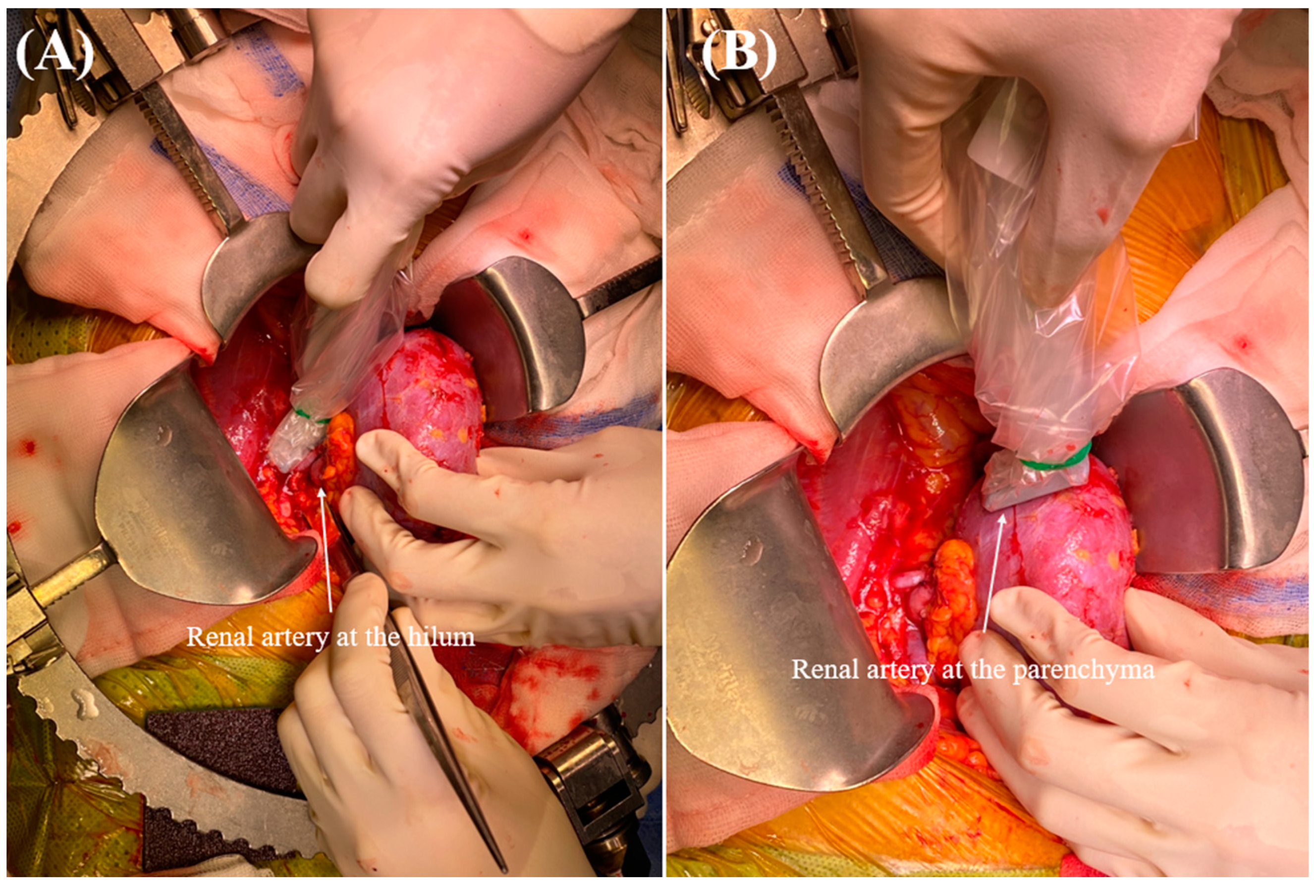
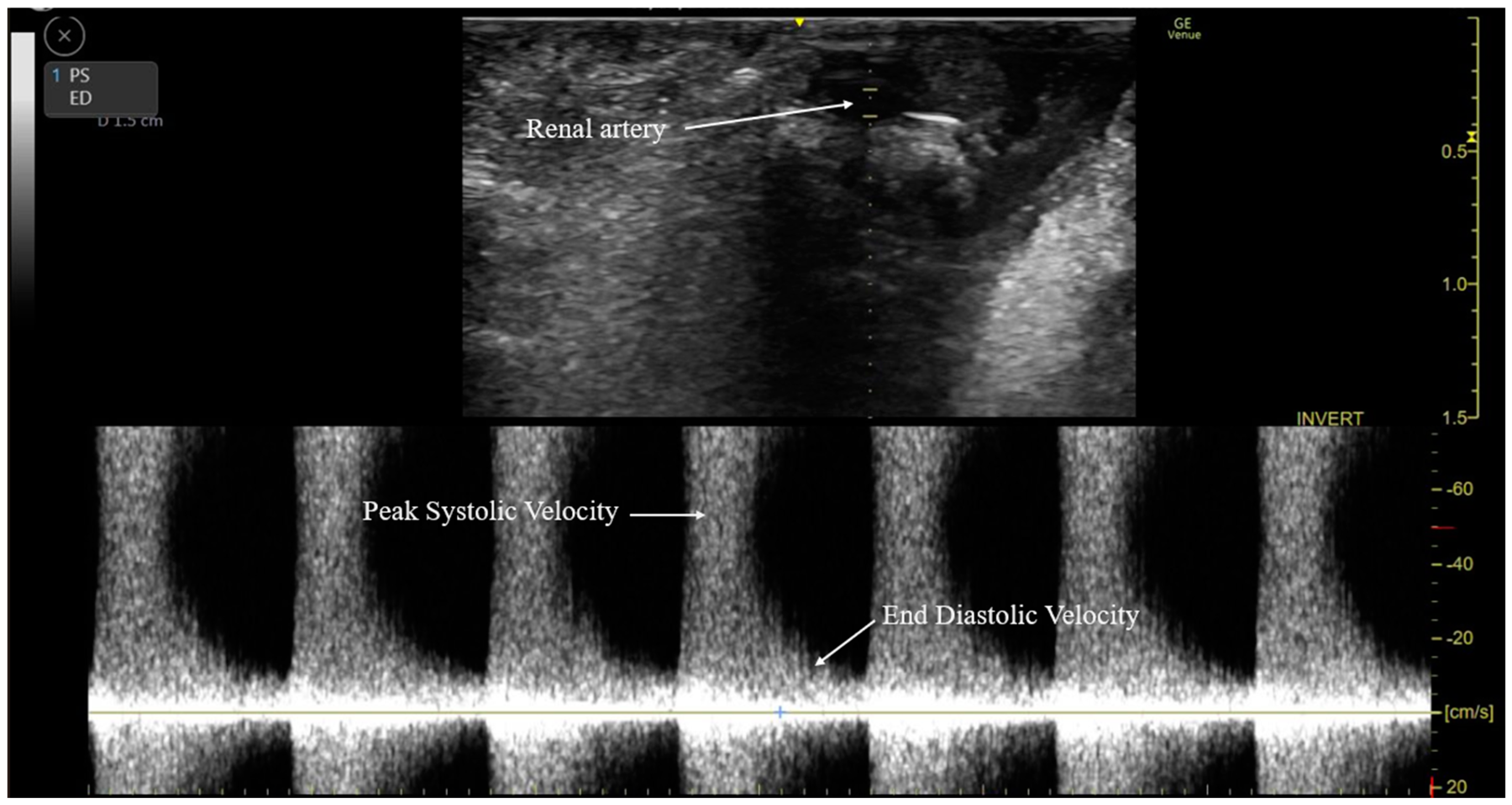
2.5. Intraoperative RARI Measurement

Following graft vascular and ureteral anastomosis, Doppler ultrasonography (Venue Go, GE Healthcare) was performed by the surgeon to measure the RARI and evaluate blood flow through the renal arteries at the renal hilum and parenchyma (Figure 2). The procedure began with the preparation of the ultrasound device, which was outfitted with a hockey stick-shaped transducer (L8-18i ultrasound transducer, GE Healthcare). Conductive gel was applied to the targeted areas on the renal artery and parenchyma to ensure the optimal transmission of ultrasound waves (pulsed wave Doppler) (Figure 3). The transducer was placed over the kidneys to locate the renal artery. The Doppler gate was positioned within the arterial lumen to capture optimal blood flow velocities during the cardiac cycle. The RARI was calculated as (peak systolic velocity − end-diastolic velocity)/peak systolic velocity [34]. This formula provides a dimensionless value indicative of the resistance in renal artery blood flow, with values > 0.8 defined as a high RARI [35]. The derived values were analyzed considering the clinical context of each patient, with all the pertinent findings and interpretations meticulously documented.

2.6. Clinical Variables for PSM

To ensure comparability between the dopamine and norepinephrine groups, we employed PSM based on a comprehensive set of preoperative, intraoperative, and donor graft factors. The preoperative recipient factors included sex, age, body mass index (BMI), and the presence of comorbidities such as diabetes mellitus (DM) and hypertension (HBP). Cardiac function markers such as ejection fraction and left ventricular mass index, along with systolic and diastolic blood pressures, heart rate, and various laboratory variables, including white blood cell count, neutrophil percentage, lymphocyte percentage, hemoglobin, albumin, electrolytes (sodium and potassium), creatinine, brain natriuretic peptide, high-sensitivity troponin I, corrected QT interval, and platelet count, were also considered. The intraoperative parameters included surgery duration, hourly fluid infusion rate, and the volume of blood loss. We also recorded serial intraoperative measurements of systolic, diastolic, and mean arterial pressures, heart rate, central venous pressure, and brain natriuretic peptide level. Donor and graft characteristics included donor sex (female), age, BMI, hemoglobin level, graft weight, and total ischemic time of the graft.

2.7. Postoperative Variables

The postoperative clinical factors included the RARI values recorded in the operating room and the ward, eGFR [36], hourly urine output, the need for rescue dialysis therapy, and the duration of intensive care unit (ICU) and hospital stays.

2.8. Statistical Analysis

The Shapiro–Wilk test was used to assess the normality of the distribution of continuous variables. The normally distributed variables are presented as means with standard deviations (SDs), whereas the non-normally distributed variables are presented as medians with interquartile ranges. The categorical variables are presented as numbers with percentages. To mitigate the effects of potential confounding factors on differences between groups, PSM was performed based on propensity scores, matching the patients on a one-to-one basis using greedy matching algorithms without replacement. The perioperative recipient and donor graft factors were compared between groups using the Mann–Whitney U test for continuous variables and the χ2 test or Fisher’s exact test for categorical variables as appropriate. The influence of intraoperative norepinephrine use on the intraoperative RARI was evaluated using multivariable logistic regression analysis after adjusting for propensity scores. The results are presented as odds ratios (ORs) with 95% confidence intervals (CIs). All statistical tests were two-sided. p-values < 0.05 were considered indicative of statistical significance. Statistical analyses were performed using R (version 2.10.1; R Foundation for Statistical Computing, Vienna, Austria) and SPSS for Windows (version 24.0; IBM Corp., Armonk, NY, USA).

3. Results

3.1. Demographic Characteristics

Of the 347 study participants, 173 (49.9%) were female. The mean age of the recipients was 49.7 ± 11.9 years, and the mean BMI was 23.5 ± 4.0 kg/m2. DM and HBP were present in 119 (34.3%) and 161 (46.4%) of the recipients, respectively. The mean serum level of creatinine prior to transplantation was 7.9 ± 2.7 mg/dL, and the mean hourly urine output was 2.1 ± 1.3 mL/kg/h. Among the living donors, 173 (49.9%) were female. The donors had a mean age of 48.4 ± 12.8 years and a mean BMI of 24.1 ± 3.1 kg/m2. The mean ischemic duration of the grafts was 58.8 ± 18.5 min, and the mean graft weight was 181.4 ± 39.7 g.

3.2. Comparison of Perioperative Factors before and after PSM

Before PSM, significant differences were observed between the two groups in terms of the preoperative characteristics of the recipients, particularly in the left ventricular mass index and hemoglobin level, as well as in the donors’ age (Table 1). However, after applying PSM to match the patients in the groups based on the aforementioned and potentially other relevant factors, the previously significant differences in perioperative recipient characteristics and donor graft parameters were rendered nonsignificant.

3.3. RARI in the Dopamine and Norepinephrine Groups in PS-Matched Patients

The RARI at the renal hilum and parenchyma were higher in the dopamine group than that in the norepinephrine group (Table 2). In addition, the proportion of patients with a high RARI (>0.8) was higher in the dopamine group than it was in the norepinephrine group. However, on postoperative day 7, these differences were not significant.

3.4. Association between Norepinephrine Infusion and a High RARI during Pre-Emptive LDKT

Norepinephrine infusion was significantly associated with a lower likelihood of a high RARI during pre-emptive LDKT, even after adjusting for the propensity scores. Specifically, the adjusted odds of achieving an RARI > 0.8 were significantly lower with norepinephrine infusion both at the renal hilum (adjusted OR = 0.214, 95% CI = 0.12–0.382, p < 0.001) and at the renal parenchyma (adjusted OR = 0.1, 95% CI = 0.029–0.348, p < 0.001) (Table 3).

3.5. Intraoperative Vital Signs and Brain Natriuretic Peptide Level in PSM Patients

During the surgical procedure, the heart rate observed after vascular graft and ureteral anastomosis, as well as at the conclusion of the surgery, was higher in the dopamine group than that in the norepinephrine group (Table 4). However, the other vital signs, including blood pressure and respiratory rate, along with the brain natriuretic peptide level, were comparable between the two groups.

3.6. Postoperative Kidney Graft Outcomes in PSM Patients

On the first postoperative day, both the eGFR and hourly urine output were higher in the norepinephrine group than that in the dopamine group (Table 5). However, subsequent evaluations of the postoperative variables, including eGFR, hourly urine output, the need for rescue dialysis therapy, and the duration of ICU and hospital stay, revealed no significant differences between the two groups.

4. Discussion

The novelty of our study lies in its focus on the intraoperative use of norepinephrine and dopamine during pre-emptive LDKT and their impact on renal arterial hemodynamics measured using the RARI. This topic has not been extensively explored in the literature. Our study utilizes PSM to minimize selection bias, ensuring a more accurate comparison between the effects of these two vasopressors. By demonstrating that norepinephrine is associated with more favorable intraoperative renal arterial hemodynamics and improved early postoperative renal function compared to those of dopamine, our findings provide critical insights that can enhance perioperative management in LDKT recipients.
We found that intraoperative norepinephrine infusion is more effective than dopamine in improving renal arterial hemodynamics during pre-emptive LDKT, as measured using the RARI at the renal hilum and parenchyma. After PSM, the norepinephrine infusion was associated with a significant decrease in the OR for a high RARI (>0.8) at the renal hilum (OR = 0.214) and parenchyma (OR = 0.1). While both norepinephrine and dopamine were capable of preventing intraoperative hypotension, the dopamine infusion was linked to an increased heart rate, an effect not observed with the norepinephrine infusion. In terms of postoperative kidney graft function, the patients receiving norepinephrine demonstrated a higher eGFR and hourly urine output on the first postoperative day. However, by the seventh postoperative day, outcomes such as the RARI, eGFR, hourly urine output, the need for rescue dialysis therapy, and the duration of ICU and hospital stays were similar between the two groups. Our findings suggest that a norepinephrine infusion may be a preferable option for maintaining systemic and renal hemodynamics during surgery and for early postoperative recovery, particularly when the denervated kidney allograft may impair the hemodynamic response to sympathomimetics.
Norepinephrine, which has strong α-adrenergic agonist properties and some β-adrenergic activity, plays a crucial role in increasing systemic vascular resistance, and to a lesser degree, cardiac output. Its ability to maintain renal perfusion pressure is particularly crucial, especially for transplanted kidneys, which often cannot adequately respond to physiological and pharmacological stresses due to their diminished autoregulatory capabilities. The denervation of kidney allografts removes the normal sympathetic nervous system inputs, necessitating the use of agents such as norepinephrine to ensure sufficient hemodynamic support [21,37]. Despite the effectiveness of vasopressors in managing such conditions, the optimal first-line vasopressor remains unclear, largely due to a scarcity of direct evidence. Nonetheless, there is a consensus that carefully titrating vasopressor dosages to avoid hypotension is preferable, given that the risks associated with low blood pressure exceed those of potential renal vasoconstriction [13,14]. Ensuring that the intraoperative MAP remains above 70 mmHg during kidney transplantation is crucial to prevent delayed graft function, which is often associated with lower MAP levels. The clinical recommendations suggest aiming for an MAP of 80–110 mmHg to protect kidney function, particularly during the critical reperfusion phase when an inadequate MAP can exacerbate kidney damage [14,38]. While direct comparisons between the responses of denervated kidney grafts to norepinephrine and those with intact autonomic regulation are challenging, several studies have indicated improvements in renal perfusion with norepinephrine administration, albeit within a broad spectrum of acceptable blood pressure levels [18,21,22,39]. In patients with septic shock, increasing the MAP from 65 to 75 mmHg using norepinephrine significantly increases the urinary output and RARI [22]. However, raising the MAP beyond this level does not result in additional benefits. In a study that explored the effects of varying MAP levels induced by norepinephrine on renal blood flow, eGFR, renal oxygen consumption, and oxygenation, the patients with norepinephrine-dependent vasodilatory shock and acute kidney injury following cardiac surgery demonstrated improved renal oxygen delivery, eGFR, and oxygenation when the MAP was increased from 60 to 75 mmHg. However, further elevation to 90 mmHg using norepinephrine only increased the renal vascular resistance without further improving perfusion, filtration, or oxygenation [21].
In patients with systemic inflammatory response syndrome due to an allograft, sepsis, or other triggers, hypotension may persist despite aggressive fluid resuscitation, often necessitating the use of a vasopressor. Traditionally, dopamine is preferred for its perceived ability to preserve blood flow to key organs such as the kidneys, brain, heart, and digestive system. In addition, concerns exist regarding norepinephrine use due to its intense vasoconstrictive effects. However, the renal-protective properties of dopamine have been disproven, revealing that it can reduce the blood flow to the mucosal lining of the digestive tract [19,20,40,41]. Contrary to the initial concerns, norepinephrine demonstrates more significant positive effects on renal blood flow and urine production than dopamine. While dopamine increases the mean renal blood flow by 20% without affecting the overall hemodynamics, norepinephrine significantly increases the MAP, cardiac output, and coronary and renal blood flow. In one study, dopamine increased the urine output, but did not improve creatinine clearance, whereas norepinephrine significantly enhanced urine output compared to low doses of dopamine, without significantly altering creatinine clearance [18]. Moreover, the harmful effects previously attributed to norepinephrine have not been substantiated. In fact, some studies have demonstrated that at clinically relevant doses, norepinephrine can enhance the blood flow to organs and tissues in various diseases. The concerns regarding severe vasoconstriction with norepinephrine administration have largely been linked to direct renal artery infusion in animal models at doses far exceeding those used in clinical practice [42,43,44].
Albanèse et al. demonstrated that norepinephrine, at doses typically used in hospitals, can improve or maintain kidney function in patients with pathological vasodilation and those who are well hydrated, but have normal systemic vascular resistance [23]. The benefits of norepinephrine include increased cyclooxygenase-2 expression in the kidney, leading to the increased production of cyclooxygenase-2-derived prostaglandins, such as PGE2 and PGI2 [45,46,47,48]. These substances mitigate norepinephrine-induced renal vasoconstriction and promote the dilation of the afferent arterioles of the kidneys, supporting glomerular filtration [46,48,49]. In addition, an increase in systemic blood pressure can trigger renal vasodilation by reducing renal sympathetic tone via a baroreceptor feedback mechanism [42]. Albanèse et al. found that norepinephrine administered at an average dose of up to 1.3 ± 0.3 µg/kg/min effectively restored the MAP and urine production in patients with septic shock and oliguria [23]. This observation is in line with other clinical studies [50,51,52] and experimental models demonstrating that norepinephrine can restore kidney function and blood flow during endotoxemia [25,26,53]. While norepinephrine is effective for the treatment of septic shock, its safety in other clinical conditions is uncertain [54]. However, Albanèse et al. demonstrated that norepinephrine, even at doses up to 0.5 µg/kg/min, is safe and effective for increasing the MAP and cerebral perfusion pressure without causing kidney dysfunction in well-hydrated patients with head injuries [23]. This finding is in line with the results of Morimatsu et al., who demonstrated that norepinephrine can maintain kidney function in hypotensive post-cardiac surgery patients [24]. Our results further support the greater effectiveness of norepinephrine than that of dopamine in maintaining renal regional arterial inflow and parenchymal function during LDKT.
While norepinephrine has shown benefits in maintaining renal perfusion and supporting early postoperative graft function, it is important to consider its potential adverse effects. Norepinephrine, primarily through its α-adrenergic activity, can cause vasoconstriction, which might reduce the blood flow to other vital organs and tissues, potentially leading to ischemic complications. This is particularly concerning in patients with compromised vascular integrity. Additionally, norepinephrine can increase the cardiac afterload, which may exacerbate heart conditions in susceptible individuals. There is also a risk of tissue ischemia and gangrene, especially in the extremities, due to prolonged or high-dose infusion [21,55,56]. Therefore, the careful monitoring and titration of norepinephrine are crucial to balancing its benefits and risks during and after surgery.
The intraoperative ultrasound imaging of renal artery flow is notably effective for the early detection of acute kidney injury (AKI), boasting high sensitivity and specificity [57]. Suggested for use in hemodynamically stable patients before sternotomy by Kajar et al., transesophageal echocardiography has proven instrumental in linking increased RARI values to AKI post-coronary artery bypass graft surgery, with a 26% diagnosis rate among patients. Particularly, an RARI above 0.7 was associated with higher AKI rates, establishing an elevated RARI as an independent AKI predictor post-surgery [58]. This predictive capacity extends to non-cardiac procedures and sepsis, with an RARI greater than 0.7 indicating an AKI with high accuracy in orthopedic surgery patients [59]. In kidney transplantation, transit time flowmetry and graft arterial flowmetry have been employed to assess renal artery flow and the RARI, which are vital for forecasting the immediate and delayed graft functions. Some studies, including those by Król et al. and Pravisani et al., have demonstrated significant correlations between the RARI levels and graft function outcomes, underscoring the importance of the RARI in evaluating transplant success [60,61]. Our research, while not primarily focused on the RARI’s direct link to AKI, explored the effects of norepinephrine and dopamine on the RARI during surgery. Our findings reveal that a high RARI (>0.8) could adversely affect early graft function recovery, emphasizing the significance of RARI monitoring and management in the intraoperative phase to enhance postoperative results.
Our study had several limitations that must be acknowledged. First, we did not measure the graft renal artery length or inner radius in the operating room, which could have influenced the graft flow and renal flow outcomes in both the groups. To mitigate this, we exclusively selected left-sided kidneys for grafting to reduce the selection bias associated with arterial length differences between left and right kidneys. However, this choice limits the generalizability of our findings to right-sided grafts or grafts with multiple arterial branches. Second, we only included grafts with a single artery, preventing us from assessing the potential differences in drug responses in grafts with multiple arterial branches. Third, although atherosclerosis is prevalent among patients undergoing kidney transplantation, we were unable to determine the effects of the two drugs on the RARI in individuals with such vascular characteristics. Fourth, the retrospective study design inherently limits our ability to establish causality and control for all the confounding variables. While propensity score matching (PSM) was employed to minimize the selection bias, residual confounding cannot be entirely eliminated. Additionally, the inability to randomize patients may introduce a selection bias, as treatment decisions were based on clinical judgment rather than random assignment. Furthermore, due to the retrospective nature of the study, we could not standardize the doses of norepinephrine and dopamine or establish target blood pressure levels, which may have influenced the outcomes. Our study also excluded patients who had undergone dialysis, limiting the generalizability of our findings to this population. Lastly, we focused exclusively on pre-emptive LDKT, thereby excluding any potential hemodynamic changes in patients who had previously undergone dialysis. Despite these limitations, our study provides valuable insights into the comparative effects of norepinephrine and dopamine on renal arterial hemodynamics and early postoperative renal function during pre-emptive LDKT. Nonetheless, further prospective randomized controlled trials are necessary to verify our findings and enhance our understanding of the optimal vasopressor for kidney transplant patients.

5. Conclusions

Our investigation into the intraoperative use of vasopressors during pre-emptive LDKT reveals the superiority of norepinephrine over dopamine in enhancing renal arterial hemodynamics, as evidenced by a significantly lower OR for a high RARI. Our results suggest that norepinephrine can effectively maintain both systemic and renal hemodynamics, without increasing the heart rate associated with dopamine. Despite similar outcomes by the seventh postoperative day, norepinephrine use is associated with a higher eGFR and urine output in the immediate postoperative period, suggesting that is preferable for early graft function recovery. These findings, while highlighting the need for further prospective trials, provide valuable guidance for vasopressor selection in LDKT, enhancing postoperative management and patient care.

Supplementary Materials

The following supporting information can be downloaded at: https://www.mdpi.com/article/10.3390/medicina60071066/s1, Table S1: Inclusion and exclusion criteria.

Author Contributions

Conceptualization, J.H. and M.S.C.; methodology, J.H. and M.S.C.; software, J.H. and M.S.C.; validation, J.H. and M.S.C.; formal analysis, J.H. and M.S.C.; investigation, J.H. and M.S.C.; resources, J.H. and M.S.C.; data curation, J.H. and M.S.C.; writing—original draft preparation, J.H. and M.S.C.; writing—review and editing, J.H., H.K., H.P., S.C.P., S.S.Y. and M.S.C.; project administration, J.H. and M.S.C.; All authors have read and agreed to the published version of the manuscript.

Funding

This research received no external funding.

Institutional Review Board Statement

The protocol for this retrospective observational cohort study received approval from the Institutional Review Board and Ethics Committee of Seoul St. Mary’s Hospital (approval no. KC22RISI0395) on 7 June 2022. For the purposes of this study, authorization to access the data was granted for the timeframe spanning from 7 June 2022 through 6 June 2023. Before initiating data collection, all the collected research data were anonymized (assigned random study numbers) to ensure the privacy of the participants. This anonymization process was maintained throughout and after data collection for all analyses conducted, adhering to the principles outlined in the Declaration of Helsinki. Given the retrospective nature of the study, it was deemed unnecessary to obtain informed consent from the participants. We have reported our findings following the STROBE Statement guidelines to ensure clarity, transparency, and rigor in reporting observational studies.

Informed Consent Statement

Given the retrospective nature of the study, it was deemed unnecessary to obtain informed consent from the participants.

Data Availability Statement

Data are contained within this article and the Supplemental File.

Conflicts of Interest

The authors declared no conflict of interest.

Abbreviations

RARI: renal arterial resistive index; LDKT: living donor kidney transplantation; ESRD: end-stage renal disease; PSM: propensity score matching; DM: diabetes mellitus; HBP: hypertension; eGFR: estimated glomerular filtration; BMI: body mass index; ICU: intensive care unit; AKI acute kidney injury.

References

  1. Kohei, N.; Sawada, Y.; Hirai, T.; Omoto, K.; Ishida, H.; Tanabe, K. Influence of dialysis duration on the outcome of living kidney transplantation. Ther. Apher. Dial. 2014, 18, 481–488. [Google Scholar] [CrossRef]
  2. Abecassis, M.; Bartlett, S.T.; Collins, A.J.; Davis, C.L.; Delmonico, F.L.; Friedewald, J.J.; Hays, R.; Howard, A.; Jones, E.; Leichtman, A.B.; et al. Kidney transplantation as primary therapy for end-stage renal disease: A National Kidney Foundation/Kidney Disease Outcomes Quality Initiative (NKF/KDOQITM) conference. Clin. J. Am. Soc. Nephrol. 2008, 3, 471–480. [Google Scholar] [CrossRef]
  3. Chadban, S.J.; Ahn, C.; Axelrod, D.A.; Foster, B.J.; Kasiske, B.L.; Kher, V.; Kumar, D.; Oberbauer, R.; Pascual, J.; Pilmore, H.L.; et al. KDIGO Clinical Practice Guideline on the Evaluation and Management of Candidates for Kidney Transplantation. Transplantation 2020, 104, S11–S103. [Google Scholar] [CrossRef]
  4. Andrews, P.A.; Burnapp, L. British Transplantation Society/Renal Association UK Guidelines for Living Donor Kidney Transplantation 2018: Summary of Updated Guidance. Transplantation 2018, 102, e307. [Google Scholar] [CrossRef]
  5. Kasiske, B.L.; Snyder, J.J.; Matas, A.J.; Ellison, M.D.; Gill, J.S.; Kausz, A.T. Preemptive kidney transplantation: The advantage and the advantaged. J. Am. Soc. Nephrol. 2002, 13, 1358–1364. [Google Scholar] [CrossRef]
  6. Papalois, V.E.; Moss, A.; Gillingham, K.J.; Sutherland, D.E.; Matas, A.J.; Humar, A. Pre-emptive transplants for patients with renal failure: An argument against waiting until dialysis. Transplantation 2000, 70, 625–631. [Google Scholar] [CrossRef]
  7. Auneau-Enjalbert, L.; Hardouin, J.B.; Blanchin, M.; Giral, M.; Morelon, E.; Cassuto, E.; Meurette, A.; Sébille, V. Comparison of longitudinal quality of life outcomes in preemptive and dialyzed patients on waiting list for kidney transplantation. Qual. Life Res. 2020, 29, 959–970. [Google Scholar] [CrossRef]
  8. Huang, Y.; Samaniego, M. Preemptive kidney transplantation: Has it come of age? Nephrol. Ther. 2012, 8, 428–432. [Google Scholar] [CrossRef]
  9. Liem, Y.S.; Weimar, W. Early living-donor kidney transplantation: A review of the associated survival benefit. Transplantation 2009, 87, 317–318. [Google Scholar] [CrossRef]
  10. Dinavahi, R.; Akalin, E. Preemptive kidney transplantation in patients with diabetes mellitus. Endocrinol. Metab. Clin. N. Am. 2007, 36, 1039–1049. [Google Scholar] [CrossRef]
  11. Renew, J.R.; Pai, S.L. A simple protocol to improve safety and reduce cost in hemodialysis patients undergoing elective surgery. Middle East. J. Anaesthesiol. 2014, 22, 487–492. [Google Scholar]
  12. Folsom, A.R.; Lutsey, P.L.; Astor, B.C.; Wattanakit, K.; Heckbert, S.R.; Cushman, M. Chronic kidney disease and venous thromboembolism: A prospective study. Nephrol. Dial. Transplant. 2010, 25, 3296–3301. [Google Scholar] [CrossRef]
  13. Schmid, S.; Jungwirth, B. Anaesthesia for renal transplant surgery: An update. Eur. J. Anaesthesiol. 2012, 29, 552–558. [Google Scholar] [CrossRef]
  14. Ricaurte, L.; Vargas, J.; Lozano, E.; Díaz, L. Anesthesia and kidney transplantation. Transplant. Proc. 2013, 45, 1386–1391. [Google Scholar] [CrossRef]
  15. Tena, B.; Vendrell, M. Perioperative considerations for kidney and pancreas-kidney transplantation. Best. Pract. Res. Clin. Anaesthesiol. 2020, 34, 3–14. [Google Scholar] [CrossRef]
  16. Mittel, A.M.; Wagener, G. Anesthesia for Kidney and Pancreas Transplantation. Anesthesiol. Clin. 2017, 35, 439–452. [Google Scholar] [CrossRef]
  17. Ciapetti, M.; di Valvasone, S.; di Filippo, A.; Cecchi, A.; Bonizzoli, M.; Peris, A. Low-dose dopamine in kidney transplantation. Transplant. Proc. 2009, 41, 4165–4168. [Google Scholar] [CrossRef]
  18. Di Giantomasso, D.; Morimatsu, H.; May, C.N.; Bellomo, R. Increasing renal blood flow: Low-dose dopamine or medium-dose norepinephrine. Chest 2004, 125, 2260–2267. [Google Scholar] [CrossRef]
  19. Marik, P.E. Low-dose dopamine: A systematic review. Intensive Care Med. 2002, 28, 877–883. [Google Scholar] [CrossRef]
  20. Kellum, J.A.; Decker, J.M. Use of dopamine in acute renal failure: A meta-analysis. Crit. Care Med. 2001, 29, 1526–1531. [Google Scholar] [CrossRef]
  21. Redfors, B.; Bragadottir, G.; Sellgren, J.; Swärd, K.; Ricksten, S.E. Effects of norepinephrine on renal perfusion, filtration and oxygenation in vasodilatory shock and acute kidney injury. Intensive Care Med. 2011, 37, 60–67. [Google Scholar] [CrossRef]
  22. Deruddre, S.; Cheisson, G.; Mazoit, J.X.; Vicaut, E.; Benhamou, D.; Duranteau, J. Renal arterial resistance in septic shock: Effects of increasing mean arterial pressure with norepinephrine on the renal resistive index assessed with Doppler ultrasonography. Intensive Care Med. 2007, 33, 1557–1562. [Google Scholar] [CrossRef]
  23. Albanèse, J.; Leone, M.; Garnier, F.; Bourgoin, A.; Antonini, F.; Martin, C. Renal effects of norepinephrine in septic and nonseptic patients. Chest 2004, 126, 534–539. [Google Scholar] [CrossRef]
  24. Morimatsu, H.; Uchino, S.; Chung, J.; Bellomo, R.; Raman, J.; Buxton, B. Norepinephrine for hypotensive vasodilatation after cardiac surgery: Impact on renal function. Intensive Care Med. 2003, 29, 1106–1112. [Google Scholar] [CrossRef]
  25. Di Giantomasso, D.; Morimatsu, H.; May, C.N.; Bellomo, R. Intrarenal blood flow distribution in hyperdynamic septic shock: Effect of norepinephrine. Crit. Care Med. 2003, 31, 2509–2513. [Google Scholar] [CrossRef]
  26. Treggiari, M.M.; Romand, J.A.; Burgener, D.; Suter, P.M.; Aneman, A. Effect of increasing norepinephrine dosage on regional blood flow in a porcine model of endotoxin shock. Crit. Care Med. 2002, 30, 1334–1339. [Google Scholar] [CrossRef]
  27. Enhesari, A.; Mardpour, S.; Makki, Z.; Mardpour, S. Early ultrasound assessment of renal transplantation as the valuable biomarker of long lasting graft survival: A cross-sectional study. Iran. J. Radiol. 2014, 11, e11492. [Google Scholar] [CrossRef]
  28. Radermacher, J.; Mengel, M.; Ellis, S.; Stuht, S.; Hiss, M.; Schwarz, A.; Eisenberger, U.; Burg, M.; Luft, F.C.; Gwinner, W.; et al. The renal arterial resistance index and renal allograft survival. N. Engl. J. Med. 2003, 349, 115–124. [Google Scholar] [CrossRef]
  29. Krumme, B. Renal Doppler sonography—Update in clinical nephrology. Nephron Clin. Pract. 2006, 103, c24–c28. [Google Scholar] [CrossRef]
  30. Pape, L.; Mengel, M.; Offner, G.; Melter, M.; Ehrich, J.H.; Strehlau, J. Renal arterial resistance index and computerized quantification of fibrosis as a combined predictive tool in chronic allograft nephropathy. Pediatr. Transplant. 2004, 8, 565–570. [Google Scholar] [CrossRef]
  31. Heine, G.H.; Girndt, M.; Sester, U.; Köhler, H. No rise in renal Doppler resistance indices at peak serum levels of cyclosporin A in stable kidney transplant patients. Nephrol. Dial. Transplant. 2003, 18, 1639–1643. [Google Scholar] [CrossRef]
  32. Naesens, M.; Heylen, L.; Lerut, E.; Claes, K.; De Wever, L.; Claus, F.; Oyen, R.; Kuypers, D.; Evenepoel, P.; Bammens, B.; et al. Intrarenal resistive index after renal transplantation. N. Engl. J. Med. 2013, 369, 1797–1806. [Google Scholar] [CrossRef]
  33. Go, J.; Park, S.C.; Yun, S.S.; Ku, J.; Park, J.; Shim, J.W.; Lee, H.M.; Kim, Y.S.; Moon, Y.E.; Hong, S.H.; et al. Exposure to Hyperchloremia Is Associated with Poor Early Recovery of Kidney Graft Function after Living-Donor Kidney Transplantation: A Propensity Score-Matching Analysis. J. Clin. Med. 2019, 8, 955. [Google Scholar] [CrossRef]
  34. Regolisti, G.; Maggiore, U.; Cademartiri, C.; Belli, L.; Gherli, T.; Cabassi, A.; Morabito, S.; Castellano, G.; Gesualdo, L.; Fiaccadori, E. Renal resistive index by transesophageal and transparietal echo-doppler imaging for the prediction of acute kidney injury in patients undergoing major heart surgery. J. Nephrol. 2017, 30, 243–253. [Google Scholar] [CrossRef]
  35. Radermacher, J.; Ellis, S.; Haller, H. Renal resistance index and progression of renal disease. Hypertension 2002, 39, 699–703. [Google Scholar] [CrossRef]
  36. Levey, A.S.; Coresh, J.; Greene, T.; Stevens, L.A.; Zhang, Y.L.; Hendriksen, S.; Kusek, J.W.; Van Lente, F. Using standardized serum creatinine values in the modification of diet in renal disease study equation for estimating glomerular filtration rate. Ann. Intern. Med. 2006, 145, 247–254. [Google Scholar] [CrossRef]
  37. Jaszczuk, S.; Natarajan, S.; Papalois, V. Anaesthetic Approach to Enhanced Recovery after Surgery for Kidney Transplantation: A Narrative Review. J. Clin. Med. 2022, 11, 3435. [Google Scholar] [CrossRef]
  38. Smudla, A.; Trimmel, D.; Szabó, G.; Fazakas, J. Systolic Blood Pressure Pattern: The Tick Mark Signal of Delayed Renal Graft Function. Transplant. Proc. 2019, 51, 1226–1230. [Google Scholar] [CrossRef]
  39. Gordon, A.C.; Mason, A.J.; Thirunavukkarasu, N.; Perkins, G.D.; Cecconi, M.; Cepkova, M.; Pogson, D.G.; Aya, H.D.; Anjum, A.; Frazier, G.J.; et al. Effect of Early Vasopressin vs Norepinephrine on Kidney Failure in Patients With Septic Shock: The VANISH Randomized Clinical Trial. JAMA 2016, 316, 509–518. [Google Scholar] [CrossRef]
  40. Marik, P.E.; Mohedin, M. The contrasting effects of dopamine and norepinephrine on systemic and splanchnic oxygen utilization in hyperdynamic sepsis. JAMA 1994, 272, 1354–1357. [Google Scholar] [CrossRef]
  41. Segal, J.M.; Phang, P.T.; Walley, K.R. Low-dose dopamine hastens onset of gut ischemia in a porcine model of hemorrhagic shock. J. Appl. Physiol. 1992, 73, 1159–1164. [Google Scholar] [CrossRef] [PubMed]
  42. Anderson, W.P.; Korner, P.I.; Selig, S.E. Mechanisms involved in the renal responses to intravenous and renal artery infusions of noradrenaline in conscious dogs. J. Physiol. 1981, 321, 21–30. [Google Scholar] [CrossRef] [PubMed]
  43. Cronin, R.E.; Erickson, A.M.; de Torrente, A.; McDonald, K.M.; Schrier, R.W. Norepinephrine-induced acute renal failure: A reversible ischemic model of acute renal failure. Kidney Int. 1978, 14, 187–190. [Google Scholar] [CrossRef] [PubMed]
  44. Cronin, R.E.; de Torrente, A.; Miller, P.D.; Bulger, R.E.; Burke, T.J.; Schrier, R.W. Pathogenic mechanisms in early norepinephrine-induced acute renal failure: Functional and histological correlates of protection. Kidney Int. 1978, 14, 115–125. [Google Scholar] [CrossRef] [PubMed]
  45. Llinás, M.T.; López, R.; Rodríguez, F.; Roig, F.; Salazar, F.J. Role of COX-2-derived metabolites in regulation of the renal hemodynamic response to norepinephrine. Am. J. Physiol. Renal Physiol. 2001, 281, F975–F982. [Google Scholar] [CrossRef] [PubMed]
  46. Imig, J.D.; Deichmann, P.C. Afferent arteriolar responses to ANG II involve activation of PLA2 and modulation by lipoxygenase and P-450 pathways. Am. J. Physiol. 1997, 273, F274–F282. [Google Scholar] [CrossRef] [PubMed]
  47. Chatziantoniou, C.; Arendshorst, W.J. Prostaglandin interactions with angiotensin, norepinephrine, and thromboxane in rat renal vasculature. Am. J. Physiol. 1992, 262, F68–F76. [Google Scholar] [CrossRef] [PubMed]
  48. Pelayo, J.C. Renal adrenergic effector mechanisms: Glomerular sites for prostaglandin interaction. Am. J. Physiol. 1988, 254, F184–F190. [Google Scholar] [CrossRef]
  49. Inscho, E.W.; Carmines, P.K.; Navar, L.G. Prostaglandin influences on afferent arteriolar responses to vasoconstrictor agonists. Am. J. Physiol. 1990, 259, F157–F163. [Google Scholar] [CrossRef]
  50. LeDoux, D.; Astiz, M.E.; Carpati, C.M.; Rackow, E.C. Effects of perfusion pressure on tissue perfusion in septic shock. Crit. Care Med. 2000, 28, 2729–2732. [Google Scholar] [CrossRef]
  51. Martin, C.; Papazian, L.; Perrin, G.; Saux, P.; Gouin, F. Norepinephrine or dopamine for the treatment of hyperdynamic septic shock? Chest 1993, 103, 1826–1831. [Google Scholar] [CrossRef] [PubMed]
  52. Desjars, P.; Pinaud, M.; Bugnon, D.; Tasseau, F. Norepinephrine therapy has no deleterious renal effects in human septic shock. Crit. Care Med. 1989, 17, 426–429. [Google Scholar] [CrossRef] [PubMed]
  53. Zhang, H.; Smail, N.; Cabral, A.; Rogiers, P.; Vincent, J.L. Effects of norepinephrine on regional blood flow and oxygen extraction capabilities during endotoxic shock. Am. J. Respir. Crit. Care Med. 1997, 155, 1965–1971. [Google Scholar] [CrossRef] [PubMed]
  54. Martin, C.; Viviand, X.; Leone, M.; Thirion, X. Effect of norepinephrine on the outcome of septic shock. Crit. Care Med. 2000, 28, 2758–2765. [Google Scholar] [CrossRef] [PubMed]
  55. Owen, V.S.; Rosgen, B.K.; Cherak, S.J.; Ferland, A.; Stelfox, H.T.; Fiest, K.M.; Niven, D.J. Adverse events associated with administration of vasopressor medications through a peripheral intravenous catheter: A systematic review and meta-analysis. Crit. Care 2021, 25, 146. [Google Scholar] [CrossRef] [PubMed]
  56. Russell, J.A. Vasopressor therapy in critically ill patients with shock. Intensive Care Med. 2019, 45, 1503–1517. [Google Scholar] [CrossRef] [PubMed]
  57. Darmon, M.; Schortgen, F.; Vargas, F.; Liazydi, A.; Schlemmer, B.; Brun-Buisson, C.; Brochard, L. Diagnostic accuracy of Doppler renal resistive index for reversibility of acute kidney injury in critically ill patients. Intensive Care Med. 2011, 37, 68–76. [Google Scholar] [CrossRef] [PubMed]
  58. Kajal, K.; Chauhan, R.; Negi, S.L.; Gourav, K.P.; Panda, P.; Mahajan, S.; Sarna, R. Intraoperative evaluation of renal resistive index with transesophageal echocardiography for the assessment of acute renal injury in patients undergoing coronary artery bypass grafting surgery: A prospective observational study. Ann. Card. Anaesth. 2022, 25, 158–163. [Google Scholar] [PubMed]
  59. Marty, P.; Szatjnic, S.; Ferre, F.; Conil, J.M.; Mayeur, N.; Fourcade, O.; Silva, S.; Minville, V. Doppler renal resistive index for early detection of acute kidney injury after major orthopaedic surgery: A prospective observational study. Eur. J. Anaesthesiol. 2015, 32, 37–43. [Google Scholar] [CrossRef]
  60. Pravisani, R.; Baccarani, U.; Langiano, N.; Meroi, F.; Avital, I.; Bove, T.; Adani, G.L. Predictive Value of Intraoperative Doppler Flowmetry for Delayed Graft Function in Kidney Transplantation: A Pilot Study. Transplant. Proc. 2020, 52, 1556–1558. [Google Scholar] [CrossRef]
  61. Król, R.; Chudek, J.; Kolonko, A.; Ziaja, J.; Pawlicki, J.; Wiecek, A.; Cierpka, L. Intraoperative resistance index measured with transsonic flowmeter on kidney graft artery can predict early and long-term graft function. Transplant. Proc. 2011, 43, 2926–2929. [Google Scholar] [CrossRef] [PubMed]
Figure 1. Study flow diagram.
Medicina 60 01066 g001
Figure 2. Doppler ultrasonography for renal arterial resistive index (RARI) measurement. RARI measurement at (A) the artery of the renal hilum and (B) the renal parenchyma.
Medicina 60 01066 g002
Figure 3. Measurement of renal arterial resistive index by using pulsed wave Doppler ultrasonography.
Medicina 60 01066 g003
Table 1. Comparison of perioperative factors between the dopamine and norepinephrine groups before and after propensity score matching.
Before Propensity Score MatchingAfter Propensity Score Matching
GroupDopamineNorepinephrinepSDDopamineNorepinephrinepSD
n177170 167167
Preoperative recipient factors
Sex (female)85 (48.0%)88 (51,8%)0.4860.07581 (48.5%)86 (51.5%)0.5840.06
Age (years)49.9 ± 12.749.5 ± 10.90.734−0.0449.7 ± 12.949.5 ± 10.90.905−0.014
Body mass index (kg/m2)23.3 ± 3.923.7 ± 4.10.2980.10923.4 ± 3.923.8 ± 4.10.3680.096
Comorbidity
Diabetes mellitus62 (35.0%)57 (33.5%)0.769−0.03259 (35.3%)55 (32.9%)0.644−0.051
Hypertension87 (49.2%)74 (43.5%)0.294−0.11383 (49.7%)72 (43.1%)0.227−0.132
Echocardiography
Ejection fraction (%)61.1 ± 5.5 59.8 ± 7.00.043−0.19661.0 ± 5.560.0 ± 6.70.16−0.136
Left ventricular mass index (g/m2)129.9 ± 83.8 127.4 ± 44.20.728−0.056123.4 ± 36.2127.6 ± 44.60.350.094
Vital sign
Systolic blood pressure (mmHg)133.7 ± 13.2131.0 ± 13.50.059−0.201133.4 ± 13.2130.9 ± 13.50.092−0.183
Diastolic blood pressure (mmHg)82.0 ± 9.881.0 ± 9.30.327−0.10881.8 ± 9.981.0 ± 9.30.437−0.088
Heart rate (beats/min)78.5 ± 9.278.6 ± 9.40.8590.01978.8 ± 9.378.6 ± 9.30.874−0.017
Hourly urine output (mL/kg/h)2.1 ± 1.42.0 ± 1.30.638−0.0522.1 ± 1.32.0 ± 1.30.799−0.029
Brain natriuretic peptide (pg/mL)245.0 ± 483.8278.6 ± 718.40.610.047226.6 ± 442.2278.6 ± 723.60.4290.072
High-sensitivity troponin I (pg/mL)45.5 ± 71.062.3 ± 199.00.2910.08545.6 ± 72.646.4 ± 84.70.9230.004
Corrected QT interval (ms)451.1 ± 29.3450.5 ± 31.00.843−0.021451.0 ± 29.5450.2 ± 31.00.793−0.028
Laboratory variables
White blood cell count (×109/L)6.7 ± 3.16.8 ± 2.60.880.0186.7 ± 3.26.8 ± 2.60.8340.026
Neutrophil (%)72.7 ± 13.171.4 ± 15.10.411−0.08372.2 ± 13.171.5 ± 15.20.633−0.049
Lymphocyte (%)17.8 ± 9.018.4 ± 9.00.590.05818.0 ± 9.118.3 ± 9.00.7480.036
Hemoglobin (g/dL)10.7 ± 1.410.4 ± 1.40.034−0.2310.6 ± 1.410.4 ± 1.40.113−0.173
Albumin (g/dL)4.1 ± 0.44.0 ± 0.50.115−0.1634.1 ± 0.44.0 ± 0.50.239−0.123
Sodium (mEq/L)136.5 ± 3.9137.2 ± 4.60.1120.158136.7 ± 3.7137.2 ± 4.70.3090.101
Potassium (mEq/L)4.6 ± 0.54.5 ± 0.50.234−0.1254.6 ± 0.54.6 ± 0.50.322−0.107
Chloride (mEq/L)99.9 ± 5.6100.4 ± 6.30.3810.089100.2 ± 5.6100.4 ± 6.40.7280.036
Platelet count (×109/L)182.3 ± 61.4186.5 ± 66.20.5440.063183.3 ± 61.7187.6 ± 66.10.5460.064
International normalized ratio1.1 ± 0.91.1 ± 0.30.583−0.1271.1 ± 0.91.1 ± 0.30.597−0.129
Creatinine (mg/dL)7.7 ± 2.78.1 ± 2.70.220.1327.7 ± 2.78.1 ± 2.60.2610.123
Intraoperative recipient factors
Operation time (min)226.3 ± 44.3230.0 ± 43.20.4320.085227.6 ± 44.8230.1 ± 43.50.6010.058
Hourly fluid infusion (mL/kg/h)9.6 ± 3.710.0 ± 3.20.2470.1339.7 ± 3.710.0 ± 3.20.4690.086
Hemorrhage (mL)171.1 ± 69.6160.5 ± 66.80.151−0.158171.3 ± 68.6160.4 ± 66.20.143−0.162
Donor and graft factors
Sex (female)85 (48.0%)88 (51.8%)0.4860.07581 (48.5%)86 (51.5%)0.5840.06
Age (years)49.7 ± 13.047.0 ± 12.40.049−0.21749.1 ± 13.146.9 ± 12.50.117−0.177
Body mass index (kg/m2)24.1 ± 3.224.1 ± 3.10.8940.01524.1 ± 3.224.1 ± 3.10.9440.008
Hemoglobin (g/dL)13.8 ± 1.113.8 ± 1.20.9170.01113.8 ± 1.113.8 ± 1.20.9030.013
Graft weight (g)185.2 ± 41.3177.4 ± 37.60.063−0.21183.9 ± 41.2177.1 ± 37.80.12−0.18
Total ischemic time (min)57.7 ± 15.059.9 ± 21.50.2610.10458.3 ± 15.259.9 ± 21.70.4340.074
Abbreviations: SD, standard deviation. Values are expressed as mean (standard deviation) and number (percentage).
Table 2. Comparison of the renal arterial resistive index between dopamine and norepinephrine groups in PS-matched patients.
GroupDopamineNorepinephrinep
167167
In the operating room (after renal vascular and ureter anastomosis)
Renal arterial resistive index
at renal hilum0.77 ± 0.110.66 ± 0.13<0.001
at renal parenchyma 0.71 ± 0.10.6 ± 0.1<0.001
Renal arterial resistive index (>0.8)
at renal hilum66 (39.5%)20 (12.0%)<0.001
at renal parenchyma 25 (15.0%)3 (1.8%)<0.001
In the ward (on postoperative 7)
Renal arterial resistive index
at renal hilum0.64 ± 0.110.64 ± 0.050.945
at renal parenchyma 0.56 ± 0.110.55 ± 0.050.382
Renal arterial resistive index (>0.8)
at renal hilum4 (2.4%)0 (0.0%)0.123
at renal parenchyma3 (1.8%)0 (0.0%)0.248
Abbreviations: PS, propensity score. Values are expressed as median (interquartile) and number (proportion).
Table 3. Association between norepinephrine infusion and renal arterial resistive index > 0.8, adjusted for propensity scores during pre-emptive living donor kidney transplantation.
βOdds Ratio95% CIp
In the operating room (after renal vascular and ureter anastomosis)
Norepinephrine adjusted for PS
Renal arterial resistive index (>0.8)
 at renal hilum−1.5430.2140.12–0.382<0.001
 at renal parenchyma−2.3010.10.029–0.348<0.001
Abbreviations: CI, confidence interval; PS, propensity score.
Table 4. Comparison of intraoperative vital signs and the brain natriuretic peptide level among PS-matched patients from the dopamine and norepinephrine groups.
GroupDopamineNorepinephrinep
167167
At the beginning of the surgery
Systolic blood pressure (mmHg)126.5 ± 13.4124.8 ± 9.80.185
Diastolic blood pressure (mmHg)77.6 ± 11.075.4 ± 13.30.091
Mean blood pressure (mmHg)93.9 ± 10.391.8 ± 11.00.075
Heart rate (beats/min)73.2 ± 9.374.5 ± 7.40.158
Central venous pressure (mmHg)5.6 ± 2.05.7 ± 1.80.841
Brain natriuretic peptide (pg/mL)147.8 (94.8–251.2)139.6 (81.9–295.9)0.583
After vascular graft and ureteral anastomosis
Systolic blood pressure (mmHg)145.7 ± 6.9144.5 ± 6.70.137
Diastolic blood pressure (mmHg)80.0 ± 7.880.1 ± 6.90.888
Mean blood pressure (mmHg)94.5 ± 10.193.5 ± 9.00.526
Heart rate (beats/min)96.5 ± 10.579.9 ± 11.2<0.001
Central venous pressure (mmHg)11.7 ± 2.111.9 ± 3.00.425
At the end of the surgery
Systolic blood pressure (mmHg)132.9 ± 16.7131.5 ± 15.60.428
Diastolic blood pressure (mmHg)75.2 ± 10.674.9 ± 9.60.787
Mean blood pressure (mmHg)94.5 ± 10.193.8 ± 9.00.526
Heart rate (beats/min)96.2 ± 15.088.8 ± 14.2<0.001
Central venous pressure (mmHg)7.3 ± 2.97.4 ± 3.30.793
Brain natriuretic peptide (pg/mL)119.0 (67.7–198.6)101.5 (62.2–174.4)0.14
Abbreviations: PS, propensity score. Values are expressed as mean (standard deviation) and median (interquartile).
Table 5. Comparison of postoperative kidney graft outcomes among PS-matched patients in the dopamine and norepinephrine groups.
GroupDopamineNorepinephrinep
167167
Estimated glomerular filtration rate (mL/min/1.73 m2)
Postoperative day 125.1 ± 17.430.0 ± 13.30.004
Postoperative day 260.2 ± 30.060.8 ± 31.80.841
Postoperative day 370.7 ± 32.269.4 ± 32.10.702
Postoperative day 784.3 ± 34.380.9 ± 31.20.34
Hourly urine output (mL/kg/h)
Postoperative day 136.5 ± 14.441.8 ± 16.90.002
Postoperative day 229.0 ± 11.929.3 ± 10.60.804
Postoperative day 326.4 ± 10.826.5 ± 9.50.949
Postoperative day 715.4 ± 5.815.3 ± 7.50.921
Rescue dialysis therapy8 (4.8%)11 (6.6%)0.479
ICU stay (day)2 (2–2)2 (2–2)0.206
Hospital stay (day)13 (12–14)13 (12–14)0.476
Values are expressed as mean (standard deviation) and median (interquartile).
Disclaimer/Publisher’s Note: The statements, opinions and data contained in all publications are solely those of the individual author(s) and contributor(s) and not of MDPI and/or the editor(s). MDPI and/or the editor(s) disclaim responsibility for any injury to people or property resulting from any ideas, methods, instructions or products referred to in the content.

