Article

Sanger Sequencing Reveals Novel Variants in GLO-1, ACE, and CBR1 Genes in Patients of Early and Severe Diabetic Nephropathy

by
Syed Zubair Hussain Shah
1,*,
Amir Rashid
1,
Asifa Majeed
1,
Tariq Ghafoor
2 and
Nadeem Azam
3
1
Department of Biochemistry and Molecular Biology, Army Medical College, National University of Medical Sciences, Rawalpindi 46000, Pakistan
2
Armed Forces Bone Marrow Transplant Center, Rawalpindi 46000, Pakistan
3
Pak Emirates Military Hospital, Rawalpindi 46000, Pakistan
*
Author to whom correspondence should be addressed.
Medicina 2024, 60(9), 1540; https://doi.org/10.3390/medicina60091540
Submission received: 13 August 2024 / Revised: 8 September 2024 / Accepted: 18 September 2024 / Published: 20 September 2024
(This article belongs to the Section Urology & Nephrology)

Abstract

:
Background and Objectives: Diabetes is a global health issue, with approximately 50% of patients developing diabetic nephropathy (DN) and 25% experiencing early and severe forms of the disease. The genetic factors contributing to rapid disease progression in a subset of these patients are unclear. This study investigates genetic variations in the GLO-1, CBR-1, and ACE genes associated with early and severe DN. Materials and Methods: Sanger DNA sequencing of the exons of CBR1, GLO1, and ACE genes was conducted in 113 patients with early and severe DN (defined as occurring within 10 years of the diagnosis of diabetes and with eGFR < 45 mL/min/1.73 m2) and 100 controls. The impact of identified genetic variations was analyzed using computational protein models created in silico with SWISS-Model and SWISS-Dock for ligand binding interactions. Results: In GLO1, two heterozygous missense mutations, c.102G>T and c.147C>G, and one heterozygous nonsense mutation, c.148G>T, were identified in patients. The SNP rs1049346 (G>A) at location 6:38703061 (GRCh38) was clinically significant. The c.147C>G mutation (C19S) was associated with ligand binding disruption in the GLO1 protein, while the nonsense mutation resulted in a truncated, non-functional protein. In CBR1, two heterozygous variations, one missense c.358G>A, and one silent mutation c.311G>C were observed, with the former (D120N) affecting the active site. No significant changes were noted in ACE gene variants concerning protein structure or function. Conclusions: The study identifies four novel and five recurrent mutations/polymorphisms in GLO1, ACE, and CBR1 genes associated with severe DN in Pakistani patients. Notably, a nonsense mutation in GLO1 led to a truncated, non-functional protein, while missense mutations in GLO1 and CBR1 potentially disrupt enzyme function, possibly accelerating DN progression.

1. Introduction

Diabetes mellitus is a major global healthcare challenge and one of the fastest-growing diseases worldwide. In 2021, an estimated 536.6 million people (10.5% of the global population) were living with diabetes, a number projected to rise to 783.2 million (12.2%) by 2045, according to the International Diabetes Federation (IDF) [1]. Diabetes mellitus can result in a serious complication, diabetic nephropathy (DN), which manifests in approximately 50% of patients with type 2 diabetes mellitus [2]. There are multiple factors implicated in the development of diabetes and its complications, i.e., poor diet, genetic makeup, environmental factors, an unhealthy lifestyle, and lack of exercise [3,4]. The underlying biochemical basis has been shortlisted to insulin resistance, chronic low-grade inflammation, oxidative stress, and genetic mutations or variations [3,5]. Complications resulting from diabetes, including DN, are among the leading causes of morbidity and mortality in the affected population [6].
DN is characterized by a reduction in the glomerular filtration rate (GFR) or an increase in albuminuria, and it can progress to end-stage renal disease (ESRD), necessitating hemodialysis or kidney transplantation [7]. Even with treatment, such as with angiotensin receptor blockers, almost half of the patients with diabetic nephropathy progress from microalbuminuria and a maintained GFR to advanced renal failure and frank proteinuria [8]. The mechanisms behind this progression are unclear; however, one implicated factor is the production of advanced glycation end products (AGEs) [9] triggered by methylglyoxal (MG) [10]. MG is an unwanted byproduct of glycolysis being produced normally and needs to be metabolized to relatively inert substances or it can lead to the formation of AGEs. AGEs result from the non-enzymatic covalent binding of sugars to DNA, proteins, or lipids, which are produced physiologically, leading to aging and pathologically leading to diseases such as diabetes [11]. Reactive oxygen species (ROS) cause protein carbonylation, as a major mechanism of ROS damage [12]. Reactive carbonyl species (RCS) are produced when lipids, amino acids, and carbohydrates are continuously oxidized by free oxygen radicals produced normally as a result of metabolism [13]. RCS, including MG, are byproducts of the oxidation of lipids, amino acids, and carbohydrates and are associated with various chronic conditions, such as diabetes and its complications, atherosclerosis, neurodegenerative diseases, and inflammatory disorders [13]. Normally, MG is detoxified by protective enzymes such as glyoxalase-1 (GLO1) and glyoxalase-2 (GLO2) [14]. Previous research has indicated significantly lower levels of glyoxalase-1 in DN patients who experienced accelerated disease progression [15]. However, the SNP rs4746 in the GLO1 gene in the same patients was not associated with this rapid progression [15]. Given the protective role of GLO1, our study aimed to investigate genetic variations in the GLO1 gene that might influence enzyme function and contribute to the early onset of DN.
The CBR1 gene, located on chromosome 21q22.13, codes for the carbonyl reductase-1 enzyme (E.C.1.1.1.184, CBR1), which detoxifies carbonyl compounds including methylglyoxal (MG) [16]. Genetic variations in the CBR1 gene may modulate the activity of this enzyme and influence susceptibility to DN. Emerging studies have suggested that CBR1 gene polymorphism is linked with decreased enzyme activity, increased MG, and other carbonyl species, underscoring their potential role in DN pathogenesis [13].
Recent genome-wide association studies (GWAS) have identified genetic variations in the angiotensin-converting enzyme (ACE) gene as significant contributors to the pathogenesis of DN, especially insertion/deletion (I/D) polymorphism in intron 16 [17,18]. However, our study employs a more comprehensive approach by examining other segments of the ACE gene using Sanger sequencing, specifically targeting early and severe forms of DN in a different population. Sanger sequencing is chosen for its high accuracy, low cost, long read lengths, and ability to handle low-quality DNA samples, making it the gold standard for detecting single nucleotide variations [19]. The ACE gene is involved in renal hemodynamics, blood pressure regulation, and glomerular basement membrane integrity [20]. ACE inhibitors have a proven efficacy in diabetic kidney disease and are the first-line drugs in diabetic nephropathy even in normotensive patients, further highlighting the gene’s role in DN pathogenesis [20,21]. The exact mechanisms by which the ACE gene influences the pathogenesis of DN remain unclear [19,22]. Research focusing on ACE gene variations, particularly in populations that are underrepresented in genetic studies, such as the Pakistani population, is essential. Moreover, there is a need to specifically investigate those DN patients who rapidly progress to severe disease, as this subgroup may reveal critical insights into the genetic underpinnings of DN. The exploration of genetic variations in GLO1, CBR1, and ACE genes and their effect on enzyme structure, function, or ligand interaction is imperative in this regard and the main focus of this study.

2. Materials and Methods

2.1. Patients/Participants

In this case–control study, 113 patients and 100 healthy adults were enrolled. Patients who had diabetes (type 1 or 2) diagnosed for less than 10 years and developed severe renal disease [23] were selected. Both male and female patients between the ages of 18 and 80 years having glycosylated hemoglobin (HbA1c) > 6.5% (as determined by an ELISA) [24] and eGFR < 45 mL/min/1.73 m2 [25] were enrolled. Patients who were dependent on hemodialysis within 10 years since diabetes diagnosis were also included [23]. Patients in a coma and those with concomitant illnesses were excluded. The healthy adults participating after providing written informed consent having HbA1c < 6.5% [26], serum urea < 7.1 mmol/L [27], serum creatinine (61.9 to 114.9 µmol/L) for men and (53 to 97.2 µmol/L) for women [27], and eGFR > 60 mL/min/1.73 m2 [25] were included in the control group.

2.2. Ethics and Compliance

The health of the patients/participants was given foremost priority, and the confidentiality of his/her personal and medical details was ensured. After informed consent was provided in writing, the medical history/records and 10 mL of blood samples for research purposes were obtained. The institutional ethical review committee approved the consent form and information sheet printed in two languages (English and Urdu).

2.3. Biochemical Analysis

The concentrations of urea, creatinine, and other biochemical parameters were determined using a fully automatic chemical analyzer (Cobas c311 Productos Roche Inter Americana S.A. (PRISA) Panama) that operates on the principle of the spectrophotometer. The Modification of Diet in Renal Disease (MDRD) equation [28] was used to determine eGFR.

2.4. Genetic Analysis

DNA was extracted using the phenol-chloroform method and kit (FavorPrep Blood Genomic DNA Extraction Mini Kit Cat No. FABGK 001-1 by Favorgen Biotech Corp, Taiwan, China) and checked using 1% agarose gel electrophoresis (Figure 1a). A PCR was carried out after obtaining primers from the Macrogen company (10F,254, Beotkkot-ro, Geumcheon-gu, Seoul, Republic of Korea). The parameters that were targeted while selecting primers online are given in Table 1. The primers used for a few exons are presented in Table 2. The reaction mixture contained reaction buffer at 1X, MgCl2 at 0.2 mM, dNTPs at 200 µM, primers at 25 pM each, Taq DNA polymerase at 0.05 units/µL, NF water, and template DNA at approx. 100 ng/µL. These reagents were procured from Thermo Fisher Scientific through NeoTech, an authorized distributor in Lahore, Pakistan.
The PCR product (50 µL) was checked using 2% agarose gel electrophoresis (Figure 1b) and was purified using the kit (Favor Prep PCR Clean-up Mini Kit Cat. No. FAPCK001) and a sequencing PCR was carried out. Genetic analysis was performed using the Sanger sequencing method on the ABI Genetic Analyzer 3500.

2.5. Statistical Analysis and Software

SPPS version 29.0 was used to analyze the biochemical data. Using Primer 3 Plus software [29], primers for exon sequencing were created. The National Center for Bioinformatics Primer Basic Local Alignment Search Tool (NCBI Primer-BLAST available at https://www.ncbi.nlm.nih.gov/tools/primer-blast/ accessed on 13 February 2024) service was then used to confirm the correctness and single hits of the primers. The chromatograms were interpreted using biological software Finch TV version 1.4 and Bio Edit version 7.2 [30].

2.6. In Silico Analysis

To look for changes in protein structure and ligand interaction, the computational modeling of identified variations was performed using the online biological software Swiss-Model [31], Swiss Dock [32,33], and Pymol [34]. The amino acid residue conservation in different species over the process of evolution was checked using the conservation alignment tool of the UCSC genome browser available online at (https://genome.ucsc.edu/cgi-bin/hgTracks?db=hg38&lastVirtModeType=default&lastVirtModeExtraState=&virtModeType=default&virtMode=0&nonVirtPosition=&position=chr6%3A38703019%2D38703069&hgsid=2344666432_V7We2em7u7htpAaWN2AHmK74KPkF accessed on 5 September 2024).

3. Results

3.1. Demographic Profile

A total of 113 DN patients and 100 age- and gender-matched controls were enrolled in the study. The demographic data are presented in Table 3, while the baseline parameters are already reported in our previous article [15].

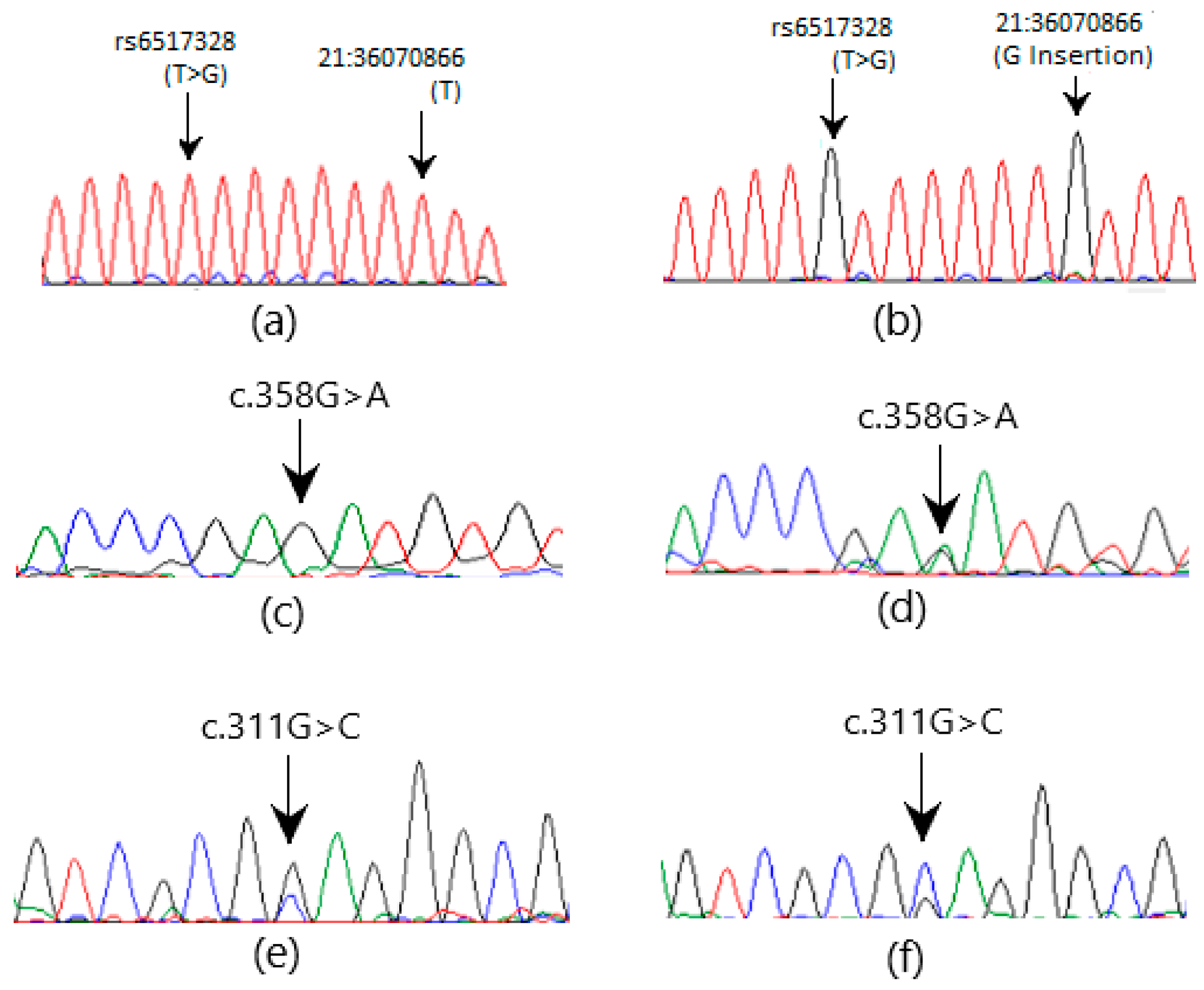
3.2. Sanger Sequencing

The Sanger sequencing of GLO1 exons revealed two heterozygous missense mutations, c.102G>T and c.147C>G, and one heterozygous nonsense mutation c.148G>T in three (2.65%), eight (7.08%), and eight (7.08%) patients, respectively (Figure 2). SNP rs1049346 at location 6:38703061 (G>A) in intron 1-2 of GLO1 (GRCh38) is another clinically important homozygous SNV observed in 13 (11.5%) patients (Figure 2e,f). In CBR1, two heterozygous variations, one missense c.358G>A, and one silent mutation c.311G>C were observed in three (2.65%) and five (4.42%) patients, respectively (Figure 3). A novel change (G insertion) at position 21: 36070866 GRCh38 (NM_001757.4) and rs6517328 (T>G) polymorphisms were observed in intron 1-2 in five (4.42%) and seven (6.19%) patients, respectively (Figure 3). The sequencing of ACE exons revealed one heterozygous missense mutation c.337A>C (Figure 2g,h) in three (2.65%) patients. The NM_000789.4 transcript in the human genome database was used as a reference and the NCBI BLAST service was used to check variation. The SNVs observed in the coding and noncoding regions of these genes are presented in Table 4 and Table 5.

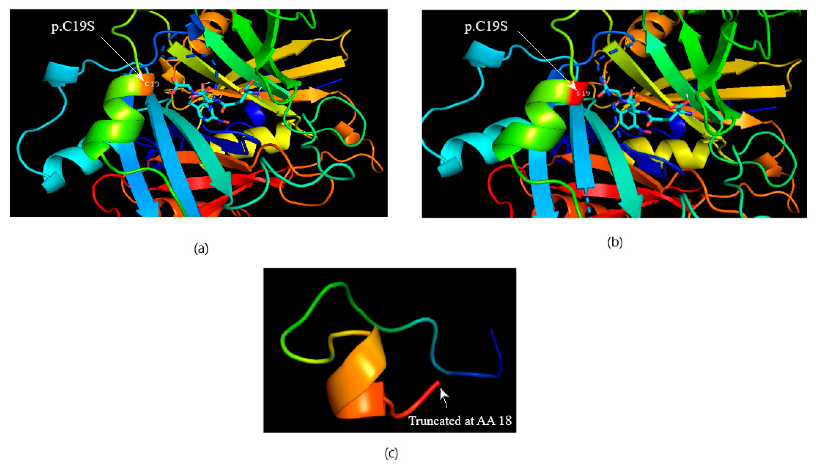
3.3. Computational Protein–Ligand Docking

The computational protein model of c.147C>G (C19S) revealed close coordination of this amino acid with the ligand (S-P-Nitrobenzyloxycarbonyl-glutathione) in the protein binding pocket (Figure 4a,b) without an apparent change in protein structure and that of the nonsense mutation revealed truncated protein unable to bind the ligand to the substrate (Figure 4c). Missense variation in the CBR1 enzyme, D120N (c.358G>A), was found to be near the active site/binding pocket of the CBR1 enzyme for ligands (glutathione and NADPH), as shown in the protein in silico structure (Figure 5). Missense variation in ACE T113M (c.337A>C) was not close to its binding site for its ligand (angiotensin 1) and no change in the protein in silico structure was observed in the computational protein model and in silico protein docking with the ligand (Figure 6).

4. Discussion

Diabetic nephropathy (DN) remains a critical complication of diabetes mellitus, contributing significantly to morbidity and mortality worldwide. Genetic predisposition plays a pivotal role in the pathogenesis of DN, and investigating the genetic variants and disease susceptibility is imperative.
A novel heterozygous variant c.148G>T (6:38702998) found in GLO1 was linked with a known heterozygous missense variant at the adjacent position c.147C>G (6:38702999). Both variations were found together in patient samples, and the latter has already been well-reported as a SNP (rs17855424 C>T) at 6:38702999 (GRCh38). However, in this study, a different change (C>G) was found at the same position instead of (C>T), which is again a missense change. In the reported SNV (C>T), the codon changes from UGC to UAC, leading to a change in the amino acid residue cysteine to tyrosine in the glyoxalase-1 protein. However, in this study, the SNV (C>G) led to a change in the codon from UGC to UCC, leading to a change in the amino acid residue from cysteine to serine. This is a novel change and has not been reported before. Cysteine at position 19 changes to serine, which is present near the ligand binding site of the glyoxalase 1 enzyme and may affect the binding, stability, or activity of the enzyme. Moreover, this cysteine residue at position 19 is conserved in different species (when checked using the conservation tool of the UCSC genome browser), indicating its importance for glyoxalase 1 function. Low plasma activity of this protective enzyme has already been reported in patients with severe diabetic nephropathy [15,35,36].
Another missense variation found at 6:38703044 (GRCh38) is a known SNP rs1168871721 (G>A), which causes a change from codon CCG to CUG and a change in amino acids from proline (non-polar residue) to leucine (non-polar residue) in the glyoxalase-1 enzyme. However, in this study, a different novel change from G>T instead of G>A at the same location was observed, which led to the formation of a new codon, CAG, instead of CUG, and the amino acid residue changed to glutamine (polar residue) instead of leucine (non-polar residue) in place of proline (non-polar residue). This SNP is present at the transcription factor-binding site for transcription factors PAX4, HOXB2::PAX1, HOXB2::PAX5, and HOXB2::PAX9 according to NCBI SNP data and the Ensembl genome browser 112 [37]. The amino acid residue proline is conserved at this position in different species (e.g., Rhesus, mouse, dog, elephant) over the evolutionary process, indicating its importance for normal protein structure and function. This change may lead to the ineffective binding of transcription factors or a reduced concentration of the glyoxalase 1 enzyme in circulation, an effect already reported in diabetic nephropathy. This finding is novel, and the related SNP is also not much reported in the literature.
The SNP rs1049346 at location 6:38703061 (GRCh38), found in patients, has already been reported to be linked with decreased levels of circulating glyoxalase-1 and renders carriers susceptible to diseases such as autism [38]. The variant A of the same SNP has been implicated in late-onset epilepsy [39] and acute coronary syndrome (ACS) in diabetic South Indians [40]. However, some newer studies on this subject propose that polymorphisms in the GLO-1 gene are not linked to low circulating levels of the enzyme or alterations in markers of methylglyoxal-related stress [41].
A novel heterozygous SNV (c. 358 G>A) at position 21:36071018 (GRCh38) in CBR1 has not been reported in the literature before. This missense variation, D120N, causes codon GAU to change to AAU and the amino acid aspartate (acidic residue) to be replaced by asparagine (polar residue). The computational mutated protein model and in silico ligand interaction show the proximity of this amino acid to the active site and thus may have consequences concerning enzyme activity. Aspartate residue at position 120 is conserved in different species listed in UCSC except in elephants and chickens. The significance of this variation is, however, not listed in ClinVar. A heterozygous change c.311G>C at position 21:36070334 in CBR1 is a reported SNP (rs25678). Although it is a synonymous variant (L73L), it has been linked with poor enzyme function in acute myeloid leukemia patients [42]. Leucine residue at position 73 is conserved in mammals except elephants and zebrafish. Methyl glyoxal is metabolized by carbonyl reductase 1 as well, and this enzyme’s reduced activity may lead to an accelerated progression of diabetic nephropathy.
It is shown that the CBR1 gene is helpful in ameliorating the damaging effects of ROS and carbonyl stress and different mechanisms have been demonstrated [12]. The up-regulation of CBR1 is proposed to reduce lung injury caused by ROS [43] through its protective effects against ROS-triggered carbonyl stress and advanced glycation end products. Newer protein modifiers, e.g., carbonylation, are gaining attention and are aspiring to be enrolled as early markers of DN [44]. This study aimed to elucidate the potential involvement of genetic variants in the CBR1 gene in patients with early and severe DN.
Two SNVs, 21:36070859 (T>G) and 21:36070866 (G insertion), in CBR1 were found together in all positive samples and are likely to be independently disease-causing. The former is a reported SNP rs6517328, which is also reported as a cosmic mutation (COSV51737954). These are intron variants and may affect splice site and protein features as per the Ensembl genome browser 112 (available at https://www.ensembl.org/index.html?redirect=no accessed on 20 July 2024). These findings are not reported before and only generally, the CBR1 gene is implicated in oxidative stress and metabolic syndrome in a few recent studies like [45]. A similar study in mice demonstrated the role of reduced CBR1 gene expression in mouse liver was causative for diabetes and its effects [46]. The role of carbonyl reductase 1 expression in anti-oxidative mechanisms is proven in vitro as well [47]. On the contrary, a recent study reported that carbonyl reductase-1 enhances glucocorticoid action and thus causes hyperglycemia and is a risk factor for diabetes and hyperglycemia in mice [48]. Polymorphism and genetic variations in this gene have not been studied well for association with diseases, especially diabetic nephropathy.
Several studies have reported polymorphism in the ACE gene, e.g., rs267604983 [18,49] and I/D polymorphism [50], linked with DN. Another study reported that a GG genotype of ACE c. 2350 G>A and DD polymorphism of ACE I/D were linked with early tubular injury [51]. Though these studies suggest polymorphism in ACE is linked to DN, none have demonstrated the novel SNV observed in this study. At position 17:63478018, heterozygous SNV c.337A>C is a novel missense change (T113M) near the N terminal and is far from the ligand binding site and unlikely to affect ligand binding, as revealed by the in silico mutated protein model. This SNV is not reported in the literature. The structure of ACE is preserved in many different species during evolution and its N terminal active site is of prime importance [52]. Changes in protein structure near the N terminal can affect the active site in many ways [52].

5. Conclusions

In conclusion, two missense, c.102G>T and c.147C>G, and one nonsense c.148G>T SNVs were found in GLO1 in patients of early and severe diabetic nephropathy, the latter being most deleterious, resulting in a truncated protein. Missense SNVs c.358G>A in CBR1 and c.337A>C in ACE were observed in patients, leading to potential effects on protein–ligand interaction and enzyme function. The nonsense mutation led to the formation of a truncated protein (GLO1) unable to bind the ligand while two missense mutations altered the protein structure near the active site in CBR1. The affected GLO1 and CBR1 enzymes may lead to the rapid progression of diabetic nephropathy.

Limitations of the Study

The sample size and sampling technique are not adequate to generalize the findings of this research to the DN population. Only Pakistani subjects were included, so there may be a possibility of ethnic or environmental effects. The identified mutations may be tested in larger cohorts and in other populations to ascertain a clear association of risk.

Author Contributions

Conceptualization, S.Z.H.S. and N.A.; methodology, S.Z.H.S.; software, S.Z.H.S. and A.M.; validation, A.R., A.M. and T.G.; formal analysis, S.Z.H.S.; investigation, S.Z.H.S.; resources, T.G., N.A. and A.M.; data curation, A.M.; writing—original draft preparation, S.Z.H.S.; writing—review and editing, A.R. and T.G.; visualization, S.Z.H.S. and T.G.; supervision, A.R. and N.A.; project administration, N.A.; funding acquisition, S.Z.H.S. All authors have read and agreed to the published version of the manuscript.

Funding

This research was funded by the National University of Medical Sciences (NUMS) Pakistan, grant number IRF-22005.

Institutional Review Board Statement

The study was conducted in accordance with the Declaration of Helsinki and approved by the Institutional Review Board constituent of Medical College, National University of Medical Sciences, Pakistan (ERC/ID/96 dated 12 November 2020).

Informed Consent Statement

Informed written consent was obtained from all participants involved in the study. Written informed consent has been obtained from the patients to publish this paper.

Data Availability Statement

The data presented in this study are available upon request from the corresponding authors due to government institutional restrictions.

Acknowledgments

The help and support provided by Khalid Raja as consultant nephrologist, Department of Nephrology, Ali Rathore, the consultant hematologist, and Nadia from a tertiary care-affiliated hospital of the National University of Medical Sciences, Rawalpindi, Pakistan, in the enrolment of patients and controls, clinical workup, lab work, and Sanger sequencing are appreciated.

Conflicts of Interest

The authors declare no conflicts of interest. The funders had no role in the design of the study; in the collection, analyses, or interpretation of data; in the writing of the manuscript; or in the decision to publish the results.

References

  1. Sun, H.; Saeedi, P.; Karuranga, S.; Pinkepank, M.; Ogurtsova, K.; Duncan, B.B.; Stein, C.; Basit, A.; Chan, J.C.N.; Mbanya, J.C.; et al. IDF Diabetes Atlas: Global, regional and country-level diabetes prevalence estimates for 2021 and projections for 2045. Diabetes Res. Clin. Pract. 2022, 183, 109119. [Google Scholar] [CrossRef] [PubMed]
  2. Selby, N.M.; Taal, M.W. Obesity, Metabolism, An updated overview of diabetic nephropathy: Diagnosis, prognosis, treatment goals and latest guidelines. Diabetes Obes. Metab. 2020, 22, 3–15. [Google Scholar] [CrossRef] [PubMed]
  3. Alam, S.; Hasan, M.K.; Neaz, S.; Hussain, N.; Hossain, M.F.; Rahman, T. Diabetes Mellitus: Insights from epidemiology, biochemistry, risk factors, diagnosis, complications and comprehensive management. Diabetology 2021, 2, 36–50. [Google Scholar] [CrossRef]
  4. Shin, J.; Zhou, X.; Tan, J.T.; Hyppönen, E.; Benyamin, B.; Lee, S.H. Lifestyle modifies the diabetes-related metabolic risk, conditional on individual genetic differences. Front. Genet. 2022, 13, 759309. [Google Scholar] [CrossRef] [PubMed]
  5. Yahaya, T.O.; Salisu, T.F. A review of type 2 diabetes mellitus predisposing genes. Curr. Diabetes Rev. 2020, 16, 52–61. [Google Scholar] [CrossRef] [PubMed]
  6. Tomic, D.; Shaw, J.E.; Magliano, D.J. The burden and risks of emerging complications of diabetes mellitus. Nat. Rev. Endocrinol. 2022, 18, 525–539. [Google Scholar] [CrossRef]
  7. Foreman, K.J.; Marquez, N.; Dolgert, A.; Fukutaki, K.; Fullman, N.; McGaughey, M.; Pletcher, M.A.; Smith, A.E.; Tang, K.; Yuan, C.-W.; et al. Forecasting life expectancy, years of life lost, and all-cause and cause-specific mortality for 250 causes of death: Reference and alternative scenarios for 2016-40 for 195 countries and territories. Lancet 2018, 392, 2052–2090. [Google Scholar] [CrossRef]
  8. Imtiaz, S.; Alam, A. Epidemiology and demography of Chronic Kidney Disease in Pakistan-A review of Pakistani literature. Pak. J. Kidney Dis. 2023, 7, 2–7. [Google Scholar] [CrossRef]
  9. Kato, S.; Matsumura, T.; Sugawa, H.; Nagai, R. Correlation between serum advanced glycation end-products and vascular complications in patient with type 2 diabetes. Sci. Rep. 2024, 14, 18722. [Google Scholar] [CrossRef]
  10. Wu, X.-Q.; Zhang, D.-D.; Wang, Y.-N.; Tan, Y.-Q.; Yu, X.-Y.; Zhao, Y.-Y. AGE/RAGE in diabetic kidney disease and ageing kidney. Free. Radic. Biol. Med. 2021, 171, 260–271. [Google Scholar] [CrossRef]
  11. Saeed, M.; Kausar, M.A.; Singh, R.; Siddiqui, A.J.; Akhter, A. The role of glyoxalase in glycation and carbonyl stress induced metabolic disorders. Curr. Protein Pept. Sci. 2020, 21, 846–859. [Google Scholar] [CrossRef] [PubMed]
  12. Geng, S.L.; Li, H.Y.; Zhang, X.S.; Wang, T.; Zhou, S.P.; Xu, W.H. CBR1 decreases protein carbonyl levels via the ROS/Akt/CREB pathway to extend lifespan in the cotton bollworm. Helicoverpa Armigera 2023, 290, 2127–2145. [Google Scholar] [CrossRef] [PubMed]
  13. Fuloria, S.; Subramaniyan, V.; Karupiah, S.; Kumari, U.; Sathasivam, K.; Meenakshi, D.U.; Wu, Y.S.; Guad, R.M.; Udupa, K.; Fuloria, N.K. A Comprehensive Review on Source, Types, Effects, Nanotechnology, Detection, and Therapeutic Management of Reactive Carbonyl Species Associated with Various Chronic Diseases. Antioxidants 2020, 9, 1075. [Google Scholar] [CrossRef] [PubMed]
  14. Hanssen, N.M.; Stehouwer, C.D.; Schalkwijk, C.G. Methylglyoxal stress, the glyoxalase system, and diabetic chronic kidney disease. Curr. Opin. Nephrol. Hypertension 2019, 28, 26–33. [Google Scholar] [CrossRef] [PubMed]
  15. Shah, S.Z.H.; Rashid, A.; Majeed, A. Determination of Glyoxalase-1 levels and Identification of Genetic Variants in GLO1 Gene in Patients of Diabetic Nephropathy. Pak. J. Med. Sci. 2024, 40, 652–656. [Google Scholar] [CrossRef]
  16. Corredor, Z.; Filho, M.; Rodriguez-Ribera, L.; Velazquez, A.; Hernandez, A.; Catalano, C.; Hemminki, K.; Coll, E.; Silva, I.; Diaz, J.M.; et al. Genetic Variants Associated with Chronic Kidney Disease in a Spanish Population. Sci. Rep. 2020, 10, 144. [Google Scholar] [CrossRef]
  17. Tziastoudi, M.; Stefanidis, I.; Zintzaras, E. The genetic map of diabetic nephropathy: Evidence from a systematic review and meta-analysis of genetic association studies. Clin. Kidney J. 2020, 13, 768–781. [Google Scholar] [CrossRef]
  18. Pai, D.; Adiga, S.; Suresh, G.; Adiga, U.; Chaitra, D.; Honalli, N.M. The association of ACE gene polymorphism and serum ACE levels with diabetic nephropathy-a cross-sectional study. J. Pharm. Sci. Appl. 2024, 14, 210–217. [Google Scholar] [CrossRef]
  19. Jankovic, M.; Novakovic, I.; Nikolic, D.; Mitrovic Maksic, J.; Brankovic, S.; Petronic, I.; Cirovic, D.; Ducic, S.; Grajic, M.; Bogicevic, D.; et al. Genetic and Epigenomic Modifiers of Diabetic Neuropathy. Int. J. Mol. Sci. 2021, 22, 4887. [Google Scholar] [CrossRef]
  20. Giani, J.F.; Veiras, L.C.; Shen, J.Z.; Bernstein, E.A.; Cao, D.; Okwan-Duodu, D.; Khan, Z.; Gonzalez-Villalobos, R.A.; Bernstein, K.E. Novel roles of the renal angiotensin-converting enzyme. Mol. Cell Endocrinol. 2021, 529, 111257. [Google Scholar] [CrossRef]
  21. Deng, X.; Li, D.; Tang, Q.; Chen, Y. ACEI and ARB lower the incidence of end-stage renal disease among patients with diabetic nephropathy: A meta-analysis. Comput. Math. Methods Med. 2022, 2022, 6962654. [Google Scholar] [CrossRef] [PubMed]
  22. Juin, S.K.; Ouseph, R.; Gondim, D.D.; Jala, V.R.; Sen, U. Diabetic Nephropathy and Gaseous Modulators. Antioxidants 2023, 12, 1088. [Google Scholar] [CrossRef] [PubMed]
  23. Mallik, R.; Chowdhury, T.A. Pharmacotherapy to delay the progression of diabetic kidney disease in people with type 2 diabetes: Past, present and future. Ther. Adv. Endocrinol. Metabolism. 2022, 13, 20420188221081601. [Google Scholar] [CrossRef] [PubMed]
  24. Tanaka, T.; Tsukube, S.; Izawa, K.; Okochi, M.; Lim, T.-K.; Watanabe, S.; Harada, M.; Matsunaga, T. Electrochemical detection of HbA1c, a maker for diabetes, using a flow immunoassay system. Biosens. Bioelectron. 2007, 22, 2051–2056. [Google Scholar] [CrossRef] [PubMed]
  25. Li, M.; Pezzolesi, M.G. Advances in understanding the genetic basis of diabetic kidney disease. Acta Diabetol. 2018, 55, 1093–1104. [Google Scholar] [CrossRef]
  26. Bonora, E.; Kiechl, S.; Mayr, A.; Zoppini, G.; Targher, G.; Bonadonna, R.C.; Willeit, J. High-Normal HbA1c Is a Strong Predictor of Type 2 Diabetes in the General Population. Diabetes Care 2011, 34, 1038–1040. [Google Scholar] [CrossRef]
  27. Walker, H.K.; Hall, W.D.; Hurst, J.W. (Eds.) Clinical Methods: The History, Physical, and Laboratory Examinations, 3rd ed.; Butterworths: Boston, UK, 1990. [Google Scholar]
  28. Umeukeje, E.M.; Koonce, T.Y.; Kusnoor, S.V.; Ulasi, I.I.; Kostelanetz, S.; Williams, A.M.; Blasingame, M.N.; Epelbaum, M.I.; Giuse, D.A.; Apple, A.N.; et al. Systematic review of international studies evaluating MDRD and CKD-EPI estimated glomerular filtration rate (eGFR) equations in Black adults. PLoS ONE 2022, 17, e0276252. [Google Scholar] [CrossRef]
  29. Untergasser, A.; Nijveen, H.; Rao, X.; Bisseling, T.; Geurts, R.; Leunissen, J.A.M. Primer3Plus, an enhanced web interface to Primer3. Nucleic Acids Res. 2007, 35 (Suppl. S2), W71–W74. [Google Scholar] [CrossRef]
  30. Al-Shuhaib, M.B.S.; Hashim, H.O. Mastering DNA chromatogram analysis in Sanger sequencing for reliable clinical analysis. J. Genet. Eng. Biotechnol. 2023, 21, 115. [Google Scholar] [CrossRef]
  31. Waterhouse, A.; Bertoni, M.; Bienert, S.; Studer, G.; Tauriello, G.; Gumienny, R.; Heer, F.T.; De Beer, T.A.P.; Rempfer, C.; Bordoli, L.; et al. SWISS-MODEL: Homology modelling of protein structures and complexes. Nucleic Acids Res. 2018, 46, W296–W303. [Google Scholar] [CrossRef]
  32. Bugnon, M.; Röhrig, U.F. SwissDock 2024: Major enhancements for small-molecule docking with Attracting Cavities and AutoDock Vina. Nucleic Acids Res. 2024, 52, W324–W332. [Google Scholar] [CrossRef] [PubMed]
  33. Eberhardt, J.; Santos-Martins, D. AutoDock Vina 1.2.0: New Docking Methods, Expanded Force Field, and Python Bindings. J. Chem. Inf. Model. 2021, 61, 3891–3898. [Google Scholar] [CrossRef] [PubMed]
  34. Schrodinger, LLC. The PyMOL Molecular Graphics System, Version 1.8; Schrodinger: New York, NY, USA, 2015. [Google Scholar]
  35. Mazani, M.; Mahdavifard, S.; Koohi, A. Crocetin ameliorative effect on diabetic nephropathy in rats through a decrease in transforming growth factor-β and an increase in glyoxalase-I activity. Clin. Nutr. ESPEN 2023, 58, 61–66. [Google Scholar] [CrossRef] [PubMed]
  36. Chen, Y.J.; Kong, L.; Tang, Z.Z.; Zhang, Y.M.; Liu, Y.; Wang, T.Y.; Liu, Y.W. Hesperetin ameliorates diabetic nephropathy in rats by activating Nrf2/ARE/glyoxalase 1 pathway. Biomed. Pharmacother. 2019, 111, 1166–1175. [Google Scholar] [CrossRef] [PubMed]
  37. Ensemble. Ensemble Human Gene Browser 11. 2024. Available online: https://asia.ensembl.org/Homo_sapiens/Variation/Mappings?db=core;g=ENSG00000124767;r=6:38675925-38703145;t=ENST00000373365;v=rs1168871721;vdb=variation;vf=429570578 (accessed on 18 February 2024).
  38. Peculis, R.; Konrade, I.; Skapare, E.; Fridmanis, D.; Nikitina-Zake, L.; Lejnieks, A.; Pirags, V.; Dambrova, M.; Klovins, J. Identification of glyoxalase 1 polymorphisms associated with enzyme activity. Gene 2013, 515, 140–143. [Google Scholar] [CrossRef]
  39. Tao, H.; Si, L.; Zhou, X.; Liu, Z.; Ma, Z.; Zhou, H.; Zhong, W.; Cui, L.; Zhang, S.; Li, Y.; et al. Role of glyoxalase I gene polymorphisms in late-onset epilepsy and drug-resistant epilepsy. J. Neurol. Sci. 2016, 363, 200–206. [Google Scholar] [CrossRef]
  40. Bora, S.; Adole, P.S.; Vinod, K.V.; Pillai, A.A.; Ahmed, S. The genetic polymorphisms and activity of glyoxalase 1 as a risk factor for acute coronary syndrome in South Indians with type 2 diabetes mellitus. Gene 2023, 885, 147701. [Google Scholar] [CrossRef]
  41. Maasen, K.; Hanssen, N.M.J.; van der Kallen, C.J.H.; Stehouwer, C.D.A.; van Greevenbroek, M.M.J.; Schalkwijk, C.G. Polymorphisms in Glyoxalase I Gene Are Not Associated with Glyoxalase I Expression in Whole Blood or Markers of Methylglyoxal Stress: The CODAM Study. Antioxidants 2021, 10, 219. [Google Scholar] [CrossRef]
  42. Megias-Vericat, J.E.; Martinez-Cuadron, D.; Herrero, M.J.; Alino, S.F.; Poveda, J.L.; Sanz, M.A.; Montesinos, P. Pharmacogenetics of metabolic genes of anthracyclines in acute myeloid leukemia. Curr. Drug Metab. 2018, 19, 55–74. [Google Scholar] [CrossRef]
  43. Jiang, Q.; Chen, Z.; Jiang, H. Flufenamic acid alleviates sepsis-induced lung injury by up-regulating CBR1. Drug Dev. Res. 2020, 81, 885–892. [Google Scholar] [CrossRef]
  44. Yanar, K.; Atayik, M.C.; Simsek, B.; Çakatay, U. Novel biomarkers for the evaluation of aging-induced proteinopathies. Biogerontology 2020, 21, 531–548. [Google Scholar] [CrossRef] [PubMed]
  45. Zheng, J.; Liu, X.; Zheng, B.; Zheng, Z.; Zhang, H.; Zheng, J.; Sun, C.; Chen, H.; Yang, J.; Wang, Z.; et al. Maternal 25-hydroxyvitamin D deficiency promoted metabolic syndrome and downregulated Nrf2/CBR1 pathway in offspring. Front. Pharmacol. 2020, 11, 97. [Google Scholar] [CrossRef] [PubMed]
  46. Ge, Q.; Feng, F.; Liu, L.; Chen, L.; Lv, P.; Ma, S.; Chen, K.; Yao, Q. RNA-Seq analysis of the pathogenesis of STZ-induced male diabetic mouse liver. J. Diabetes Its Complicat. 2019, 34, 107444. [Google Scholar] [CrossRef] [PubMed]
  47. Zhang, Z.; Song, W.; Yan, R. Gbp3 is associated with the progression of lupus nephritis by regulating cell proliferation, inflammation and pyroptosis. Autoimmunity 2023, 56, 2250095. [Google Scholar] [CrossRef]
  48. Bell, R.M.; Villalobos, E.; Nixon, M.; Miguelez-Crespo, A.; Murphy, L.; Fawkes, A.; Coutts, A.; Sharp, M.G.; Koerner, M.V.; Allan, E.; et al. Carbonyl reductase 1 amplifies glucocorticoid action in adipose tissue and impairs glucose tolerance in lean mice. Mol. Metab. 2021, 48, 101225. [Google Scholar] [CrossRef]
  49. Ma, H.; Yu, C.; Wang, R. Association of ACE polymorphism and diabetic nephropathy susceptibility. Int. J. Clin. Exp. Med. 2015, 8, 2962–2965. [Google Scholar]
  50. Deepashree, G.A.; Ramprasad, E.; Jayakumar, M.; Paul, S.F.; Gnanasambandan, R. ACE ID gene polymorphism contributes to chronic kidney disease progression but not NOS3 gene among Type 2 diabetes with nephropathy patients. Endocrine Metab. Sci. 2021, 4, 100100. [Google Scholar]
  51. Taha, M.M.; Mahdy-Abdallah, H.; Shahy, E.M.; Helmy, M.A.; ElLaithy, L.S. Diagnostic efficacy of cystatin-c in association with different ACE genes predicting renal insufficiency in T2DM. Sci. Rep. 2023, 13, 5288. [Google Scholar] [CrossRef]
  52. Lubbe, L.; Cozier, G.E.; Oosthuizen, D.; Acharya, K.R.; Sturrock, E.D. ACE2 and ACE: Structure-based insights into mechanism, regulation and receptor recognition by SARS-CoV. Clin. Sci. 2020, 134, 2851–2871. [Google Scholar] [CrossRef]
Figure 1. (a) A picture of 1% agarose gel showing positive DNA bands on gel electrophoresis. (b) A picture of 2% agarose gel showing 325 bp fragments on gel electrophoresis amplified using a PCR.
Medicina 60 01540 g001
Figure 2. The electrochromatogram shows SNVs in GLO1 and ACE genes. (a,c,e,g) are wild-type sequences, while (b,d,f,h) show mutated sequences (A = Green, G = Black, T = Red, C = Blue).
Medicina 60 01540 g002
Figure 3. The electrochromatogram shows SNVs in the CBR1 gene. (a,c,e) are wild-type sequences, while (b,d,f) show mutated sequences (A = Green, G = Black, T = Red, C = Blue).
Medicina 60 01540 g003
Figure 4. The protein–ligand docking of GLO1 and S-P-Nitrobenzyloxycarbonylglutathione. (a) Wild-type protein docking with ligand. (b) Mutated C19S. (c) Truncated 18AA peptide (stop codon gained).
Medicina 60 01540 g004
Figure 5. The protein–ligand docking of CBR1 and glutathione with NADPH. (a) Normal protein docking with ligand. (b) D120N.
Medicina 60 01540 g005
Figure 6. The protein–ligand docking of ACE and angiotensin 1. (a) Wild-type protein docking with the ligand. (b) T113M novel mutation.
Medicina 60 01540 g006
Table 1. Points adhered to while selecting primers online.
ParameterTarget
Tool usedNCBI primer BLAST and Primer 3 plus
GenomeRefSeq representative genome (GRCh38)
Maximum hits in genome1
5′ self-complementarity<6
Single base repetition (max)3 (no 4 Gs together)
Product size500 to 900 bp
Minimum length around the target sequence at both ends150 bp
Primer annealing temperature Tm55–63 °C
The maximum difference in Tm of the two primers4 °C
GC content45 to 60%
Table 2. Some primers and their annealing temperatures.
TargetPrimerPrimer SequenceProduct SizePrimer
Tm (°C)
Reaction Tm (°C)
GLO1 Exon 1Forward5′ TTCTACCAAATTGCAGCCCTGA 3′725 bp61.061.8
Reverse5′ CAGCCACCGTCGCAACATA 3′ 62.3
GLO1 Exon 2Forward5′ TTGCAAGTTGTAGGTGGTAGGTT 3′280 bp60.361.2
Reverse5′ AAGATGGGTCTGAAAACACTCTC 3′ 59.4
GLO1 Exon 2
(second set)
Forward5′ TCTGACACTTTGGACTTGCATCA 3′761 bp60.761.2
Reverse5′ TTTCAGGCTGGCTGGGATAGA 3′ 63.6
CBR1 Exon 1Forward5′ GTCCATAACGCCTCCCTAGG 3′413 bps61.361.2
Reverse5′ GTCCATAACGCCTCCCTAGG 3′ 59.3
CBR1 Exon 2Forward5′ AACTTTGTGTTTCCCTGGCTGGG 3′812 bp63.664.8
Reverse5′ GGATGGACTCCCACGCAGAG 3′ 62.3
ACE Exon 1Forward5′ AGAGGAGGCCCTTTCTCCAGCT 3′716 bp68.665.0,
Reverse5′ ACCCTCATCCATCCAACTCG 3′ 62.966.0
ACE Exon 2Forward5′ TCCGCAAACTAAGGTCTCCC 3′549 bp62.762.0
Reverse5′ TTGGCTTCCTACTCCAGAATGC 3′ 61.7
ACE Exon 2
(second set)
Forward5′ AAGCCCTTGGCCTTCCTC 3′325 bp64.267.0
Reverse5′ CACGATGGGGCACTAGGAG 3′ 63.9
Table 3. Demographic factors.
Parameters 1Control
Mean (SD)
(n = 100)
Diabetic Nephropathy
Mean (SD)
(n = 113)
p Value
Age(years)54.6 ± 10.357.2 ± 11.40.27
GenderMale n (%)59 (59)68 (60.2)0.13
Female n (%)41 (41)45 (39.8)0.32
BMI25.5 ± 4.727.3 ± 3.20.19
1 Biochemical parameters are already reported in our previous article [15].
Table 4. Genetic variations in coding regions of GLO1, CBR1, and ACE genes (NM_000789.4).
GeneDNA Sequence Change
(GRCh38)
Amino Acid Residue ChangeStatusLocus/SiteType of ChangeControl
n = 100
n (%)
DN
n = 113
n (%)
GLO1c.102G>Tp.P4Qrs11688717216:38703044Missense0 (0)3 (2.65)
c.147C>Gp.C19Srs178554246:38702999Missense0 (0)8 (7.08)
c.148G>Tp.C19XNovel6:38702998Nonsense0 (0)8 (7.08)
CBR1c.358G>Ap.D120NNovel21:36071018Missense0 (0)3 (2.65)
c.311G>Cp.L73Lrs2567821:36070334Silent0 (0)5 (4.42)
ACEc.337A>Cp.T113MNovel17:63478018Missense0 (0)3 (2.65)
Table 5. Single nucleotide variations in the noncoding regions of GLO1, CBR1, and ACE genes (NM_000789.4).
GeneLocation
(GRCh38)
VariationStatusControl
n = 100
n (%)
DN
n = 113
n (%)
GLO16:38703061G>Ars10493460 (0)13 (11.5)
6:38703141G>Trs17617344270 (0)1 (0.9)
6:38703186C>ANovel0 (0)1 (0.9)
6:38686709G>CNovel0 (0)1 (0.9)
6:38686760G>ANovel0 (0)1 (0.9)
6:38686799G>ANovel0 (0)1 (0.9)
6:38686823G>ANovel0 (0)1 (0.9)
6:38686886T>CNovel0 (0)1 (0.9)
CBR121:36070068G>Ars115421680 (0)1 (0.9)
21:36070774G>ANovel0 (0)1 (0.9)
21:36070859T>Grs65173280 (0)7 (6.19)
21:36070866X>GNovel0 (0)5 (4.42)
21:36070872X>GNovel0 (0)1 (0.9)
21:36070881A>Trs14696928240 (0)1 (0.9)
21:36070907A>CNovel0 (0)1 (0.9)
ACE17:63477094C>Grs8873055220 (0)1 (0.9)
17:63477454C>XNovel0 (0)1 (0.9)
17:63477926A>CNovel0 (0)1 (0.9)
17:63477929X>CNovel0 (0)2 (1.7)
Disclaimer/Publisher’s Note: The statements, opinions and data contained in all publications are solely those of the individual author(s) and contributor(s) and not of MDPI and/or the editor(s). MDPI and/or the editor(s) disclaim responsibility for any injury to people or property resulting from any ideas, methods, instructions or products referred to in the content.

